Embed Size (px)
Citation preview
291Copyright © 2014 The Korean Society of Cardiology
Korean Circulation Journal
Introduction
Catheter ablation has been evolving rapidly for the management of atrial fibrillation (AF) with improvements in both efficacy and safety.1) The efficacy of radiofrequency (RF) catheter ablation for maintaining sinus rhythm is superior to the current antiarrhythmic drug therapy in selected patients.2-4) The goal of catheter ablation is to eliminate AF. The development of AF requires both a trigger and a susceptible substrate. The most common trigger initiating AF comes from the myocardial sleeve of the left atrium (LA), which ex-tends into the pulmonary veins (PV).5) Unique anatomical and elec-trophysiological features of the PV and LA-PV junction (LA-PVJ) play major roles in AF. Therefore, PV isolation (PVI) or LA-PVJ ablation is the cornerstone of various catheter ablation strategies.1)
Review Article
http://dx.doi.org/10.4070/kcj.2014.44.5.291Print ISSN 1738-5520 • On-line ISSN 1738-5555
How to Achieve Complete and Permanent Pulmonary Vein Isolation without ComplicationsSeongwook Han, MD1,2 and Chun Hwang, MD1
1Central Utah Clinic-Cardiology, Utah Valley Regional Medical Center, Provo, UT, USA 2Department of Cardiology, Dongsan Medical Center, Keimyung University, Daegu, Korea
The efficacy and safety of catheter ablation for the management of atrial fibrillation (AF) has been improved in recent years. Radiofrequency (RF) catheter ablation for maintaining sinus rhythm is superior to the current antiarrhythmic drug therapy in selected patients. Pulmonary vein isolation (PVI) is the cornerstone of various catheter ablation strategies. It is well recognized that pulmonary vein (PV) antrum contrib-utes to the AF initiation and/or perpetuation. Since PV stenosis is a complication of ablation within a PV, the ablation site for PVI has shift-ed to the junction between the left atrium and the PV rather than the ostium of the PV. However, PV reconnection after ablation is the major cause of recurrence of AF. The recovery of PV conduction could be caused by anatomical variations such as the failure to produce complete transmural lesion or gaps at the ablation line due to the transient electrophysiologic effects from the RF ablation. In this review, we discussed several factors to be considered for the achievement of the best PVI, including clinical aspects and technical aspects. (Korean Circ J 2014;44(5):291-300)
KEY WORDS: Atrial fibrillation; Catheter ablation, radiofrequency; Pulmonary veins; Isolation.
Correspondence: Chun Hwang, MD, Central Utah Medical Clinic-Cardiology, Utah Valley Regional Medical Center, 1055 North 500 West, Provo, UT 84604, USATel: 1-801-429-8048, Fax: 1-801-818-0331E-mail: [email protected]
• The authors have no financial conflicts of interest.
This is an Open Access article distributed under the terms of the Creative Commons Attribution Non-Commercial License (http://creativecommons.org/licenses/by-nc/3.0) which permits unrestricted non-commercial use, distribution, and reproduction in any medium, provided the original work is properly cited.
Techniques used for PVI vary widely from center to center. Elec-trical isolation introduced by Haissaguerre et al.6) involves the se-quential identification and ablation of the PV myocardial sleeve close to the earliest site of PV activation. However, PV antrum also contributes to AF initiation and/or perpetuation.7-10) In addition, PV stenosis could be a complication of ablation within a PV. Therefore, ablation strategies have shifted to target the antrum rather than the PV proper. Although PVI is the most common AF ablation proce-dure performed to treat various forms of AF, its results varies widely among institutions. There are many possible reasons contributing to different results. Among them, the most important one is the lack of a standardized and systematic method and technique to achieve PVI. In addition, reliable acute predictor during PVI for long-term success is unavailable.
Atrial fibrillation catheter ablation has risks of major complica-tions. International survey and European multinational registry re-ported a 4.5–7.7% incidence of major complications associated with AF catheter ablation.11)12) Many of these complications could be avoided with careful pre-procedural assessment of patients and de-tailed planning of the procedure. The majority of the collateral dam-ages as complications of the ablation attributed to individual ana-tomical variations. Although these complications are not frequent, they have been observed several times even in experienced centers. The current safety and efficacy measures used during the AF ablation
292 How to Achieve PVI
http://dx.doi.org/10.4070/kcj.2014.44.5.291 www.e-kcj.org
have limitations due to the lack of real-time visualization and char-acterization of the cardiac tissue, especially in lesion formation. There are three major techniques to achieve electrical PVI during AF catheter ablation: 1) Segmental ablation, the earliest PV potential guided segmental and ostial ablation; 2) LA-PVJ ablation, a PVI by circumferential ablation at LA-PVJ; 3) Wide antral circumferential ablation (WACA), an ablation at the LA outside the LA-PVJ.
In this review article, we aimed to characterize each PVI tech-nique so the best PVI result could be achieved in each patient within the given condition of electrophysiology laboratory. It is important to keep in mind that these techniques are not exclusive but rather mutual complementary in some patients. We also included our ex-periences with a few useful clinical observations to provide better understanding of AF ablation procedures.
Clinical Consideration
Pre-procedure general evaluationPre-procedure evaluation starts with the first clinical encounter
which should include a clear documentation of clinical events to justify the PVI. It is important to assess clinical status of not only cardiac conditions, such as congestive heart failure or history of rheumatic fever, but also other associated clinical conditions that
may influence the outcome or may increase the risks during PVI. Clinical condition examples include: 1) Chronic obstructive lung dis-ease, which may require special respiratory support during the se-dation; 2) Diabetes mellitus, which may require close monitoring of blood glucose; 3) Renal disease, which may require judicious fluid management as well as hemodynamic monitoring. These clinical conditions may help avoid fluid overload, therefore minimizing the risk for respiratory distresses or exacerbation of renal failure due to hypotension during PVI. It is also important to share and discuss these concerns with the patients.
Pre-procedure physical examinationPhysical evaluation should be performed in consideration of AF
ablation procedure which should include the exam of the vascular access site. Some patients may have extensive scar over the vascu-lar access site due to pre-existing conditions or previous procedures or surgeries. In patients with pre-existing scar tissues, a generous skin incision after the venous access followed by a good dilatation prior to the insertion of the sheath is recommended. A small skin incision might create a significant resistance during the sheath and catheter manipulations. Generous skin incision is particularly im-portant during trans-septal catheterization which requires delicate sheath manipulation in order to puncture the ideal site within the
A
C
B
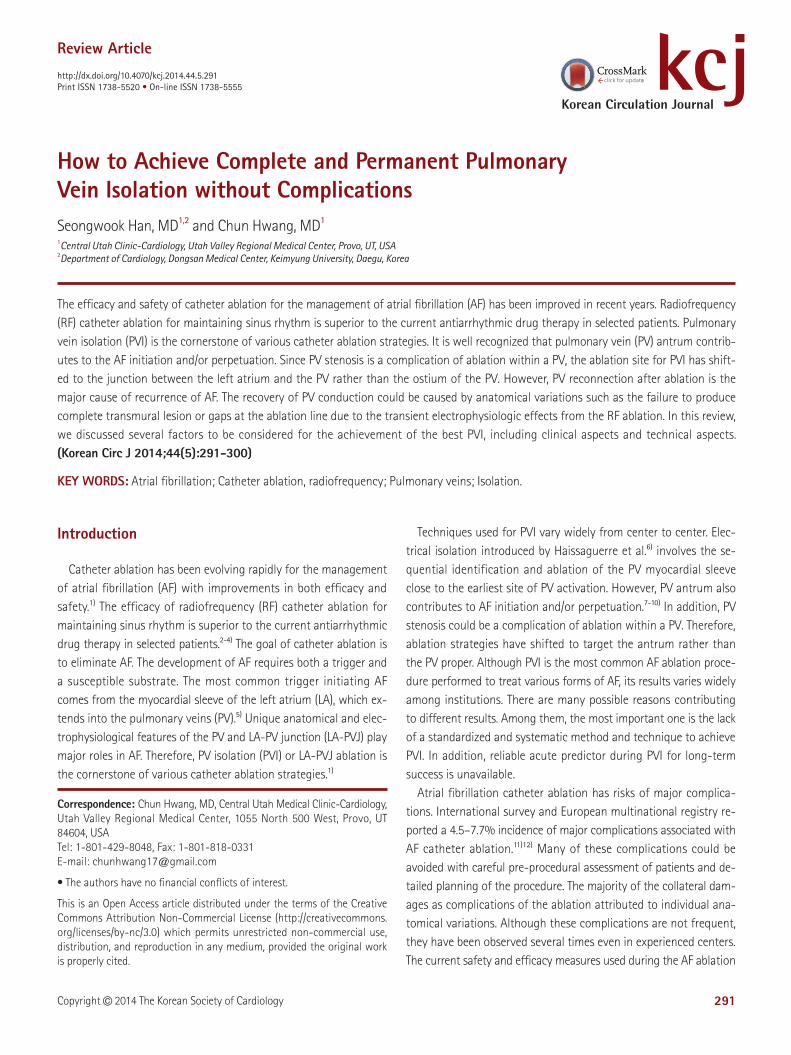
D Fig. 1. Three-dimensional reconstructed images of a left atrium. A and B: the left atrium of a patient with Pectus Excavatum. A shows the relationship be-tween the ascending aorta and the left atrial anterior wall. B shows the indentation of the anterior wall by the aorta and the flattening of the left atrium. C and D: the left atrium of a patient with Marfan syndrome, which shows significant indentation of the posterior wall by the vertebral column.
293Seongwook Han, et al.
http://dx.doi.org/10.4070/kcj.2014.44.5.291www.e-kcj.org
fossa ovale.Special clinical circumstances may require additional consider-
ation for PVI. If the patient presents with significant chest deformi-ties such as scoliosis, lordosis, pectus excavatum (Fig. 1A and B), or Marfan syndrome (Fig. 1C and D), they can create difficulties dur-ing trans-septal catheterization as well as catheter manipulation to achieve successful PVI. Other clinical conditions such as prior pneu-monectomy or hemi-diaphragm paralysis can also change attitu-dinal cardiac anatomy.
Pre-procedure 3-dimensional cardiac imaging evaluationPre-procedural evaluation of 3-D cardiac imaging has become an
essential part in AF ablation. Cardiac CT angiography (cCTA) is the most widely used imaging modality. Modern cCTA provides detailed anatomy, including the relationship of LA, PVs, LA-PVJ, and LA ap-pendage. Cardiac MRI (cMRI) is the other 3-D imaging modality that provides not only anatomical information for cardiac function, but also pre-existing myocardial fibrosis with its delayed enhance-ment imaging technology.
The objective of 3-D cardiac imaging is to not only evaluate the patient’s detailed cardiac anatomy, but also recognize the relation-ship between LA and its neighboring structures that are subject to collateral damages during PVI, such as esophagus, major proximal arteries, and pulmonary parenchyma. However, some neighboring structures could not be imaged with cCTA or cMRI, such as phrenic and recurrent laryngeal nerves. In addition, they could be injured during PVI despite best efforts.
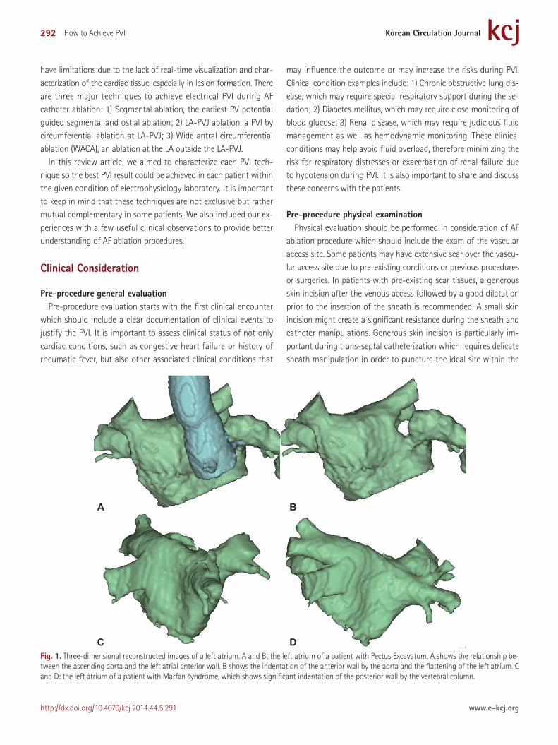
3-D reconstructed LA image can be used to guide the catheter manipulation during PVI. The potential ablation sites or lines can be pre-selected and planned prior to PVI. The anatomy of LA-PVJ and its relationship with the esophagus or descending aorta may force a change in the ablation strategy to avoid collateral damages. For example, if the left PV posterior wall is in close contact with the esophagus, segmental ostial PVI of the posterior wall combined with the continuous ablation at the remaining part of LA-PVJ or the antrum could be planned (Fig. 2).
From our past experiences, we have learned that CT and MRI may diagnose unsuspected non-cardiac conditions such as lung mass or
A
C
B
D Fig. 2. The proximity of esophagus to the left pulmonary veins. The line on A shows the location of the esophagus during the CT imaging, which was lo-cated close to the ablation line (B). Continuous ablation was done in the left lateral ridge, superior, inferior, and carina of the ridge side (C). Afterwards seg-mental isolation of the pulmonary veins was performed following the earliest activation site of the pulmonary veins to avoid possible esophageal injury (D).
294 How to Achieve PVI
http://dx.doi.org/10.4070/kcj.2014.44.5.291 www.e-kcj.org
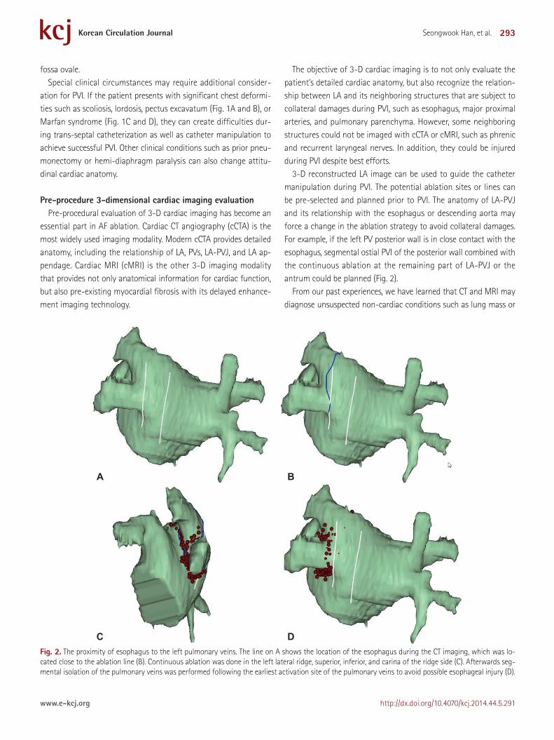
cancer, large hiatal hernia (Fig. 3), or significant vertebral deformi-ties, which may change the planned ablation strategies or abandon the ablation completely.
Procedural Consideration
Of the three major ablation techniques, the best one is determined by the LA anatomy and its surrounding structures. Understanding the anatomy and the histology of PV and LA-PVJ are very important for effective catheter ablation.
Anatomical and histological considerationHistological and embryological studies of the human LA revealed
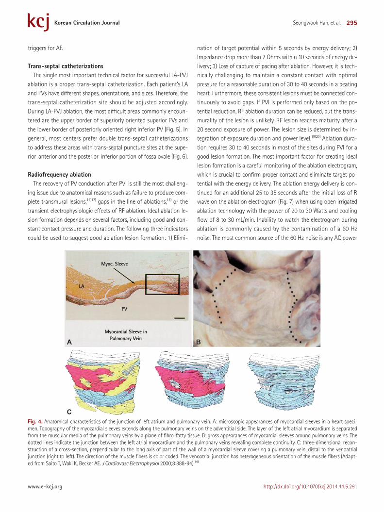
complex and wide variations during the development of LA and PVs from primordial LA and venous system, which may explain the wide anatomical variations at the LA-PVJ and PVs observed in patients with AF. These variations may also occur not only at gross anatomi-cal level but also at histological level, resulting in the differences in the length, orientation, and thickness of the atrial myocardial fi-bers.13) The myocardial muscle fibers with a circumferential and spi-ral orientation form the thick heterogeneous ostial muscle sleeves, which may progressively become thinner toward the distal PV and end up being organized to parallel along the PV long axis (Fig. 4). It
is important to note that these myocardial sleeves are predomi-nantly epicardial structures and that their lengths vary widely be-tween the veins as well as the diameters of the PVs.14) These histo-logical characteristics of myocardial sleeve directly influence the ablation lesion formation and ultimately the long-term PVI. Al-though it is easy to achieve transmural lesions inside the PV with a small amount of ablation due to the thin and homogenously orient-ed myocardial fibers, it has a high risk of PV stenosis and collateral damages. In contrast, the WACA technique requires a much larger number of ablation and higher energy to achieve complete isolation because thick atrial myocardial sleeves with multiple muscle layers present in most of the PV antrum, which is less likely to achieve ho-mogenous transmural lesions in the entire circumference with the current available ablation technologies. Therefore, those circum-stances favor the use of LA-PVJ ablation technique. LA-PVJ ablation could also eliminate other possible triggers, such as Marshall bun-dle and posterior LA wall. In addition, LA-PVJ ablation might alter the arrhythmogenic substrate that may generate or perpetuate AF and/or reduce the mass of atrial tissue needed to sustain reentry. Furthermore, neuro-anatomical studies at the LA-PVJ revealed cir-cumferential distribution of a high number of autonomic parasym-pathetic and sympathetic nerves in LA-PVJ.15) Therefore, LA-PVJ ablation may interrupt the autonomic innervations, the potential
Fig. 3. Images of hiatal hernia of a stomach. A and B: the thoracic portion of the stomach (arrow) has very close contact to the posterior wall of the left atrium on CT images. C and D show 3-dimensional relationship between the herniated stomach (arrow) with the esophagus and the left atrium.
A
C
B
D
LALA
295Seongwook Han, et al.
http://dx.doi.org/10.4070/kcj.2014.44.5.291www.e-kcj.org
triggers for AF.
Trans-septal catheterizationsThe single most important technical factor for successful LA-PVJ
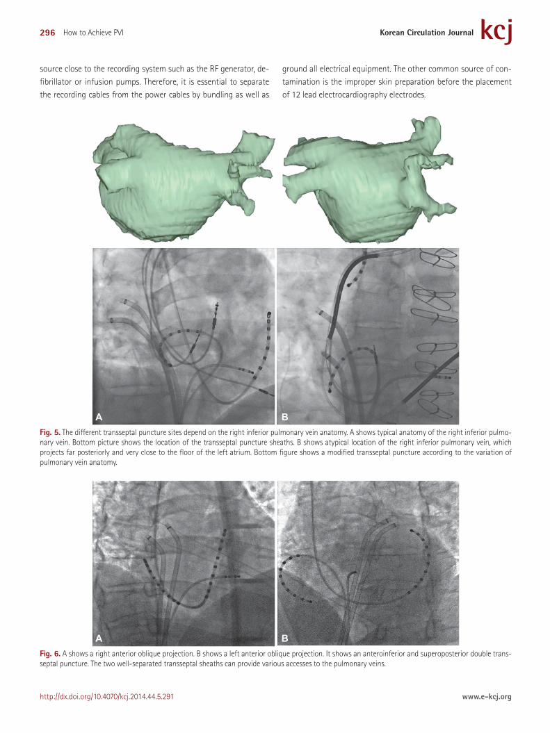
ablation is a proper trans-septal catheterization. Each patient’s LA and PVs have different shapes, orientations, and sizes. Therefore, the trans-septal catheterization site should be adjusted accordingly. During LA-PVJ ablation, the most difficult areas commonly encoun-tered are the upper border of superiorly oriented superior PVs and the lower border of posteriorly oriented right inferior PV (Fig. 5). In general, most centers prefer double trans-septal catheterizations to address these areas with trans-septal puncture sites at the supe-rior-anterior and the posterior-inferior portion of fossa ovale (Fig. 6).
Radiofrequency ablation The recovery of PV conduction after PVI is still the most challeng-
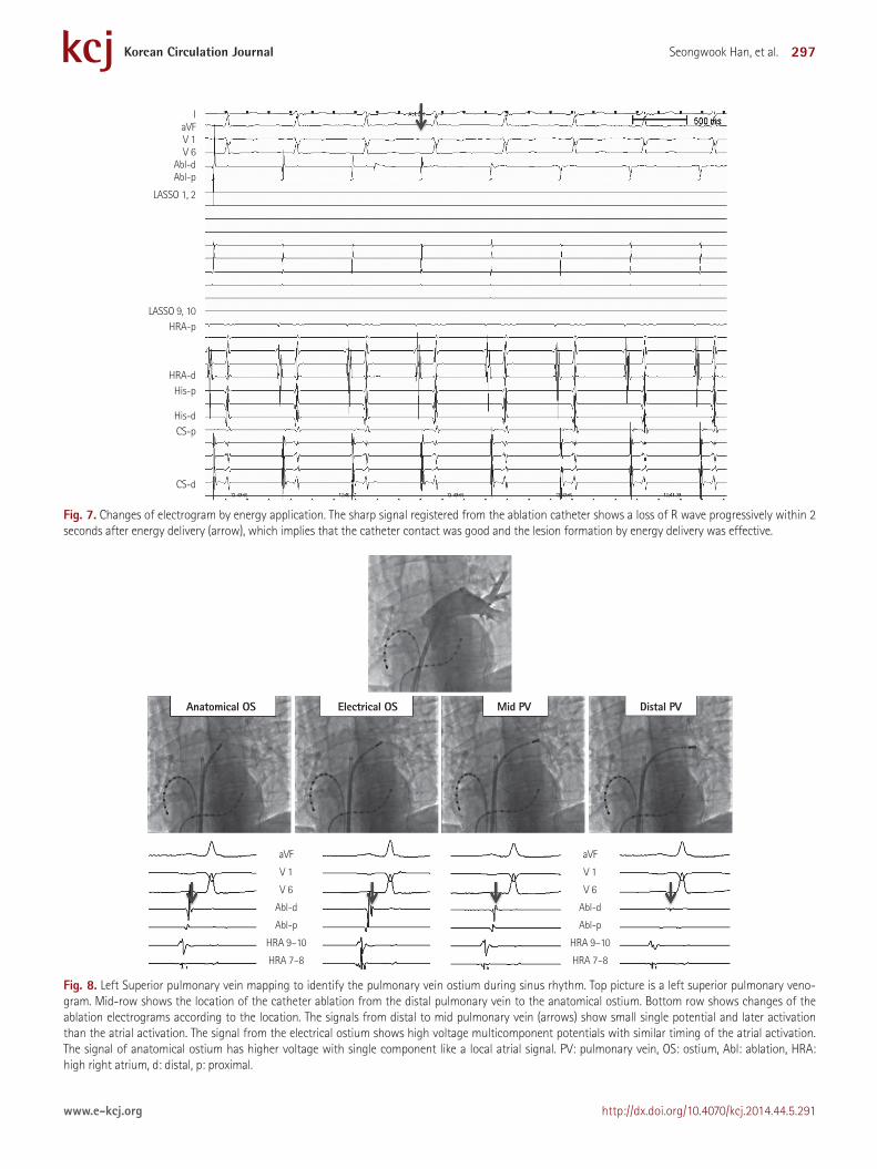
ing issue due to anatomical reasons such as failure to produce com-plete transmural lesions,16)17) gaps in the line of ablations,18) or the transient electrophysiologic effects of RF ablation. Ideal ablation le-sion formation depends on several factors, including good and con-stant contact pressure and duration. The following three indicators could be used to suggest good ablation lesion formation: 1) Elimi-
nation of target potential within 5 seconds by energy delivery; 2) Impedance drop more than 7 Ohms within 10 seconds of energy de-livery; 3) Loss of capture of pacing after ablation. However, it is tech-nically challenging to maintain a constant contact with optimal pressure for a reasonable duration of 30 to 40 seconds in a beating heart. Furthermore, these consistent lesions must be connected con-tinuously to avoid gaps. If PVI is performed only based on the po-tential reduction, RF ablation duration can be reduced, but the trans-murality of the lesion is unlikely. RF lesion reaches maturity after a 20 second exposure of power. The lesion size is determined by in-tegration of exposure duration and power level.19)20) Ablation dura-tion requires 30 to 40 seconds in most of the sites during PVI for a good lesion formation. The most important factor for creating ideal lesion formation is a careful monitoring of the ablation electrogram, which is crucial to confirm proper contact and eliminate target po-tential with the energy delivery. The ablation energy delivery is con-tinued for an additional 25 to 35 seconds after the initial loss of R wave on the ablation electrogram (Fig. 7) when using open irrigated ablation technology with the power of 20 to 30 Watts and cooling flow of 8 to 30 mL/min. Inability to watch the electrogram during ablation is commonly caused by the contamination of a 60 Hz noise. The most common source of the 60 Hz noise is any AC power
Fig. 4. Anatomical characteristics of the junction of left atrium and pulmonary vein. A: microscopic appearances of myocardial sleeves in a heart speci-men. Topography of the myocardial sleeves extends along the pulmonary veins on the adventitial side. The layer of the left atrial myocardium is separated from the muscular media of the pulmonary veins by a plane of fibro-fatty tissue. B: gross appearances of myocardial sleeves around pulmonary veins. The dotted lines indicate the junction between the left atrial myocardium and the pulmonary veins revealing complete continuity. C: three-dimensional recon-struction of a cross-section, perpendicular to the long axis of part of the wall of a myocardial sleeve covering a pulmonary vein, distal to the venoatrial junction (right to left). The direction of the muscle fibers is color coded. The venoatrial junction has heterogeneous orientation of the muscle fibers (Adapt-ed from Saito T, Waki K, Becker AE. J Cardiovasc Electrophysiol 2000;8:888-94).14)
A
C
B
Myoc. Sleeve
LA
PV
Myocardial Sleeve inPulmonary Vein
296 How to Achieve PVI
http://dx.doi.org/10.4070/kcj.2014.44.5.291 www.e-kcj.org
source close to the recording system such as the RF generator, de-fibrillator or infusion pumps. Therefore, it is essential to separate the recording cables from the power cables by bundling as well as
ground all electrical equipment. The other common source of con-tamination is the improper skin preparation before the placement of 12 lead electrocardiography electrodes.
A B Fig. 5. The different transseptal puncture sites depend on the right inferior pulmonary vein anatomy. A shows typical anatomy of the right inferior pulmo-nary vein. Bottom picture shows the location of the transseptal puncture sheaths. B shows atypical location of the right inferior pulmonary vein, which projects far posteriorly and very close to the floor of the left atrium. Bottom figure shows a modified transseptal puncture according to the variation of pulmonary vein anatomy.
A B Fig. 6. A shows a right anterior oblique projection. B shows a left anterior oblique projection. It shows an anteroinferior and superoposterior double trans-septal puncture. The two well-separated transseptal sheaths can provide various accesses to the pulmonary veins.
297Seongwook Han, et al.
http://dx.doi.org/10.4070/kcj.2014.44.5.291www.e-kcj.org
Fig. 7. Changes of electrogram by energy application. The sharp signal registered from the ablation catheter shows a loss of R wave progressively within 2 seconds after energy delivery (arrow), which implies that the catheter contact was good and the lesion formation by energy delivery was effective.
IaVFV 1V 6
Abl-dAbl-p
LASSO 1, 2
LASSO 9, 10HRA-p
HRA-dHis-p
His-dCS-p
CS-d
Fig. 8. Left Superior pulmonary vein mapping to identify the pulmonary vein ostium during sinus rhythm. Top picture is a left superior pulmonary veno-gram. Mid-row shows the location of the catheter ablation from the distal pulmonary vein to the anatomical ostium. Bottom row shows changes of the ablation electrograms according to the location. The signals from distal to mid pulmonary vein (arrows) show small single potential and later activation than the atrial activation. The signal from the electrical ostium shows high voltage multicomponent potentials with similar timing of the atrial activation. The signal of anatomical ostium has higher voltage with single component like a local atrial signal. PV: pulmonary vein, OS: ostium, Abl: ablation, HRA: high right atrium, d: distal, p: proximal.
Anatomical OS Electrical OS Mid PV Distal PV
aVF
V 1
V 6
Abl-d
Abl-p
HRA 9–10
HRA 7–8
aVF
V 1
V 6
Abl-d
Abl-p
HRA 9–10
HRA 7–8
298 How to Achieve PVI
http://dx.doi.org/10.4070/kcj.2014.44.5.291 www.e-kcj.org
Left atrium-pulmonary vein junction identification and ablation
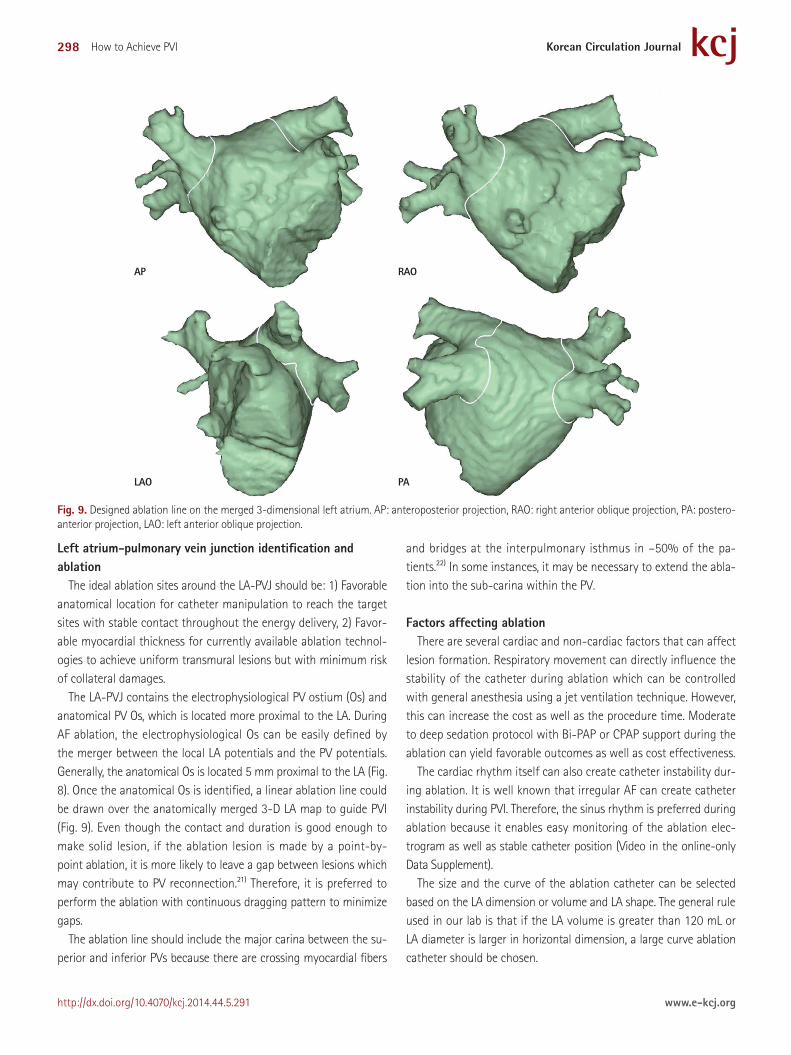
The ideal ablation sites around the LA-PVJ should be: 1) Favorable anatomical location for catheter manipulation to reach the target sites with stable contact throughout the energy delivery, 2) Favor-able myocardial thickness for currently available ablation technol-ogies to achieve uniform transmural lesions but with minimum risk of collateral damages.
The LA-PVJ contains the electrophysiological PV ostium (Os) and anatomical PV Os, which is located more proximal to the LA. During AF ablation, the electrophysiological Os can be easily defined by the merger between the local LA potentials and the PV potentials. Generally, the anatomical Os is located 5 mm proximal to the LA (Fig. 8). Once the anatomical Os is identified, a linear ablation line could be drawn over the anatomically merged 3-D LA map to guide PVI (Fig. 9). Even though the contact and duration is good enough to make solid lesion, if the ablation lesion is made by a point-by-point ablation, it is more likely to leave a gap between lesions which may contribute to PV reconnection.21) Therefore, it is preferred to perform the ablation with continuous dragging pattern to minimize gaps.
The ablation line should include the major carina between the su-perior and inferior PVs because there are crossing myocardial fibers
and bridges at the interpulmonary isthmus in –50% of the pa-tients.22) In some instances, it may be necessary to extend the abla-tion into the sub-carina within the PV.
Factors affecting ablationThere are several cardiac and non-cardiac factors that can affect
lesion formation. Respiratory movement can directly influence the stability of the catheter during ablation which can be controlled with general anesthesia using a jet ventilation technique. However, this can increase the cost as well as the procedure time. Moderate to deep sedation protocol with Bi-PAP or CPAP support during the ablation can yield favorable outcomes as well as cost effectiveness.
The cardiac rhythm itself can also create catheter instability dur-ing ablation. It is well known that irregular AF can create catheter instability during PVI. Therefore, the sinus rhythm is preferred during ablation because it enables easy monitoring of the ablation elec-trogram as well as stable catheter position (Video in the online-only Data Supplement).
The size and the curve of the ablation catheter can be selected based on the LA dimension or volume and LA shape. The general rule used in our lab is that if the LA volume is greater than 120 mL or LA diameter is larger in horizontal dimension, a large curve ablation catheter should be chosen.
Fig. 9. Designed ablation line on the merged 3-dimensional left atrium. AP: anteroposterior projection, RAO: right anterior oblique projection, PA: postero-anterior projection, LAO: left anterior oblique projection.
AP
LAO
RAO
PA
299Seongwook Han, et al.
http://dx.doi.org/10.4070/kcj.2014.44.5.291www.e-kcj.org
Acute pulmonary vein isolation confirmation and provocation testing
Pulmonary vein isolation is confirmed with a circular catheter once the ablation is completed. Elimination of PV potentials is the primary endpoint for PV ablation procedures in most centers. PVI can be confirmed when the dissociated PV potentials are observed. Some centers routinely perform pacing from inside PV to confirm the exit block after the elimination of the PV potentials.1) High out pacing from the ablation site to confirm the loss of capture has also been proposed as a good and acute predictor.23)
Acute PV reconnection can be observed up to 50% in 30 minutes following completion of PVI. However, it appears to occur continu-ously throughout 90 days even though majority of them are re-connected during the first 30 days after PVI. There are two phar-macological methods to detect acute PV reconnections. The most commonly used method is a high dose isoproterenol infusion for 10 to 15 minutes and a waiting time of 30 minutes after PVI. The more recent method is a high dose adenosine bolus injection to facilitate the acute reconnections. Regardless of the method used, acute PV reconnection is due to non-transmural lesions or gaps be-tween the ablation lesions. Therefore, reconfirmation of PVI at 20–30 minutes after initial PVI with or without pharmacologic chal-lenging is important to minimize the recurrences.
Late pulmonary vein reconduction after pulmonary vein isolation
Two hypothesized mechanisms are involved in post PVI reconduc-tion: 1) Acute reconnection, defined as first 90 days after the PVI, is the main reason of recurrences. This is primarily due to the failure to achieve continuous and complete trans-mural lesions; 2) Delayed recurrences which are observed over several years after the PVI.24) This is most likely due to the longitudinal ultra-structural and elec-trophysiological propriety changes from the healing mechanisms observed in scar tissues. However, there is no clinical data available to confirm the 2nd hypothesis. Both mechanisms may play roles in a given clinical setting.
In summary, PV reconnection is still the major clinical challenge in any form of AF ablation. Major limitations to successful PVI are: 1) The lack of good acute predictor for the continuous uniform trans-mural lesions; 2) The lack of ablation procedure endpoints to pre-dict long term success of PVI. Technologies that can improve the real time monitoring and the visualizing of the ablation lesion for-mation will improve the safety as well as the efficacy of AF ablation, which will ultimately improve the long-term outcome of PVI. Further clinical researches are merited to overcome these limitations.
Conclusion
Pulmonary vein isolation can be approached systematically from clinical to procedural aspects. One needs to develop the PVI protocol that is practical and feasible within the given health-care environ-ment so that it has reliable and reproducible results without com-plications or with very low complications.
AcknowledgmentsWe sincerely thank Janette Lancaster and Shannon Logsdon for
their assistance and contribution.
Supplementary MaterialsThe online-only Data Supplement is available with this article at
http://dx.doi.org/10.4070/kcj.2014.44.5.291.
References1. Calkins H, Kuck KH, Cappato R, et al. 2012 HRS/EHRA/ECAS expert
consensus statement on catheter and surgical ablation of atrial fibril-lation: recommendations for patient selection, procedural techniques, patient management and follow-up, definitions, endpoints, and re-search trial design: a report of the Heart Rhythm Society (HRS) Task Force on Catheter and Surgical Ablation of Atrial Fibrillation. Devel-oped in partnership with the European Heart Rhythm Association (EHRA), a registered branch of the European Society of Cardiology (ESC) and the European Cardiac Arrhythmia Society (ECAS); and in collaboration with the American College of Cardiology (ACC), Ameri-can Heart Association (AHA), the Asia Pacific Heart Rhythm Society (APHRS), and the Society of Thoracic Surgeons (STS). Endorsed by the governing bodies of the American College of Cardiology Foundation, the American Heart Association, the European Cardiac Arrhythmia So-ciety, the European Heart Rhythm Association, the Society of Thoracic Surgeons, the Asia Pacific Heart Rhythm Society, and the Heart Rhythm Society. Heart Rhythm 2012;9:632-96.
2. Calkins H, Reynolds MR, Spector P, et al. Treatment of atrial fibrillation with antiarrhythmic drugs or radiofrequency ablation: two systematic literature reviews and meta-analyses. Circ Arrhythm Electrophysiol 2009;2:349-61.
3. Piccini JP, Lopes RD, Kong MH, Hasselblad V, Jackson K, Al-Khatib SM. Pulmonary vein isolation for the maintenance of sinus rhythm in pa-tients with atrial fibrillation: a meta-analysis of randomized, controlled trials. Circ Arrhythm Electrophysiol 2009;2:626-33.
4. Parkash R, Tang AS, Sapp JL, Wells G. Approach to the catheter ablation technique of paroxysmal and persistent atrial fibrillation: a meta-anal-ysis of the randomized controlled trials. J Cardiovasc Electrophysiol 2011;22:729-38.
5. Haïssaguerre M, Jaïs P, Shah DC, et al. Spontaneous initiation of atrial fibrillation by ectopic beats originating in the pulmonary veins. N Engl J Med 1998;339:659-66.

300 How to Achieve PVI
http://dx.doi.org/10.4070/kcj.2014.44.5.291 www.e-kcj.org
6. Haïssaguerre M, Jaïs P, Shah DC, et al. Electrophysiological end point for catheter ablation of atrial fibrillation initiated from multiple pul-monary venous foci. Circulation 2000;101:1409-17.
7. Kalifa J, Jalife J, Zaitsev AV, et al. Intra-atrial pressure increases rate and organization of waves emanating from the superior pulmonary veins during atrial fibrillation. Circulation 2003;108:668-71.
8. Kumagai K, Ogawa M, Noguchi H, Yasuda T, Nakashima H, Saku K. Electrophysiologic properties of pulmonary veins assessed using a mul-tielectrode basket catheter. J Am Coll Cardiol 2004;43:2281-9.
9. Sanders P, Berenfeld O, Hocini M, et al. Spectral analysis identifies sites of high-frequency activity maintaining atrial fibrillation in humans. Circulation 2005;112:789-97.
10. Lin YJ, Tsao HM, Chang SL, et al. Role of high dominant frequency sites in nonparoxysmal atrial fibrillation patients: insights from high-density frequency and fractionation mapping. Heart Rhythm 2010;7:1255-62.
11. Cappato R, Calkins H, Chen SA, et al. Updated worldwide survey on the methods, efficacy, and safety of catheter ablation for human atrial fibrillation. Circ Arrhythm Electrophysiol 2010;3:32-8.
12. Arbelo E, Brugada J, Hindricks G, et al. ESC-EURObservational Research Programme: the Atrial Fibrillation Ablation Pilot Study, conducted by the European Heart Rhythm Association. Europace 2012;14:1094-103.
13. Hassink RJ, Aretz HT, Ruskin J, Keane D. Morphology of atrial myocar-dium in human pulmonary veins: a postmortem analysis in patients with and without atrial fibrillation. J Am Coll Cardiol 2003;42:1108-14.
14. Saito T, Waki K, Becker AE. Left atrial myocardial extension onto pul-monary veins in humans: anatomic observations relevant for atrial arrhythmias. J Cardiovasc Electrophysiol 2000;11:888-94.
15. Tan AY, Li H, Wachsmann-Hogiu S, Chen LS, Chen PS, Fishbein MC. Au-tonomic innervation and segmental muscular disconnections at the human pulmonary vein-atrial junction: implications for catheter abla-tion of atrial-pulmonary vein junction. J Am Coll Cardiol 2006;48:132-43.
16. McGann CJ, Kholmovski EG, Oakes RS, et al. New magnetic resonance imaging-based method for defining the extent of left atrial wall injury after the ablation of atrial fibrillation. J Am Coll Cardiol 2008;52: 1263-71.
17. Kowalski M, Grimes MM, Perez FJ, et al. Histopathologic characteriza-tion of chronic radiofrequency ablation lesions for pulmonary vein iso-lation. J Am Coll Cardiol 2012;59:930-8.
18. Ranjan R, Kato R, Zviman MM, et al. Gaps in the ablation line as a po-tential cause of recovery from electrical isolation and their visualiza-tion using MRI. Circ Arrhythm Electrophysiol 2011;4:279-86.
19. Wittkampf FH, Hauer RN, Robles de Medina EO. Control of radiofre-quency lesion size by power regulation. Circulation 1989;80:962-8.
20. Jain MK, Wolf PD. Temperature-controlled and constant-power radio-frequency ablation: what affects lesion growth? IEEE Trans Biomed Eng 1999;46:1405-12.
21. Kautzner J, Neuzil P, Peichl P, et al. Contact force, force time integral and lesion continuity are critical to improve durable PV isolation: EFFI-CAS II results. Heart Rhythm 2012;9:S28.
22. Cabrera JA, Ho SY, Climent V, Fuertes B, Murillo M, Sánchez-Quintana D. Morphological evidence of muscular connections between contigu-ous pulmonary venous orifices: relevance of the interpulmonary isth-mus for catheter ablation in atrial fibrillation. Heart Rhythm 2009;6: 1192-8.
23. Andrade JG, Pollak SJ, Monir G, et al. Pulmonary vein isolation using a pace-capture-guided versus an adenosine-guided approach: effect on dormant conduction and long-term freedom from recurrent atrial fi-brillation--a prospective study. Circ Arrhythm Electrophysiol 2013;6: 1103-8.
24. Sotomi Y, Inoue K, Ito N, et al. Cause of very late recurrence of atrial fi-brillation or flutter after catheter ablation for atrial fibrillation. Am J Cardiol 2013;111:552-6.