Embed Size (px)
Citation preview
University of Tartu
Faculty of Mathematics and Computer Science
Institute of Computer Science
Hodgkin-Huxley model simulator
MTAT.03.291 Introduction to Computational Neuroscience
Katrin Valdson
Kristiina Pokk
Marit Asula
Tartu 2015
Contents
Introduction
Part I: Equilibrium Potential
Part II: Membrane Potential
Part III: The Action Potential
Part IV: The Fast Sodium Channel
Drugs TTX, TEA and Pronase
Step Current Input
References
1
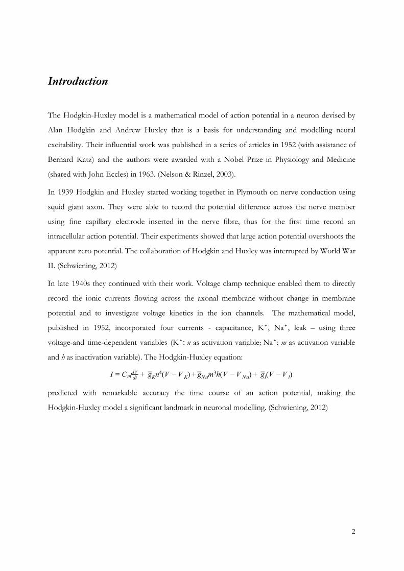
Introduction
The Hodgkin-Huxley model is a mathematical model of action potential in a neuron devised by
Alan Hodgkin and Andrew Huxley that is a basis for understanding and modelling neural
excitability. Their influential work was published in a series of articles in 1952 (with assistance of
Bernard Katz) and the authors were awarded with a Nobel Prize in Physiology and Medicine
(shared with John Eccles) in 1963. (Nelson & Rinzel, 2003).
In 1939 Hodgkin and Huxley started working together in Plymouth on nerve conduction using
squid giant axon. They were able to record the potential difference across the nerve member
using fine capillary electrode inserted in the nerve fibre, thus for the first time record an
intracellular action potential. Their experiments showed that large action potential overshoots the
apparent zero potential. The collaboration of Hodgkin and Huxley was interrupted by World War
II. (Schwiening, 2012)
In late 1940s they continued with their work. Voltage clamp technique enabled them to directly
record the ionic currents flowing across the axonal membrane without change in membrane
potential and to investigate voltage kinetics in the ion channels. The mathematical model,
published in 1952, incorporated four currents - capacitance, K⁺, Na⁺, leak – using three
voltage-and time-dependent variables (K⁺: n as activation variable; Na⁺: m as activation variable
and h as inactivation variable). The Hodgkin-Huxley equation:
g n (V ) m h(V ) g (V )I = Cm dtdV + K
4 − V K + gNa 3 − V Na + l − V l
predicted with remarkable accuracy the time course of an action potential, making the
Hodgkin-Huxley model a significant landmark in neuronal modelling. (Schwiening, 2012)
2
Part I: Equilibrium Potential
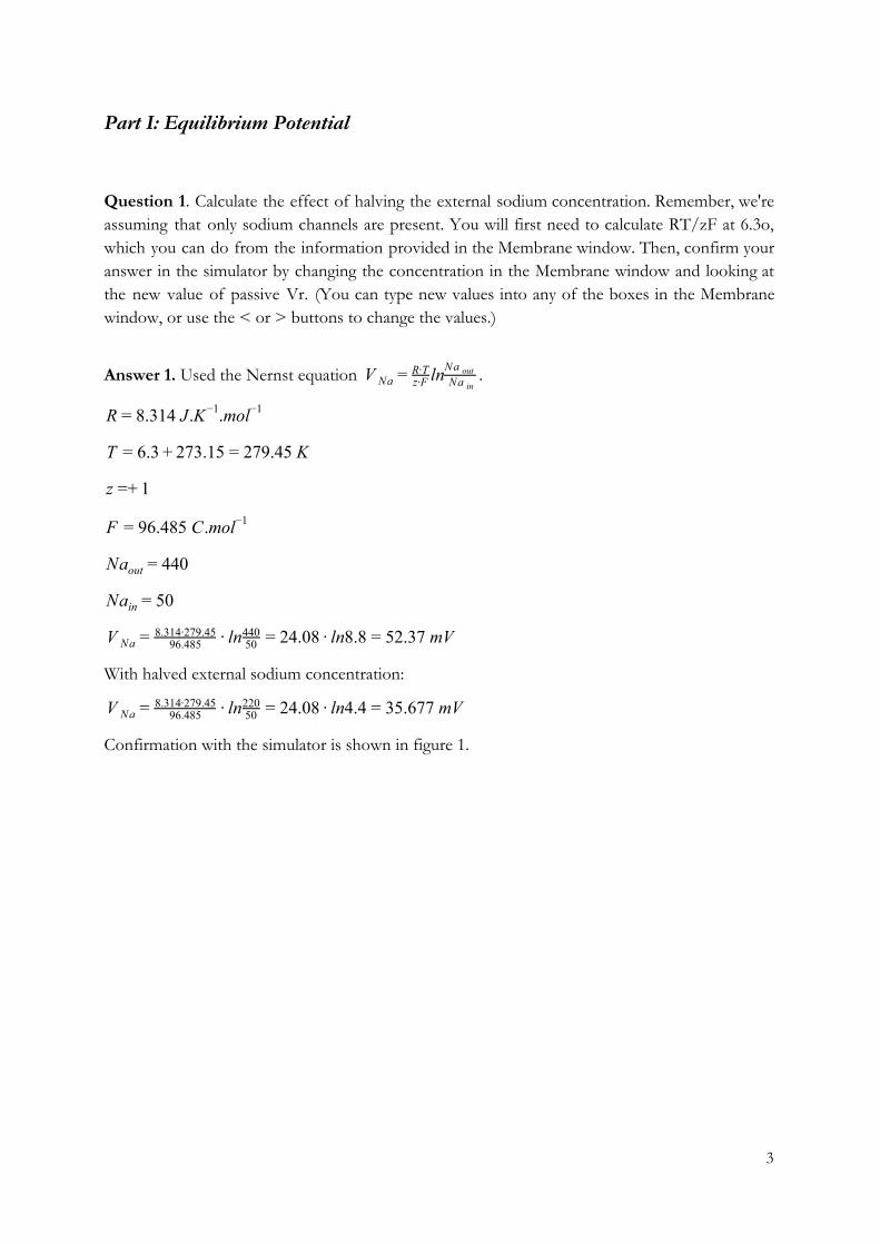
Question 1. Calculate the effect of halving the external sodium concentration. Remember, we're assuming that only sodium channels are present. You will first need to calculate RT/zF at 6.3o, which you can do from the information provided in the Membrane window. Then, confirm your answer in the simulator by changing the concentration in the Membrane window and looking at the new value of passive Vr. (You can type new values into any of the boxes in the Membrane window, or use the < or > buttons to change the values.)
Answer 1. Used the Nernst equation .ln V Na = z∙FR∙T
Na inNa out
.314 J.K .mol R = 8 −1 −1
.3 73.15 79.45 K T = 6 + 2 = 2
+ z = 1
6.485 C.mol F = 9 −1
40 Naout = 4
0 Nain = 5
n 4.08 n8.8 2.37 mV V Na = 96.4858.314∙279.45 ∙ l 50
440 = 2 ∙ l = 5
With halved external sodium concentration:
n 4.08 n4.4 5.677 mV V Na = 96.4858.314∙279.45 ∙ l 50
220 = 2 ∙ l = 3
Confirmation with the simulator is shown in figure 1.
3
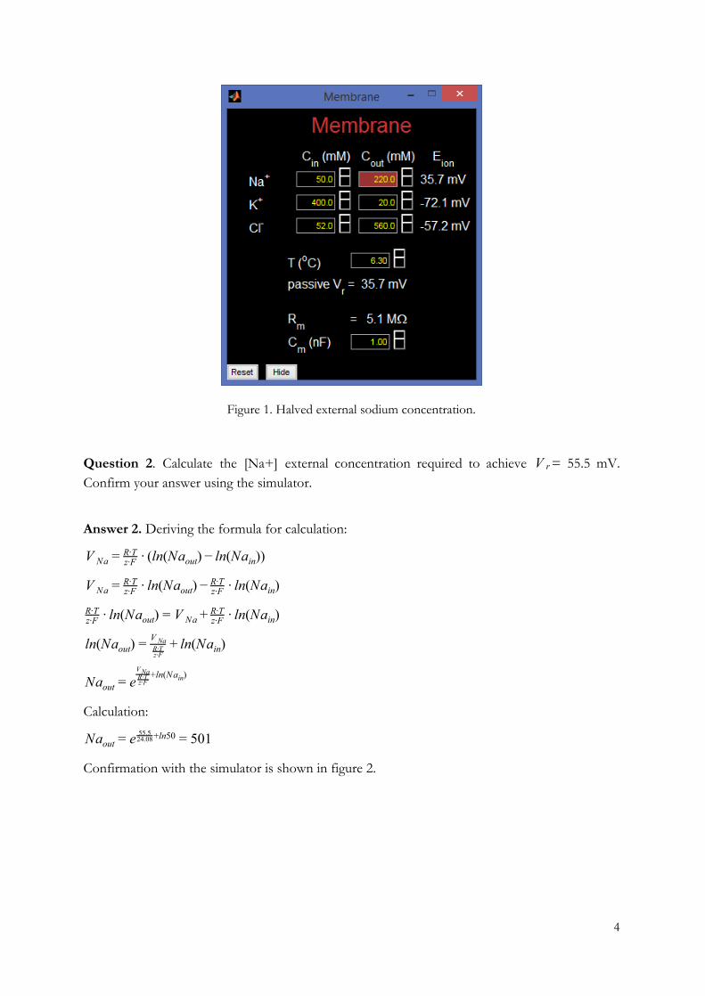
Figure 1. Halved external sodium concentration.
Question 2. Calculate the [Na+] external concentration required to achieve = 55.5 mV. V r Confirm your answer using the simulator.
Answer 2. Deriving the formula for calculation:
ln(Na ) n(Na )) V Na = z∙FR∙T ∙ ( out − l in
n(Na ) n(Na ) V Na = z∙FR∙T ∙ l out − z∙F
R∙T ∙ l in
n(Na ) n(Na ) z∙FR∙T ∙ l out = V Na + z∙F
R∙T ∙ l in
n(Na ) n(Na )l out =z∙FR∙TV Na + l in
Naout = e+ln(Na )
z∙FR∙TVNa
in
Calculation:
01 Naout = e +ln5055.524.08 = 5
Confirmation with the simulator is shown in figure 2.
4
Figure 2. [Na+] external concentration required to achieve = 55.5 mVV r
Question 3. Suppose we double the temperature from 6.3o C to 12.6o C. What is the new value of Vr? Explain why Vr doesn't double.
Answer 3.
n 4.623 n8.8 3.55 mV V Na = 96.485
8.314∙285.75 ∙ l 50440 = 2 ∙ l = 5
does not double because the temperature is in K not Celsius in the formula. SinceV R , multiplying the Celsius degrees by 2 does not multiply K by 2:73.15 elsius K = 2 +C
73.15 elsius = 273.15 elsius) 2 + 2 ∙C / 2 ∙ ( +C
Thus, the temperature value in K changes only by a little.
Part II: Membrane Potential
Question 1. Write down the parallel conductance equation for the resting potential as a function
of equilibrium potentials and conductances. Given the equilibrium potentials shown in the
5
Membrane window and the conductances shown in the Channels window, calculate the resting
potential . V r
Answer 1. = 0 is equal to the flow of current through the passive channels and current I leak
through one leak channel can be calculated from the equation:
,g V ) I i = i ∙ ( m − Ei
where denotes the given ion channel (Na, K or Cl), denotes the conductance per unit area, i gi
denotes membrane potential and denotes reversal potential of the i-th ion channel. V m Ei
In our case , where denotes the resting potential. The current through all the leak V m = V r V r
channels can be calculated from the equation:
, 0 g ∙ (V ) g ∙ (V ) g ∙ (V ) I leak = = Na r − ENa + K r − EK + Cl r − ECl
from where we can find followingly: V r
0 g ∙ V g ∙ E g ∙ V ∙ E g ∙ V ∙ E I leak = = Na r − Na Na + K r − gK K + Cl r − gCl Cl
∙ V g ∙ V g ∙ V g ∙ E g ∙ E g ∙ E gNa r + K r + Cl r = Na Na + K K + Cl Cl
∙ (g g g ) g ∙ E g ∙ E g ∙ E V r Na + K + Cl = Na Na + K K + Cl Cl
V r = g + g + gNa K Cl
g ∙ E + g ∙ E + g ∙ ENa Na K K Cl Cl
V r = 0.0265 + 0.07 + 0.1 0.0265 ∙ 52.4 + 0.07 ∙ (−72.1) + 0.1 ∙ (−57.2)
7.7 V r = − 4
Confirmation with the simulator is shown in figure 3 (although the simulator value is slightly
different).
6
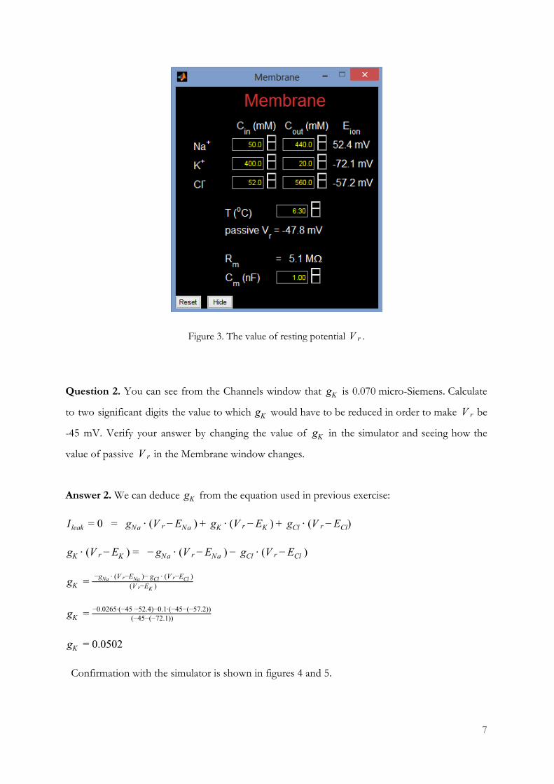
Figure 3. The value of resting potential .V r
Question 2. You can see from the Channels window that is 0.070 micro-Siemens. Calculate gK
to two significant digits the value to which would have to be reduced in order to make be gK V r
-45 mV. Verify your answer by changing the value of in the simulator and seeing how the gK
value of passive in the Membrane window changes. V r
Answer 2. We can deduce from the equation used in previous exercise: gK
g ∙ (V ) g ∙ (V ) g ∙ (V ) I leak = 0 = Na r − ENa + K r − EK + Cl r − ECl
∙ (V ) − ∙ (V ) g ∙ (V ) gK r − EK = gNa r − ENa − Cl r − ECl
gK = (V −E )r K
−g ∙ (V −E )− g ∙ (V −E )Na r Na Cl r Cl
gK = (−45−(−72.1))−0.0265∙(−45 −52.4)−0.1∙(−45−(−57.2))
.0502 gK = 0
Confirmation with the simulator is shown in figures 4 and 5.
7
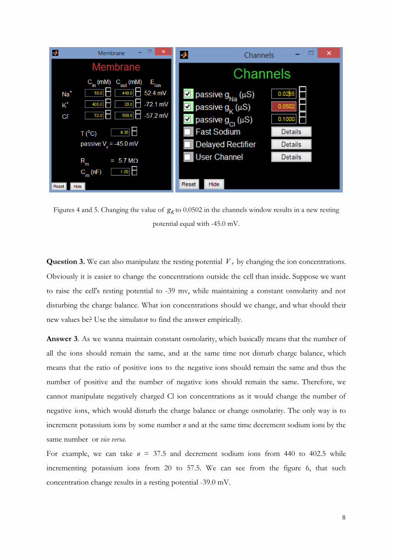
Figures 4 and 5. Changing the value of to 0.0502 in the channels window results in a new restinggK
potential equal with -45.0 mV.
Question 3. We can also manipulate the resting potential by changing the ion concentrations. V r
Obviously it is easier to change the concentrations outside the cell than inside. Suppose we want
to raise the cell's resting potential to -39 mv, while maintaining a constant osmolarity and not
disturbing the charge balance. What ion concentrations should we change, and what should their
new values be? Use the simulator to find the answer empirically.
Answer 3. As we wanna maintain constant osmolarity, which basically means that the number of
all the ions should remain the same, and at the same time not disturb charge balance, which
means that the ratio of positive ions to the negative ions should remain the same and thus the
number of positive and the number of negative ions should remain the same. Therefore, we
cannot manipulate negatively charged Cl ion concentrations as it would change the number of
negative ions, which would disturb the charge balance or change osmolarity. The only way is to
increment potassium ions by some number n and at the same time decrement sodium ions by the
same number or vice versa.
For example, we can take n = 37.5 and decrement sodium ions from 440 to 402.5 while
incrementing potassium ions from 20 to 57.5. We can see from the figure 6, that such
concentration change results in a resting potential -39.0 mV.
8
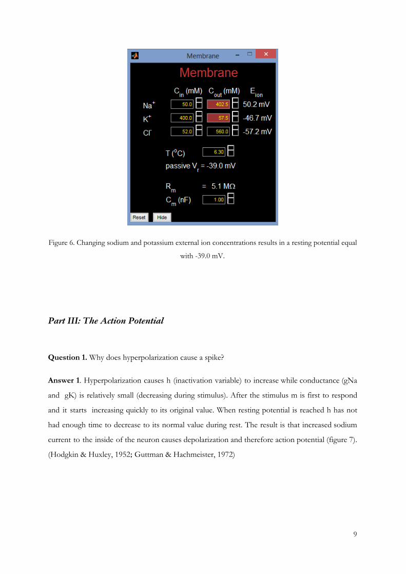
Figure 6. Changing sodium and potassium external ion concentrations results in a resting potential equal
with -39.0 mV.
Part III: The Action Potential
Question 1. Why does hyperpolarization cause a spike?
Answer 1. Hyperpolarization causes h (inactivation variable) to increase while conductance (gNa
and gK) is relatively small (decreasing during stimulus). After the stimulus m is first to respond
and it starts increasing quickly to its original value. When resting potential is reached h has not
had enough time to decrease to its normal value during rest. The result is that increased sodium
current to the inside of the neuron causes depolarization and therefore action potential (figure 7).
(Hodgkin & Huxley, 1952; Guttman & Hachmeister, 1972)
9
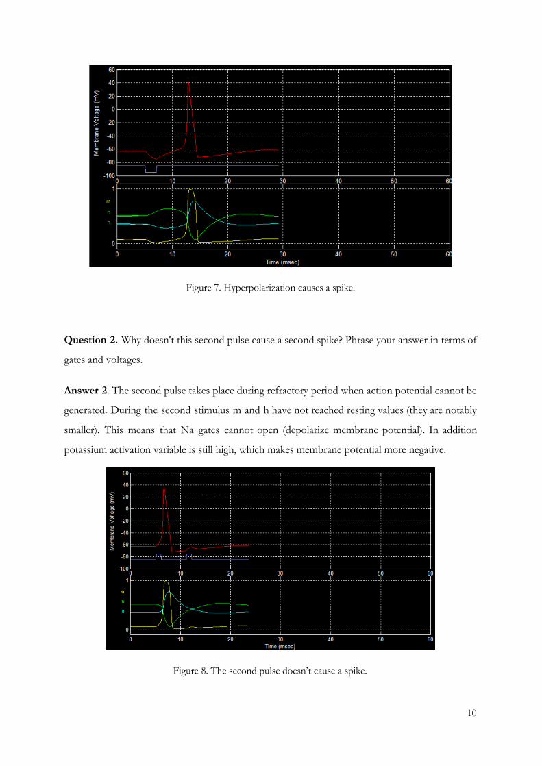
Figure 7. Hyperpolarization causes a spike.
Question 2. Why doesn't this second pulse cause a second spike? Phrase your answer in terms of
gates and voltages.
Answer 2. The second pulse takes place during refractory period when action potential cannot be
generated. During the second stimulus m and h have not reached resting values (they are notably
smaller). This means that Na gates cannot open (depolarize membrane potential). In addition
potassium activation variable is still high, which makes membrane potential more negative.
Figure 8. The second pulse doesn’t cause a spike.
10
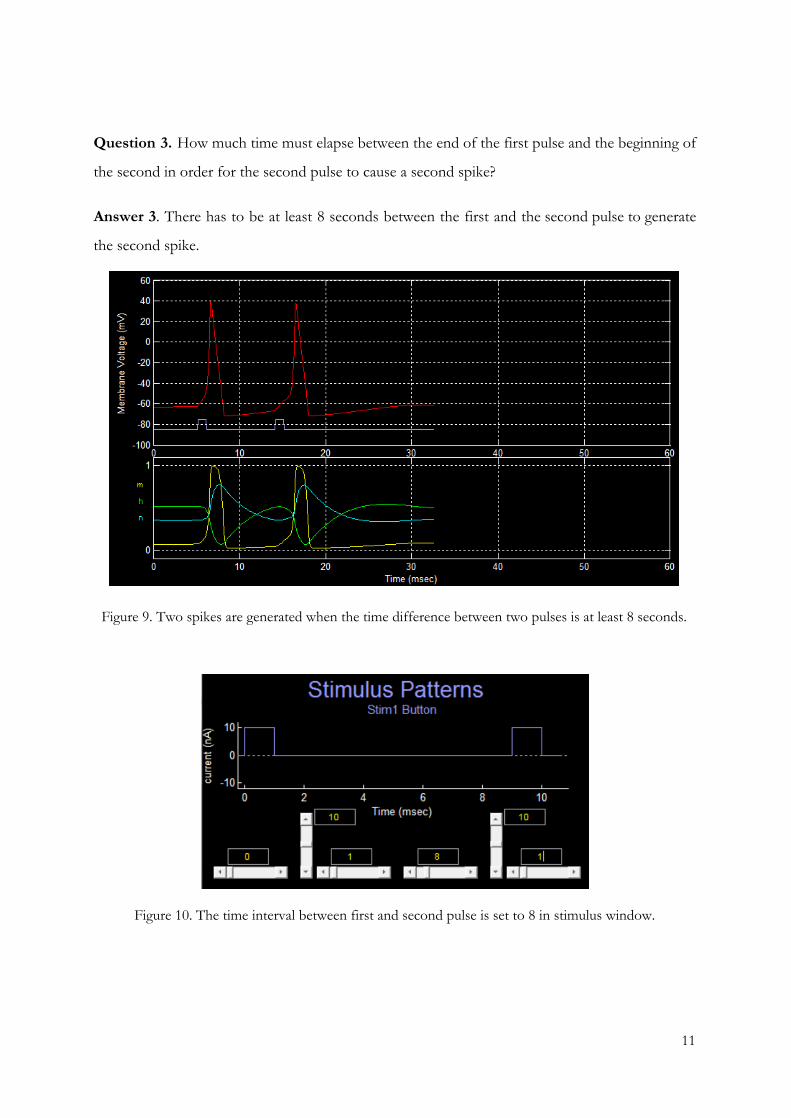
Question 3. How much time must elapse between the end of the first pulse and the beginning of
the second in order for the second pulse to cause a second spike?
Answer 3. There has to be at least 8 seconds between the first and the second pulse to generate
the second spike.
Figure 9. Two spikes are generated when the time difference between two pulses is at least 8 seconds.
Figure 10. The time interval between first and second pulse is set to 8 in stimulus window.
11
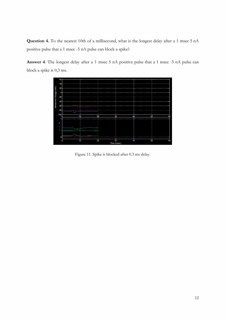
Question 4. To the nearest 10th of a millisecond, what is the longest delay after a 1 msec 5 nA
positive pulse that a 1 msec -5 nA pulse can block a spike?
Answer 4. The longest delay after a 1 msec 5 nA positive pulse that a 1 msec -5 nA pulse can
block a spike is 0,3 ms.
Figure 11. Spike is blocked after 0.3 ms delay.
12
Part IV: The Fast Sodium Channel
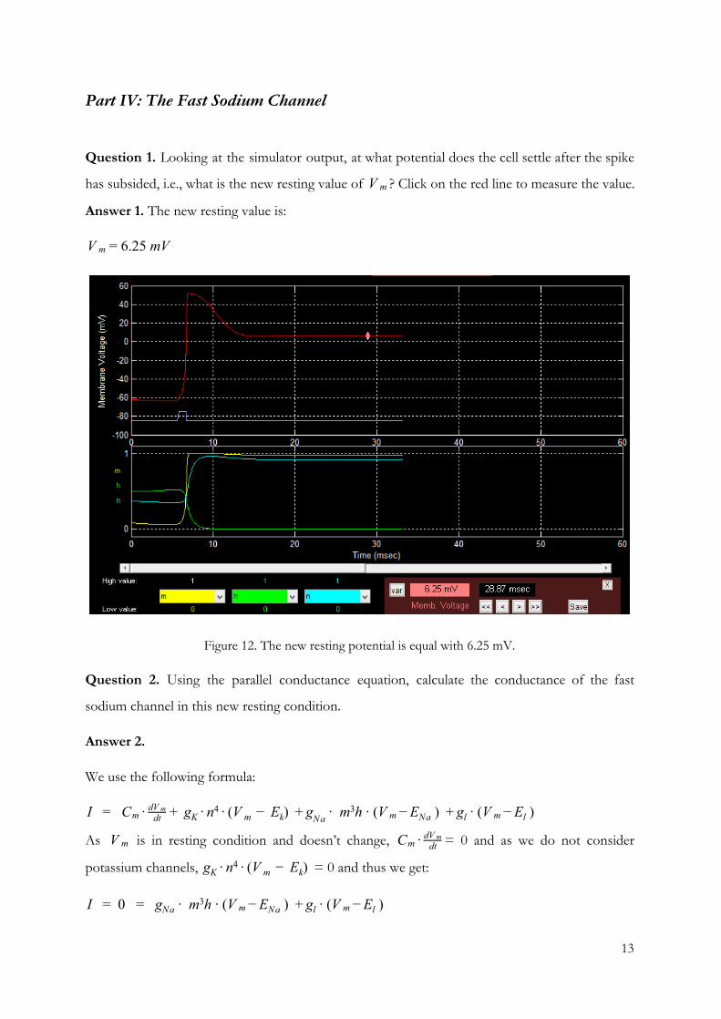
Question 1. Looking at the simulator output, at what potential does the cell settle after the spike
has subsided, i.e., what is the new resting value of ? Click on the red line to measure the value. V m
Answer 1. The new resting value is:
.25 mV V m = 6
Figure 12. The new resting potential is equal with 6.25 mV.
Question 2. Using the parallel conductance equation, calculate the conductance of the fast
sodium channel in this new resting condition.
Answer 2.
We use the following formula:
C g V E ) ∙ m h ∙ (V ) ∙ (V ) I = m ∙ dtdV m + K ∙ n4 ∙ ( m − k + gNa
3m − ENa + gl m − El
As is in resting condition and doesn’t change, = 0 and as we do not consider V m Cm ∙ dtdV m
potassium channels, = 0 and thus we get:V E ) gK ∙ n4 ∙ ( m − k
0 g ∙ m h ∙ (V ) ∙ (V ) I = = Na3
m − ENa + gl m − El
13
0 = g ∙ m h ∙ (V ) ∙ (V ) g ∙ (V ) g ∙ (V ) (x)Na3
m − ENa + gNa m − ENa + K m − EK + Cl m − ECl
, whereg(x)Na = m h ∙ (V −E )3 m Na
− (g ∙ (V − E ) + g ∙ (V − E ) + g ∙ (V − E ))Na m Na K m K Cl m Cl
= 0.0265 gNa
= 0.0700 gK
= 0.1 gCl
= 6.25 V m
= 52.4ENa
= -72.1EK
= -57.2 ECl
m = 0.984
h = 0.00201
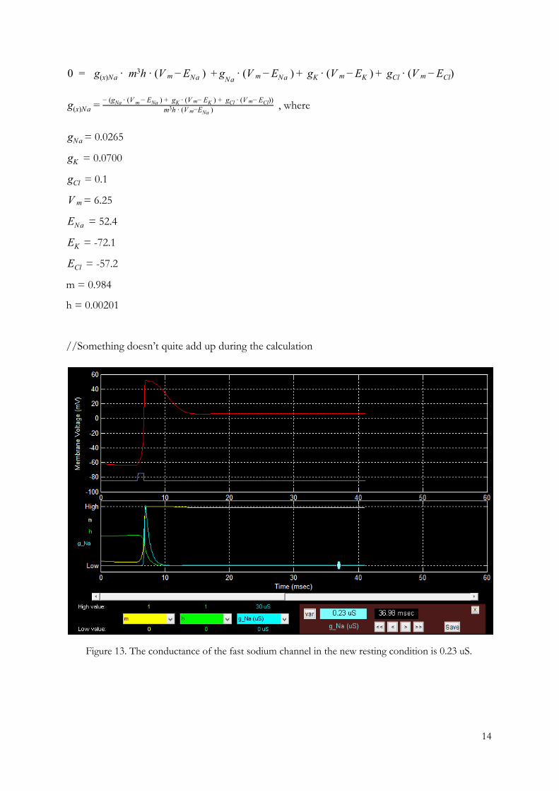
//Something doesn’t quite add up during the calculation
Figure 13. The conductance of the fast sodium channel in the new resting condition is 0.23 uS.
14

Question 3. Stimulating the cell again (using the Stim1 button) in this condition will not cause
another spike. Even if you raise the stimulus intensity to 20 nA and the duration to 10 msec, the
cell will not spike. (Try it. Turn off the cyan plot by selecting "-" in the pop-up menu so you can
see the yellow and green plots clearly when you hit Stim1.) What is the explanation for this?
Answer 3. Activation variable m and inactivation variable h are almost not affected by the
stimulation, which means that they are balanced and thus spikes cannot occur.
Part V: The Delayed Rectifier
Question 1. Based on the plot (red line), what is the peak value reached by the membrane V m
voltage?
Answer 1. Peak value reached by the membrane voltage is -15,9 mV
Figure 14. The peak value of membrane potential is -15.9 mV.
Question 2. After reaching its peak, the membrane voltage quickly declines again, even though
the stimulus is still on. What is causing this?
Answer 2. K⁺channels open (n increases), allowing K⁺to flow out of the neuron, making
membrane potential more negative.
15
Question 3. When the stimulus ends, the cell does not simply return to its resting value; it
undershoots it and then approaches the value from below. Why doesn't it just return to its resting
value?
Answer 3. Potassium channels remain open to the point when membrane potential is reaching
the equilibrium potential of K⁺ (about -70mV).
Part VI: Voltage-Gated Channel Parameters
Question 1. How does g_max for the fast sodium channel compare with the passive sodium conductance?
Answer 1. The passive sodium conductance is a lot smaller than the value of.0265 gNa = 0 .20 gmax = 1
Question 2. What relationship must hold between the red and blue lines for m to increase? And, what is the approximate value of Vm at which this condition occurs (if there were no other mechanisms affecting the cell)? How does this compare to the cell's normal resting potential?
Answer 2. The rate at which m changes is expressed by the formula . Because(1 ) m dtdm = α −m − β
m is the fraction of channels with open activation gates and is the rate at which segments moveα from closed to open, we multiply with . is the rate at which segments move from openα 1 −m β to closed, thus we multiply with . Since we are looking at the case where m increases, we getβ m the following inequality:
(1 ) m dtdm = α −m − β > 0
So the relationship between and is the following:α β
1 ) ( −m ∙ α > m ∙ β
The value for this condition depends on the value of m. For instance if , then for m to V m m = 0 increase, , which applies for all values of . If , then for m to increase, ,α > 0 V m .5 m = 0 α > β which occurs at around . And if m=1, then m can not increase further, thus the formula0 mV − 4 says , which does not happen.β < 0
The normal resting potential is lower than the value when in the case where . V m α > β .5 m = 0
Question 3. In order to stop the cell from oscillating on its own, we can change the passive channel conductance. Hit the Run button in the main simulator window to continue the simulation. By playing with the value of g_K in the Channels window, find a value close to the
16
original value of 0.07 micro-Siemens that prevents the cell from spiking spontaneously. The cell should of course still spike in reponse to a stimulus from the Stim1 button. Report the g_K value you find, to two significant digits.
Answer 3. To stop the oscillation, it is necessary to increase the value. At 0.17 the spikes still gK do not stop, the closest value to 0.07 which made the random spiking stop was 0.18.
Drugs TTX, TEA and Pronase
Question 1. Simulate with the "Drug window" the role of drugs TTX, TEA, and pronase on the
voltage of the neuron. Describe where these drugs are found, which channel affect, and how do
they affect the action potential.
TTX
Tetrodotoxin
Tetrodotoxin or TTX is a potent neurotoxin, being about 1200 times more dangerous to humans
than cyanide. It’s most well-known source is the pufferfish (fugu) and the toxin was even named
after it (Tetraodontidae). It has been later discovered that other animals such as blue-ringed
octopuses, marine worms, star fish and some terrestrial animals: salamanders, frogs. It is likely
that TTX is provided by bacteria residing in the animals that are poisonous. (Map of Life -
"Tetrodotoxin", 2015)
17
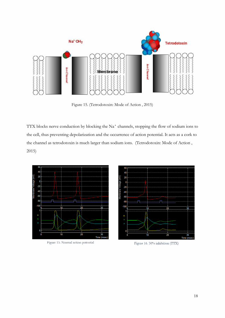
Figure 15. (Tetrodotoxin: Mode of Action , 2015)
TTX blocks nerve conduction by blocking the Na⁺ channels, stopping the flow of sodium ions to
the cell, thus preventing depolarization and the occurrence of action potential. It acts as a cork to
the channel as tetrodotoxin is much larger than sodium ions. (Tetrodotoxin: Mode of Action ,
2015)
18
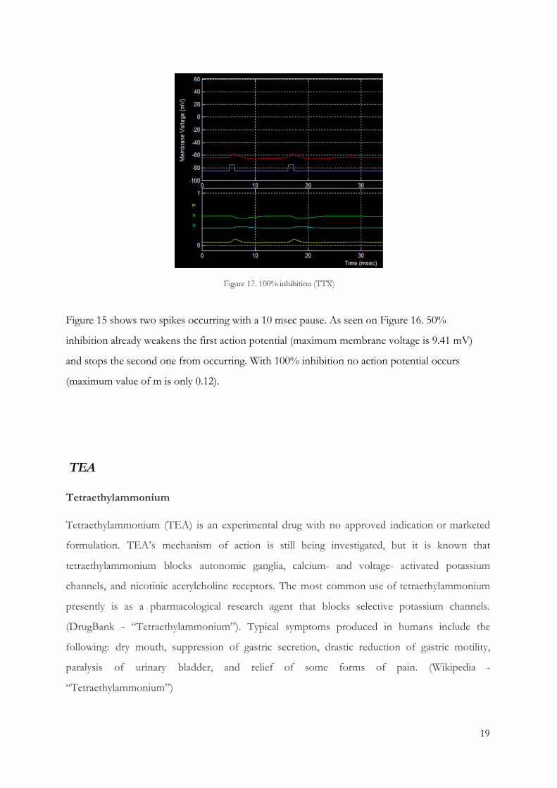
Figure 15 shows two spikes occurring with a 10 msec pause. As seen on Figure 16. 50%
inhibition already weakens the first action potential (maximum membrane voltage is 9.41 mV)
and stops the second one from occurring. With 100% inhibition no action potential occurs
(maximum value of m is only 0.12).
TEA
Tetraethylammonium
Tetraethylammonium (TEA) is an experimental drug with no approved indication or marketed
formulation. TEA’s mechanism of action is still being investigated, but it is known that
tetraethylammonium blocks autonomic ganglia, calcium- and voltage- activated potassium
channels, and nicotinic acetylcholine receptors. The most common use of tetraethylammonium
presently is as a pharmacological research agent that blocks selective potassium channels.
(DrugBank - “Tetraethylammonium”). Typical symptoms produced in humans include the
following: dry mouth, suppression of gastric secretion, drastic reduction of gastric motility,
paralysis of urinary bladder, and relief of some forms of pain. (Wikipedia -
“Tetraethylammonium”)
19
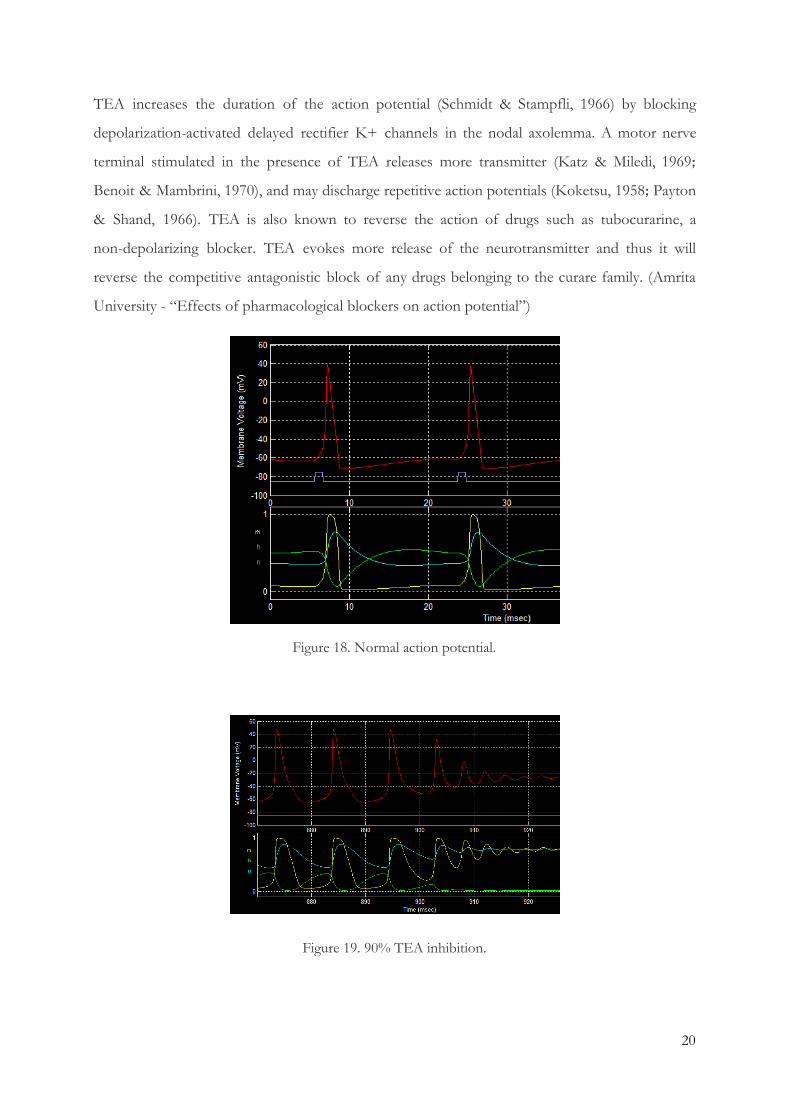
TEA increases the duration of the action potential (Schmidt & Stampfli, 1966) by blocking
depolarization-activated delayed rectifier K+ channels in the nodal axolemma. A motor nerve
terminal stimulated in the presence of TEA releases more transmitter (Katz & Miledi, 1969;
Benoit & Mambrini, 1970), and may discharge repetitive action potentials (Koketsu, 1958; Payton
& Shand, 1966). TEA is also known to reverse the action of drugs such as tubocurarine, a
non-depolarizing blocker. TEA evokes more release of the neurotransmitter and thus it will
reverse the competitive antagonistic block of any drugs belonging to the curare family. (Amrita
University - “Effects of pharmacological blockers on action potential”)
Figure 18. Normal action potential.
Figure 19. 90% TEA inhibition.
20
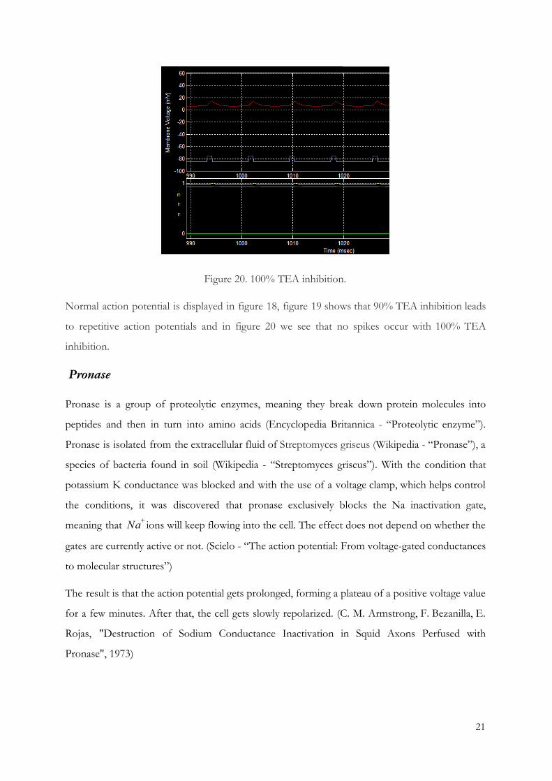
Figure 20. 100% TEA inhibition.
Normal action potential is displayed in figure 18, figure 19 shows that 90% TEA inhibition leads
to repetitive action potentials and in figure 20 we see that no spikes occur with 100% TEA
inhibition.
Pronase
Pronase is a group of proteolytic enzymes, meaning they break down protein molecules into
peptides and then in turn into amino acids (Encyclopedia Britannica - “Proteolytic enzyme”).
Pronase is isolated from the extracellular fluid of Streptomyces griseus (Wikipedia - “Pronase”), a
species of bacteria found in soil (Wikipedia - “Streptomyces griseus”). With the condition that
potassium K conductance was blocked and with the use of a voltage clamp, which helps control
the conditions, it was discovered that pronase exclusively blocks the Na inactivation gate,
meaning that ions will keep flowing into the cell. The effect does not depend on whether the Na+
gates are currently active or not. (Scielo - “The action potential: From voltage-gated conductances
to molecular structures”)
The result is that the action potential gets prolonged, forming a plateau of a positive voltage value
for a few minutes. After that, the cell gets slowly repolarized. (C. M. Armstrong, F. Bezanilla, E.
Rojas, "Destruction of Sodium Conductance Inactivation in Squid Axons Perfused with
Pronase", 1973)
21
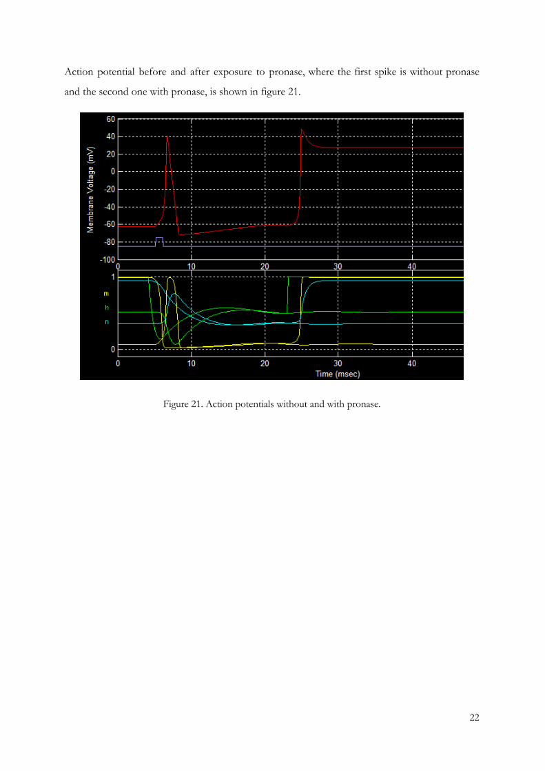
Action potential before and after exposure to pronase, where the first spike is without pronase
and the second one with pronase, is shown in figure 21.
Figure 21. Action potentials without and with pronase.
22
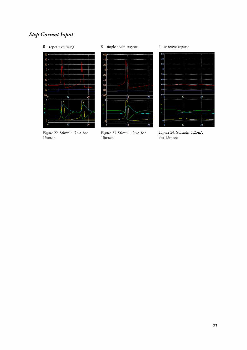
Step Current Input
23
References
Guttman, R., & Hachmeister, L. (1972). Anode Break Excitation in Space-Clamped Squid Axons.
Biophysical Journal, 12(5), 552-563.
Hodgkin, A. L., & Huxley, A. F. (1952). A quantitative description of membrane current and its
application to conduction and excitation in nerve. The Journal of Physiology, 117(4), 500-544.
Nelson, M., & Rinzel, J. (2003). The Hodgkin-Huxley Model. In M. J. Bower, & D. Beeman, The
Book of GENESIS. Exploring Realistic Neural Models with the GEneral NEural SImulation System (pp.
29-50).
Schwiening, C. J. (2012). A brief historical perspective: Hodgkin and Huxley. The Journal of
Physiology, 590(11), 2571-2575.
Map of Life - "Tetrodotoxin". Retrieved from
http://www.mapoflife.org/topics/topic_396_Tetrodotoxin/ (Last visit: 2015, May 22)
Tetrodotoxin: Mode of Action . Retrieved from
http://www.life.umd.edu/grad/mlfsc/zctsim/ionchannel.html (Last visit: 2015, May 22)
DrugBank - “Tetraethylammonium”. Retrieved from
http://www.drugbank.ca/drugs/DB08837 (Last visit: 2015, May 24)
Amrita University - “Effects of pharmacological blockers on action potential”. Retrieved from
http://vlab.amrita.edu/?sub=3&brch=212&sim=1310&cnt=1 (Last visit: 2015, May 24)
Encyclopedia Britannica - “Proteolytic enzyme”. Retrieved from
http://www.britannica.com/EBchecked/topic/479818/proteolytic-enzyme (Last visit: 2015,
May 24)
Wikipedia - “Pronase”. Retrieved from
http://en.wikipedia.org/wiki/Pronase (Last visit: 2015, May 24)
Wikipedia - “Streptomyces griseus”. Retrieved from
http://en.wikipedia.org/wiki/Streptomyces_griseus (Last visit: 2015, May 24)
Scielo - “The action potential: From voltage-gated conductances to molecular structures” . Retrieved from
http://www.scielo.cl/scielo.php?pid=S0716-97602006000300005&script=sci_arttext (Last visit:
2015, May 24)
24
C. M. Armstrong, F. Bezanilla, E. Rojas, "Destruction of Sodium Conductance Inactivation in Squid
Axons Perfused with Pronase", 1973, Retrieved from:
http://jgp.rupress.org/content/62/4/375.full.pdf (Last visit: 2015, May 24)
Wikipedia - “Tetraethylammonium”. Retrieved from:
http://en.wikipedia.org/wiki/Tetraethylammonium (Last visit: 2015, May 25)
25


![A Review of Biologically Plausible Neuron Models for ...personal.psu.edu/lnl/papers/aiaa_2010_3540.pdf · A. Hodgkin-Huxley (HH) Model In 1952 Hodgkin and Huxley [1] proposed a mathematical](https://img.pdfslide.us/doc/110x75/5ea83e430643df317d18e4db/a-review-of-biologically-plausible-neuron-models-for-a-hodgkin-huxley-hh.jpg)