Embed Size (px)
Citation preview

High-fidelity color reproduction and multispectral medical imaging
Masahiro Yamaguchi Global Scientific Information and Computing Center
Tokyo Institute of Technology
1

2

3
High-fidelity color reproduction: Questions
Is “high-fidelity color” significant in medicine?
How is it valuable?
4
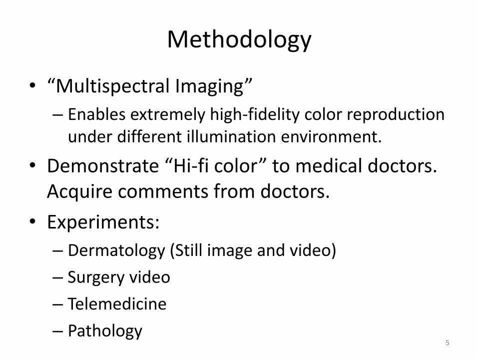
Methodology
• “Multispectral Imaging”
– Enables extremely high-fidelity color reproduction under different illumination environment.
• Demonstrate “Hi-fi color” to medical doctors. Acquire comments from doctors.
• Experiments:
– Dermatology (Still image and video)
– Surgery video
– Telemedicine
– Pathology 5
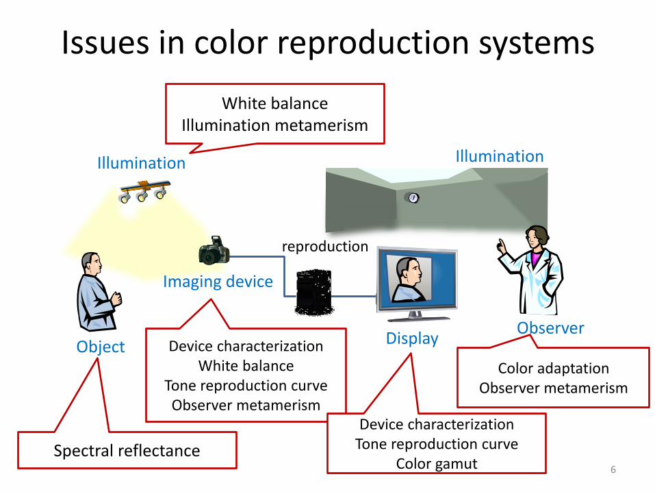
Issues in color reproduction systems
Illumination Illumination
Object
Imaging device
Display Observer
White balance Illumination metamerism
Spectral reflectance
Device characterization White balance
Tone reproduction curve Observer metamerism
Device characterization Tone reproduction curve
Color gamut
Color adaptation Observer metamerism
reproduction
6
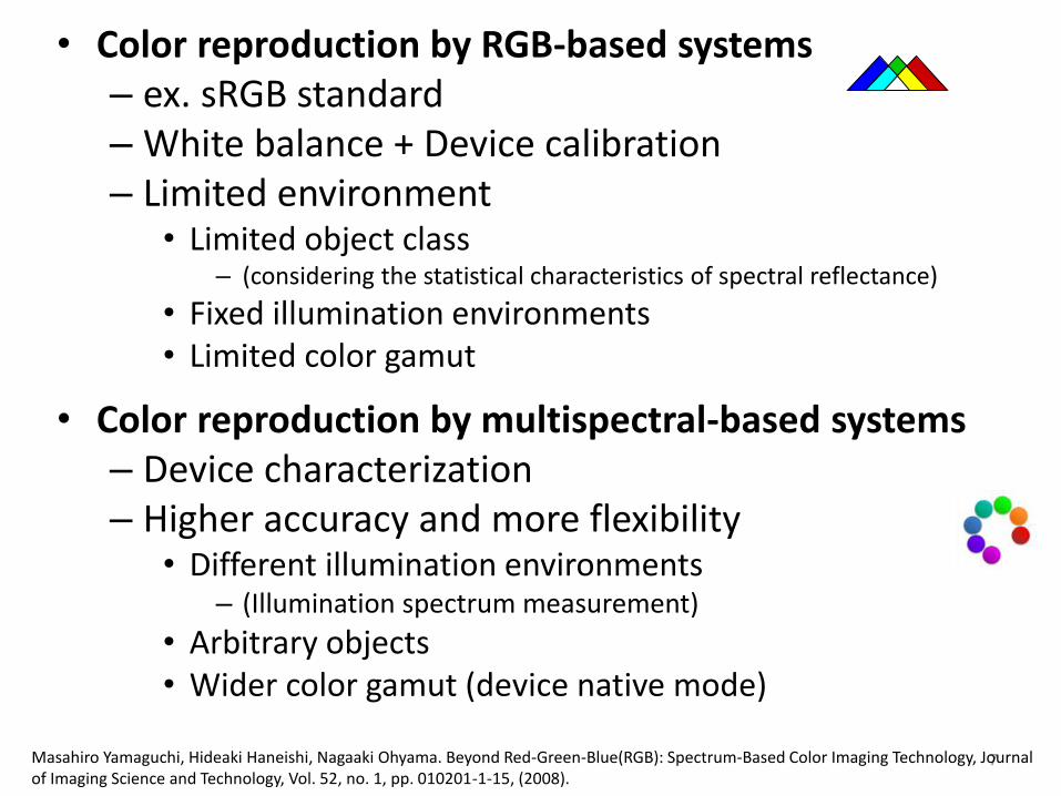
• Color reproduction by RGB-based systems – ex. sRGB standard – White balance + Device calibration – Limited environment
• Limited object class – (considering the statistical characteristics of spectral reflectance)
• Fixed illumination environments • Limited color gamut
• Color reproduction by multispectral-based systems – Device characterization – Higher accuracy and more flexibility
• Different illumination environments – (Illumination spectrum measurement)
• Arbitrary objects • Wider color gamut (device native mode)
7 Masahiro Yamaguchi, Hideaki Haneishi, Nagaaki Ohyama. Beyond Red-Green-Blue(RGB): Spectrum-Based Color Imaging Technology, Journal
of Imaging Science and Technology, Vol. 52, no. 1, pp. 010201-1-15, (2008).
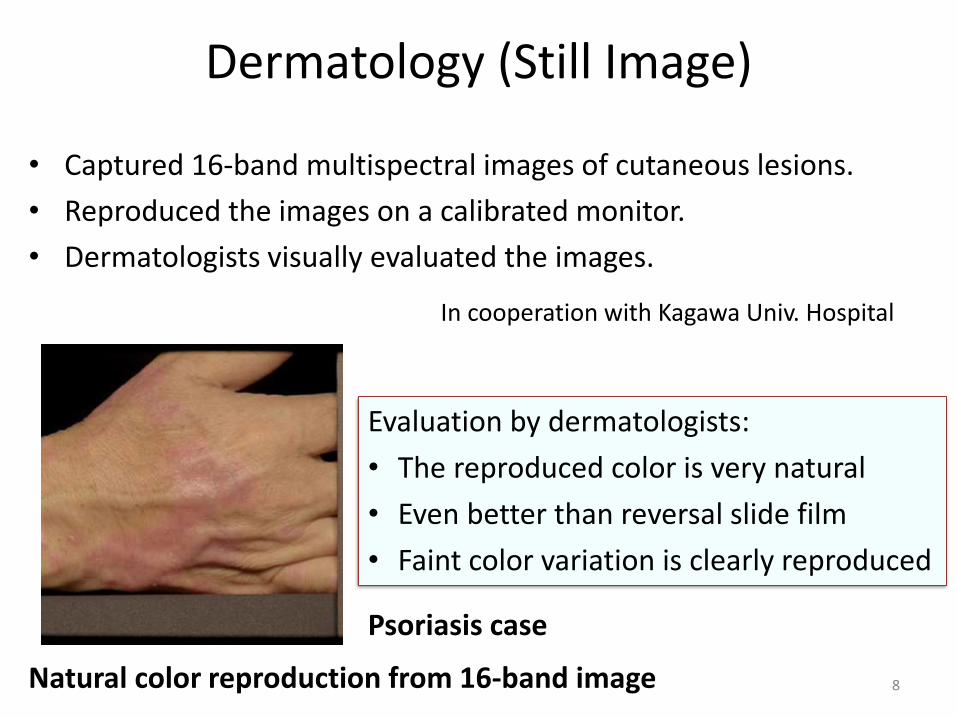
Dermatology (Still Image)
• Captured 16-band multispectral images of cutaneous lesions.
• Reproduced the images on a calibrated monitor.
• Dermatologists visually evaluated the images.
Natural color reproduction from 16-band image
Psoriasis case
In cooperation with Kagawa Univ. Hospital
Evaluation by dermatologists:
• The reproduced color is very natural
• Even better than reversal slide film
• Faint color variation is clearly reproduced
8

Dermatology (Still Image) Utilizing quantitative color information
(550nm enhancement)
High-fidelity color and Spectral information is also useful for Computer Aided Diagnosis - Quantification of skin disease - Assessment of treatment - Explanation to patient
Psoriasis: Spectrum-based color enhancement
Before treatment
3 weeks after
< 20% > 80% 20% - 50%
0% 20% 40% 60% 80% 100%
後
前Before
After
50% - 80%
Quantification of skin lesions
9

Dermatology (Still Image) Discussions
• The accuracy of color reproduction from 3-channel image is worse in some cases;
– Scleroderma and dermatomyositis give visually apparent color difference
• Hi-fi color reproduction is suitable for case DB, teledermatology…
• High-fidelity reproduction is required for specific types of diseases
– Dermatomyositis ex. “heliotrope rash,” Pigmented spots ex. nevus spilus
• Possibilities of skin disease quantification or grading using color: – Quantitative evaluation: effect of treatment, explanation to patient – Identification of inflammatory and immunologic disease: ex. support
general physicians – Acne grading
10 Masahiro Yamaguchi, et. al., “Multispectral color imaging for dermatoligy:application in inflammatory and immunologic deseases,”
Proc. IS&T/SID 13th Color Imaging Conference, pp. 52-58, (2005).

6-band HDTV Conventional
HDTV
Oversight 0 8
Total observation (3 dermatologists)
80 60
Dermatology (Video)
• Experiments using 6-band and conventional-RGB HD videos
• Artificial erythema - Simulating typical flare, ex. Urticaria
– "Prick tests" of histamine, cedar pollen, and mite allergen
0
2
4
6
8
10
12
14
16
6B RGB 6B RGB
Co
lor
diffe
ren
ce
(D
E* a
b)
Max
Average
Normal skin Erythema
Color difference between real skin and reproduced image
• Dermatologists were asked to measure erythema size when it found.
• Real Skin, 6-band, and conventional RGB.
• Result: – No significant difference between RGB and
6B systems in Erythema size measurement – Oversight in the 3B system (Erythema sizes were not measured)
13% 11

Dermatology (Video) Comments from dermatologists
• The color reproduction by conventional RGB system is not sufficient especially in reddish colors, not suitable for the diagnosis of subtle flare such as measles, virus infection, and drug allergy.
• The image color in 6B system looks natural, and the reddish and yellowish colors can be easily discriminated.
• The profile of the erythema, the dilatation of blood capillary are clearly observed in 6B system.
• Expected applications: – Monitoring or recording of surgery – The observation of circulatory disorders. – The condition and the area of inflammation in inflammatory diseases. – Intraepidermal carcinoma, ex., Paget's disease, the subtle color variation is
important to determine the extent of tumor. – Precancerous skin diseases, which depend on the color appearance of dark
or light tan, pink, and red colors. 12
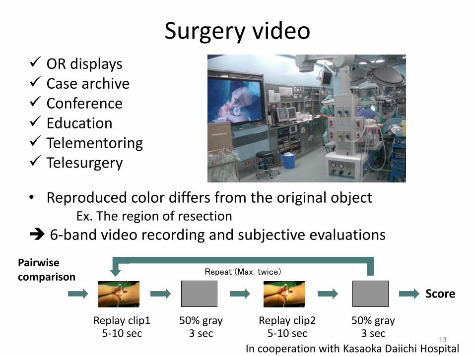
Surgery video OR displays Case archive Conference Education Telementoring Telesurgery
• Reproduced color differs from the original object Ex. The region of resection
6-band video recording and subjective evaluations
Score
Repeat (Max. twice)
Replay clip1 5-10 sec
50% gray 3 sec
Replay clip2 5-10 sec
50% gray 3 sec
Pairwise comparison
In cooperation with Kasaoka Daiichi Hospital 13
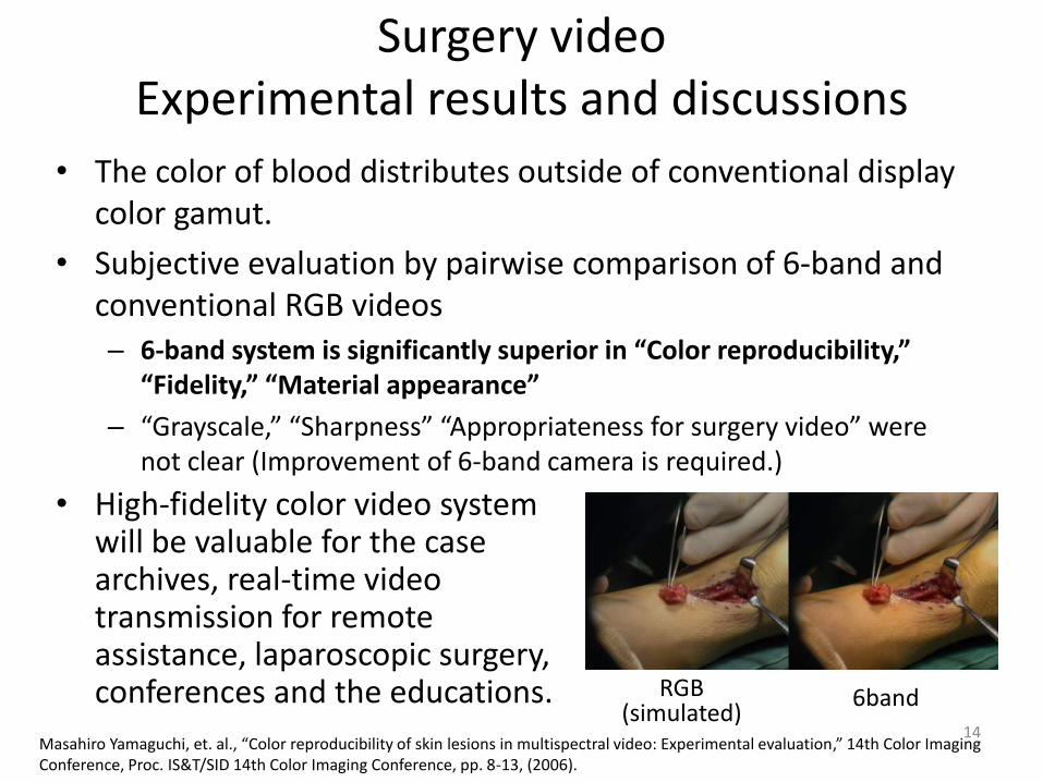
Surgery video Experimental results and discussions
• The color of blood distributes outside of conventional display color gamut.
• Subjective evaluation by pairwise comparison of 6-band and conventional RGB videos – 6-band system is significantly superior in “Color reproducibility,”
“Fidelity,” “Material appearance”
– “Grayscale,” “Sharpness” “Appropriateness for surgery video” were not clear (Improvement of 6-band camera is required.)
RGB (simulated)
6band
• High-fidelity color video system will be valuable for the case archives, real-time video transmission for remote assistance, laparoscopic surgery, conferences and the educations.
14 Masahiro Yamaguchi, et. al., “Color reproducibility of skin lesions in multispectral video: Experimental evaluation,” 14th Color Imaging Conference, Proc. IS&T/SID 14th Color Imaging Conference, pp. 8-13, (2006).
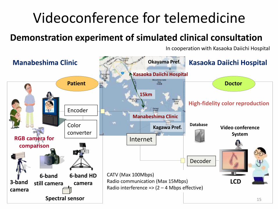
Videoconference for telemedicine Demonstration experiment of simulated clinical consultation
In cooperation with Kasaoka Daiichi Hospital
Manabeshima Clinic
6-band still camera 3-band
camera
Database
LCD 6-band HD
camera
Video conference System
Kasaoka Daiichi Hospital
Decoder
RGB camera for comparison
High-fidelity color reproduction
Spectral sensor
Kagawa Pref.
Okayama Pref.
Kasaoka Daiichi Hospital
Manabeshima Clinic
15km
Internet
Color converter
Encoder
Patient Doctor
CATV (Max 100Mbps) Radio communication (Max 15Mbps) Radio interference => (2 – 4 Mbps effective)
15
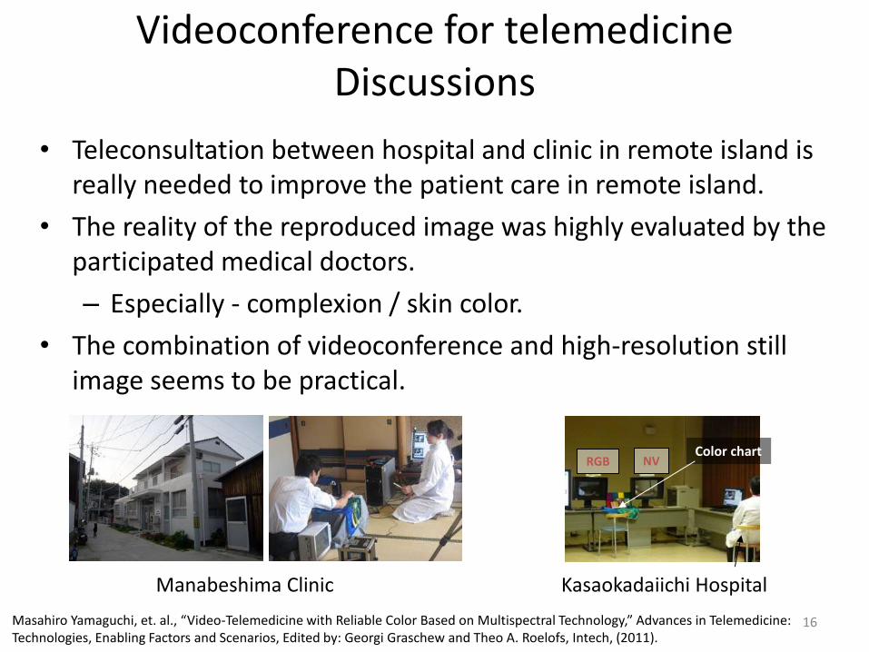
Videoconference for telemedicine Discussions
• Teleconsultation between hospital and clinic in remote island is really needed to improve the patient care in remote island.
• The reality of the reproduced image was highly evaluated by the participated medical doctors.
– Especially - complexion / skin color.
• The combination of videoconference and high-resolution still image seems to be practical.
Manabeshima Clinic
NV RGB Color chart
Kasaokadaiichi Hospital
16 Masahiro Yamaguchi, et. al., “Video-Telemedicine with Reliable Color Based on Multispectral Technology,” Advances in Telemedicine: Technologies, Enabling Factors and Scenarios, Edited by: Georgi Graschew and Theo A. Roelofs, Intech, (2011).
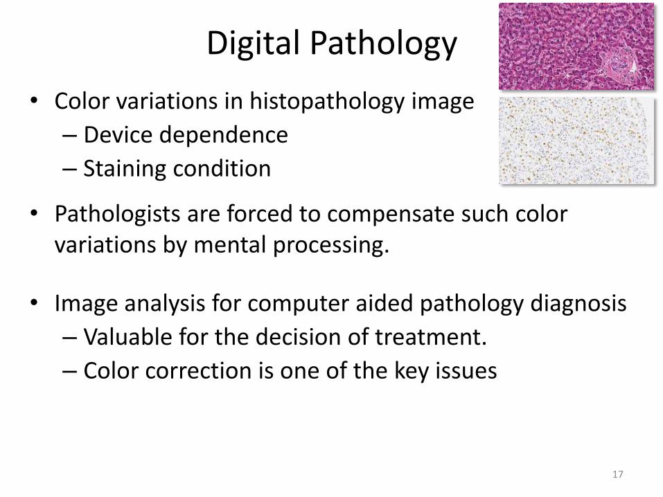
Digital Pathology
• Color variations in histopathology image
– Device dependence
– Staining condition
• Pathologists are forced to compensate such color variations by mental processing.
• Image analysis for computer aided pathology diagnosis
– Valuable for the decision of treatment.
– Color correction is one of the key issues
17

Digital pathology Color correction experiment
• Device characterization – Color calibration slides
• Color chart slide • HE stained mouse embryo
– Gold standard: captured by multispectral microscope – Regression model applied.
• Results of color correction
(Multispctral)
Average color difference
18
Yuri Murakami, et. al., "Color Correction in Whole Slide Digital Pathology," 20th Color and Imaging Conference, 253-258 (2012)

Digital Pathology Correction of staining variation
• Color unmixing – Estimation of dye amount image
• Adjustment of weighting factors for dye amount images • Re-mix a color image
Standardization of staining condition
HE stained image H component E component Residual Not actual data
H staining time
E stainin
g time
standardization
Target
19
Multispectral image
Multispectral image
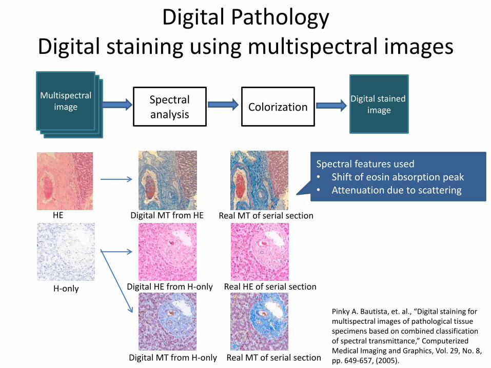
Digital Pathology Digital staining using multispectral images
H-only Digital HE from H-only
Digital MT from H-only
Real HE of serial section
Real MT of serial section
HE Digital MT from HE Real MT of serial section
Spectral features used • Shift of eosin absorption peak • Attenuation due to scattering
Multispectral image
Spectral analysis
Colorization Digital stained
image
20
Pinky A. Bautista, et. al., “Digital staining for multispectral images of pathological tissue specimens based on combined classification of spectral transmittance,” Computerized Medical Imaging and Graphics, Vol. 29, No. 8, pp. 649-657, (2005).
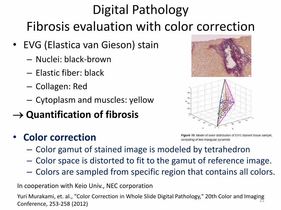
Digital Pathology Fibrosis evaluation with color correction
• EVG (Elastica van Gieson) stain
– Nuclei: black-brown
– Elastic fiber: black
– Collagen: Red
– Cytoplasm and muscles: yellow
Quantification of fibrosis
• Color correction – Color gamut of stained image is modeled by tetrahedron – Color space is distorted to fit to the gamut of reference image. – Colors are sampled from specific region that contains all colors.
Yuri Murakami, et. al., "Color Correction in Whole Slide Digital Pathology," 20th Color and Imaging Conference, 253-258 (2012)
In cooperation with Keio Univ., NEC corporation
21
Discussions and Summary
• Significance of “high-fidelity color” and “quantitative color”
– Dermatology
– Surgery video
– Teleconsultation
– Digital pathology
• Not only for telemedicine, but the introduction of digital color imaging technology to visible light images will provide much benefits.
– Database
– Quantitative analysis for supporting diagnosis, explanation to patients
22
What aspect of color is most important for telemedicine: accuracy, consistency or discrimination?
• Accuracy – “To be” in near future.
• Required in the observation of complexion, or face color. • Specific types of lesions in dermatology, such as pigmented spots,
heliotrope rash of dermatomyositis.
• Discrimination – Crucial in many cases of color imaging
• Evaluation of the area of skin lesion, • Visual or automatic measurement of histology or cytology images • Decision of resection area based on faint color difference of tumor
in surgery operation.
• Consistency – Without consistency, the color information is
meaningless.
23
What one step (if any) would you suggest we should take in order to improve the handling of color within your area of expertise?
Methods and Criteria for the color reproduction capabilities of input and display devices
Then users will be able to choose appropriate system for respective purposes.
Acknowledgements • The works related to dermatology, surgery, and multispectral pathology were supported by National Institute of
Information and Communications Technology (NICT), and Special Coordination Funds for Promoting Science and Technology by MEXT, and Natural Vision Promotion Council ( http://www.nvision.jp ).
• Multispectral pathology is a joint work with Dr. Yukako Yagi at MGH. • Quantitative pathology is collaborative work with Keio Univ., Saitama Medical School, and NEC Corporation,
supported by the New Energy and Industrial Technology Development Organization (NEDO) of Japan. 24