Embed Size (px)
Citation preview
RESEARCH ARTICLE
Helicobacter pylori bab Paralog Distributionand Association with cagA, vacA, and homA/B Genotypes in American and South KoreanClinical IsolatesAeryun Kim1,2☯, Stephanie L. Servetas3☯, Jieun Kang1,2, Jinmoon Kim1,2, Sungil Jang1, HoJin Cha1, Wan Jin Lee1, June Kim1, Judith Romero-Gallo4, Richard M. Peek, Jr.4, D.Scott Merrell3*, Jeong-Heon Cha1,2*
1 Department of Oral Biology, Oral Science Research Center, Yonsei University College of Dentistry, Seoul,South Korea, 2 Department of Applied Life Science, BK21 Plus Project, Yonsei University College ofDentistry, Seoul, South Korea, 3 Department of Microbiology and Immunology, Uniformed ServicesUniversity of the Health Sciences, 4301 Jones Bridge Rd., Bethesda, Maryland, 20814, United States ofAmerica, 4 Departments of Cancer Biology and Medicine, Vanderbilt University, Nashville, Tennessee,37240, United States of America
☯ These authors contributed equally to this work.* [email protected] (DSM); [email protected] (JHC)
AbstractHelicobacter pylori genetic variation is a crucial component of colonization and persistence
within the inhospitable niche of the gastric mucosa. As such, numerous H. pylori geneshave been shown to vary in terms of presence and genomic location within this pathogen.
Among the variable factors, the Bab family of outer membrane proteins (OMPs) has been
shown to differ within subsets of strains. To better understand genetic variation among the
bab genes and to determine whether this variation differed among isolates obtained from
different geographic locations, we characterized the distribution of the Bab family members
in 80 American H. pylori clinical isolates (AH) and 80 South Korean H. pylori clinical isolates(KH). Overall, we identified 23 different bab genotypes (19 in AH and 11 in KH), but only 5
occurred in greater than 5 isolates. Regardless of strain origin, a strain in which locus A and
locus B were both occupied by a bab gene was the most common (85%); locus C was only
occupied in those isolates that carried bab paralog at locus A and B. While the babA/babB/-genotype predominated in the KH (78.8%), no single genotype could account for greater
than 40% in the AH collection. In addition to basic genotyping, we also identified associa-
tions between bab genotype and well known virulence factors cagA and vacA. Specifically,significant associations between babA at locus A and the cagA EPIYA-ABDmotif
(P<0.0001) and the vacA s1/i1/m1 allele (P<0.0001) were identified. Log-linear modeling
further revealed a three-way association between bab carried at locus A, vacA, and number
of OMPs from the HOM family (P<0.002). En masse this study provides a detailed charac-
terization of the bab genotypes from two distinct populations. Our analysis suggests greater
variability in the AH, perhaps due to adaptation to a more diverse host population. Further-
more, when considering the presence or absence of both the bab and homA/B paralogs at
PLOS ONE | DOI:10.1371/journal.pone.0137078 August 28, 2015 1 / 19
a11111
OPEN ACCESS
Citation: Kim A, Servetas SL, Kang J, Kim J, Jang S,Cha HJ, et al. (2015) Helicobacter pylori bab ParalogDistribution and Association with cagA, vacA, andhomA/B Genotypes in American and South KoreanClinical Isolates. PLoS ONE 10(8): e0137078.doi:10.1371/journal.pone.0137078
Editor: Daniela Flavia Hozbor, Universidad Nacionalde La Plata., ARGENTINA
Received: May 6, 2015
Accepted: August 13, 2015
Published: August 28, 2015
Copyright: This is an open access article, free of allcopyright, and may be freely reproduced, distributed,transmitted, modified, built upon, or otherwise usedby anyone for any lawful purpose. The work is madeavailable under the Creative Commons CC0 publicdomain dedication.
Data Availability Statement: The sequences for babgenotypes in American and South Korean strainswere deposited in the NCBI GenBank database withaccession numbers KP339308 to KP33949.
Funding: This research was supported by BasicScience Research Program through the NationalResearch Foundation of Korea (NRF) funded by theMinistry of Education (2014R1A2A1A11051054).
Competing Interests: The authors have declaredthat no competing interests exist.
their given loci and the vacA genotype, an association was observed. Our results highlight
the multifactorial nature of H. pylorimediated disease and the importance of considering
how the specific combinations of H. pylori virulence genes and their multiple interactions
with the host will collectively impact disease progression.
IntroductionHelicobacter pylori (H. pylori) is a successful pathogen, colonizing the gastric mucosa of over50% of the world’s population [1, 2]. This pathogen has gained notoriety for its ability to colo-nize the inhospitable niche of the stomach and to cause gastric diseases [1, 2].H. pylori causepersistent, potentially lifelong infections; however, only about 20% of individuals infected willdevelop symptomatic infection. Although clinical manifestations occur only in a subset ofinfected individuals, these can be severe and include peptic ulcers and gastric cancer. Rates ofH. pylori infection and gastric cancer mortality vary geographically. East Asian countries suchas China, Japan, and South Korea have high rates ofH. pylori infection, and these populationsalso have some of the highest rates of gastric cancer [3–6].
Environmental, host, and bacterial factors are known to influence H. pylori-associated dis-ease outcome and also vary geographically [7]. Interestingly, H. pylori is a bacterial species thatshows exceptionally high rates of genetic variability and intra-species diversity. Indeed, thesepolymorphisms have been used to determine the geographic origin of H. pylori isolates [8, 9].Furthermore, it is highly probable that these genetic differences influence virulence. Previousstudies have shown that severe disease outcomes such as gastric cancer and ulcers are associ-ated with polymorphism inH. pylori virulence factors such as the cytotoxin-associated gene A(cagA) and vacuolating cytotoxin (vacA) [4, 6, 10–12]. In addition, disease outcome has beenassociated with several outer membrane proteins (OMPs). Specifically, OMPs such as AlpAand B (adherence-associated lipoprotein A and B), BabA (blood group antigen binding adhe-sin), HomB (Helicobacter outer membrane B), HopZ (Helicobacter outer membrane proteinZ), OipA (outer membrane inflammatory protein A) and SabA (sialic acid binding adhesion)are all associated with variable disease outcomes [13–18]. In comparison to the polymorphismswithin cagA and vacA, genetic variability within the OMP families is based upon presence andabsence of different closely related paralogs. For example, the bab-family of genes is made upof three paralogs babA, babB and babC, which can be located at three different H. pylori chro-mosomal loci referred to as locus A, B and C [19–21]. Recently, we presented detailed epidemi-ological studies of cagA, vacA, and homB from a collection of 260 H. pylori clinical isolatesfrom South Korea [11, 12, 17, 22]. Our studies showed a significant association between infec-tion withH. pylori strains carrying the EPIYA-ABD cagA genotype and the development ofgastric cancer [22]. Moreover, the majority of H. pylori isolates from the South Korean popula-tion encoded the most virulent toxins, CagA EPIYA-ABD motif and VacA s1/i1/m1 allele [11].While, we found no association between the presence of homB and the progression to severegastric disease [17], other groups working with isolates from different geographic regions haveshown that the presence of homB was associated with development of peptic ulcer disease inchildren and young adults [23, 24] and with gastric cancer development and the presence ofcagA [25]. The variations in these results suggest that the impact of the homB gene on disease isgeographically dependent [17].
Our studies and others have revealed that certain polymorphisms are more prevalent withinspecific populations [11, 12, 17, 22, 26–29]. Thus, it is possible that the combination of
Importance of bab pParalog in H. pylori Clinical Isolates
PLOS ONE | DOI:10.1371/journal.pone.0137078 August 28, 2015 2 / 19
virulence factor polymorphisms present within a population may influence the rate of severedisease outcome. Therefore, the polymorphism of each virulence factor must be evaluatedwithin various populations to help understand the relationship between these genotypes andtheir associations with gastric diseases including cancer.
The bab gene family has been shown to have a positive association with gastric cancer andduodenal ulcers [6, 30–32]. BabA, one member of this family, was the first identified adhesin inH. pylori and mediates the binding of bacteria to the fucosylated blood group O antigen Lewisb (Leb), which is highly expressed in gastrointestinal epithelial cells [33, 34]. Similar to severalH. pylori OMPs, BabA has two closely related paralogous, BabB (HopT) and BabC (HopU);however, the functions of BabB and BabC are unknown. These OMPs, particularly BabA andBabB, have nearly identical N- and C-terminal domains but a divergent middle region, suggest-ing that the middle variable region of BabA is most likely responsible for the specific adhesinfunction [19, 34–36]. Interestingly, it has been shown that BabB expression is modulated bychanges in the number of cytosine-thymidine (CT) dinucleotide repeats in the 50 region of thebabB gene resulting in slipped-strand mispairing [37–39]. Changes in the number of CTrepeats results in a frame shift; ‘IN’-frame bab genes are translated into a full functional Bab or‘OUT’-of-frame bab genes into a premature partial Bab [37–39]. Furthermore, bab expressioncan be modulated by gene conversion between the loci, most likely by intragenomic homolo-gous recombination [35, 40]. This gene conversion results in chimeric bab genes where the CTrepeats of the babB 50 region become associated with babA or babC subjecting these genes tophase variation. These recombination events may play an important role in regulation ofadherence. For example, babBA chimera results in the regulation of babA by changes in CTrepeats that lead to changes in the binding to Leb on gastric epithelial cells [41].
To gain further insight into genetic diversity ofH. pylori isolates within the South Koreanpopulation, we analyzed the bab gene family. In addition, we expanded our analysis to includeclinical isolates obtained from an American hospital to allow for strain comparisons betweendifferent geographic regions.
Materials and Methods
Bacterial Strains and Culture ConditionsA total of 160H. pylori clinical isolates were used in our analysis. The 80 South Korean H.pylori clinical isolates (KH) were a subset of a collection of South Korean strains previouslycharacterized for distribution of cagA EPIYA polymorphism, vacA s/i/m polymorphism, andhomA/B paralog [11, 12, 17, 22], while the other 80 isolates were obtained from patients atVanderbilt University Medical Center, Nashville TN, USA. Written informed consent wasreceived from each patient, and the protocol was approved by the Vanderbilt University andthe Nashville Department of Veterans Affairs Institutional Review Board (IRB# 5190). Gastricbiopsies were harvested from individuals at the Veterans Affairs Medical Center in Nashvilleundergoing upper endoscopy, and used for bacterial culture. Isolates from biopsies were con-firmed to beH. pylori by positive urease, catalase, and oxidase tests, and typical appearance onGram stain. These strains are referred to as AmericanH. pylori clinical isolates (AH). For 80AH, polymorphisms in cagA and vacA, and homB paralog were determined using the strategiespreviously developed for characterization of KH [11, 12, 17, 22]. The 160 KH and AH aredescribed in detail in S1 Table. All H. pylori clinical isolates were cultured as previouslydescribed [22]. Briefly, bacterial isolates preserved at -80°C, were grown and expanded on anti-biotic-supplemented horse blood agar plates under microaerophilic conditions created in Gas-Pak jars with Campypak generators (Mitsubishi Gas Chemical Company, Inc, Tokyo, Japan).
Importance of bab pParalog in H. pylori Clinical Isolates
PLOS ONE | DOI:10.1371/journal.pone.0137078 August 28, 2015 3 / 19
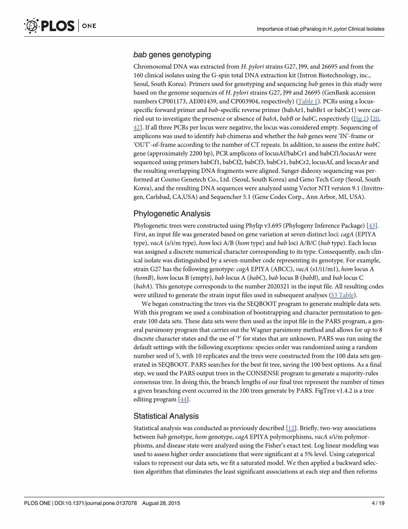
bab genes genotypingChromosomal DNA was extracted fromH. pylori strains G27, J99, and 26695 and from the160 clinical isolates using the G-spin total DNA extraction kit (Intron Biotechnology, inc.,Seoul, South Korea). Primers used for genotyping and sequencing bab genes in this study werebased on the genome sequences of H. pylori strains G27, J99 and 26695 (GenBank accessionnumbers CP001173, AE001439, and CP003904, respectively) (Table 1). PCRs using a locus-specific forward primer and bab-specific reverse primer (babAr1, babBr1 or babCr1) were car-ried out to investigate the presence or absence of babA, babB or babC, respectively (Fig 1) [20,42]. If all three PCRs per locus were negative, the locus was considered empty. Sequencing ofamplicons was used to identify bab chimeras and whether the bab genes were ‘IN’-frame or‘OUT’-of-frame according to the number of CT repeats. In addition, to assess the entire babCgene (approximately 2200 bp), PCR amplicons of locusAf/babCr1 and babCf1/locusAr weresequenced using primers babCf1, babCf2, babCf3, babCr1, babCr2, locusAf, and locusAr andthe resulting overlapping DNA fragments were aligned. Sanger dideoxy sequencing was per-formed at Cosmo Genetech Co., Ltd. (Seoul, South Korea) and Geno Tech Corp (Seoul, SouthKorea), and the resulting DNA sequences were analyzed using Vector NTI version 9.1 (Invitro-gen, Carlsbad, CA,USA) and Sequencher 5.1 (Gene Codes Corp., Ann Arbor, MI, USA).
Phylogenetic AnalysisPhylogenetic trees were constructed using Phylip v3.695 (Phylogeny Inference Package) [43].First, an input file was generated based on gene variation at seven distinct loci: cagA (EPIYAtype), vacA (s/i/m type), hom loci A/B (hom type) and bab loci A/B/C (bab type). Each locuswas assigned a discrete numerical character corresponding to its type. Consequently, each clin-ical isolate was distinguished by a seven-number code representing its genotype. For example,strain G27 has the following genotype: cagA EPIYA (ABCC), vacA (s1/i1/m1), hom locus A(homB), hom locus B (empty), bab locus A (babC), bab locus B (babB), and bab locus C(babA). This genotype corresponds to the number 2020321 in the input file. All resulting codeswere utilized to generate the strain input files used in subsequent analyses (S3 Table).
We began constructing the trees via the SEQBOOT program to generate multiple data sets.With this program we used a combination of bootstrapping and character permutation to gen-erate 100 data sets. These data sets were then used as the input file in the PARS program, a gen-eral parsimony program that carries out the Wagner parsimony method and allows for up to 8discrete character states and the use of ‘?’ for states that are unknown. PARS was run using thedefault settings with the following exceptions: species order was randomized using a randomnumber seed of 5, with 10 replicates and the trees were constructed from the 100 data sets gen-erated in SEQBOOT. PARS searches for the best fit tree, saving the 100 best options. As a finalstep, we used the PARS output trees in the CONSENSE program to generate a majority-rulesconsensus tree. In doing this, the branch lengths of our final tree represent the number of timesa given branching event occurred in the 100 trees generate by PARS. FigTree v1.4.2 is a treeediting program [44].
Statistical AnalysisStatistical analysis was conducted as previously described [12]. Briefly, two-way associationsbetween bab genotype, hom genotype, cagA EPIYA polymorphisms, vacA s/i/m polymor-phisms, and disease state were analyzed using the Fisher’s exact test. Log linear modeling wasused to assess higher order associations that were significant at a 5% level. Using categoricalvalues to represent our data sets, we fit a saturated model. We then applied a backward selec-tion algorithm that eliminates the least significant associations at each step and then reforms
Importance of bab pParalog in H. pylori Clinical Isolates
PLOS ONE | DOI:10.1371/journal.pone.0137078 August 28, 2015 4 / 19
the model to look for associations. Data analysis was conducted using SPSS version 22 software(SPSS Inc., Chicago, IL, USA) or SAS version 9.3 software (SAS Institute Inc., Cary, NC, USA).
Nucleotide Sequence Accession NumbersThe sequences for bab genotypes in American and South Korean strains were deposited in theNCBI GenBank database with accession numbers KP339308 to KP339491.
Results
Sample PopulationComplete demographic data was available for all 80 AH (S1A and S2A Tables). For the Ameri-can population, the mean patient age was 57 years, with an age range of 27–84 years. 20.0% (16patients) of the population was black, and 80.0% (64 patients) was white. Finally, 79 of the
Table 1. Primer sequences for analysis ofH. pylori bab genes.
Primer name Primer sequence (5’!3’) Reference
locusAf TTTTGAGCCGGTGGATATATTAG HypDF1 [42] d
locusBf CTTTAATCCCCTACATTGTGGA S18F1 [42] d
locusCf a ACCCTAGTGGGCATGTGGTA Hp1-AS [20] d
locusAr b GGAAATGCGCACACGAGGG this study
babAr1 b TTTGCCGTCTATGGTTTGG BabAR1 [42] d
babAr2 b GAAAGGATGTGTTTTTTCATG this study
babBr1 b TCGCTTGTTTTAAAAGCTCTTGA BabBR1 [42] d
babBr2 b CTGCCAGGACCACAAGCAGT this study
babCf1 b GCGGCAAACATCATGCAAGTC this study
babCr1 b GACTTGCATGATGTTTGCCGC this study
babCf2 b CGGGTTTGCTCAAAGAAAAAAYC c this study
babCr2 b GRTTTTTTCTTTGAGCAAACCCG c this study
babCf3 b AAAGAATAACCCCTATAGCC this study
a One nucleotide in the primer was modified from the indicated primer in reference.b Primer was used for sequencing.c Underlined Y indicates C or T, and underlined R indicates G or A.d Primer name was used in the indicated reference.
doi:10.1371/journal.pone.0137078.t001
Fig 1. Outline of bab genotyping by PCR. (Top) Schematic representation of the three loci where the bab genes are generally detected: Locus A, B and C.The annealing positions (arrows) and names of each locus-specific forward are shown. (Bottom) Annealing positions (arrows) and names of bab-specificreverse primer are indicated with their respective bab gene: babA depicted by the black box, babB depicted by the white box, babC depicted by the grey box.Primers are listed in Table 1, and a full explanation of the genotyping scheme can be found in the Materials and Methods.
doi:10.1371/journal.pone.0137078.g001
Importance of bab pParalog in H. pylori Clinical Isolates
PLOS ONE | DOI:10.1371/journal.pone.0137078 August 28, 2015 5 / 19
patients were male; there was one white female patient, age 50. Of these clinical isolates, 7.5%were from patients with cancer/pre-malignant lesions (2.5% with gastric carcinoma and 5.0%with Barrett’s Esophagus), 43.8% were from patients with peptic ulcer disease (31.3% with duo-denal ulcers and 12.5% with gastric ulcers), 32.5% were from patients with gastritis, and 16.3%were from patients with esophagitis. Within the South Korean population, age and genderwere missing for 2 individuals. Of the remaining 78, the mean age was 48 years, ranging from20–86 years of age. The South Korean population had a more even distribution between malesand females, with 48.7% (38 patients) female and 51.3% (40 patients) male. Within the SouthKorean female patient group, the mean age was 51, with an age range of 21–86 years. Withinthe South Korean male patient group, the mean age was 46, with an age range of 20–76 years.Of the 80 KH samples analyzed, 18.8% were from patients with gastric cancer, 33.8% werefrom patients with peptic ulcer disease (32.5% with duodenal ulcers and 1.3% with gastriculcers), and 47.5% were from patients with gastritis.
bab genes genotypingThe distribution of identified bab genes at locus A, B and C is shown in Fig 2. Genotypic analysisrevealed that in addition to babA, babB and babC, three types of chimeric bab genes were alsopresent: babAB, babBA and babBC. A total of 23 different bab genotypes, including a singlestrain where babwas absent from the three known loci (-/-/-), were identified within the 160 iso-lates chosen for this study. While 6.9% of strains (11/160) carried a single bab gene, the majorityof the strains (85.0%, 136/160) carried two bab genes, one at locus A and one at locus B (Fig 2).Of note, locus C was only occupied if both other loci also carried a bab gene (Fig 2). In the KH,11 of the 23 genotypes were identified, and isolates carrying two bab genes were most common,accounting for 87.5% of the isolates. The genotype babA/babB/-, position 1, 2, and 3 correspondto locus A, B and C, respectively, occurred in the 78.8% of the isolates (Fig 2). In comparison,there were many more bab genotypes within the AH. In fact, 19 of the 23 genotypes were identi-fied. Twelve genotypes were unique to the AH while 4 were unique to the KH. Similar to theKH, isolates carrying two bab genes were most common among AH, accounting for 82.5% ofthe isolates; however, while the most common genotype of babA/babB/- occurred in only38.8%, genotypes of babAB/babB/- (16.3%) and babC/babB/- (15.0%) showed a higher fre-quency in this population (Fig 2). Whereas locus A of KH was occupied by babA in greater than90% of isolates (74/80), most of the diversity within the bab genotypes for the AH was driven bylocus A, which was significantly more variable (P<0.0001) than seen in KH. Indeed, babAoccurred at locus A only 47.5% of the time (S1 Fig). The next most frequent occupants of locusA were chimeric bab genes (28.8%), followed by babC (15.0%) and babB (7.5%).
In total, we identified 3 chimeric bab genes, babAB, babBA and babBC that were the resultof gene conversion. This gene conversion likely occurs in the 50region (~60 to 240 bp from theinitiation codon) and in the 30region (~1200 to 2100 bp) but this later event is hard to detectdue to the homology in this region. It can be speculated that the characteristic of the chimerawould follow that of the second bab gene, which would constitute the majority of the chimericgene. bab chimeric forms were relatively common in the AH group, occurring at one or moreloci in 26/80 (32.5%) of isolates versus 6/80 (7.5%) in the KH group (Fig 2 and S2 Table). Themost common chimeric gene was babAB, which accounted for 88.9% of chimeric genes; 90.0%of chimeras in AH (27/30) and 83.3% in KH (5/6) (Fig 2 and S2 Table). Within the babB cod-ing sequence there are variable CT repeats approximately 56 bp downstream from the initia-tion codon that are responsible for phase variation. In the babAB chimera, this region isdisplaced with the corresponding part of babA [41], resulting in loss of phase variable babB.Although it appeared much less frequently, babBA and babBC chimeras were also present (Fig
Importance of bab pParalog in H. pylori Clinical Isolates
PLOS ONE | DOI:10.1371/journal.pone.0137078 August 28, 2015 6 / 19
Fig 2. Distribution of the bab genotypes in AH and KH (n = 160).Genotypes of one, two, three, or none bab genes are grouped into I, II, III, and IV,respectively. These classifications were made on the basis of number of bab genes present at locus A, B, and C. A black, white, or grey box indicated babA,babB, and babC, respectively. ‘-’ indicates the absence of bab gene at the designated loci. ‘AH%’ and ‘KH%’ indicate the frequency of each individualgenotype within the specified population (n = 80) and ‘Total%’ shows the percent of the respective genotype in the overall population (n = 160). Bracketednumbers indicate the number of isolates possessing each respective genotype out of the population examined. ‘*’ indicates genotypes containing chimericform(s).
doi:10.1371/journal.pone.0137078.g002
Importance of bab pParalog in H. pylori Clinical Isolates
PLOS ONE | DOI:10.1371/journal.pone.0137078 August 28, 2015 7 / 19
2 and S1 Fig). In these instances, CT repeats from the 50region of babB were introduced tobabA and babC, respectively, resulting in phase variable babA and babC.
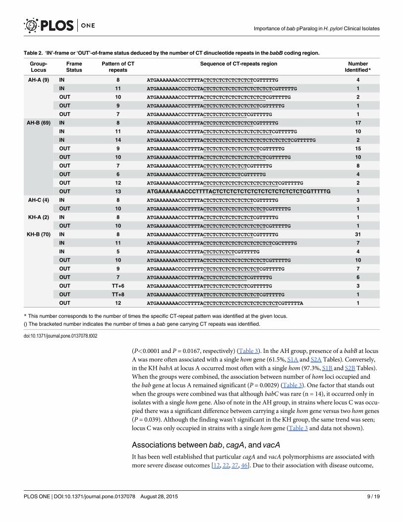
Eighty-two bab genes from the AH group and 72 bab genes from the KH group contained arun of CT repeats (5–14 CT repeats) (Table 2). In all cases, the CT repeats were associated withthe 50 region of babB genes; therefore, babBC and babBA also carry CT repeats. CT repeatswere found at all three loci within the AH, with the majority (84.2%) occurring at locus B(Table 2). Similarly, in the KH group a majority of the bab genes containing CT repeats werefound at locus B (97.2%) (Table 2). Interestingly, when comparing locus B in the AH group tothe KH, significantly more babB genes were ‘IN’-frame in the KH group than in the AH(P = 0.0341, data not shown). Although not significant, the trend was similar for all babB genesregardless of loci, where more babB genes were ‘OUT’-of-frame (54.9%) than ‘IN’-frame(45.1%) in the AH group (S2 Fig). The opposite trend was observed in the KH group; babBgenes tended to be ‘IN’-frame (59.7%) as compared to ‘OUT’-of-frame (40.3%) (S2 Fig).
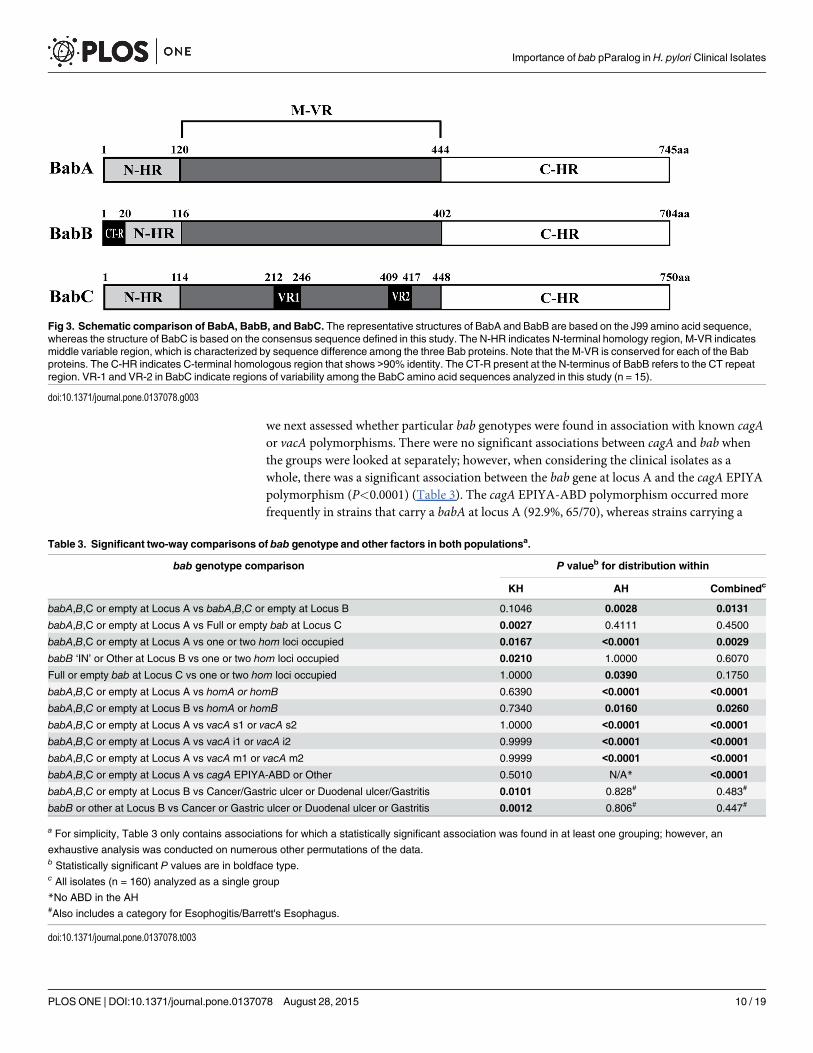
Since very little is known about babC, we took this opportunity to sequence 15 babC genesfrom our collection and compare the deduced BabCs. As shown in S3 Fig we defined a BabCconsensus sequence by comparing 14 BabCs and one BabBC from this study along with theBabC from G27. According to the babC sequences, we identified only one chimeric babBCgene (AH-J68) and no babAC gene. Furthermore, we found that the N-terminus of BabC wasmost similar to that of BabA, and the C-terminus of BabC was homologous to that of bothBabA and BabB (Fig 3). The amino acid sequence of BabC contained two variable regions, VR1spanning from amino acid 212 to 246 and VR2 spanning from amino acid 409 to 417. TheBabC sequences, including VR1 and VR2, shared over 70% identities (Fig 3 and S3 Fig).
To further understand the bab genotypes in these populations, we next sought to determineif carrying a specific bab gene at one locus was associated with the presence of bab genes at theother loci. Given the difference in bab genotypes between the two populations, we first exam-ined each population separately. A significant association was found in the KH group betweenthe genotype at locus A and locus C (P = 0.0027) (Table 3). Conversely, in the AH group a sig-nificant association was found between locus A and locus B (P = 0.0028). When KH and AHwere combined, there was also a significant association between locus A and locus B(P = 0.0131) (Table 3). Of note, in the AH/KH-combined analysis babC was more likely tooccur at locus A when babB at locus B was ‘OUT’-of-frame (78.6%, 11/14). Furthermore, ifBabB was being expressed from locus A, then babB at locus B was more likely to be ‘OUT’-of-frame (60.0%, 15/25). Yet interestingly, if babA occupied locus A, then babB at locus B wasmore likely to be ‘IN’-frame (58.9%, 66/112).
Associations between bab and hom genesGiven the associations among bab loci, we next assessed whether bab genotype was associatedwith other outer membrane proteins. We have previously looked at the association betweenhomB and disease in the South Korean population [17]. hom paralog (homA and homB) fromthe Hom protein family, which shares over 90% identity, are known to occupy two possible loci(also called locus A and B) [45]. Therefore, we chose to evaluate any association between thebab genes, and homA and homB. Once again, given the differences in the two populations, eachwas analyzed individually as well as combined. There were no significant associations whencomparing strains for presence of homA versus homB. Given that homA and homB are homolo-gous and share two conserved loci, locus A and locus B, we also considered hom gene number inwhich we looked at whether or not both hom locus A and locus B were occupied regardless of ifit was homA or homB. We found a significant association with the bab paralog present at locusA and number of hom loci occupied. This association was seen in both the AH and KH groups
Importance of bab pParalog in H. pylori Clinical Isolates
PLOS ONE | DOI:10.1371/journal.pone.0137078 August 28, 2015 8 / 19
(P<0.0001 and P = 0.0167, respectively) (Table 3). In the AH group, presence of a babB at locusA was more often associated with a single hom gene (61.5%, S1A and S2A Tables). Conversely,in the KH babA at locus A occurred most often with a single hom (97.3%, S1B and S2B Tables).When the groups were combined, the association between number of hom loci occupied andthe bab gene at locus A remained significant (P = 0.0029) (Table 3). One factor that stands outwhen the groups were combined was that although babC was rare (n = 14), it occurred only inisolates with a single hom gene. Also of note in the AH group, in strains where locus C was occu-pied there was a significant difference between carrying a single hom gene versus two hom genes(P = 0.039). Although the finding wasn’t significant in the KH group, the same trend was seen;locus C was only occupied in strains with a single hom gene (Table 3 and data not shown).
Associations between bab, cagA, and vacAIt has been well established that particular cagA and vacA polymorphisms are associated withmore severe disease outcomes [12, 22, 27, 46]. Due to their association with disease outcome,
Table 2. ‘IN’-frame or ‘OUT’-of-frame status deduced by the number of CT dinucleotide repeats in the babB coding region.
Group-Locus
FrameStatus
Pattern of CTrepeats
Sequence of CT-repeats region NumberIdentified*
AH-A (9) IN 8 ATGAAAAAAACCCTTTTACTCTCTCTCTCTCTCTCGTTTTTG 4
IN 11 ATGAAAAAAACCCTCCTACTCTCTCTCTCTCTCTCTCTCTCGTTTTTG 1
OUT 10 ATGAAAAAAACCCTTTTACTCTCTCTCTCTCTCTCTCTCGTTTTTG 2
OUT 9 ATGAAAAAAACCCTTTTACTCTCTCTCTCTCTCTCTCGTTTTTG 1
OUT 7 ATGAAAAAAACCCTTTTACTCTCTCTCTCTCTCGTTTTTG 1
AH-B (69) IN 8 ATGAAAAAAACCCTTTTACTCTCTCTCTCTCTCTCGTTTTTG 17
IN 11 ATGAAAAAAACCCTTTTACTCTCTCTCTCTCTCTCTCTCTCGTTTTTG 10
IN 14 ATGAAAAAAACCCTTTTACTCTCTCTCTCTCTCTCTCTCTCTCTCTCGTTTTTG 2
OUT 9 ATGAAAAAAACCCTTTTACTCTCTCTCTCTCTCTCTCGTTTTTG 15
OUT 10 ATGAAAAAAACCCTTTTACTCTCTCTCTCTCTCTCTCTCGTTTTTG 10
OUT 7 ATGAAAAAAACCCTTTTACTCTCTCTCTCTCTCGTTTTTG 8
OUT 6 ATGAAAAAAACCCTTTTACTCTCTCTCTCTCGTTTTTG 4
OUT 12 ATGAAAAAAACCCTTTTACTCTCTCTCTCTCTCTCTCTCTCTCGTTTTTG 2
OUT 13 ATGAAAAAAACCCTTTTACTCTCTCTCTCTCTCTCTCTCTCTCTCGTTTTTG 1
AH-C (4) IN 8 ATGAAAAAAACCCTTTTACTCTCTCTCTCTCTCTCGTTTTTG 3
OUT 10 ATGAAAAAAACCCTTTTACTCTCTCTCTCTCTCTCTCTCGTTTTTG 1
KH-A (2) IN 8 ATGAAAAAAACCCTTTTACTCTCTCTCTCTCTCTCGTTTTTG 1
OUT 10 ATGAAAAAAACCCTTTTACTCTCTCTCTCTCTCTCTCTCGTTTTTG 1
KH-B (70) IN 8 ATGAAAAAAACCCTTTTACTCTCTCTCTCTCTCTCGTTTTTG 31
IN 11 ATGAAAAAAACCCTTTTACTCTCTCTCTCTCTCTCTCTCTCGCTTTTG 7
IN 5 ATGAAAAAAACCCTTTTACTCTCTCTCTCGTTTTTG 4
OUT 10 ATGAAAAAAATCCTTTTACTCTCTCTCTCTCTCTCTCTCGTTTTTG 10
OUT 9 ATGAAAAAACCCCTTTTTCTCTCTCTCTCTCTCTCTCGTTTTTG 7
OUT 7 ATGAAAAAAACCCTTTTACTCTCTCTCTCTCTCGTTTTTG 6
OUT TT+6 ATGAAAAAAACCCTTTTATTCTCTCTCTCTCTCGTTTTTG 3
OUT TT+8 ATGAAAAAAACCCTTTTATTCTCTCTCTCTCTCTCTCGTTTTTG 1
OUT 12 ATGAAAAAAACCCTTTTACTCTCTCTCTCTCTCTCTCTCTCTCGTTTTTA 1
* This number corresponds to the number of times the specific CT-repeat pattern was identified at the given locus.
() The bracketed number indicates the number of times a bab gene carrying CT repeats was identified.
doi:10.1371/journal.pone.0137078.t002
Importance of bab pParalog in H. pylori Clinical Isolates
PLOS ONE | DOI:10.1371/journal.pone.0137078 August 28, 2015 9 / 19
we next assessed whether particular bab genotypes were found in association with known cagAor vacA polymorphisms. There were no significant associations between cagA and bab whenthe groups were looked at separately; however, when considering the clinical isolates as awhole, there was a significant association between the bab gene at locus A and the cagA EPIYApolymorphism (P<0.0001) (Table 3). The cagA EPIYA-ABD polymorphism occurred morefrequently in strains that carry a babA at locus A (92.9%, 65/70), whereas strains carrying a
Fig 3. Schematic comparison of BabA, BabB, and BabC. The representative structures of BabA and BabB are based on the J99 amino acid sequence,whereas the structure of BabC is based on the consensus sequence defined in this study. The N-HR indicates N-terminal homology region, M-VR indicatesmiddle variable region, which is characterized by sequence difference among the three Bab proteins. Note that the M-VR is conserved for each of the Babproteins. The C-HR indicates C-terminal homologous region that shows >90% identity. The CT-R present at the N-terminus of BabB refers to the CT repeatregion. VR-1 and VR-2 in BabC indicate regions of variability among the BabC amino acid sequences analyzed in this study (n = 15).
doi:10.1371/journal.pone.0137078.g003
Table 3. Significant two-way comparisons of bab genotype and other factors in both populationsa.
bab genotype comparison P valueb for distribution within
KH AH Combinedc
babA,B,C or empty at Locus A vs babA,B,C or empty at Locus B 0.1046 0.0028 0.0131
babA,B,C or empty at Locus A vs Full or empty bab at Locus C 0.0027 0.4111 0.4500
babA,B,C or empty at Locus A vs one or two hom loci occupied 0.0167 <0.0001 0.0029
babB ‘IN’ or Other at Locus B vs one or two hom loci occupied 0.0210 1.0000 0.6070
Full or empty bab at Locus C vs one or two hom loci occupied 1.0000 0.0390 0.1750
babA,B,C or empty at Locus A vs homA or homB 0.6390 <0.0001 <0.0001
babA,B,C or empty at Locus B vs homA or homB 0.7340 0.0160 0.0260
babA,B,C or empty at Locus A vs vacA s1 or vacA s2 1.0000 <0.0001 <0.0001
babA,B,C or empty at Locus A vs vacA i1 or vacA i2 0.9999 <0.0001 <0.0001
babA,B,C or empty at Locus A vs vacA m1 or vacA m2 0.9999 <0.0001 <0.0001
babA,B,C or empty at Locus A vs cagA EPIYA-ABD or Other 0.5010 N/A* <0.0001
babA,B,C or empty at Locus B vs Cancer/Gastric ulcer or Duodenal ulcer/Gastritis 0.0101 0.828# 0.483#
babB or other at Locus B vs Cancer or Gastric ulcer or Duodenal ulcer or Gastritis 0.0012 0.806# 0.447#
a For simplicity, Table 3 only contains associations for which a statistically significant association was found in at least one grouping; however, an
exhaustive analysis was conducted on numerous other permutations of the data.b Statistically significant P values are in boldface type.c All isolates (n = 160) analyzed as a single group
*No ABD in the AH#Also includes a category for Esophogitis/Barrett's Esophagus.
doi:10.1371/journal.pone.0137078.t003
Importance of bab pParalog in H. pylori Clinical Isolates
PLOS ONE | DOI:10.1371/journal.pone.0137078 August 28, 2015 10 / 19
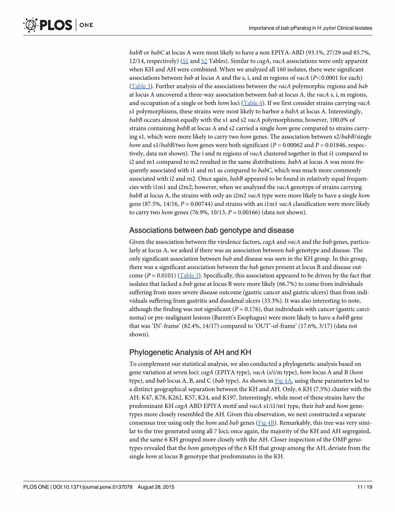
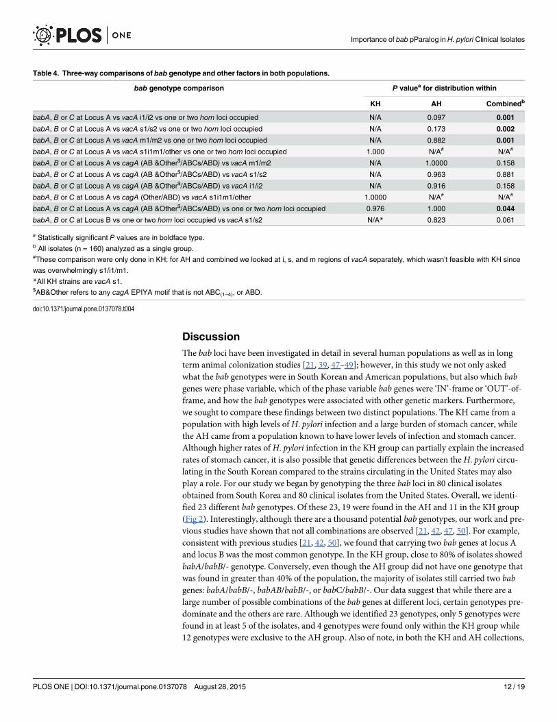
babB or babC at locus A were most likely to have a non EPIYA-ABD (93.1%, 27/29 and 85.7%,12/14, respectively) (S1 and S2 Tables). Similar to cagA, vacA associations were only apparentwhen KH and AH were combined. When we analyzed all 160 isolates, there were significantassociations between bab at locus A and the s, i, and m regions of vacA (P<0.0001 for each)(Table 3). Further analysis of the associations between the vacA polymorphic regions and babat locus A uncovered a three-way association between bab at locus A, the vacA s, i, m regions,and occupation of a single or both hom loci (Table 4). If we first consider strains carrying vacAs1 polymorphisms, these strains were most likely to harbor a babA at locus A. Interestingly,babB occurs almost equally with the s1 and s2 vacA polymorphisms; however, 100.0% ofstrains containing babB at locus A and s2 carried a single hom gene compared to strains carry-ing s1, which were more likely to carry two hom genes. The association between s2/babB/singlehom and s1/babB/two hom genes were both significant (P = 0.00062 and P = 0.01846, respec-tively, data not shown). The i and m regions of vacA clustered together in that i1 compared toi2 and m1 compared to m2 resulted in the same distributions. babA at locus A was more fre-quently associated with i1 and m1 as compared to babC, which was much more commonlyassociated with i2 and m2. Once again, babB appeared to be found in relatively equal frequen-cies with i1m1 and i2m2; however, when we analyzed the vacA genotype of strains carryingbabB at locus A, the strains with only an i2m2 vacA type were more likely to have a single homgene (87.5%, 14/16, P = 0.00744) and strains with an i1m1 vacA classification were more likelyto carry two hom genes (76.9%, 10/13, P = 0.00166) (data not shown).
Associations between bab genotype and diseaseGiven the association between the virulence factors, cagA and vacA and the bab genes, particu-larly at locus A, we asked if there was an association between bab genotype and disease. Theonly significant association between bab and disease was seen in the KH group. In this group,there was a significant association between the bab genes present at locus B and disease out-come (P = 0.0101) (Table 3). Specifically, this association appeared to be driven by the fact thatisolates that lacked a bab gene at locus B were more likely (66.7%) to come from individualssuffering from more severe disease outcome (gastric cancer and gastric ulcers) than from indi-viduals suffering from gastritis and duodenal ulcers (33.3%). It was also interesting to note,although the finding was not significant (P = 0.176), that individuals with cancer (gastric carci-noma) or pre-malignant lesions (Barrett’s Esophagus) were more likely to have a babB genethat was ‘IN’-frame’ (82.4%, 14/17) compared to ‘OUT’-of-frame’ (17.6%, 3/17) (data notshown).
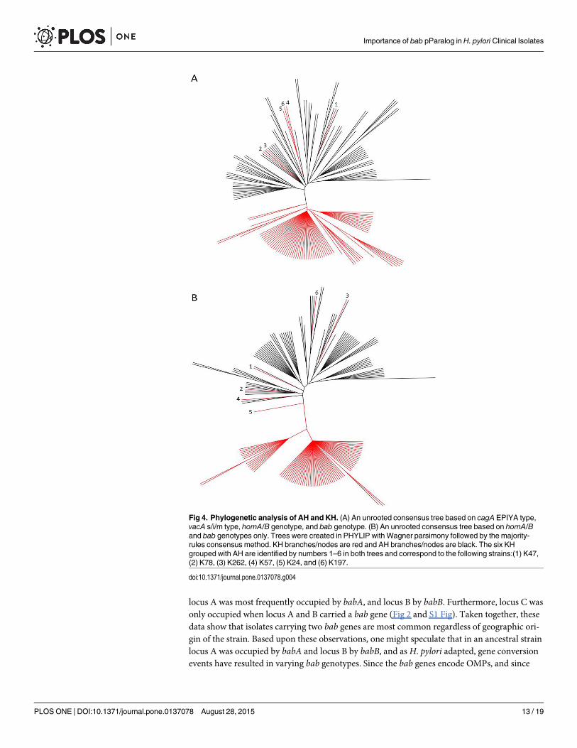
Phylogenetic Analysis of AH and KHTo complement our statistical analysis, we also conducted a phylogenetic analysis based ongene variation at seven loci: cagA (EPIYA type), vacA (s/i/m type), hom locus A and B (homtype), and bab locus A, B, and C (bab type). As shown in Fig 4A, using these parameters led toa distinct geographical separation between the KH and AH. Only, 6 KH (7.5%) cluster with theAH: K47, K78, K262, K57, K24, and K197. Interestingly, while most of these strains have thepredominant KH cagA ABD EPIYA motif and vacA s1/i1/m1 type, their bab and hom geno-types more closely resembled the AH. Given this observation, we next constructed a separateconsensus tree using only the hom and bab genes (Fig 4B). Remarkably, this tree was very simi-lar to the tree generated using all 7 loci; once again, the majority of the KH and AH segregated,and the same 6 KH grouped more closely with the AH. Closer inspection of the OMP geno-types revealed that the hom genotypes of the 6 KH that group among the AH, deviate from thesingle hom at locus B genotype that predominates in the KH.
Importance of bab pParalog in H. pylori Clinical Isolates
PLOS ONE | DOI:10.1371/journal.pone.0137078 August 28, 2015 11 / 19
DiscussionThe bab loci have been investigated in detail in several human populations as well as in longterm animal colonization studies [21, 39, 47–49]; however, in this study we not only askedwhat the bab genotypes were in South Korean and American populations, but also which babgenes were phase variable, which of the phase variable bab genes were ‘IN’-frame or ‘OUT’-of-frame, and how the bab genotypes were associated with other genetic markers. Furthermore,we sought to compare these findings between two distinct populations. The KH came from apopulation with high levels ofH. pylori infection and a large burden of stomach cancer, whilethe AH came from a population known to have lower levels of infection and stomach cancer.Although higher rates of H. pylori infection in the KH group can partially explain the increasedrates of stomach cancer, it is also possible that genetic differences between theH. pylori circu-lating in the South Korean compared to the strains circulating in the United States may alsoplay a role. For our study we began by genotyping the three bab loci in 80 clinical isolatesobtained from South Korea and 80 clinical isolates from the United States. Overall, we identi-fied 23 different bab genotypes. Of these 23, 19 were found in the AH and 11 in the KH group(Fig 2). Interestingly, although there are a thousand potential bab genotypes, our work and pre-vious studies have shown that not all combinations are observed [21, 42, 47, 50]. For example,consistent with previous studies [21, 42, 50], we found that carrying two bab genes at locus Aand locus B was the most common genotype. In the KH group, close to 80% of isolates showedbabA/babB/- genotype. Conversely, even though the AH group did not have one genotype thatwas found in greater than 40% of the population, the majority of isolates still carried two babgenes: babA/babB/-, babAB/babB/-, or babC/babB/-. Our data suggest that while there are alarge number of possible combinations of the bab genes at different loci, certain genotypes pre-dominate and the others are rare. Although we identified 23 genotypes, only 5 genotypes werefound in at least 5 of the isolates, and 4 genotypes were found only within the KH group while12 genotypes were exclusive to the AH group. Also of note, in both the KH and AH collections,
Table 4. Three-way comparisons of bab genotype and other factors in both populations.
bab genotype comparison P valuea for distribution within
KH AH Combinedb
babA, B or C at Locus A vs vacA i1/i2 vs one or two hom loci occupied N/A 0.097 0.001
babA, B or C at Locus A vs vacA s1/s2 vs one or two hom loci occupied N/A 0.173 0.002
babA, B or C at Locus A vs vacA m1/m2 vs one or two hom loci occupied N/A 0.882 0.001
babA, B or C at Locus A vs vacA s1i1m1/other vs one or two hom loci occupied 1.000 N/A# N/A#
babA, B or C at Locus A vs cagA (AB &Other$/ABCs/ABD) vs vacA m1/m2 N/A 1.0000 0.158
babA, B or C at Locus A vs cagA (AB &Other$/ABCs/ABD) vs vacA s1/s2 N/A 0.963 0.881
babA, B or C at Locus A vs cagA (AB &Other$/ABCs/ABD) vs vacA i1/i2 N/A 0.916 0.158
babA, B or C at Locus A vs cagA (Other/ABD) vs vacA s1i1m1/other 1.0000 N/A# N/A#
babA, B or C at Locus A vs cagA (AB &Other$/ABCs/ABD) vs one or two hom loci occupied 0.976 1.000 0.044
babA, B or C at Locus B vs one or two hom loci occupied vs vacA s1/s2 N/A* 0.823 0.061
a Statistically significant P values are in boldface type.b All isolates (n = 160) analyzed as a single group.#These comparison were only done in KH; for AH and combined we looked at i, s, and m regions of vacA separately, which wasn’t feasible with KH since
was overwhelmingly s1/i1/m1.
*All KH strains are vacA s1.$AB&Other refers to any cagA EPIYA motif that is not ABC(1–4), or ABD.
doi:10.1371/journal.pone.0137078.t004
Importance of bab pParalog in H. pylori Clinical Isolates
PLOS ONE | DOI:10.1371/journal.pone.0137078 August 28, 2015 12 / 19
locus A was most frequently occupied by babA, and locus B by babB. Furthermore, locus C wasonly occupied when locus A and B carried a bab gene (Fig 2 and S1 Fig). Taken together, thesedata show that isolates carrying two bab genes are most common regardless of geographic ori-gin of the strain. Based upon these observations, one might speculate that in an ancestral strainlocus A was occupied by babA and locus B by babB, and as H. pylori adapted, gene conversionevents have resulted in varying bab genotypes. Since the bab genes encode OMPs, and since
Fig 4. Phylogenetic analysis of AH and KH. (A) An unrooted consensus tree based on cagA EPIYA type,vacA s/i/m type, homA/B genotype, and bab genotype. (B) An unrooted consensus tree based on homA/Band bab genotypes only. Trees were created in PHYLIP with Wagner parsimony followed by the majority-rules consensus method. KH branches/nodes are red and AH branches/nodes are black. The six KHgrouped with AH are identified by numbers 1–6 in both trees and correspond to the following strains:(1) K47,(2) K78, (3) K262, (4) K57, (5) K24, and (6) K197.
doi:10.1371/journal.pone.0137078.g004
Importance of bab pParalog in H. pylori Clinical Isolates
PLOS ONE | DOI:10.1371/journal.pone.0137078 August 28, 2015 13 / 19
BabA is known to act as an adhesin, it is likely that differences in the bab genotype affect howthe bacteria interacts with the host. Therefore, perhaps the genotypes that appear to occur inonly 1 or 2 individuals reflect adaptation ofH. pylori to an individual host, or across gastricregions within a host, and also within an individual over time [42].
In addition to bab locus genotyping, we also examined which bab genes were subject to phasevariation. As previously shown, CT repeats associated with the 50 region of babB can result inslipped-strand mispairing leading to a premature stop in translation [37–39]. In the KH group,babB at locus B was significantly more likely to be ‘IN’ frame’ compared to the AH which mayreflect a difference in the host populations sinceH. pylori likely uses phase variation as aresponse to host changes. Probably through gene conversion, CT repeats were also identified inassociation with babA and babC at locus A. These data suggest that gene conversion occurred inthese strains and resulted in phase variable babA and babC, impartingH. pylori with additionalmeans of regulation over these genes. Interestingly, the most common gene conversion ofbabAB among 3 types of chimeric bab genes deprived babB of the regulation of phase variation.
Evidence from this study and others suggest that varying the bab genotype is a mechanismofH. pylori adaptation [33, 41]. To try and gain a better understanding of these adaptations, weasked if there were associations among the bab loci. In both populations, babB was more likelyto be expressed from locus B when babA occupied locus A. Conversely, if babB was expressedfrom locus A then the babB at locus B was more likely to be ‘OUT’ of frame. Through theseanalyses we also noted that the babC gene was far more likely to occur in strains that also carrya babB. Furthermore, in strains containing both babB and babC, babB was more likely to be‘OUT’ of frame. Taken together, these data suggest thatH. pylori actively adjusts its repertoireof bab genes, not only through gene replacement, but also through the formation of chimericproteins and phase variation supporting the finding of other authors [35, 42]. Although thefunction of babB and babC are unknown, it could be speculated that the Bab OMPs share over-lapping functions, perhaps acting as adhesins similar to BabA. The differences between babA,babB, and babCmay affect their function. For instance, if they are functioning as adhesins,these differences may result from different binding targets. Alternatively, the differencebetween these three genes may also make them antigenically distinct and allow them to escapethe immune system.
To this end, we expanded our analysis to include other OMPs from the Hom family. Therewere no statistically significant differences when we compared strains carrying homA to thosecarrying homB; however, given that homA and homB are greater than 90% identical we decidedto ask whether or not we saw an difference in vacA, cagA and bab genotypes between strainscarrying a single hom gene at locus A or locus B versus strains in which both loci were occu-pied, regardless of whether it was homA or homB. We analyzed each population individually aswell as combined, and observed an association between the bab gene carried at locus A andnumber of homA and/or B genes. In both population, babA occurred most frequently with asingle homA or homB gene, babB occurred relatively equally whether a single or both hom lociwere occupied, and babC was only found in strains with a single homA or homB gene. If oneconsiders the environment in whichH. pylori persist, having a variable repertoire of OMPs islikely paramount to the success of the organism. While H. pylori has greater than 50 OMPs, toour knowledge this is the first evidence showing an association between the Hom and Bab fam-ilies [16, 37, 45]. Since only the function of babA is known, it is difficult to speculate how theseproteins may interact functionally [33]; however, one can surmise that H. pylori finds the rightcombination of OMPs as a means to balance between the adherent population, which is closeto nutrients, and a free population, which is less susceptible to immune clearance. Throughmechanisms such as phase variation and gene conversion H. pylori is able to fine tune its OMPrepertoire in response to changes in the environment.
Importance of bab pParalog in H. pylori Clinical Isolates
PLOS ONE | DOI:10.1371/journal.pone.0137078 August 28, 2015 14 / 19
In addition to investigating the relationship between the bab genes and other OMPs, wewere also interested in how bab genotypes related to known virulence factors such as cagA andvacA in KH [11, 12, 22, 46] and AH (S1A Table). In case of cagA genotypes, interestingly theAH group contained none of the EPIYA-D motif while KH included 7 strains possessing theEPIYA-C motifs. Importantly, we discovered several associations. A previous study found thatstrains with babA were more likely to have a cagA gene than strains that lacked babA [20].While all of the strains in our collection contained a cagA gene [22], we did find that there wasa positive association between having babA at locus A and having a cagA EPIYA-ABD motif.Strains that carried babB or babC at locus A were significantly more likely to have a nonEPIYA-ABD motif. Carrying a babA at locus A was also significantly more likely to occur withvacA type s1/i1/m1. Our group has previously shown that cagA EPIYA-ABD and vacA s1/i1/m1 are associated with more severe disease in the KH [11]. Our current data suggest that car-riage of babA at locus A may also be associated with these more virulent genotypes. Further-more, we observed a three-way association between bab genotype at locus A, occupation of oneor both hom loci and the vacA s1/i1/m1 genotype. Based on these three-way associations, weobserved some interesting patterns. For instance, strains harboring a single homA or homBgene were more likely to be associated with vacA s1, i1 and m1 if there was a babA at locus A;however, if there was a babB at locus A, then strains with a single homA or homB gene weremore likely to be found with a strain possessing s2/i2/m2, supporting the idea that babAmaybe associated with more virulent genotypes. However, if both hom loci are occupied and babBis at locus A, then this strain was more likely to be associated with s1, i1, and m1. These find-ings perhaps demonstrate that it is a specific combination of virulence factors that combine toresult in more severe disease outcomes. Although we did not see higher order associations withdisease, it could be that our sample size was not adequate to obtain statistical significance overthe number of variables that were included.
Taken en masse, theH. pylori isolates obtained from the American population showed alarger degree of diversity in bab genotypes compared to the South Korean population. Thismay reflect adaptions to a more diverse human population. Furthermore, difference seenbetween the South Korean and American isolates due to babB phase variation may also be dueto differences in the populations. Given that difference in host genetics are known to exist, per-haps babB is beneficial toH. pylori in the South Korean population but not in the Americanpopulation. Even though the populations were distinct, we identified associations that wereseen in both populations, as well as associations that became more apparent when we com-bined the populations and increased the sample size. Based on our analyses it appears thatbabAmay be associated with more virulent genotypes of cagA and vacA. We also identified anassociation between OMPs from the Hom family of proteins with the bab genes, suggesting apotential coordination between OMP families. Interestingly, our phylogenetic analysis suggeststhat the using only the hom and bab genotypes segregated strains as well as the full genotypingscheme. Specifically the hom genotype appears to influence whether the strains cluster withAH or KH. Previous studies have demonstrated that strains with the same cagA EPIYA typecluster geographically [46]; however, to our knowledge this is the first report in which the num-ber and type of OMP genes have been shown to segregate strains geographically. AlthoughhomA and homB sequence variation has been shown to cluster strains [51], the prior workfocused on strains with a single homA or homB gene, whereas our study focused on which homloci were occupied and not sequence differences. Thus, our observations suggest that perhapsin addition to geographic variation of homA and homB sequence, there is also a geographic dif-ference in location of the hom genes, as well as number of loci occupied (1 vs. 2). Furthermore,these data suggest that OMPs may be a valuable tool to help develop a comprehensive classifi-cation system to identify H. pylori populations and sub-populations. In all, this study has
Importance of bab pParalog in H. pylori Clinical Isolates
PLOS ONE | DOI:10.1371/journal.pone.0137078 August 28, 2015 15 / 19
expanded the profile of bab possible genotypes and characterized a new population. Further-more, it highlights several associations that should be investigated next at a functional level.
Supporting InformationS1 Fig. Comparison of the presence of bab genes at loci A, B and C between 80 AH and 80KH.(TIF)
S2 Fig. Status of babB reading frame. (A) Percentage of ‘IN’ and ‘OUT’ babB gene readingframe in each population. (B) Number of ‘IN’ and ‘OUT’ babB gene reading frame at eachlocus.(TIF)
S3 Fig. Alignment of the amino acid sequences of 13 BabCs and 1 BabBC (AH-J68). Vari-able regions 1 and 2 are indicated, VR1 and VR2, respectively, above the BabC amino acidsequence.(PDF)
S1 Table. A. cagA EPIYA polymorphism, vacA s/i/m polymorphism and homA/B genotypeof 80 AH. B. cagA EPIYA polymorphism, vacA s/i/m polymorphism and homA/B genotypeof 80 KH.(XLSX)
S2 Table. A. bab genotype of 80 AH. B. bab genotype of 80 KH.(XLSX)
S3 Table. Discrete character code for phylogenetic analysis.(XLSX)
AcknowledgmentsWe thank Dr. In-Sik Chung for the collection of Korean H. pylori clinical strain and miss himdeeply.
Author ContributionsConceived and designed the experiments: JHC AK SLS DSM. Performed the experiments: AKSLS J. Kang Jinmoon Kim SJ HJCWJL June Kim. Analyzed the data: JHC AK SLS DSM. Con-tributed reagents/materials/analysis tools: JRG RMP. Wrote the paper: JHC AK SLS DSM JRGRMP. Deposited data collection for gene accession numbers: AK HJCWJL June Kim.
References1. Ahuja V, Sharma MP. High recurrence rate of Helicobacter pylori infection in developing countries.
Gastroenterology. 2002; 123: 653–654.
2. Suerbaum S, Michetti P. Medical progress:Helicobacter pylori infection. New England Journal of Medi-cine. 2002; 347: 1175–1186. PMID: 12374879
3. Shin A, Shin HR, Kang D, Park SK, Kim CS, Yoo KY. A nested case-control study of the association ofHelicobacter pylori infection with gastric adenocarcinoma in Korea. Br J Cancer. 2005; 92: 1273–1275.PMID: 15756269
4. Gwack J, Shin A, Kim CS, Ko KP, Kim Y, Jun JK, et al. CagA-producing Helicobacter pylori andincreased risk of gastric cancer: a nested case-control study in Korea. Br J Cancer. 2006; 95: 639–641.PMID: 16909137
Importance of bab pParalog in H. pylori Clinical Isolates
PLOS ONE | DOI:10.1371/journal.pone.0137078 August 28, 2015 16 / 19
5. Tokudome S, Ando R, Ghadimi R, Tanaka T, Hattori N, Yang Z, et al. Are there any real Helicobacterpylori infection-negative gastric cancers in Asia? Asian Pac J Cancer Prev. 2007; 8: 462–463. PMID:18159988
6. Torres LE, Melian K, Moreno A, Alonso J, Sabatier CA, Hernandez M, et al. Prevalence of vacA, cagAand babA2 genes in CubanHelicobacter pylori isolates. World J Gastroenterol. 2009; 15: 204–210.PMID: 19132771
7. Beswick EJ, Suarez G, Reyes VE. H pylori and host interactions that influence pathogenesis. World JGastroenterol. 2006; 12: 5599–5605. PMID: 17007010
8. Kawai M, Furuta Y, Yahara K, Tsuru T, Oshima K, Handa N, et al. Evolution in an oncogenic bacterialspecies with extreme genome plasticity:Helicobacter pylori East Asian genomes. BMCMicrobiol.2011; 11: 104. doi: 10.1186/1471-2180-11-104 PMID: 21575176
9. Nagasawa S, Motani-Saitoh H, Inoue H, Iwase H. Geographic diversity of Helicobacter pylori in cadav-ers: forensic estimation of geographical origin. Forensic Sci Int. 2013; 229: 7–12. doi: 10.1016/j.forsciint.2013.02.028 PMID: 23683903
10. Wada A, Yamasaki E, Hirayama T. Helicobacter pylori vacuolating cytotoxin, VacA, is responsible forgastric ulceration. J Biochem. 2004; 136: 741–746. PMID: 15671482
11. Jang S, Jones KR, Olsen CH, Joo YM, Yoo YJ, Chung IS, et al. Epidemiological link between gastricdisease and polymorphisms in VacA and CagA. J Clin Microbiol. 2010; 48: 559–567. doi: 10.1128/JCM.01501-09 PMID: 19955279
12. Jones KR, Jang S, Chang JY, Kim J, Chung IS, Olsen CH, et al. Polymorphisms in the intermediateregion of VacA impact Helicobacter pylori-induced disease development. J Clin Microbiol. 2011; 49:101–110. doi: 10.1128/JCM.01782-10 PMID: 21084502
13. Mahdavi J, Sonden B, Hurtig M, Olfat FO, Forsberg L, Roche N, et al.Helicobacter pylori SabA adhesinin persistent infection and chronic inflammation. Science. 2002; 297: 573–578. PMID: 12142529
14. Yamaoka Y, Kita M, Kodama T, Imamura S, Ohno T, Sawai N, et al.Helicobacter pylori infection inmice: Role of outer membrane proteins in colonization and inflammation. Gastroenterology. 2002; 123:1992–2004. PMID: 12454856
15. de Jonge R, Durrani Z, Rijpkema SG, Kuipers EJ, van Vliet AH, Kusters JG. Role of the Helicobacterpylori outer-membrane proteins AlpA and AlpB in colonization of the guinea pig stomach. J Med Micro-biol. 2004; 53: 375–379. PMID: 15096545
16. Yamaoka Y, Ojo O, Fujimoto S, Odenbreit S, Haas R, Gutierrez O, et al. Helicobacter pylori outer mem-brane proteins and gastroduodenal disease. Gut. 2006; 55: 775–781. PMID: 16322107
17. Kang J, Jones KR, Jang S, Olsen CH, Yoo YJ, Merrell DS, et al. The geographic origin of Helicobacterpylori influences the association of the homB gene with gastric cancer. J Clin Microbiol. 2012; 50:1082–1085. doi: 10.1128/JCM.06293-11 PMID: 22205793
18. Talebi Bezmin Abadi A, Taghvaei T, Mohabbati Mobarez A, Vaira G, Vaira D. High correlation ofbabA2-positive strains of Helicobacter pylori with the presence of gastric cancer. Intern Emerg Med.2013; 8: 497–501. doi: 10.1007/s11739-011-0631-6 PMID: 21604199
19. Pride DT, Meinersmann RJ, Blaser MJ. Allelic Variation withinHelicobacter pylori babA and babB.Infect Immun. 2001; 69: 1160–1171. PMID: 11160014
20. Hennig EE, Allen JM, Cover TL. Multiple chromosomal loci for the babA gene inHelicobacter pylori.Infect Immun. 2006; 74: 3046–3051. PMID: 16622249
21. Armitano RI, Matteo MJ, Goldman C, Wonaga A, Viola LA, De Palma GZ, et al.Helicobacter pylori het-erogeneity in patients with gastritis and peptic ulcer disease. Infect Genet Evol. 2013; 16: 377–385. doi:10.1016/j.meegid.2013.02.024 PMID: 23523597
22. Jones KR, Joo YM, Jang S, Yoo YJ, Lee HS, Chung IS, et al. Polymorphism in the CagA EPIYAmotifimpacts development of gastric cancer. J Clin Microbiol. 2009; 47: 959–968. doi: 10.1128/JCM.02330-08 PMID: 19158258
23. Oleastro M, Monteiro L, Lehours P, Megraud F, Menard A. Identification of markers for Helicobacterpylori strains isolated from children with peptic ulcer disease by suppressive subtractive hybridization.Infect Immun. 2006; 74: 4064–4074. PMID: 16790780
24. Oleastro M, Cordeiro R, Ferrand J, Nunes B, Lehours P, Carvalho-Oliveira I, et al. Evaluation of the clin-ical significance of homB, a novel candidate marker of Helicobacter pylori strains associated with pep-tic ulcer disease. J Infect Dis. 2008; 198: 1379–1387. doi: 10.1086/592166 PMID: 18811585
25. Jung SW, Sugimoto M, Graham DY, Yamaoka Y. homB status of Helicobacter pylori as a novel markerto distinguish gastric cancer from duodenal ulcer. J Clin Microbiol. 2009; 47: 3241–3245. doi: 10.1128/JCM.00293-09 PMID: 19710266
Importance of bab pParalog in H. pylori Clinical Isolates
PLOS ONE | DOI:10.1371/journal.pone.0137078 August 28, 2015 17 / 19
26. Ando T, Peek RM, Pride D, Levine SM, Takata T, Lee YC, et al. Polymorphisms of Helicobacter pyloriHP0638 reflect geographic origin and correlate with cagA status. J Clin Microbiol. 2002; 40: 239–246.PMID: 11773122
27. Alfizah H, Ramelah M, Rizal AM, Anwar AS, Isa MR. Association of MalaysianHelicobacter pylori viru-lence polymorphisms with severity of gastritis and patients' ethnicity. Helicobacter. 2012; 17: 340–349.doi: 10.1111/j.1523-5378.2012.00956.x PMID: 22967117
28. Kalaf EA, Al-Khafaji ZM, Yassen NY, Al-Abbudi FA, Sadwen SN. Study of the cytoxin-associated genea (CagA gene) inHelicobacter pylori using gastric biopsies of Iraqi patients. Saudi J Gastroenterol.2013; 19: 69–74. doi: 10.4103/1319-3767.108474 PMID: 23481132
29. Zhang XS, Tegtmeyer N, Traube L, Jindal S, Perez-Perez G, Sticht H, et al. A Specific A/T Polymor-phism in Western Tyrosine Phosphorylation B-Motifs RegulatesHelicobacter pylori CagA EpithelialCell Interactions. PLoS Pathog. 2015; 11: e1004621. doi: 10.1371/journal.ppat.1004621 PMID:25646814
30. Gerhard M, Lehn N, Neumayer N, Boren T, Rad R, ScheppW, et al. Clinical relevance of theHelico-bacter pylori gene for blood-group antigen-binding adhesin. Proc Natl Acad Sci U S A. 1999; 96:12778–12783. PMID: 10535999
31. Abdullah SM, Hussein NR, Salih AM, Merza MA, Goreal AA, Odeesh OY, et al. Infection with Helico-bacter pylori strains carrying babA2 and cagA is associated with an increased risk of peptic ulcer dis-ease development in Iraq. Arab J Gastroenterol. 2012; 13: 166–169. doi: 10.1016/j.ajg.2012.12.001PMID: 23432983
32. Sheu SM, Sheu BS, ChiangWC, Kao CY, Wu HM, Yang HB, et al. H. pylori clinical isolates havediverse babAB genotype distributions over different topographic sites of stomach with correlation toclinical disease outcomes. BMCMicrobiol. 2012; 12: 89. doi: 10.1186/1471-2180-12-89 PMID:22646246
33. Boren T, Falk P, Roth KA, Larson G, Normark S. Attachment ofHelicobacter pylori to human gastricepitheliummediated by blood group antigens. Science. 1993; 262: 1892–1895. PMID: 8018146
34. Ilver D, Arnqvist A, Ogren J, Frick IM, Kersulyte D, Incecik ET, et al.Helicobacter pylori adhesin bindingfucosylated histo-blood group antigens revealed by retagging. Science. 1998; 279: 373–377. PMID:9430586
35. Pride DT, Blaser MJ. Concerted evolution between duplicated genetic elements in Helicobacter pylori.J Mol Biol. 2002; 316: 629–642. PMID: 11866522
36. Aspholm-Hurtig M, Dailide G, Lahmann M, Kalia A, Ilver D, Roche N, et al. Functional adaptation ofBabA, the H. pylori ABO blood group antigen binding adhesin. Science. 2004; 305: 519–522. PMID:15273394
37. Tomb JF, White O, Kerlavage AR, Clayton RA, Sutton GG, Fleischmann RD, et al. The completegenome sequence of the gastric pathogenHelicobacter pylori. Nature. 1997; 388: 539–547. PMID:9252185
38. Alm RA, Ling LS, Moir DT, King BL, Brown ED, Doig PC, et al. Genomic-sequence comparison of twounrelated isolates of the human gastric pathogenHelicobacter pylori. Nature. 1999; 397: 176–180.PMID: 9923682
39. Solnick JV, Hansen LM, Salama NR, Boonjakuakul JK, Syvanen M. Modification of Helicobacter pyloriouter membrane protein expression during experimental infection of rhesus macaques. Proc Natl AcadSci U S A. 2004; 101: 2106–2111. PMID: 14762173
40. Oleastro M, Menard A. The Role ofHelicobacter pyloriOuter Membrane Proteins in Adherence andPathogenesis. Biology (Basel). 2013; 2: 1110–1134.
41. Backstrom A, Lundberg C, Kersulyte D, Berg DE, Boren T, Arnqvist A. Metastability of Helicobacterpylori bab adhesin genes and dynamics in Lewis b antigen binding. Proc Natl Acad Sci U S A. 2004;101: 16923–16928. PMID: 15557006
42. Colbeck JC, Hansen LM, Fong JM, Solnick JV. Genotypic profile of the outer membrane proteins BabAand BabB in clinical isolates of Helicobacter pylori. Infect Immun. 2006; 74: 4375–4378. PMID:16790815
43. Felsenstein J. PHYLIP (Phylogeny Inference Package). Version 3.7a. Distributed by the author.Department of Genome Sciences, University of Washington, Seattle. 2009
44. Rambaut A. 2014. Available: http://tree.bio.ed.ac.uk/software/figtree.
45. Alm RA, Bina J, Andrews BM, Doig P, Hancock RE, Trust TJ. Comparative genomics of Helicobacterpylori: analysis of the outer membrane protein families. Infect Immun. 2000; 68: 4155–4168. PMID:10858232
46. Bridge DR, Merrell DS. Polymorphism in the Helicobacter pylori CagA and VacA toxins and disease.Gut Microbes. 2013; 4: 101–117. doi: 10.4161/gmic.23797 PMID: 23380646
Importance of bab pParalog in H. pylori Clinical Isolates
PLOS ONE | DOI:10.1371/journal.pone.0137078 August 28, 2015 18 / 19
47. Matteo MJ, Armitano RI, RomeoM, Wonaga A, Olmos M, Catalano M. Helicobacter pylori bab genesduring chronic colonization. Int J Mol Epidemiol Genet. 2011; 2: 286–291. PMID: 21915366
48. Ohno T, Vallstrom A, Rugge M, Ota H, Graham DY, Arnqvist A, et al. Effects of blood group antigen-binding adhesin expression duringHelicobacter pylori infection of Mongolian gerbils. J Infect Dis. 2011;203: 726–735. doi: 10.1093/infdis/jiq090 PMID: 21227917
49. Liu H, Fero JB, Mendez M, Carpenter BM, Servetas SL, Rahman A, et al. Analysis of a singleHelico-bacter pylori strain over a 10-year period in a primate model. Int J Med Microbiol. 2015. doi: 10.1016/j.ijmm.2015.03.002
50. Nell S, Kennemann L, Schwarz S, Josenhans C, Suerbaum S. Dynamics of Lewis b binding andsequence variation of the babA adhesin gene during chronicHelicobacter pylori infection in humans.MBio. 2014; 5.
51. Oleastro M, Cordeiro R, Menard A, Yamaoka Y, Queiroz D, Megraud F, et al. Allelic diversity and phy-logeny of homB, a novel co-virulence marker of Helicobacter pylori. BMCMicrobiol. 2009; 9: 248. doi:10.1186/1471-2180-9-248 PMID: 19954539
Importance of bab pParalog in H. pylori Clinical Isolates
PLOS ONE | DOI:10.1371/journal.pone.0137078 August 28, 2015 19 / 19