Embed Size (px)
Citation preview
HLX 06-03 AMDT 3 15-JAN-2016 GORE® HELEX® Septal Occluder / GORE® CARDIOFORM Septal Occluder
CONFIDENTIAL INFORMATION TMPLT 07-11-19 PROT
Nothing herein is to be disclosed in any way without the prior express written permission of W.L. Gore & Associates, Inc.
GORE® HELEX® Septal Occluder / GORE® CARDIOFORM Septal Occluder and Antiplatelet Medical Management for Reduction of Recurrent Stroke or
Imaging-Confirmed TIA in Patients with Patent Foramen Ovale (PFO)
The Gore REDUCE Clinical Study
W.L. Gore & Associates, Inc.
Medical Products Division 4250 West Kiltie Lane
Flagstaff, AZ 86001 USA Telephone: (01) 1-800-437-8181 Facsimile: (01) 1-623-234-5730
Protocol Number: HLX 06-03 Amendment [3]: 15-JAN-2016 Amendment [2]: 15-MAY-2012 Amendment [1]: 01-DEC-2009
Original Protocol: 07-APR-2008
HLX 06-03 AMDT 3 15-JAN-2016 GORE® HELEX® Septal Occluder / GORE® CARDIOFORM Septal Occluder
CONFIDENTIAL INFORMATION TMPLT 07-11-19 PROT ii
SIGNATURE PAGE Device: GORE® HELEX® Septal Occluder / GORE® CARDIOFORM Septal Occluder Title: GORE® HELEX® Septal Occluder / GORE® CARDIOFORM Septal Occluder and Antiplatelet Medical Management for Reduction of Recurrent Stroke or Imaging-Confirmed TIA in Patients with Patent Foramen Ovale (PFO) – The Gore REDUCE Clinical Study Protocol Version: Amendment No. 3, January 15, 2016 I, the undersigned, have read and understood the specified Protocol and agree with the contents. The Protocol, the Clinical Study Agreement, and any additional information provided by the Sponsor will serve as a basis for cooperation in the study. Investigator: Please complete the following. Sign and date the bottom of the page and return the original to the Clinical Study Manager at W. L. Gore & Associates. Retain a copy with the Protocol at the study site.
Name and Title (print): Address: Phone: _________________________ ___
Fax: Please sign and date this page. Signature: _________________________ Date: __________________
HLX 06-03 AMDT 3 15-JAN-2016 GORE® HELEX® Septal Occluder / GORE® CARDIOFORM Septal Occluder
CONFIDENTIAL INFORMATION TMPLT 07-11-19 PROT iii
Study Summary
Please refer to the main protocol for comprehensive descriptions of study entrance criteria and endpoints.
Study Title GORE® HELEX® Septal Occluder / GORE® CARDIOFORM Septal Occluder and Antiplatelet Medical Management for Reduction of Recurrent Stroke or Imaging-Confirmed TIA in Patients with Patent Foramen Ovale (PFO) – The Gore REDUCE Clinical Study
Sponsor W.L. Gore & Associates, Inc. Medical Products Division 4250 West Kiltie Lane Flagstaff, AZ 86001 USA US Telephone: (011) 800-437-8181 US Facsimile: (011) 623-234-5730
Study Number HLX 06-03 IDE Number G070185 Reimbursement Code
B-4
Study Objective Demonstrate that patent foramen ovale (PFO) closure with the GORE® HELEX® Septal Occluder / GORE® CARDIOFORM Septal Occluder plus antiplatelet medical management is safe and effective and reduces the risk of recurrent stroke or imaging-confirmed transient ischemic attack (TIA) when compared to antiplatelet medical management alone in patients with a PFO and history of cryptogenic stroke or imaging-confirmed TIA. A co-primary objective is to demonstrate that medical management plus closure with the study device reduces the risk of new brain infarct compared to medical management alone.
Study Design
Overview HLX 06-03 is a prospective, randomized, multicenter, multinational clinical trial. Approximately 664 subjects will be randomized to either the test or control arm using a 2:1 randomization scheme. A maximum of eighty (80) Investigational Sites in the United States, Canada and Europe will participate in the study with no per-site subject limit.
The anticipated accrual rate is approximately 10 subjects per month for a total accrual period of approximately 60-66 months. Randomized subjects will be followed for 60 months with follow-up evaluations at 1, 6, 12, 18, 24, 36, 48, and 60 months.
Study Endpoints Co-Primary Endpoint 1: freedom from recurrent ischemic stroke or imaging-confirmed TIA through at least 24 months post-randomization.
HLX 06-03 AMDT 3 15-JAN-2016 GORE® HELEX® Septal Occluder / GORE® CARDIOFORM Septal Occluder
CONFIDENTIAL INFORMATION TMPLT 07-11-19 PROT iv
Co-primary Endpoint 2: incidence of subjects with new brain infarct or clinical findings of ischemic stroke from screening through 24 months or last follow-up visit, whichever occurs first. Secondary endpoints: Safety:
Adverse events (AEs) directly related to the device, procedure, and/or antiplatelet medical therapy
Efficacy: Assessment of PFO closure in test (device) arm subjects by transthoracic
echocardiography (TTE) or transesophageal echocardiography (TEE) Additional secondary endpoints:
Clinical Success– Test Arm - defined as the composite of Device Success, PFO
closure, and absence of a recurrent stroke or imaging-confirmed TIA at 24 months post-procedure
Control Arm - defined as the freedom from a recurrent stroke or imaging-confirmed TIA at 24 months post-randomization
Overall Survival – time from randomization to death from any cause or last known contact
Time to any stroke/TIA Device Success – the proportion of test arm subjects with successful
implant and retention after procedure of the GORE® HELEX® Septal Occluder / GORE® CARDIOFORM Septal Occluder
Statistical Hypotheses This study is designed to test the null hypothesis that the hazard of a recurrent stroke or imaging-confirmed TIA in subjects treated with antiplatelet medical management and percutaneous PFO closure is equal to or higher than subjects treated with antiplatelet medical management alone. The alternative hypothesis is that the hazard of a recurrent neurologic embolic event is lower in subjects treated with percutaneous PFO closure plus antiplatelet medical management compared to antiplatelet medical management alone. In statistical terms:
tallfortHRHtallfortHRH
CTA
CTO
0.1)(:0.1)(:
/
/
where: HRT/C = hazard ratio comparing the test (T) arm (PFO closure) to the control (C) arm (antiplatelet medical management only). In addition, this study will test the null hypothesis that the incidence of brain infarct at 24 months in subjects treated with percutaneous PFO closure plus antiplatelet medical management is equal to or higher than subjects treated with antiplatelet medical management alone. The alternative hypothesis is that the brain infarct incidence is lower in subjects treated with percutaneous PFO closure plus antiplatelet medical management compared to antiplatelet medical management alone. In statistical terms: 𝐻0: 𝑃𝐶 − 𝑃𝑇 ≤ 0𝐻1: 𝑃𝐶 − 𝑃𝑇 > 0
HLX 06-03 AMDT 3 15-JAN-2016 GORE® HELEX® Septal Occluder / GORE® CARDIOFORM Septal Occluder
CONFIDENTIAL INFORMATION TMPLT 07-11-19 PROT v
where: PC = true proportion of subjects with incident brain infarct in the control group PT = true proportion of subjects with incident brain infarct in the test group
HLX 06-03 AMDT 3 15-JAN-2016 GORE® HELEX® Septal Occluder / GORE® CARDIOFORM Septal Occluder
CONFIDENTIAL INFORMATION TMPLT 07-11-19 PROT vi
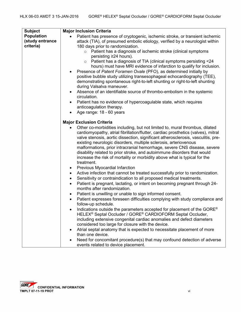
Subject Population (study entrance criteria)
Major Inclusion Criteria Patient has presence of cryptogenic, ischemic stroke, or transient ischemic
attack (TIA), of presumed embolic etiology, verified by a neurologist within 180 days prior to randomization.
o Patient has a diagnosis of ischemic stroke (clinical symptoms persisting ≥24 hours).
o Patient has a diagnosis of TIA (clinical symptoms persisting <24 hours) must have MRI evidence of infarction to qualify for inclusion.
Presence of Patent Foramen Ovale (PFO), as determined initially by positive bubble study utilizing transesophageal echocardiography (TEE), demonstrating spontaneous right-to-left shunting or right-to-left shunting during Valsalva maneuver.
Absence of an identifiable source of thrombo-embolism in the systemic circulation.
Patient has no evidence of hypercoagulable state, which requires anticoagulation therapy.
Age range: 18 - 60 years
Major Exclusion Criteria Other co-morbidities including, but not limited to, mural thrombus, dilated
cardiomyopathy, atrial fibrillation/flutter, cardiac prosthetics (valves), mitral valve stenosis, aortic dissection, significant atherosclerosis, vasculitis, pre-existing neurologic disorders, multiple sclerosis, arteriovenous malformations, prior intracranial hemorrhage, severe CNS disease, severe disability related to prior stroke, and autoimmune disorders that would increase the risk of mortality or morbidity above what is typical for the treatment.
Previous Myocardial Infarction Active infection that cannot be treated successfully prior to randomization. Sensitivity or contraindication to all proposed medical treatments. Patient is pregnant, lactating, or intent on becoming pregnant through 24-
months after randomization. Patient is unwilling or unable to sign informed consent. Patient expresses foreseen difficulties complying with study compliance and
follow-up schedule. Indications outside the parameters accepted for placement of the GORE®
HELEX® Septal Occluder / GORE® CARDIOFORM Septal Occluder, including extensive congenital cardiac anomalies and defect diameters considered too large for closure with the device.
Atrial septal anatomy that is expected to necessitate placement of more than one device.
Need for concomitant procedure(s) that may confound detection of adverse events related to device placement.
HLX 06-03 AMDT 3 15-JAN-2016 GORE® HELEX® Septal Occluder / GORE® CARDIOFORM Septal Occluder
CONFIDENTIAL INFORMATION TMPLT 07-11-19 PROT vii
Contact Information Principal Investigators
Scott Eric Kasner, MD, FAHA Neurologist Philadelphia, Pennsylvania
Lars Thomassen, MD Neurologist Bergen, Norway
John F. Rhodes, MD Cardiologist Miami, Florida
Lars Søndergaard, MD Cardiologist Copenhagen, Denmark
Contract Research Organization
United States, Canada and Europe:
cc
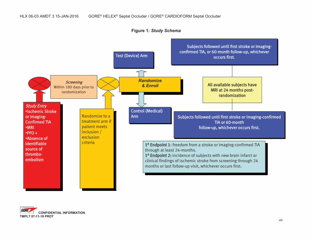
Study EEntry Ischemic Stroke
or Imaging-Confirmed TIA MRI PFO + Absence of
identifiable ssource of thrombo-embolism
Screening Within 180 days prior to
randomization
1° Endpoint 1: freedom from a stroke or imaging-confirmed TIA through at least 24-months. 1° Endpoint 2: incidence of subjects with new brain infarct or clinical findings of ischemic stroke from screening through 24 months or last follow-up visit, whichever occurs first.
Randomize to a treatment arm if patient meets inclusion / exclusion criteria
Test (Device) Arm
Control (Medical) AArm
All available subjects have MRI at 24 months post-
randomization
Subjects followed until first stroke or imaging-confirmed TIA, or 60-month follow-up, whichever
occurs first.
Subjects followed until first stroke or imaging-confirmed TIA or 60-month
follow-up, whichever occurs first.
HLX 06-03 AMDT 3 15-JAN-2016 GORE® HELEX® Septal Occluder / GORE® CARDIOFORM Septal Occluder
CONFIDENTIAL INFORMATION Page ix of 54 TMPLT 07-11-19 PROT
TABLE OF CONTENTS STUDY SUMMARY ..................................................................................................................................... III
1. INTRODUCTION ............................................................................................................................. 1
1.1. BACKGROUND ....................................................................................................................... 1 1.2. SUMMARY OF CLINICAL EXPERIENCE WITH GORE® HELEX® SEPTAL OCCLUDER ................... 5 1.3. DEVICE DESCRIPTION ............................................................................................................ 7
1.3.1. GORE® HELEX® Septal Occluder ......................................................................... 7 1.3.2. GORE® Delivery System........................................................................................ 7 1.3.3. GORE® HELEX® Septal Occluder Package Configuration .................................... 8 1.3.4. GORE® CARDIOFORM Septal Occluder .............................................................. 9 1.3.5. GORE® CARDIOFORM Septal Occluder Delivery System .................................. 9
2. STUDY DESIGN AND STATISTICAL CONSIDERATIONS ......................................................... 11
2.1. STUDY OBJECTIVES ............................................................................................................ 11 2.2. STUDY DESIGN OVERVIEW .................................................................................................. 11 2.3. PRIMARY ENDPOINTS .......................................................................................................... 11 2.4. SECONDARY ENDPOINTS ..................................................................................................... 12 2.5. PRIMARY STUDY HYPOTHESES ............................................................................................ 13 2.6. SAMPLE SIZE DETERMINATION ............................................................................................. 13
2.6.1. Sample Size Assumptions ................................................................................... 13 2.6.2. Randomization and Enrollment ............................................................................ 14 2.6.3. Sample Size Calculations .................................................................................... 14 2.6.4. Power Calculations for Brain Infarct Hypothesis Test ......................................... 15
2.7. RANDOMIZATION SCHEME .................................................................................................... 16 2.8. STATISTICAL ANALYSIS OF PRIMARY ENDPOINTS .................................................................. 16 2.9. INTERIM ANALYSES ............................................................................................................. 17 2.10. STATISTICAL ANALYSIS OF SECONDARY ENDPOINTS ............................................................. 17 2.11. HYPOTHESIS TESTING STRUCTURE ...................................................................................... 17 2.12. EVALUATION OF SEPTAL OCCLUDER DEVICE POOLABILITY .................................................... 18
3. CLINICAL STUDY PLAN .............................................................................................................. 20
3.1. ETHICAL CONSIDERATIONS .................................................................................................. 20 3.2. STUDY INITIATION ................................................................................................................ 20 3.3. SUBJECT POPULATION......................................................................................................... 20 3.4. INCLUSION/EXCLUSION CRITERIA ......................................................................................... 20
3.4.1. General Inclusion Criteria .................................................................................... 20 3.4.2. General Exclusion Criteria ................................................................................... 21
3.5. SCREENING EVALUATION AND CONSENT .............................................................................. 22 3.6. SCREEN FAILURES .............................................................................................................. 25 3.7. RANDOMIZATION AND ENROLLMENT ..................................................................................... 25 3.8. TREATMENT GROUPS .......................................................................................................... 25 3.9. INITIATION AND ADMINISTRATION OF ANTIPLATELET THERAPY FOR ALL SUBJECTS .................. 28 3.10. PFO CLOSURE ................................................................................................................... 28
3.10.1. Pre-Procedure Evaluation – Within 72 hours of PFO Closure .......................... 28 3.10.2. Subject Preparation ........................................................................................... 28 3.10.3. Pre-medication ................................................................................................... 28 3.10.4. Procedure .......................................................................................................... 29 3.10.5. Post-Device Placement Echocardiogram .......................................................... 29 3.10.6. Access Site Management .................................................................................. 30 3.10.7. Post-Procedural Care ........................................................................................ 30
3.11. EARLY POST-PROCEDURE EVALUATIONS – 4-72 HOURS POST PFO CLOSURE ...................... 30 3.12. FOLLOW-UP EVALUATIONS – TEST ARM ............................................................................... 30 3.13. FOLLOW-UP TIME ALLOWANCES – TEST ARM ....................................................................... 32 3.14. FOLLOW-UP EVALUATIONS – CONTROL ARM ......................................................................... 33 3.15. FOLLOW-UP TIME ALLOWANCES – CONTROL ARM ................................................................. 34
HLX 06-03 AMDT 3 15-JAN-2016 GORE® HELEX® Septal Occluder / GORE® CARDIOFORM Septal Occluder
CONFIDENTIAL INFORMATION Page x of 54 TMPLT 07-11-19 PROT
3.16. TREATMENT CHANGES POST-RANDOMIZATION ..................................................................... 34 3.17. STUDY FAILURES ................................................................................................................ 34 3.18. SUBJECT WITHDRAWAL OR DISCONTINUATION FROM THE CLINICAL STUDY ............................ 35 3.19. IMAGING ANALYSIS .............................................................................................................. 35
3.19.1. MRI / CT ............................................................................................................. 35 3.19.2. Echocardiography .............................................................................................. 36
3.20. DATA SAFETY MONITORING BOARD ..................................................................................... 36 3.21. CLINICAL EVENTS COMMITTEE ............................................................................................. 37
4. ADVERSE EVENTS ...................................................................................................................... 38
4.1. ANTICIPATED ADVERSE EVENTS........................................................................................... 38 4.2. SERIOUS ADVERSE DEVICE EFFECTS / SERIOUS ADVERSE EVENTS ....................................... 40
5. DATA COLLECTION AND EVALUATION ................................................................................... 42
5.1. PROTOCOL DEVIATIONS AND VIOLATIONS ............................................................................. 43 5.1.1. Protocol Violations ............................................................................................... 44
6. RISK ANALYSIS ........................................................................................................................... 45
6.1. MINIMIZATION OF RISKS ....................................................................................................... 47 6.2. EXPECTED BENEFITS ........................................................................................................... 47
7. INVESTIGATOR RESPONSIBILITIES ......................................................................................... 48
7.1. SPECIFIC INVESTIGATOR RESPONSIBILITIES .......................................................................... 48 7.1.1. Subject Informed Consent ................................................................................... 48 7.1.2. Compliance .......................................................................................................... 49 7.1.3. GORE® HELEX® Septal Occluder / GORE® CARDIOFORM Septal Occluder Use 49 7.1.4. GORE® HELEX® Septal Occluder / GORE® CARDIOFORM Septal Occluder Disposal 49 7.1.5. Occluder Explant.................................................................................................. 49 7.1.6. Investigator Records ............................................................................................ 49 7.1.7. Investigator Reports ............................................................................................. 50
8. SPONSOR AND MONITOR RESPONSIBILITIES ....................................................................... 51
8.1. SPONSOR RESPONSIBILITIES ............................................................................................... 51 8.2. MONITOR RESPONSIBILITIES ................................................................................................ 51
8.2.1. Pre-investigation Site Visit ................................................................................... 52 8.2.2. Periodic Site Visits ............................................................................................... 52 8.2.3. Clinical Monitor Reports ....................................................................................... 53
9. STEERING COMMITTEE AND PUBLICATION OF RESULTS ................................................... 54
HLX 06-03 AMDT 3 15-JAN-2016 GORE® HELEX® Septal Occluder / GORE® CARDIOFORM Septal Occluder
CONFIDENTIAL INFORMATION Page 1 of 54 TMPLT 07-11-19 PROT
1. Introduction
1.1. Background In the United States (U.S.), stroke is been recognized as a leading cause of both death and disability (CDC 2001, Rosamond 2007). Approximately 700,000 individuals experience new or recurrent strokes annually, which translates to an estimated socioeconomic impact of more than $67 billion in lost productivity and total health care costs (Rosamond 2007). Approximately 87% of all strokes are ischemic, resulting from a blockage in a blood vessel. Even after extensive investigation, up to 40% of all ischemic strokes have no clear pathogenesis and are considered cryptogenic (Halperin 2002). The historical treatment for cryptogenic stroke is medical therapy, including antiplatelet and/or anticoagulation medication. These regimens, however, can increase the risk of bleeding complications and may not prevent recurrent ischemic neurological events (McBride 1994, Stone 1996). Thus, research has turned to identifying potential etiologies of ischemic stroke having no detectable thromboembolic source. Increasingly, attention has been focused on the potential relationship between Patent Foramen Ovale (PFO) and cryptogenic stroke, when there is no other detected thromboembolic source. Studies of cryptogenic stroke patients have found that those with PFO are younger and less likely to have traditional risk factors for stroke than those without PFO, suggesting a different stroke mechanism within this subset (Lamy 2002). Proposed mechanisms of PFO-induced stroke include embolism from the peripheral venous system through the PFO and genesis of thromboembolism directly from the endocardial surface of the atrial septum [Halperin 2002]. Although rare, additional documented cases of thromboembolism have been observed within a PFO (Caes 1995, Falk 1997). The frequency of PFO in patients with presumed normal hearts has been estimated at approximately 25%, based on autopsy studies (Hagen 1984). Controlled studies in the current literature consistently report statistically higher prevalence of PFO among cryptogenic stroke populations (between 40-75%) compared to (between 5-30%) non-cryptogenic stroke populations (Lechat 1988, Webster 1988, Di Tullio 1992, Itoh 1994, Job 1994, Chant 2001, Chen 1991, and Homma 2002). These findings suggest that embolization through the PFO may provide a plausible etiology of stroke in some patients. Nevertheless, a causal relationship between PFO and cryptogenic stroke remains open to debate, since other studies have reported statistically insignificant differences in the occurrence of PFO in patients with cryptogenic stroke versus control populations (Petty 2006, Meissner 2006). Contributing to this uncertainty, the extent of increased risk of recurrent embolic events for cryptogenic stroke patients with a PFO remains unclear compared to cryptogenic stroke patients without a PFO. Recurrent-event rates for patients with and without a PFO reported in the literature vary from 0% to 15% and include many confounding factors (Mas 1995, De Castro 2000, Mas 2001, Homma 2002, and Messe 2004). Further, several of these studies found similar recurrent stroke rates in patients regardless of the presence of a PFO (Mas 2001, Homma 2002, and De Castro 2000). For example, the Patent Foramen Ovale in Cryptogenic Stroke Study (PICSS), reported on 265 patients (42.1%) with cryptogenic stroke and 365 patients (57.9%) with strokes of known
HLX 06-03 AMDT 3 15-JAN-2016 GORE® HELEX® Septal Occluder / GORE® CARDIOFORM Septal Occluder
CONFIDENTIAL INFORMATION Page 2 of 54 TMPLT 07-11-19 PROT
etiology. The average annual risks of death or subsequent stroke in the entire cohort among patients with and without PFO were 7.4% and 7.7%, respectively, and 7.15% and 6.35% among patients with and without PFO in the cryptogenic stroke subset (Homma 2002). Although a direct association of PFO with cryptogenic stroke or its recurrence has yet to be validated, additional existing evidence suggests a combination of PFO and any one of various synergistic factors may exacerbate the risk of stroke. Such features include increased shunt through the PFO and the presence of an atrial septal aneurysm. Other evidence suggests that certain features of the PFO, atrial septal aneurysm and degree of shunting, as well as multiple cerebral infarcts/TIA, Valsalva preceding the presenting event, and age also may contribute to increased stroke risk (Webster 1988, Cabanes 1993, Hausmann 1995, Stone 1996, and De Castro 2000). As before, consensus on each of these factors also still is lacking. With regard to the morphological characteristics of the PFO and the atrial endocardium, De Castro, et al. (2000) found that “high-risk” patients with both right-to-left shunting at rest and increased septal mobility had significantly higher risk of recurrent stroke at three years compared to PFO patients with no shunting at rest (12.5% versus 4.3%; p=0.05). Additionally, presence of an atrial septal aneurysm accompanying a PFO has been associated with higher stroke risk in several studies (Cabanes 1993, Mas 1995, De Castro 2000, and Mas 2001). Although Mas, et al. (2001) did not find an increased risk of recurrent stroke for patients with PFO only, they did report increased risk for patients with PFO plus an atrial-septal aneurysm (4-year recurrent-event rate of 2.3% for patients with PFO, 15.2% for patients with PFO and atrial-septal aneurysm, and 4.2% for patients with neither of these cardiac abnormalities). In the PICSS analysis (Homma 2002) and prospective studies by Martin, et al. (2002) and Harrer, et al. (2006), however, presence of an atrial-septal aneurysm was not associated with increased incidence of recurrent embolic events after either medical treatment or closure of the PFO with a device (p > 0.05). Finally, with regard to age, the PICSS study revealed that stroke recurrence was not dependent on presence of a PFO in patients younger than 55 (p=0.15) or in patients 55 to 64 years old (p=0.48). In contrast, patients with a PFO who were older than 65 years were 4.14 times more likely to experience a recurrent stroke than those without a PFO (p=0.01). Other studies have indicated, however, that young cryptogenic stroke patients with a PFO are at increased risk (Lechat 1988, Ranoux 1993) and recurrence rates of up to 15% in patients younger than 50 years remain a serious concern (Sievert 2004). To summarize, several plausible etiologies and risk factors for cryptogenic stroke have been considered. However, associated data still are inconclusive as to whether a difference in recurrent embolic event rates exists between cryptogenic stroke patients with and without a PFO, as well as with or without other putative risk factors. Based on available data, many physicians already have accepted a causal relationship between PFO and stroke, particularly in younger patients (Ranoux 1993, Bogousslavsky 1996). Indeed, many consider transcatheter/percutaneous closure the treatment of choice for these patients in light of the risks associated with medical therapy, i.e., lifelong anticoagulation and surgical PFO closure (Sievert 2004). In addition, various studies have been, or currently are, underway to determine if transcatheter closure can further reduce the risk of recurrent embolic events for cryptogenic stroke patients with a PFO. Although these studies are not randomized, controlled multicenter trials, the data are promising and suggest that a reduction in recurrent-events occurs after PFO closure.
HLX 06-03 AMDT 3 15-JAN-2016 GORE® HELEX® Septal Occluder / GORE® CARDIOFORM Septal Occluder
CONFIDENTIAL INFORMATION Page 3 of 54 TMPLT 07-11-19 PROT
In a primary example of risk reduction following PFO closure, Schuchlenz, et al. (2005) studied 280 consecutive patients with cryptogenic cerebrovascular events and PFO. The annual recurrence rates of patients treated with medical therapy (13% for patients treated with platelet inhibitors, 5.6% for those on oral anticoagulation) and for those treated with device closure (0.6%) were statistically different (p < 0.001). In this study, PFO diameters > 4 mm and multiple previous cerebral vascular accidents (CVAs) also were found to be independent predictors of recurrent stroke. Similarly, Windecker, et al. (2004) compared risk of recurrent events in 308 patients with cryptogenic stroke and PFO who were treated either medically or with percutaneous PFO closure. At four years of follow-up, percutaneous PFO closure resulted in a nonsignificant trend (p=0.08) toward risk reduction of recurrent stroke or TIA compared with medical treatment (7.8% versus 22.2%, respectively). Further, patients with more than one cerebrovascular event at baseline and those with complete PFO occlusion were at significantly lower risk for recurrent stroke or TIA when compared with medically treated patients (7.3% versus 33.2%, p=0.01). Although limited by uncontrolled data and baseline differences, a meta-analysis (Khairy, et al., 2003) of ten studies of transcatheter closure (1355 patients) and six studies of medical therapy (895 patients) demonstrated that transcatheter closure of patent foramen ovale may prevent a substantial proportion of cryptogenic strokes. Overall, the one-year rate of recurrent neurologic thromboembolism with transcatheter intervention was 0% to 4.9%, while medical management was associated with one-year recurrence rates of 3.8% to 12.0%. A recently published study (Harrer, et al. 2006), however, did not show a difference in recurrent-event rates among 124 patients with cryptogenic stroke and PFO who were treated either medically or had PFO closure (34 transcatheter, 7 surgical). Annual stroke recurrence rates were generally low and comparable among all groups (2.9% percutaneous closure, 2.1% medical closure, 0% surgical closure, 2.2% no therapy). Of the two patients who experienced recurrent events after transcatheter closure, both were found to have residual shunts. Recurrent-event rates after transcatheter closure of PFOs in non-controlled studies have ranged between 0-6%. In initial and intermediate reports from the AGA U.S. Multicenter Clinical Trial, of a total of 94 patients, no recurrent thromboembolic complications were reported following implantation of the Amplatzer® PFO occluder (Hong 2003, Du 2002, and Bruch 2002). In a prospective study, (Braun, et al. 2002), 276 patients underwent PFO closure using the PFO Star device. The mean follow-up time was 15.1 months. During the follow-up period, only six patients (1.7%) had recurrent events, all of which were TIAs occurring within the first six months after implantation. Martin, et al. (2002) also conducted a prospective study to investigate immediate and long-term outcomes of 110 patients who, after paradoxical embolism, underwent percutaneous PFO closure with either a Sideris Button or CardioSEAL® occluder. An annual risk of recurrence of 0.9% based on a mean follow-up of 2.3 years was reported. Other studies have reported embolic recurrence rates following percutaneous PFO closure of 3.2% with mean follow-up of 2.6 years (Hung 2000); 3.4% with mean follow-up of 1.6 years (Windecker 2000); and 0.0% with mean follow-up of 8.4 months (Bridges 1992). Differences in the outcomes of these PFO closure studies may be due to factors specific to the devices
HLX 06-03 AMDT 3 15-JAN-2016 GORE® HELEX® Septal Occluder / GORE® CARDIOFORM Septal Occluder
CONFIDENTIAL INFORMATION Page 4 of 54 TMPLT 07-11-19 PROT
used, e.g. thrombus formation, closure rates, and device complications or to patient variability, atrial-septal aneurysm (ASA), size of shunt, multiple cerebral infarcts/TIA, and patient age. Randomized multicenter clinical trials with appropriate rigorous patient selection need to be conducted to provide definitive objective data to clarify the relationship between presence of a PFO in cryptogenic stroke patients and increased risk of recurrent embolic events. Even with these considerations, current data suggest that transcatheter closure of PFO has the potential for reducing the rate of recurrent ischemic events. In the United States, three additional clinical trials comparing PFO closure to medical management for recurrent stroke prevention are in various stages: the CLOSURE-I study sponsored by NMT Medical; the RESPECT trial sponsored by St. Jude Medical Corp.; and the CARDIA trial sponsored by CARDIA medical. Each of these clinical trials have faced, major enrollment issues and the consensus among the medical community is that enrollment difficulties stem from patient and physician bias toward closure. Also, it has been suggested that patients with significant concern for a paradoxical event are often unwilling to risk randomization to the medical treatment control arm. Further complicating slow enrollment in these studies are the low risk of recurrent embolic events (generally < 4%) and the consequent large numbers of patients required for statistically powering studies. If strokes alone (excluding TIAs) are included in the primary endpoint to reduce confounding factors, annual risk of recurrent embolic events is even less, generally < 2%, and an even larger number of patients is required. For patients being treated with antiaggregants or anticoagulants, Bogousslavsky, et al. (1996) reported an average annual recurrence rate of 1.9% for stroke alone and 3.8% for stroke and TIA. For patients with a PFO and no atrial septal aneurysm, Mas, et al. (2001) reported that the risk of stroke alone at two years was 1.8%, while the risk of either stroke or TIA was 4.6%. In the Mas study, presence of an atrial septal aneurysm in PFO patients also increased the risk of recurrent events. The risk of stroke at two years was 4.0% while the risk of either stroke or TIA was 8.0% in that group. The study design described in the Clinical Study Plan (Section 4.0 of this document) addresses the difficulties encountered in other studies in several ways. Patients will be enrolled into the study from European countries where device availability outside of trials is less of an obstacle to enrollment than in the U.S. and patients are more willing to participate in randomized trials. The study design employs a 2:1, device (test) arm to medical treatment (control) arm, randomization scheme. Because this study allows more patients in the device treatment arm, it provides incentive for patients to be randomized. Finally, a comparison of screening Magnetic Resonance Images (MRIs) or Computed Tomographs (CTs) with end-of-study MRIs or CTs will yield more definitive information on the nature and etiology of recurrent embolic events in the study population. The original closure device selected for this trial is the GORE® HELEX® Septal Occluder, manufactured by W. L. Gore & Associates, Inc. It is comprised of an implantable prosthesis (Occluder) and a catheter delivery system. The Occluder is designed to be soft, atraumatic to surrounding tissue, and to conform to septal anatomy in order to provide safe and effective PFO closure. The safety and efficacy of the GORE® HELEX® Septal Occluder for closing small to medium atrial septal defects (ASDs) has been demonstrated in U.S. studies (Zahn 2001, Latson 2006, and Jones 2007). The excellent closure rates and few complications provide the rationale for use of the GORE® HELEX® Septal Occluder in the present study.
HLX 06-03 AMDT 3 15-JAN-2016 GORE® HELEX® Septal Occluder / GORE® CARDIOFORM Septal Occluder
CONFIDENTIAL INFORMATION Page 5 of 54 TMPLT 07-11-19 PROT
In Europe, nearly a decade of experience regarding the safety and efficacy of PFO closure with the GORE® HELEX® Septal Occluder has been reported in the literature (Sievert 2001, Krumsdorf 2004, Wahl 2005, Billinger 2006, and Schrale 2007). Based on information available to date, the GORE® HELEX® Septal Occluder represents an ideal device for use in a study of the association between PFO closure and recurrent stroke reduction because of its low rate of mild clinical complications and established efficacy for defect closure. During the course of the Gore REDUCE Clinical Study, W.L. Gore & Associates, Inc. developed a new septal defect occluder. As with the GORE® HELEX® Septal Occluder, the GORE® CARDIOFORM Septal Occluder provides a soft, atraumatic, conformable option for the closure of patent foramen ovale. Initial experience with the device, through its development and observations from its initial clinical use, demonstrate that this device is easier to use than the GORE® HELEX® Septal Occluder. It additionally is thought to provide quicker closure of the defect than the GORE® HELEX® Septal Occluder, while maintaining a similar safety profile. Thus, upon its availability, this device will be used as the device of choice within the test arm of the study.
1.2. Summary of Clinical Experience with GORE® HELEX® Septal Occluder Considerable clinical experience has been gained with the GORE® HELEX® Septal Occluder since it became commercially available in Europe in June 1999 and in the US in August 2006. Published data demonstrate that the GORE® HELEX® Septal Occluder provides a reliable and safe system for occlusion of small and moderate ASDs and PFOs with minimal risk of major complications (Sievert 2001, 2002, Pedra 2003, Billinger 2006, Jones 2007). FDA-reviewed studies leading to approval of the GORE® HELEX® Septal Occluder for closure of ASDs included the GORE Feasibility study (two-center, single-arm), the GORE Pivotal study (multicenter, surgical control arm, nonrandomized), and the GORE Continued Access study (multicenter, single-arm, prospective), with a total of 388 patients receiving a device. The clinical success outcomes from the GORE Pivotal study satisfied the primary, noninferiority hypothesis (p <0.001; two-sample binomial proportions test with noninferiority margin = 10%). The Composite Clinical Success of the Continued Access study, defined as no major adverse events or repeat procedures and clinical closure success at 12 months, was 92.6%. To date, portions of these data have been presented in the peer-reviewed literature (Vincent 2003, Latson 2006, and Jones 2007). In Europe, the GORE® HELEX® Septal Occluder is indicated for closure of both ASDs and PFOs. Over 3000 devices have been sold. Several peer-reviewed articles have discussed the safety and efficacy of the GORE® HELEX® Septal Occluder for transcatheter closure of PFOs (Sievert 2001, Krumsdorf 2001, 2004, Wahl 2005, and Billinger 2006). Billinger, et al. (2006) demonstrated the feasibility and safety of the GORE® HELEX® Septal Occluder for transcatheter PFO closure in a large series of patients (128) receiving the HELEX® device. Their follow-up included evaluations of residual right-to-left shunt, device-related adverse events, and recurrent embolic events. Right-to-left shunt was resolved completely in 90% of the patients, a result similar to occlusion rates reported for other septal occluders (Wahl 2001, Beitzke 2001, Martin 2002, and Braun 2002). Furthermore, there were no reported strokes and only one reported TIA (0.8% recurrence) after a mean follow-up of 21 months. This mean recurrence rate of 0.9% per year compares favorably with recurrence rates of up to 3.4% per year observed in closure studies utilizing other devices (Windecker 2000, Beitzke 2001, Martin 2002, Braun 2002, and Wahl 2005).
HLX 06-03 AMDT 3 15-JAN-2016 GORE® HELEX® Septal Occluder / GORE® CARDIOFORM Septal Occluder
CONFIDENTIAL INFORMATION Page 6 of 54 TMPLT 07-11-19 PROT
Wahl, et al. (2005) also reported on effectiveness and safety of several septal occluder devices, including the HELEX® device, for transcatheter PFO closure in patients with cryptogenic stroke. In this study, there were no complications for patients receiving a HELEX® implant, the rate of residual shunt for the HELEX® device was 5% (the lowest of all of devices tested, e.g. compared to 9%, the next lowest rate). The rate of recurrent embolic events, 0%, was among the lowest of all devices tested. In an earlier study, Sievert, et al. (2001), seven types of septal occluders were implanted in 281 patients; 33 received HELEX® Septal Occluders. In the HELEX® device group, no adverse events were reported. During mean follow-up of 12 months, the average annual embolic recurrence rate was 3.3% across all devices, but 0% for patients who received HELEX Septal Occluders. Thrombus formation on atrial septal occluders generally is rare, but is a potential clinical complication that could cause an embolic event. The expanded polytetrafluoroethylene (ePTFE) material used in the GORE® HELEX® Septal Occluder is less thrombogenic than most other materials across a multitude of implanted medical devices (vascular patches, stent-grafts, surgical patches, etc). This low thrombogenicity also has been demonstrated in the GORE® HELEX® Septal Occluder. To date, there has been only one documented report of thrombus formation associated with a GORE® HELEX® Septal Occluder (Krumsdorf 2004). In that study, thrombus formation was evaluated on nine different types of septal occluders implanted in a total of 1000 patients, 161 of whom received a HELEX® device. The rate of thrombus formation for the HELEX® device was 0.8% (one patient), compared to rates as high as 7% for other occluders. To conclude, multiple studies reported in the scientific literature provide many examples and data that have implicated PFO as a contributing factor to increased risk of ischemic embolic event. This putative correlation is supported further by the reduction of recurrent events in patients receiving transcatheter closure of PFO compared to controls. Clinical experience with the GORE® HELEX® Septal and the GORE® CARDIOFORM Septal Occluder illustrate these devices’ potential for demonstrating a reduction of recurrent ischemic embolic events. With the Gore REDUCE Clinical Study, W.L. Gore & Associates will provide the clinical basis to support this claim by demonstrating that transcatheter closure of PFO with the GORE® HELEX® Septal Occluder / GORE® CARDIOFORM Septal Occluder reduces the risk of recurrent stroke or TIA while maintaining a minimal complication rate.
HLX 06-03 AMDT 3 15-JAN-2016 GORE® HELEX® Septal Occluder / GORE® CARDIOFORM Septal Occluder
CONFIDENTIAL INFORMATION Page 7 of 54 TMPLT 07-11-19 PROT
1.3. Device Description
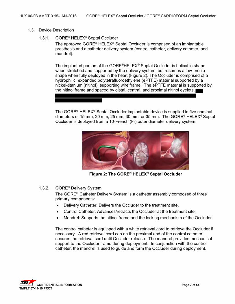
1.3.1. GORE® HELEX® Septal Occluder The approved GORE® HELEX® Septal Occluder is comprised of an implantable prosthesis and a catheter delivery system (control catheter, delivery catheter, and mandrel). The implanted portion of the GORE®HELEX® Septal Occluder is helical in shape when stretched and supported by the delivery system, but resumes a low-profile shape when fully deployed in the heart (Figure 2). The Occluder is comprised of a hydrophilic, expanded polytetrafluoroethylene (ePTFE) material supported by a nickel-titanium (nitinol), supporting wire frame. The ePTFE material is supported by the nitinol frame and spaced by distal, central, and proximal nitinol eyelets.
The GORE® HELEX® Septal Occluder implantable device is supplied in five nominal diameters of 15 mm, 20 mm, 25 mm, 30 mm, or 35 mm. The GORE® HELEX® Septal Occluder is deployed from a 10-French (Fr) outer diameter delivery system.
Figure 2: The GORE® HELEX® Septal Occluder
1.3.2. GORE® Delivery System The GORE® Catheter Delivery System is a catheter assembly composed of three primary components: Delivery Catheter: Delivers the Occluder to the treatment site. Control Catheter: Advances/retracts the Occluder at the treatment site. Mandrel: Supports the nitinol frame and the locking mechanism of the Occluder.
The control catheter is equipped with a white retrieval cord to retrieve the Occluder if necessary. A red retrieval cord cap on the proximal end of the control catheter secures the retrieval cord until Occluder release. The mandrel provides mechanical support to the Occluder frame during deployment. In conjunction with the control catheter, the mandrel is used to guide and form the Occluder during deployment.
HLX 06-03 AMDT 3 15-JAN-2016 GORE® HELEX® Septal Occluder / GORE® CARDIOFORM Septal Occluder
CONFIDENTIAL INFORMATION Page 8 of 54 TMPLT 07-11-19 PROT
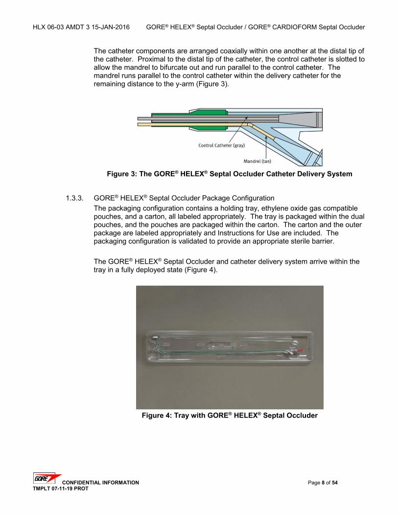
The catheter components are arranged coaxially within one another at the distal tip of the catheter. Proximal to the distal tip of the catheter, the control catheter is slotted to allow the mandrel to bifurcate out and run parallel to the control catheter. The mandrel runs parallel to the control catheter within the delivery catheter for the remaining distance to the y-arm (Figure 3).
Figure 3: The GORE® HELEX® Septal Occluder Catheter Delivery System
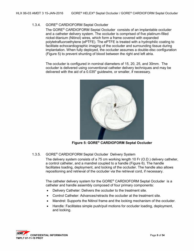
1.3.3. GORE® HELEX® Septal Occluder Package Configuration The packaging configuration contains a holding tray, ethylene oxide gas compatible pouches, and a carton, all labeled appropriately. The tray is packaged within the dual pouches, and the pouches are packaged within the carton. The carton and the outer package are labeled appropriately and Instructions for Use are included. The packaging configuration is validated to provide an appropriate sterile barrier. The GORE® HELEX® Septal Occluder and catheter delivery system arrive within the tray in a fully deployed state (Figure 4).
Figure 4: Tray with GORE® HELEX® Septal Occluder
HLX 06-03 AMDT 3 15-JAN-2016 GORE® HELEX® Septal Occluder / GORE® CARDIOFORM Septal Occluder
CONFIDENTIAL INFORMATION Page 9 of 54 TMPLT 07-11-19 PROT
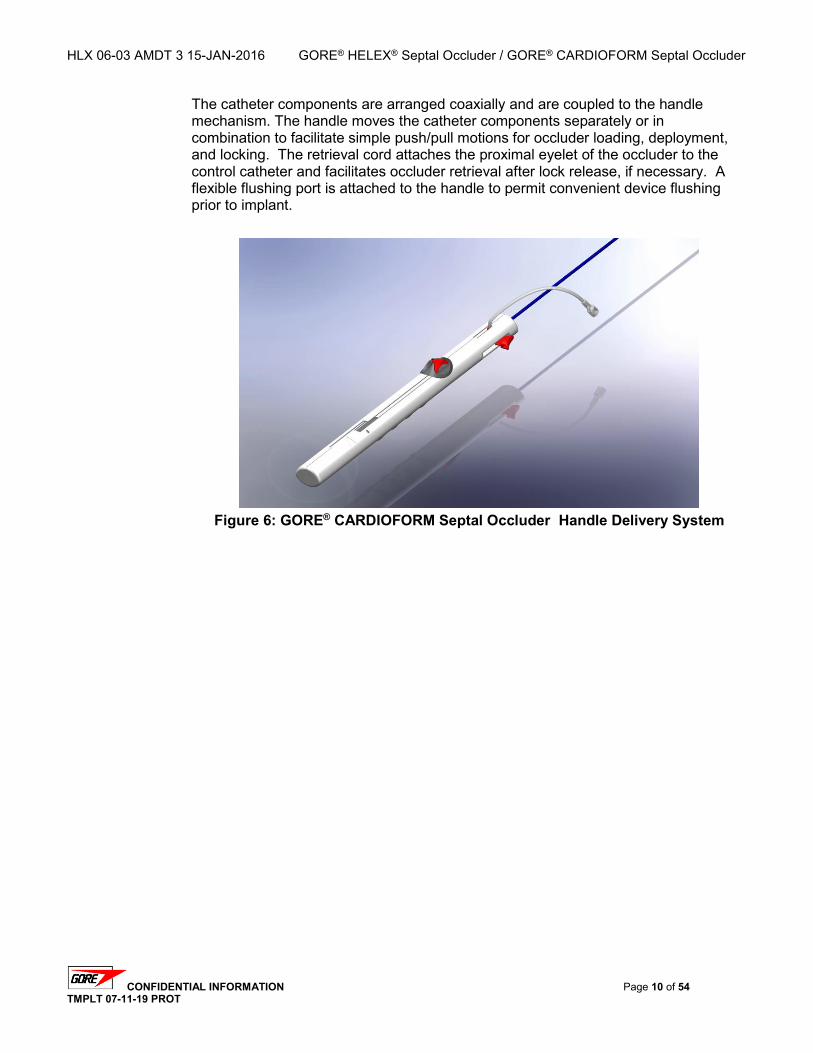
1.3.4. GORE® CARDIOFORM Septal Occluder The GORE® CARDIOFORM Septal Occluder consists of an implantable occluder and a catheter delivery system. The occluder is comprised of five platinum-filled nickel-titanium (Nitinol) wires, which form a frame covered with expanded polytetrafluoroethylene (ePTFE). The ePTFE is treated with a hydrophilic coating to facilitate echocardiographic imaging of the occluder and surrounding tissue during implantation. When fully deployed, the occluder assumes a double-disc configuration (Figure 5) to prevent shunting of blood between the right and left atria. The occluder is configured in nominal diameters of 15, 20, 25, and 30mm. The occluder is delivered using conventional catheter delivery techniques and may be delivered with the aid of a 0.035" guidewire, or smaller, if necessary.
Figure 5: GORE® CARDIOFORM Septal Occluder
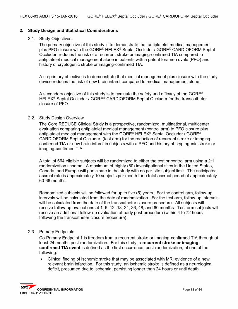
1.3.5. GORE® CARDIOFORM Septal Occluder Delivery System The delivery system consists of a 75 cm working length 10 Fr (O.D.) delivery catheter, a control catheter, and a mandrel coupled to a handle (Figure 6). The handle facilitates loading, deployment, and locking of the occluder. The handle also allows repositioning and retrieval of the occluder via the retrieval cord, if necessary. The catheter delivery system for the GORE® CARDIOFORM Septal Occluder is a catheter and handle assembly composed of four primary components: Delivery Catheter: Delivers the occluder to the treatment site. Control Catheter: Advances/retracts the occluder at the treatment site. Mandrel: Supports the Nitinol frame and the locking mechanism of the occluder. Handle: Facilitates simple push/pull motions for occluder loading, deployment,
and locking.
HLX 06-03 AMDT 3 15-JAN-2016 GORE® HELEX® Septal Occluder / GORE® CARDIOFORM Septal Occluder
CONFIDENTIAL INFORMATION Page 10 of 54 TMPLT 07-11-19 PROT
The catheter components are arranged coaxially and are coupled to the handle mechanism. The handle moves the catheter components separately or in combination to facilitate simple push/pull motions for occluder loading, deployment, and locking. The retrieval cord attaches the proximal eyelet of the occluder to the control catheter and facilitates occluder retrieval after lock release, if necessary. A flexible flushing port is attached to the handle to permit convenient device flushing prior to implant.
Figure 6: GORE® CARDIOFORM Septal Occluder Handle Delivery System
HLX 06-03 AMDT 3 15-JAN-2016 GORE® HELEX® Septal Occluder / GORE® CARDIOFORM Septal Occluder
CONFIDENTIAL INFORMATION Page 11 of 54 TMPLT 07-11-19 PROT
2. Study Design and Statistical Considerations
2.1. Study Objectives The primary objective of this study is to demonstrate that antiplatelet medical management plus PFO closure with the GORE® HELEX® Septal Occluder / GORE® CARDIOFORM Septal Occluder reduces the risk of a recurrent stroke or imaging-confirmed TIA compared to antiplatelet medical management alone in patients with a patent foramen ovale (PFO) and history of cryptogenic stroke or imaging-confirmed TIA. A co-primary objective is to demonstrate that medical management plus closure with the study device reduces the risk of new brain infarct compared to medical management alone. A secondary objective of this study is to evaluate the safety and efficacy of the GORE® HELEX® Septal Occluder / GORE® CARDIOFORM Septal Occluder for the transcatheter closure of PFO.
2.2. Study Design Overview The Gore REDUCE Clinical Study is a prospective, randomized, multinational, multicenter evaluation comparing antiplatelet medical management (control arm) to PFO closure plus antiplatelet medical management with the GORE® HELEX® Septal Occluder / GORE® CARDIOFORM Septal Occluder (test arm) for the reduction of recurrent stroke or imaging-confirmed TIA or new brain infarct in subjects with a PFO and history of cryptogenic stroke or imaging-confirmed TIA. A total of 664 eligible subjects will be randomized to either the test or control arm using a 2:1 randomization scheme. A maximum of eighty (80) investigational sites in the United States, Canada, and Europe will participate in the study with no per-site subject limit. The anticipated accrual rate is approximately 10 subjects per month for a total accrual period of approximately 60-66 months. Randomized subjects will be followed for up to five (5) years. For the control arm, follow-up intervals will be calculated from the date of randomization. For the test arm, follow-up intervals will be calculated from the date of the transcatheter closure procedure. All subjects will receive follow-up evaluations at 1, 6, 12, 18, 24, 36, 48, and 60 months. Test arm subjects will receive an additional follow-up evaluation at early post-procedure (within 4 to 72 hours following the transcatheter closure procedure).
2.3. Primary Endpoints Co-Primary Endpoint 1 is freedom from a recurrent stroke or imaging-confirmed TIA through at least 24 months post-randomization. For this study, a recurrent stroke or imaging-confirmed TIA event is defined as the first occurrence, post-randomization, of one of the following: Clinical finding of ischemic stroke that may be associated with MRI evidence of a new
relevant brain infarction. For this study, an ischemic stroke is defined as a neurological deficit, presumed due to ischemia, persisting longer than 24 hours or until death.
HLX 06-03 AMDT 3 15-JAN-2016 GORE® HELEX® Septal Occluder / GORE® CARDIOFORM Septal Occluder
CONFIDENTIAL INFORMATION Page 12 of 54 TMPLT 07-11-19 PROT
Clinical finding of TIA that also has MRI evidence of a new relevant brain infarction. For this study, a TIA is defined as a transient neurological deficit, presumed due to ischemia, persisting less than 24 hours.
Clinical findings of stroke/TIA may include: Sudden numbness or weakness of the face, arm or leg (especially on one side of the
body) Sudden confusion, trouble speaking or understanding speech Sudden trouble seeing in one or both eyes Sudden trouble walking, dizziness, loss of balance or coordination Sudden severe headache with no known cause
Co-Primary Endpoint 1 will be calculated as the time from randomization to the first recurrent event. Subjects free from a recurrent event will be censored at the date of last known contact. All deaths and suspected recurrent stroke/TIA events will be reviewed and adjudicated by a Clinical Events Committee (CEC). In the event of subject death, all possible efforts will be made to obtain relevant records from the hospital or the subject’s primary care physician, including a death certificate or autopsy report, to determine the cause of death. Co-primary Endpoint 2 is the incidence of subjects with new brain infarct or stroke from screening through 24 months or last follow-up visit, whichever occurs first, hereinafter referred to as brain infarct. A responder is defined as any subject with at least one new T2 hyperintense MRI lesion with diameter ≥ 3 mm from screening or clinical findings of ischemic stroke, through 24 months or last follow-up visit, whichever occurs first. It will be calculated as a subject-based binomial proportion.
2.4. Secondary Endpoints Safety Endpoints will include the proportion of subjects who experience adverse events (AEs) that are determined to be related to device, procedure, and/or antiplatelet medical management. This will include specific adverse events and groups of adverse events such as all-cause adverse events, device-related events, procedure-related events, antiplatelet medical therapy-related events, and any serious adverse events. The safety endpoints may also be analyzed using time-to-event methods to estimate the percentage of subjects free from the event at time points of interest, such as 30 days and 24 months post-randomization (or post-procedure for the test arm). Efficacy Endpoints will evaluate the success of the device in achieving PFO closure in subjects randomized to the test arm. PFO closure success will be measured by assessing the degree of residual right-to-left shunt after device implant. Time points for the assessment of PFO closure include early post-procedure, 1 month, 12 months, and 24 months. Additional Secondary Endpoints will include: 1. Clinical Success –
HLX 06-03 AMDT 3 15-JAN-2016 GORE® HELEX® Septal Occluder / GORE® CARDIOFORM Septal Occluder
CONFIDENTIAL INFORMATION Page 13 of 54 TMPLT 07-11-19 PROT
a. Test Arm - defined as the composite of Device Success, PFO closure, and absence of a recurrent stroke or imaging-confirmed TIA at 24 months post-procedure
b. Control Arm - defined as the freedom from a recurrent stroke or imaging-confirmed TIA at 24 months post-randomization
2. Overall Survival – defined as time from randomization to death from any cause or last known contact
3. Time to any stroke/TIA – defined as time from randomization to first occurrence of stroke or TIA
4. Device Success – defined as the proportion of device arm subjects with successful implant and retention of the device after procedure (test arm only)
2.5. Primary Study Hypotheses This study is designed to test the null hypothesis that the hazard of a recurrent stroke or imaging-confirmed TIA in subjects treated with percutaneous PFO closure plus antiplatelet medical management is equal to or higher than subjects treated with antiplatelet medical management alone. The alternative hypothesis is that the hazard of a recurrent stroke/imaging-confirmed TIA is lower in subjects treated with percutaneous PFO closure plus antiplatelet medical management compared to antiplatelet medical management alone. In statistical terms:
tallfortHRHtallfortHRH
CTA
CTO
0.1)(:0.1)(:
/
/
where: HRT/C = hazard ratio comparing the test (T) arm (PFO closure) to the control (C) arm (antiplatelet medical management only). In addition, this study will test the null hypothesis that the incidence of brain infarct at 24 months in subjects treated with percutaneous PFO closure plus antiplatelet medical management is equal to or higher than subjects treated with antiplatelet medical management alone. The alternative hypothesis is that the brain infarct incidence is lower in subjects treated with percutaneous PFO closure plus antiplatelet medical management compared to antiplatelet medical management alone. In statistical terms: 𝐻0: 𝑃𝐶 − 𝑃𝑇 ≤ 0𝐻1: 𝑃𝐶 − 𝑃𝑇 > 0
where: PC = true proportion of subjects with incident brain infarct in the control group PT = true proportion of subjects with incident brain infarct in the test group
2.6. Sample Size Determination
2.6.1. Sample Size Assumptions Based on literature available at the study’s initiation (see Section 1.0 - Introduction), the proportion of PFO patients free from a recurrent stroke or imaging-confirmed TIA at 24 months after initial, cryptogenic stroke or imaging-confirmed TIA is assumed to be approximately 92%, with a range of 86% to 94%. Antiplatelet medical management plus PFO closure will be considered superior to antiplatelet medical management alone if there is at least a 55% reduction in the hazard of a recurrent stroke or imaging-confirmed TIA.
HLX 06-03 AMDT 3 15-JAN-2016 GORE® HELEX® Septal Occluder / GORE® CARDIOFORM Septal Occluder
CONFIDENTIAL INFORMATION Page 14 of 54 TMPLT 07-11-19 PROT
At the time this study was designed, Co-primary Endpoint 2 was considered a secondary endpoint and was not relevant to the sample size assumptions.
2.6.2. Randomization and Enrollment Subjects will be randomized in a 2:1 allocation ratio with the greater proportion of subjects randomized to the test arm. Enrollment of 120 to 140 subjects per year is anticipated, for a total enrollment period of 60-66 months (5 to 5.5 years).
2.6.3.
HLX 06-03 AMDT 3 15-JAN-2016 GORE® HELEX® Septal Occluder / GORE® CARDIOFORM Septal Occluder
CONFIDENTIAL INFORMATION Page 15 of 54 TMPLT 07-11-19 PROT
HLX 06-03 AMDT 3 15-JAN-2016 GORE® HELEX® Septal Occluder / GORE® CARDIOFORM Septal Occluder
CONFIDENTIAL INFORMATION Page 16 of 54 TMPLT 07-11-19 PROT
2.7. Randomization Scheme Patients who meet the study eligibility criteria will be randomized to one of the two study treatment arms. Randomization will be weighted 2:1 in favor of the test arm.
2.8. Statistical Analysis of Primary Endpoints The primary endpoint analyses will be performed when the last subject enrolled completes 24 months of follow-up. Co-primary Endpoint 1, freedom from a recurrent event, will be compared between treatment groups using an unadjusted log-rank test and presented using Kaplan-Meier methods. All follow-up data through 5 years will be included on subjects continuing follow-up past the 24-month evaluation. As part of a simultaneous test with the brain infarct co-primary endpoint hypothesis a multiplicity-adjusted p-value for the hazard ratio test of 0.025 or less will be considered evidence to reject the freedom from recurrent event null hypothesis. The analysis sample will consist of randomized subjects with valid MRI core lab data at screening and an appropriate follow-up, where follow-up will be at 24 months or immediately following a recurrent event (suspected stroke or TIA), whichever occurs first, as well as randomized subjects who experience a confirmed recurrent event through 24 months regardless of MRI data status. Responders are subjects who show one or more new infarction(s) on MRI since screening or experience a confirmed recurrent event. Nonresponders are subjects who do not show new infarction on MRI since screening and do not experience a confirmed recurrent event. The primary analysis will be a two-sample comparison of the binomial proportion of subjects with brain infarct between the two treatment groups. Each binomial proportion will be calculated as the count of responders divided by the count of evaluable subjects (sum of the responder and nonresponder counts). The hypothesis will be tested using a two-sample binomial proportions test:
𝑧 =𝑝𝐶 − 𝑝𝑇
√𝑝𝐶(1 − 𝑝𝐶)
𝑛𝐶+
𝑝𝑇(1 − 𝑝𝑇)𝑛𝑇
where:
HLX 06-03 AMDT 3 15-JAN-2016 GORE® HELEX® Septal Occluder / GORE® CARDIOFORM Septal Occluder
CONFIDENTIAL INFORMATION Page 17 of 54 TMPLT 07-11-19 PROT
pC = observed brain infarct proportion in control group nC = number of evaluable subjects in control group pT = observed brain infarct proportion in test group nT = number of evaluable subjects in test group The test statistic z is assumed to have a standard Normal distribution. The significance level for this test will be set at a 1-sided = 0.025 , but the p-value will be adjusted for multiplicity with the 1-sided = 0.025 test performed simultaneously on the primary endpoint. Therefore, pC − pT > 0 and a multiplicity-adjusted p-value 0.025 will result in rejection of the null hypothesis in favor of the alternative hypothesis and a conclusion that the test treatment reduces the rate of brain infarct compared to the control treatment. The 1-sided multiplicity-adjusted p-value and unadjusted 2-sided 95% confidence interval for the difference in proportions will be reported.
2.9. Interim Analyses
The original protocol specified that, in addition to standard study monitoring, an interim analysis will be performed after approximately 50% of the total expected recurrent stroke or imaging-confirmed TIA events have occurred. This milestone event had not occurred as of the completion of full enrollment in February 2015. Under the plan described herein, the original interim analysis plan no longer serves its purpose and is rescinded.
A new interim analysis that does not involve any statistical hypothesis testing is anticipated to occur around April 2016. No analyses that would require alpha spending are planned for this interim analysis.
2.10. Statistical Analysis of Secondary Endpoints The secondary endpoint analysis will be performed in conjunction with the primary endpoint analysis. Statistical methods for testing multiple endpoints will be utilized in the comparison of secondary endpoints across test and control to preserve the overall Type I error rate.
p
HLX 06-03 AMDT 3 15-JAN-2016 GORE® HELEX® Septal Occluder / GORE® CARDIOFORM Septal Occluder
CONFIDENTIAL INFORMATION Page 18 of 54 TMPLT 07-11-19 PROT
Based on the final analysis of correlation between the two endpoints and the unadjusted p-values obtained from the two test statistics, the appropriate adjusted p-values will be compared to the overall 1-sided = 0.025 for the experiment.
2.12. Evaluation of Septal Occluder Device Poolability It is expected that shortly after approximately one-third of the subjects have been enrolled, the new study device (GORE® CARDIOFORM Septal Occluder ) will become available and will be used as the device of choice within the test arm of the study. As a regulatory requirement, an assessment of poolability will be conducted for the two study device subgroups in the test arm. The statistical plan for this assessment will consist of two stages: baseline homogeneity and primary outcome comparability. For baseline homogeneity, the two test device subgroups will be compared on the following six baseline demographic and predictor covariates: age, gender, qualifying cerebrovascular event, balloon-sized PFO diameter, PFO tunnel length, and presence of atrial septal aneurysm. These subgroup comparisons will use the two-sample t-test, chi-square test, or Fisher’s Exact test, depending on the distribution of the covariate. For primary outcome comparability, covariate-by-device interactions will be assessed using Cox proportional hazards regression models where main effects for device and the covariate and the covariate-by-device interaction term will be regressed on the 24-month freedom from recurrent stroke or imaging-confirmed TIA primary endpoint; this will be performed individually for each of the six baseline covariates. Statistically significant interactions will be assessed
HLX 06-03 AMDT 3 15-JAN-2016 GORE® HELEX® Septal Occluder / GORE® CARDIOFORM Septal Occluder
CONFIDENTIAL INFORMATION Page 19 of 54 TMPLT 07-11-19 PROT
graphically for the nature of the interaction (quantitative vs. qualitative). Qualitative interactions (difference in direction of device effect across levels of covariate) will suggest differences between the device subgroups, leading to analyses performed separately for each device subgroup. A similar approach will be used for brain infarct comparability, but using a logistic regression model for this binary endpoint. Finally, device subgroup and any baseline covariates deemed to be different between device subgroups will be included as main effects in a Cox regression model on the primary endpoint. If the device subgroup term is statistically significant and the observed hazard reduction (test vs. control) for either of the device subgroups is less than the hypothesized 55%, then the device subgroups will not be considered outcome comparable, leading to analyses performed separately for each device subgroup. A similar approach will be used for brain infarct comparability, but using a logistic regression model for this binary endpoint. A significance level of =0.15 will be used for these poolability tests, without correction for multiplicity. Since this evaluation plan calls for a minimum of 13 significance tests per endpoint, the overall Type-I error rate per endpoint for this analysis may exceed 1 − (1 − 0.15)13 = 0.88.
HLX 06-03 AMDT 3 15-JAN-2016 GORE® HELEX® Septal Occluder / GORE® CARDIOFORM Septal Occluder
CONFIDENTIAL INFORMATION Page 20 of 54 TMPLT 07-11-19 PROT
3. Clinical Study Plan
3.1. Ethical Considerations The clinical study will be conducted in accordance with the applicable national regulatory requirements, the US Investigational Device Exemption (IDE) regulations (21 CFR § 812) and the current European ISO standard, as required. In addition, the clinical study will comply with the ethical principles of the Declaration of Helsinki.
3.2. Study Initiation Initiation of the clinical study can begin following: FDA approval of the Investigational Device Exemption (IDE), and Sponsor receipt of notification of the investigational site Institutional Review Board (IRB)
or Ethics Committee (EC) approval, and Sponsor’s receipt of an executed Clinical Study Agreement (CSA) or Clinical Trial Letter of
Agreement (CTLA) for the investigational site.
3.3. Subject Population Subjects will be enrolled at a maximum of eighty (80) investigational sites in the United States, Canada and Europe. All potential subjects must have been diagnosed with cryptogenic stroke or imaging-confirmed TIA and the presence of a PFO and be at risk for recurrent stroke or TIA event. Eligible subjects will be randomized to a treatment arm in a 2:1 fashion (device-to-antiplatelet medical therapy). This randomization will result in approximately 443 in the test (device) arm and 221 subjects in the control (antiplatelet medical therapy) arm.
3.4. Inclusion/Exclusion Criteria
3.4.1. General Inclusion Criteria The criteria listed below shall be used to determine whether a patient is eligible for entry into the trial. The patient must meet all inclusion criteria to be enrolled in the study. In other words, all inclusion criteria must be answered “yes” for the patient to be eligible for the study. 1. Patient has had a cryptogenic, ischemic stroke, or transient ischemic attack
(TIA), of presumed embolic etiology, verified by a neurologist within 180 days prior to randomization, meeting either criteria a or b: a) Patient has a diagnosis of ischemic stroke (clinical symptoms persisting
≥24 hours). OR
b) Patient has a diagnosis of TIA (clinical symptoms persisting <24 hours) and has MRI evidence of infarction. For MRI-incompatible patients (i.e., patients that are claustrophobic and/or have implants that are contraindicated for MR) a CT scan of the brain will be accepted.
2. Patient is diagnosed with a patent foramen ovale (PFO), confirmation of which is achieved by transesophageal echocardiography (TEE) with bubble study demonstrating spontaneous right-to-left shunting or right-to-left shunting during Valsalva maneuver.
HLX 06-03 AMDT 3 15-JAN-2016 GORE® HELEX® Septal Occluder / GORE® CARDIOFORM Septal Occluder
CONFIDENTIAL INFORMATION Page 21 of 54 TMPLT 07-11-19 PROT
3. There is an absence of an identifiable source of thromboembolism in the systemic arterial circulation.
4. Patient is at least 18 years and less than 60 years of age (subjects cannot have reached their 60th birthday prior to randomization/enrollment).
5. Patient has vascular imaging that rules out other potential sources of cerebral thromboembolism (e.g., dissection of the aorta or neck vessels, carotid stenosis > 50% and/or presence of ulcerated plaques, or intracranial stenosis > 50%).
6. Patient has no evidence of hypercoagulable state, which requires anticoagulation therapy. This determination will be based on the evaluation of, at a minimum: platelet count, Prothrombin Time (PT) or International Normalized Ratio (INR), Activated Partial Thromboplastin Time (aPTT), and Antiphospholipid Antibodies. All test results are to be evaluated based on the laboratory normals established at the institution. A thorough history of thromboembolic events in first degree family members must be obtained for all patients. For patients who have a first degree family member with such an event prior to age 55, or whose family history is unknown, the following additional tests are required and must be interpreted as normal: Factor V Leiden mutation, Prothrombin Gene G20210A mutation, protein C, protein S, and Antithrombin III.
7. Patient is willing and capable of complying with the study protocol requirements, including the specified follow-up period, and can be contacted by telephone.
8. Patient or patient’s legal representative (or person designated acceptable under local Ethics Committee requirements) is willing to provide written informed consent prior to enrollment in study.
3.4.2. General Exclusion Criteria All exclusion criteria must be answered “no” for subject inclusion in the study. 1. Patient has a life expectancy of less than one year. 2. Patient is experiencing severe disability, defined as modified Rankin Scale
(mRS) score greater than or equal to 3, at the time of randomization. 3. Patient has neurological deficits not due to stroke that may affect the patient’s
neurologic assessments. 4. Patient has other potential source(s) of cardio-embolism, for example: atrial
fibrillation (AFib) or atrial flutter (AFlu), prosthetic heart valve, severe native valve disease, left ventricular ejection fraction of <40%, severe ventricular wall motion abnormalities (akinesis, severe hypokinesis), intracardiac thrombus, mitral valve stenosis, prior cardiac surgery, other major congenital cardiac abnormality.
5. Patient has had a prior myocardial infarction. 6. Patient has uncontrolled diabetes mellitus at the time of randomization, in the
opinion of the investigator. 7. Patient has pulmonary hypertension (mean pulmonary artery pressure >25
mmHg). 8. Patient has uncontrolled systemic hypertension at the time of screening, in the
opinion of the investigator. 9. Patient presented with a lacunar stroke syndrome (e.g., small deep infarction
<1.5 cm in diameter and/or a typical lacunar syndrome such as pure motor hemiparesis, pure sensory stroke, clumsy hand-dysarthria syndrome, or ataxic-hemiparesis syndrome).
HLX 06-03 AMDT 3 15-JAN-2016 GORE® HELEX® Septal Occluder / GORE® CARDIOFORM Septal Occluder
CONFIDENTIAL INFORMATION Page 22 of 54 TMPLT 07-11-19 PROT
10. Patient has intracranial pathology that makes the patient inappropriate for study participation based on discretion of the Investigator (e.g., brain tumor other than meningioma, arterio-venous malformation (AVM) or cerebral hemorrhage, cerebral venous sinus thrombosis on CT or MRI, or cerebral aneurysm > 7 mm).
11. Patient has active autoimmune disease (e.g., lupus erythematosus disseminata, rheumatoid arthritis, polyarteritis nodosa, primary cerebral vasculitis).
12. Patient has active infection that cannot be treated successfully prior to randomization.
13. Patient abuses alcohol and/or drugs [e.g., on average >5 units or drinks (60 grams) of alcohol/day] or abuses alcohol and/or drugs in the opinion of the Investigator.
14. Patient is pregnant, lactating, or intent on becoming pregnant through 24-months after randomization.
15. Patient has contraindication to study medications, including antiplatelet therapy. 16. Patient requires chronic anticoagulation therapy that cannot be discontinued
prior to randomization, in the opinion of the Investigator. Testing for prothrombotic disorders may be performed at the discretion of the treating physicians but is not required for this study.
17. Patient is currently participating in another clinical device or drug trial that has not completed its primary endpoint or that will clinically confound the current study endpoints or does not permit subjects to participate in other studies. Typically, subjects that are involved in the long-term surveillance phase of a clinical study are eligible.
18. Patient has other anatomic or co-morbid conditions that could, in the Investigator’s opinion, limit the patient’s ability to participate in the study or to comply with follow-up requirements, or impact the scientific soundness of the study results.
19. Patient has a known sensitivity to contrast media that cannot be controlled adequately with pre-medication.
20. Patient has had any major surgical procedure within 30 days preceding randomization.
21. Patient plans to have a major elective surgical procedure within 30 days after randomization or within 30 days of a PFO closure procedure.
22. Patient has the need for any concomitant procedure, based on the results of the screening evaluations, during the PFO closure procedure that may confound detection of device-related adverse events.
23. In the opinion of the Investigator, patient has anatomic criteria identified during the screening evaluation and/or the screening transesophageal echocardiogram (TEE) that are unfavorable for successful placement of the GORE® HELEX® Septal Occluder / GORE® CARDIOFORM Septal Occluder or the patient has contraindications for device placement, which may include:
Inability to accommodate a 10 Fr delivery catheter The need for trans-septal puncture Requires placement of more than one device PFO estimated to be too large for successful device placement Device would impinge on cardiac structure(s) Anatomy would likely prevent discs from apposing the septal tissue.
3.5. Screening Evaluation and Consent
HLX 06-03 AMDT 3 15-JAN-2016 GORE® HELEX® Septal Occluder / GORE® CARDIOFORM Septal Occluder
CONFIDENTIAL INFORMATION Page 23 of 54 TMPLT 07-11-19 PROT
Informed consent must be obtained prior to performing study-related screening tests or procedures that are not standard of care. Patients will be screened by a Neurologist Investigator or designee for initial study eligibility. Patients who meet the initial criteria for entry (i.e., previous cryptogenic stroke or imaging-confirmed TIA and the presence of PFO with right-to-left shunting) will be informed of his/her potential eligibility. An Investigator or designee will discuss the clinical study with the patient including risks, benefits, and required follow-up procedures before obtaining informed consent. (See Appendix F for the Informed Consent Form template.) The patient will be required to read and sign an Informed Consent Form prior to completing study-related tests and procedures. A legal representative (or legal guardian or person designated acceptable under local Ethics Committee requirements) may provide consent if the patient is unable to do so. Patients with a signed Informed Consent will be scheduled for screening evaluation. Patients who consent will complete the screening tests and procedures (e.g. vascular imaging, neurologic assessments, ECG, and blood tests) within 180 days of the qualifying stroke or TIA and prior to final determination of study entrance (e.g., meeting all inclusion and exclusion criteria) by the Investigator. The following will be included in the overall screening evaluation: Initial eligibility assessment, to include confirmation of
o previous cryptogenic stroke or imaging-confirmed TIA All patients must have a brain MRI obtained for future comparison.
For patients with a diagnosis of TIA, MRI must be positive for infarction.
For patients with CT evidence of infarction a MRI must also be obtained as part of the baseline screening assessments.
For MRI-incompatible patients only (i.e., patients that are claustrophobic and/or have metal implants that are contraindicated for MR) a CT scan of the brain will be accepted.
o presence of a patent foramen ovale (PFO) with positive bubble study utilizing transesophageal echocardiography TEE demonstrating spontaneous right-to-left shunting or right-to-left shunting during Valsalva maneuver
o absence of left atrial appendage thrombus or other mural thrombus during the TEE Physical examination MRA, MRI, CTA, duplex color Doppler, carotid ultrasound, transcranial Doppler (TCD), or
conventional angiography of the head, neck, and aortic arch (aortic arch may also be evaluated by TEE)
ECG Obtain patient demographics Record patient’s medical history Neurologic assessments performed by the Neurologist Investigator, or designee, qualified
to score NIHSS and modified Rankin Scales, to include all of the following
HLX 06-03 AMDT 3 15-JAN-2016 GORE® HELEX® Septal Occluder / GORE® CARDIOFORM Septal Occluder
CONFIDENTIAL INFORMATION Page 24 of 54 TMPLT 07-11-19 PROT
o Modified Rankin Scale (mRS) o National Institute of Health Stroke Scale (NIHSS), and o Supplemental neurologic examination (captures assessments not gathered by the
NIHSS and should be performed by a Neurologist Investigator only) Blood pressure Laboratory Evaluations
o Blood drawn for determination of hypercoagulable state by the Investigator, to include, but not limited to:
Platelet count Prothrombin Time (PT) or International Normalized Ratio (INR) Activated Partial Thromboplastin Time (aPTT) Antiphospholipid Antibodies Additional testing for patients who have a first degree family member with a
history of thromboembolic events prior to age 55, or whose family history is unknown:
Factor V Leiden Mutation Prothrombin Gene G20210A Mutation Protein C Protein S Antithrombin III
o Pregnancy test (if applicable) The MRI scan must include Fluid Attenuated Inversion Recovery (FLAIR) sequencing and diffusion-weighted images (transverse and coronal planes, with 5 mm or thinner section thicknesses recommended, and no gaps) of the brain to detect and record any evidence of clinical and/or silent lesions. This MRI will serve as the screening image for later comparison with a follow-up image at 24 months or recurrent event, whichever occurs first. For MRI-incompatible patients (i.e., patients that are claustrophobic and/or have metal implants that are contraindicated for MR) a CT scan of the brain will be accepted. An independent Core Lab will be utilized to compare all MRI and CT screening scans with recurrent event or 24-month scans for determination of endpoints. In addition, a vascular examination of the head, neck, and aortic arch must have been performed using one or more of the following diagnostic tools: magnetic resonance angiography (MRA), magnetic resonance imaging (MRI), computed tomography angiography (CTA), duplex color Doppler, carotid ultrasound, transcranial Doppler (TCD) ultrasound, and/or conventional angiography to rule out other potential sources of emboli in the systemic circulation. Transesophageal echocardiography (TEE) may also be used for the evaluation of the aortic arch.
HLX 06-03 AMDT 3 15-JAN-2016 GORE® HELEX® Septal Occluder / GORE® CARDIOFORM Septal Occluder
CONFIDENTIAL INFORMATION Page 25 of 54 TMPLT 07-11-19 PROT
3.6. Screen Failures Patients who consent to participate in the study, but who are not randomized to treatment, or who withdraw consent prior to randomization will be considered screen failures and will not be followed in this study. Screen failures will be entered in the Electronic Data Capture (EDC) system.
3.7. Randomization and Enrollment Upon completion of screening tests and confirmation of study eligibility, consented patients will be randomized to a treatment arm. Using the EDC database approved by the Sponsor, the patient will be assigned to a treatment arm in the study. Patients will have a 2-in-3 chance of being randomized to the test (device) arm. Because of the inclusion criteria of prior cryptogenic stroke, patients may have already initiated antiplatelet medical therapy prior to randomization. All patients will be instructed to continue antiplatelet medical therapy or begin antiplatelet medical therapy, according to the regimen prescribed in this protocol and upon instruction of their Neurologist Investigator. For all patients randomized, if the subject has already initiated antiplatelet medical therapy prior to randomization, treatment should continue based on the antiplatelet regimen prescribed in this protocol (see Section 3.9 – Initiation and Administration of Antiplatelet Therapy). If antiplatelet medical therapy is not in progress or a regimen change is required, treatment should commence promptly (within 48 hours of randomization). Patients randomized to the test (device) arm must be treated (have a PFO closure attempt) within 90 days of randomization. Patients are considered “subjects” enrolled in the study following randomization to a treatment arm. A patient is considered enrolled into the clinical study when: he/she signs the consent form, and he/she completes all required screening tests and evaluations, and he/she meets inclusion/exclusion criteria, and he/she is randomized to a treatment arm.
3.8. Treatment Groups The Schedules of Events for each treatment group, both test and control arms, are presented in Table 1: Test Arm Schedule of Events and Table 2: Control Arm Schedule of Events.
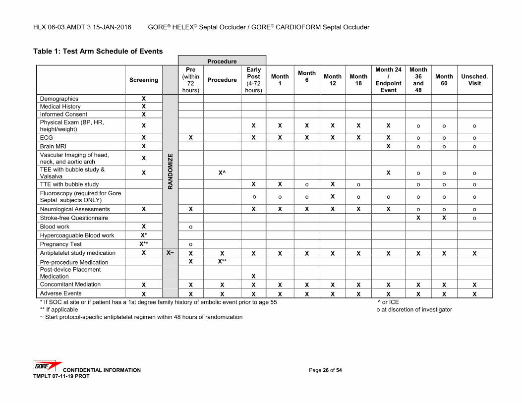
HLX 06-03 AMDT 3 15-JAN-2016 GORE® HELEX® Septal Occluder / GORE® CARDIOFORM Septal Occluder
CONFIDENTIAL INFORMATION Page 26 of 54 TMPLT 07-11-19 PROT
Table 1: Test Arm Schedule of Events Procedure
Screening
Pre (within
72 hours)
Procedure Early Post (4-72 hours)
Month 1
Month 6
Month
12 Month
18
Month 24 /
Endpoint Event
Month 36
and 48
Month 60
Unsched. Visit
Demographics X
RA
ND
OM
IZE
Medical History X Informed Consent X Physical Exam (BP, HR, height/weight) X X X X X X X o o o
ECG X X X X X X X X o o o Brain MRI X X o o o Vascular Imaging of head, neck, and aortic arch X
TEE with bubble study & Valsalva X X^ X o o o
TTE with bubble study X X o X o o o o Fluoroscopy (required for Gore Septal subjects ONLY) o o o X o o o o o
Neurological Assessments X X X X X X X X o o o Stroke-free Questionnaire X X o Blood work X o Hypercoaguable Blood work X* Pregnancy Test X** o Antiplatelet study medication X X~ X X X X X X X X X X X Pre-procedure Medication X X** Post-device Placement Medication X Concomitant Mediation X X X X X X X X X X X X Adverse Events X X X X X X X X X X X X * If SOC at site or if patient has a 1st degree family history of embolic event prior to age 55 ^ or ICE ** If applicable o at discretion of investigator ~ Start protocol-specific antiplatelet regimen within 48 hours of randomization
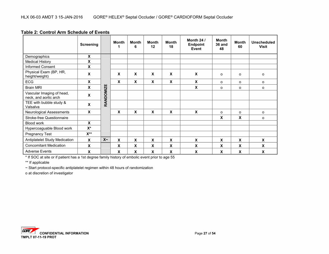
HLX 06-03 AMDT 3 15-JAN-2016 GORE® HELEX® Septal Occluder / GORE® CARDIOFORM Septal Occluder
CONFIDENTIAL INFORMATION Page 27 of 54 TMPLT 07-11-19 PROT
Table 2: Control Arm Schedule of Events
Screening Month 1
Month 6
Month 12
Month 18
Month 24 / Endpoint
Event
Month 36 and
48 Month
60 Unscheduled
Visit
Demographics X
RA
ND
OM
IZE
Medical History X Informed Consent X Physical Exam (BP, HR, height/weight) X X X X X X o o o
ECG X X X X X X o o o Brain MRI X X o o o Vascular Imaging of head, neck, and aortic arch X
TEE with bubble study & Valsalva X
Neurological Assessments X X X X X X o o o Stroke-free Questionnaire X X o Blood work X Hypercoaguable Blood work X* Pregnancy Test X** Antiplatelet Study Medication X X~ X X X X X X X X Concomitant Medication X
X X X X X X X X
Adverse Events X X X X X X X X X * If SOC at site or if patient has a 1st degree family history of embolic event prior to age 55 ** If applicable ~ Start protocol-specific antiplatelet regimen within 48 hours of randomization o at discretion of investigator
HLX 06-03 AMDT 3 15-JAN-2016 GORE® HELEX® Septal Occluder / GORE® CARDIOFORM Septal Occluder
CONFIDENTIAL INFORMATION Page 28 of 54 TMPLT 07-11-19 PROT
3.9. Initiation and Administration of Antiplatelet Therapy for All Subjects Investigators will prescribe the same or similar antiplatelet medical therapy regimens for all subjects, test and control, participating in the study at their site, based on their best medical judgment. All subjects will receive an antiplatelet therapy regimen throughout the course of the study, whether randomized to the test (device plus antiplatelet medical therapy) arm or control (antiplatelet medical therapy alone) arm. Investigators will choose one of the options listed below. Use of more than one of the options below is not permitted according to this protocol: Aspirin alone. A dosage between 75 - 325 mg once daily. Aggrenox™ or generic equivalent. A total daily dosage of 50-100 mg of aspirin and
225-400 mg of dipyridamole. Clopidogrel (Plavix®) or generic equivalent. A dosage of 75 mg once daily.
All subjects will be maintained on one of these antiplatelet medication options, as prescribed, through study completion. If applicable, subjects should be instructed to continue taking antihypertensive medications to minimize hypertension and fluctuations in blood pressure. If for any reason a subject is noncompliant with the antiplatelet therapy, or must change antiplatelet medications, change medical therapy, or can no longer tolerate antiplatelet medication, record the reason on the appropriate CRF and continue to follow all subjects per protocol.
3.10. PFO Closure
3.10.1. Pre-Procedure Evaluation – Within 72 hours of PFO Closure Within 72 hours prior to the transcatheter PFO closure procedure, a neurologic examination will be performed to assess the subject’s neurological status. The following assessments will be performed within 72 hours prior to the PFO-closure procedure (pre-procedure): NIHSS, supplemental neurologic examination, and modified Rankin Scale Antiplatelet study medication and pre-medication Concomitant medications ECG Pregnancy test (if applicable)
3.10.2. Subject Preparation If applicable, subjects should be instructed to continue taking antihypertensive medications to minimize hypertension and fluctuations in blood pressure.
3.10.3. Pre-medication
HLX 06-03 AMDT 3 15-JAN-2016 GORE® HELEX® Septal Occluder / GORE® CARDIOFORM Septal Occluder
CONFIDENTIAL INFORMATION Page 29 of 54 TMPLT 07-11-19 PROT
To minimize the risk of neurological embolic events related to thrombosis during the transcatheter procedure, subjects should remain on the antiplatelet medical therapy regime chosen by the investigator at randomization until time of scheduled procedure. Pre-procedural antiplatelet therapy is recommended per the institutional standard of care or physician discretion.
3.10.4. Procedure Subjects will undergo percutaneous transcatheter PFO closure with the GORE® HELEX® Septal Occluder / GORE® CARDIOFORM Septal Occluder (or alternative closure method if closure with the GORE® HELEX® Septal Occluder / GORE® CARDIOFORM Septal Occluder cannot be achieved). The procedure starts upon venous cannulation and the procedure ends upon removal of the final catheter. Procedure start and stop times will be recorded for this study. Anesthesia times, whether general anesthesia or conscious sedation, and fluoroscopy times will be recorded during the procedure. Anesthesia starts when the subject is intubated and stops when the subject is extubated. Sedation starts at the time of intravenous (IV) administration and stops when the subject becomes responsive (or termination of subject monitoring). Fluoroscopy time is the total fluoroscopy exposure time recorded during the procedure. The Investigator should follow the Instructions for Use (IFU) of the GORE® HELEX® Septal Occluder / GORE® CARDIOFORM Septal Occluder (see Appendix E: Instructions for Use) when performing this procedure. (Note: Protamine is not to be given to reverse heparin). Subjects who have a delivery attempt with the GORE® HELEX® Septal Occluder / GORE® CARDIOFORM Septal Occluder but whose PFO cannot be closed with the device (e.g., closed with an alternative occluder device, surgery, or no closure) will be considered technical failures. Subjects who are technical failures will remain in the study and should be followed per the protocol based on the original treatment assignment received (reference Section 3.16 Treatment Changes Post-Randomization).
3.10.5. Post-Device Placement Echocardiogram Post-device-placement echocardiograms [TEE or intracardiac echocardiography (ICE)] should be performed in the catheterization laboratory during the original procedure and in accordance with the Echocardiographic Core Laboratory (Echo Core Lab) guidelines to check for complications and possible residual shunting. Following device placement, the Investigator should evaluate the subject for the presence of any residual shunting using bubble contrast.
HLX 06-03 AMDT 3 15-JAN-2016 GORE® HELEX® Septal Occluder / GORE® CARDIOFORM Septal Occluder
CONFIDENTIAL INFORMATION Page 30 of 54 TMPLT 07-11-19 PROT
3.10.6. Access Site Management Closure of the femoral venous puncture site should be performed per hospital standards. Access site hemostasis may be achieved by manual compression, or by utilizing alternative methods, such as a suture closure device.
3.10.7. Post-Procedural Care Subjects will be monitored in the recovery room (per the investigational site’s standard post-treatment procedures) until stable. Subjects are to be provided with Clopidogrel (Plavix), 75 mg daily for three days post-procedure. No additional antiplatelet therapy should be provided during this time period. Subjects should continue their prescribed antiplatelet regimen, as specified in section 3.9, through at least 24 months post-randomization. Subjects will be encouraged to stay on their prescribed medication throughout the five (5) year follow-up period. Antibiotic therapy may be administered at the discretion of the Investigator.
3.11. Early Post-procedure Evaluations – 4-72 Hours Post PFO Closure Prior to leaving the hospital, typically within 4 to 72 hours after treatment, test arm subjects will be evaluated for concomitant medications, adverse events (AEs), and device-related or procedure-related events. The following evaluations will be performed at the early post-procedure visit: Physical examination ECG Transthoracic echocardiogram (TTE) with bubble study Fluoroscopy examination without contrast may be performed at the discretion of the
Investigator. NIHSS, supplemental neurologic examination, and modified Rankin Scale Antiplatelet study medication Concomitant medications
3.12. Follow-up Evaluations – Test Arm The follow-up schedule for enrolled test arm subjects is presented in Table 3. All test subjects will be required to return for regular follow-up visits with their Neurologist Investigator through 24 months (see Table 4: Follow-Up Contact Requirements). Table 3: Test Arm Follow-up Schedule
Subject Population Follow-up Schedule Subjects Randomized to Test Arm
and who have a:
Successful GORE® HELEX® Septal Occluder / GORE® CARDIOFORM
Septal Occluder implant
Follow-up at Months 1, 6, 12, 18, 24, 36, 48, and 60 post-procedure
HLX 06-03 AMDT 3 15-JAN-2016 GORE® HELEX® Septal Occluder / GORE® CARDIOFORM Septal Occluder
CONFIDENTIAL INFORMATION Page 31 of 54 TMPLT 07-11-19 PROT
Subject Population Follow-up Schedule Successful alternative transcatheter
occluder device implant Follow-up at Months 1, 6, 12, 18, 24, 36, 48, and 60 post-
procedure
Subjects with no occluder implanted Follow-up at Months 1, 6, 12, 18, 24, 36, 48, and 60 post-procedure
Subjects with surgical PFO closure Follow-up at Months 1, 6, 12, 18, 24, 36, 48, and 60 post-procedure
Table 4: Test Arm Follow-up Contact Requirements
Interval Contact Month 1 Neurology, Cardiology Month 6 Neurology, Cardiology**
Month 12 Neurology, Cardiology Month 18 Neurology, Cardiology** Month 24 Neurology, Cardiology
Year 3 Neurology (phone), Cardiology** Year 4 Neurology (phone), Cardiology** Year 5 Neurology (phone), Cardiology**
** Cardiology Visit– may be scheduled at the discretion of an Investigator All test arm subjects will be evaluated at 24 months by MRI for changes in brain lesions. Test arm subjects also will be evaluated by TEE for residual leak status at 24 months. In addition, all test arm subjects will then be followed through five (5) years post-procedure or until recurrent stroke/imaging-confirmed TIA, whichever occurs first. Follow-up, occurring annually during Years 3, 4, and 5, will be conducted by telephone. The Investigator may request that a subject schedule an office appointment with his/her Neurologist or Cardiologist Investigator at any time the Investigator deems necessary. An MRI scan of the brain with Fluid Attenuated Inversion Recovery (FLAIR) sequencing and diffusion-weighted images (transverse and coronal planes, with 5 mm or thinner sections recommended, and no gaps) is required for all subjects following the clinical diagnosis of a stroke or TIA. Additionally, subjects who have not experienced a recurrent stroke or imaging-confirmed TIA will complete the MRI evaluation at the 24-month follow-up visit. This image will be evaluated by the MRI Core Lab and compared with the screening image. Subjects who are free from stroke or imaging-confirmed TIA at the 24-month follow-up visit but have a subsequent clinical diagnosis of stroke or TIA through five years should have an MRI for imaging-confirmation of the event. At each Neurology follow-up visit through 24 months (see Table 1: Test Arm Schedule of Events), all subjects will undergo: Physical examination ECG NIH Stroke Scale, supplemental neurologic examination, and modified Rankin Scale
(mRS) MRI – required for all subjects at 24 months Antiplatelet study medication
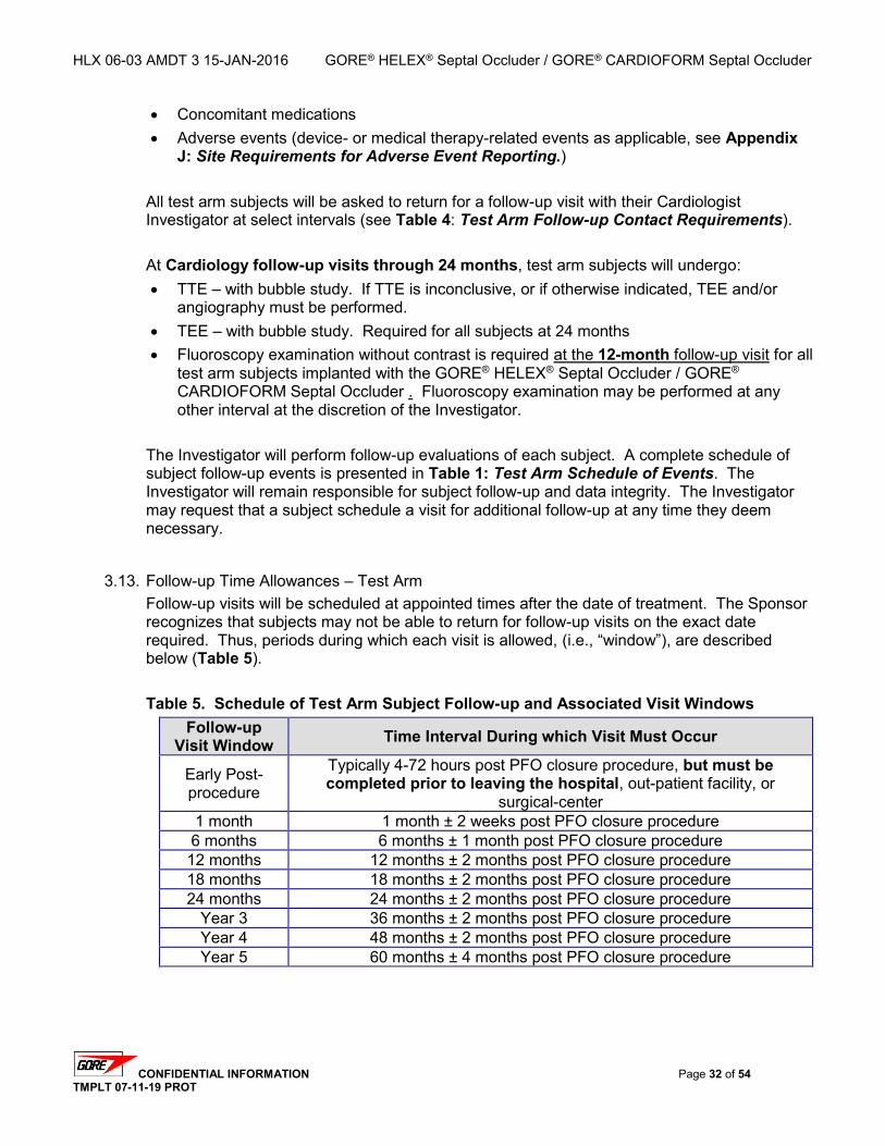
HLX 06-03 AMDT 3 15-JAN-2016 GORE® HELEX® Septal Occluder / GORE® CARDIOFORM Septal Occluder
CONFIDENTIAL INFORMATION Page 32 of 54 TMPLT 07-11-19 PROT
Concomitant medications Adverse events (device- or medical therapy-related events as applicable, see Appendix
J: Site Requirements for Adverse Event Reporting.) All test arm subjects will be asked to return for a follow-up visit with their Cardiologist Investigator at select intervals (see Table 4: Test Arm Follow-up Contact Requirements). At Cardiology follow-up visits through 24 months, test arm subjects will undergo: TTE – with bubble study. If TTE is inconclusive, or if otherwise indicated, TEE and/or
angiography must be performed. TEE – with bubble study. Required for all subjects at 24 months Fluoroscopy examination without contrast is required at the 12-month follow-up visit for all
test arm subjects implanted with the GORE® HELEX® Septal Occluder / GORE® CARDIOFORM Septal Occluder . Fluoroscopy examination may be performed at any other interval at the discretion of the Investigator.
The Investigator will perform follow-up evaluations of each subject. A complete schedule of subject follow-up events is presented in Table 1: Test Arm Schedule of Events. The Investigator will remain responsible for subject follow-up and data integrity. The Investigator may request that a subject schedule a visit for additional follow-up at any time they deem necessary.
3.13. Follow-up Time Allowances – Test Arm Follow-up visits will be scheduled at appointed times after the date of treatment. The Sponsor recognizes that subjects may not be able to return for follow-up visits on the exact date required. Thus, periods during which each visit is allowed, (i.e., “window”), are described below (Table 5). Table 5. Schedule of Test Arm Subject Follow-up and Associated Visit Windows
Follow-up Visit Window Time Interval During which Visit Must Occur
Early Post-procedure
Typically 4-72 hours post PFO closure procedure, but must be completed prior to leaving the hospital, out-patient facility, or
surgical-center 1 month 1 month ± 2 weeks post PFO closure procedure 6 months 6 months ± 1 month post PFO closure procedure 12 months 12 months ± 2 months post PFO closure procedure 18 months 18 months ± 2 months post PFO closure procedure 24 months 24 months ± 2 months post PFO closure procedure
Year 3 36 months ± 2 months post PFO closure procedure Year 4 48 months ± 2 months post PFO closure procedure Year 5 60 months ± 4 months post PFO closure procedure
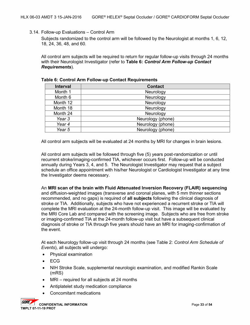
HLX 06-03 AMDT 3 15-JAN-2016 GORE® HELEX® Septal Occluder / GORE® CARDIOFORM Septal Occluder
CONFIDENTIAL INFORMATION Page 33 of 54 TMPLT 07-11-19 PROT
3.14. Follow-up Evaluations – Control Arm Subjects randomized to the control arm will be followed by the Neurologist at months 1, 6, 12, 18, 24, 36, 48, and 60. All control arm subjects will be required to return for regular follow-up visits through 24 months with their Neurologist Investigator (refer to Table 6: Control Arm Follow-up Contact Requirements). Table 6: Control Arm Follow-up Contact Requirements
Interval Contact Month 1 Neurology Month 6 Neurology Month 12 Neurology Month 18 Neurology Month 24 Neurology
Year 3 Neurology (phone) Year 4 Neurology (phone) Year 5 Neurology (phone)
All control arm subjects will be evaluated at 24 months by MRI for changes in brain lesions. All control arm subjects will be followed through five (5) years post-randomization or until recurrent stroke/imaging-confirmed TIA, whichever occurs first. Follow-up will be conducted annually during Years 3, 4, and 5. The Neurologist Investigator may request that a subject schedule an office appointment with his/her Neurologist or Cardiologist Investigator at any time the Investigator deems necessary. An MRI scan of the brain with Fluid Attenuated Inversion Recovery (FLAIR) sequencing and diffusion-weighted images (transverse and coronal planes, with 5 mm thinner sections recommended, and no gaps) is required of all subjects following the clinical diagnosis of stroke or TIA. Additionally, subjects who have not experienced a recurrent stroke or TIA will complete the MRI evaluation at the 24-month follow-up visit. This image will be evaluated by the MRI Core Lab and compared with the screening image. Subjects who are free from stroke or imaging-confirmed TIA at the 24-month follow-up visit but have a subsequent clinical diagnosis of stroke or TIA through five years should have an MRI for imaging-confirmation of the event. At each Neurology follow-up visit through 24 months (see Table 2: Control Arm Schedule of Events), all subjects will undergo: Physical examination ECG NIH Stroke Scale, supplemental neurologic examination, and modified Rankin Scale
(mRS) MRI – required for all subjects at 24 months Antiplatelet study medication compliance Concomitant medications
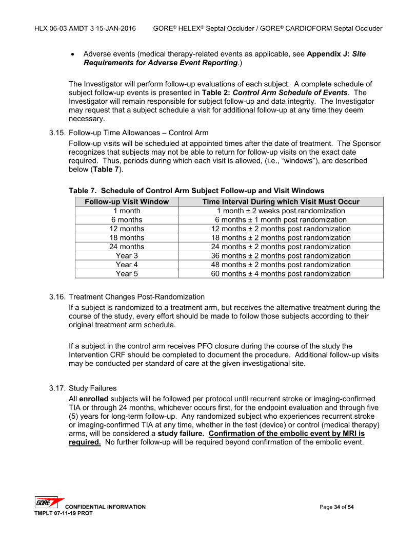
HLX 06-03 AMDT 3 15-JAN-2016 GORE® HELEX® Septal Occluder / GORE® CARDIOFORM Septal Occluder
CONFIDENTIAL INFORMATION Page 34 of 54 TMPLT 07-11-19 PROT
Adverse events (medical therapy-related events as applicable, see Appendix J: Site Requirements for Adverse Event Reporting.)
The Investigator will perform follow-up evaluations of each subject. A complete schedule of subject follow-up events is presented in Table 2: Control Arm Schedule of Events. The Investigator will remain responsible for subject follow-up and data integrity. The Investigator may request that a subject schedule a visit for additional follow-up at any time they deem necessary.
3.15. Follow-up Time Allowances – Control Arm Follow-up visits will be scheduled at appointed times after the date of treatment. The Sponsor recognizes that subjects may not be able to return for follow-up visits on the exact date required. Thus, periods during which each visit is allowed, (i.e., “windows”), are described below (Table 7). Table 7. Schedule of Control Arm Subject Follow-up and Visit Windows
Follow-up Visit Window Time Interval During which Visit Must Occur 1 month 1 month ± 2 weeks post randomization 6 months 6 months ± 1 month post randomization
12 months 12 months ± 2 months post randomization 18 months 18 months ± 2 months post randomization 24 months 24 months ± 2 months post randomization
Year 3 36 months ± 2 months post randomization Year 4 48 months ± 2 months post randomization Year 5 60 months ± 4 months post randomization
3.16. Treatment Changes Post-Randomization If a subject is randomized to a treatment arm, but receives the alternative treatment during the course of the study, every effort should be made to follow those subjects according to their original treatment arm schedule. If a subject in the control arm receives PFO closure during the course of the study the Intervention CRF should be completed to document the procedure. Additional follow-up visits may be conducted per standard of care at the given investigational site.
3.17. Study Failures All enrolled subjects will be followed per protocol until recurrent stroke or imaging-confirmed TIA or through 24 months, whichever occurs first, for the endpoint evaluation and through five (5) years for long-term follow-up. Any randomized subject who experiences recurrent stroke or imaging-confirmed TIA at any time, whether in the test (device) or control (medical therapy) arms, will be considered a study failure. Confirmation of the embolic event by MRI is required. No further follow-up will be required beyond confirmation of the embolic event.
HLX 06-03 AMDT 3 15-JAN-2016 GORE® HELEX® Septal Occluder / GORE® CARDIOFORM Septal Occluder
CONFIDENTIAL INFORMATION Page 35 of 54 TMPLT 07-11-19 PROT
3.18. Subject Withdrawal or Discontinuation from the Clinical Study The Investigator should encourage subjects to return for the required follow-up visits. These measures are important as the clinical study objectives may not be realized if a large number of subjects are lost to follow-up (LTF). A subject may withdraw from the clinical study at any time and should notify the Investigator in this event. If a subject refuses to return for the follow-up visits, the Investigator will attempt to obtain at least vital status and complete the Stroke-Free Questionnaire. If the Stroke-Free Questionnaire suggests a possible event, then records and images will be requested for adjudication in the same manner as all suspected endpoints. In the event of subject death, a death certificate or autopsy report should be obtained to confirm cause of death and reported on the Subject Completion / Discontinuation Form. A subject will be considered lost to follow-up and discontinued from the study when both of the following criteria have been met: the subject has missed two consecutive follow-up visits, and the Investigator has documented three failed attempts to contact the subject. One of the three documented attempts must include a certified (traceable) letter. The Investigator will ensure that the appropriate CRF (Study Completion / Discontinuation Form) is completed, documenting the subject’s discontinuation (i.e., lost to follow-up) from the clinical study. If a subject is considered lost to follow-up, the Investigator will attempt to obtain vital status and complete the Stroke-Free Questionnaire annually from the point of last contact. Any information collected after the time of discontinuation should be captured in the study database under an Unscheduled visit.
The Investigator may also withdraw the subject from the clinical study at any time based on the Investigator’s medical judgment. The study Monitor will ensure that subject withdrawals are documented properly, discussed with the Investigator, and reported to the Sponsor (ISO 14155 – 1:2003, Section 9.1(j.).
3.19. Imaging Analysis
3.19.1. MRI / CT For all subjects, an independent MRI Core Laboratory will compare the screening MRI with the 24 month follow-up or recurrent event MRI for a determination of study imaging endpoints. Variables assessed by the MRI Core Laboratory will include: Screening lesion evaluation Change in focal lesion on follow-up scan
MRI Guidelines are provided for reference for the MRI technician and MRI Core Laboratory at Appendix H: MRI Imaging Guidelines.
HLX 06-03 AMDT 3 15-JAN-2016 GORE® HELEX® Septal Occluder / GORE® CARDIOFORM Septal Occluder
CONFIDENTIAL INFORMATION Page 36 of 54 TMPLT 07-11-19 PROT
3.19.2. Echocardiography An independent Echocardiography Core Laboratory will conduct analyses of pre-procedure/pre-treatment (required screening) echocardiography data for all (test and control) subjects, and procedure and follow-up echocardiography data for test (device) subjects only. Variables assessed by the Echocardiography Core Laboratory will include: Residual shunting, as determined by TEE with Valsalva maneuver and contrast
bubble study Residual shunting, as determined by TTE ordered by an Investigator Thrombus on device
Residual shunt will be evaluated based on maximum number of agitated saline or microbubble particles seen in the left atrial chamber in any single frame during first three cardiac cycles, during a resting state or during/after Valsalva maneuver. The scale for measuring the significance of the shunt is as follows:
Occluded = 0 particles Trivial or small = 1 – 5 particles
Moderate = 6 – 25 particles Large = > 25 particles
Echocardiography Guidelines are provided for reference by the echocardiographer and Echocardiography Core Laboratory at Appendix D – Echocardiography Imaging Guidelines. De-identified and appropriately labeled media (e.g., CDs, DICOM, videotape, or other format) of the required imaging evaluations should be forwarded to the appropriate Core Lab along with any technical worksheets and/or transmittal forms at the designated intervals (refer to Table 1: Test Arm Schedule of Events and Table 2: Control Arm Schedule of Events). All media should be forwarded to the Core Lab, in DICOM format, as soon as possible, but no later than 10 days following the Screening and Procedure Evaluations and no later than 30 days following all subsequent follow-up visits.
3.20. Data Safety Monitoring Board An independent Data and Safety Monitoring Board (DSMB) will be comprised of an interdisciplinary team of five individuals, four physicians and a biostatistician, who are not directly involved in the conduct of the study. The team shall include, at minimum, one stroke neurologist, one interventional cardiologist, and a biostatistician to assist with formulation of stopping rules and direction of interim analyses.
HLX 06-03 AMDT 3 15-JAN-2016 GORE® HELEX® Septal Occluder / GORE® CARDIOFORM Septal Occluder
CONFIDENTIAL INFORMATION Page 37 of 54 TMPLT 07-11-19 PROT
The members may be compensated for their participation in the DSMB, including reimbursement for reasonable travel expenses to attend meetings. Members will not have any business or financial affiliation with the study sponsor, the core laboratories, or the study investigators. The DSMB is responsible for conducting periodic reviews of aggregate data on a prescribed schedule. Based on the safety data, the DSMB will make recommendations to the Sponsor. Recommendations may include modifying the study, stopping the study, or continuing the study. All final decisions regarding study modifications or study continuation, however, will rest with the Sponsor.
3.21. Clinical Events Committee The independent Clinical Events Committee (CEC) will be comprised of at least three physicians who are not participating in the study and do not have any conflicts of interest with the study Sponsor. The CEC will include representatives from at least three of the following specialties/disciplines: stroke neurology interventional cardiology echocardiography neuro-radiology
Input from other disciplines shall be solicited, if required. The members may be compensated for their involvement in the CEC including reimbursement for reasonable travel expenses to attend meetings. The committee will be responsible for: Review definitions of clinical endpoints in the study in conjunction with the Sponsor. Review and adjudication of adverse events that have the potential to be study endpoint
events including death, stroke, or TIA; AND Subsequent classification of these adverse events as related to the study device,
procedure, or medications.
HLX 06-03 AMDT 3 15-JAN-2016 GORE® HELEX® Septal Occluder / GORE® CARDIOFORM Septal Occluder
CONFIDENTIAL INFORMATION Page 38 of 54 TMPLT 07-11-19 PROT
4. Adverse Events This study will adhere to current ISO requirements and 21 CFR 812, regarding the definitions and reporting of adverse events (AEs). Adverse events (AEs) are defined as any untoward medical occurrence in a subject. An adverse device effect (ADE) is an untoward and unintended response to a medical device. This includes ADEs resulting from insufficiencies or inadequacies in the Instructions for Use (IFU), the deployment of the device, or any response that is a result of a user error (in accordance with the current ISO standard) The severity of an AE or ADE will be rated as follows: Mild – subject is aware of event or symptom, but event/symptom is easily tolerated. Moderate - subject experiences sufficient discomfort to interfere with or reduce their usual level
of activity Severe – significant impairment of functioning; subject is unable to carry out usual activities
Adverse events or adverse device effects are anticipated, but usually are not expected (i.e., not previously reported in relation to the study device; expected events are those that are referred to in the protocol, Investigator’s Brochure, consent form, or IFU when applicable). All AEs will be recorded on the appropriate internet case report form (CRF) and documented in the subject’s permanent medical record. The CRFs to be used for data collection during the study are in Appendix B: Case Report Forms. All AEs will be reported to the FDA, the IRBs and ECs, Competent Authorities per local requirements, participating Investigators, and/or safety monitoring boards via annual study progress updates. (in accordance with the current ISO standard)
4.1. Anticipated Adverse Events Study complications, whether device-, procedure-, or antiplatelet medical therapy-related, that may be anticipated in subjects undergoing PFO closure with the GORE® HELEX® Septal Occluder / GORE® CARDIOFORM Septal Occluder , another occluder, or in subjects being treated with study-specified medications (i.e., antiplatelets) are listed below and under Section 6.0, Risk Analysis. These complications are considered anticipated AEs. Access site complications
o Emboli (air, tissue, or thrombotic emboli) o Groin hematoma, with or without surgical repair o Infection and pain at insertion site o Pseudoaneurysm, femoral o Retro-peritoneal bleeding o Thrombosis at access puncture site or remote site o Venous occlusion o Vessel dissection, perforation, or rupture
HLX 06-03 AMDT 3 15-JAN-2016 GORE® HELEX® Septal Occluder / GORE® CARDIOFORM Septal Occluder
CONFIDENTIAL INFORMATION Page 39 of 54 TMPLT 07-11-19 PROT
Allergic response o To a study medication o To nickel in the Occluder o To contrast media o To latex
Arrhythmia Bleeding, hemorrhage, or bruising
o Related to study (antiplatelet) medications o With or without transfusion
Cerebral hemorrhage Chest pain or palpitations Death Effusion, pericardial or pleural, with or without drainage Headache, including
o Migraine, with or without aura o Pain in the head or neck o Severe unilateral headache
Hospitalization (in-patient) required to treat a complication Hypertension Hypotension Infection, with or without fever
o Bacteremia o Septicemia
Ischemic events o Cerebral ischemia o Cerebrovascular accident (CVA) o Myocardial infarction (MI) o Stroke o TIA with or without motor weakness
Medications, study-related o Allergic responses
Rash, itching, or papules o Excessive bruising o Gastro-intestinal problems
stomach ache ulcer diarrhea
HLX 06-03 AMDT 3 15-JAN-2016 GORE® HELEX® Septal Occluder / GORE® CARDIOFORM Septal Occluder
CONFIDENTIAL INFORMATION Page 40 of 54 TMPLT 07-11-19 PROT
Occluder-related issues o Component detachment o Damage to delivery system or occluder during prep or delivery o Embolization o Erosion of cardiac tissue o Malposition o Migration o Perforation of a cardiac structure o Removal of the device post-implant o Thrombosis o Wire frame fracture
Procedure-related issues o Brachial plexus stretch injury o Back pain resulting from lengthy procedures o Excessive radiation exposure during imaging o Sore throat (intubation / extubation / probe)
Prolonged medication required to treat a complication Seizure
The Investigator at each investigative site is ultimately responsible for reporting AEs to the Sponsor. The Investigator is required to complete the appropriate CRF to report the occurrence of AEs. The CRFs for a given visit must list all AEs that have occurred since the last documented visit.
4.2. Serious Adverse Device Effects / Serious Adverse Events A SERIOUS adverse event (SAE) is an event that: Led to death, Led to a serious deterioration in the health of the subject that:
o Resulted in a life-threatening illness or injury, o Resulted in a permanent impairment of a body structure or a body function o Resulted in in-patient hospitalization or prolongation of existing hospitalization o Resulted in a medical or surgical intervention to prevent permanent impairment of a
body structure or a body function, or led to: Led to fetal distress, fetal death, or a congenital anomaly or birth defect.
Any serious adverse event (SAE), whether considered to be related to the investigational product or not, whether foreseen or unforeseen, anticipated or unanticipated, must be reported by the Investigator to the Sponsor, the CRO (European sites only), the IRB or EC, and Competent Authorities if applicable. A list of SAEs and their definitions are provided in Appendix I: Reportable Adverse Event Definitions to assist Investigators in determining when an adverse event becomes serious. The European countries may require reporting these events as soon as possible, but in no event later than 10 working days after the Investigator first learns of the event.
HLX 06-03 AMDT 3 15-JAN-2016 GORE® HELEX® Septal Occluder / GORE® CARDIOFORM Septal Occluder
CONFIDENTIAL INFORMATION Page 41 of 54 TMPLT 07-11-19 PROT
A SERIOUS adverse device effect (SADE) is an effect that results in any of the consequences of characteristics of a SAE, or that might have led to death, or a serious deterioration in the health of a subject or any other person if suitable action had not been taken, or intervention had not been made, or if circumstances had been less opportune. These events must be reported by the Investigator to the Sponsor, the CRO (European sites only), the IRB or EC, and Competent Authorities if applicable, as soon as possible, but in no event later than 10 working days after the Investigator first learns of the event. Unanticipated adverse device effects (UADEs) means any serious adverse effect on health or safety or any life-threatening problem or death caused by, or associated with, a device, if that effect, problem, or death was not previous identified in nature, severity, or degree of incidence in the Investigational Plan or application, or any other unanticipated serious problem associated with a device that relates to the rights, safety, or welfare of subjects (21 CFR § 812.3(s) and 150(a)(1)). These events are encompassed by the definition of serious adverse events (SAEs) and must be reported by the Investigator to the Sponsor, the CRO (European sites only), the IRB or EC, and Competent Authorities if applicable, as soon as possible, but in no event later than 10 working days after the Investigator first learns of the event. If applicable to local requirements of the reviewing IRB or EC, the Investigator must submit to the Sponsor and to the IRB or EC a report of any SADE, UADEs, and SAEs that occur during the clinical study, no later than 10 working days after the Investigator first learns of the event. All SADEs and UADEs must be documented by the Investigator, including the date of onset, a complete description of the event, possible reason(s) for the event, severity, duration, actions taken and outcome. Copies of all supporting documents should accompany the appropriate CRF, where applicable. Emergency contact information for the United States and European sites for the required reporting of SAEs/SADEs/UADEs can be found in Appendix J: Site Requirements for Adverse Event Reporting. The Sponsor will immediately conduct an evaluation of any event determined to be unanticipated (i.e., UADE) and report the results of the evaluation to the FDA, all IRBs and ECs, and participating Investigators within 10 working days after the Sponsor first receives notice of the effect by the investigative site (§812.46(b), 812.150(b)(1)) If the event or effect is determined to present unreasonable risk to subjects, the Sponsor shall terminate all investigations or part of investigations presenting that risk as soon as possible, but no later than 5 working days after making the determination and no later than 15 days after first learning of the effect (21 CFR § 812.46(2).
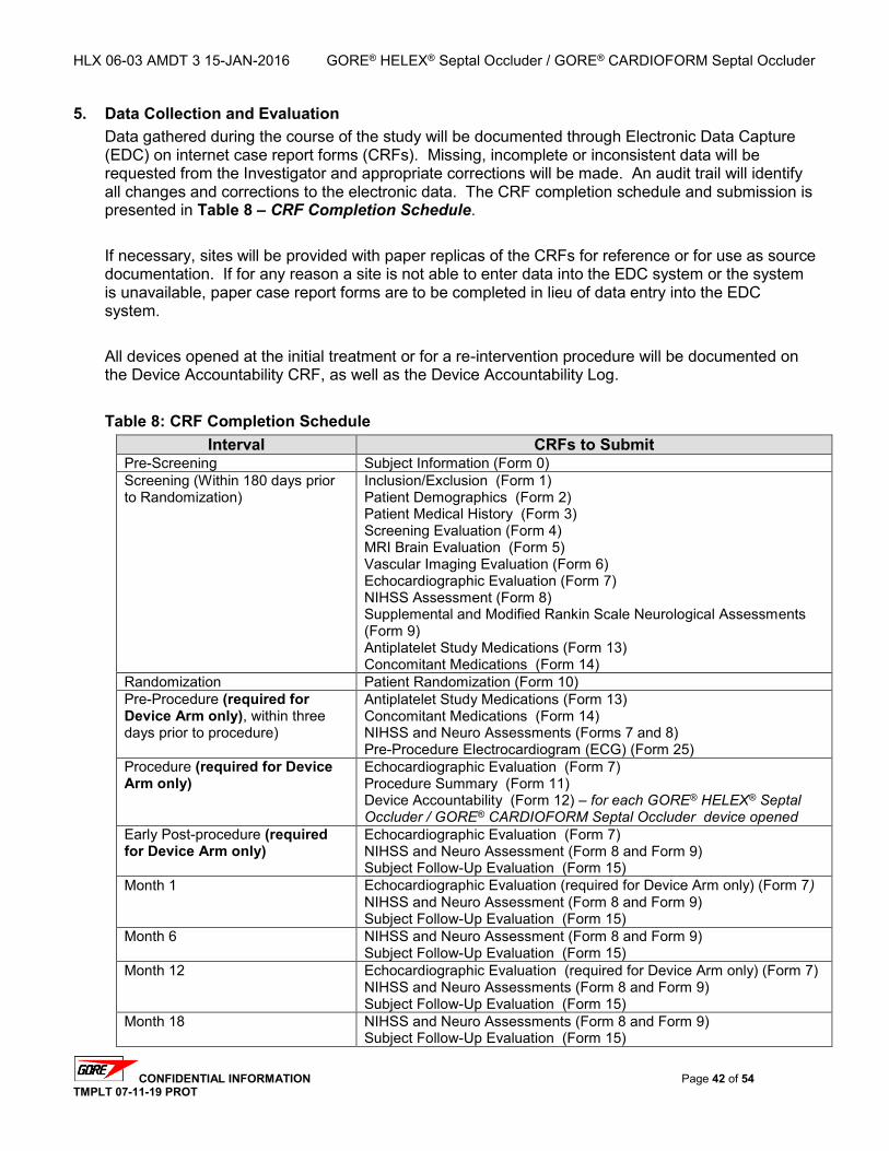
HLX 06-03 AMDT 3 15-JAN-2016 GORE® HELEX® Septal Occluder / GORE® CARDIOFORM Septal Occluder
CONFIDENTIAL INFORMATION Page 42 of 54 TMPLT 07-11-19 PROT
5. Data Collection and Evaluation Data gathered during the course of the study will be documented through Electronic Data Capture (EDC) on internet case report forms (CRFs). Missing, incomplete or inconsistent data will be requested from the Investigator and appropriate corrections will be made. An audit trail will identify all changes and corrections to the electronic data. The CRF completion schedule and submission is presented in Table 8 – CRF Completion Schedule. If necessary, sites will be provided with paper replicas of the CRFs for reference or for use as source documentation. If for any reason a site is not able to enter data into the EDC system or the system is unavailable, paper case report forms are to be completed in lieu of data entry into the EDC system. All devices opened at the initial treatment or for a re-intervention procedure will be documented on the Device Accountability CRF, as well as the Device Accountability Log. Table 8: CRF Completion Schedule
Interval CRFs to Submit Pre-Screening Subject Information (Form 0) Screening (Within 180 days prior to Randomization)
Inclusion/Exclusion (Form 1) Patient Demographics (Form 2) Patient Medical History (Form 3) Screening Evaluation (Form 4) MRI Brain Evaluation (Form 5) Vascular Imaging Evaluation (Form 6) Echocardiographic Evaluation (Form 7) NIHSS Assessment (Form 8) Supplemental and Modified Rankin Scale Neurological Assessments (Form 9) Antiplatelet Study Medications (Form 13) Concomitant Medications (Form 14)
Randomization Patient Randomization (Form 10) Pre-Procedure (required for Device Arm only), within three days prior to procedure)
Antiplatelet Study Medications (Form 13) Concomitant Medications (Form 14) NIHSS and Neuro Assessments (Forms 7 and 8) Pre-Procedure Electrocardiogram (ECG) (Form 25)
Procedure (required for Device Arm only)
Echocardiographic Evaluation (Form 7) Procedure Summary (Form 11) Device Accountability (Form 12) – for each GORE® HELEX® Septal Occluder / GORE® CARDIOFORM Septal Occluder device opened
Early Post-procedure (required for Device Arm only)
Echocardiographic Evaluation (Form 7) NIHSS and Neuro Assessment (Form 8 and Form 9) Subject Follow-Up Evaluation (Form 15)
Month 1
Echocardiographic Evaluation (required for Device Arm only) (Form 7) NIHSS and Neuro Assessment (Form 8 and Form 9) Subject Follow-Up Evaluation (Form 15)
Month 6
NIHSS and Neuro Assessment (Form 8 and Form 9) Subject Follow-Up Evaluation (Form 15)
Month 12
Echocardiographic Evaluation (required for Device Arm only) (Form 7) NIHSS and Neuro Assessments (Form 8 and Form 9) Subject Follow-Up Evaluation (Form 15)
Month 18
NIHSS and Neuro Assessments (Form 8 and Form 9) Subject Follow-Up Evaluation (Form 15)
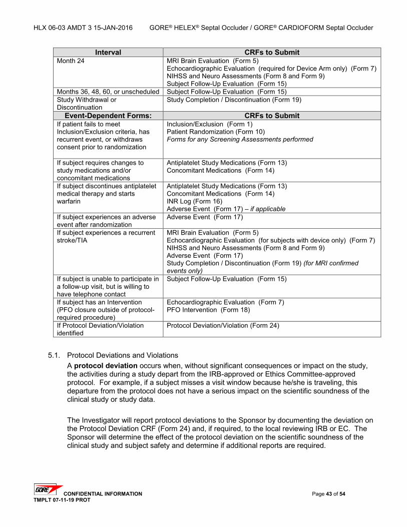
HLX 06-03 AMDT 3 15-JAN-2016 GORE® HELEX® Septal Occluder / GORE® CARDIOFORM Septal Occluder
CONFIDENTIAL INFORMATION Page 43 of 54 TMPLT 07-11-19 PROT
Interval CRFs to Submit Month 24
MRI Brain Evaluation (Form 5) Echocardiographic Evaluation (required for Device Arm only) (Form 7) NIHSS and Neuro Assessments (Form 8 and Form 9) Subject Follow-Up Evaluation (Form 15)
Months 36, 48, 60, or unscheduled Subject Follow-Up Evaluation (Form 15) Study Withdrawal or Discontinuation
Study Completion / Discontinuation (Form 19)
Event-Dependent Forms: CRFs to Submit If patient fails to meet Inclusion/Exclusion criteria, has recurrent event, or withdraws consent prior to randomization
Inclusion/Exclusion (Form 1) Patient Randomization (Form 10) Forms for any Screening Assessments performed
If subject requires changes to study medications and/or concomitant medications
Antiplatelet Study Medications (Form 13) Concomitant Medications (Form 14)
If subject discontinues antiplatelet medical therapy and starts warfarin
Antiplatelet Study Medications (Form 13) Concomitant Medications (Form 14) INR Log (Form 16) Adverse Event (Form 17) – if applicable
If subject experiences an adverse event after randomization
Adverse Event (Form 17)
If subject experiences a recurrent stroke/TIA
MRI Brain Evaluation (Form 5) Echocardiographic Evaluation (for subjects with device only) (Form 7) NIHSS and Neuro Assessments (Form 8 and Form 9) Adverse Event (Form 17) Study Completion / Discontinuation (Form 19) (for MRI confirmed events only)
If subject is unable to participate in a follow-up visit, but is willing to have telephone contact
Subject Follow-Up Evaluation (Form 15)
If subject has an Intervention (PFO closure outside of protocol-required procedure)
Echocardiographic Evaluation (Form 7) PFO Intervention (Form 18)
If Protocol Deviation/Violation identified
Protocol Deviation/Violation (Form 24)
5.1. Protocol Deviations and Violations A protocol deviation occurs when, without significant consequences or impact on the study, the activities during a study depart from the IRB-approved or Ethics Committee-approved protocol. For example, if a subject misses a visit window because he/she is traveling, this departure from the protocol does not have a serious impact on the scientific soundness of the clinical study or study data. The Investigator will report protocol deviations to the Sponsor by documenting the deviation on the Protocol Deviation CRF (Form 24) and, if required, to the local reviewing IRB or EC. The Sponsor will determine the effect of the protocol deviation on the scientific soundness of the clinical study and subject safety and determine if additional reports are required.
HLX 06-03 AMDT 3 15-JAN-2016 GORE® HELEX® Septal Occluder / GORE® CARDIOFORM Septal Occluder
CONFIDENTIAL INFORMATION Page 44 of 54 TMPLT 07-11-19 PROT
5.1.1. Protocol Violations A protocol violation is defined as a divergence from the protocol that materially: (a) reduces the quality or completeness of the data, (b) renders the informed consent document inaccurate, or (c) impacts a subject’s safety, rights or welfare. Examples of protocol violations may include, but are not limited to: Inadequate or delinquent informed consent Inclusion/exclusion criteria not met Unreported serious adverse events (SAEs) Incorrect or missing tests that may impact the primary endpoint Multiple visits missed or outside permissible windows (and not reported as a lost
to follow-up) Materially inadequate record-keeping Intentional deviation from protocol, good clinical practices (GCP), or regulations
by study personnel The Investigator will report protocol violations within five (5) days to the Sponsor by documenting the deviation on the Protocol Deviation CRF (Form 24) and, if required, to the local reviewing IRB or EC. The Sponsor will determine the effect of the protocol violation on the scientific soundness of the clinical study and subject safety and determine if additional reports are required.
HLX 06-03 AMDT 3 15-JAN-2016 GORE® HELEX® Septal Occluder / GORE® CARDIOFORM Septal Occluder
CONFIDENTIAL INFORMATION Page 45 of 54 TMPLT 07-11-19 PROT
6. Risk Analysis The purpose of this study is to assess the safety and effectiveness of the GORE® HELEX® Septal Occluder / GORE® CARDIOFORM Septal Occluder when used in conjunction with antiplatelet therapy to close PFOs in subjects who are at high risk for recurrent embolic (stroke or TIA) events. The adverse events listed here are considered anticipated adverse events because they have been known to occur with the use of occluder devices or during the administration of the study medications. An adverse event that is anticipated or foreseen does not preclude the event from being a serious adverse event (SAE or SADE). Complications associated with the use of the GORE® HELEX® Septal Occluder / GORE® CARDIOFORM Septal Occluder or the study medications may be related to the device, the procedure, or medical (antiplatelet) therapy and may include, but are not limited to: Access site complications
o Emboli (air, tissue, or thrombotic emboli) o Groin hematoma, with or without surgical repair o Infection and pain at insertion site o Pseudoaneurysm, femoral o Retro-peritoneal bleeding o Thrombosis at access puncture site or remote site o Venous occlusion o Vessel dissection, perforation, or rupture
Allergic response o To a study medication o To nickel in the Occluder o To contrast media o To latex
Arrhythmia Bleeding, hemorrhage, or bruising
o Related to study (antiplatelet) medications o With or without transfusion
Cerebral hemorrhage Chest pain or palpitations Death Effusion, pericardial or pleural, with or without drainage Headache, including
o Migraine, with or without aura o Pain in the head or neck o Severe unilateral headache
Hospitalization (in-patient) required to treat a complication Hypertension Hypotension
HLX 06-03 AMDT 3 15-JAN-2016 GORE® HELEX® Septal Occluder / GORE® CARDIOFORM Septal Occluder
CONFIDENTIAL INFORMATION Page 46 of 54 TMPLT 07-11-19 PROT
Infection, with or without fever o Bacteremia o Septicemia
Ischemic events o Cerebral ischemia o Cerebrovascular accident (CVA) o Myocardial infarction (MI) o Stroke o TIA with or without motor weakness
Medications, study-related o Allergic responses
Rash, itching, or papules o Excessive bruising o Gastro-intestinal problems
stomach ache ulcer diarrhea
Occluder-related issues o Component detachment o Damage to delivery system or occluder during prep or delivery o Embolization o Erosion of cardiac tissue o Malposition o Migration o Perforation of a cardiac structure o Removal of the device post-implant o Thrombosis o Wire frame fracture
Procedure-related issues o Brachial plexus stretch injury o Back pain resulting from lengthy procedures o Excessive radiation exposure during imaging o Sore throat (intubation / extubation / probe)
Prolonged medication required to treat a complication Seizure
HLX 06-03 AMDT 3 15-JAN-2016 GORE® HELEX® Septal Occluder / GORE® CARDIOFORM Septal Occluder
CONFIDENTIAL INFORMATION Page 47 of 54 TMPLT 07-11-19 PROT
6.1. Minimization of Risks Potential risks associated with the use of the GORE® HELEX® Septal Occluder / GORE® CARDIOFORM Septal Occluder or the study medications may be minimized by the following activities: Investigator and staff training will be conducted to share information regarding design of
the device, its application, and preliminary clinical results from any pre-clinical and clinical studies.
Investigators will be reminded of the importance of complex anatomies when using the device.
Investigators will be reminded of the importance of monitoring subjects on antiplatelet medications
An evaluation will be completed by the Investigator for each subject against the inclusion/exclusion criteria before study entry to ensure the subject’s medical and anatomical status is appropriate. Procedural angiography will be used to assess the morphology of the heart and the characteristics of the PFO. Antiplatelet therapy will be administered to reduce the risk of thrombosis. Procedural anticoagulation therapy will be administered to reduce thromboembolic risk. (Note: Protamine is not to be given to reverse heparin). Subjects will be assessed at discharge and periodically during the follow-up period. Evaluations will include, but will not be limited to, physical examination performed by the Neurologist Investigator and/or the Cardiologist Investigator , neurologic assessments, and transthoracic or transesophageal echocardiography (TTE or TEE). The Investigator or designee will evaluate the subject for device-related events or AEs related to the procedure or medical treatment. Data submitted from the investigative sites will be monitored. An initiation site visit and interim site visit(s) will be conducted as appropriate to evaluate protocol compliance, accuracy, and subject safety. Safety data obtained during the clinical study will be shared with the Investigators to aid understanding of the device and potential complications associated with its use. All echos performed will be reviewed by the Echocardiography Core Laboratory to evaluate the heart and any residual shunting across the PFO. A DSMB and CEC will be utilized to further mitigate risk to subjects during the study.
6.2. Expected Benefits A major expected benefit from the use of the GORE® HELEX® Septal Occluder / GORE®
CARDIOFORM Septal Occluder is the reduction of risk from recurrent embolic (stroke or TIA) events when compared with antiplatelet medical therapy alone.
HLX 06-03 AMDT 3 15-JAN-2016 GORE® HELEX® Septal Occluder / GORE® CARDIOFORM Septal Occluder
CONFIDENTIAL INFORMATION Page 48 of 54 TMPLT 07-11-19 PROT
7. Investigator Responsibilities The Investigator is responsible for ensuring the clinical study is conducted according to the Clinical Study Agreement (CSA) or Clinical Trial Letter of Agreement (CTLA), the study protocol, all conditions of the FDA and IRB or EC approvals and applicable FDA and European regulations. Written confirmation of IRB or EC approval must be provided to the Sponsor before the start of the clinical study. A copy of the current curriculum vitae (CV) for the Investigator and any Sub-Investigators with information concerning their prior experience will be provided to the Sponsor at the beginning of the study and may be updated periodically on the request of the Sponsor. The Investigator is responsible for protecting the rights, safety, and welfare of subjects under the Investigator’s care and for the control of devices under investigation. The Investigator or trained designee is also responsible for ensuring that informed consent is properly obtained. (CFR § 812 Subpart E and current ISO standard)
7.1. Specific Investigator Responsibilities
7.1.1. Subject Informed Consent Each potential subject that meets the inclusion/exclusion criteria for this clinical study will be informed of the requirements for participating in the clinical study. Subjects will be provided with a copy of the Informed Consent Form and an opportunity to discuss any questions with the Investigator. Subjects will be informed that their medical records will be subject to review by the Sponsor, its representatives and the regulatory authorities (i.e., the FDA and European regulatory authorities), and will be asked to sign an Authorization for the Use and Disclosure of Protected Health Information. Subjects will be informed that they may refuse to participate in this clinical study without loss of benefits to which they are otherwise entitled, and that if they choose to participate, they may withdraw at any time without prejudice to future care. Once a subject agrees to the study requirements, the Informed Consent Form and Authorization for the Use and Disclosure of Protected Health Information must be signed and dated by the subject or subject’s guardian or legal representative and Investigator (or Investigator’s designee). The original signed Informed Consent Form for each subject will be retained by the investigative site and is subject to review by the regulatory authorities, the Sponsor, and its representatives. A copy of the signed Informed Consent Form will be provided to the subject. The Sponsor’s representatives will review a copy of the signed Informed Consent Form. A copy of the Sponsor’s Informed Consent Form template is provided in Appendix F: Informed Consent Template. The Investigator or trained designee will be required to obtain informed consent from the subject, the subject’s legal representative, or a person designated acceptable by local Ethics Committee requirements, as appropriate.
HLX 06-03 AMDT 3 15-JAN-2016 GORE® HELEX® Septal Occluder / GORE® CARDIOFORM Septal Occluder
CONFIDENTIAL INFORMATION Page 49 of 54 TMPLT 07-11-19 PROT
7.1.2. Compliance The Investigator will adhere to the signed Clinical Study Agreement (CSA), the clinical study protocol, all applicable FDA or European regulations, and any conditions of approval imposed by the FDA or IRB or EC or European Regulatory Bodies.
7.1.3. GORE® HELEX® Septal Occluder / GORE® CARDIOFORM Septal Occluder Use The Investigator will ensure that patients meeting the study entrance criteria, specifically confirmation of the presence of a PFO, and who become subjects enrolled in this clinical study, will have access to the GORE® HELEX® Septal Occluder / GORE®CARDIOFORM Septal Occluder for PFO closure. The Investigator will not supply a study device or device component to any person not authorized to participate in the clinical study or to any other person who does not have legal access to the device.
7.1.4. GORE® HELEX® Septal Occluder / GORE® CARDIOFORM Septal Occluder Disposal Upon completion or termination either of the clinical study or the Investigator’s participation in the clinical study, or at the Sponsor’s request, the Investigator will return any remaining supply of the device(s) or dispose of the device(s) as directed by the Sponsor, including devices that are opened but not used.
7.1.5. Occluder Explant In the event that an Occluder must be explanted or there is an opportunity to retrieve a device secondary to autopsy, the device should be returned to W.L. Gore and Associates for pathological and mechanical analyses. Ideally, the Occluder and 1-2 cm of native tissue would be excised and returned in 10% neutral buffered Formalin. If possible, physical damage to the device should be avoided. Operative photographs and clinical/pathological impressions are helpful and appreciated. Instructions for obtaining a return kit and processing explanted devices are provided in Appendix C – Device Explant and Return Procedure.
7.1.6. Investigator Records The Investigator will maintain complete, accurate and current study records. Records will be maintained during the clinical study and for a minimum of two (2) years after the latter of the date on which the study is terminated or completed, or the date the records are no longer required to support FDA approval of the new indication for the device. Clinical study records will not be disposed of or custody of the records will not be transferred without prior Sponsor approval. Investigator records will include the following: 1. All correspondence with another Investigator, the IRB, the EC, the Sponsor,
DSMB, a monitor, FDA, a European regulatory body, or a study subject.
HLX 06-03 AMDT 3 15-JAN-2016 GORE® HELEX® Septal Occluder / GORE® CARDIOFORM Septal Occluder
CONFIDENTIAL INFORMATION Page 50 of 54 TMPLT 07-11-19 PROT
2. Accountability records of receipt, use and disposition of all devices including the type and quantity of devices received, the dates of their receipt, and the lot numbers. In addition, the names of all persons who received, used or disposed of each device must be recorded.
3. Subject records that include the subject’s case history and exposure to the device. This information will be documented on standardized CRFs provided by the Sponsor. Other subject records that should be retained by the Investigator include the signed informed consent and supporting documents, e.g., catheterization reports, diagnostic tests, and laboratory reports.
4. Documentation of device use without informed consent. In the event of such an occurrence, the IRB or EC and Sponsor must be notified within five (5) working days. The Investigator must provide a written concurrence from a physician who is not participating in the clinical study explaining why informed consent was not obtained.
5. A copy of the study protocol and all amendments. 6. Reports and information pertaining to ADEs, SADEs, SAEs, and UADEs. 7. All information pertaining to IRB or EC review and approval of the clinical study.
This includes a copy of the IRB or EC approval, the IRB- or EC-approved Informed Consent Form, and a certification stating that the IRB or EC is in compliance with FDA regulations and applicable European regulations.
8. An original signed Clinical Study Agreement (CSA) or Clinical Trial Letter of Agreement (CTLA).
9. Any other records as required by the FDA, European regulatory requirements, and the Sponsor.
7.1.7. Investigator Reports The Investigator will prepare and submit the following reports: 1. CRFs documenting any SAE/SADE occurrence will be submitted to the IRB or
EC and Sponsor no later than 10 working days after the Investigator is notified. The Sponsor will submit a report to the FDA and, if applicable, to the appropriate Competent Authority in the European countries, no later than 10 working days after receipt of notification from the site.
2. Information pertaining to withdrawal of IRB or EC approval will be submitted to the Sponsor within five (5) working days after the Investigator has been notified of the withdrawal.
3. Progress reports documenting the procedure, AEs and follow-up data concerning individual subjects will be submitted to the Sponsor on standardized CRFs. The Investigator may also be required to submit progress reports to the IRB or EC and to the Sponsor summarizing the Investigator’s experience during the study.
4. Information concerning deviations/violations from the clinical study plan will be reported. The Investigator will notify the Sponsor, and when applicable, the reviewing IRB or EC, of any violation of the clinical study no later than five (5) working days after the violation occurred (refer to Section 5.1 for definitions).
HLX 06-03 AMDT 3 15-JAN-2016 GORE® HELEX® Septal Occluder / GORE® CARDIOFORM Septal Occluder
CONFIDENTIAL INFORMATION Page 51 of 54 TMPLT 07-11-19 PROT
8. Sponsor and Monitor Responsibilities
8.1. Sponsor Responsibilities The Sponsor will be responsible for ensuring the Investigators have the necessary skills, training, and information to properly conduct the clinical study. The Sponsor will ensure proper monitoring of the clinical study, ensure IRB or EC approval is obtained, and provide information to the Investigators, the reviewing IRBs or ECs, and the FDA and Regulatory Bodies concerning the progress and new information about the clinical study. The Sponsor will conduct the clinical study in accordance with applicable national regulatory requirements. In addition, the clinical study will comply with the ethical principles of the Declaration of Helsinki. The Sponsor of this study is: W.L. Gore & Associates, Inc. Medical Products Division 4250 West Kiltie Lane Flagstaff, AZ 86001 USA US Telephone: 800.437.8181 US Facsimile: 928.864.4952 Outside US Telephone: 011.928.779.2771 Outside US Facsimile: 011.928.864.4952
8.2. Monitor Responsibilities The Clinical Monitor is qualified by training and experience to oversee the progress of the study and will ensure that the Investigators and their staff understand and adhere to both the regulatory requirements and the study protocol. In addition, the Clinical Monitor and Clinical Study Managers will assist in resolution of any problems that may arise during the study. The Sponsor’s Clinical Monitor may work with a Contract Research Organization (CRO) to assist in investigative site monitoring for this study. The CRO will be managed and coordinated by the Sponsor’s Clinical Monitor and/or designee. The Sponsor’s Clinical Study Managers or designees for this study can be contacted at the following: W.L. Gore & Associates, Inc. Clinical Research 4250 West Kiltie Lane Flagstaff, AZ 86001 USA Telephone: (011) 800-437-8181 Facsimile: (011) 623-234-5730 E-mail: [email protected]
HLX 06-03 AMDT 3 15-JAN-2016 GORE® HELEX® Septal Occluder / GORE® CARDIOFORM Septal Occluder
CONFIDENTIAL INFORMATION Page 52 of 54 TMPLT 07-11-19 PROT
8.2.1. Pre-investigation Site Visit A pre-investigation visit will be made to each investigative site at or before study initiation to assure that each Investigator and his/her staff understand the protocol, the Investigator’s obligations, and have adequate time, subject population, facilities, and staff support to properly perform the study. Topics to be discussed will include: 1. FDA or European regulations pertaining to the clinical study, including
obligations of Investigators and inspection procedures by FDA or European regulatory bodies, and the Sponsor. (21 CFR § 812 Subpart C and current ISO standard)
2. IRB or EC approval, continuing annual review and approval, and documentation to be provided.
3. Adequacy of subject population, facilities, and support staff. 4. The Investigator's availability and commitment to the clinical study. 5. Identification of the Clinical Coordinator. 6. Training of Investigators and coordinators to ensure site understanding of
responsibilities and FDA or European regulations pertaining to the clinical study and the protocol.
7. Informed consent requirements for each subject participating in the clinical study. Written informed consent must be obtained using the Informed Consent Form approved by the reviewing IRB or EC, FDA, and Sponsor.
8. Record keeping requirements including documentation, traceability and inventory control for device accountability.
9. Completion and timely submission of CRFs. 10. Administrative, AEs, ADEs, SADEs, SAEs, UADEs and other reports and time
frames.
8.2.2. Periodic Site Visits Periodic site visits will occur as necessary to ensure continuing adequacy of facilities, adherence to the clinical study, maintenance of proper records, adherence to regulations that pertain to the conduct of the clinical study, and the timely submission of accurate records to the Sponsor, as well as a review of the CRFs and source documentation for the duration of the study. Other visits will occur as necessary depending on subject enrollment and problems encountered. Periodic site visits may include: Informed consent form review for completion and accuracy A review of subject CRFs, source documents pertinent to the data requested on
the forms, subject enrollment log, compliance with Eligibility criteria, review of adverse events, verification of attendance at study visits, and records of receipt and disposition of the device. (in accordance with the current ISO standard)
Resolution of missing, incomplete, and/or inconsistent data. Confirmation of continued compliance with the IDE regulations and adherence to
the clinical study, including adequate documentation of any deviations from the protocol.
Verification of continued adequacy of the facilities and staff support.
HLX 06-03 AMDT 3 15-JAN-2016 GORE® HELEX® Septal Occluder / GORE® CARDIOFORM Septal Occluder
CONFIDENTIAL INFORMATION Page 53 of 54 TMPLT 07-11-19 PROT
8.2.3. Clinical Monitor Reports The Sponsor’s Clinical Monitor(s) or Sponsor’s designee will prepare a written report for the Sponsor following investigative site visits. A summary of the report will be given to the Investigator at each site. Reports will include specific information regarding the site visit, including staff members contacted, records reviewed, subject enrollment and general progress of the study, status of any problems identified and any corrective action recommended.
HLX 06-03 AMDT 3 15-JAN-2016 GORE® HELEX® Septal Occluder / GORE® CARDIOFORM Septal Occluder
CONFIDENTIAL INFORMATION Page 54 of 54 TMPLT 07-11-19 PROT
9. Steering committee and publication of results The REDUCE Steering Committee is a multidisciplinary group of sponsor representatives and study principal investigators who collectively have the scientific, medical, and clinical trial management experience to conduct and evaluate the Gore REDUCE Clinical Study. The Committee will serve as liaison to the REDUCE Data and Safety Monitoring Board (DSMB) and Gore study management on issues of study design, conduct, and analysis. The REDUCE Steering Committee consists of the four National Principle Investigators and two representatives from the sponsor. Publication of the manuscript reporting the main results will be authored by the Steering Committee on behalf of all REDUCE study investigators.