1
THYROID Volume 11, Number 9, 2001 Mary Ann Liebert, Inc. Images in Thyroidology * Section Editor: Yaron Tomer Hashimoto’s Thyroiditis Presenting as a Functioning Adenoma 893 A 51-YEAR-OLD WOMAN was referred for evaluation of a thyroid nodule. She had become aware of a painless neck swelling 6 months earlier. At that time, she had watery diarrhea for several weeks and later complained of fatigue. When first seen, the fatigue was her primary complaint, and the painless swelling was unchanged in size. Clinically, an autonomic functioning thyroid nodule leading to thyrotoxi- cosis was suspected. A subsequent 123 I scintigraphy of the thyroid gland revealed a hot nodule in the right lobe (Fig. 1). However, thyroid hormones were low (thyroid stimulat- ing hormone [TSH] 9.7 mU/L (reference range 0.3–5.0 mU/L), free thyroxine (T 4 ) 9.2 pmol/L (reference range, 9–16 pmol/L) and free triiodothyronine T 3 4.3 pmol/L (reference range, 3.0–8.2 pmol/L). Microsomal antibodies were strongly positive in a semiquantitative assay. After 3 months, TSH increased to 15.4 mU/L and levothyroxine replacement was started, initially at a dose of 50 mg/d and later increased to 100 mg/d. The complaints resolved and the nodule de- creased in volume. The patient was dismissed from the out- patient clinic visits with the diagnosis of Hashimoto’s thy- roiditis presenting with a thyroid nodule. Characteristically, the radioactive iodine scintigraphy re- veals a nonhomogenous and patchy uptake of the isotope in Hashimoto’s thyroiditis. This has been ascribed to the vari- able degrees of lymphocytic infiltration, fibrosis, and follic- ular degeneration in the thyroid gland. Although Hashimoto’s disease may present with a goiter, often after decreasing levothyroxine replacement, the manifestation of a well-delineated hot nodule in primary autoimmune hy- pothyroidism is extremely rare (1,2). The case, hereby, illus- trates one side of the heterogeneous findings that can be en- countered with thyroid imaging in Hashimoto’s disease. References 1. Bialas P, Marks S, Dekicer A, Field JB 1976 Hashimoto’s thy- roiditis presenting as a solitary functioning thyroid nodule. J Clin Endocrinol Metab 43: 1365–1368. 2. Sulimani RA, el-Desouki M 1990 Hashimoto’s thyroiditis pre- senting as hot and cold nodules. Clin Nucl Med 15: 315–316. Address reprint requests to: K. Hoogenberg,M.D., Ph.D. K.M. van Tol Department of Internal Medicine Divisionof Endocrinology PO Box 30.001 9700 RB Groningen The Netherlands E-mail: [email protected] *If you would like to submit an image for publication in “Images in Thyroidology” please send two copies of high-quality black-and- white images with a short legend. In special circumstances color figures will be published. Please inquire with the Section Editor for de- tails. All material must be original and neither published nor submitted elsewhere. The legend should give relevant clinical information. The entire legend should be typed double-spaced, and should be no more than 200 words. Send all submissions to Yaron Tomer, M.D., Box 1055, Mount Sinai School of Medicine, One Gustave L. Levy Place, New York, NY 10029; E-mail: [email protected]

Hashimoto's Thyroiditis Presenting as a Functioning Adenoma

  • Upload
    km

  • View
    214

  • Download
    0

Embed Size (px)

Citation preview

THYROIDVolume 11, Number 9, 2001Mary Ann Liebert, Inc.

Images in Thyroidology*

Section Editor: Yaron Tomer

Hashimoto’s Thyroiditis Presenting as a Functioning Adenoma

893

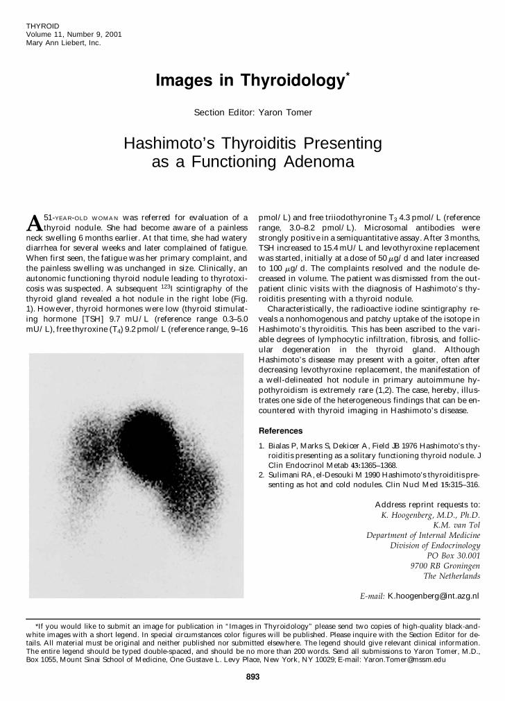
A51-YEAR-OLD WOMAN was referred for evaluation of athyroid nodule. She had become aware of a painless

neck swelling 6 months earlier. At that time, she had waterydiarrhea for several weeks and later complained of fatigue.When first seen, the fatigue was her primary complaint, andthe painless swelling was unchanged in size. Clinically, anautonomic functioning thyroid nodule leading to thyrotoxi-cosis was suspected. A subsequent 123I scintigraphy of thethyroid gland revealed a hot nodule in the right lobe (Fig.1). However, thyroid hormones were low (thyroid stimulat-ing hormone [TSH] 9.7 mU/L (reference range 0.3–5.0mU/L), free thyroxine (T4) 9.2 pmol/L (reference range, 9–16

pmol/L) and free triiodothyronine T3 4.3 pmol/L (referencerange, 3.0–8.2 pmol/L). Microsomal antibodies werestrongly positive in a semiquantitative assay. After 3 months,TSH increased to 15.4 mU/L and levothyroxine replacementwas started, initially at a dose of 50 mg/d and later increasedto 100 mg/d. The complaints resolved and the nodule de-creased in volume. The patient was dismissed from the out-patient clinic visits with the diagnosis of Hashimoto’s thy-roiditis presenting with a thyroid nodule.

Characteristically, the radioactive iodine scintigraphy re-veals a nonhomogenous and patchy uptake of the isotope inHashimoto’s thyroiditis. This has been ascribed to the vari-able degrees of lymphocytic infiltration, fibrosis, and follic-ular degeneration in the thyroid gland. AlthoughHashimoto’s disease may present with a goiter, often afterdecreasing levothyroxine replacement, the manifestation ofa well-delineated hot nodule in primary autoimmune hy-pothyroidism is extremely rare (1,2). The case, hereby, illus-trates one side of the heterogeneous findings that can be en-countered with thyroid imaging in Hashimoto’s disease.

References

1. Bialas P, Marks S, Dekicer A, Field JB 1976 Hashimoto’s thy-roiditis presenting as a solitary functioning thyroid nodule. JClin Endocrinol Metab 43:1365–1368.

2. Sulimani RA, el-Desouki M 1990 Hashimoto’s thyroiditis pre-senting as hot and cold nodules. Clin Nucl Med 15:315–316.

Address reprint requests to:K. Hoogenberg, M.D., Ph.D.

K.M. van TolDepartment of Internal Medicine

Division of EndocrinologyPO Box 30.001

9700 RB GroningenThe Netherlands

E-mail: [email protected]

*If you would like to submit an image for publication in “Images in Thyroidology” please send two copies of high-quality black-and-white images with a short legend. In special circumstances color figures will be published. Please inquire with the Section Editor for de-tails. All material must be original and neither published nor submitted elsewhere. The legend should give relevant clinical information.The entire legend should be typed double-spaced, and should be no more than 200 words. Send all submissions to Yaron Tomer, M.D.,Box 1055, Mount Sinai School of Medicine, One Gustave L. Levy Place, New York, NY 10029; E-mail: [email protected]