Embed Size (px)
Citation preview
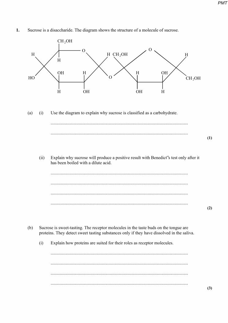
1. Sucrose is a disaccharide. The diagram shows the structure of a molecule of sucrose.
CH OH
CH OH
CH OH
O
O
O
OH
OH OH
OH
HH H
H
H H
H
H
HO
2
2
2
(a) (i) Use the diagram to explain why sucrose is classified as a carbohydrate.
...........................................................................................................................
........................................................................................................................... (1)
(ii) Explain why sucrose will produce a positive result with Benedict‟s test only after it has been boiled with a dilute acid.
...........................................................................................................................
...........................................................................................................................
...........................................................................................................................
........................................................................................................................... (2)
(b) Sucrose is sweet-tasting. The receptor molecules in the taste buds on the tongue are proteins. They detect sweet tasting substances only if they have dissolved in the saliva.
(i) Explain how proteins are suited for their roles as receptor molecules.
...........................................................................................................................
...........................................................................................................................
...........................................................................................................................
........................................................................................................................... (3)
PMT
(ii) Explain why glucose and maltose both taste sweet but starch does not.
...........................................................................................................................
........................................................................................................................... (1)
(iii) Saccharin, cyclamates and sucrose are chemically different but they all taste sweet. Suggest why.
...........................................................................................................................
...........................................................................................................................
........................................................................................................................... (2)
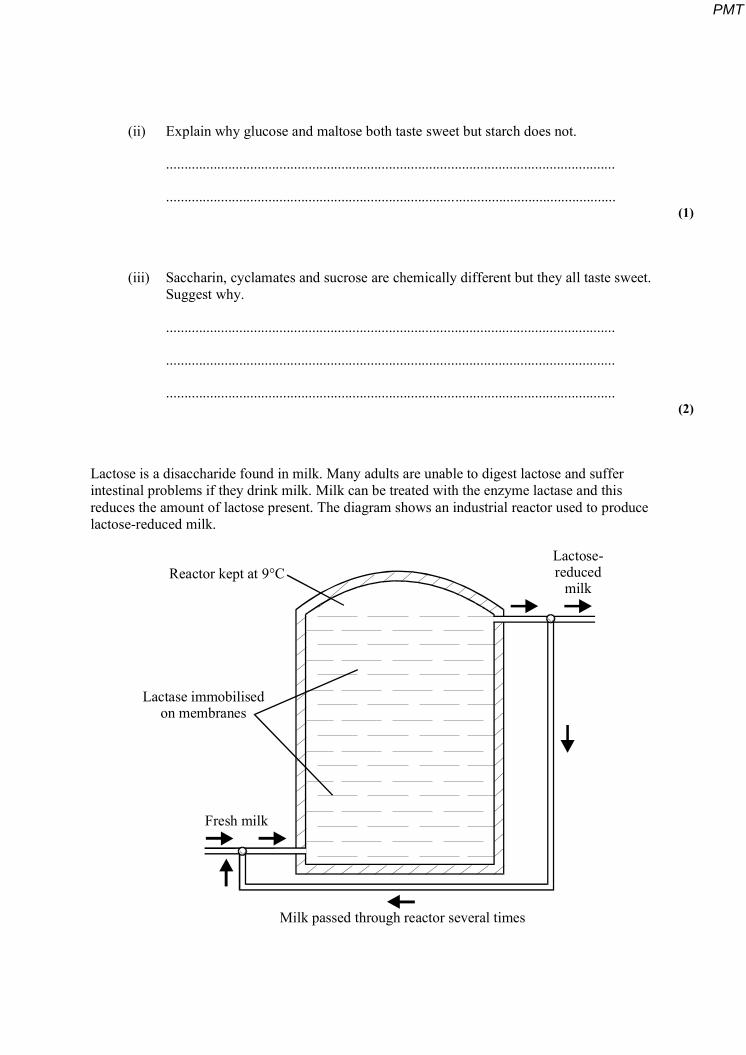
Lactose is a disaccharide found in milk. Many adults are unable to digest lactose and suffer intestinal problems if they drink milk. Milk can be treated with the enzyme lactase and this reduces the amount of lactose present. The diagram shows an industrial reactor used to produce lactose-reduced milk.
Lactose-reduced
milkReactor kept at 9°C
Fresh milk
Milk passed through reactor several times
Lactase immobilisedon membranes
PMT
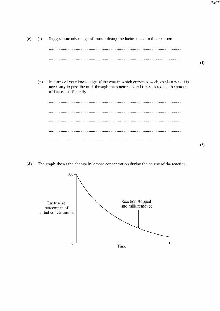
(c) (i) Suggest one advantage of immobilising the lactase used in this reaction.
...........................................................................................................................
........................................................................................................................... (1)
(ii) In terms of your knowledge of the way in which enzymes work, explain why it is necessary to pass the milk through the reactor several times to reduce the amount of lactose sufficiently.
...........................................................................................................................
...........................................................................................................................
...........................................................................................................................
...........................................................................................................................
........................................................................................................................... (3)
(d) The graph shows the change in lactose concentration during the course of the reaction.
Lactose aspercentage of
initial concentration
100
0
Reaction stoppedand milk removed
Time
PMT
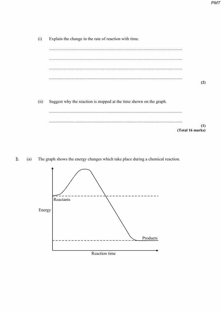
(i) Explain the change in the rate of reaction with time.
...........................................................................................................................
...........................................................................................................................
...........................................................................................................................
........................................................................................................................... (2)
(ii) Suggest why the reaction is stopped at the time shown on the graph.
...........................................................................................................................
........................................................................................................................... (1)
(Total 16 marks)
2. (a) The graph shows the energy changes which take place during a chemical reaction.
Energy
Reactants
Products
Reaction time
PMT
(i) Use the graph to explain what is meant by the term activation energy.
...........................................................................................................................
...........................................................................................................................
........................................................................................................................... (1)
(ii) Draw a curve on the graph to show the energy changes which would take place if the same chemical reaction were catalysed by an enzyme.
(2)
The flow chart shows the way in which fructose is produced from starch in the food industry.
starch
glucose
glucose andfructose
modified starch,dextrins and
glucose
100°C
55° C
50° C
-amylase
glucoamylase
glucose isomerase
(b) Describe a biochemical test which could be used to show that reducing sugars were produced in the first stage of this process.
.....................................................................................................................................
............................................................................................................................. ........
.....................................................................................................................................
............................................................................................................................. ........ (2)
PMT
(c) Acid could have been used in place of the -amylase in the first stage of this process. Suggest why:
(i) acid could have been used;
...........................................................................................................................
........................................................................................................................... (1)
(ii) acid was not used.
...........................................................................................................................
........................................................................................................................... (1)
(d) In the laboratory, the optimal conditions for bacterial -amylase are a pH of 7 and a temperature of 80°C.
In terms of your knowledge of the way in which enzymes work, explain why the rate of reaction would change if:
(i) the temperature fell by 10°C;
...........................................................................................................................
...........................................................................................................................
...........................................................................................................................
........................................................................................................................... (2)
(ii) the pH changed substantially.
...........................................................................................................................
...........................................................................................................................
...........................................................................................................................
...........................................................................................................................
...........................................................................................................................
........................................................................................................................... (3)
(Total 12 marks)
3. A test for glucose relies on two enzyme-controlled reactions.
PMT
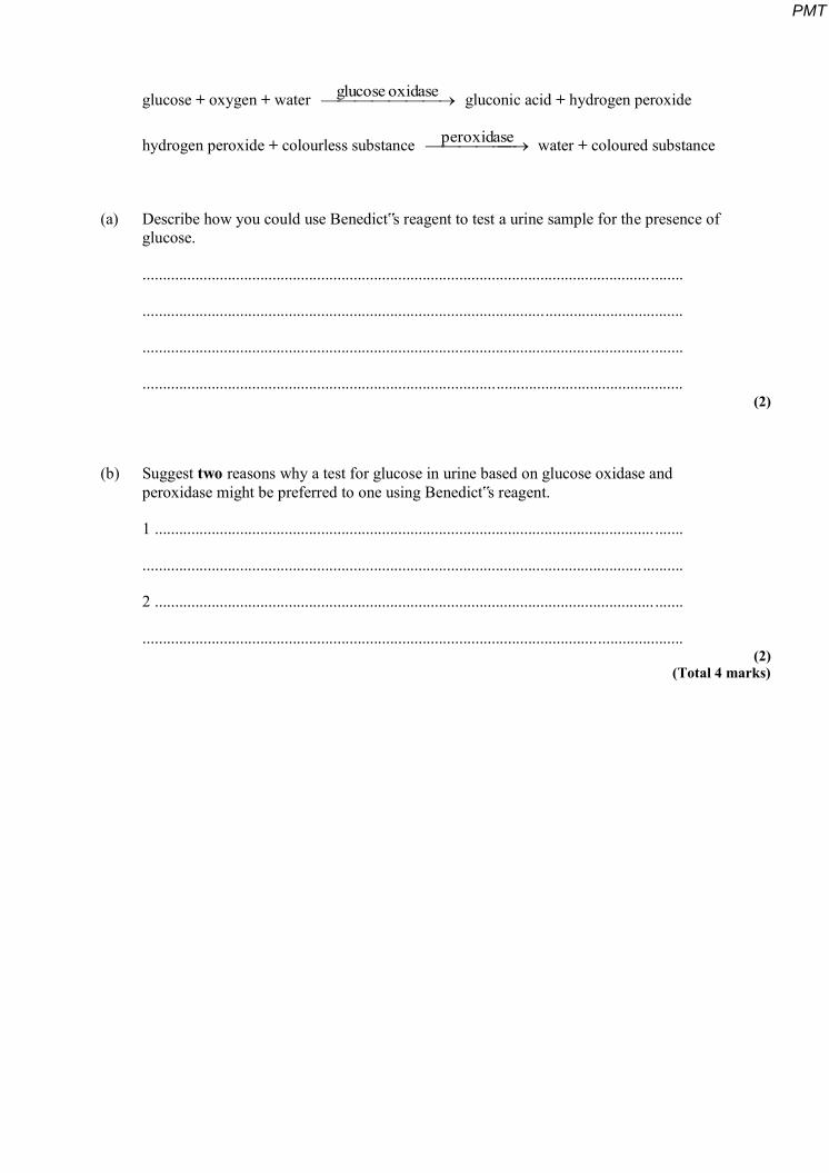
glucose + oxygen + water oxidaseglucose gluconic acid + hydrogen peroxide
hydrogen peroxide + colourless substance peroxidase water + coloured substance
(a) Describe how you could use Benedict‟s reagent to test a urine sample for the presence of glucose.
............................................................................................................................. ........
.....................................................................................................................................
............................................................................................................................. ........
..................................................................................................................................... (2)
(b) Suggest two reasons why a test for glucose in urine based on glucose oxidase and peroxidase might be preferred to one using Benedict‟s reagent.
1 ........................................................................................................................... .......
........................................................................................................................... ..........
2 ........................................................................................................................... .......
..................................................................................................................................... (2)
(Total 4 marks)
PMT
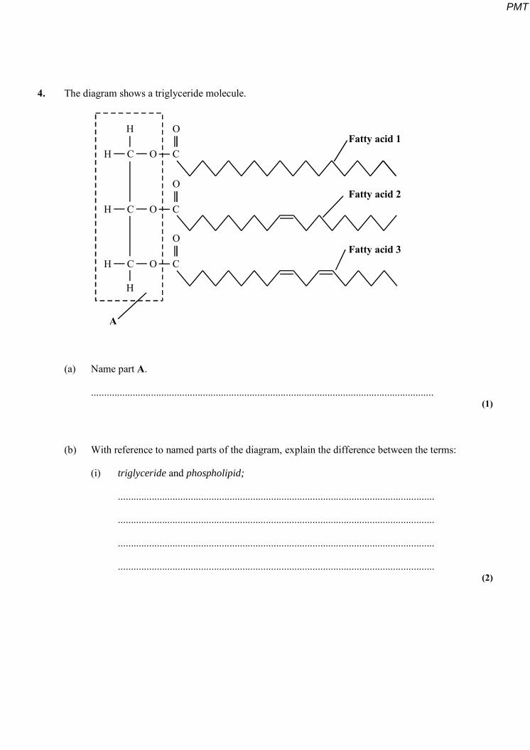
4. The diagram shows a triglyceride molecule.
H C O C
H C O C
H C O C
H
H
O
O
O
A
Fatty acid 1
Fatty acid 2
Fatty acid 3
(a) Name part A.
.................................................................................................................................... (1)
(b) With reference to named parts of the diagram, explain the difference between the terms:
(i) triglyceride and phospholipid;
..........................................................................................................................
..........................................................................................................................
..........................................................................................................................
.......................................................................................................................... (2)
PMT
(ii) saturated and unsaturated.
..........................................................................................................................
..........................................................................................................................
.......................................................................................................................... (2)
(Total 5 marks)
5. (a) Starch is an important storage substance in plants. Give two features of starch molecules and explain how each enables starch to act as an efficient storage substance.
1 Feature..............................................................................................................
Explanation.......................................................................................................
..........................................................................................................................
2 Feature..............................................................................................................
Explanation.......................................................................................................
.......................................................................................................................... (2)
Glucose syrup is used in the production of many human foods. It is produced from starch in a series of enzyme-controlled reactions.
(b) One way of monitoring the progress of these reactions is to measure the amount of reducing sugar produced.
(i) Describe a chemical test that would enable you to show that glucose syrup contained reducing sugar.
...........................................................................................................................
...........................................................................................................................
...........................................................................................................................
...........................................................................................................................
PMT
(ii) Suggest how you could use this test to compare the amount of reducing sugar in two solutions.
...........................................................................................................................
...........................................................................................................................
...........................................................................................................................
........................................................................................................................... (4)
(c) The progress of these reactions can also be monitored by finding the dextrose equivalent (DE). Dextrose equivalent can be calculated from the formula:
DE = starchinpresentbondsglycosidicofnumber
100hydrolisedbondsglycosidicofnumber
Explain why pure glucose obtained from starch has a dextrose equivalent of 100.
.....................................................................................................................................
............................................................................................................................. ........ (1)
(Total 7 marks)
PMT
6. Read the following passage.
Proteins have many different functions. These include catalysing chemical reactions and transporting substances across membranes. Many of these functions rely on the specific shape of their molecules. Molecules of a particular protein always fold into the same shape.
5 Although different proteins have different shapes, they share a number of structural features. They are formed from 20 different types of amino acid, each containing the same four chemical elements. Unlike triglycerides, proteins are polymers. Their chains are linear and never branched. The primary structure is the term used to refer to the sequence of amino acids which makes up a particular 10 protein. These amino acids are linked by peptide bonds. The side-chains or R-groups of different amino acids may form chemical bonds with each other. It is these bonds which allow the formation of protein molecules with specific tertiary shapes.
The amino acid sequences of over 100000 proteins are known but, so far, we only 15 know the tertiary structure of about 5000 of these. We have recently discovered that the folding of polypeptide chains is controlled by a group of proteins called chaperones. Chaperones bind to unfolded regions of polypeptide chains as they are being synthesised and prevent them from binding to other proteins. Once folded, the protein and chaperone separate allowing the chaperone to affect the 20 folding of more polypeptide chains.
Use information from the passage and your own knowledge to answer the following questions.
(a) (i) What are the “same four chemical elements” found in all amino acids (line 7)?
........................................................................................................................... (1)
(ii) Explain why “unlike triglycerides, proteins are polymers” (lines 7 – 8).
...........................................................................................................................
...........................................................................................................................
...........................................................................................................................
........................................................................................................................... (2)
PMT
(iii) Glycogen is also a polymer. Explain how many different sorts of protein can be produced but only one sort of glycogen.
...........................................................................................................................
...........................................................................................................................
...........................................................................................................................
........................................................................................................................... (2)
(b) Describe two ways in which chaperones (line 17) are similar to enzymes.
1............................................................................................................................ .......
.....................................................................................................................................
2............................................................................................................................ .......
..................................................................................................................................... (2)
(c) (i) Explain what causes molecules of a particular protein always to fold into the same shape.
...........................................................................................................................
...........................................................................................................................
...........................................................................................................................
........................................................................................................................... (2)
PMT
(ii) Describe how molecular shape is important in explaining the way in which enzymes may be affected by inhibitors.
...........................................................................................................................
...........................................................................................................................
...........................................................................................................................
...........................................................................................................................
........................................................................................................................... (6)
(Total 15 marks)
7. (a) Describe a chemical test you could carry out to show that a piece of coconut contains lipids.
............................................................................................................................. ........
............................................................................................................................. ........
.......................................................................................................................... ...........
............................................................................................................................. ........
.....................................................................................................................................
............................................................................................................................. ........ (3)
(b) The diagram shows the structure of a phospholipid molecule,
Y
PMT
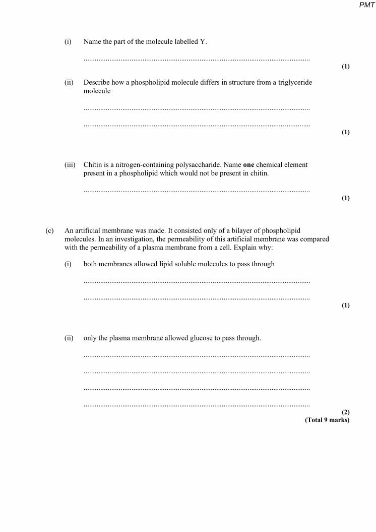
(i) Name the part of the molecule labelled Y.
........................................................................................................................... (1)
(ii) Describe how a phospholipid molecule differs in structure from a triglyceride molecule
...........................................................................................................................
........................................................................................................................... (1)
(iii) Chitin is a nitrogen-containing polysaccharide. Name one chemical element present in a phospholipid which would not be present in chitin.
........................................................................................................................... (1)
(c) An artificial membrane was made. It consisted only of a bilayer of phospholipid molecules. In an investigation, the permeability of this artificial membrane was compared with the permeability of a plasma membrane from a cell. Explain why:
(i) both membranes allowed lipid soluble molecules to pass through
...........................................................................................................................
........................................................................................................................... (1)
(ii) only the plasma membrane allowed glucose to pass through.
...........................................................................................................................
...........................................................................................................................
...........................................................................................................................
........................................................................................................................... (2)
(Total 9 marks)
PMT
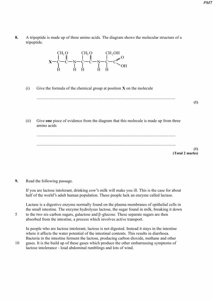
8. A tripeptide is made up of three amino acids. The diagram shows the molecular structure of a tripeptide.
X C C N C C N C C
CH CH CH OHO OO
OH
3 3 2
H H H H H
(i) Give the formula of the chemical group at position X on the molecule
..................................................................................................................................... (1)
(ii) Give one piece of evidence from the diagram that this molecule is made up from three amino acids
.....................................................................................................................................
............................................................................................................................. ........ (1)
(Total 2 marks)
9. Read the following passage.
If you are lactose intolerant, drinking cow‟s milk will make you ill. This is the case for about half of the world‟s adult human population. These people lack an enzyme called lactase.
Lactase is a digestive enzyme normally found on the plasma membranes of epithelial cells in the small intestine. The enzyme hydrolyses lactose, the sugar found in milk, breaking it down 5 to the two six-carbon sugars, galactose and -glucose. These separate sugars are then absorbed from the intestine, a process which involves active transport.
In people who are lactose intolerant, lactose is not digested. Instead it stays in the intestine where it affects the water potential of the intestinal contents. This results in diarrhoea. Bacteria in the intestine ferment the lactose, producing carbon dioxide, methane and other 10 gases. It is the build up of these gases which produce the other embarrassing symptoms of lactose intolerance - loud abdominal rumblings and lots of wind.
PMT
Use information from the passage and your own knowledge to answer the following questions.
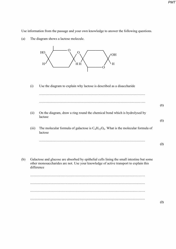
(a) The diagram shows a lactose molecule.
HO HO
H HH H
OO
O
(i) Use the diagram to explain why lactose is described as a disaccharide
...........................................................................................................................
........................................................................................................................... (1)
(ii) On the diagram, draw a ring round the chemical bond which is hydrolysed by lactase
(1)
(iii) The molecular formula of galactose is C6H12O6. What is the molecular formula of lactose
........................................................................................................................... (2)
(b) Galactose and glucose are absorbed by epithelial cells lining the small intestine but some other monosaccharides are not. Use your knowledge of active transport to explain this difference
............................................................................................................................. ........
.....................................................................................................................................
............................................................................................................................. ........
..................................................................................................................................... (2)
PMT
(c) Diarrhoea involves the production of large amounts of watery faeces. Explain the link between the presence of lactose in the intestine and diarrhoea.
................................................................................................................................... ..
............................................................................................................................. ........
............................................................................................................................. ........
.....................................................................................................................................
............................................................................................................................. ........
..................................................................................................................................... (3)
(d) The bacteria in the intestine are prokaryotic cells. The epithelial cells which line the small intestine are eukaryotic cells. Describe the ways in which prokaryotic cells and eukaryotic cells differ
............................................................................................................................. ........
.......................................................................................................................... ...........
............................................................................................................................. ........
.....................................................................................................................................
............................................................................................................................. ........ (6)
(Total 15 marks)
10. Sucrase is an enzyme. It hydrolyses a molecule of sucrose to give a molecule of glucose and a molecule of fructose. This is shown in the equation.
sucrose + water sucrase glucose + fructose
(a) The molecular formula of fructose is C6H12O6. What is the molecular formula of sucrose?
..................................................................................................................................... (2)
PMT
A solution containing the enzyme sucrase was added to a sucrose solution. The mixture was incubated in a test tube at 40°C for 1 hour. Sample A was removed from the tube at the start. Sample B was removed after 1 hour.
(b) A biuret test was carried out on sample A. It gave a positive result.
(i) Describe what you would expect to see if the biuret test gave a positive result.
..........................................................................................................................
.......................................................................................................................... (1)
(ii) Explain why the biuret test gave a positive result with sample A.
..........................................................................................................................
.......................................................................................................................... (1)
(c) Describe how you would use a biochemical test to show that sample B contained reducing sugar.
.....................................................................................................................................
............................................................................................................................. ........
.....................................................................................................................................
............................................................................................................................. ........ (2)
(Total 6 marks)
11. Read the following passage.
Many different processes essential to life depend on proteins. These include enzyme controlled reactions, transport across plasma membranes and the binding of hormones to receptor molecules on their target cells. Every protein molecule has a tertiary structure which gives it a precise three-dimensional shape. The function of the protein depends on this shape,
5 and the shape depends on the pH of the surrounding solution.
PMT
Changes in pH affect different proteins in different ways. This is because the amino acid molecules from which they are built have different structures. Some of these amino acids have different charges at different pH values. Unless they have the correct charges, the protein molecule will not have its correct three-dimensional shape.
10 If hydrogen or hydroxyl ions are added to a solution, its pH will normally change. A buffer solution is one which maintains a constant pH when hydrogen or hydroxyl ions are added to it. Buffers also occur naturally and play an important role in keeping conditions inside living organisms constant.
Use information from the passage and your own knowledge to answer the following questions.
(a) The receptor molecules to which hormones bind are proteins. Glucagon is a hormone.
(i) Use the information in the first paragraph to explain why glucagon will only bind to one particular type of receptor molecule.
..........................................................................................................................
..........................................................................................................................
..........................................................................................................................
.......................................................................................................................... (2)
(ii) Suggest why glucagon is able to bind to liver cells but not to cells in other parts of the body.
..........................................................................................................................
.......................................................................................................................... (1)
(b) Explain how the amino acids from which proteins are built (lines 6–7) differ in structure from each other.
............................................................................................................................. ........
..................................................................................................................................... (1)
PMT
(c) Amylase is an enzyme, found in saliva, which breaks down starch. It works best at a pH of 8. Explain why amylase does not function in the stomach where the pH is approximately 3.
............................................................................................................................. ........
.....................................................................................................................................
............................................................................................................................. ........
.....................................................................................................................................
............................................................................................................................. ........
............................................................................................................................. ........ (3)
(d) When a suspension of mitochondria is prepared from liver, the tissue is ground in a buffer solution, then centrifuged. Explain why a buffer solution is used.
............................................................................................................................. ........
.....................................................................................................................................
............................................................................................................................. ........
..................................................................................................................................... (2)
PMT
(e) Describe how proteins are arranged in a plasma membrane and the part they play in transporting substances into and out of cells.
.....................................................................................................................................
............................................................................................................................. ........
.....................................................................................................................................
............................................................................................................................. ........
.....................................................................................................................................
............................................................................................................................. ........
.....................................................................................................................................
............................................................................................................................. ........
............................................................................................................................. ........
......................................................................................................................... ............
............................................................................................................................. ........
............................................................................................................................. ........
.......................................................................................................................... ...........
............................................................................................................................. ........
..................................................................................................................................... (6)
(Total 15 marks)
PMT
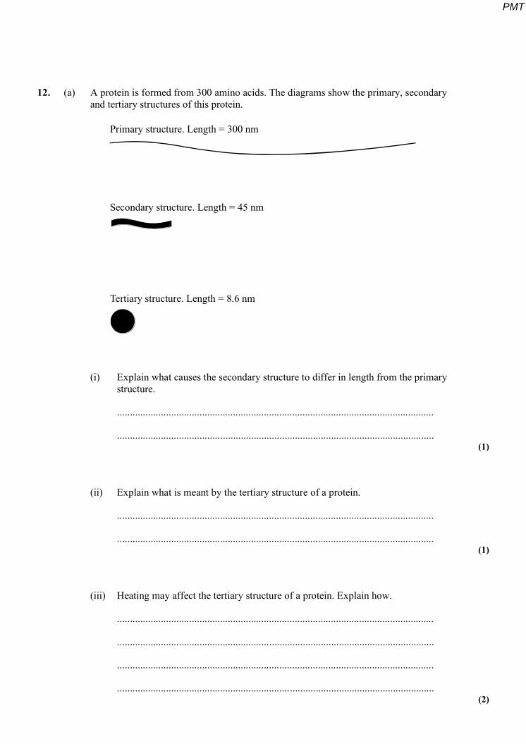
12. (a) A protein is formed from 300 amino acids. The diagrams show the primary, secondary and tertiary structures of this protein.
Primary structure. Length = 300 nm
Secondary structure. Length = 45 nm
Tertiary structure. Length = 8.6 nm
(i) Explain what causes the secondary structure to differ in length from the primary structure.
...........................................................................................................................
........................................................................................................................... (1)
(ii) Explain what is meant by the tertiary structure of a protein.
...........................................................................................................................
........................................................................................................................... (1)
(iii) Heating may affect the tertiary structure of a protein. Explain how.
...........................................................................................................................
...........................................................................................................................
...........................................................................................................................
........................................................................................................................... (2)
PMT
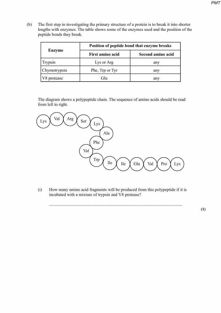
(b) The first step in investigating the primary structure of a protein is to break it into shorter lengths with enzymes. The table shows some of the enzymes used and the position of the peptide bonds they break.
Enzyme
Trypsin
Chymotrypsin
V8 protease
Lys or Arg
Phe, Trp or Tyr
Glu
First amino acid Second amino acid
any
any
any
Position of peptide bond that enzyme breaks
The diagram shows a polypeptide chain. The sequence of amino acids should be read from left to right.
Lys Ser Lys
Phe
TrpIle Ile Glu Val Pro Lys
Val
Ala
Val Arg
(i) How many amino acid fragments will be produced from this polypeptide if it is incubated with a mixture of trypsin and V8 protease?
........................................................................................................................... (1)
PMT
(ii) Explain why trypsin and chymotrypsin break peptide bonds between different amino acids.
...........................................................................................................................
...........................................................................................................................
...........................................................................................................................
........................................................................................................................... (3)
(Total 8 marks)
13. (a) Describe how phospholipid molecules are arranged in a plasma membrane.
............................................................................................................................. ........
.......................................................................................................................... ...........
............................................................................................................................. ........
..................................................................................................................................... (2)
PMT
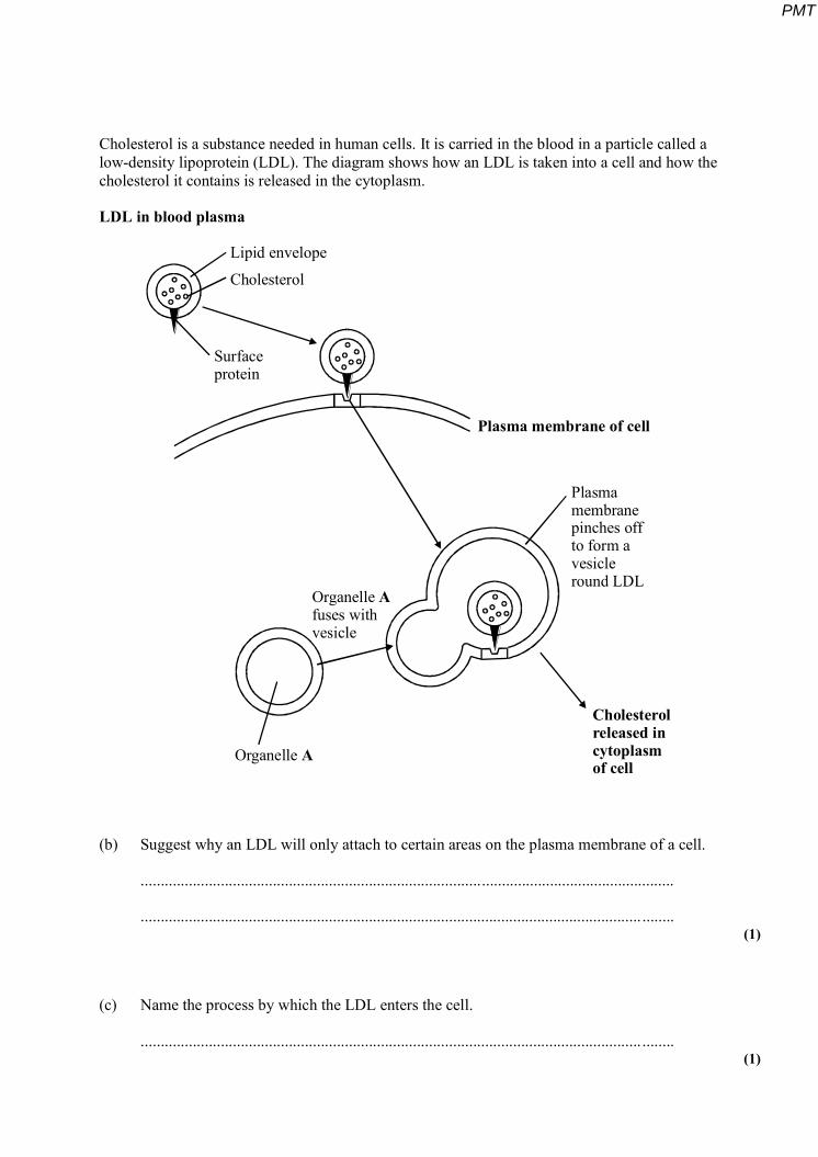
Cholesterol is a substance needed in human cells. It is carried in the blood in a particle called a low-density lipoprotein (LDL). The diagram shows how an LDL is taken into a cell and how the cholesterol it contains is released in the cytoplasm.
LDL in blood plasma
Lipid envelope
Cholesterol
Surfaceprotein
Plasma membrane of cell
Plasmamembranepinches offto form avesicleround LDL
Cholesterolreleased incytoplasmof cell
Organelle A
Organelle fuses withvesicle
A
(b) Suggest why an LDL will only attach to certain areas on the plasma membrane of a cell.
.....................................................................................................................................
............................................................................................................................. ........ (1)
(c) Name the process by which the LDL enters the cell.
............................................................................................................................. ........ (1)
PMT
(d) (i) Name organelle A.
........................................................................................................................... (1)
(ii) Explain how this organelle is involved in the release of cholesterol from the vesicle.
...........................................................................................................................
...........................................................................................................................
...........................................................................................................................
........................................................................................................................... (2)
(Total 7 marks)
14. Read the following passage.
If you read a sports magazine, it will not be long before you come across an advert for a sports drink. These adverts often claim that performance can be improved by consuming such drinks. Is this just a sales gimmick or is there a scientific basis for these claims?
Most sports drinks have a similar composition. Apart from water, the main ingredient is 5 carbohydrate. This is usually a mixture of different sugars – sucrose and the two monomers
from which it is formed by condensation – glucose and fructose. This combination improves taste and ensures efficient water absorption from the intestine. Most commercially available drinks are advertised as isotonic. They have the same water potential as the body fluids. When sugars are transported into the cells lining the intestine, water will also be absorbed.
10 Recently there has been an interest in the addition of particular amino acids to these drinks. Glutamine has been added because it is supposed to help protect the body from minor illness and infection. As well as glutamine, amino acids with a branched R-group may be added. These appear to be linked with the delay of biochemical processes in the body which cause fatigue.
PMT
Use the information from the passage and your own knowledge to answer the questions.
(a) Glucose and fructose both have the same molecular formula, C6H12O6.
(i) Suggest how two molecules can have the same formula but a different structure.
..........................................................................................................................
.......................................................................................................................... (1)
(ii) What is the molecular formula of a molecule of sucrose?
.......................................................................................................................... (2)
(b) (i) The uptake of sugars from the intestine involves facilitated diffusion and active transport. Give two ways in which facilitated diffusion differs from active transport.
1 ........................................................................................................................
...........................................................................................................................
2 ........................................................................................................................
........................................................................................................................... (2)
(ii) Explain how transport of sugars into cells lining the intestine (lines 8-9) leads to water being absorbed.
...........................................................................................................................
...........................................................................................................................
...........................................................................................................................
........................................................................................................................... (2)
PMT
(c) Give two ways in which the structure of a glutamine molecule (line11) is identical to the structure of an amino acid with a branched R-group (line 12).
1 ..................................................................................................................................
............................................................................................................................. ........
2 ..................................................................................................................................
............................................................................................................................. ........ (2)
(Total 9 marks)
15. (a) (i) How many molecules are produced when a triglyceride molecule is completely hydrolysed?
........................................................................................................................... (1)
(ii) Many large biological molecules are polymers. Explain why triglycerides are not polymers.
...........................................................................................................................
........................................................................................................................... (1)
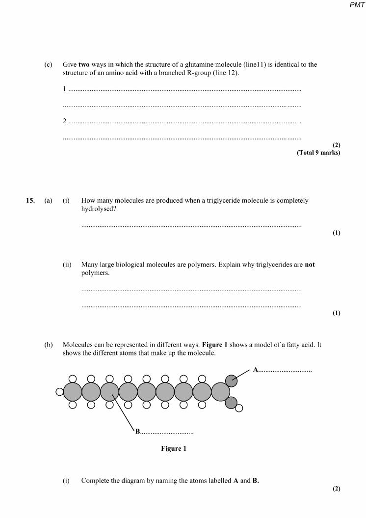
(b) Molecules can be represented in different ways. Figure 1 shows a model of a fatty acid. It shows the different atoms that make up the molecule.
A..............................
B..............................
Figure 1
(i) Complete the diagram by naming the atoms labelled A and B. (2)
PMT
(ii) This molecule is a saturated fatty acid. Explain the meaning of saturated.
...........................................................................................................................
........................................................................................................................... (1)
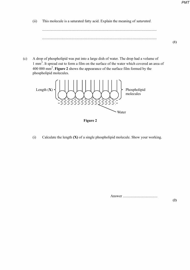
(c) A drop of phospholipid was put into a large dish of water. The drop had a volume of 1 mm3. It spread out to form a film on the surface of the water which covered an area of 400 000 mm2. Figure 2 shows the appearance of the surface film formed by the phospholipid molecules.
Length ( )X Phospholipidmolecules
Water
Figure 2
(i) Calculate the length (X) of a single phospholipid molecule. Show your working.
Answer ..................................... (2)
PMT
(ii) Explain what causes the phospholipid molecules to be arranged in the way shown in Figure 2.
...........................................................................................................................
...........................................................................................................................
...........................................................................................................................
........................................................................................................................... (2)
(Total 9 marks)
16. Read the following passage.
Bananas go through a series of changes as they ripen. The skin goes from green through yellow to brown. Biochemical changes take place in the fruit pulp. In an unripe banana, the main carbohydrate in the fruit pulp is starch. As the fruit ripens, this starch is largely replaced by reducing sugars such as glucose. The mass of cellulose in the fruit pulp, however, does not
5 change and remains more or less constant at between 1 and 2%.
Bananas are picked when they are green and transported in refrigerated ships. A major problem with shipping bananas in this way is “chilling”. Chilling results from exposing fruit to temperatures below a critical low value for longer than a critical time. In general, the longer the voyage, the higher the temperature bananas must be kept at to avoid chilling. Chilled
10 bananas are poor in quality. They are not as sweet as usual because starch hydrolysis is slow.
Use information from the passage and your own knowledge to answer the questions.
(a) Suggest how you could use Benedict‟s solution to show that a ripe banana contained more reducing sugar than an unripe banana.
.....................................................................................................................................
............................................................................................................................. ........
.....................................................................................................................................
............................................................................................................................. ........
.....................................................................................................................................
............................................................................................................................. ........ (3)
PMT
(b) Explain what causes the water potential of the banana pulp to decrease as the fruit ripens.
............................................................................................................................. ........
.....................................................................................................................................
............................................................................................................................. ........
..................................................................................................................................... (2)
(c) (i) Explain what causes starch hydrolysis to be slow in chilled bananas (line 10).
.....................................................................................................................................
............................................................................................................................. ........
.....................................................................................................................................
............................................................................................................................. ........
.....................................................................................................................................
............................................................................................................................. ........ (3)
(ii) Explain why “In general, the longer the voyage, the higher the temperature bananas must be kept at to avoid chilling.” (lines 8 - 9).
.....................................................................................................................................
............................................................................................................................. ........ (1)
(Total 9 marks)
PMT
17. Read the following passage.
Human milk contains all the nutrients a young baby needs in exactly the right proportions. It is formed in the mammary glands by small groups of milk-producing cells. These cells absorb substances from the blood and use them to synthesise the lipids, carbohydrates and proteins found in milk. Milk-producing cells are roughly cube-shaped
5 and have a height to breadth ratio of approximately 1.2 : 1.
The main carbohydrate in milk is lactose. Lactose is a disaccharide formed by the condensation of two monosaccharides, glucose and galactose. (A molecule of galactose has the same formula as a molecule of glucose – the atoms are just arranged in a different way.)
10 Lactose is synthesised in the Golgi apparatus and transported in vesicles through the cytoplasm. Because lactose is unable to escape from these vesicles, they increase in diameter as they move towards the plasma membrane. The vesicle membranes fuse with the plasma membrane and the vesicles empty their contents out of the cell.
Use the information from the passage and your own knowledge to answer the following questions.
(a) (i) The breadth of a milk-producing cell is 26 µm. Calculate the height of this cell.
Height = .......................... µm (1)
(ii) Describe and explain how you would expect the height to breadth ratio of an epithelial cell from a lung alveolus to differ from the height to breadth ratio of a milk-producing cell.
.....................................................................................................................................
............................................................................................................................. ........
.....................................................................................................................................
............................................................................................................................. ........ (2)
PMT
(b) How many oxygen atoms are there in a molecule of
(i) galactose;
............................................................................................................................. ........ (1)
(ii) lactose?
............................................................................................................................. ........ (1)
(c) The lactose-containing vesicles increase in diameter as they move towards the plasma membrane of the milk-producing cell (lines 11-12). Use your knowledge of water potential to explain why.
............................................................................................................................. ........
.....................................................................................................................................
............................................................................................................................. ........
..................................................................................................................................... (2)
(d) Suggest one advantage of milk-producing cells containing large numbers of mitochondria.
.....................................................................................................................................
............................................................................................................................. ........
.....................................................................................................................................
............................................................................................................................. ........ (2)
PMT
(e) Some substances pass through the plasma membrane of a milk-producing cell by diffusion. Describe the structure of a plasma membrane and explain how different substances are able to pass through the membrane by diffusion.
.....................................................................................................................................
............................................................................................................................. ........
.....................................................................................................................................
............................................................................................................................. ........
.....................................................................................................................................
............................................................................................................................. ........
............................................................................................................................. ........
......................................................................................................................... ............
............................................................................................................................. ........
.....................................................................................................................................
............................................................................................................................. ........ (6)
(Total 15 marks)
18. (a) Describe how you would use a biochemical test to show that a solution contained protein.
..............................................................................................................….....................
..............................................................................................................….....................
..............................................................................................................….....................
..............................................................................................................…..................... (2)
PMT
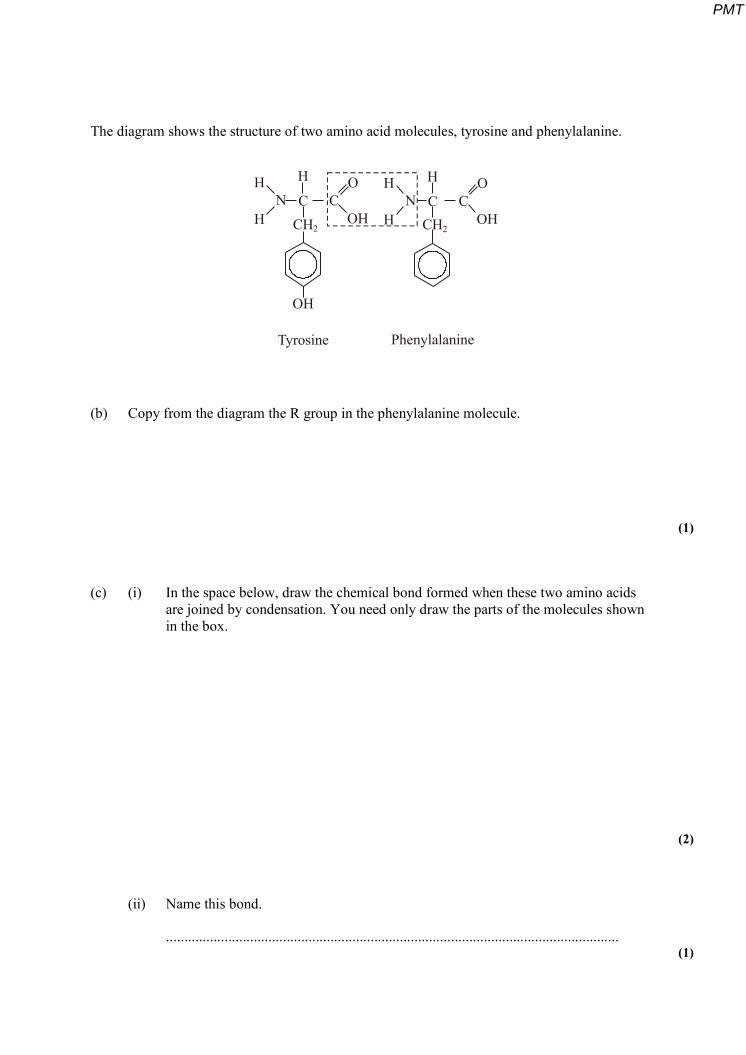
The diagram shows the structure of two amino acid molecules, tyrosine and phenylalanine.
H H
H HN N
H HC C
CH CHC C
O O
OH
OH
OH2 2
Tyrosine Phenylalanine
(b) Copy from the diagram the R group in the phenylalanine molecule.
(1)
(c) (i) In the space below, draw the chemical bond formed when these two amino acids are joined by condensation. You need only draw the parts of the molecules shown in the box.
(2)
(ii) Name this bond.
............................................................................................................................ (1)
PMT
(d) Tyrosine can be made in the body by hydroxylating phenylalanine. Use the diagram to explain the meaning of hydroxylating.
..............................................................................................................….....................
..............................................................................................................…............... ...... (1)
(Total 7 marks)
19. Read the following passage.
Job‟s Tears is a cereal plant which grows in the tropics. An unusual protein has been found in its grains. This protein is unusual because it has two functions. It acts as both an enzyme inhibitor and as an enzyme. As an inhibitor, the protein reduces the activity of starch-digesting enzymes. The protein acts as an enzyme by breaking down chitin, a polysaccharide found in
5 the walls of many fungi, to its monomers. Because of the resulting more negative water potential in the cytoplasm of the fungus, this effectively leads to “death by osmosis” of any fungus attacking the grain.
Our knowledge of the relationship between protein structure and function has led to the development of the new technology of protein engineering. This involves changing the amino
10 acid sequence of a protein and altering its tertiary structure. Altering the tertiary structure changes the protein‟s properties. So far, we have been unable to produce a protein with more than one function such as that found in Job‟s Tears. We have had success, though, in making some enzymes more stable and less prone to heat denaturation. We have done this by substituting amino acids and allowing the formation of additional chemical bonds.
Use information from the passage and your own knowledge to answer the following questions.
(a) (i) The protein found in Job‟s Tears breaks down chitin (line 4). What type of chemical reaction is involved in breaking down chitin?
............................................................................................................................ (1)
(ii) Breakdown of chitin leads to “death by osmosis” of fungi attacking the grain (lines 6 - 7). Explain how.
............................................................................................................................
............................................................................................................................
............................................................................................................................
............................................................................................................................ (2)
PMT
(iii) This protein does not break down the cell walls of the Job‟s Tears plant. Explain why.
............................................................................................................................
............................................................................................................................ (1)
(b) Explain what is meant by the tertiary structure of a protein (line 10).
..............................................................................................................….....................
..............................................................................................................…............ ......... (1)
(c) (i) Explain how heating an enzyme leads to it being denatured.
............................................................................................................................
............................................................................................................................
............................................................................................................................
............................................................................................................................ (2)
(ii) How can protein engineering make enzymes more stable and less prone to heat denaturation (line 13)?
............................................................................................................................
............................................................................................................................
............................................................................................................................
............................................................................................................................ (2)
PMT
(d) Describe how the sequence of amino acids in part of the protein from Job‟s Tears could enable this protein to act as an enzyme inhibitor.
..............................................................................................................….....................
..............................................................................................................….....................
..............................................................................................................….....................
..............................................................................................................….....................
..............................................................................................................….....................
..............................................................................................................….....................
..............................................................................................................….....................
..............................................................................................................….....................
..............................................................................................................….....................
..............................................................................................................….....................
..............................................................................................................….....................
..............................................................................................................…..................... (6)
(Total 15 marks)
20. (a) Starch and protein are biologically important polymers.
(i) Explain what is meant by a polymer.
...........................................................................................................................
........................................................................................................................... (1)
(ii) Give one example of a biologically important polymer other than starch or protein.
........................................................................................................................... (1)
PMT
(b) In an investigation, the enzyme amylase was mixed in a test tube with a buffer solution and a suspension of starch. The amylase broke down the starch to maltose. When all the starch had been broken down, a sample was removed from the test tube and tested with biuret reagent.
(i) Explain why a buffer solution was added to the amylase-starch mixture.
...........................................................................................................................
...........................................................................................................................
...........................................................................................................................
………………………………………………………………………………... (2)
(ii) What colour would you expect the sample to go when tested with biuret reagent?
........................................................................................................................... (1)
(iii) Give an explanation for your answer to part (ii)
...........................................................................................................................
...........................................................................................................................
...........................................................................................................................
………………………………………………………………………………... (2)
(Total 7 marks)
PMT

21. Read the following passage.
During the course of a day, we come into contact with many poisonous substances. These include industrial and household chemicals. The skin acts as a barrier and prevents many of these substances entering and harming the body.
The skin is one of the largest organs in the body. It is composed of several layers of 5 tissue. The outer layer consists of dead cells packed with keratins. Keratins are a group of
proteins that differ from each other in their primary structure. Each keratin molecule consists of several polypeptide chains, each individual chain wound into a spiral or helix. The polypeptide chains include many sulphur-containing amino acids and these help to give the keratin molecules their characteristic strength.
Use information from the passage and your own knowledge to answer the questions.
(a) What is the evidence from the passage that keratin molecules have a quaternary structure?
.....................................................................................................................................
............................................................................................................................. ........ (1)
(b) Explain how sulphur-containing amino acids help to give keratin molecules their characteristic strength (lines 8–9).
............................................................................................................................. ........
.....................................................................................................................................
............................................................................................................................. ........
..................................................................................................................................... (2)
(c) Explain why differences in primary structure result in keratins with different properties (line 6).
............................................................................................................................. ........
.......................................................................................................................... ...........
............................................................................................................................. ........
..................................................................................................................................... (2)
PMT
(d) The skin prevents poisonous substances entering and harming the body (line 3). Explain why these substances are unable to pass through the outer layer of skin cells by active transport.
.....................................................................................................................................
............................................................................................................................. ........
.....................................................................................................................................
............................................................................................................................. ........
.....................................................................................................................................
............................................................................................................................. ........ (3)
(e) Skin cells may be studied with a transmission electron microscope or an optical microscope. Explain the advantages and limitations of using a transmission electron microscope to study cells.
.....................................................................................................................................
............................................................................................................................. ........
.....................................................................................................................................
............................................................................................................................. ........
.....................................................................................................................................
............................................................................................................................. ........
............................................................................................................................. ........
.......................................................................................................................... ...........
............................................................................................................................. ........
.....................................................................................................................................
............................................................................................................................. ........
.....................................................................................................................................
............................................................................................................................. ........
..................................................................................................................................... (6)
(Total 14 marks)
PMT
22. Some enymes digest protein. They hydrolyse the peptide bonds between amino acids. The extent to which a protein is digested is called the degree of hydrolysis (DH). The DH value may be calculated from the equation:
presentbondspeptideofnumberTotalhydrolysedbondspeptideofNumber100DH
(a) (i) A protein molecule contains 151 amino acids. What is the total number of peptide bonds in this molecule?
........................................................................................................................... (1)
(ii) A molecule of this protein is digested. The DH value of the digested protein is 18. Calculate the number of peptide bonds that have been hydrolysed.
Answer ...................................... (1)
(b) What would be the DH value of a protein if it were completely hydrolysed to amino acids? Explain how you arrived at your answer.
DH value .....................................................................................................................
Explanation .................................................................................................................
............................................................................................................................. ........
.......................................................................................................................... ........... (2)
PMT
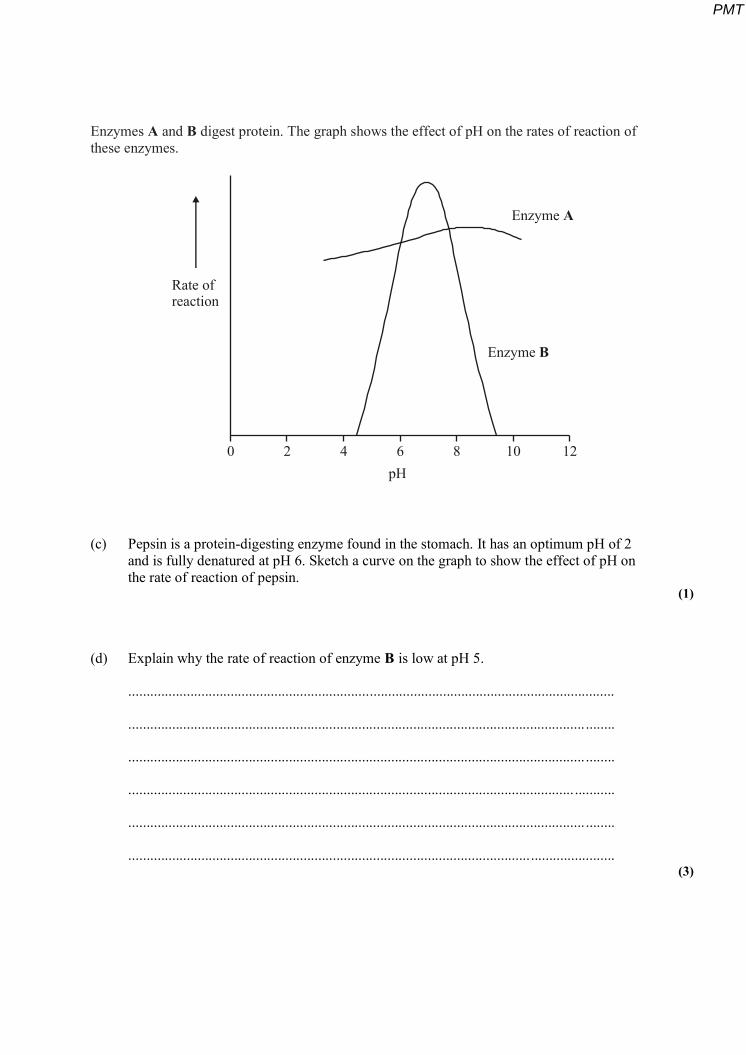
Enzymes A and B digest protein. The graph shows the effect of pH on the rates of reaction of these enzymes.
0 2 4 6 8 10 12
Enzyme A
Enzyme B
Rate ofreaction
pH
(c) Pepsin is a protein-digesting enzyme found in the stomach. It has an optimum pH of 2 and is fully denatured at pH 6. Sketch a curve on the graph to show the effect of pH on the rate of reaction of pepsin.
(1)
(d) Explain why the rate of reaction of enzyme B is low at pH 5.
.....................................................................................................................................
............................................................................................................................. ........
............................................................................................................................. ........
.......................................................................................................................... ...........
............................................................................................................................. ........
..................................................................................................................................... (3)
PMT
(e) Enzyme A is present in some washing powders used for cleaning clothes. Use the graph to suggest why enzyme A would be of more use in washing clothes than enzyme B.
.....................................................................................................................................
............................................................................................................................. ........
..................................................................................................................................... (1)
(f) Use your knowledge of protein structure to explain why enzymes are specific and may be affected by non-competitive inhibitors.
.....................................................................................................................................
............................................................................................................................. ........
.....................................................................................................................................
............................................................................................................................. ........
.....................................................................................................................................
............................................................................................................................. ........
............................................................................................................................. ........
.....................................................................................................................................
............................................................................................................................. ........
.....................................................................................................................................
............................................................................................................................. ........
.....................................................................................................................................
............................................................................................................................. ........
.....................................................................................................................................
............................................................................................................................. ........ (6)
(Total 15 marks)
PMT
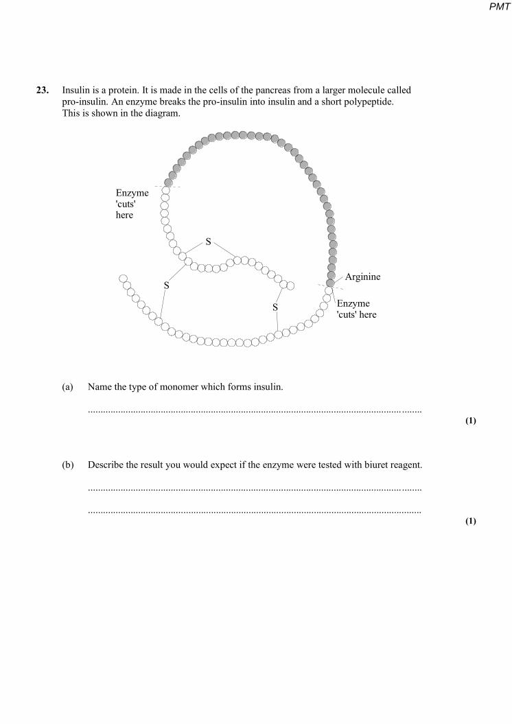
23. Insulin is a protein. It is made in the cells of the pancreas from a larger molecule called pro-insulin. An enzyme breaks the pro-insulin into insulin and a short polypeptide. This is shown in the diagram.
Enzyme'cuts'here
S
S
S
Arginine
Enzyme'cuts' here
(a) Name the type of monomer which forms insulin.
............................................................................................................................. ........ (1)
(b) Describe the result you would expect if the enzyme were tested with biuret reagent.
............................................................................................................................. ........
..................................................................................................................................... (1)
PMT
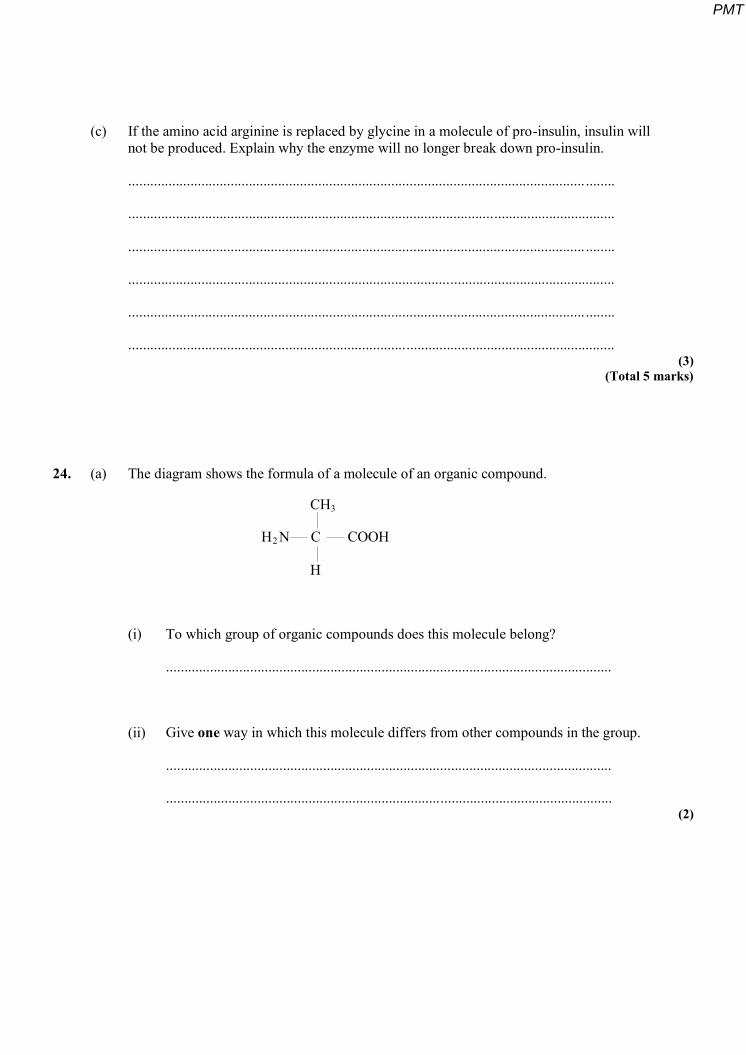
(c) If the amino acid arginine is replaced by glycine in a molecule of pro-insulin, insulin will not be produced. Explain why the enzyme will no longer break down pro-insulin.
............................................................................................................................. ........
.....................................................................................................................................
............................................................................................................................. ........
.....................................................................................................................................
............................................................................................................................. ........
..................................................................................................................................... (3)
(Total 5 marks)
24. (a) The diagram shows the formula of a molecule of an organic compound.
2
3
H N COOH
CH
H
C
(i) To which group of organic compounds does this molecule belong?
..........................................................................................................................
(ii) Give one way in which this molecule differs from other compounds in the group.
..........................................................................................................................
.......................................................................................................................... (2)
PMT
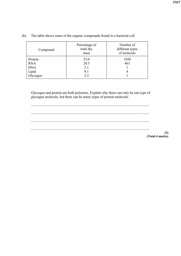
(b) The table shows some of the organic compounds found in a bacterial cell.
Compound Percentage of
total dry mass
Number of different types
of molecule
Protein RNA DNA Lipid Glycogen
55.0 20.5 3.1 9.1 2.5
1050 463
1 4 1
Glycogen and protein are both polymers. Explain why there can only be one type of glycogen molecule, but there can be many types of protein molecule.
............................................................................................................................. ........
.......................................................................................................................... ...........
............................................................................................................................. ........
..................................................................................................................................... (2)
(Total 4 marks)
PMT
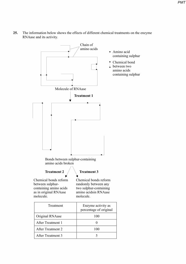
25. The information below shows the effects of different chemical treatments on the enzyme RNAase and its activity.
Chain ofamino acids
Molecule of RNAase
Treatment 1
Treatment 2 Treatment 3
Amino acidcontaining sulphur
Chemical bondbetween twoamino acidscontaining sulphur
Bonds between sulphur-containingamino acids broken
Chemical bonds reformbetween sulphur-containing amino acidsas in original RNAasemolecule.
Chemical bonds reformrandomly between anytwo sulphur-containingamino acidsin RNAasemolecule.
Treatment Enzyme activity as percentage of original
Original RNAase 100
After Treatment 1 0
After Treatment 2 100
After Treatment 3 5
PMT
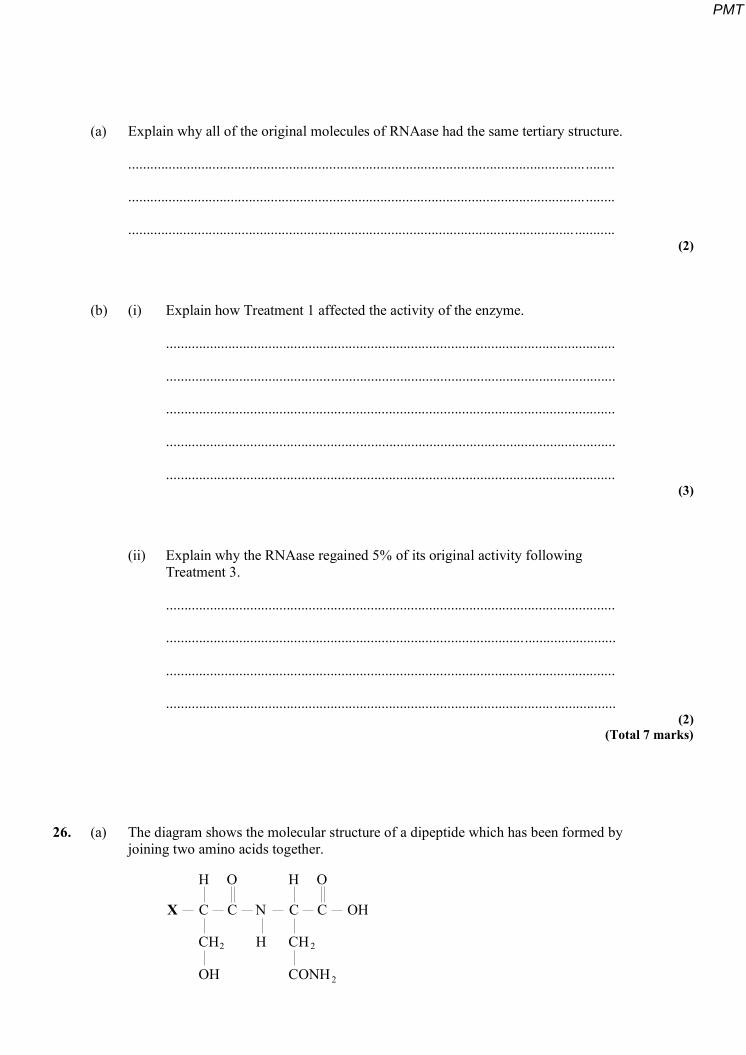
(a) Explain why all of the original molecules of RNAase had the same tertiary structure.
............................................................................................................................. ........
............................................................................................................................. ........
.......................................................................................................................... ........... (2)
(b) (i) Explain how Treatment 1 affected the activity of the enzyme.
...........................................................................................................................
...........................................................................................................................
...........................................................................................................................
...........................................................................................................................
........................................................................................................................... (3)
(ii) Explain why the RNAase regained 5% of its original activity following Treatment 3.
...........................................................................................................................
...........................................................................................................................
...........................................................................................................................
........................................................................................................................... (2)
(Total 7 marks)
26. (a) The diagram shows the molecular structure of a dipeptide which has been formed by joining two amino acids together.
CH
OH
CH
CONH
H H
NX
H
O O
C C OHC C
2 2
2
PMT
(i) Give the formula of the chemical group present at position X on this molecule.
.......................................................................................................................... (1)
(ii) Draw a circle round the chemical bond which has been formed as the result of the condensation reaction between the two amino acids.
(1)
(iii) The table shows the chemical structure of the R–group in a number of different amino acids.
Amino acid Structure of R-group
Alanine CH3
Asparagine CH2CONH2
Aspartic acid CH2COOH
Glutamine (CH2)2CONH2
Serine CH2OH
Use the information in the table to name the two amino acids from which the dipeptide was formed.
First amino acid ................................................................................................
Second amino acid .......................................................................................... (1)
(b) The relative molecular mass (Mr) of a molecule is a measure of its size.
(i) The mean Mr of an amino acid is 110. The Mr of a particular protein is 40 000. Calculate the number of amino acids that make up this protein. Show your working.
Answer = .............................. (1)
PMT
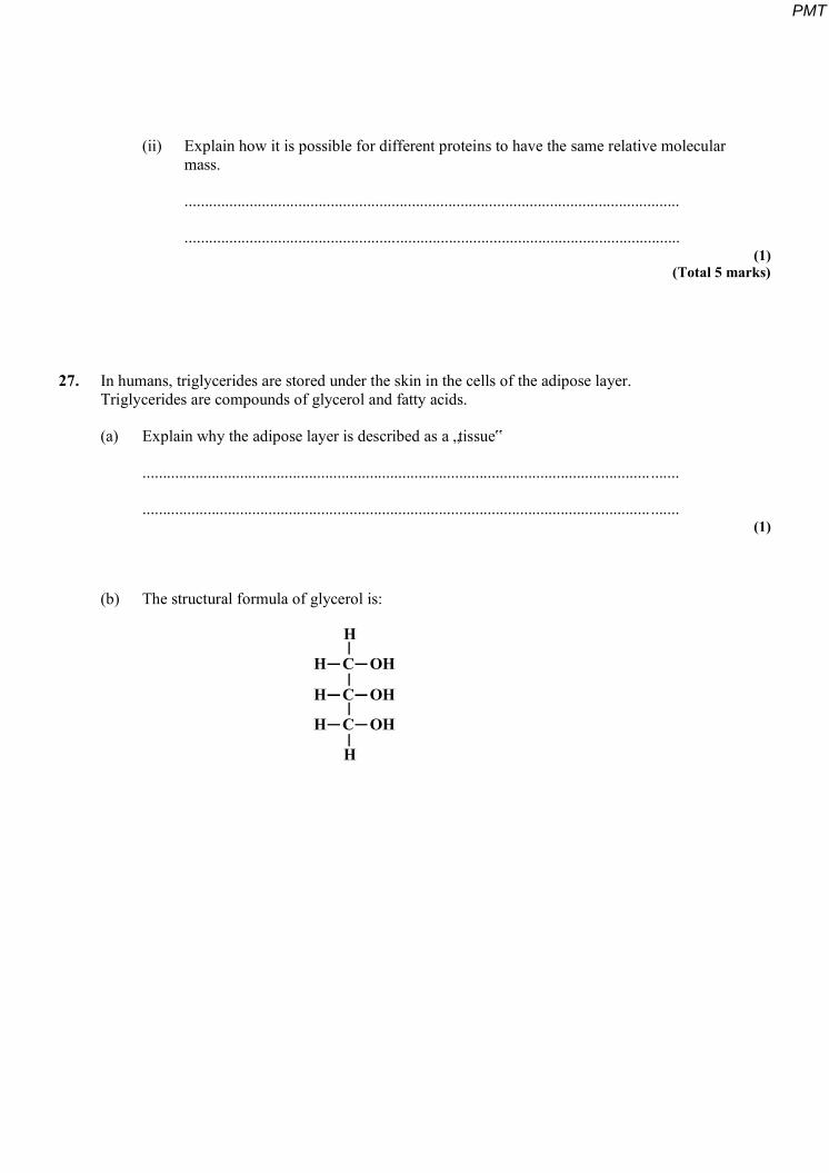
(ii) Explain how it is possible for different proteins to have the same relative molecular mass.
..........................................................................................................................
.......................................................................................................................... (1)
(Total 5 marks)
27. In humans, triglycerides are stored under the skin in the cells of the adipose layer. Triglycerides are compounds of glycerol and fatty acids.
(a) Explain why the adipose layer is described as a „tissue‟
............................................................................................................................. .......
............................................................................................................................. ....... (1)
(b) The structural formula of glycerol is:
H C OH
H C OH
H C OH
H
H
PMT
The structural formula of a fatty acid is: R - COOH.
(i) In the space below, draw the structural formula of a triglyceride.
(2)
(ii) Name the process in which fatty acids are combined with glycerol.
........................................................................................................................... (1)
(c) The triglycerides in adipose tissue occur as oily droplets, not as solid fats. The three fatty acids in a triglyceride may be all the same or a mixture of different fatty acids. The table shows the melting points of triglycerides with various combinations of fatty acids. It gives the number of carbon atoms in each fatty acid and whether the fatty acid is saturated (S) or unsaturated (U).
Number of carbon atom in each fatty acid 1st 2nd 3rd
Melting Point / ºC
16 (S)16 (S)18 (U)16 (S)18 (U)18 (S)
16 (S)18 (U)16 (S)18 (U)16 (S)18 (S)
16 (S)16 (S)16 (S)18 (U)18 (U)18 (U)
663635181842
(i) Describe the effect on melting point of including unsaturated fatty acids in a triglyceride.
...........................................................................................................................
........................................................................................................................... (1)
PMT
(ii) Analysis of the triglycerides in adipose tissue shows that about 50% of the fatty acids are unsaturated. Suggest the advantage of this.
...........................................................................................................................
...........................................................................................................................
...........................................................................................................................
........................................................................................................................... (2)
(Total 7 marks)
28. (a) Describe a biochemical test to find out if a substance contains a protein.
............................................................................................................................. ........
.......................................................................................................................... ...........
............................................................................................................................. ........
..................................................................................................................................... (2)
(b) The diagram shows the structural formulae of two amino acids.
H HO O
C CH N H N
H CH
SH
C COH OH2 2
2
(i) Name one chemical element found in all amino acids, but not in monosaccharides.
.......................................................................................................................... (1)
(ii) What type of chemical reaction occurs to form a dipeptide?
.......................................................................................................................... (1)
PMT

(iii) Draw the structural formula of the dipeptide formed when these two amino acids combine.
(1) (Total 5 marks)
29. Explain how the structure of fibrous proteins is related to their functions.
............................................................................................................................. ..................
...............................................................................................................................................
............................................................................................................................. ..................
............................................................................................................................. ..................
...............................................................................................................................................
............................................................................................................................. ..................
...............................................................................................................................................
............................................................................................................................. ..................
...............................................................................................................................................
............................................................................................................................. ..................
............................................................................................................................................. ..
............................................................................................................................. .................. (Total 5 marks)
PMT
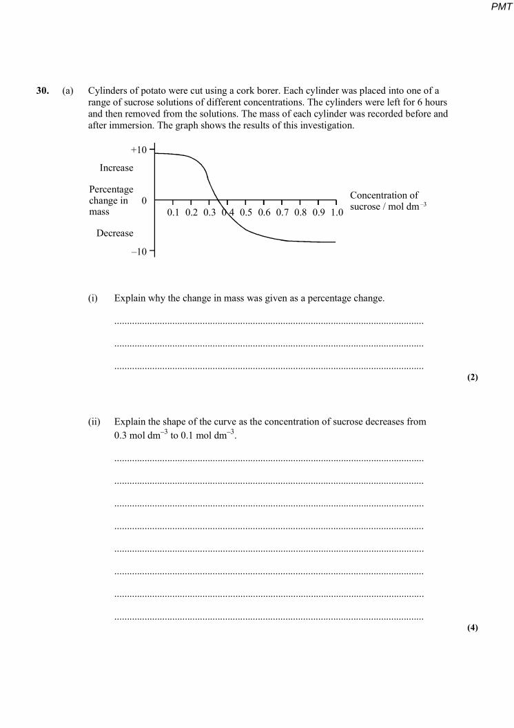
30. (a) Cylinders of potato were cut using a cork borer. Each cylinder was placed into one of a range of sucrose solutions of different concentrations. The cylinders were left for 6 hours and then removed from the solutions. The mass of each cylinder was recorded before and after immersion. The graph shows the results of this investigation.
+10
0
–10
Increase
Percentagechange inmass
Decrease
0.1 0.2 0.3 0 4 0.5 0.6 0.7 0.8 0.9 1.0
Concentration ofsucrose / mol dm –3
(i) Explain why the change in mass was given as a percentage change.
...........................................................................................................................
...........................................................................................................................
........................................................................................................................... (2)
(ii) Explain the shape of the curve as the concentration of sucrose decreases from 0.3 mol dm–3 to 0.1 mol dm–3.
...........................................................................................................................
...........................................................................................................................
...........................................................................................................................
...........................................................................................................................
...........................................................................................................................
...........................................................................................................................
...........................................................................................................................
........................................................................................................................... (4)
PMT
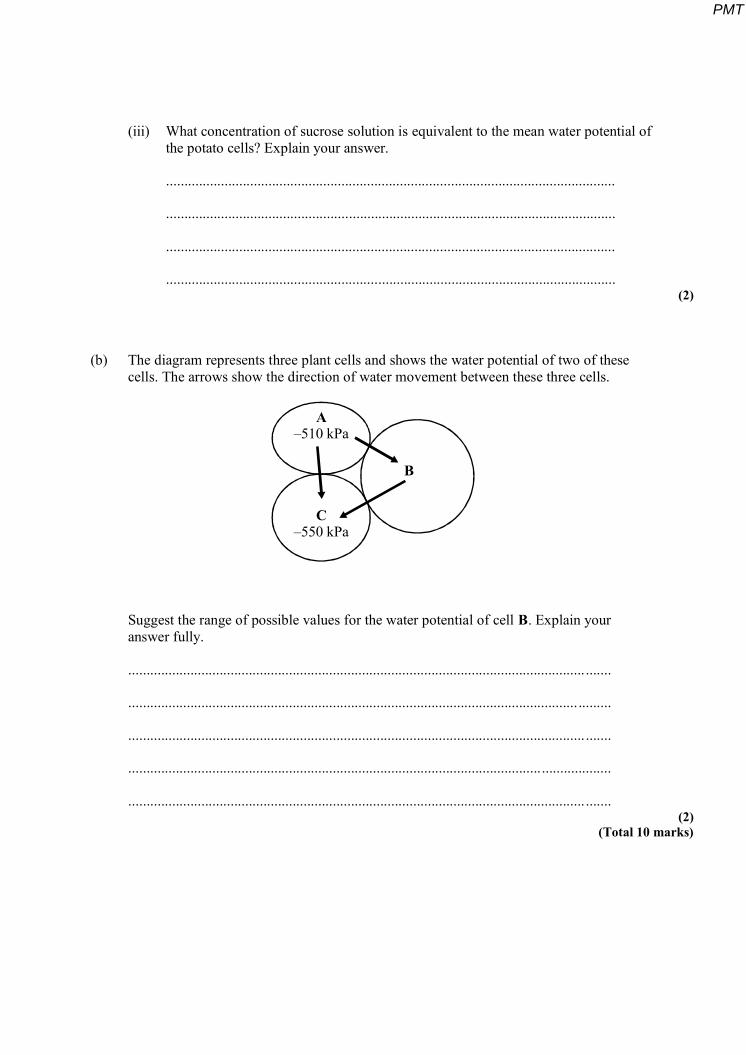
(iii) What concentration of sucrose solution is equivalent to the mean water potential of the potato cells? Explain your answer.
...........................................................................................................................
...........................................................................................................................
...........................................................................................................................
........................................................................................................................... (2)
(b) The diagram represents three plant cells and shows the water potential of two of these cells. The arrows show the direction of water movement between these three cells.
A–510 kPa
C–550 kPa
B
Suggest the range of possible values for the water potential of cell B. Explain your answer fully.
............................................................................................................................. .......
........................................................................................................................... .........
............................................................................................................................. .......
....................................................................................................................................
............................................................................................................................. ....... (2)
(Total 10 marks)
PMT

31.S In an investigation, the storage tissue in a plant root was tested for the presence of reducing sugar. Four cylinders were cut from the plant root with a cork borer. Each cylinder was exactly 3 cm long. These cylinders were washed thoroughly. They were then treated as shown in the table, before being put into 5 cm3 of distilled water in a test tube. After 5 minutes, a sample of the water from each tube was tested with Benedict‟s solution. The results are shown in the table.
Tube Treatment of cylinders of root Colour after testing with Benedict’s solution
A B C D
Cut into 5 equal lengths Cut into 10 equal lengths Cut into 15 equal lengths Uncut
Greenish yellow Reddish yellow Brick red Blue
(a) (i) Explain the results for tubes A, B and C.
...........................................................................................................................
...........................................................................................................................
...........................................................................................................................
........................................................................................................................... (2)
(ii) Explain the result for tube D.
...........................................................................................................................
........................................................................................................................... (1)
PMT
(b) To estimate the concentration in each sample, standard solutions of reducing sugar were used. Describe how standard solutions could be used to estimate the concentration of reducing sugar in the samples.
.....................................................................................................................................
............................................................................................................................. ........
.....................................................................................................................................
............................................................................................................................. ........
............................................................................................................................. ........
.......................................................................................................................... ........... (3)
(c) Tube D was placed in a bath of boiling water. After 5 minutes another sample of water was removed from the tube and tested with Benedict‟s solution. What result would you expect? Explain your answer.
Result: .........................................................................................................................
Explanation: ................................................................................................................
.....................................................................................................................................
............................................................................................................................. ........ (2)
PMT
(d) In autumn the shoots of the plants die, leaving the root as a storage organ in the soil. New shoots grow from the storage organ in spring. When the storage organs were tested after the shoots had died in late autumn, they were found to contain large amounts of storage polysaccharide and very little reducing sugar. When tested in spring as the new shoots were developing, much higher concentrations of reducing sugar were found. Suggest an explanation for the high concentration of reducing sugar in the spring.
.....................................................................................................................................
............................................................................................................................. ........
............................................................................................................................. ........
......................................................................................................................... ............ (2)
(Total 10 marks)
PMT
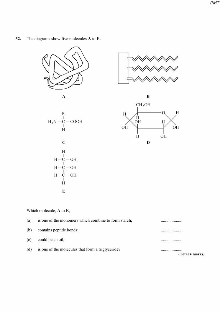
32. The diagrams show five molecules A to E.
A
C
E
B
D
R
H
C
C
H N
H
H
H
H
CH OH
H
OH
H
OH OH
CH
CH
H
COOH
OH
OH
OH
2
2
HOH
O
Which molecule, A to E,
(a) is one of the monomers which combine to form starch; ....................
(b) contains peptide bonds: ....................
(c) could be an oil; ....................
(d) is one of the molecules that form a triglyceride? .................... (Total 4 marks)
PMT
33. (a) Many reactions take place in living cells at temperatures far lower than those required for the same reactions in a laboratory. Explain how enzymes enable this to happen.
.....................................................................................................................................
............................................................................................................................. ........
............................................................................................................................. ........
.....................................................................................................................................
............................................................................................................................. ........
..................................................................................................................................... (3)
An amylase enzyme converts starch to maltose syrup which is used in the brewing industry.
(b) Describe a biochemical test to identify
(i) starch;
...........................................................................................................................
...........................................................................................................................
...........................................................................................................................
........................................................................................................................... (2)
(ii) a reducing sugar such as maltose.
...........................................................................................................................
...........................................................................................................................
...........................................................................................................................
........................................................................................................................... (2)
PMT
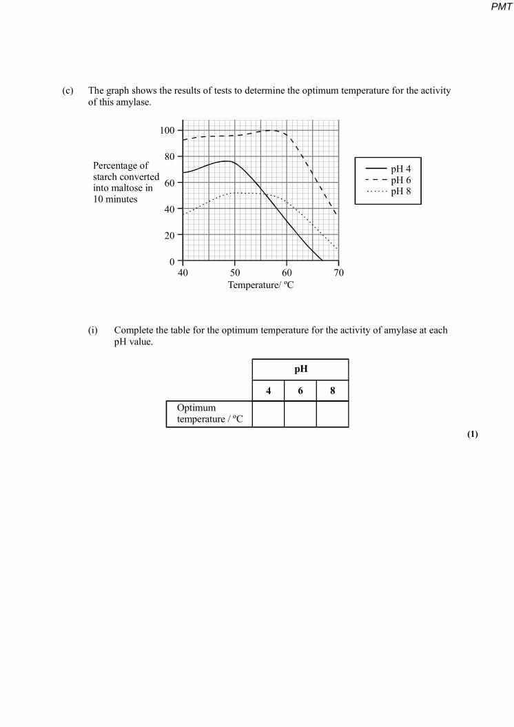
(c) The graph shows the results of tests to determine the optimum temperature for the activity of this amylase.
100
80
60
40
20
040 50 60 70
Temperature/ ºC
Percentage ofstarch convertedinto maltose in10 minutes
pH 4pH 6pH 8
(i) Complete the table for the optimum temperature for the activity of amylase at each pH value.
Optimumtemperature / ºC
pH
4 6 8
(1)
PMT
(ii) Describe and explain the effect of temperature on the rate of reaction of this enzyme at pH 4.
...........................................................................................................................
...........................................................................................................................
...........................................................................................................................
...........................................................................................................................
...........................................................................................................................
...........................................................................................................................
...........................................................................................................................
...........................................................................................................................
...........................................................................................................................
...........................................................................................................................
...........................................................................................................................
........................................................................................................................... (6)
(Total 14 marks)
34. The table shows the results of biochemical tests on substances found in food.
Substance Colour with Benedict’s
solution test
Colour with iodine solution
test
Colour with biuret test
Appearance with emulsion
test
A Blue Yellow-brown Violet Clear
B Blue Yellow-brown Blue Milky
C Brick-red Yellow-brown Blue Clear
D Blue Yellow-brown Blue Clear
PMT
(a) Identify substances A, B and C.
A...................................................................
B ...................................................................
C ................................................................... (3)
(b) Describe a further biochemical test to find out if substance D is a non-reducing sugar.
............................................................................................................................. ........
.....................................................................................................................................
............................................................................................................................. ........
..................................................................................................................................... (2)
(c) Name the chemical elements in a non-reducing sugar.
.....................................................................................................................................
............................................................................................................................. ........ (1)
(Total 6 marks)
35. (a) Figure 1 shows the structure of a molecule of glycerol and a molecule of fatty acid.
H
CH
CH
C RH
H
OH
OH
OH COOH
Glycerol Fatty acid
Figure 1
PMT
Draw a diagram to show the structure of a triglyceride molecule.
(2)
(b) Explain why triglycerides are not considered to be polymers.
............................................................................................................................. ........
..................................................................................................................................... (1)
(c) Figure 2 shows two types of fat storage cell. Mammals living in cold conditions have more brown fat cells than mammals living in tropical conditions.
Brown fat cell White fat cell
Nucleus
Triglyceride
Cytoplasm
Mitochondria
Figure 2
PMT
Using evidence from Figure 2 to support your answer, suggest how the function of brown fat cells differs from that of white fat cells.
.....................................................................................................................................
............................................................................................................................. ........
.....................................................................................................................................
............................................................................................................................. ........
.....................................................................................................................................
............................................................................................................................. ........ (3)
(Total 6 marks)
36. Lactose is a disaccharide sugar which can be broken down by the enzyme lactase into two monosaccharides, glucose and galactose.
lactose + water glucose + galactoselactase
(a) The formula for galactose is C6H12O6. What is the formula for lactose?
..................................................................................................................................... (2)
(b) A solution containing the enzyme lactase was added to a lactose solution. The solution was incubated at 40 °C for one hour. Sample A was removed from the tube before incubation. Sample B was removed after one hour.
(i) Describe a chemical test you could carry out on sample A to show that lactose is a reducing sugar.
...........................................................................................................................
...........................................................................................................................
...........................................................................................................................
........................................................................................................................... (2)
PMT
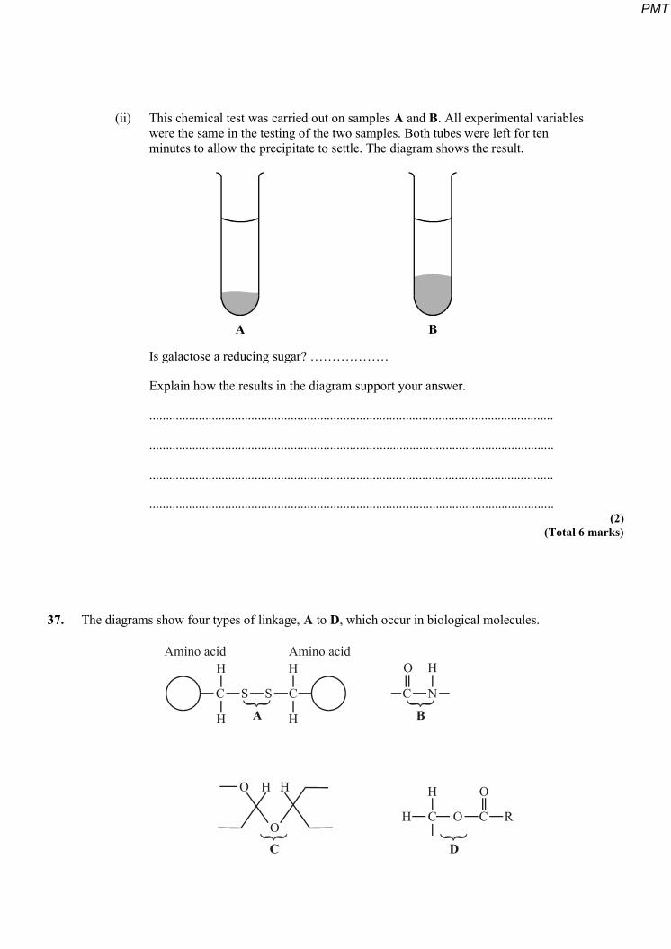
(ii) This chemical test was carried out on samples A and B. All experimental variables were the same in the testing of the two samples. Both tubes were left for ten minutes to allow the precipitate to settle. The diagram shows the result.
A B
Is galactose a reducing sugar? ………………
Explain how the results in the diagram support your answer.
...........................................................................................................................
...........................................................................................................................
...........................................................................................................................
........................................................................................................................... (2)
(Total 6 marks)
37. The diagrams show four types of linkage, A to D, which occur in biological molecules.
C S S C
H C O C R
C N
H
H
H
H
O
HA
Amino acid Amino acidH
H H
O
O
O
B
C D
PMT
(a) Name the chemical process involved in the formation of linkage B.
............................................................................................................................. ........ (1)
(b) Give the letter of the linkage which
(i) occurs in a triglyceride molecule;
......................................................................................................................…. (1)
(ii) might be broken down by the enzyme amylase;
......................................................................................................................…. (1)
(iii) may occur in the tertiary, but not the primary structure of protein.
......................................................................................................................…. (1)
(c) Describe how a saturated fatty acid differs in molecular structure from an unsaturated fatty acid.
............................................................................................................................. ...….
..................................................................................................................................…
............................................................................................................................. ........
............................................................................................................................. ........ (2)
(Total 6 marks)
PMT
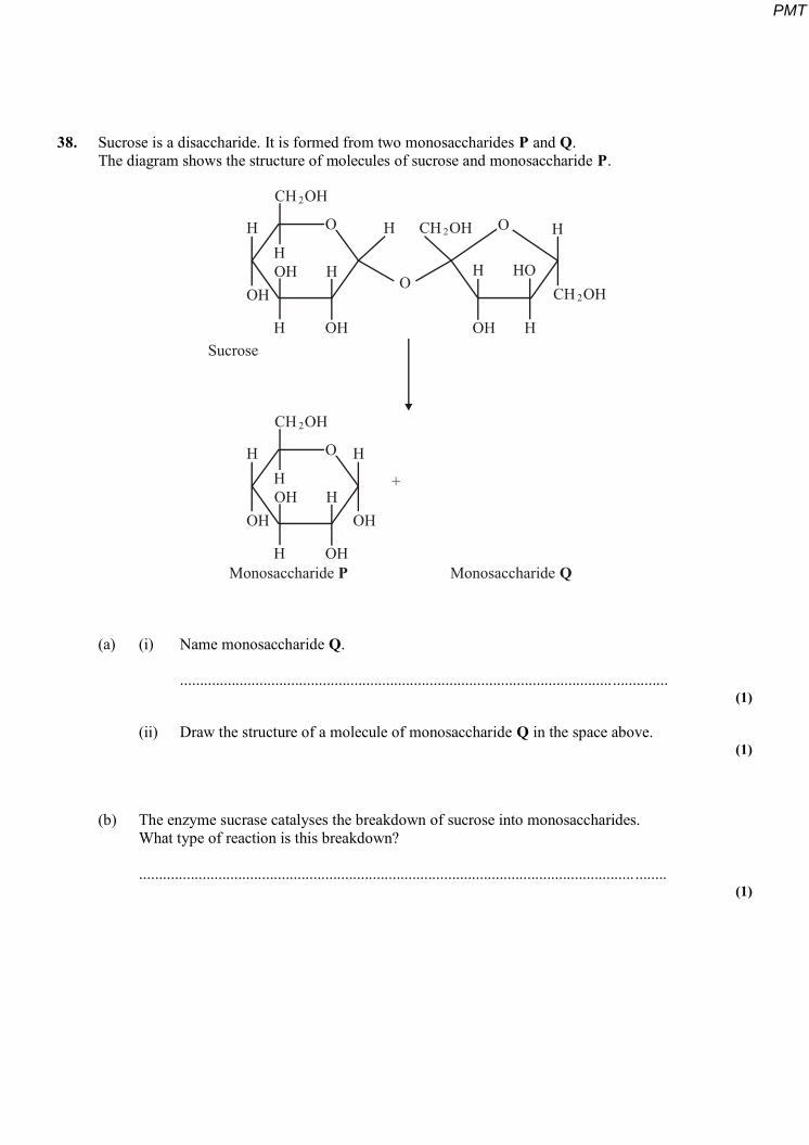
38. Sucrose is a disaccharide. It is formed from two monosaccharides P and Q. The diagram shows the structure of molecules of sucrose and monosaccharide P.
OH
OH OH
OH
OH
OH
OH
OH
H
H H
H
H
H
H
H
H
H
H
H
H
CH OH
CH OH
CH OH
CH OHHO
O
O
O
O
2
2
2
2
Sucrose
+
Monosaccharide MonosaccharideP Q
(a) (i) Name monosaccharide Q.
........................................................................................................................... (1)
(ii) Draw the structure of a molecule of monosaccharide Q in the space above. (1)
(b) The enzyme sucrase catalyses the breakdown of sucrose into monosaccharides. What type of reaction is this breakdown?
............................................................................................................................. ........ (1)
PMT
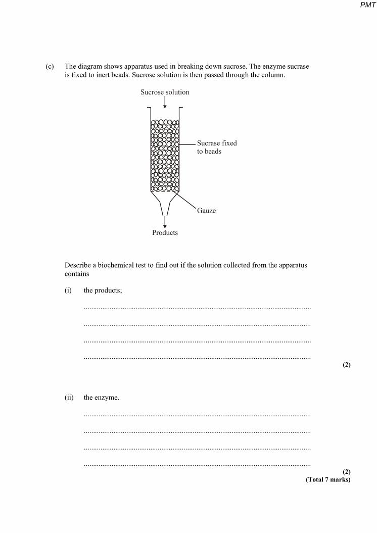
(c) The diagram shows apparatus used in breaking down sucrose. The enzyme sucrase is fixed to inert beads. Sucrose solution is then passed through the column.
Sucrose solution
Gauze
Products
Sucrase fixedto beads
Describe a biochemical test to find out if the solution collected from the apparatus contains
(i) the products;
...........................................................................................................................
...........................................................................................................................
...........................................................................................................................
........................................................................................................................... (2)
(ii) the enzyme.
...........................................................................................................................
...........................................................................................................................
...........................................................................................................................
........................................................................................................................... (2)
(Total 7 marks)
PMT
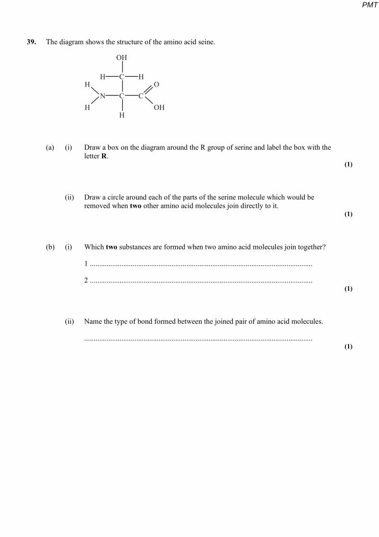
39. The diagram shows the structure of the amino acid seine.
H
NH
HC
OH
C
H
H
CO
OH
(a) (i) Draw a box on the diagram around the R group of serine and label the box with the letter R.
(1)
(ii) Draw a circle around each of the parts of the serine molecule which would be removed when two other amino acid molecules join directly to it.
(1)
(b) (i) Which two substances are formed when two amino acid molecules join together?
1 ........................................................................................................................
2 ........................................................................................................................ (1)
(ii) Name the type of bond formed between the joined pair of amino acid molecules.
........................................................................................................................... (1)
PMT
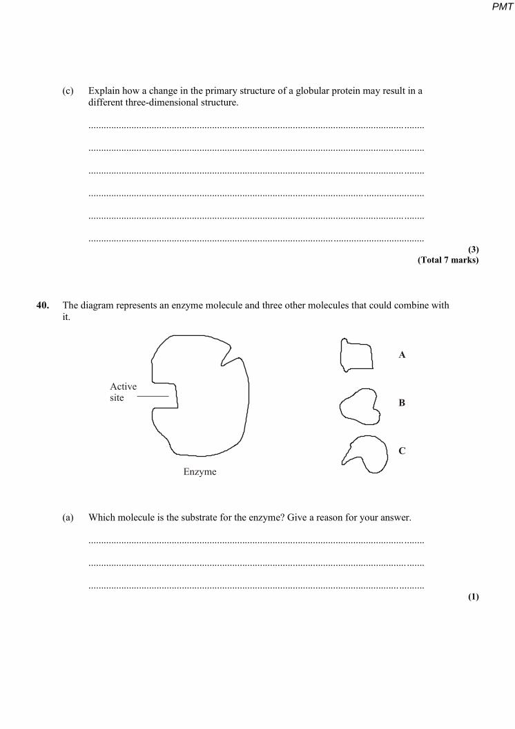
(c) Explain how a change in the primary structure of a globular protein may result in a different three-dimensional structure.
............................................................................................................................. ........
......................................................................................................................... ............
............................................................................................................................. ........
.....................................................................................................................................
............................................................................................................................. ........
..................................................................................................................................... (3)
(Total 7 marks)
40. The diagram represents an enzyme molecule and three other molecules that could combine with
it.
Activesite
Enzyme
A
B
C
(a) Which molecule is the substrate for the enzyme? Give a reason for your answer.
............................................................................................................................. ........
.............................................................................................................................. .......
........................................................................................................................... .......... (1)
PMT
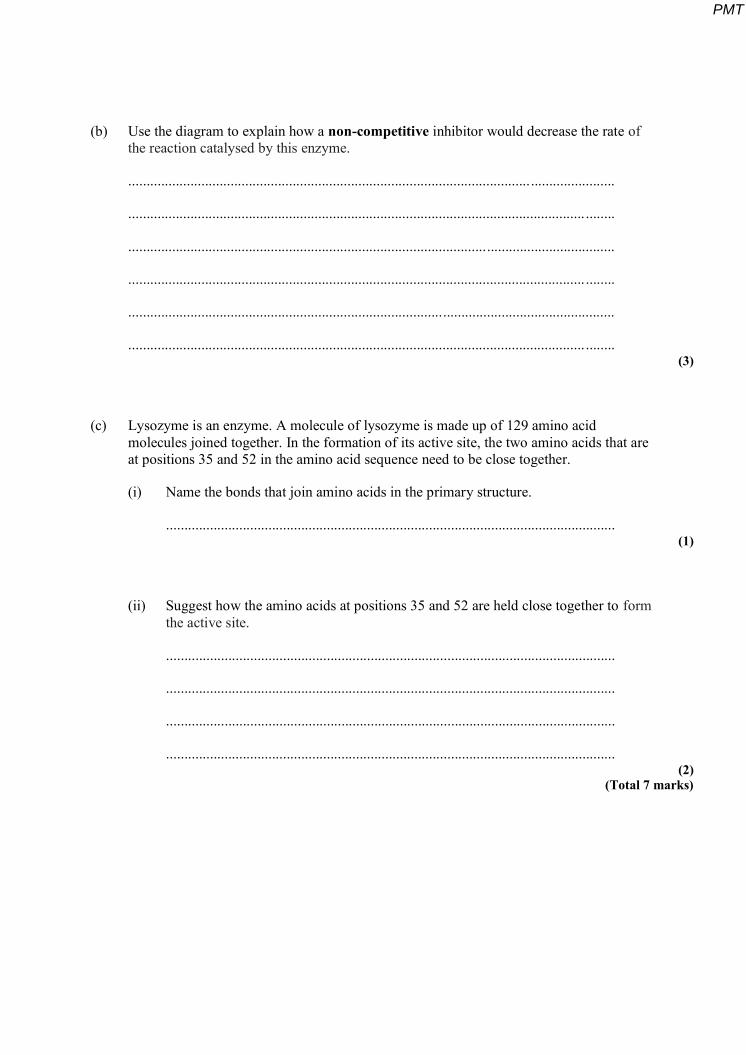
(b) Use the diagram to explain how a non-competitive inhibitor would decrease the rate of the reaction catalysed by this enzyme.
.....................................................................................................................................
............................................................................................................................. ........
.....................................................................................................................................
............................................................................................................................. ........
.....................................................................................................................................
............................................................................................................................. ........ (3)
(c) Lysozyme is an enzyme. A molecule of lysozyme is made up of 129 amino acid molecules joined together. In the formation of its active site, the two amino acids that are at positions 35 and 52 in the amino acid sequence need to be close together.
(i) Name the bonds that join amino acids in the primary structure.
........................................................................................................................... (1)
(ii) Suggest how the amino acids at positions 35 and 52 are held close together to form the active site.
...........................................................................................................................
...........................................................................................................................
...........................................................................................................................
........................................................................................................................... (2)
(Total 7 marks)
PMT


![(8.3) Acid-Base Properties of Salt Solutions. pH Review Recall Acidic [H 3 O + ] > [OH - ] Basic [H 3 O + ] < [OH - ] Neutral [H 3 O + ] = [OH - ]](https://img.pdfslide.us/doc/110x75/56649e165503460f94b00c9e/83-acid-base-properties-of-salt-solutions-ph-review-recall-acidic-h.jpg)