Embed Size (px)
Citation preview

Guideline-Driven Care in Cardio-
Oncology: Utilizing
Recommendations Across
Disciplines
Jennifer Liu, MD FACC FASE
Director of CV Laboratories
Associate Professor of Clinical Medicine
Memorial Sloan Kettering Cancer Center
Weill Cornell Medical College

• Summarize the key points
• Science behind the recommendations
• Apply the document to clinical practice
• Take home messages
I. CTRCD – definition, classification and mechanisms of
toxicity
II. Echocardiographic evaluation of the patient undergoing
cancer therapy
III. Early detection of toxicity
IV. Other imaging modalities
V. Integrated approach
I. CTRCD – definition, classification and
mechanisms of toxicity

Forms of Cardiotoxicity
• LV dysfunction
– Cancer therapeutics-related cardiac dysfunction
or CTRCD
• Ischemia
• HTN
• Arrhythmia
• Thromboembolism
• QT prolongation
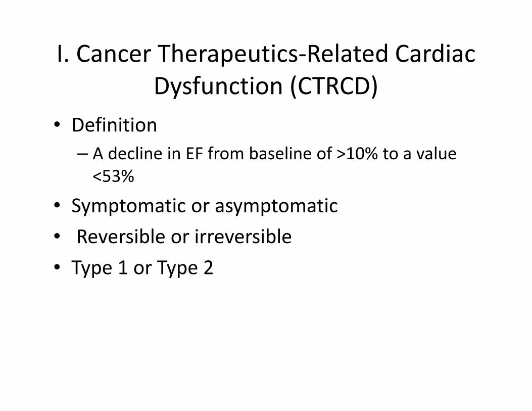
I. Cancer Therapeutics-Related Cardiac
Dysfunction (CTRCD)
• Definition
– A decline in EF from baseline of >10% to a value
<53%
• Symptomatic or asymptomatic
• Reversible or irreversible
• Type 1 or Type 2

Classification of CTRCD
Type 1 vs. Type 2

Type I CTRCD
• Prototype: doxorubicin
– Cumulative dose-
dependent
– Irreversible damage
– Cellular
apoptosis/necrosis
– Ultrastructural changes
on biopsy
Type II CTRCD
• Prototype: trastuzumab
– Not cumulative dose
dependent
– mostly reversible LV
dysfunction
– Cellular dysfunction
– No biopsy changes
Consensus Statement:
Differentiation of CTRCD into Type 1 and Type 2
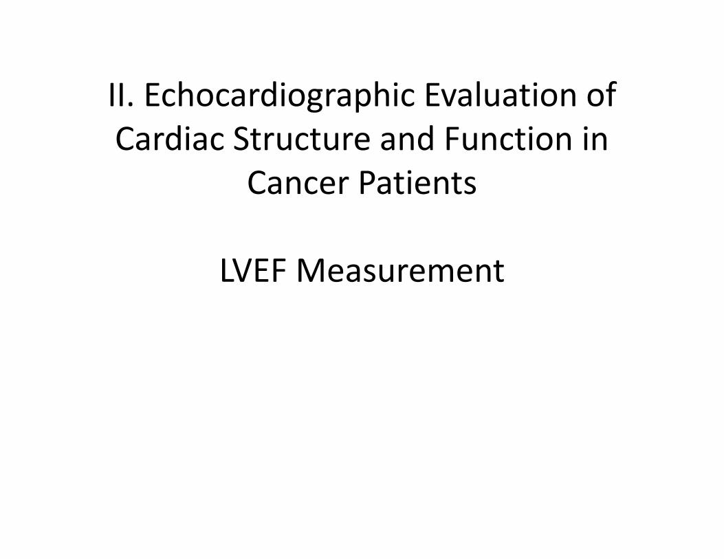
II. Echocardiographic Evaluation of
Cardiac Structure and Function in
Cancer Patients
LVEF Measurement
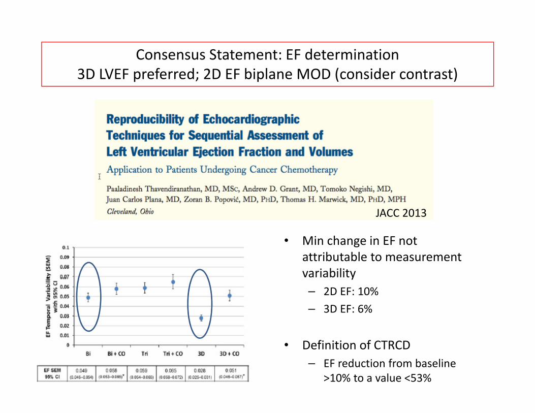
• Min change in EF not
attributable to measurement
variability
– 2D EF: 10%
– 3D EF: 6%
• Definition of CTRCD
– EF reduction from baseline
>10% to a value <53%
JACC 2013
Consensus Statement: EF determination
3D LVEF preferred; 2D EF biplane MOD (consider contrast)

• Advantages:
– More accurate and reproducible than 2D
– No geometric assumptions
– Minimize foreshortening
– Semi-automated border detection
• Disadvantages:
– Dependent on image quality
– Learning curve; experience with image acquisition and analysis
– Availability with equipment and expertise*
EF Determination: 3D Echo
Walker et al, JCO 2010
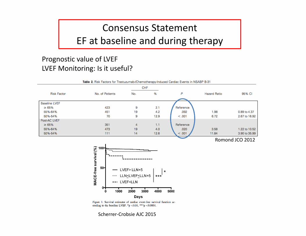
Romond JCO 2012
Scherrer-Crobsie AJC 2015
Prognostic value of LVEF
LVEF Monitoring: Is it useful?
Consensus Statement
EF at baseline and during therapy

III. Detection of Subclinical LV
Dysfunction
• Imaging
– Global longitudinal strain (GLS) with 2D echo
• Serum cardiac biomarkers
– Troponin
JACC 2013
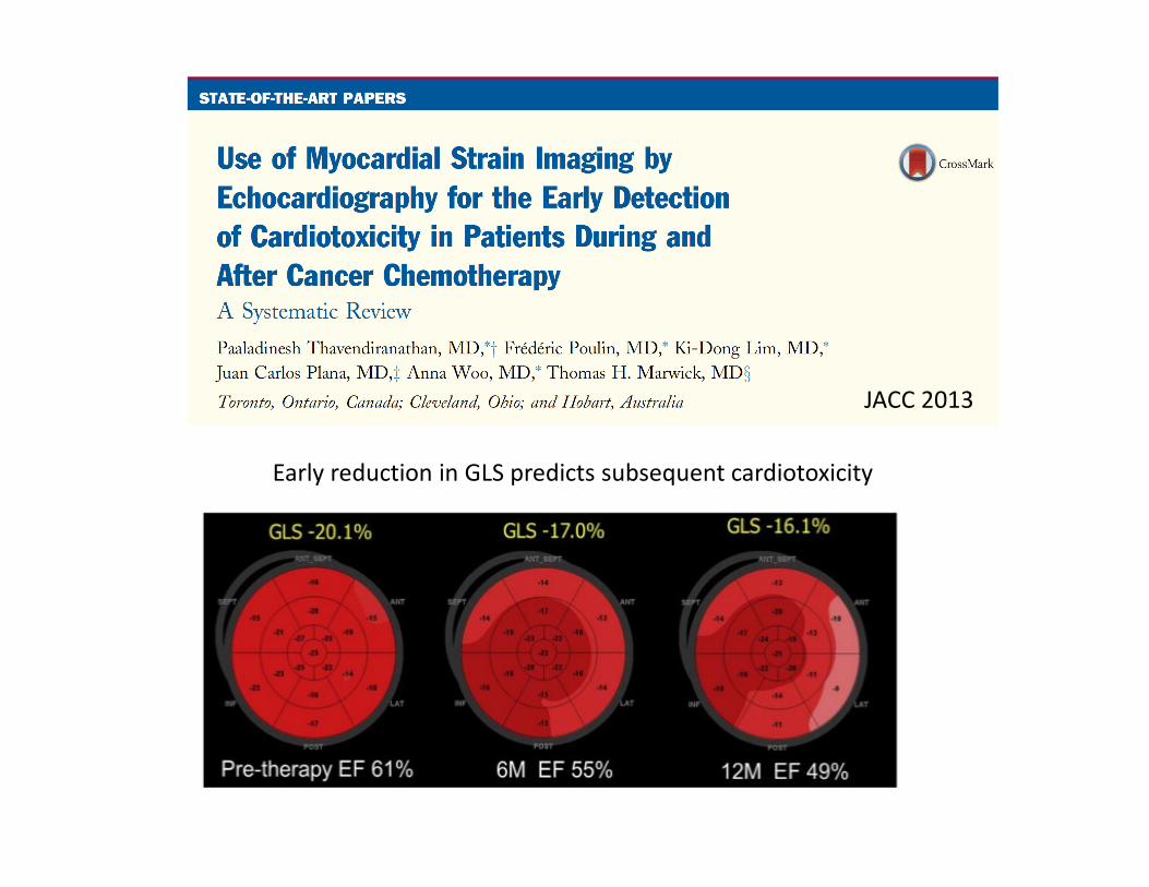
Early reduction in GLS predicts subsequent cardiotoxicity

Negichi K, Marwick T JASE 2013
� Decrease of 11% predictive
95% CI (8.3% - 14.6%)
81 BC patients w/ trastuzumab +/- AC
Consensus Statement:• Decrease GLS >15%, subclinical LV dysfunction likely
• Decrease <8%, no evidence of subclinical LV dysfunction

Prognostic value of biomarkers: troponin
Ky B, J Am Coll Cardiol 2014
Cardinale D, Circ 2002
703 pts receiving high dose chemotherapy
78 BC pts receiving AC + Trastuzuamb

IV Other Imaging Modalities
• MUGA
• Cardiac MRI
Consensus Statement:
Important to keep imaging modality consistent for baseline and follow-up studies.
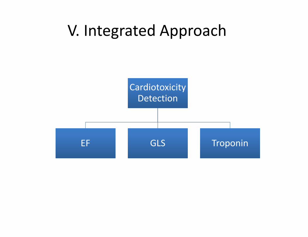
V. Integrated Approach
Cardiotoxicity Detection
Cardiotoxicity Detection
EFEF GLSGLS TroponinTroponin

Baseline Assessment and Monitoring
Type I Agents Type II agents
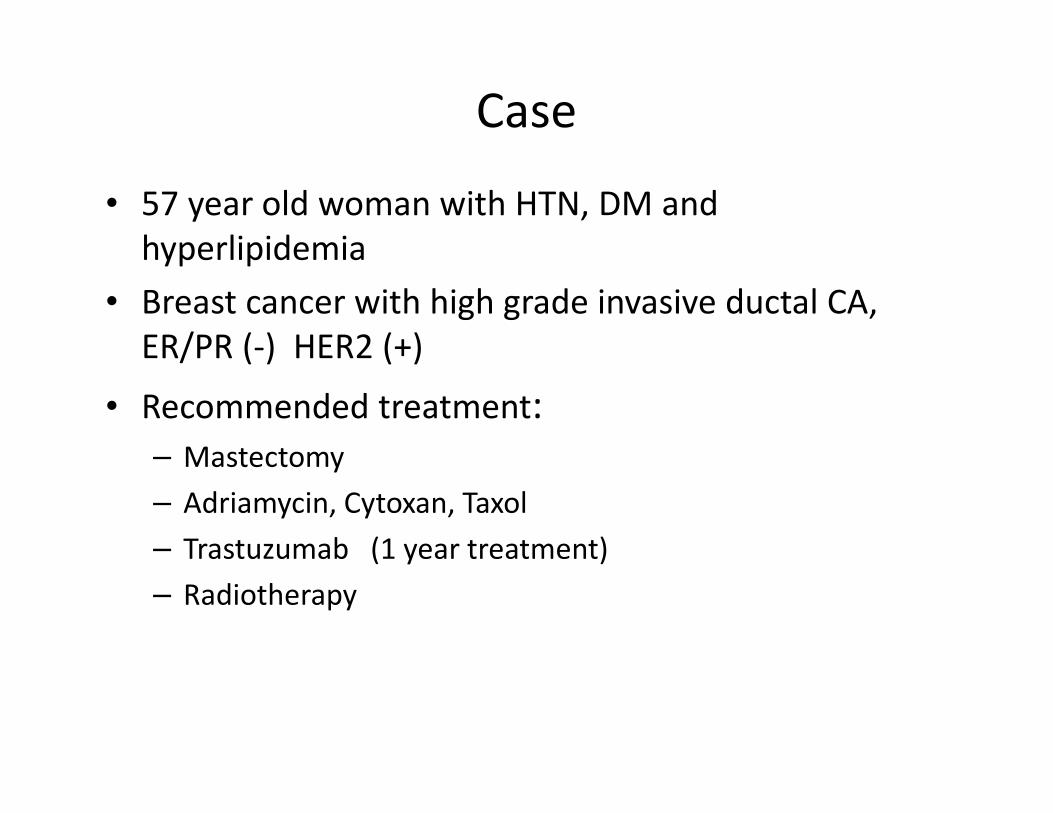
Case
• 57 year old woman with HTN, DM and
hyperlipidemia
• Breast cancer with high grade invasive ductal CA,
ER/PR (-) HER2 (+)
• Recommended treatment:
– Mastectomy
– Adriamycin, Cytoxan, Taxol
– Trastuzumab (1 year treatment)
– Radiotherapy
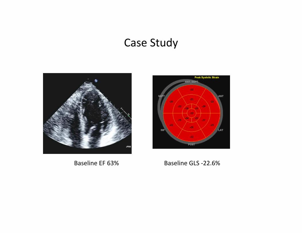
Case Study
Baseline EF 63% Baseline GLS -22.6%
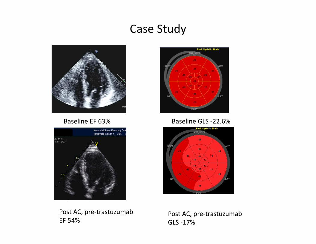
Case Study
Baseline EF 63% Baseline GLS -22.6%
Post AC, pre-trastuzumab
EF 54%Post AC, pre-trastuzumab
GLS -17%
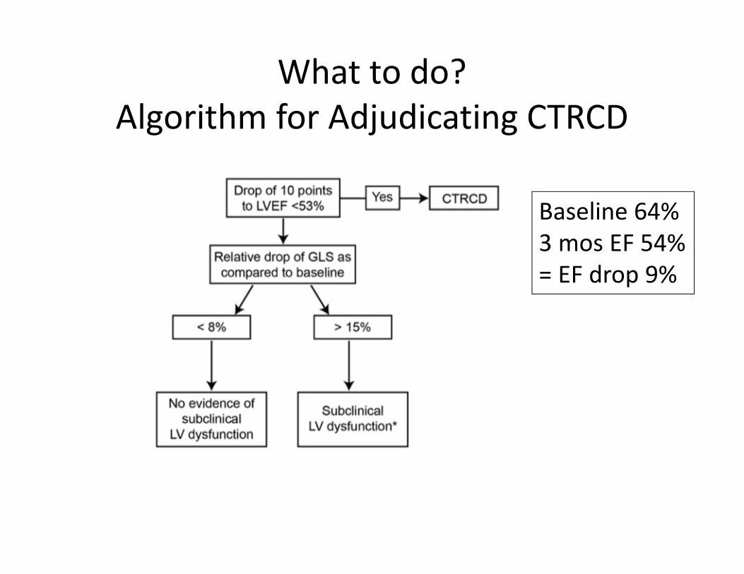
Baseline 64%
3 mos EF 54%
= EF drop 9%
What to do?
Algorithm for Adjudicating CTRCD
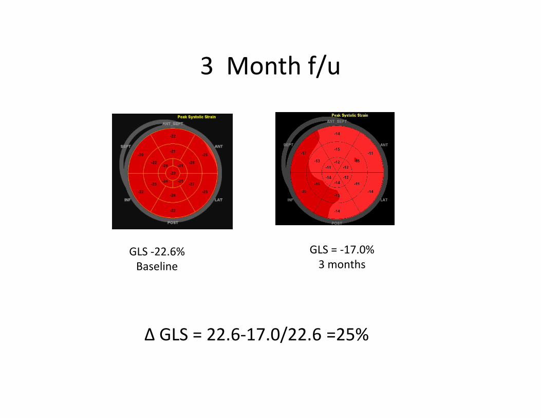
3 Month f/u
GLS -22.6%
Baseline
GLS = -17.0%
3 months
∆ GLS = 22.6-17.0/22.6 =25%
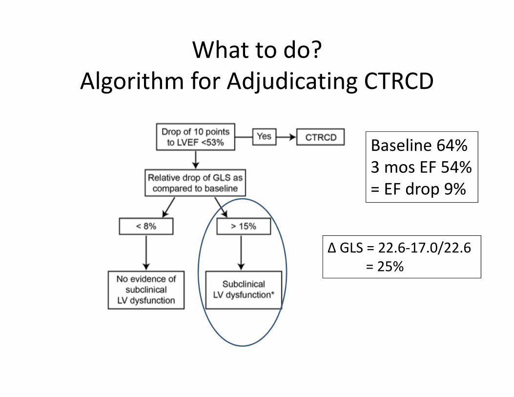
Baseline 64%
3 mos EF 54%
= EF drop 9%
What to do?
Algorithm for Adjudicating CTRCD
∆ GLS = 22.6-17.0/22.6
= 25%

Management of Heart Failure
ACCF/AHA Guideline
Abnl GLS
Troponin??
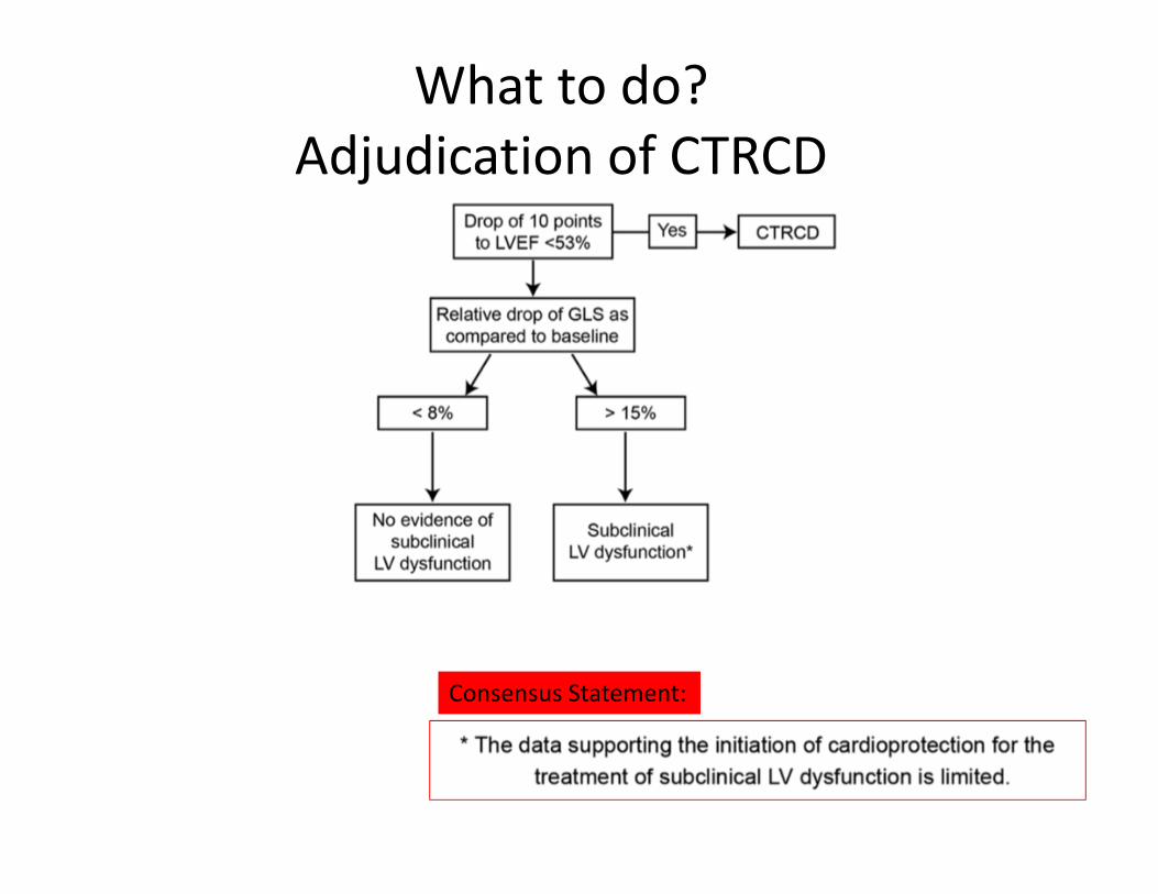
What to do?
Adjudication of CTRCD
Consensus Statement:

Take Home Messages
• Two categories of CTRCD: Type 1 exemplified by doxorubicin and Type 2 exemplified by trastuzumab.
• Cardiotoxicity or CTRCD is defined as a drop in EF >10% from baseline to a value <53%.
• LVEF measurement at baseline, during and after therapy:
– 3D echo preferred or 2D Biplane MOD +/- contrast
– Keep imaging modality consistent in follow-up
• Strategy of early detection of cardiotoxicity with GLS and/or and troponin
• Cardiology consultation recommended for abnormal echo or troponin
• Cooperation between cardiologists and oncologists is absolutely essential.