Embed Size (px)
Citation preview
Ana Teresa Oliveira Rufino
GLUCOSE SENSING AND MODULATION OF HUMAN CHONDROCYTE FUNCTIONS
BY HYPERGLYCEMIA: RELEVANCE AS PHARMACOLOGICAL TARGETS FOR
DIABETES–ASSOCIATED OA
Tese de Doutoramento em Farmácia, Especialidade de Farmacologia e Farmacoterapia, orientada por
Alexandrina Maria Ferreira dos Santos Pinto Mendes e Carlos Manuel Freire Cavaleiro
e apresentada à Faculdade de Farmácia da Universidade de Coimbra
Julho de 2014
GLUCOSE SENSING AND MODULATION OF HUMAN
CHONDROCYTE FUNCTIONS BY HYPERGLYCEMIA:
RELEVANCE AS PHARMACOLOGICAL TARGETS FOR
DIABETES – ASSOCIATED OA
Dissertação apresentada à Faculdade de Farmácia da Universidade de Coimbra
para prestação de Provas de Doutoramento em Farmácia, na Especialidade De
Farmacologia e Farmacoterapia
Este trabalho foi realizado ao abrigo da bolsa de Doutoramento SFRH/BD/47470/2008
concedida pela Fundação para a Ciência e Tecnologia
Ana Teresa Oliveira Rufino
Universidade de Coimbra
2014

AGRADECIMENTOS
Esta tese marca o fim de mais uma etapa… Talvez esta a mais dura por que passe até
agora… Feita de desilusões e frustrações mas muitas mais conquistas e descobertas!
Cada nova tentativa trazia mais esperança que medo… e havia sempre quem
dissesse “Faz parte…”!
Dessas pessoas há muitas que estiveram ao meu lado, me apoiaram sempre e que
por isso merecem o meu reconhecimento e agradecimento.
Em primeiro lugar queria agradecer aos meus orientadores Dra. Alexandrina Mendes e
Dr. Carlos Cavaleiro.
Alexandrina, foi um prazer trabalhar consigo, obrigada por ter acreditado em mim, por
me ter ajudado e motivado. Por ter sempre mais uma sugestão e um conselho, por
encontrar sempre um caminho alternativo quando parecia que não existia mais
nenhum. Obrigada pelo empenho e minúcia com que encarou cada assunto deste
trabalho e também agora na elaboração desta tese. Obrigada por ter sido mais que
uma orientadora e se ter tornado também uma amiga.
Dr. Cavaleiro, obrigada também pela sua disponibilidade e por ter aceite este desafio a
meio do percurso e ainda assim, com todas as suas solicitações acreditar e empenhar-
se nele.
Quero também agradecer ao Professor Doutor Fernando Judas pela sua
disponibilidade e colaboração, crucial para o desenvolvimento deste trabalho bem
como a todos os funcionários, médicos e enfermeiros do Banco de Ossos pela
simpatia e ajuda durante as colheitas de cartilagem.
Aos meus colegas de laboratório,
à Joana, à Ana, ao João, à Isabel, à Madalena, à Cátia, à Mónica, à Dra. Teresa Cruz,
ao Bruno, à Su, à Verinha e ao João Demétrio. Vocês são o máximo e sinto que juntos
temos aquilo a que podemos chamar um verdadeiro espírito de grupo. Obrigada por
todas as conversas, por todas as gargalhadas, por todos os desabafos e por todas as
vezes que precisei da vossa ajuda e vocês estavam prontos a ajudar. Obrigada por me
aturarem as neuras e por partilharem as vossas! Obrigada por serem verdadeiros
amigos!
À minha Família,
em primeiro aos meus Pais, Obrigada por TUDO! Sem vocês, sem o vosso empenho,
sem o vosso apoio incondicional, sem a vossa ajuda e sem o vosso amor, nada disto
seria possível. Todos os agradecimentos serão sempre poucos! Obrigada por todas as
vezes que me deram coragem, que me secaram as lágrimas, que partilharam sorrisos,
que relevaram as minhas más disposições e que toleraram a minha falta de tempo.
Obrigada por pensarem sempre em mim antes de pensarem em vocês. Eu serei
sempre a vossa menina!
Ao meu irmão, obrigada Manito! Por estares sempre por perto, por teres orgulho e por
acreditares em mim. Por torceres por mim sempre. Por continuares a ser sempre o
meu mano protector e eu a tua “garota”.
Por último, mas nunca menos importante, ao André!
Obrigada por partilhares cada dia comigo! Obrigada por cada palavra de força, de
incentivo para superar cada etapa e por acreditares em mim mais do que eu própria.
Por me “abanares” e me fazeres reagir mesmo sabendo que não é essa a minha
natureza. Obrigada pela paciência que insistes em dizer que não tens. Obrigada por
me fazeres sentir especial e me fazeres sorrir, todos os dias. Obrigada pelo amor e
pelo carinho. Obrigada por te orgulhares de mim. Obrigada por cada abraço, longe,
mas sempre perto. Obrigada por me compreenderes e por me fazeres uma pessoa
melhor, e contigo, muito mais forte.
Obrigada!
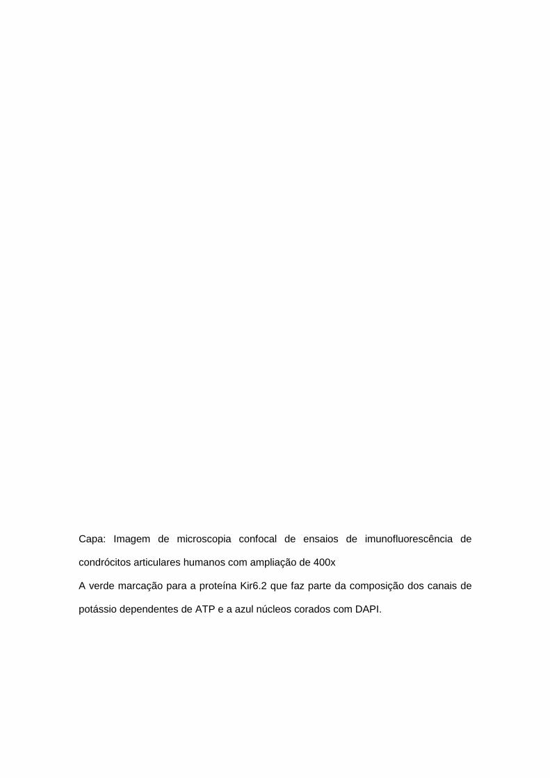
Capa: Imagem de microscopia confocal de ensaios de imunofluorescência de
condrócitos articulares humanos com ampliação de 400x
A verde marcação para a proteína Kir6.2 que faz parte da composição dos canais de
potássio dependentes de ATP e a azul núcleos corados com DAPI.

1
INDEX
ABBREVIATIONS ...................................................................................................................... 7
ABSTRACT ............................................................................................................................... 11
RESUMO ................................................................................................................................... 13
CHAPTER 1. ............................................................................................................................. 17
INTRODUCTION ...................................................................................................................... 17
1.1 SYNOVIAL JOINTS AND ARTICULAR CARTILAGE ...................................... 19
1.1.1 Structure and Composition of Articular Cartilage ................................. 20
1.1.1.1 Collagens and Proteoglycans ................................................................. 21
1.1.1.2 Chondrocyte ................................................................................................ 24
1.2 OSTEOARTHRITIS RISK FACTORS AND SPECIFIC PHENOTYPES ......... 26
1.2.1 Diabetes Mellitus and Osteoarthritis: Role of Extracellular glucose
concentrations ................................................................................................................ 27
1.2.1.1 Glucose sensing and transport .............................................................. 30
1.3 CARTILAGE CHANGES IN OSTEOARTHRITIS ............................................... 32
1.3.1 Regulation of Catabolic and Anabolic Pathways ................................... 34
1.3.1.1 Matrix Metalloproteinases and Aggrecanases ................................... 34
1.3.2 Inflammatory Process in OA ....................................................................... 36
1.3.2.1 Regulation and Function of Inflammatory Mediators in
Osteoarthritis ................................................................................................................... 37
1.3.2.1.1 Cytokines ...................................................................................................... 37
1.3.2.1.2 Nitric Oxide .................................................................................................. 40
1.3.2.1.3 Nuclear Factor – κB (NF-κB) .................................................................... 41
1.3.2.1.4 Mitogen Activated Protein Kinases (MAPK) ........................................ 44
1.4 OA TREATMENT ..................................................................................................... 46
1.4.1 Currently Available Treatment .................................................................... 47
1.4.2 Disease Modifying OA Treatment .............................................................. 51
1.5 OBJECTIVES AND ORGANIZATION OF THE THESIS ................................... 54
CHAPTER 2. ............................................................................................................................. 57
MATERIAL AND METHODS ................................................................................................. 57
2.1 Cartilage Samples .................................................................................................. 57
2.2 Chondrocyte Isolation and Culture ................................................................... 57
2.3 Assessment of Cell Viability ................................................................................ 58
2.4 Western Blot ............................................................................................................ 58
2.5 Immunohistochemistry ......................................................................................... 59
INDEX
2
2.6 Total RNA Extraction and Quantitative Real-Time PCR (qRT-PCR) .......... 59
2.7 2-Deoxy-D-Glucose uptake Assay ..................................................................... 62
2.8 Nitric Oxide Production ........................................................................................ 62
2.9 NF-κB Transcription Factor Assay .................................................................... 62
2.10 Essential oil identification, Fractionation and Analysis .......................... 63
2.11 Statistical Analysis ............................................................................................ 64
CHAPTER 3. ............................................................................................................................. 67
EXPRESSION AND FUNCTION OF K(ATP) CHANNELS IN NORMAL AND
OSTEOARTHRITIC HUMAN CHONDROCYTES: POSSIBLE ROLE IN GLUCOSE
SENSING ................................................................................................................................... 67
ABSTRACT ............................................................................................................................... 68
3.1 INTRODUCTION ...................................................................................................... 69
3.2 RESULTS .................................................................................................................. 72
3.2.1 Characterization of the subunit composition of (KATP) channels
expression in human chondrocytes .......................................................................... 72
3.2.2 Immunofluorescence staining of K(ATP) channels expressed in
human chondrocytes .................................................................................................... 74
3.2.3 Role of exposure to high glucose on K(ATP) channel subunit protein
and mRNA expression .................................................................................................. 76
3.2.4 Regulation of GLUT-1 and GLUT-3 protein levels by the K(ATP)
channel blocker Glibenclamide .................................................................................. 76
3.2.5 Role of Glibenclamide in regulating GLUT-1 and GLUT-3 mRNA
levels in OA grade 0-1 chondrocytes ........................................................................ 79
3.2.6 Role of glibenclamide in regulating cellular glucose transport ......... 79
3.3 DISCUSSION ............................................................................................................ 80
CHAPTER 4. ............................................................................................................................. 85
ROLE OF HIGH GLUCOSE AS MODULATOR OF INFLAMMATION IN HUMAN
CHONDROCYTES ................................................................................................................... 85
ABSTRACT ............................................................................................................................... 86
4.1 INTRODUCTION ...................................................................................................... 87
4.2 RESULTS .................................................................................................................. 88
4.2.1 Induction of pro-inflammatory cytokines, iNOS expression and NO
production by high glucose ........................................................................................ 88
4.2.2 High glucose induces NF-κB p65 translocation to the nucleus ........ 88
4.2.3 High Insulin modulates NF-κB translocation to the nucleus .............. 89
4.3 DISCUSSION ............................................................................................................ 91
CHAPTER 5. ............................................................................................................................. 93
INDEX
3
DIFFERENTIAL EFFECTS OF LAVANDULA LUISIERI AND ERYNGIUM DURAEI
SUBSP JURESIANUM ESSENTIAL OILS IN CELL MODELS OF TWO CHRONIC
INFLAMMATORY DISEASES ............................................................................................... 93
ABSTRACT ............................................................................................................................... 94
5.1 INTRODUCTION ...................................................................................................... 95
5.2 RESULTS .................................................................................................................. 97
5.2.1 Composition of the essential oils .............................................................. 97
5.2.2 Evaluation of cytotoxicity and selection of non cytotoxic
concentrations of the essential oils .......................................................................... 99
5.2.3 Effect of the EO’s on cytokine induced iNOS expression and NO
production ...................................................................................................................... 100
5.2.4 Effect of the EO’s of Lavandula luisieri and Eryngium duriaei subsp.
juresianum on NF-κB activation ............................................................................... 103
5.3 DISCUSSION .......................................................................................................... 107
CHAPTER 6. ........................................................................................................................... 111
ANTI-INFLAMMATORY AND CHONDROPROTECTIVE ACTIVITY OF (+)-Α-PINENE:
STRUCTURAL AND ENANTIOMERIC SELECTIVITY ................................................... 111
ABSTRACT ............................................................................................................................. 112
6.1 INTRODUCTION .................................................................................................... 113
6.2 RESULTS AND DISCUSSION............................................................................. 115
CHAPTER 7. ........................................................................................................................... 123
EVALUATION OF THE ANTI-INFLAMMATORY, ANTI-CATABOLIC AND PRO-
ANABOLIC EFFECTS OF E-CARYOPHYLLENE, MYRCENE AND LIMONENE IN A
CELL MODEL OF OSTEOARTHRITIS .............................................................................. 123
ABSTRACT ............................................................................................................................. 124
7.1 INTRODUCTION .................................................................................................... 125
7.2 RESULTS AND DISCUSSION............................................................................. 127
CHAPTER 8. ........................................................................................................................... 139
DISCUSSION AND CONCLUSION .................................................................................... 139
8.1 GENERAL DISCUSSION ..................................................................................... 139
CHAPTER 9. ........................................................................................................................... 145
REFERENCES ....................................................................................................................... 145
INDEX
4
FIGURES AND TABLES INDEX
Figure 1.1 Graphical representation of the major structures of a synovial joint ........... 20
Figure 1.2 Schematic diagram of collagen type II fiber structure ................................. 22
Figure 1.3 Schematic representation of proteoglycans linked to Hyaluronic acid. ....... 23
Figure 1.4 Schematic representation of the different cartilage zones and chondrocytes
distribution .................................................................................................................... 25
Figure 1.5 Schematic representation of the cytokine role on the OA pathogenesis .... 39
Figure 1.6 Schematic representation of the canonical NF-κB activation in response to
pro-inflammatory cytokines ........................................................................................... 42
Figure 1.7 Schematic representation of the MAPK activation in response to several
stimulus ......................................................................................................................... 45
Figure 1.8 Current treatment options for osteoarthritis ................................................ 50
Table 2.1 Antibodies used for western blot and immunohistochemistry analysis ........ 59
Table 2.2 Oligonucleotide Primer Pairs Used for qRT-PCR ......................................... 61
Figure 3.1 Protein and mRNA levels of K(ATP) channel subunits expressed in human
chondrocytes ................................................................................................................ 73
Figure 3.2 In situ immunofluorescence staining of K(ATP) channel subunits in normal
(OA grades 0–1) human cartilage ................................................................................. 75
Table 3.1 Basal protein levels of GLUTs-1 and -3 in human chondrocytes according to
OA grade ...................................................................................................................... 77
Figure 3.3 Regulation of total GLUT‐1 and GLUT‐3 protein content by glibenclamide in
chondrocytes from different OA grades ........................................................................ 78
Table 3.2 Relative 2-DG uptake (% of untreated cells) ................................................ 79
INDEX
5
Figure 4.1 Role of High glucose in modulating cytokines, iNOS expression and NO
production ..................................................................................................................... 88
Figure 4.2 Role of glucose and insulin concentrations on nuclear NF-κB p65 levels. . 89
Figure 5.1 Classes of compounds and major constituintes of the essential oils .......... 98
Figure 5.1 Assessement of cell viability in human chondrocytes and C2BBe1 intestinal
cell line ........................................................................................................................ 100
Figure 5.2 Effect of the EOs on iNOS protein expression in human chondrocytes ... 101
Figure 5.3 Effect of the EOs on iNOS protein expression in C2BBe1 cells ............... 102
Figure 5.4 Effect of the EOs on IL-1β- induced NO production in human
chondrocytes .............................................................................................................. 103
Figure 5.5 Effect of the EOs of L. luisieri and E. duriaei subsp. juresianum on IL1β-
induced IκB-α phosphorylation and degradation in chondrocytes .............................. 104
Figure 5.6 Effect of the EO of L. luisieri on CytMix-induced IκB-α phosphorylation and
degradation in intestinal C2BBe1 cells ....................................................................... 105
Figure 6.0 Graphical Abstract “Anti-Inflammatory and Chondroprotective Activity Of
(+)-α–Pinene: Structural and Enantiomeric Selectivity” .............................................. 111
Supporting Information Figure 6. Chondrocyte viability in human chondrocytes ... 115
Figure 6.1 Effect of α-pinene, β- pinene and other pinane derivatives on IL-1β induced
NO production and effect of the active test compounds on iNOS protei and mRNA
expression levels in primary human chondrocytes ..................................................... 116
Figure 6.2 Effect of α-pinene enatiomers on IL-1β induced NF-κB activation ........... 118
Figure 6.3 Effect of α-pinene enatiomers on IL-1β induced JNK and p38 MAPK
phosphorylation .......................................................................................................... 119
Figure 6.4 Effect of (+)-α-pinene on IL-1β induced changes in gene expression ...... 121
INDEX
6
Figure 7.0 Graphical Abstract “Evaluation of the Anti-inflammatory, Anti-catabolic and
Pro-anabolic Effects of E- caryophyllene, Myrcene and Limonene in a cell model of
Osteoarthritis” ............................................................................................................. 123
Supporting Information Figure 7. Effect of the fractions isolated from the essential
oils of Eryngium duriaei subsp. juresianum and Lavandula luisieri and of the isolated
compounds E-Caryophyllene, myrcene and limonene on human
chondrocyte viability ................................................................................................... 128
Table 7.1 Composition of Eryngium duriaei subsp. juresianum and Lavandula luisieri
Essential oils fractions ................................................................................................ 129
Figure 7.1 Effect of the fractions isolated from the essential oils of Eryngium duriaei
subsp. juresianum and Lavandula luisieri and of the isolated compounds E-
Caryophyllene, myrcene and limonene on IL-1β- induced NO production ................ 130
Figure 7.2 Effect of myrcene and limonene on IL-1β- induced iNOS protein and mRNA
levels in human chondrocytes ................................................................................... 132
Figure 7.3 Effect of the myrcene and limonene on IL-1β- induced NF-κB activation 133
Figure 7.4 Effect of the myrcene and limonene on IL-1β- induced activation of JNK p38
and ERK1/2 in human chondrocytes .......................................................................... 134
Figure 7.5 Effect of the myrcene and limonene on IL-1β- induced changes in the
expression of catabolic and anti-catabolic genes ....................................................... 136
Figure 7.6 Effect of the myrcene and limonene on IL-1β- induced changes in the
expression of extracellular matrix genes .................................................................... 137
ABBREVIATIONS
7
ABBREVIATIONS
2-DG: 2-Deoxy-D-Glucose
ADAMTS: A Desintegrin and Metalloproteinase with TromboSpondin Motif
ADAM: A Desintegrin and Metalloproteinase
ADP: Adenosine diphosphate
AGEs: Advanced Glycation End Products
AP-1: Activation Protein-1
ATF-2: Activating Transcription Factor 2
ATP: Adenosine triphosphate
Bay: Bay-11-7082
BMP-2: Bone Morphogenic Protein 2
BSA: Bovine Serum Albumin
COMP: Cartilage Oligomeric Matrix Protein
CS: Chondroitin Sulphate
DAPI: 4',6-diamidino-2-phenylindole
DM: Diabetes Mellitus
DMEM: Dulbecco’s Modified Eagle Medium
DMOAD: Disease Modifying Osteoarthritis Drugs
DMSO: Dimethilsulphoxide
ECM: Extracellular Matrix
ELF3: E74-Like Factor
ELK-1: ETS domain-containing protein
EMA: European Medicine Agency
eNOS: Endothelial form of Nitric Oxide Synthase
EO: Essential oil
ERK: Extracellular regulated Kinase
EULAR: European League Against Rheumatism
FBS: Fetal Bovine Serum
FDA: Food and Drug Administration
GAG: Glycosaminoglycans
GlcN: Glucosamine
GLUT: Glucose transporter
HA: Hyaluronic Acid
HIF-2: Hypoxia- inducible Factor 2
ICE: Interleukin-1 Coverting Enzyme (caspase-1)
ABBREVIATIONS
8
IGF-1: Insulin Growth factor-1
IKK: IκB Kinase
IL: Interleukin
IL-1β: Interleukin-1β
iNOS: Inducible form of Nitric Oxide Synthase
IκBα: nuclear factor kappa B inhibitor, alpha
JNK: c-Jun NH2-terminal Kinase
K (ATP) channel: ATP-sensitive potassium channel
LPS: Lipopolissacaride
MAPK: Mitogen Activated Protein Kinase
MEF-2: Myocyte Enhancer Factor-2
MKK: Mitogen Activated Protein Kinase Kinase
MMPs: Matrix Metalloproteinases
MTT: 3-(4,5-dimethylthiazol-2-yl)-2,5-diphenyltetrazolium bromide
NF-κB: Nuclear Factor κB
NLS: Nuclear Localization Sequence
nNOS: Neuronal form of Nitric Oxide Synthase
NO: Nitric Oxide
NSAIDS: Non- Steroidal Anti-inflammatory drugs
OA: Osteoarthritis
OARSI: Osteoarthritis Research Society International
OCT: Optimal Cutting Temperature compound
PAGE: Poliacrilamide gel Electrophoresis
PBS: Phosphate Buffered Saline
PGE2: Prostaglandin E2
PI9: Protease Inhibitor 9
PRAC: Pharmacovigilance Risk Assessment Committee
PVDF: Polyvinylidene fluoride
RA: Rheumatoid Arthritis
ROS: Reactive Oxygen Species
RUNX2: Runt-related Transcription Factor 2
SASP: Senescence Associated Secretory Phenotype
SDS: Sodium Dodecilsulphate
Ser: Serine
SYSADOA: Symptomatic Slow Acting Drugs in Osteoarthritis

ABBREVIATIONS
9
TBS-T: Tris Buffered Saline Tween
Thr: Threonine
TIMPs: Tissue Inhibitor of Metalloproteinases
TNF-α: Tumor Necrosis Factor α
Tyr: Tyrosine
VGCC: Voltage-gated Calcium Channel
WHO: World Health organization

11
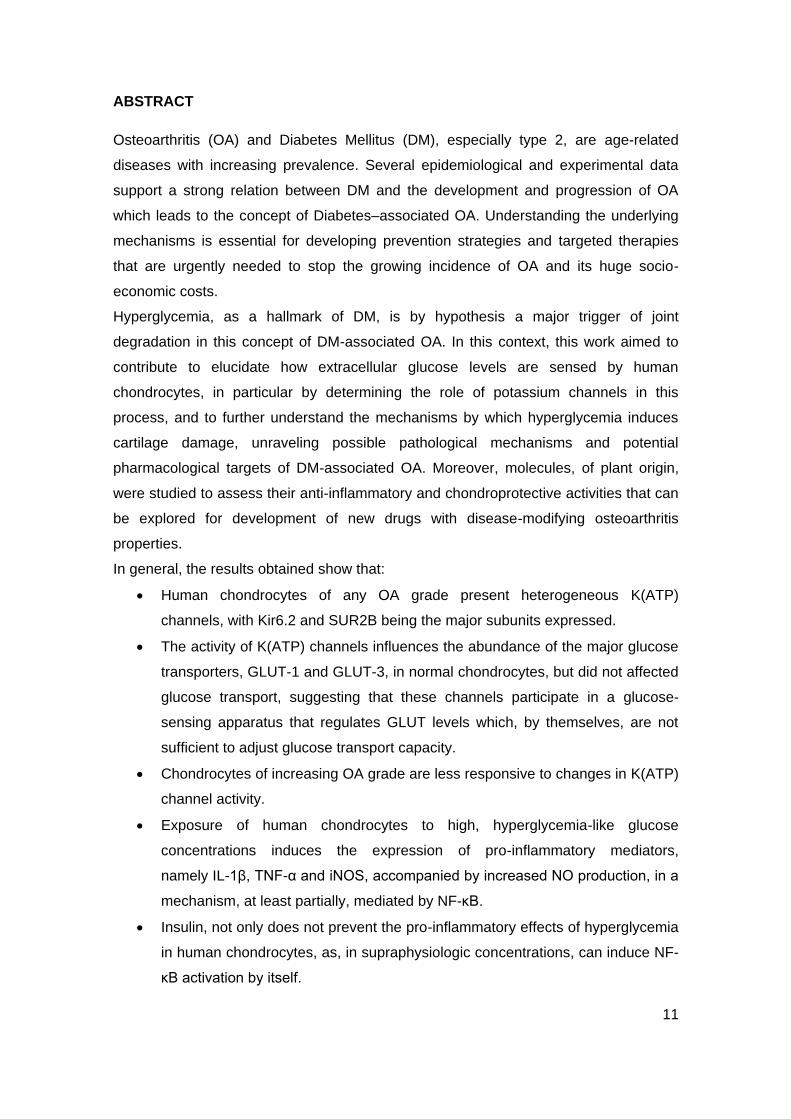
ABSTRACT
Osteoarthritis (OA) and Diabetes Mellitus (DM), especially type 2, are age-related
diseases with increasing prevalence. Several epidemiological and experimental data
support a strong relation between DM and the development and progression of OA
which leads to the concept of Diabetes–associated OA. Understanding the underlying
mechanisms is essential for developing prevention strategies and targeted therapies
that are urgently needed to stop the growing incidence of OA and its huge socio-
economic costs.
Hyperglycemia, as a hallmark of DM, is by hypothesis a major trigger of joint
degradation in this concept of DM-associated OA. In this context, this work aimed to
contribute to elucidate how extracellular glucose levels are sensed by human
chondrocytes, in particular by determining the role of potassium channels in this
process, and to further understand the mechanisms by which hyperglycemia induces
cartilage damage, unraveling possible pathological mechanisms and potential
pharmacological targets of DM-associated OA. Moreover, molecules, of plant origin,
were studied to assess their anti-inflammatory and chondroprotective activities that can
be explored for development of new drugs with disease-modifying osteoarthritis
properties.
In general, the results obtained show that:
Human chondrocytes of any OA grade present heterogeneous K(ATP)
channels, with Kir6.2 and SUR2B being the major subunits expressed.
The activity of K(ATP) channels influences the abundance of the major glucose
transporters, GLUT-1 and GLUT-3, in normal chondrocytes, but did not affected
glucose transport, suggesting that these channels participate in a glucose-
sensing apparatus that regulates GLUT levels which, by themselves, are not
sufficient to adjust glucose transport capacity.
Chondrocytes of increasing OA grade are less responsive to changes in K(ATP)
channel activity.
Exposure of human chondrocytes to high, hyperglycemia-like glucose
concentrations induces the expression of pro-inflammatory mediators,
namely IL-1β, TNF-α and iNOS, accompanied by increased NO production, in a
mechanism, at least partially, mediated by NF-κB.
Insulin, not only does not prevent the pro-inflammatory effects of hyperglycemia
in human chondrocytes, as, in supraphysiologic concentrations, can induce NF-
κB activation by itself.
ABSTRACT
12
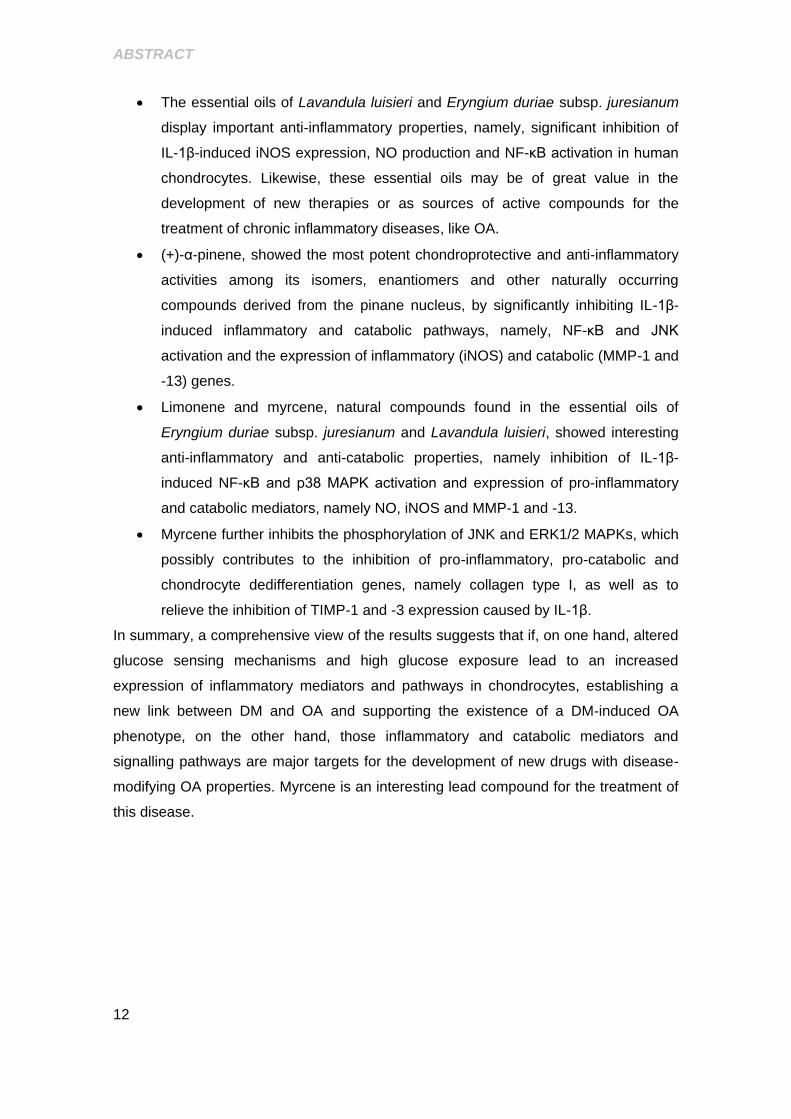
The essential oils of Lavandula luisieri and Eryngium duriae subsp. juresianum
display important anti-inflammatory properties, namely, significant inhibition of
IL-1β-induced iNOS expression, NO production and NF-κB activation in human
chondrocytes. Likewise, these essential oils may be of great value in the
development of new therapies or as sources of active compounds for the
treatment of chronic inflammatory diseases, like OA.
(+)-α-pinene, showed the most potent chondroprotective and anti-inflammatory
activities among its isomers, enantiomers and other naturally occurring
compounds derived from the pinane nucleus, by significantly inhibiting IL-1β-
induced inflammatory and catabolic pathways, namely, NF-κB and JNK
activation and the expression of inflammatory (iNOS) and catabolic (MMP-1 and
-13) genes.
Limonene and myrcene, natural compounds found in the essential oils of
Eryngium duriae subsp. juresianum and Lavandula luisieri, showed interesting
anti-inflammatory and anti-catabolic properties, namely inhibition of IL-1β-
induced NF-κB and p38 MAPK activation and expression of pro-inflammatory
and catabolic mediators, namely NO, iNOS and MMP-1 and -13.
Myrcene further inhibits the phosphorylation of JNK and ERK1/2 MAPKs, which
possibly contributes to the inhibition of pro-inflammatory, pro-catabolic and
chondrocyte dedifferentiation genes, namely collagen type I, as well as to
relieve the inhibition of TIMP-1 and -3 expression caused by IL-1β.
In summary, a comprehensive view of the results suggests that if, on one hand, altered
glucose sensing mechanisms and high glucose exposure lead to an increased
expression of inflammatory mediators and pathways in chondrocytes, establishing a
new link between DM and OA and supporting the existence of a DM-induced OA
phenotype, on the other hand, those inflammatory and catabolic mediators and
signalling pathways are major targets for the development of new drugs with disease-
modifying OA properties. Myrcene is an interesting lead compound for the treatment of
this disease.
RESUMO
13
RESUMO
A Osteoartrite (OA) e a Diabetes Mellitus (DM), especialmente tipo 2, são doenças
relacionadas com o envelhecimento e, por isso, cada vez mais prevalentes. Estudos
epidemiológicos e experimentais suportam a forte relação entre a DM e o
desenvolvimento e progressão da OA, conduzindo ao aparecimento do conceito de OA
associada à diabetes. A compreensão dos mecanismos subjacentes é essencial para
o desenvolvimento de estratégias preventivas e terapêuticas específicas que são
urgentemente necessárias para travar a crescente incidência da OA e os seus
enormes custos socioeconómicos.
A hiperglicémia, como característica fundamental da DM, é por hipótese um importante
promotor da degradação da articulação, neste conceito de OA associada à diabetes.
Neste contexto, o principal objectivo deste trabalho foi contribuir para o esclarecimento
dos mecanismos pelos quais os níveis extracelulares de glucose são detectados pelos
condrócitos, em particular a clarificação do papel dos canais de potássio dependentes
de ATP [K(ATP)] neste processo e compreender os mecanismos através dos quais a
hiperglicemia induz dano nos condrócitos. Compreender estes processos é essencial
para evidenciar possíveis mecanismos patológicos e potenciais alvos farmacológicos
para a OA associada à DM. Adicionalmente procurou-se identificar compostos com
propriedades anti-inflamatórias e anti-catabólicas que justifiquem a sua exploração
para o desenvolvimento de novos fármacos anti-osteoartríticos. Como fontes dos
compostos a estudar, utilizaram-se extractos de plantas, a maioria constituintes da
flora endémica da Península Ibérica, de modo a contribuir também para a valorização
do nosso património biológico.
Em geral, os resultados obtidos mostram que:
Condrócitos humanos de todos os graus de OA apresentam canais de potássio
ATP dependentes [K(ATP)] heterogéneos, com múltiplas combinações de
subunidades possíveis, mas parecendo a Kir6.2 e a SUR2B as subunidades
mais expressas.
A actividade dos canais K(ATP) influencia a abundância dos principais
transportadores de glucose, GLUT-1 e -3, nos condrócitos normais, mas não
afecta o transporte de glucose. Isto sugere que estes canais participam num
mecanismo sensor de glucose que regula os níveis dos GLUT, mas a
modulação da sua actividade isoladamente não é suficiente para ajustar a
capacidade de transporte de glucose em condrócitos.
RESUMO
14
Os condrócitos de graus de OA mais elevados, isto é, em que a degradação da
cartilagem é progressivamente mais extensa, respondem menos eficazmente
ou não respondem a alterações na actividade dos canais K(ATP).
A exposição dos condrócitos a concentrações elevadas de glucose induz a
expressão de mediadores pro-inflamatórios, nomeadamente a IL-1β, o TNF-α e
a iNOS, acompanhada pelo aumento da produção de NO, através de um
mecanismo, pelo menos parcialmente, mediado pelo NF-κB.
A insulina não só não previne os efeitos pro-inflamatórios da hiperglicemia nos
condrócitos humanos mas ainda, em concentrações supra-fisiológicas, induz
por si só a activação do NF-κB.
Os óleos essenciais de Lavandula luisieri e Eryngium duriaei subsp. juresianum
apresentam importantes actividades anti-inflamatórias, nomeadamente a
inibição significativa da expressão da iNOS, da produção de NO e da activação
do NF-κB induzidos pela IL-1β. Assim, estes óleos podem ser de grande valor
no desenvolvimento de novas terapias ou como fonte de compostos activos
para o tratamento da OA
O (+)-α-pinene apresenta o maior potencial condroprotector e anti-inflamatório
de entre os seus isómeros, enantiomeros e outros compostos naturais de
núcleo pinano por inibir significativamente a activação de vias de sinalização
inflamatórias e catabólicas induzidas pela IL-1β, nomeadamente a activação do
NF-κB e da JNK e a expressão de genes inflamatórios (iNOS) e catabólicos
(MMP-1 e -13).
O mirceno e o limoneno, compostos naturais encontrados nos óleos essenciais
de Eryngium duriaei subsp. juresianum e Lavandula luisieri, mostraram
interessantes propriedades anti-inflamatórias e anti-catabólicas,
nomeadamente a inibição da expressão de mediadores pro-inflamatórios e
catabólicos como o NO, iNOS, MMP-1 e -13 e da activação de vias de
sinalização como a do NF-κB e a da p38MAPK.
Adicionalmente, o mirceno ainda inibe a fosforilação das MAPK, JNK e
ERK1/2, o que possivelmente contribui para a inibição da expressão de genes
pro-inflamatórios, pro-catabólicos e de desdiferenciação do condrócito,
nomeadamente o colagénio I, e atenua a inibição da expressão dos genes anti-
catabólicos TIMP-1 e -3 causadas pela IL-1β.
Em resumo, uma visão geral dos resultados sugere por um lado, que a alteração dos
mecanismos de sensibilidade à glucose e a exposição a concentrações de glucose
elevada conduzem a um aumento da expressão de mediadores e vias de sinalização
RESUMO
15
inflamatórias nos condrócitos, estabelecendo uma nova ligação entre a OA e a DM e
suportando a existência de um fenótipo específico de OA induzida pela DM. Por outro
lado, estes mediadores e vias de sinalização inflamatórios e catabólicos são alvos
importantes para o desenvolvimento de novos fármacos com capacidade de modificar
o curso da doença. O mirceno aparece como um composto lead interessante a ser
explorado para o desenvolvimento de novas terapêuticas para a OA

17
CHAPTER 1.
INTRODUCTION
Rheumatic and musculoskeletal conditions comprise over 150 diseases and
syndromes which are usually progressive and associated with pain. The World Health
Organization (WHO) classifies them as conditions that mostly affect joints, bones,
cartilage, tendons, ligaments and muscles yielding a reduction of the range of motion in
one or more areas of the musculoskeletal system. Included in this group are joint
diseases such as osteoarthritis (OA) and rheumatoid arthritis (RA); back and neck pain;
osteoporosis and fragility fractures; soft tissue rheumatologic diseases as bursitis and
tendinitis; injuries due to trauma (sports, accidents) and autoimmune diseases as
Lupus erythematosus. Rheumatic diseases associated with age, like OA, are leading
causes of morbidity and disability, yielding a great socio-economic burden whose
prevalence continues to increase. Recent epidemiologic data [Arden and Nevitt, 2006;
Neogi and Zhang, 2013; Zhang and Jordan, 2010] indicate that OA is the most
common joint disorder in the world. According to the WHO, by 2050 people aged over
60 will account for more than 20% of the world’s population. Of these 20%, it is
estimated that about 15% will have symptomatic OA, and one-third of these people will
be severely disabled. This means that by 2050, 130 million people will suffer from OA
worldwide, of whom 40 million will be severely disabled by the disease [Goldring, 2006;
Heidari, 2011; Kaplan et al., 2013].
The diagnosis and definition of OA are complicated; especially due to the discrepancy
between the symptomatic and the radiographic evidences of the disease which are the
major factors considered for diagnosis. For instance, 50% of subjects in the general
population with radiographic evidence of knee OA do not have pain and 50% of
subjects who complain of knee pain, and who are at or above the age when
osteoarthritis starts to become common(about 55 years), have no definite radiographic
evidence of OA [Heidari, 2011; Neogi and Zhang, 2013].
In 1986, the American College of Rheumatology defined OA as “a heterogeneous
group of conditions that lead to joint symptoms and signs, which are associated with
defective integrity of articular cartilage, in addition to related changes in the underlying
bone at the joint margins” [Altman et al., 1986].
CHAPTER 1
18
Since then, there was consensus that OA is usually a progressive disease affecting the
whole synovial joint [Lane et al., 2011] and generally it can be characterized by a local
chronic inflammation, pain, cartilage destruction and loss of joint function.
Traditionally, OA was thought to be a normal consequence of mechanical use and
ageing. With age it is unequivocal the accumulation of changes in the joint tissue
composition and structure, reason why it is considered a primary risk factor for OA.
Despite its importance, there are also elderly people that do not develop OA, so the
recent paradigm for disease definition considers that, more than just an “age-related”
and “wear and tear” disease, OA results from an interplay of multiple factors, including
trauma, genetic pre-disposition, local inflammation, congenital malalignement of joints
and cellular and biochemical insults resulting from metabolic conditions [Goldring and
Goldring, 2007; Lane et al., 2011].
Generally, OA is subdivided by aetiology into either, idiopathic, if no identifiable cause
exists for development of the disease, which corresponds to the majority of the cases
and does not affect people under 40 years old, or secondary, when it is caused or
associated with an identifiable underlying condition that promotes joint vulnerability and
development of OA lesions at younger age. In children or younger adults, trauma or
joint insults resulting from sports practice or accidents as well as congenital
malalignement of joints and consequent biomechanical instability are strongly
implicated in the initiation of OA. Also hereditary predisposition, and metabolic and
inflammatory disorders, as obesity and gout, can result in an earlier onset of the
disease and be considered causes of secondary OA [Buckwalter and Martin, 2006;
Goldring, 2006].
It is likely however, that for idiopathic OA, several risk factors contribute to the initiation
and progression of the disease, including ageing, hereditary predisposition, mechanical
factors (joint malalignement and overuse, trauma, etc.), gender, obesity and other
metabolic factors as malnutrition and Diabetes Mellitus [Buckwalter and Martin, 2006;
Goldring and Goldring, 2007; Katz et al., 2010].
Even though the clinical manifestations and symptoms [Goldring and Goldring, 2007]
are common to the multiple OA aetiologies, it is reasonable that each risk factor
influences the joint in a particular way and increases the susceptibility to the disease
and its progression through a variety of mechanisms [Felson, 2010; Kerkhof et al.,
2010]. This complexity and variability of OA aetiologies suggests the need of identifying
risk factors, establishing distinct phenotypes and understanding in each way they
contribute to the initiation and progression of the disease. Uncovering the molecular or
biomechanical processes affected in the joint and the mechanisms underlying the
CHAPTER 1
19
primary trigger are critical for the development of new treatments and new preventing
strategies for OA [Berenbaum, 2008; Felson, 2010; Kerkhof et al., 2010; Wenham and
Conaghan, 2013].
The factors that initiate OA likely vary depending on the joint site [Lane et al., 2011].
They can affect a single joint, a few joints or be generalized. The most commonly
affected joints are the knees, hips, spine and hands [Goldring and Marcu, 2009].
Although most tissues of the joint, including subchondral bone, synovial membrane,
joint capsule, periarticular muscles and tendons, are definitely involved in and may be
affected by the disease pathogenesis [Attur et al., 2010; Brandt et al., 2006; Goldring
and Goldring, 2010; Hulejova et al., 2007; Stone et al., 2013], it is the cartilage that is
affected to the major extent and its alterations are the most “visible” face of the
disease.
In the first part of this chapter, we will make a brief overview to the anatomy and
physiology of the synovial joint by presenting the structure and composition of the
articular cartilage and the role of chondrocytes, the only cell type in the articular
cartilage, in the maintenance of the tissue homeostasis. Then, the known OA risk
factors and specific phenotypes already identified were also briefly described. Special
emphasis will be given to Diabetes Mellitus (DM) and particularly to glucose
physiological and pathological effects and its sensing and transporting mechanisms.
Then, the major cartilage changes observed in OA will also be addressed, as well as
the function and regulation of the catabolic and anabolic processes involved in OA
pathology.
Finally, we will make a short description of the current OA therapies, including the
currently available treatments and the current investigation on drugs with capacity to
stop OA progression.
The second part of this chapter will be dedicated to presenting the objectives that
determined this work, as well as the content and organization of this thesis.
1.1 SYNOVIAL JOINTS AND ARTICULAR CARTILAGE
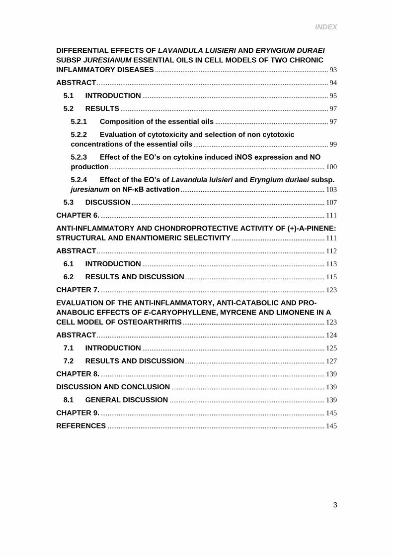
Synovial joints allow movement at the point of contact of articulating bones. Joints of
the knees, hips and hands are general examples of synovial joints that are the most
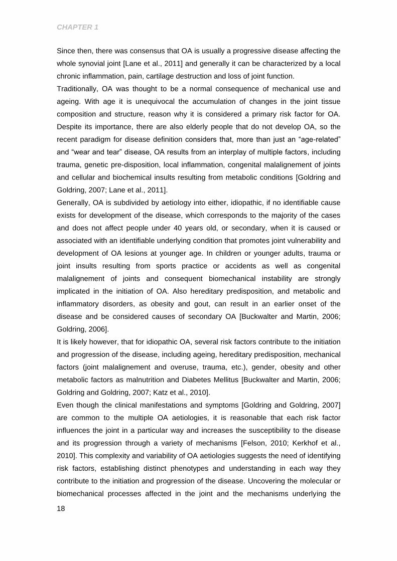
affected in OA. These joints, as represented in figure 1.1, are characterized by the
presence of a joint cavity surrounded by a capsule around the bone extremities,
covered inside by the synovial membrane, and by the presence of a lubricating
CHAPTER 1
20
synovial fluid. The bones extremities in a synovial joint are covered by a layer of
hyaline cartilage, the articular cartilage, which is a stiff but at the same time flexible and
elastic tissue.
Figure 1.1 Schematic representation of the major structures of a synovial joint
1.1.1 Structure and Composition of Articular Cartilage
Articular cartilage is a specialized connective tissue that covers the bony surface of
synovial joints. It is avascular and aneural and serves as a viscoelastic, load-bearing
support tissue that absorbs impacts, resists to compression and provides a smooth
surface with almost no friction during joint movement, without losing its original shape
[Martel-Pelletier et al., 2008]. These unique biomechanical properties are given by its
extracellular matrix composition and structure, namely by the collagen and
proteoglycan network [Goldring, 2006].
The chondrocyte is the only cell type found in mature articular cartilage, thus it is
responsible for the homeostatic process involving the synthesis and degradation of the
cartilage matrix components and consequently for its biomechanical properties. The
physical function of joints is critically dependent on the integrity of the matrix and so
dependents on normal chondrocyte function.
Proteoglycans and collagens are the most abundant macromolecules of the
extracellular cartilage matrix and are ordered in distinct layers from the superficial to
the deepest zones of the cartilage, reflecting different proportions, structural
CHAPTER 1
21
organization and function. The superficial layer is the thinnest and consists of fine
collagen fibrils with tangential orientation and low proteoglycan content. This zone is in
contact with synovial fluid, and is responsible for most of the tensile properties of
cartilage. The middle zone represents 40–60% of the total cartilage height. It is formed
by proteoglycans and thicker collagen fibrils organized into radial layers. The deep
zone contains the largest collagen fibrils in a radial disposition, and the highest
aggrecan content. Finally, the calcified cartilage is divided from the other zones by the
tidemark, and separates – physically and mechanically – the hyaline cartilage from the
subchondral bone [Martel-Pelletier et al., 2008].
Despite the importance of collagen, proteoglycans and other supporting glycoproteins,
they only represent 20% of the tissue wet weight. Chondrocytes represent 5% and the
remaining is water and inorganic salts. This enormous water content, almost all
associated with proteoglycans, is extremely important for the mechanical properties,
especially elasticity and capacity to resist to compressive forces, but also for the
nutrition and lubrication of the tissue [Martel-Pelletier et al., 2008; Mobasheri, 1998;
Muir, 1995].
1.1.1.1 Collagens and Proteoglycans
The collagen network provides the shape and form of the articular cartilage and is
composed by several types of collagens, including collagen type II, which represents
the major collagen type in the tissue (90-98% of total collagen), and collagens type XI,
type IX and type XXVII. Collagen type VI, characteristic of the connective tissue is also
present in a small percentage [Eyre, 2002; Goldring and Marcu, 2009]. It constitutes
the collagen network close to the cells in the so-called territorial matrix and acts as an
interface between the rigid interterritorial cartilage matrix and the chondrocyte. It is also
involved in cell anchoring, as well as in matrix-cell signalling [Soder et al., 2002].
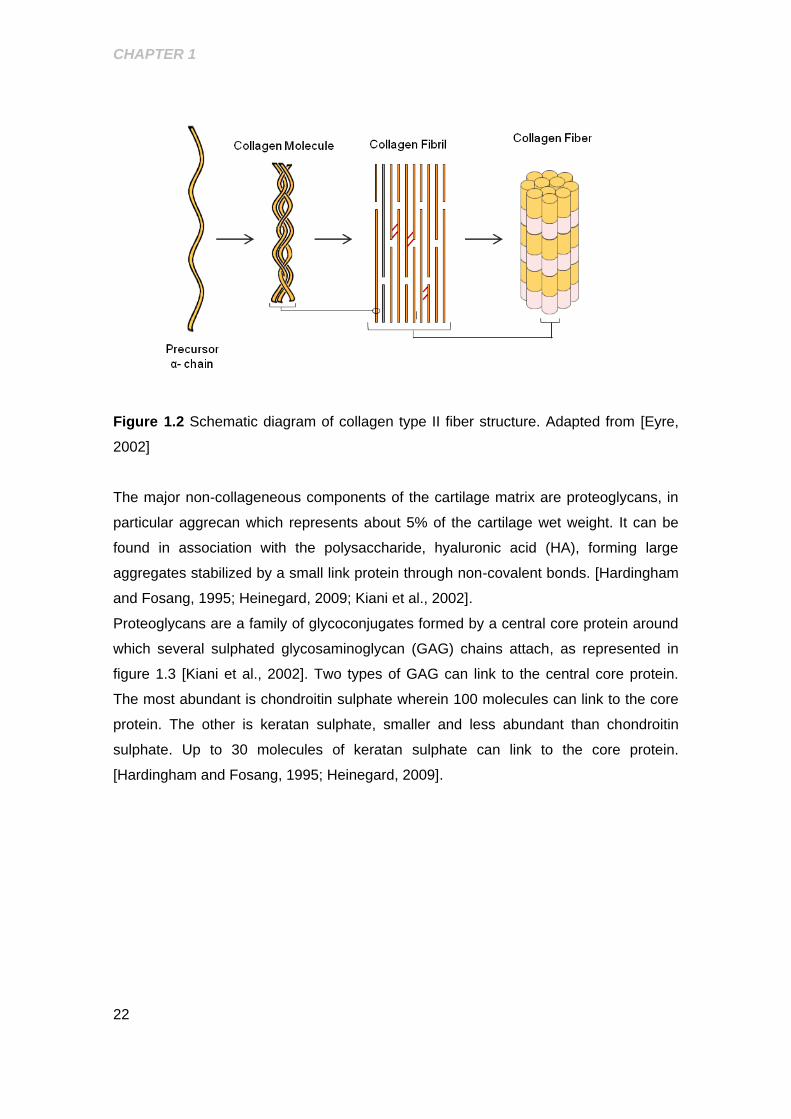
The collagen type II macromolecule is composed of 3 identical polypeptide chains – α1
(II) chains – interconnected to form a triple helix, as represented in figure 1.2. Those
helixes do not exist in an isolated form, but as fibrils resulting from the association of
adjacent collagen molecules within the extracellular matrix and stabilized by crosslinks
between the N-terminal telopeptide of one collagen molecule and the triple helix of
another [Eyre, 2002]. Collagens type XI and type IX are also associated with the
collagen type II fibrils, the first lying within the fibril and the second integrated in the
fibril surface, providing protection to collagen type II and enabling interactions with the
other non-collagen elements of the matrix, especially proteoglycans [Eyre et al., 2006].
CHAPTER 1
22
Figure 1.2 Schematic diagram of collagen type II fiber structure. Adapted from [Eyre,
2002]
The major non-collageneous components of the cartilage matrix are proteoglycans, in
particular aggrecan which represents about 5% of the cartilage wet weight. It can be
found in association with the polysaccharide, hyaluronic acid (HA), forming large
aggregates stabilized by a small link protein through non-covalent bonds. [Hardingham
and Fosang, 1995; Heinegard, 2009; Kiani et al., 2002].
Proteoglycans are a family of glycoconjugates formed by a central core protein around
which several sulphated glycosaminoglycan (GAG) chains attach, as represented in
figure 1.3 [Kiani et al., 2002]. Two types of GAG can link to the central core protein.
The most abundant is chondroitin sulphate wherein 100 molecules can link to the core
protein. The other is keratan sulphate, smaller and less abundant than chondroitin
sulphate. Up to 30 molecules of keratan sulphate can link to the core protein.
[Hardingham and Fosang, 1995; Heinegard, 2009].

CHAPTER 1
23
Figure 1.3 Schematic representation of proteoglycans linked to Hyaluronic acid.
Adapted from [Hardingham and Fosang, 1995]
At high concentrations, proteoglycans, and in particular the aggregates formed with
HA, create a large osmotic swelling pressure that draws water into the cartilage. This
osmotic pressure occurs because all of the negatively charged sulphated groups of
chondroitin and keratan sulphates on the GAG chains of aggrecan carry with them
positive ions such as Na+, generating a huge difference in ion concentration between
the cartilage and surrounding milieu. The osmotic imbalance generated is responsible
for water to flow and to be retained within the cartilage. The addition of water causes
the aggrecan-rich matrix to swell and expand which is essential to the biomechanical
properties of cartilage, namely its visco-elastic properties that allow the impact loading
without permanent deformation [Hardingham and Fosang, 1995; Kiani et al., 2002]. In
an exclusively biophysic perspective of the process, when the cartilage is submitted to
a compressive force, an amount of the water retained in the tissue is released, in
proportion to the load applied [Kiani et al., 2002]. Moreover, some authors also
postulate that aggrecan, through the chondroitin sulphate chains, can protect collagen
type II fibrils from degradation by collagenases, possibly via direct blocking of their
binding sites [Hedlund et al., 1999].
CHAPTER 1
24
Small amounts of other non-collagen molecules, including biglycan, decorin,
fibromodulin, the matrilins and cartilage oligomeric matrix protein (COMP), are also
present in the matrix. COMP acts as a catalyst in the formation of the collagen fibrils.
Interactions between type IX collagen and COMP or matrilin-3 are essential for proper
formation and maintenance of the articular cartilage matrix [Shakibaei et al., 2008].
The collagen/aggrecan network is a resilient, viscoelastic load-bearing structure that
allows smooth movement and distributes loads applied to the joint. The type II collagen
network gives cartilage its shape and tensile strength and provides a framework to
resist the swelling pressure of aggrecan. On its hand, aggrecan resists to any fluid flow
and is able to efficiently distribute water. Proteolysis of any of the components of the
cartilage not followed by its respective resynthesis will imbalance the network
compromising the tissue biomechanical properties [Eyre, 2002; Goldring and Marcu,
2009; Hedlund et al., 1999; Kiani et al., 2002; Martel-Pelletier et al., 2008].
1.1.1.2 Chondrocyte
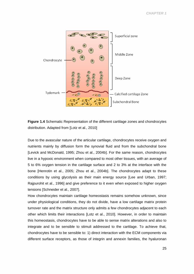
Similarly to the other cartilage components, chondrocytes are distributed unevenly
throughout the cartilage depth, being in general more abundant at the surface.
Differences in the morphology and organization of chondrocytes can be a reflex of the
mechanical environment given by the collagen architecture in the interterritorial matrix,
and can be the reason for the different matrix compositions found along the tissue.
Chondrocytes in the superficial zone (SZ) are small and flattened; those of the middle
zone (MZ) are rounded with no organized orientation while those of the deep zone (DZ)
are grouped in columns or clusters, as represented in figure 1.4. These cell distribution
patterns are thought to derive from the formation and specific organization of the
Extracellular Matrix (ECM) during joint development and maturation [Lotz et al., 2010].
CHAPTER 1
25
Figure 1.4 Schematic Representation of the different cartilage zones and chondrocytes
distribution. Adapted from [Lotz et al., 2010]
Due to the avascular nature of the articular cartilage, chondrocytes receive oxygen and
nutrients mainly by diffusion form the synovial fluid and from the subchondral bone
[Levick and McDonald, 1995; Zhou et al., 2004b]. For the same reason, chondrocytes
live in a hypoxic environment when compared to most other tissues, with an average of
5 to 6% oxygen tension in the cartilage surface and 2 to 3% at the interface with the
bone [Henrotin et al., 2005; Zhou et al., 2004b]. The chondrocytes adapt to these
conditions by using glycolysis as their main energy source [Lee and Urban, 1997;
Rajpurohit et al., 1996] and give preference to it even when exposed to higher oxygen
tensions [Schneider et al., 2007].
How chondrocytes maintain cartilage homeostasis remains somehow unknown, since
under physiological conditions, they do not divide, have a low cartilage matrix protein
turnover rate and the matrix structure only admits a few chondrocytes adjacent to each
other which limits their interactions [Lotz et al., 2010]. However, in order to maintain
this homeostasis, chondrocytes have to be able to sense matrix alterations and also to
integrate and to be sensible to stimuli addressed to the cartilage. To achieve that,
chondrocytes have to be sensible to: 1) direct interaction with the ECM components via
different surface receptors, as those of integrin and annexin families, the hyaluronan
CHAPTER 1
26
receptor, CD44, or the Discoidin Domain receptor 2 (DDR2) which binds collagen II
when proteoglycans are depleted [van der Kraan et al., 2002; Xu et al., 2007]; 2)
soluble mediators produced by the chondrocytes themselves or by any other articular
or non-articular tissue, and, finally, 3) to mechanical stimuli, [Goldring and Marcu, 2009;
Shakibaei et al., 2008; van der Kraan et al., 2002].
Moreover, several studies indicate that although chondrocytes are normally quiescent,
in response to any injury or abnormal stimulus they can shift to an activated form and
enter on a remodelling program that includes catabolic (destructive) and anabolic
(synthetic) processes. The normal function of chondrocytes and consequently the
integrity of the cartilage are dependent on a variety of signals including mechanical
forces and chemical mediators (cytokines, growth factors, hormones), and on the
capacity of chondrocytes to integrate those signals without disrupting the homeostatic
balance of the tissue [Goldring and Marcu, 2009; Martel-Pelletier et al., 2008].
1.2 OSTEOARTHRITIS RISK FACTORS AND SPECIFIC PHENOTYPES
Epidemiological studies have been transforming the way how OA is seen. Firstly saw
as an age-related and a “wear and tear” disease of the cartilage, OA is now considered
a multifactorial disease affecting the whole joint where the identification of several risk
factors, both endogenous and exogenous, played an unquestionable role [Michael et
al., 2010].
No longer misconceived as a uniform disease, OA is an heterogeneous disease with
the failure of the joint function as the major outcome [Goldring and Goldring, 2007].
Different pathophysiological aetiologies resulting in the final common consequence of
cartilage destruction have driven the division of OA into different phenotypes according
to the respective major related risk factor. The definition of a phenotype should be
confined to those factors that have a primary effect on disease biology and aetiology,
and that can be significant to treatment and prevention [Felson, 2010]. Traditional risk
factors for OA, namely age, trauma, genetic predisposition and obesity by inducing
direct effects on cartilage destruction, through destabilizing catabolic and anabolic
homeostasis and creating a low grade inflammatory environment, have already been
associated with specific OA phenotypes [Neogi and Zhang, 2013]. Although they can
be seen as independent, most of the times OA onset and progression does not depend
on an individual risk factor alone but on a synergy of multiple factors. Genetics, for
instance, plays an indubitable role in OA development and progression [Spector et al.,
1996]. However, its role is more likely due to an interaction among multiple genes, in
CHAPTER 1
27
combination with further risk factors, as ageing. Moreover, the effects of obesity on the
joint have been admitted, historically, as simply resulting from excessive mechanical
loading. However, there is increasing evidence of multifactorial, systemic links between
obesity and OA, since non-loading joints, as elbows and hands, are also affected by
OA in obese patients [Sowers and Karvonen-Gutierrez, 2010].
The evidence of systemic factors involved in obesity related-OA also contributed to the
definition of a metabolic-induced OA. This phenotype shares several similar
biochemical and inflammatory features with the metabolic syndrome. Hypertension,
dyslipidemias and conditions with altered glucose metabolism, as diabetes mellitus
(DM), and their major characteristics, increased expression of adipokines, varying
glucose concentrations and hormonal imbalance characterize the metabolic syndrome.
When OA is also present it has been described as metabolic-OA. These characteristics
have already been, either directly or indirectly, related with the mechanisms of OA
pathology [Zhuo et al., 2012].
Besides its relationship with obesity and the metabolic syndrome, DM is increasingly
recognized as an independent risk factor for OA development and progression. Thus, it
can define a new specific OA phenotype [Berenbaum, 2011]. Recent epidemiologic
data further demonstrate a higher OA prevalence with an earlier onset and more
severe manifestations in diabetic vs. non-diabetic patients [King et al., 2013; Martinez-
Huedo et al., 2013; Nieves-Plaza et al., 2013; Schett et al., 2012a]. Moreover, several
experimental evidences show that hyperglycemia, one of the features of DM, can
trigger events that can be related to cartilage destruction, as Reactive Oxygen Species
(ROS) production and a low grade systemic inflammation further linking DM and
hyperglycemia to OA incidence [Berenbaum, 2011].
1.2.1 Diabetes Mellitus and Osteoarthritis: Role of Extracellular glucose
concentrations
Diabetes mellitus (DM) is a group of chronic diseases characterized generally by high
blood glucose levels that result from defects in the body’s ability to produce and/or use
insulin efficiently. Together with OA, DM type 2 is one of the most important causes of
mortality and morbidity in older individuals [Kaplan et al., 2013]. Injurious effects
induced by DM can be related with several factors, including insulin or other pancreatic
hormone levels, but hyperglycemia is probably the most significant [Yan and Li, 2013].
At normal physiological conditions, due to the avascular nature of cartilage,
chondrocytes are under a particular environment where nutrients and oxygen only
CHAPTER 1
28
arrive by diffusion from the synovial fluid and subchondral bone. Consequently
chondrocytes must have the capacity to survive in an extracellular matrix with limited
nutrients and low oxygen tensions. Several studies showed that these particular
conditions of low oxygen tension are important for the normal synthesis of matrix
components and to the maintenance of the differentiated chondrocytic phenotype [Das
et al., 2010; McNulty et al., 2005] and that their disruption results on negative
outcomes, namely contributing to the altered synthesis of structural and regulatory
proteins [Das et al., 2010; Grimshaw and Mason, 2000; Grimshaw and Mason, 2001].
In terms of energetic needs, chondrocytes adapted to this limited availability of oxygen
by using the glycolytic pathway as their major energetic source [Lee and Urban, 1997]
and they use it even in the presence of high oxygen tensions [Lee and Urban, 1997;
Schneider et al., 2007]. However, glucose serves not only as the major energy source,
but also as an essential precursor for glycosaminoglycan synthesis. Thus, a stable
supply of glucose is important for the tissue homeostasis [Goldring, 2006].
In pathological conditions, glucose and oxygen supply may be affected by different
factors. For example, studies of Otte and Hernvann (1992) relate the importance of
glucose as a metabolic substrate and also emphasize the fact that glucose uptake is
stimulated by catabolic cytokines in chondrocytes in the concentrations easily found in
OA joints [Hernvann et al., 1992; Otte, 1991]. In addition, since chondrocytes receive
glucose especially from the synovial fluid whose glucose concentrations reflect, in the
majority of cases, the plasma concentrations, conditions like DM that affect glycemia
will probably also affect the availability of glucose to articular chondrocytes [Brannan
and Jerrard, 2006].
Despite these observations, the role of the extracellular glucose concentrations on the
chondrocyte functions is still largely unknown. From the in vitro and in vivo data
available, however, it is possible to understand that both hypo- and hyperglycemic
conditions may contribute to cartilage homeostasis breakdown and thus to OA onset
and progression. Glucose deprivation has been shown to increase the expression of
the Matrix Metalloproteinase 2 (MMP-2) [Richardson et al., 2003], that participates in
cartilage degradation in late OA, and was associated with decreased expression of
collagen type II and proteoglycans [Yang et al., 2005]. The effects of hyperglycemia in
cartilage have been associated with the formation of advanced glycation end products
(AGEs) [Verzijl et al., 2003], as mediators of matrix stiffness, subchondral bone
destruction and chondrocyte dysfunction. Chronic hyperglycemia increases the
occurrence of non-enzymatic glycation reactions, which are associated with oxidative
stress, as illustrated in patients with diabetes [Gillery, 2006]. AGEs have been shown to
CHAPTER 1
29
accumulate within the cartilage in amounts that increase with age [Verzijl et al., 2001].
Hiraiwa and Nah [2011] showed that AGEs mediate the expression of matrix-degrading
enzymes and pro-inflammatory and catabolic mediators (MMP-1, MMP-13, COX-2;
NO) [Hiraiwa et al., 2011; Nah et al., 2008; Nah et al., 2007] and DeGroot [2001 and
1999] showed that they decrease collagen type II and proteoglycan expression in
bovine chondrocytes.
Notwithstanding the importance of AGEs on OA pathogenesis, it is important to note
that the development of OA with aging is not inevitable, even though changes in the
composition and structure of the cartilage matrix are. So, studies focused only on older
individuals and in AGEs, generated in increased amounts during aging, would not
completely explain the role of hyperglycemia in OA development [DeGroot et al., 1999;
Goldring and Goldring, 2007; Verzijl et al., 2003].
Recent studies have focused on the direct effects of high glucose on the chondrocyte
functions. Those studies clearly show that direct exposure of chondrocytes to high
glucose affects the cartilage homeostasis, namely by interfering with the synthesis of
ECM components. In one study, hypo- and hyperglycemia-like conditions induced
resistance to Insulin-like Growth factor (IGF-1), an important anabolic factor in
chondrocytes, thus contributing to decreased proteoglycan synthesis [Kelley et al.,
1999] and in another, exposure of human chondrocytes to high glucose concentrations,
was shown to decrease dehydroascorbate transport, which can compromise the
synthesis of collagen type II [McNulty et al., 2005]. On the other hand, another study
from our group showed that collagen II mRNA was similarly increased in normal and
OA chondrocytes, exposed to hyperglycemia-like conditions, but the increase lasted
longer in the OA group [Rosa et al., 2011b]. Moreover, this study also showed that that
exposure to high glucose (30 mM) increased the mRNA levels of matrix-degrading
enzymes in OA chondrocytes, whereas in normal ones only MMP-1 was increased.
Furthermore, exposure of human chondrocytes to high glucose also prevented anti-
catabolic effects of TGF-β both in normal and OA chondrocytes [Rosa et al., 2011b].
Finally, culture of chondrocytes under high glucose elicited a increase in ROS
production. In OA chondrocytes the increase in ROS production was sustained for a
longer period than in normal ones [Rosa et al., 2009]. Since ROS are involved in the
pathophysiology of OA, high glucose-induced ROS production is likely to directly
contribute to high-glucose induced cartilage damage. In vivo data corroborate these
findings. Indeed, diabetic rats were associated with major alterations in matrix
composition in the cartilage of the knee joints, namely with decreased expression of
proteoglycans and collagen type II, the main structural component of the articular
CHAPTER 1
30
cartilage, and increased expression of collagen type XI, that is not normally present in
articular cartilage and is a marker of chondrocyte progression to hypertrophy and
subsequent calcification [Atayde et al., 2012]. This finding may contribute to
understanding the functional limitations of diabetic patients and the association of DM
with OA. Although these observations of glucose effects on chondrocytes and cartilage
may contribute to establish a correlation between DM and OA, strengthening the
existence of a specific DM-induced OA phenotype, more studies are needed to
understand how DM affects the development and progression of OA. Finally, a better
knowledge of the specificities of a DM-induced OA phenotype should allow a targeted
approach for the development of preventive and curative treatments for OA
[Berenbaum, 2011].
1.2.1.1 Glucose sensing and transport
As glycolytic cells, chondrocytes must be able to sense the quantities of oxygen and
glucose available to them in the extracellular matrix and respond appropriately by
adjusting cellular metabolism [Mobasheri et al., 2005]. As the concentrations of glucose
available may vary, depending for instance on the pathological conditions, immobility
and age, glucose transport becomes a critical step on the utilization and on the effects
of this nutrient on chondrocyte functions [Mobasheri et al., 2002b; Shikhman et al.,
2001].
Glucose consumption seems to be dependent on the extracellular glucose
concentrations [Schneider et al., 2007], suggesting that chondrocytes may adjust
glucose transport and increase glucose storage, glycolysis, and production of
proteoglycans [Lee and Urban, 1997] to the extracellular glucose concentration.
Several reports show that high, hyperglycemia-like glucose concentrations play an
important role in the development of diabetic complications. High glucose can induce
the release of pro-inflammatory cytokines as Interleukin-1β (IL-1β), Interleukin-6 (IL-6),
Tumor necrosis factor –α (TNF-α), and interleukin-18 (IL-18) [Asakawa et al., 1997;
Esposito et al., 2002; Jiang et al., 2012] and other pro-inflammatory mediators, as Nitric
Oxide (NO) [Askwith et al., 2011; Jia et al., 2013], in different cell types, activate pro-
inflammatory and pro-catabolic signalling pathways, as those of Nuclear Factor κB (NF-
κB) and p38 Mitogen Activated Protein Kinase (MAPK) [Igarashi et al., 1999; Yerneni et
al., 1999], and induce the production of ROS [Busik et al., 2008; Yao and Brownlee,
2010] and AGEs [Brownlee, 2005]. Chondrocytes express multiple isoforms of the
facilitative glucose or solute carrier (GLUT/SLC2A) family of glucose/polyol transporters
CHAPTER 1
31
[Mobasheri et al., 2008], including GLUT-1, GLUT-3, GLUT-5, GLUT-9, GLUT-10 and
GLUT-11 which are responsible for the majority of the glucose transport capacity of
these cells [Mobasheri et al., 2008]. Among those, GLUT-1 and GLUT-3, constitutively
expressed in chondrocytes, are the isoforms better characterized, and both have high
glucose affinity [Colville et al., 1993]. GLUT-1 seems to be regulated by anabolic and
catabolic stimuli, while GLUT-3, is less affected by [Mobasheri et al., 2008;
Peansukmanee et al., 2009; Shikhman et al., 2001]. Specifically, GLUT-1 was found to
be increased in equine articular chondrocytes stimulated with TNF-α, IL-1β, IGF-I,
TGF-β and insulin, while GLUT-3 was only upregulated by IGF-I [Phillips et al., 2005;
Richardson et al., 2003]. GLUT-1 and GLUT-3 together with hypoxia-inducible factor
alpha (HIF-1alpha) were proposed as potential components of the glucose and oxygen
sensing apparatus in chondrocytes by promoting anaerobic glycolysis and favoring
oxidative phosphorylation, which allow chondrocytes to survive in the adverse
conditions of low glucose and oxygen supply [Mobasheri et al., 2008; Mobasheri et al.,
2005]. However, how human chondrocytes sense and modulate glucose transport, in
health and disease and in the presence of different extracellular glucose
concentrations, remains unclear.
Recent studies showed that normal human chondrocytes respond to high extracellular
glucose concentrations by decreasing glucose uptake, which occurs through
downregulation of GLUT-1 content [Rosa et al., 2009]. This mechanism seems to
protect the cells against excessive intracellular glucose accumulation. Moreover, this
study showed that a different effect occurs in OA chondrocytes, which when exposed
to high glucose concentrations were unable to downregulate GLUT-1 and glucose
transport, leading to increased glucose accumulation and prolonged ROS production
[Rosa et al., 2009]. Increased ROS production and, thus oxidative stress, are seen as a
major deleterious mechanism in OA pathogenesis by mediating inflammatory and
catabolic responses in chondrocytes [Grishko et al., 2009; Mendes et al., 2003a;
Mendes et al., 2003b]. Moreover, these observations establish another link between
DM and OA development since OA chondrocytes seem to be susceptible to
hyperglycemic episodes as those occurring in DM patients, and thus particularly
subjected to the deleterious effects of high glucose.
Further research is needed to expand our understanding of glucosensing and glucose
signalling in chondrocytes, and other joint tissues, in health and disease.
Understanding how human chondrocytes sense extracellular glucose and adjust its
uptake to match the cell’s needs and whether such mechanisms remain functional in
OA chondrocytes is critical to elucidate the mechanisms by which conditions
CHAPTER 1
32
associated with impaired glucose metabolism contribute to the development and/or
progression of OA, and for further development of new therapies and identification of
new therapeutic targets for treating this joint disorder.
1.3 CARTILAGE CHANGES IN OSTEOARTHRITIS
Cartilage degradation and loss, one of the OA hallmarks, occurs due to a disturbance
of the balance between the reparative and degradative processes of the cartilage. The
initiation and progression of OA, however, appear to be enclosed in a cyclic evolution
plan where it becomes virtually impossible to understand which are the causes and/or
the consequences, i.e., any factor that disturbs the tissue can contribute to the
deregulation of the cell’s functions and to tissue alterations, which in turn will lead to
more cell dysfunction and more ECM alterations. As an example, abnormal mechanical
stress induces chondrocytes to produce Matrix Metalloproteinases (MMPs), which
degrade matrix components, destroying the cartilage, while the ECM protein fragments
generated interact with different receptors to induce the production of inflammatory
cytokines, chemokines and more MMPs, expanding further matrix destruction [Fichter
et al., 2006; Pulai et al., 2005].
Chondrocytes, because of their location and function, play a central role in OA
pathogenesis which can be described as a result of their failure to maintain the
homeostatic process, so that the rate of loss of collagens and proteoglycans from the
matrix exceeds the rate of deposition of new molecules. This, in turn, involves multiple
interactions between local and systemic factors [Buckwalter and Martin, 2006; Goldring
and Goldring, 2007; Martel-Pelletier et al., 2008] and even though the aetiology of the
disease is still largely unknown, the fact is that several risk factors, lead to the OA
phenotype. The similarities observed in the pathological progression of the disease,
even when the initiating models are different, indicate that common molecular
sequences may underlie OA onset and progression [Castaneda et al., 2013].
Regardless the nature of the factors that initiate the pathology, its progression follows a
consistent pattern which includes chondrocyte phenotype alterations, increased
general synthetic activity, increased expression of ECM degradative proteinases,
gradual loss of proteoglycans and collagen type II, fibrillation and formation of
fibrocartilage and osteophytes [Fosang and Beier, 2011]. In non-pathological
conditions, the normally quiescent chondrocytes have a very low turnover, which is
enough to ensure the replacement of long-lived ECM proteins, like aggrecan with a
CHAPTER 1
33
half-life of 3-24 years [Maroudas et al., 1998] and collagen whose half-life is even
greater of 100 years or more [Verzijl et al., 2000].
In the initial pathological conditions, mechanical stress, cytokines, matrix degradation
products, age-related AGEs or any other factor that can disrupt the cartilage
homeostasis [Hiraiwa et al., 2011] lead chondrocytes to a changed phenotype where
they become “activated”, proliferate and form clusters and suffer an inappropriate
hypertrophy-like maturation [Lotz et al., 2010]. Hypertrophic chondrocytes undergo a
stress response associated with ECM remodelling in what can be seen as an attempt
to repair the damages. In this active remodelling, however, the quality of the ECM is
compromised due to the abnormal quick turnover rate, and to the atypical composition
of the newly synthesized ECM proteins. In this period, there is expression of non-
cartilage specific collagens, like type X, normally confined to the cartilage-calcified
zones close to the subchondral bone and only produced by hypertrophic chondrocytes,
and collagen type I typically present in fibrocartilage and structurally different from type
II. [Boos et al., 1999; Eyre et al., 2006; Lahm et al., 2010; Miosge et al., 2004]. Both
collagen types have less capacity to interact with non-collagen cartilage molecules as
proteoglycans and contribute to the formation of fibrocartilage, biomechanically less
effective than the original one. In the late phases of the disease, this fibrocartilage
contributes to further cartilage matrix degradation and loss [Lorenzo et al., 2004;
Martel-Pelletier et al., 2008].
Associated with this abnormal renovation, there is decreased expression and higher
degradation of the naturally-occurring articular components, namely types IX, XI and II
collagens and proteoglycans. It has been shown that there is a significant
downregulation of aggrecan gene expression at the onset of OA in a rat model [Nam et
al., 2011] and this finding agrees with markedly low proteoglycan synthesis observed in
human cartilage samples from an early phase of the disease process, even before a
clear fibrillation of the tissue is evident [Lorenzo et al., 2004].
Evidence suggests that the degradative process of the cartilage starts most of the
times at its surface. The local loss of collagen type II results in an increased water
content and loss of the tensile strength which is often observed in the early phases of
the disease. Later, the combination of aggrecan and collagen loss leads to the
increased overall stiffness of the tissue. A relationship between this degradation and
the increased expression of proteinases, namely those responsible for the degradation
of collagen type II and aggrecans, as MMPs (MMP-1, MMP-3 and MMP-13), and the
aggrecanases, A Desintegrin And Metalloproteinase with ThromboSpondin motif
CHAPTER 1
34
(ADAMTS)-4 and -5, was established [Bau et al., 2002; Okada et al., 1992; Shinmei et
al., 1990; Yoshihara et al., 2000]
An increasing number of evidences suggest that an important inflammatory process
characterized by deregulated cytokine activities that can be triggered by any
mechanical, biomechanical or molecular factor involved in OA pathogenesis, yield
alterations of chondrocyte functions that are likely to contribute to the described
disruption of that balance between the anabolism and catabolism in OA [Goldring and
Berenbaum, 2004].
1.3.1 Regulation of Catabolic and Anabolic Pathways
1.3.1.1 Matrix Metalloproteinases and Aggrecanases
Degradation of the cartilage ECM is a feature of arthritic diseases and a consequence
of the upregulated catabolic process. The destruction seems to be driven by both
Matrix Metalloproteinases (MMPs) and aggrecanases, which degrade the two major
structural components of the cartilage extracellular matrix, the proteoglycan, aggrecan,
and type II collagen.
Several members of the ADAMTs family of enzymes (ADAMTS 1, 4, 5, 8, 9, and 15)
have been identified in articular cartilage [Bau et al., 2002] and are known to be
capable of cleaving aggrecan in different sites [Troeberg and Nagase, 2012].
ADAMTS4 and ADAMTS5 (aggrecanase-1 and aggrecanase-2, respectively) seem to
be the most active aggrecanases and their depletion [Song et al., 2007] was
associated with lower proteoglycan degradation and slower OA progression [Majumdar
et al., 2007].
Along with aggrecan breakdown, degradation of collagen is a central feature of OA, in
a process that is extensively mediated by MMPs. These are a family of zinc-dependent
proteinases and although they can, similarly to ADAMTs, participate in the degradation
of all the ECM proteins, collagen is their main target [Burrage et al., 2006; Cawston and
Wilson, 2006; Patwari et al., 2005]. They can be categorized in different groups
according to their preferable substrate and tissue location: the collagenases (MMP-1, -
8, -13), which degrade the fibrillated collagens (types I, II and III), the gelatinases
(MMP-2, -9), which target denaturated polypeptide chains of types IV and II collagen
and, finally, the stromelysins (MMP-3, -10, -11) and the membrane-type MMPs (MMP-
14, -15, -16, -17, -24, -25) which degrade non-collagen matrix proteins, as aggrecans,
fibronectin and elastin and participate in the activation of other MMPs [Burrage et al.,
CHAPTER 1
35
2006]. Generally, the collagenases (MMP-1, -8 and -13) cleave the collagen type II
triple helix allowing further degradation of the released chains by gelatinases (MMP-2
and -9). Even though all the collagenases were able to effectively cleave the triple
helix, they seem to have a substrate preference, with MMP-13 being the most
expressed in chondrocytes and preferably cleaving collagen type II. Although MMP-1
preferentially cleaves collagen III, it also effectively cleaves type II collagen and both
MMPs were found to be overexpressed in OA joints [Bau et al., 2002; Konttinen et al.,
1999; Troeberg and Nagase, 2012; Yoshihara et al., 2000].
Additionally, MMPs participate in other processes that indirectly contribute to further
cartilage breakdown, as is the case of MMP-3 that seems to be involved in the
activation of MMP-1 and MMP-13 [Troeberg and Nagase, 2012; van Meurs et al.,
1999].
Given the destructive potential of MMPs and their abnormal high levels in OA,
abolishing or even just reducing their pathologic levels are interesting strategies for
development of new disease-modifying OA drugs.
Several factors and mechanisms have been associated with increased expression of
the matrix-degrading enzymes observed in OA cartilage. The primary one is probably
the abnormal mechanical loading. In vivo studies showed that immobilization leads to
joint damage by upregulating MMP-3 and ADAMTS-5, while mechanical stimulation of
moderate intensity has the opposite effect, increasing aggrecan and decreasing MMP-
3 [Leong et al., 2010a; Leong et al., 2010b]. Nevertheless, abnormal mechanical stress
can be sensed by chondrocytes resulting in an increased stimulation of matrix-
degrading proteins [Fujisawa et al., 1999].
In addition, several other studies showed that activation of mechanical receptors is not
the only cause of the upregulated expression of these proteinases. The stronger
evidence rests on the role of pro-inflammatory mediators, namely, cytokines (IL-1β and
TNF-α) [Fernandes et al., 2002; Kapoor et al., 2011], chemokines [Lotz et al., 1992],
prostaglandins [Mohamed-Ali, 1995] and Nitric Oxide (NO) [Abramson, 2008].
Chondrocytes in OA cartilage express receptors that respond to those mediators,
produced by chondrocytes themselves or by the synovium and other periarticular joint
tissues, resulting in an increased expression of MMP-1,-3, -9 and -13 [Burrage et al.,
2006]. Moreover also integrins and matrix proteins fragments (collagen and fibronectin)
have been associated with cartilage damage through the production MMPs and
aggrecanases and through an amplification feedback by induction of the production of
pro-inflammatory cytokines [Fichter et al., 2006; Loeser et al., 2003].
CHAPTER 1
36
The synthesis of MMPs is highly regulated at the transcriptional level [Burrage et al.,
2006; Murphy and Lee, 2005] and regardless of the mechanism that triggers their
overexpression, the same signalling pathways seem to be involved. Several evidences
showed that, among others, signalling pathways of Activator Protein-1 (AP-1) and NF-
κB transcription factors together with those of MAPKs are key pieces on the regulation
of the expression of the matrix-degrading enzymes in human chondrocytes
[Barchowsky et al., 2000; Mengshol et al., 2000]. At a post-transcriptional level, MMPs
are regulated by the Tissue Inhibitors of the Metalloproteinases (TIMPs) [Cawston and
Wilson, 2006]. The inhibition of MMP activity by TIMPs involves non-covalent binding in
a 1:1 stoichiometry to the active site of the target MMP [Burrage et al., 2006].
Generally, TIMPs-1 to -4 can inhibit all MMPs, but are also effective in inhibiting
aggrecanases. For example, TIMP-3 blocks all the MMPs [Burrage et al., 2006] and
also ADAM-17 [Amour et al., 2000] and ADAMTS-4 and -5 [Burrage et al., 2006;
Kashiwagi et al., 2004].
Regardless of the natural role of MMPs in cartilage homeostasis, in the pathologic
environment of an arthritic joint, their elevated levels usually exceed those of TIMPs
[Dean et al., 1989] which contributes to cartilage degradation in OA [Martel-Pelletier et
al., 1994].
1.3.2 Inflammatory Process in OA
OA is not considered a classical inflammatory disease, especially due to the
intermittent influx of inflammatory cells into the joint and the sparse characteristic signs
of inflammation, such as redness and swelling with heat during the course of the
disease when compared to other inflammatory arthritic diseases, namely, rheumatoid
arthritis (RA). However, several reports have demonstrated the overproduction and
expression of proinflammatory mediators including NO, inducible nitric oxide synthase
(iNOS), Cycloxigenase 2 (COX-2), IL-1β, TNF-α, IL-6, and IL-8 in OA cartilage. This
inflammatory response, seen as a chronic low-grade inflammation and so called
“molecular inflammation”, even though not reflected by clinical signs of inflammation,
leads to an imbalance of cartilage homeostasis that results in progressive breakdown
[Attur et al., 2000; Fernandes et al., 2002; Sokolove and Lepus, 2013]. It is now
significantly clear that inflammation is present in OA joints well before the development
of significant radiographic changes, suggesting that the cartilage damage in OA can be
driven by an articular inflammatory process [Attur et al., 2000; Ayral, 2005; Fernandes
et al., 2002; Haywood et al., 2003; Ishii et al., 2002]. So, any factor that triggers the
CHAPTER 1
37
excessive production of proinflammatory mediators can contribute to the initiation and
progression of this pathology.
Although the mechanism by which production of inflammatory mediators is initiated is
unclear, several factors, including mechanical and metabolic, have been related with
the induction of cytokines and chemokines, ROS, in particular NO, as well as
inflammation-related enzymes, like COX-2, and iNOS in the joint tissues and,
particularly, in cartilage. Evidences from in vivo and in vitro studies indicate that
chondrocytes can produce and/or respond to a number of cytokines and chemokines
that are present in OA joint tissues and fluids [Berenbaum, 2004; Berenbaum, 2013;
Goldring and Goldring, 2007]. Synovial inflammation for instance is a factor that likely
contributes to deregulation of chondrocyte functions with the levels of catabolic
enzymes and inflammatory mediators, such as prostaglandins and NO, and the levels
of IL-1β and TNF-α in OA synovial fluids and joint tissue favoring the imbalance
between the catabolic and anabolic activities of the chondrocyte [Loeser, 2006; Loeser
et al., 2012].
The major role of the inflammatory mediators in OA pathogenesis is related with their
activity on the catabolic and anabolic programs of the chondrocyte [Berenbaum, 2013].
The upregulation of the proinflammatory genes generally results in increased levels of
catabolic enzymes, as MMPs and aggrecanases, decreased synthesis of ECM
proteins, as collagen type II and aggrecan, and anti-catabolic mediators, as TIMPs
contributing, in this way, to the progressive cartilage degradation [Clancy et al., 2004;
Goldring and Goldring, 2007; Marcu et al., 2010]. These processes involve the
activation of different transcription factors and signalling pathways, in particular those
of NF-κB and MAPK.
1.3.2.1 Regulation and Function of Inflammatory Mediators in Osteoarthritis
1.3.2.1.1 Cytokines
Pro-inflammatory cytokines were found to be highly expressed in the synovial fluid,
synovial membrane, subchondral bone and cartilage of the joints affected by OA and
can be produced not only by chondrocytes, but also by other joint tissue cells
[Berenbaum, 2013; Kapoor et al., 2011; Kobayashi et al., 2005; Loeser et al., 2012;
Meats et al., 1980].
In OA, pro-inflammatory cytokines are known to induce the catabolic and inhibit the
anabolic programs. This translates into an increase of inflammatory mediators and
CHAPTER 1
38
proteolytic enzymes, and decreased synthesis of matrix components and TIMPs.
Additionally, those cytokines also promote the induction of cell death, mainly by
apoptosis, at least in part mediated by inflammatory mediators such as NO [Abramson,
2008; Blanco et al., 1995; Fernandes et al., 2002; Kapoor et al., 2011]. These
mechanisms make them critical mediators of cartilage breakdown and of disease
amplification and progression [Kapoor et al., 2011]
Although several cytokines (i.e. IL-15, IL-17, IL-18) [Fernandes et al., 2002; Kapoor et
al., 2011] have been shown to be upregulated in OA joints and definitely contribute to
the pathology establishment [Goldring and Goldring, 2007; Melas et al., 2014], both in
vitro and in vivo studies evidence that, IL-1β and at a lower extent TNF-α are major
players [Kapoor et al., 2011], by deregulating chondrocyte anabolic and catabolic
responses. The deleterious effects of IL-1β and TNF-α are mainly associated with the
upregulation of proteolytic enzymes. In response to these cytokines, OA chondrocytes
produce a variety of matrix degrading enzymes, including MMP-1, MMP-3, MMP-8,
MMP-13 and aggrecanases, as ADAMTS-4 and ADAMTS-5 [Lefebvre et al., 1990;
Liacini et al., 2003; Reboul et al., 1996][Maldonado and Nam, 2013; Pelletier et al.,
1995; Tetlow et al., 2001] On the other hand, the synthesis and expression of the
natural MMPs inhibitors, TIMPs [Dean et al., 1989; Lefebvre et al., 1990], is inhibited,
further promoting the degradative process. Moreover, other matrix-degrading enzymes
are activated by IL-β in chondrocytes, namely glucosidases, responsible for GAG’s
degradation, which increases the spectrum of degradation sites activated by this
cytokine and that contributes to further damage [Shikhman et al., 2000].
Moreover, the negative effects of IL-1β and TNF-α are also felt in the ECM protein
synthesis. TNF-α has been shown to suppress the synthesis of proteoglycans, link
protein and collagen type II in chondrocytes [Saklatvala, 1986; Seguin and Bernier,
2003], while the expression of collagens type II and type IX and aggrecan is
downregulated in IL-1β-treated chondrocytes from different species [Chadjichristos et
al., 2003; Goldring et al., 1988; Nietfeld et al., 1990a; Nietfeld et al., 1990b; Shakibaei
et al., 2005; Stove et al., 2000]. At the same time, IL-1β upregulates the expression of
the non-cartilage specific collagens type I and type III in human chondrocytes [Goldring
et al., 1988].
IL-1β and TNF-α further promote the perpetuation of the disease by inducing the
expression of pro-inflammatory cytokines, including their own and IL-17, IL-18 [Kapoor
et al., 2011] and IL-6 [Reboul et al., 1996], and chemokines, as IL-8 [Lotz et al., 1992].
Additionally, IL-1β is responsible for the upregulated gene expression and production of
other inflammatory mediators implicated in cartilage degradation in OA. Prostaglandin
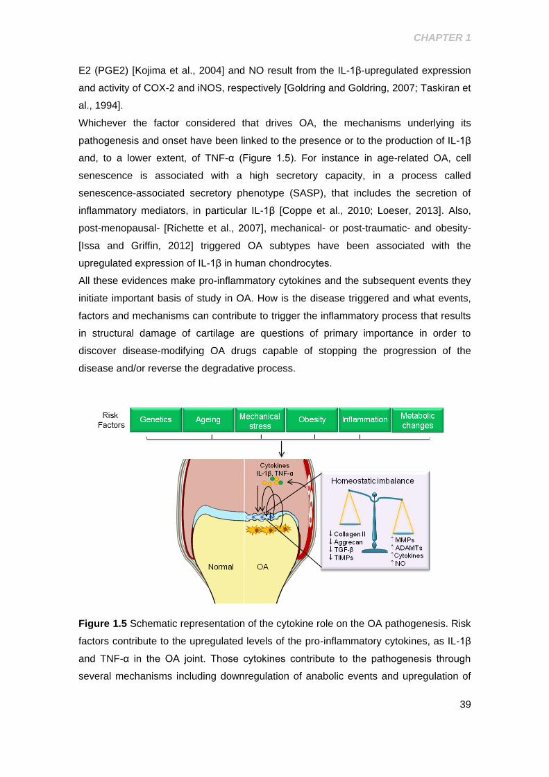
CHAPTER 1
39
E2 (PGE2) [Kojima et al., 2004] and NO result from the IL-1β-upregulated expression
and activity of COX-2 and iNOS, respectively [Goldring and Goldring, 2007; Taskiran et
al., 1994].
Whichever the factor considered that drives OA, the mechanisms underlying its
pathogenesis and onset have been linked to the presence or to the production of IL-1β
and, to a lower extent, of TNF-α (Figure 1.5). For instance in age-related OA, cell
senescence is associated with a high secretory capacity, in a process called
senescence-associated secretory phenotype (SASP), that includes the secretion of
inflammatory mediators, in particular IL-1β [Coppe et al., 2010; Loeser, 2013]. Also,
post-menopausal- [Richette et al., 2007], mechanical- or post-traumatic- and obesity-
[Issa and Griffin, 2012] triggered OA subtypes have been associated with the
upregulated expression of IL-1β in human chondrocytes.
All these evidences make pro-inflammatory cytokines and the subsequent events they
initiate important basis of study in OA. How is the disease triggered and what events,
factors and mechanisms can contribute to trigger the inflammatory process that results
in structural damage of cartilage are questions of primary importance in order to
discover disease-modifying OA drugs capable of stopping the progression of the
disease and/or reverse the degradative process.
Figure 1.5 Schematic representation of the cytokine role on the OA pathogenesis. Risk
factors contribute to the upregulated levels of the pro-inflammatory cytokines, as IL-1β
and TNF-α in the OA joint. Those cytokines contribute to the pathogenesis through
several mechanisms including downregulation of anabolic events and upregulation of
CHAPTER 1
40
catabolic and anabolic responses, effects that lead to the homeostatic imbalance that
result on structural damage of the OA joint
1.3.2.1.2 Nitric Oxide
Among the inflammatory mediators upregulated in OA chondrocytes, NO seems to
have a prominent role. Several studies suggest that it can have different functions in
normal and pathological joint conditions that appear to depend on the concentrations of
NO found. In the cartilage of the OA joint, NO production is increased, and this
excessive amount of NO has been mainly considered a catabolic factor that may be
responsible for perpetuating the release of pro-inflammatory cytokines and other
catabolic processes involved in OA process [Abramson, 2008].
Nitric oxide synthesis is catalyzed by one of the three isoforms of the nitric oxide
synthase (NOS), iNOS, eNOS and nNOS. The inducible isoform, iNOS, is upregulated
in OA chondrocytes, as result for example of mechanical stress, induced by abnormal
compressive and shear forces [Fermor et al., 2001; Lee et al., 2003], pro-inflammatory
cytokines, like TNF-α, IL-1β and IL-17 [Mendes et al., 2002a; Pacquelet et al., 2002;
Palmer et al., 1993] or even as result of the presence of collagen [Chowdhury et al.,
2010] and fibronectin fragments [Yasuda et al., 2004]. The upregulation of iNOS results
in the overproduction of NO in OA joints [Melchiorri et al., 1998]. NO itself plays a role
in perpetuating the release of inflammatory cytokines and other catabolic factors
contributing to the positive loop on the inflammatory process found in OA joints. For
instance NO is, in part, responsible for the synthesis of caspase 1 or Interleukin-1-
coverting enzyme [Boileau et al., 2002], the enzyme responsible for the activation of IL-
1β and IL-18 and for downregulation of its natural inhibitor, the protease inhibitor 9
(PI9), thus sustaining the inflammatory response by promoting the maturation of
inflammatory cytokines [Boileau et al., 2002]. It also participates in the activation of the
AP-1 [Mendes et al., 2002c] and NF-κB [Abramson, 2008], transcription factors that
critically mediate the gene expression of inflammatory mediators, like IL-1β, TNF-α and
iNOS itself, and catabolic factors, as MMPs [Boileau et al., 2002; Sasaki et al., 1998;
Xie et al., 1994].
Apart from its role in sustaining the inflammatory process in OA, NO also participates in
the modulation of proteoglycan, collagen and MMPs expression. It has been shown
that NO mediates IL-1β-induced suppression of proteoglycans and collagen synthesis
in human chondrocytes [Taskiran et al., 1994], increases the gene expression of MMP-
CHAPTER 1
41
1, -2, -3 and -9 in rabbit articular chondrocytes [Sasaki et al., 1998] and suppresses the
endogenous activity of TIMP-1 in human chondrocytes [Ridnour et al., 2007].
On the other hand, several other studies, in part, counteract the observations above
showing that NO also have an important role on the maintenance of normal joint
conditions. In human chondrocytes, it was shown that the IL-17- induced NO
production suppresses the matrix synthesis and protects cartilage from matrix
breakdown [Cai et al., 2002]. It also seems to promote procollagen synthesis in tendon
cells [Xia et al., 2006]. It is also contradictory the information regarding the pro-
inflammatory activity and the cartilage protection yield by NO. Henrotin (1998) showed
that, in human chondrocytes, NO reduces the synthesis of PGE2, IL-6 and IL-8
[Henrotin et al., 1998] and Rosa (2008) showed that NO can also help to downregulate
NF-κB activity [Rosa et al., 2008]. Moreover, in an animal model of OA where the iNOS
gene was silenced, the inflammatory symptoms were exacerbated, as well as cartilage
degradation [McInnes et al., 1998; Veihelmann et al., 2001].
Given the variety of catabolic but also protective effects of NO in the chondrocytes, its
production, as a potential target for disease-modifying therapy for OA must be carefully
managed. Both the inhibition of iNOS, for inhibition of the high amounts of NO
associated with catabolic events and the NO donation might have benefits on OA
treatment [Abramson, 2008].
1.3.2.1.3 Nuclear Factor – κB (NF-κB)
The upregulation by IL-1β and TNF-α of COX-2, MMP-13, iNOS and other central
molecules of the osteoarthritic process is mediated by the induction and activation of
transcription factors and signalling pathways including NF-κB and AP-1 which are
involved generally in stress and inflammation-induced signalling [Goldring and
Goldring, 2007]. Among the signalling pathways involved in the initiation and
amplification of inflammatory responses and tissue destruction, the transcription factor
NF-κB is regarded as its primary regulator [Tak and Firestein, 2001]. NF-κB represents
a family of proteins sharing a Rel homology domain in their N-terminus that include NF-
κB1 (p50 and its precursor p105) and NF-κB2 (p52 and its precursor p100) Rel A (p65),
Rel B, c-Rel (REL). This Rel domain is responsible for homo- and heterodimer
formation, for the interaction with inhibitory molecules and for binding to the promoter
sequences of target genes [Ge et al., 2011; Hayden and Ghosh, 2008a; Oeckinghaus
and Ghosh, 2009]. This transcription factor takes part of the normal physiology of cells;
however, the inappropriate regulation of its activity has been implicated in the
pathogenesis of diverse pathologies [Marcu et al., 2010] with several studies showing
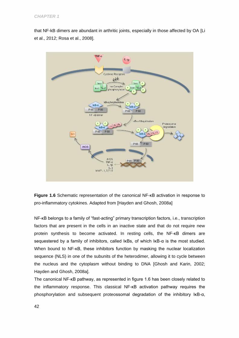
CHAPTER 1
42
that NF-kB dimers are abundant in arthritic joints, especially in those affected by OA [Li
et al., 2012; Rosa et al., 2008].
Figure 1.6 Schematic representation of the canonical NF-κB activation in response to
pro-inflammatory cytokines. Adapted from [Hayden and Ghosh, 2008a]
NF-κB belongs to a family of “fast-acting” primary transcription factors, i.e., transcription
factors that are present in the cells in an inactive state and that do not require new
protein synthesis to become activated. In resting cells, the NF-κB dimers are
sequestered by a family of inhibitors, called IκBs, of which IκB-α is the most studied.
When bound to NF-κB, these inhibitors function by masking the nuclear localization
sequence (NLS) in one of the subunits of the heterodimer, allowing it to cycle between
the nucleus and the cytoplasm without binding to DNA [Ghosh and Karin, 2002;
Hayden and Ghosh, 2008a].
The canonical NF-κB pathway, as represented in figure 1.6 has been closely related to
the inflammatory response. This classical NF-κB activation pathway requires the
phosphorylation and subsequent proteossomal degradation of the inhibitory IκB-α,
CHAPTER 1
43
which releases NF-κB dimers, commonly made of p65 (or RelA) and p50 proteins
[Hayden and Ghosh, 2008a; O'Dea and Hoffmann, 2009]. Free dimers translocate to
the nucleus to activate the transcription of target genes, particularly those of pro-
inflammatory cytokines (e.g. IL-1β, TNF-α), chemokines (e.g. CXCL-8) and
inflammation-associated enzymes (iNOS and COX-2) [Hayden and Ghosh, 2008b].
In the context of OA, NF-κB can be triggered by several distinct factors and
mechanisms generated or not within the cartilage. Mechanical stress [Agarwal et al.,
2004; Ferretti et al., 2006; Knobloch et al., 2008], pro-inflammatory mediators, in
particular IL-1β and TNF-α [Frank et al., 2013; Hayden and Ghosh, 2008a], iNOS
[Aktan, 2004] or even ECM protein fragments [Chowdhury et al., 2010; Pulai et al.,
2005] are known to activate this transcription factor [Uwe, 2008; Yasuda, 2011]. Either
acting by itself or in coordination with other mediators or signalling cascades, NF-κB
induces additional expression of inflammatory genes, impairs the anabolic activity and
directly enhances the secretion of several matrix-degrading proteinases [Fan et al.,
2006; Ge et al., 2011; Mengshol et al., 2000; Stove et al., 2000]. More specifically, NF-
κB seems to be involved in every step of the cartilage change that occurs in OA. In the
OA cartilage, the responses induced by the chondrocytes with a degradative
phenotype were NF-κB mediated and included the production of MMP-1, MMP-3,
MMP-8, MMP-13, ADAMTS-4 and ADMATS-5 [Liacini et al., 2003; Liacini et al., 2002],
increased expression of COX-2 and PGE2, iNOS and its product, NO, IL-1β, IL-6, TNF-
α and type X collagen, thus directly contributing to the increased cartilage degradation
and further chondrocyte hypertrophy [Goldring and Marcu, 2009]. Thus, it is clear that
in OA, NF-κB is part of a positive loop, resulting in a sustained activation of this
transcription factor and, in additional expression of inflammatory genes and catabolic
mediators further contributing to cartilage degradation.
Moreover, NF-κB activation also results on the expression of other regulatory factors,
including E74-like factor 3 (ELF3), hypoxia-inducible factor-2alpha (HIF-2), runt-related
transcription factor 2 (RUNX2) and Bone morphogenic protein-2 (BMP-2), which further
trigger the hyperthrophic state of the chondrocyte [Goldring, 2012].
Regarding its pivotal role in OA onset and progression, NF-κB is regarded as an
important therapeutic target. However, given its importance for cell survival and
maintenance, targeted strategies have to be weighted in order to not cause unwanted
side effects due to complete inhibition [Uwe, 2008]. Targeting specifically the canonical
activation of NF-κB, namely IκB-α phosphorylation and the upstream events may
prevent the high activation of this transcription factor that contributes to cartilage
degeneration in OA pathology [Marcu et al., 2010]
CHAPTER 1
44
1.3.2.1.4 Mitogen Activated Protein Kinases (MAPK)
Together with NF-κB, MAPKs pathways are key regulators of the chondrocyte
functions, both during development and in adult cartilage [Thalhamer et al., 2008].
MAPK are a family of Ser/Thr kinases formed by 4 subfamilies of kinases with different
isoforms, including c-Jun NH2- terminal kinase (JNK), p38 and extracellular signal-
regulated kinase (ERK), each presenting isoforms [Johnson and Lapadat, 2002].
MAPK are activated by phosphorylation of specific Thr and Tyr residues by MAPK
kinases (MKKs). Such phosphorylation can be triggered by a great range of factors,
namely cytokines, growth factors and/or integrin-mediated signals which activate
downstream events that result on the activation of one or more MAPK. Once activated,
the MAPK in turn, activate other protein kinases and several transcription regulatory
proteins, including ERK activation of Elk-1 and c-Myc, p38 activation of ATF-2 and
MEF2, and JNK activation of c-Jun (figure 1.7). These transcription factors regulate the
expression of a set of genes relevant to OA, namely some involved in the inflammatory
response, regulation of cell proliferation, and production of matrix-degrading enzymes
[Berenbaum, 2004; Loeser et al., 2008]. JNK may be particularly relevant due to its role
in the activation of c-jun [Han et al., 2001; Johnson and Nakamura, 2007; Kristof et al.,
2001], a key AP-1 component essential for the expression of MMPs and pro-
inflammatory cytokines, such as TNF-α and IL-1β [Loeser et al., 2008] and other pro-
inflammatory mediators, as iNOS and NO [Kristof et al., 2001; Mendes et al., 2003b]
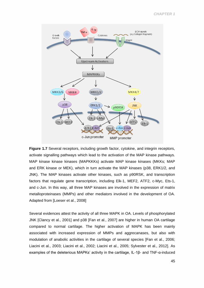
CHAPTER 1
45
Figure 1.7 Several receptors, including growth factor, cytokine, and integrin receptors,
activate signalling pathways which lead to the activation of the MAP kinase pathways.
MAP kinase kinase kinases (MAPKKKs) activate MAP kinase kinases (MKKs; MAP
and ERK kinase or MEK), which in turn activate the MAP kinases (p38, ERK1/2, and
JNK). The MAP kinases activate other kinases, such as p90RSK, and transcription
factors that regulate gene transcription, including Elk-1, MEF2, ATF2, c-Myc, Ets-1,
and c-Jun. In this way, all three MAP kinases are involved in the expression of matrix
metalloproteinases (MMPs) and other mediators involved in the development of OA.
Adapted from [Loeser et al., 2008]
Several evidences attest the activity of all three MAPK in OA. Levels of phosphorylated
JNK [Clancy et al., 2001] and p38 [Fan et al., 2007] are higher in human OA cartilage
compared to normal cartilage. The higher activation of MAPK has been mainly
associated with increased expression of MMPs and aggrecanases, but also with
modulation of anabolic activities in the cartilage of several species [Fan et al., 2006;
Liacini et al., 2003; Liacini et al., 2002; Liacini et al., 2005; Sylvester et al., 2012]. As
examples of the deleterious MAPKs’ activity in the cartilage, IL-1β- and TNF-α-induced
CHAPTER 1
46
expression of MMP-1, MMP-13 and aggrecanase 1 and downregulation of collagen
type II in chondrocytes requires JNK and p38 activation [Fan et al., 2006; Liacini et al.,
2003; Liacini et al., 2002; Liacini et al., 2005; Sylvester et al., 2012], and the
modulation of anabolic and catabolic activities by collagen type II and fibronectin
fragments is mediated by MAPK activation [Chowdhury et al., 2010; Loeser et al.,
2003; Mengshol et al., 2000; Sondergaard et al.]
All three MAPK are involved in the expression of MMPs and other factors involved in
the development of OA. In the majority of cases, the mediation of cartilage destruction
did not address a specific MAPK, but two MAPK or the three MAPK signalling
pathways. Contrariwise, the inhibition of a single MAPK seems to be, in most cases,
enough to inhibit the increased expression of MMPs or aggrecanases. For example, IL-
1β stimulation of MMP-13 production requires the activation of both JNK and p38
[Mengshol et al., 2000] and fibronectin fragments activate ERK1/2, p38 and JNK1/2,
but inhibition of one of the three MAPK is enough to inhibit the fibronectin fragments-
stimulated MMP-13 expression [Loeser et al., 2003].
Besides the role of MAPK in mediating the catabolic function of chondrocytes, several
studies also showed that they take part on deregulated anabolic processes observed in
OA. Activation of ERK and p38 was associated with inhibition of aggrecan expression
[Masuko et al., 2007] and the LPS-inhibited synthesis of proteoglycans in chondrocytes
can be partially reversed by inhibition of p38 [Bobacz et al., 2007].
1.4 OA TREATMENT
According to the World Health Organization, OA is among the high burden diseases, in
Europe particularly, for which the currently available treatment is inadequate. OA is
highly common and is increasing not only in the elderly but also among the general
population, and available treatment is ineffective in stopping or reversing disease
progression [Kaplan et al., 2013; Wieland et al., 2005].
Most of the times, the major disease outcomes, as pain, loss of the joint function and
disability [Wieland et al., 2005], are only distinctly observed in late stages of OA, being
difficult to establish a correlation between symptoms and pathology in early stages of
the disease, which together with the limited biological markers make an early
intervention in the disease difficult [Rousseau and Garnero, 2012].
Regarding these difficulties, OA pain and functional impairment of the patient, as the
major OA burden on individuals, have been the main focus of OA therapy. In addition
CHAPTER 1
47
to the first-line non-pharmacological measures, such as weight loss and physical
exercise, OA therapy includes pharmacological symptomatic treatment with analgesics,
classical non-steroidal anti-inflammatory agents and COX-2 selective inhibitors
(NSAIDs and COXIBs), glucosamine and chondroitin sulphate supplements and/or
intra-articular administration of steroids or hyaluronic acid, all of them with limited
efficacy and/or adverse side effects, especially in the long-term utilization that the
disease requires [Wieland et al., 2005]. To date, the approved treatments directed at
reducing the symptoms of OA have not been shown to prevent or reverse joint
structural damage so, in severe cases, joint replacement surgery is the last choice
effective in relieving the painful and debilitating effects of the disease.
Consequently, there is an urgent need for OA disease-modifying therapies which also
improve symptoms and are safe for clinical use over long periods of time.
As we described before, OA is not considered only an age-related or a “wear and tear”
disease, but a multiplicity of factors and conditions can influence and lead to OA onset
and progression. The complexity and variability of OA aetiology is believed to have a
further impact in the response to treatment. Thus, a better understanding of particular
pathophysiological mechanisms involved in each particular OA phenotype together with
an individual evaluation of the patient’s co-morbidities and with the evaluation of
specific biomarkers of joint destruction [Poole, 2003] is necessary to found better
targeted and effective therapies with capacity to prevent the degenerative joint process
and reverse the already established damage, without underestimate the pain [Goldring
and Goldring, 2007; Lane et al., 2011; Mobasheri, 2013].
1.4.1 Currently Available Treatment
Current treatment options for OA patients are focused on relief from pain and
improvement of joint function. Not surprisingly, since these medical interventions do not
consider the full OA illness, they have little effect, if any at all, on the structural
degradation of the joint tissue. Because of that, many patients have surgery as the only
option to improve quality of life [McAlindon et al., 2014].
General recommendations by the European League Against Rheumatism (EULAR),
the American College of Rheumatology (ACR) and the Osteoarthritis Research Society
International (OARSI) for the management of OA include a combination of non-
pharmacological interventions and pharmacological treatments (figure 1.8) [Hochberg
et al., 2012; McAlindon et al., 2014; Zhang and Doherty, 2006; Zhang et al., 2008].
OARSI has specifically developed guidelines for the non-surgical treatment of OA that

CHAPTER 1
48
are based on the stratification of patients into four groups: patients with knee-only OA
and no comorbidities, patients with knee-only OA with comorbidities, patients with
multi-joint OA and no comorbidities, and patients with multi-joint OA with comorbidities.
These comorbidities included diabetes, hypertension, cardiovascular disease, renal
failure, gastrointestinal bleeding, depression, or a physical impairment limiting activity,
like obesity, and were taken into account based on that those co-morbidities might
influence treatment choices for OA [McAlindon et al., 2014].
From the classes of medications currently used to treat OA of the knee we can refer:
analgesics/ anti-inflammatory agents, glucocorticoids, opioids and slow-acting drugs for
osteoarthritis (SYSADOA).
Acetaminophen (paracetamol) is recommended for initial management of OA, as it is
generally safe and particularly good for treatment of mild pain, being the first line
therapy for knee OA [Hochberg et al., 2012; Towheed et al., 2006]. Oral and topical
non-steroidal inflammatory drugs (NSAIDs) are considered when patients do not
respond to acetaminophen. NSAIDs are the most common treatment for rheumatologic
conditions, including OA, rheumatoid arthritis (RA) and gout. They present analgesic,
antipyretic and anti-inflammatory effects and act mostly through inhibition of the 2
isoforms of COX-1 and COX-2 enzymes, leading to the inhibition of prostaglandin
synthesis that is directly related with pain and inflammation [Wieland et al., 2005]. Most
conventional NSAIDs inhibit both COX isoforms which is directly related to their
gastrointestinal adverse side effects. More recently, these drugs have also been
associated with significant cardiovascular side-effects that warrant caution in patients
at higher risk [Soubrier et al., 2013]. Selective COX-2 inhibitors were developed in
order to avoid those side effects as this isoform is supposed to be induced at the site of
inflammation and not on the gastrointestinal system; however, they are not devoid of
side effects, as nephrotoxicity and hypertension, which significantly limit their
therapeutic use, especially in chronic conditions like OA [Hochberg, 2005]. Despite
their side effects, NSAIDs are recommended by EULAR and OARSI treatment
guidelines committee recommends NSAIDs for management of hip and knee OA
[McAlindon et al., 2014].
Opioid analgesics, such as tramadol, are useful alternatives in patients in whom
NSAIDs, including COX2-selective inhibitors, are contraindicated, ineffective and/or
poorly tolerated. The increased risk of adverse side effects, particularly in the elderly,
has to be taken seriously [Nuesch et al., 2009; Wieland et al., 2005]. Finally, the
treatment with glucocorticoids is one of the last pharmacological lines of OA treatment
and could be used as alternatives for pain relief in patients who cannot use or do not
CHAPTER 1
49
respond to other first line therapies [Towheed et al., 2006]. However, they can bring
about adverse effects, including mild ones as constipation, nausea and itching, and
more serious, as respiratory depression, and addiction [Nuesch et al., 2009].
In addition to the types of medications mentioned above, there is a heterogeneous
group of medications that, unlike the COX-2 inhibitors, do not inhibit prostaglandin
synthesis. This group includes hyaluronic acid (HA), D-glucosamine sulphate (GlcN),
chondroitin sulphate (CS), and diacerhein. These medications are collectively called
Symptomatic Slow Acting Drugs for Osteoarthritis (SYSADOA), and have been
recommended by EULAR for the treatment of pain in OA [Zhang and Doherty, 2006].
Nevertheless, their usefulness is controversial, being considered as treatments of
“uncertain appropriateness” by OARSI on the last update of Knee OA management
guidelines [McAlindon et al., 2014]. This heterogeneous group of compounds was the
first to be linked to structure-modifying effects in the disease with several evidences
showing that these drugs slow the radiographical progression of knee OA [Zhang et al.,
2008]. Their activities can range from inhibition of inflammation to a potential alteration
of the viscoelastic properties of cartilaginous tissue [Hochberg et al., 2012; Michael et
al., 2010; Towheed and Anastassiades, 2007]. Several studies have shown that they
have a slow onset of action improving OA symptoms after a couple of weeks, benefits
that last for a long period after the end of treatment [Volpi, 2004; Volpi, 2006]. In a
randomized, controlled, double-blind trial, patients with knee OA treated with intra-
articular injection of hyaluronic acid had significantly less pain and better function for up
to three weeks afterward the end of treatment with no severe side effects reported
[Petrella and Petrella, 2006]. On the other hand, several studies reported opposite
effects regarding the efficacy of CS, GlcN and HA. Among the studies that compare
glucosamine with placebo for OA treatment, one review found no statistically significant
benefit of glucosamine for pain [Wandel et al., 2010] and the other found a positive
effect for pain that did not reach statistical significance [Towheed et al., 2005]. For HA
there is only a small effect in the treatment of knee OA compared with placebo injection
[Bannuru et al., 2011; McAlindon et al., 2014; Rutjes et al., 2012] and for CS, results
differed regarding symptom relief, with some reviews finding no significant benefit over
placebo for pain and others finding large effect sizes in favor of chondroitin [Hochberg
et al., 2008].
Diacerhein is also a SYSADOA with anti-inflammatory properties used in OA treatment.
It works by blocking the action of IL-1β, and is a potent inhibitor of IL-1β-induced NF-κB
activation and NO production in chondrocytes [Mendes et al., 2002b; Pelletier et al.,
1998]. Several evidences show that diacerhein is slightly, but significantly, more
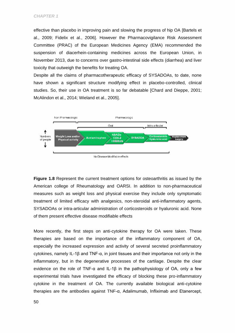
CHAPTER 1
50
effective than placebo in improving pain and slowing the progress of hip OA [Bartels et
al., 2009; Fidelix et al., 2006]. However the Pharmacovigilance Risk Assessment
Committee (PRAC) of the European Medicines Agency (EMA) recommended the
suspension of diacerhein-containing medicines across the European Union, in
November 2013, due to concerns over gastro-intestinal side effects (diarrhea) and liver
toxicity that outweigh the benefits for treating OA.
Despite all the claims of pharmacotherapeutic efficacy of SYSADOAs, to date, none
have shown a significant structure modifying effect in placebo-controlled, clinical
studies. So, their use in OA treatment is so far debatable [Chard and Dieppe, 2001;
McAlindon et al., 2014; Wieland et al., 2005].
Figure 1.8 Represent the current treatment options for osteoarthritis as issued by the
American college of Rheumatology and OARSI. In addition to non-pharmaceutical
measures such as weight loss and physical exercise they include only symptomatic
treatment of limited efficacy with analgesics, non-steroidal anti-inflammatory agents,
SYSADOAs or intra-articular administration of corticosteroids or hyaluronic acid. None
of them present effective disease modifiable effects
More recently, the first steps on anti-cytokine therapy for OA were taken. These
therapies are based on the importance of the inflammatory component of OA,
especially the increased expression and activity of several secreted proinflammatory
cytokines, namely IL-1β and TNF-α, in joint tissues and their importance not only in the
inflammatory, but in the degenerative processes of the cartilage. Despite the clear
evidence on the role of TNF-α and IL-1β in the pathophysiology of OA, only a few
experimental trials have investigated the efficacy of blocking these pro-inflammatory
cytokine in the treatment of OA. The currently available biological anti-cytokine
therapies are the antibodies against TNF-α, Adalimumab, Infliximab and Etanercept,
CHAPTER 1
51
which were initially approved for RA treatment [van der Bijl et al., 2008]. For OA, in
humans, a case report described the successful treatment of inflammatory knee OA
using Adalimumab [Grunke and Schulze-Koops, 2006] with improvement not only in
the symptoms, but also on MRI findings with an important reduction of synovitis and
synovial effusion. However, the results of the open label study with this drug in 12
patients with erosive OA of the hands revealed only a modest improvement in the
number of swollen joints, disability score, pain score, and physician global assessment
[Magnano et al., 2007].
1.4.2 Disease Modifying OA Treatment
Given the high disability burden among patients, the social-economic impact generated
by OA and the limitations of the existing non-pharmacologic/protective measures and
pharmacological agents, new treatments for OA are urgently needed. In particular,
disease-modifying OA drugs (DMOADs), capable of slowing or halting the structural
changes in joint tissues occurring in OA, are crucial to significantly reduce pain and
restore the joint function.
Cytokine cascades, nitric oxide, proteinase release and activation, senescence,
apoptosis, etc., are all processes or mediators affecting all the cells of the joint,
whether they are located in the synovium, bone or in the articular cartilage [Qvist et al.,
2008], that can be considered important targets for DMOADs.
In theory, replacing the balance between those processes would stop the progression
and eventually favor the reparative process which are the main focus of the DMOADs
principals [Alcaraz et al., 2010]. The development of DMOADs presents many
challenges that include, firstly, the complexity of the disease, namely the involvement
of several joint structures (cartilage, synovium and bone…) the complex
etiopathogenesis, the heterogeneity of clinical presentations and the slow progression
of OA, which create a complex environment for therapeutic development with disease-
modifying objectives [Hunter, 2011]. The second one seems to be the lack of sensitive
biomarkers and measurable clinical endpoints that help to connect the structural
changes to the patient response and assess the efficacy of new formulations. For
instance, the determination of joint space narrowing could be an important radiograph
measure of cartilage loss, however, its use as single endpoint considered for the
evaluation of the efficacy of a new drug could be critical. The space between the two
bones in a joint, in a radiograph view represents the overall thickness of the joint
cartilage surfaces of each opposing bone as they articulate, or come into contact with
CHAPTER 1
52
one another. In conditions like arthritis, the joint space gets narrow because of cartilage
degradation. Although this parameter can be relevant on the late phases of OA were
the cartilage degradation and loss is obvious, in early stages this parameter is not that
relevant as could be, for instance, the presence of inflammatory mediators [Qvist et al.,
2008; Sokolove and Lepus, 2013].
Strategies for development of DMOADs include anti-catabolic agents, aiming at
slowing or halting OA progression and anabolic agents aiming at inducing cartilage
regeneration. Anti-catabolic agents would target: 1) direct inhibition of MMP expression
and activity [Clutterbuck et al., 2009], as these proteinases are intrinsically involved in
cartilage breakdown. 2) Prevention of excessive proinflammatory cytokine production,
especially IL-1β and TNF-α but also IL-6, IL-8, and IL-17, also implicated in OA
pathophysiology [Goldring, 2001; Rudolphi et al., 2003] and/or 3) prevention of the
activation of major signalling pathways that drive inflammatory and catabolic
responses, as those of NF-κB [Costa et al., 2012; Marcu et al., 2010] and MAPKs
[Khalife and Zafarullah, 2011], since their activation by stress-related stimuli regulates
the expression of many cytokines, chemokines, adhesion molecules, inflammatory
mediators, and several matrix-degrading enzymes related with joint damage [Goldring,
2006]. On the other hand, pro-anabolic therapeutic agents, such as growth factors,
could be used to target several receptors and signalling pathways that[Pelletier, 2004]
increase chondrocyte anabolism [Pelletier, 2004].
Highly specific drugs, small interfering RNAs (siRNAs), or other biological inhibitors are
under the focus of current OA research [Mobasheri, 2013], but despite the large
number of active research and drug discovery programs to identify structure-modifying
approaches to inhibit joint destruction in OA, none has been conclusive or showed
significant efficacy as a DMOAD and to date, no DMOADs are approved by the Food
and Drug Administration (FDA) or the EMA. Oral administration of PG-116800, a MMP
inhibitor effective against MMP-2, -3, -8, -9, -11, -13, and -14 was associated with
severe musculoskeletal adverse effects without clear benefits for knee OA [Krzeski et
al., 2007]. Similarly, other non-selective MMP inhibitors evaluated for oncology were
associated with arthralgia, stiffness and joint swelling, as well as with other severe
musculoskeletal complications [Bissett et al., 2005; King et al., 2003; Leff et al., 2003].
Nevertheless, the MMP inhibitor, doxycycline, has demonstrated potential efficacy for
disease modification [Brandt et al., 2005; Maher et al., 2013]. Development of more
selective MMP inhibitors could potentially improve the balance between benefit and
side effects in patients with OA.
CHAPTER 1
53
Similarly, despite the clear role of inflammation in OA, no anti-inflammatory therapies
were found, and those under test, including use of biologic agents to inhibit TNFα and
IL-1β, all proved to be disappointing [Hunter, 2008]. IL-1 or IL-1R inhibitors have been
tested in preclinical and clinical trials of RA, however only a few of these have been
investigated in OA, e.g. Anakinra (IL-1R antagonist) and AMG-108 (anti-IL-1 human
antibody, anti-IL-1 huMAb). Their beneficial effects on symptoms and signs in knee OA
are still controversial [Chevalier et al., 2005; Yang et al., 2008].
Other DMOADs are under investigation namely the iNOS inhibitor, SD-6010, by Pfizer
which rationale for development rests on the inhibition of pro-catabolic and pro-
inflammatory effects of effects of NO generated by the upregulation of the synthetic
activity of enzyme, iNOS [Abramson, 2008] and the COX-inhibiting NO-donating
compound naproxcinod (formerly HCT 3012), from NiCox. Naproxcinod is a derivative
of naproxen and is the first compound in the COX-inhibiting NO-donating class, which
integrates the anti-inflammatory effects of the NSAID with the potential beneficial
effects of NO on the cardiovascular system. It is in phase III trial for the treatment of
OA [Anonimous, 2007; Qvist et al., 2008].
Since ever, natural products, as plants, are a source of compounds with
pharmacological properties. In the last decades, a great percentage of the new drug
entities registered have a natural origin or are inspired in a natural compound which
proves them as a believable source of new compounds with pharmacological
properties[Newman and Cragg, 2007]. Accordingly, research on OA therapy has
focused on these sources to found new compounds with disease modifying properties
with very promising results [Henrotin et al., 2013; Khalife and Zafarullah, 2011;
Mobasheri, 2012; Seidl, 2002; Shakibaei et al., 2012; Shakibaei et al., 2011; Shakibaei
et al., 2005], However, none has yet proceed to clinical development.
The biodiversity of the Portuguese flora opens wide possibilities for identification of
compounds to be studied in the context of chronic inflammatory diseases and
specifically in OA. From the panoply of compounds that can be obtained from plants,
literature and preliminary studies showed that small volatile and hydrophobic plant
metabolites can modulate inflammatory signalling pathways [Neves et al., 2009]. A
huge amount of compounds with such characteristics can be found in essential oils
which are typical extracts from aromatic plants, generally obtained by distillation.
CHAPTER 1
54
1.5 OBJECTIVES AND ORGANIZATION OF THE THESIS
OA and DM are age-related diseases whose prevalence is increasing exponentially.
Several studies showed that OA is a heterogeneous disease and that the multiple risk
factors associated with it are responsible for a multiplicity of OA phenotypes. Diabetes
mellitus, especially type 2, has been considered an independent risk factor for the
development and progression of OA in several epidemiological studies which indicate
that OA is more prevalent, more severe, has an earlier onset and progresses more
rapidly in diabetic- than in non-diabetic patients [King et al., 2013; Martinez-Huedo et
al., 2013; Nieves-Plaza et al., 2013; Schett et al., 2012a]. These evidences, as well as
in vitro and in vivo data, including studies by our own group [Rosa et al., 2009; Rosa et
al., 2011b], strongly suggest the existence of a specific DM-associated OA phenotype
[Berenbaum, 2011]. A better understanding of particular underlying physiological and
pathological mechanisms is essential for the design and development of new and
better targeted therapeutic strategies effective to halt OA progression, its growing
incidence and its associated huge socio-economic costs [Kaplan et al., 2013]. We
addressed this issue by hypothesizing that hyperglycemia, a hallmark of DM and other
conditions associated with glucose imbalance, is a major effector of chondrocyte
damage, accelerating age-related changes that favor OA development. In this context,
previous studies from our group [Rosa et al., 2009; Rosa et al., 2011b] showed that
hyperglycemia-like glucose concentrations induce catabolic responses and impair the
ability of chondrocytes to respond to anabolic agents. Thus, one of the objectives of
this thesis was to identify mechanisms by which chondrocytes sense and regulate
glucose transport which is the first rate limiting step for glucose utilization and thus can
be a target for preventing deleterious effects caused by hyperglycemia. Moreover, and
to further identify potential therapeutic targets, we also investigated the role of
hyperglycemia in modulating inflammatory responses in chondrocytes. The results
obtained and their discussion are presented in chapters 3 and 4.
Since every factor, including hyperglycemia as we demonstrate in chapter 4, that shifts
chondrocyte homeostasis towards a pro-catabolic state, induces an inflammatory
response in chondrocytes and this is a major effector mechanism of cartilage
destruction in OA, our last objective was to identify compounds that can halt
inflammation and catabolic responses in chondrocytes. Identification of such
compounds is crucial for the development of effective disease-modifying anti-
osteoarthritic drugs. For this, we evaluated a series of essential oils and their
compounds for their ability to inhibit the inflammatory and catabolic processes in
CHAPTER 1
55
human chondrocytes, focusing on the major signalling pathways activated in
osteoarthritic joints, namely NF-κB and the MAPKs. The results obtained are reported
and discussed in chapters 5 to 7.
Finally, even though OA involves the whole joint, cartilage degradation, a multi-faceted
and complex process that characterizes all forms of OA, is the hallmark of the
progression and irreversibility of the disease reason why this work was specifically
focused on this tissue.
The last chapter of this thesis presents a general and integrated overview of the results
reported, matching them with the major objectives defined and identifying new
questions and future perspectives elicited by this work.

CHAPTER 1
56
57
CHAPTER 2.
MATERIAL AND METHODS
2.1 Cartilage Samples
Human knee cartilage was collected, within 12 h of death, from the distal femoral
condyles of multi-organ donors or with informed consent from patients undergoing total
knee replacement surgery at the Orthopaedics Department of the University Hospital of
Coimbra (HUC). The Ethics Committee of HUC approved all procedures. Where
considered relevant for the work goals the samples were classified according to the
degree of macroscopic damage, using the classification of Outerbridge (1961). Group
0-1 includes cartilage samples with no macroscopic signs of degradation (score 0) or
presenting only a softened surface (score 1); Group 2-3 includes fibrillated or fissured
cartilage samples without evident signs of surface erosion (scores 2 and 3); and Group
4 comprises severely damaged cartilage samples, with areas of extensive full thickness
erosion that exposed the subchondral bone, corresponding to advanced OA.
2.2 Chondrocyte Isolation and Culture
Chondrocytes were isolated by enzymatic digestion from non-pooled cartilage samples.
Briefly, cartilage slices underwent sequential digestion with Pronase (Roche,
Indianapolis, IN, USA), a non-specific protease for 1h30min and collagenase A (Roche)
overnight. To avoid chondrocyte dedifferentiation, non-proliferating monolayer cultures
were setup by plating 1x10⁶ chondrocytes/ml in Ham : F-12 medium containing 3%
antibiotic/antimycotic solution and 5% Fetal Bovine Serum (Invitrogen, Life
technologies, Carlsbad, CA) and allowed to recover for 24 h at 37ºC in a humidified
atmosphere supplemented with 5% CO2.
Cells from the human chondrocytic cell line, C28/I2, kindly provided by Prof. Mary
Goldring (currently at the Hospital for Special Surgery, New York) and Harvard
University, were cultured in DMEM/ Ham’s F-12 (1:1, v/v) and passaged every
four days, as recommended. For the experiments 0,5x10⁶ chondrocytes/mL were
plated and allowed to recover for 24h in similar conditions to that of primary human
chondrocytes.
CHAPTER 2
58
Prior to any treatments, the cells were serum-starved overnight and thereafter
maintained in culture medium with 1% antibiotic/antimycotic solution, without serum.
The specific treatments conditions are indicated in figure legends.
2.3 Assessment of Cell Viability
The 3-(4,5-dimethylthiazol-2-yl)-2,5-diphenyltetrazolium (MTT; Sigma) reduction assay,
a quantitative colorimetric method based on the reduction of the MTT salt by the
mitochondrial enzymes of viable cells [Mosmann, 1983], was used to determine the
cytotoxicity and to select non-cytotoxic concentrations of each of the compounds under
test. Briefly, the cells were incubated with each compound in different concentrations
for 24h h after what the culture medium was replaced by fresh medium containing 5
μg/mL MTT and the cells further incubated for 30 min. The resulting dark blue crystals
of formazan were then dissolved in acidified isopropanol and the absorbance of the
corresponding solution, directly proportional to the number of living cells, was
measured in an automatic plate reader (SLT, Austria) set at a test wavelength of 570
nm and a reference wavelength of 620 nm. The absence of cytotoxic effects in each
cell model was defined as the concentration of tested compounds eliciting an
absorbance reading of at least 80% of the value measured in the cells treated with IL-
1β alone for same time period.
2.4 Western Blot
Total, cytoplasmatic and nuclear cell extracts were prepared and subjected to western
blot. Briefly, 25 µg of protein were separated by SDS/PAGE [10% (v/v)] and transferred
onto PVDF membranes by electroblotting. The membranes were blocked with 5% skim
milk for 2 hours in Tris-buffered saline-Tween (TBS-T, 20mM Tris-HCl, 150mM NaCl,
0,1% Tween 20) and probed with the antibodies presented on Table 2.1 and with anti-
rabbit, anti-mouse, anti-goat alkaline phosphatase-conjugated secondary antibodies
(GE Healthcare, Chalfont St Giles, UK). β-tubulin, actin and lamin B1 antibodies were
used to detect those proteins as loading controls. Immune complexes were detected
with the Enhanced ChemiFluorescence reagent (GE Healthcare) and the bands were
analyzed using ImageQuant™TL (version 7.0, GE Healthcare). The results were
normalized by calculating the ratio between the intensities of the bands corresponding
to the protein of interest and the protein used as loading control.
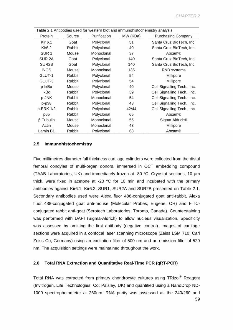
CHAPTER 2
59
Table 2.1 Antibodies used for western blot and immunohistochemistry analysis
Protein Source Purification MW (KDa) Purchasing Company
Kir 6.1 Goat Polyclonal 51 Santa Cruz BioTech, Inc.
Kir6.2 Rabbit Polyclonal 40 Santa Cruz BioTech, Inc.
SUR 1 Mouse Monoclonal 37 Abcam®
SUR 2A Goat Polyclonal 140 Santa Cruz BioTech, Inc.
SUR2B Goat Polyclonal 140 Santa Cruz BioTech, Inc.
iNOS Mouse Monoclonal 135 R&D systems
GLUT-1 Rabbit Polyclonal 54 Millipore
GLUT-3 Rabbit Polyclonal 54 Millipore
p-IκBα Mouse Polyclonal 40 Cell Signalling Tech., Inc.
IκBα Rabbit Polyclonal 39 Cell Signalling Tech., Inc.
p-JNK Rabbit Monoclonal 54 Cell Signalling Tech., Inc.
p-p38 Rabbit Polyclonal 43 Cell Signalling Tech., Inc.
p-ERK 1/2 Rabbit Polyclonal 42/44 Cell Signalling Tech., Inc.
p65 Rabbit Polyclonal 65 Abcam®
β-Tubulin Mouse Monoclonal 55 Sigma-Aldrich®
Actin Mouse Monoclonal 43 Millipore
Lamin B1 Rabbit Polyclonal 68 Abcam®
2.5 Immunohistochemistry
Five millimetres diameter full thickness cartilage cylinders were collected from the distal
femoral condyles of multi-organ donors, immersed in OCT embedding compound
(TAAB Laboratories, UK) and immediately frozen at -80 ºC. Cryostat sections, 10 µm
thick, were fixed in acetone at -20 ºC for 10 min and incubated with the primary
antibodies against Kir6.1, Kir6.2, SUR1, SUR2A and SUR2B presented on Table 2.1.
Secondary antibodies used were Alexa fluor 488-conjugated goat anti-rabbit, Alexa
fluor 488-conjugated goat anti-mouse (Molecular Probes, Eugene, OR) and FITC-
conjugated rabbit anti-goat (Serotech Laboratories; Toronto, Canada). Counterstaining
was performed with DAPI (Sigma-Aldrich) to allow nucleus visualization. Specificity
was assessed by omitting the first antibody (negative control). Images of cartilage
sections were acquired in a confocal laser scanning microscope (Zeiss LSM 710; Carl
Zeiss Co, Germany) using an excitation filter of 500 nm and an emission filter of 520
nm. The acquisition settings were maintained throughout the work.
2.6 Total RNA Extraction and Quantitative Real-Time PCR (qRT-PCR)
Total RNA was extracted from primary chondrocyte cultures using TRIzol® Reagent
(Invitrogen, Life Technologies, Co; Paisley, UK) and quantified using a NanoDrop ND-
1000 spectrophotometer at 260nm. RNA purity was assessed as the 240/260 and
CHAPTER 2
60
280/260 ratios. The cDNA was reverse transcribed using the iScript Select cDNA
Synthesis Kit (Bio-Rad), beginning with 1 µg of RNA. Specific sets of primers used
throughout the work, including housekeeping genes and summarized in Table 2.2 were
designed using Beacon Designer software (PREMIER Biosoft International, Palo Alto,
CA). PCR reactions were performed using 25 g/mL of transcribed cDNA in a final
volume of 20 µL.
The efficiency of the amplification reaction for each gene was calculated using a
standard curve of a series of diluted cDNA samples, and the specificity of the
amplification products was assessed by analyzing the melting curve generated in the
process.
Gene expression changes were analyzed using the built-in iQ5 Optical system software
v2, which enables the analysis of the results with the Pfaffl method, a variation of the
ΔΔCT method corrected for gene-specific efficiencies [Nolan et al., 2006].
The results for each gene of interest were normalized against HPRT-1 or against 18S
ribosomal protein, the housekeeping genes found to be the most stable under the
different experimental conditions used.
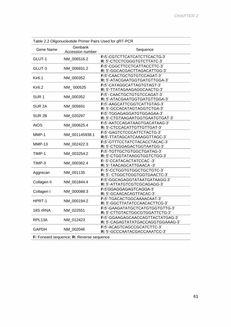
CHAPTER 2
61
Table 2.2 Oligonucleotide Primer Pairs Used for qRT-PCR
Gene Name Genbank
Accession number Sequence
GLUT-1 NM_006516.2 F:5’-CGTCTTCATCATCTTCACTG-3’ R: 5’-CTCCTCGGGTGTCTTATC-3’
GLUT-3 NM_006931.2 F:5’-CGGCTTCCTCATTACCTTC-3’ R: 5’-GGCACGACTTAGACATTGG-3’
Kir6.1 NM_000352 F:5’-CAACTGCTGTGTCCAGAT-3’ R: 5’-ATACGAATGGTGATGTTGGA-3’
Kir6.2 NM_ 000525 F:5’-CATAGGCATTAGTGTAGT-3’ R: 5’-TTATAGAAGAGGCAACTG-3’
SUR 1 NM_000352 F:5’- CAACTGCTGTGTCCAGAT-3’ R: 5’-ATACGAATGGTGATGTTGGA-3’
SUR 2A NM_005691 F:5’-AAGCATTCGGTCATTGTAG-3’ R: 5’-GCCACATAGTAGGTCTGA-3’
SUR 2B NM_020297 F:5’-TGGAGAGGATGTGGAGAA-3’ R: 5’-CTGTAAGAATGGTGAATGTGAT-3’
iNOS NM_000625.4
F:5’-AATCCAGATAAGTGACATAAG-3’ R: 5’-CTCCACATTGTTGTTGAT-3’
MMP-1 NM_001145938.1
F:5’-GAGTCTCCCATTCTACTG-3’ R:5’-TTATAGCATCAAAGGTTAGC-3’
MMP-13 NM_002422.3
F:5’-GTTTCCTATCTACACCTACAC-3’ R: 5’-CTCGGAGACTGGTAATGG-3’
TIMP-1 NM_003254.2
F:5’-TGTTGCTGTGGCTGATAG-3’ R: 5’-CTGGTATAAGGTGGTCTGG-3’
TIMP-3 NM_000362.4
F: 5'-CCATACACTATCCAC -3' R: 5'-TAACAGCATTGAACA -3'
Aggrecan NM_001135 F: 5’-CCTGGTGTGGCTGCTGTC-3’ R: 5’- CTGGCTCGGTGGTGAACTC-3’
Collagen II NM_001844.4
F:5’-GGCAGAGGTATAATGATAAGG-3’ R: 5’-ATTATGTCGTCGCAGAGG-3’
Collagen I NM_000088.3
F:5’GGAGGAGAGTCAGGA-3’ R: 5’-GCAACACAGTTACAC-3’
HPRT-1 NM_000194.2
F:5’-TGACACTGGCAAAACAAT-3’ R: 5’-GGCTTATATCCAACACTTCG-3’
18S rRNA NM_022551 F:5’-GAAGATATGCTCATGTGGTGTTG-3’ R: 5’-CTTGTACTGGCGTGGATTCTG-3’
RPL13A NM_012423 F:5’-GGAAGAGCAACCAGTTACTATGAG-3’ R: 5’-CAGAGTATATGACCAGGTGGAAAG-3’
GAPDH NM_002046 F:5’-ACAGTCAGCCGCATCTTC-3’ R: 5’-GCCCAATACGACCAAATCC-3’
F: Forward sequence; R: Reverse sequence
CHAPTER 2
62
2.7 2-Deoxy-D-Glucose uptake Assay
Glucose transport was determined by measuring the net uptake of a non-metabolizable
analogue of glucose, 2-deoxy-D-glucose (2-DG). Briefly, chondrocytes were pre-
incubated for 18 hours with Glibenclamide (10, 20, 100 nM and 20 M) and then
incubated for 30 minutes at 37oC in glucose-free DMEM containing 0.5 mM 2-DG and
0.5 Ci/mL of [2,6-3H]-2-DG. Cytochalasin B, a glucose transporter inhibitor (10 M),
was used to determine the non-specific uptake. For each sample, the non-specific
uptake was subtracted from the total uptake, before normalization to the respective
protein concentration.
2.8 Nitric Oxide Production
NO production was evaluated as the concentration of nitrite accumulated in the
supernatants of primary chondrocyte cultures treated with IL-1β in the presence or
absence of the test compounds, as indicated in figures and figure legends. Nitrite
concentration was measured using the spectrophotometric method based on the
Griess reaction [Green et al., 1982].
2.9 NF-κB Transcription Factor Assay
A colorimetric ELISA-based assay (NoShift Transcription Factor Assay kit, Novagen,
La Jolla, CA) was used to evaluate the presence of active NF-κB dimers, capable of
binding to the cognate consensus DNA sequence. For this purpose, nuclear extracts
from C-28/I2 cells were incubated with a biotinylated consensus NF-κB oligonucleotide
(NoShift NF-κB Reagents; Novagen) and the assay performed according to the
manufacturerʼs instructions. The absorbance intensity in each sample is directly
proportional to the amount of NF-κB-DNA complexes formed and, thus, to the amount
of active NF-κB dimers present in each sample. In parallel, the specificity of the
reaction was confirmed in competition assays where addition of a 10-fold molar excess
of non-biotinylated wild-type or mutant oligonucleotides (NoShift NF-κB Reagents;
Novagen) to binding reactions containing nuclear extracts from IL-1β treated cells
abrogated or did not affect, respectively, the formation of NF-κB-DNA complexes.
CHAPTER 2
63
2.10 Essential oil identification, Fractionation and Analysis
EOs from the aerial parts of Eryngium duriaei subsp. juresianum (M. Laínz) M. Laínz
(Apiaceae), from the aerial parts of Laserpitium eliasii subsp. thalictrifolium Sennen &
Pau (Apiaceae), from leaves and flowers of Lavandula luisieri (Rozeira) Rivas-Martínez
(Lamiaceae), from the aerial parts of Othantus maritimus (L.) Hoff. & Link (Asteraceae)
and from the aerial parts of Thapsia villosa L. (Apiaceae) were picked from the
collection of plant extracts of the Faculty of Pharmacy, Univ. Coimbra. Voucher
specimens of plant material are deposited at the Herbarium of the Botanic Garden,
Univ. Coimbra (COI) or at the Herbarium of the Faculty of Pharmacy, Univ. Coimbra.
All EOs were prepared at laboratory by water distillation using a Clevenger-type
apparatus (EDQM, 2007) (+)-α-Pinene (≥98% purity) was purchased from Sigma
Chemical Co. (St. Louis, MO, US).
The composition of each essential oil was established following a combined
methodology of GC and GC/MS. Analytical GC was performed in a Hewlett-Packard
6890 (Agilent Technologies, Palo Alto, CA, US) gas chromatograph with a HP GC
ChemStation Rev. A.05.04 data handling system, equipped with a single injector and
two flame ionization detectors (FID). A graphpak divider (Agilent Technologies, part no.
5021-7148) was used for simultaneous sampling to two Supelco (Supelco, Bellefonte,
PA, US) fused silica capillary columns with different stationary phases: SPB-1
(polydimethylsiloxane 30 m × 0.20 mm i.d., film thickness 0.20 μm), and SupelcoWax-
10 (polyethyleneglycol 30 m × 0.20 mm i.d., film thickness 0.20 μm). Oven temperature
program: 70–220°C (3°C/min), 220°C (15 min); injector temperature: 250°C; carrier
gas: helium adjusted to a linear velocity of 30 cm/s; split ratio 1:40; detectors
temperature: 250°C. GC-MS was performed in a Hewlett-Packard 6890 gas
chromatograph fitted with a HP1 fused silica column (polydimethylsiloxane 30 m × 0.25
mm i.d., film thickness 0.25 μm), interfaced with an Hewlett-Packard mass selective
detector 5973 (Agilent Technologies) operated by HP Enhanced ChemStation
software, version A.03.00. GC parameters were as described above; interface
temperature: 250°C; MS source temperature: 230°C; MS quadrupole temperature:
150°C; ionization energy: 70 eV; ionization current: 60 μA; scan range: 35 – 350 units;
scans/s: 4.51.
The essential oils of E. duriaei subsp. juresianum and L. luisieri were fractionated by
flash chromatography on silica gel 63-200 m (Merck) using a 2 cm x 40 cm Omnifit
(Sigma-Aldrich) glass columns. Elution was made in step gradients from 100% of n-
pentane, n-pentane / ethyl ether mixtures, till a final concentration of 100% ethyl ether.
CHAPTER 2
64
Collected fractions were analysed by gas chromatography-mass spectroscopy
(GC/MS) and combined if having similar composition. Analysis were performed using a
Hewlett-Packard 6890 gas chromatograph fitted with a HP1 fused silica column
(polydimethylsiloxane 30 m × 0.25 mm i.d., film thickness 0.25 m), interfaced with an
Hewlett-Packard mass selective detector 5973 (Agilent Technologies) operated by HP
Enhanced ChemStation software, version A.03.00. GC-MS parameters: oven
temperature program: 70–220 C (3 C.min-1), 220 C (15 min); injector temperature:
250 C; carrier gas: helium, adjusted to a linear velocity of 30 cm.s-1; splitting ratio 1:40;
interface temperature: 250 C; MS source temperature: 230 C; MS quadrupole
temperature: 150 C; ionization energy: 70 eV; ionization current: 60 A; scan range:
35–350 units; scans.s-1: 4.51.
Components of each EO and fraction were identified considering their retention indices
(RIs) on both SPB-1 and SupelcoWax-10 columns and their mass spectra. RIs,
calculated by linear interpolation relative to retention times of C8–C23 n-alkanes
(Vandendool & Kratz, 1963), were compared with those of authentic samples,
laboratory database (CEF / Faculty of Pharmacy, University of Coimbra) data available
in digital banks (Linstrom, 2013; El-Sayed, 2012; Acree, 2004 and/ or literature data..
Acquired mass spectra were compared with reference spectra from the laboratory
database, Wiley / NIST library[McLafferty, 2009] Wiley / NIST database (EDQM, 2007)
and literature data [Adams, 1995; Cavaleiro et al., 2011; Joulain, 1998; Videira et al.,
2013] (Adams, 1995; Joulain, 1998). Relative amounts of individual components were
calculated based on GC raw data without further correction. Table 5.1 summarizes the
major components found in each EO and table 7.1 summarizes the major components
found in each fraction of the Eryngium duriaei subsp. juresianum and Lavandula luisieri
essential oils
For cell assays, the EOs and isolated compounds were diluted in Dimethylsulfoxide
(DMSO) and then dispersed in the culture medium to achieve final concentrations of
10-200 µg/mL. The final DMSO concentration did not exceed 0.1% (v/v).
2.11 Statistical Analysis
Results are presented as mean ± SEM. Statistical analysis was performed using
GraphPad Prism (version 5.00). SPSS software (version 17.0) was used to assess the
normality (Kolmogorov Smirnov test) and homogeneity of variances to determine
whether the conditions required to apply parametric tests were satisfied. As in all cases
such conditions were observed, the statistical analysis was performed using the paired

CHAPTER 2
65
Student t-test for comparison of each condition with its respective positive (IL-1β
treated) or negative (untreated cells) control. Results were considered statistically
significant at P <0.05.

67
CHAPTER 3.
EXPRESSION AND FUNCTION OF K(ATP) CHANNELS IN NORMAL AND
OSTEOARTHRITIC HUMAN CHONDROCYTES: POSSIBLE ROLE IN GLUCOSE
SENSING
Ana T. Rufino, Susana C. Rosa, Fernando Judas, Ali Mobasheri, M. Celeste Lopes and
Alexandrina F. Mendes
Expression and Function of K(ATP) Channels in Normal and Osteoarthritic
Human Chondrocytes: Possible Role in Glucose Sensing
Published in Journal of Cellular Biochemistry (2013) 114:1879–1889
CHAPTER 3
68
ABSTRACT
ATP-sensitive potassium [K(ATP)] channels sense intracellular ATP/ADP levels, being
essential components of a glucose-sensing apparatus in various cells that couples
glucose metabolism, intracellular ATP/ADP levels and membrane potential. These
channels are present in human chondrocytes, but their subunit composition and
functions are unknown. This study aimed at elucidating the subunit composition of
K(ATP) channels expressed in human chondrocytes and determining whether they play
a role in regulating the abundance of major glucose transporters, GLUT-1 and GLUT-3,
and glucose transport capacity. The results obtained show that human chondrocytes
express the pore forming subunits, Kir6.1 and Kir6.2, at the mRNA and protein levels
and the regulatory sulfonylurea receptor (SUR) subunits, SUR2A and SUR2B, but not
SUR1. The expression of these subunits was no affected by culture under
hyperglycemia-like conditions. Functional impairment of the channel activity, using a
SUR blocker (glibenclamide 10 or 20 nM), reduced the protein levels of GLUT-1 and
GLUT-3 by approximately 30% in normal chondrocytes, while in cells from cartilage
with increasing osteoarthritic (OA) grade no changes were observed. Glucose transport
capacity, however, was not affected in normal or OA chondrocytes. These results show
that K(ATP) channel activity regulates the abundance of GLUT-1 and GLUT-3,
although other mechanisms are involved in regulating the overall glucose transport
capacity of human chondrocytes. Therefore, K(ATP) channels are potential
components of a broad glucose sensing apparatus that modulates glucose transporters
and allows human chondrocytes to adjust to varying extracellular glucose
concentrations. This function of K(ATP) channels seems to be impaired in OA
chondrocytes.
CHAPTER 3
69
3.1 INTRODUCTION
Glucose serves an important anabolic function in chondrocytes, being required for the
synthesis of matrix components. Due to the avascular nature of cartilage, chondrocytes
are physiologically exposed to low oxygen tensions [Grimshaw and Mason, 2000; Kay
et al., 2011; Zhou et al., 2004b], using glycolysis as the main energy source [Rajpurohit
et al., 1996], even when exposed to normoxia [Schneider et al., 2007]. The supply of
glucose to chondrocytes depends on diffusion from the synovial fluid [Maroudas, 1970]
which is facilitated by joint motion and fluid flow in the extracellular matrix (ECM). The
synovial fluid glucose concentration is identical to that in the plasma both in normal
conditions and in non-inflammatory and inflammatory types of arthritis, except in septic
arthritis where it is usually significantly reduced [Brannan and Jerrard, 2006]. Several
members of the facilitative glucose transporter family – the GLUT/SLC2A transporters
– are expressed in chondrocytes and optimize its uptake [Mobasheri et al., 2008].
GLUT-1 is especially important as both anabolic and catabolic stimuli increase its
expression, while others, like GLUT-3, which is also highly expressed constitutively,
does not seem to be regulated by those stimuli [Mobasheri et al., 2008; Phillips et al.,
2005; Richardson et al., 2003; Shikhman et al., 2004].
On the other hand, extracellular glucose concentrations both below and above
physiologic levels, have been predicted and shown to alter the pattern of gene
expression [Mobasheri et al., 2002a; Rosa et al., 2011b], as well as the availability of
GLUT-1 protein [Rosa et al., 2009] in chondrocytes. Indeed, glucose deprivation
increases the expression of GLUT-1, whereas a supra-physiologic glucose
concentration has the opposite effect, decreasing GLUT-1 content and glucose uptake
in young/healthy chondrocytes, but has no effect on those isolated from osteoarthritic
(OA) cartilage [Rosa et al., 2009]. Therefore, normal chondrocytes seem to be able to
adjust to the extracellular glucose concentration by modulating the abundance of
GLUT-1 and the total glucose transport capacity. Nonetheless, this ability seems to be
compromised in OA chondrocytes in vitro, leading to intracellular glucose
accumulation, increased production of reactive oxygen species [Rosa et al., 2009] and
expression of matrix degrading enzymes [Rosa et al., 2011b] when these cells are
exposed to supra-physiologic glucose concentrations, as those occurring in diabetes
mellitus, metabolic syndrome and other conditions characterized by impaired glucose
metabolism. These conditions are increasingly recognized as independent risk factors
for OA development and progression [Berenbaum, 2011; Del Rosso et al., 2006; Hart
et al., 1995; Katz et al., 2010; Schett et al., 2012b; Sturmer et al., 2001; Velasquez and
CHAPTER 3
70
Katz, 2010]. OA is the major musculoskeletal disorder - or most likely, a set of
disorders with distinct etiological and clinical characteristics [Berenbaum, 2011; Zhuo et
al., 2012] and a main cause of physical disability [Goldring and Marcu, 2009; Martel-
Pelletier et al., 2008]. Understanding how human chondrocytes sense extracellular
glucose and intracellular ATP concentrations and adjust their substrate uptake to
match the cell’s needs and whether such mechanisms remain functional in OA
chondrocytes is, thus, critical to elucidate the mechanisms by which conditions
associated with impaired glucose metabolism contribute to the development and/or
progression of OA.
ATP-sensitive potassium [K(ATP)] channels are weak inwardly rectifying potassium
channels that act as metabolic sensors in a number of cell types, coupling metabolic
activity to the membrane potential and to secretory activity, primarily by sensing
intracellular ATP levels [Burke et al., 2008; McTaggart et al., 2010; Miki et al., 2002;
Minami et al., 2004]. In pancreatic β-cells [Burke et al., 2008] and in subpopulations of
mediobasal hypothalamic neurons [Acosta-Martinez and Levine, 2007], for instance,
K(ATP) channels have been identified as an essential component of a glucose-sensing
mechanism, wherein uptake and metabolism of glucose leads to increased intracellular
ATP/ADP levels, closure of the channels, and increase in insulin secretion and
neuronal excitability, respectively.
Structurally, K(ATP) channels are heteromultimers of four ATP-binding cassette (ABC)
proteins that form the sulfonylurea receptor (SUR), surrounding four pore forming
potassium channel subunits (Kir6) [Babenko et al., 1998]. Kir6 and SUR have two
isoforms each, Kir6.1 and Kir6.2 and SUR1 and SUR2, respectively. SUR2 has two
major splice variants, SUR2A and SUR2B [Burke et al., 2008; Hibino et al., 2010]. ATP
binds to the SUR subunits inducing a conformational change of the Kir6 subunits that
results in channel closure, cessation of K+ efflux and cell membrane depolarization. In
contrast, decreased intracellular ATP availability allows ADP binding and channel
opening with the consequent K+ efflux and hyperpolarization [Babenko et al., 1998; Shi
et al., 2005]
The subunit composition of K(ATP) channels determines their sensitivity to intracellular
ATP and to pharmacological modulators, eliciting the functional diversity of these
channels [Acosta-Martinez and Levine, 2007; Babenko et al., 1998; Inagaki and Seino,
1998]. Kir6.1 and Kir6.2 have similar sensitivities to nucleotides (ATP and ADP), so the
differences found are believed to be due to the SUR subtype existing in each channel
or to the combination of different SUR subunits in the same channel [Chan et al., 2008;
Shi et al., 2005]. K(ATP) channels involved in coupling glucose sensing with insulin
CHAPTER 3
71
secretion in pancreatic β-cells are composed of Kir6.2 and SUR1 [Nielsen et al., 2007],
whereas channels containing Kir6.2 and SUR2A, found in cardiomyocytes, and
channels composed of Kir6.2 or Kir6.1 and SUR2B, occurring in vascular cells,
regulate the duration of the action potential and vasodilatation [Aguilar-Bryan and
Bryan, 1999; Burke et al., 2008; Chan et al., 2008; Gribble and Ashcroft, 2000; Hibino
et al., 2010].
Information regarding the subunit composition and function of K(ATP) channels
expressed in non-excitable cells is much scarcer. Functional K(ATP) channels have
been demonstrated in equine chondrocytes and Kir6.1 was detected in normal and OA
human cartilage [Mobasheri et al., 2007], but their subunit composition and
physiological roles in cartilage are not yet understood, even though these channels are
highly likely to be important for regulation of cartilage metabolism and sensing ATP
levels within the cell [Barrett-Jolley et al., 2010; Mobasheri et al., 2012]. We
hypothesize that K(ATP) channels, by responding to ATP levels, may indirectly act as
components of the glucose sensing apparatus in chondrocytes, generating signals that
allow the cell to adjust to varying extracellular glucose concentrations by regulating the
availability of its facilitative glucose transporters and hence net glucose transport
capacity. Therefore, this work was aimed at i) elucidating the subunit composition of
K(ATP) channels expressed in human chondrocytes and ii) determining whether
K(ATP) channels play a role in regulating the abundance of two major glucose
transporters in chondrocytes, GLUT-1 and GLUT-3, and the glucose transport capacity
of human chondrocytes exposed to hyperglycemia-like conditions. Furthermore, these
processes were evaluated in chondrocytes isolated from cartilage samples with
different degradation grades to determine whether they are impaired in OA.
CHAPTER 3
72
3.2 RESULTS
3.2.1 Characterization of the subunit composition of (KATP) channels
expression in human chondrocytes
To determine the subunit composition of K(ATP) channels present in human
chondrocytes, we evaluated the expression of the different Kir and SUR subunits, both
at the protein and mRNA levels, by western blot and qRT-PCR, respectively. The
results obtained show that all the pore-forming and regulatory subunits that can
compose K (ATP) channels are expressed in human chondrocytes, although with
different intensities and inter-individual variability (Fig. 3.1). Kir6.1, SUR1, SUR2A and
SUR2B are expressed both at the protein and mRNA levels (Fig. 3.1). Kir6.2 is
apparently the most abundant of the pore-forming subunits at the protein level, but its
mRNA is absent or, at least, is expressed at very low levels, which may simply indicate
that this protein has a low turnover. On the other hand, even though the mRNAs of all
the regulatory SUR subunits were detected in human chondrocytes (Fig. 3.1C), SUR2B
seems to be expressed at the protein level more intensely than SUR2A, while SUR1
was only detected at very low levels (Fig. 3.1A). Nonetheless, the apparently different
intensities of those subunits may simply be accounted for by limitations of the western
blot technique and distinct affinities of the antibodies used. Even though it was not
possible to gather a sufficient number of cartilage samples of each OA grade to allow
an adequate statistical analysis of the expression of each Kir6 and SUR subunit among
the three experimental groups defined, the western blot images shown in Fig. 3.1B
suggest that OA grade does not affect the protein levels of each subunit.
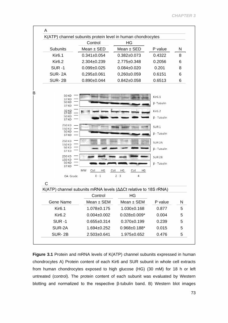
CHAPTER 3
73
A
K(ATP) channel subunits protein level in human chondrocytes
Control
HG
Subunits
Mean ± SED
Mean ± SED
P value
N
Kir6.1
0.341±0.054
0.382±0.073
0.4322
8
Kir6.2
2.304±0.239
2.775±0.348
0.2056
6
SUR -1
0.099±0.025
0.084±0.020
0.201
8
SUR- 2A
0,295±0.061
0,260±0.059
0.6151
6
SUR- 2B
0.890±0.044
0.842±0.058
0.6513
6
B
C
K(ATP) channel subunits mRNA levels (ΔΔCt relative to 18S rRNA)
Control
HG
Gene Name
Mean ± SEM
Mean ± SEM
P value
N
Kir6.1
1.078±0.175
1.030±0.168
0.877
5
Kir6.2
0.004±0.002
0.028±0.009*
0.004
5
SUR -1
0.655±0.314
0.370±0.199
0.239
5
SUR-2A
1.694±0.252
0.968±0.188*
0.015
5
SUR- 2B
2.503±0.641
1.975±0.652
0.476
5
Figure 3.1 Protein and mRNA levels of K(ATP) channel subunits expressed in human
chondrocytes A) Protein content of each Kir6 and SUR subunit in whole cell extracts
from human chondrocytes exposed to high glucose (HG) (30 mM) for 18 h or left
untreated (control). The protein content of each subunit was evaluated by Western
blotting and normalized to the respective β‐tubulin band. B) Western blot images
CHAPTER 3
74
representative of the protein content of each Kir6 and SUR subunit expressed in
human chondrocytes according to OA grade, as defined in Materials and Methods
Section. MW, molecular weight protein marker. C) mRNA content of each Kir6 and
SUR subunit in human chondrocytes exposed to high glucose (HG) (30 mM) for 18 h or
left untreated (control). The mRNA content of each subunit was evaluated by qRT‐PCR
and normalized to the respective 18S rRNA content.
3.2.2 Immunofluorescence staining of K(ATP) channels expressed in human
chondrocytes
To further confirm the results obtained in isolated chondrocytes (Fig. 3.1), the presence
of each subunit in full depth normal (grade 0-1) cartilage samples was assessed by
immunofluorescence.
Fig. 3.2 shows the presence of chondrocytes staining positively for Kir6.1, Kir6.2,
SUR2A and SUR2B scattered throughout the cartilage sections and in the midst of
non-stained chondrocytes. No cells staining positively for SUR1 could be detected in
any of the cartilage sections analysed. Although no quantitative analysis of the
immunofluorescence images was performed, these results are in agreement with those
obtained in cell extracts of isolated chondrocytes where SUR1 was detected at very
low levels by western blot, while Kir6.2 and SUR2B seem to be more abundant.
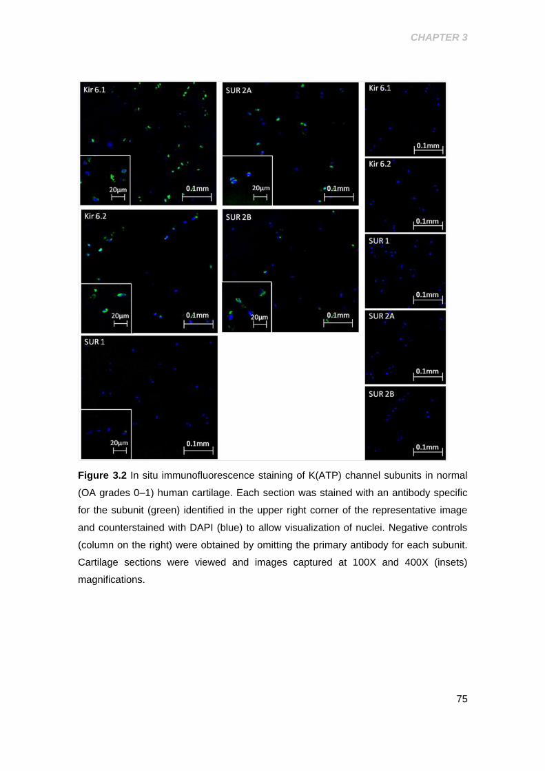
CHAPTER 3
75
Figure 3.2 In situ immunofluorescence staining of K(ATP) channel subunits in normal
(OA grades 0–1) human cartilage. Each section was stained with an antibody specific
for the subunit (green) identified in the upper right corner of the representative image
and counterstained with DAPI (blue) to allow visualization of nuclei. Negative controls
(column on the right) were obtained by omitting the primary antibody for each subunit.
Cartilage sections were viewed and images captured at 100X and 400X (insets)
magnifications.
CHAPTER 3
76
3.2.3 Role of exposure to high glucose on K(ATP) channel subunit protein and
mRNA expression
To elucidate whether exposure to a hyperglycemia-like glucose concentration
influences the subunit composition of K(ATP) channels, chondrocytes, classified
according to OA grade, were treated with 30 mM glucose for 18 h and the protein and
mRNA levels of each subunit were analysed by western blot and qRT-PCR,
respectively. No differences in MTT reduction capacity were found between
chondrocytes of either OA grade, exposed or not to high glucose for 18 h (data not
shown), thus showing that exposure to high glucose did not affect chondrocyte viability.
The results presented in Fig. 3.1 show that exposure to high glucose did not
significantly affect the protein levels of SUR1, SUR2A, SUR2B, Kir6.1 or Kir6.2 relative
to chondrocytes maintained in regular culture medium containing 10 mM glucose.
However, as shown in Fig. 3.1C, a small, but statistically significant difference was
found in the mRNA level of SUR2A between cells treated with 30 mM glucose
(0.97±0.19) and control cells maintained in regular medium (10 mM glucose)
(1.69±0.25). On the other hand, the mRNA levels of Kir6.2 were approximately 6.5 fold
higher in cells cultured in high glucose for 18 h (0.028±0.009) than in those kept in
regular medium (0.004±0.002), although the absolute levels in either condition were
very low.
3.2.4 Regulation of GLUT-1 and GLUT-3 protein levels by the K(ATP) channel
blocker Glibenclamide
To determine whether the activity of K(ATP) channels is involved in the regulation of
glucose transporters in human chondrocytes, the cells were treated for 18 h with
glibenclamide, a widely used SUR inhibitor that blocks K(ATP) channels [Burke et al.,
2008], in concentrations ranging from 10 nM to 20 µM.
Data presented on Table 3.1 show that there are no significant differences in the basal
protein levels of GLUT-1 and GLUT3 among the three groups defined according to the
macroscopic OA grade, as defined in Materials and Methods.
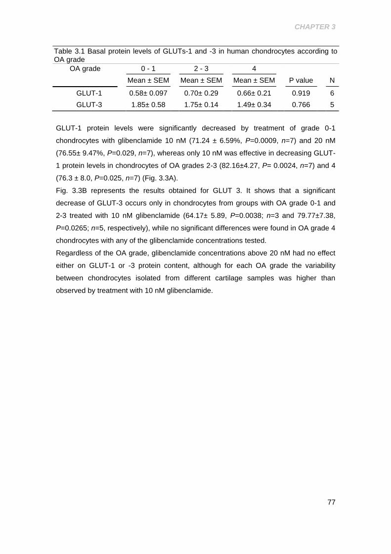
CHAPTER 3
77
Table 3.1 Basal protein levels of GLUTs-1 and -3 in human chondrocytes according to OA grade
OA grade
0 - 1
2 - 3
4
Mean ± SEM
Mean ± SEM
Mean ± SEM
P value
N
GLUT-1 0.58± 0.097
0.70± 0.29
0.66± 0.21
0.919
6
GLUT-3 1.85± 0.58
1.75± 0.14
1.49± 0.34
0.766
5
GLUT-1 protein levels were significantly decreased by treatment of grade 0-1
chondrocytes with glibenclamide 10 nM (71.24 ± 6.59%, P=0.0009, n=7) and 20 nM
(76.55± 9.47%, P=0.029, n=7), whereas only 10 nM was effective in decreasing GLUT-
1 protein levels in chondrocytes of OA grades 2-3 (82.16±4.27, P= 0.0024, n=7) and 4
(76.3 ± 8.0, P=0.025, n=7) (Fig. 3.3A).
Fig. 3.3B represents the results obtained for GLUT 3. It shows that a significant
decrease of GLUT-3 occurs only in chondrocytes from groups with OA grade 0-1 and
2-3 treated with 10 nM glibenclamide (64.17± 5.89, P=0.0038; n=3 and 79.77±7.38,
P=0.0265; n=5, respectively), while no significant differences were found in OA grade 4
chondrocytes with any of the glibenclamide concentrations tested.
Regardless of the OA grade, glibenclamide concentrations above 20 nM had no effect
either on GLUT-1 or -3 protein content, although for each OA grade the variability
between chondrocytes isolated from different cartilage samples was higher than
observed by treatment with 10 nM glibenclamide.
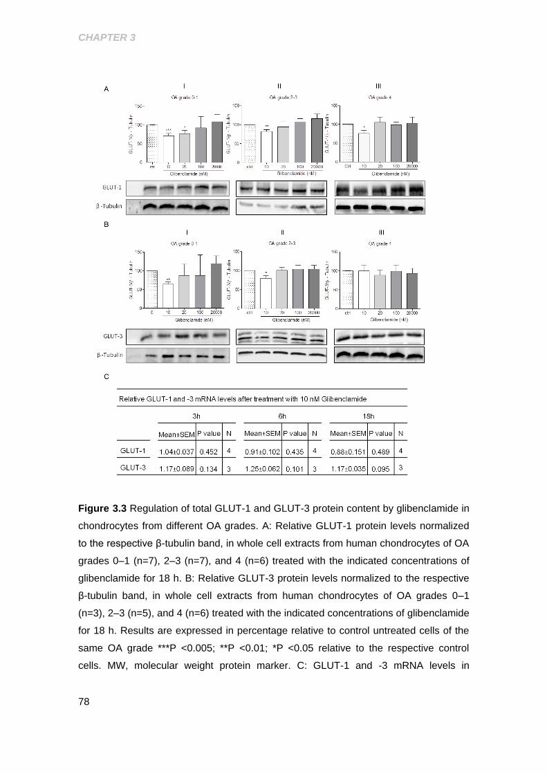
CHAPTER 3
78
Figure 3.3 Regulation of total GLUT-1 and GLUT-3 protein content by glibenclamide in
chondrocytes from different OA grades. A: Relative GLUT-1 protein levels normalized
to the respective β-tubulin band, in whole cell extracts from human chondrocytes of OA
grades 0–1 (n=7), 2–3 (n=7), and 4 (n=6) treated with the indicated concentrations of
glibenclamide for 18 h. B: Relative GLUT-3 protein levels normalized to the respective
β-tubulin band, in whole cell extracts from human chondrocytes of OA grades 0–1
(n=3), 2–3 (n=5), and 4 (n=6) treated with the indicated concentrations of glibenclamide
for 18 h. Results are expressed in percentage relative to control untreated cells of the
same OA grade ***P <0.005; **P <0.01; *P <0.05 relative to the respective control
cells. MW, molecular weight protein marker. C: GLUT-1 and -3 mRNA levels in
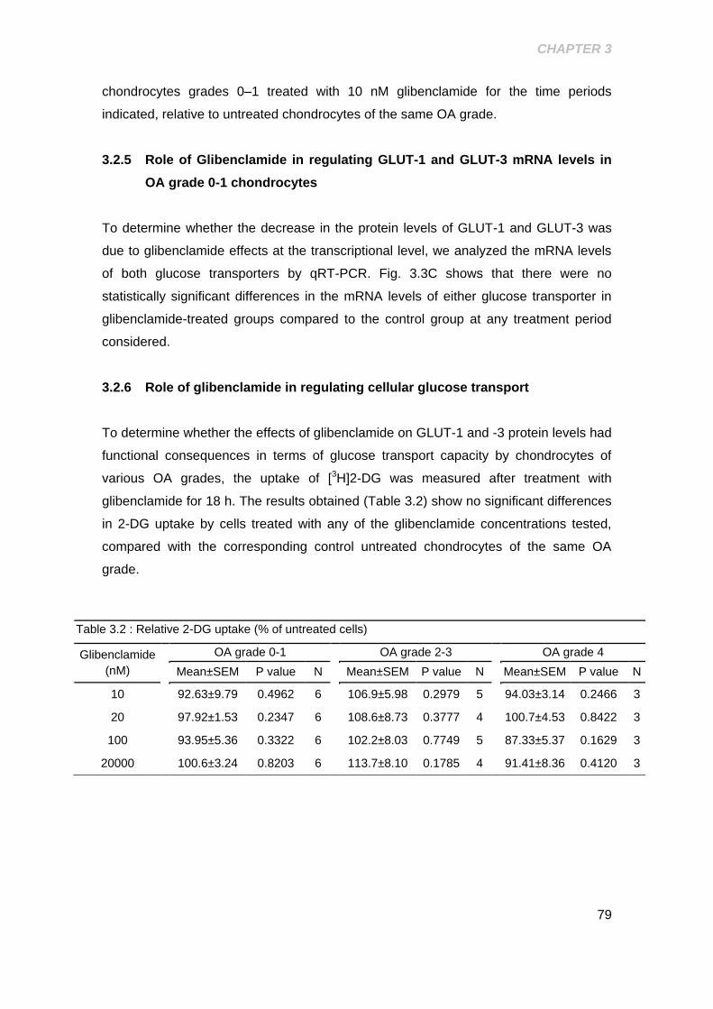
CHAPTER 3
79
chondrocytes grades 0–1 treated with 10 nM glibenclamide for the time periods
indicated, relative to untreated chondrocytes of the same OA grade.
3.2.5 Role of Glibenclamide in regulating GLUT-1 and GLUT-3 mRNA levels in
OA grade 0-1 chondrocytes
To determine whether the decrease in the protein levels of GLUT-1 and GLUT-3 was
due to glibenclamide effects at the transcriptional level, we analyzed the mRNA levels
of both glucose transporters by qRT-PCR. Fig. 3.3C shows that there were no
statistically significant differences in the mRNA levels of either glucose transporter in
glibenclamide-treated groups compared to the control group at any treatment period
considered.
3.2.6 Role of glibenclamide in regulating cellular glucose transport
To determine whether the effects of glibenclamide on GLUT-1 and -3 protein levels had
functional consequences in terms of glucose transport capacity by chondrocytes of
various OA grades, the uptake of [3H]2-DG was measured after treatment with
glibenclamide for 18 h. The results obtained (Table 3.2) show no significant differences
in 2-DG uptake by cells treated with any of the glibenclamide concentrations tested,
compared with the corresponding control untreated chondrocytes of the same OA
grade.
Table 3.2 : Relative 2-DG uptake (% of untreated cells)
Glibenclamide
(nM)
OA grade 0-1
OA grade 2-3
OA grade 4
Mean±SEM P value N
Mean±SEM P value N
Mean±SEM P value N
10
92.63±9.79 0.4962 6
106.9±5.98 0.2979 5
94.03±3.14 0.2466 3
20
97.92±1.53 0.2347 6
108.6±8.73 0.3777 4
100.7±4.53 0.8422 3
100
93.95±5.36 0.3322 6
102.2±8.03 0.7749 5
87.33±5.37 0.1629 3
20000
100.6±3.24 0.8203 6
113.7±8.10 0.1785 4
91.41±8.36 0.4120 3
CHAPTER 3
80
3.3 DISCUSSION
Although the presence of Kir6.1 in human and equine chondrocytes has already been
demonstrated [Mobasheri et al., 2007], this study represents the first comprehensive
analysis of Kir6.x and SUR subunits expressed in human articular chondrocytes, both
at the protein and mRNA levels. The results obtained (Figs. 3.1 and 3.2) suggest that
Kir6.2 and SUR2B are major K(ATP) channel subunits expressed in human
chondrocytes, while Kir6.1 and SUR2A are likely present at much lower levels and
SUR1 is barely detectable or even undetectable. How the various Kir and SUR
subunits expressed in human chondrocytes assemble to form functional K(ATP)
channels is beyond the scope of this study and was not investigated. Nonetheless, it is
possible that K(ATP) channels containing more than one SUR subtype co-exist with
others containing only one SUR isoform. The relative abundance of each channel type
would depend on the expression level of each subunit, as observed in other cells [Chan
et al., 2008; Wheeler et al., 2008]. Since Kir 6.2 and SUR2B seem to be more
abundant than the other Kir6 and SUR subunits, it seems likely that K(ATP) channels
containing these subunits, namely octamers composed of Kir6.2 and SUR2B,
predominate in human articular chondrocytes, although all other possible combinations
of Kir6 and SUR subunits can also be present. Even though the scarcity of human
cartilage samples did not allow us to determine the influence of the OA grade on the
subunit composition of K(ATP) channels in human chondrocytes, the results presented
in Fig. 3.1 suggest that none of the Kir6 or SUR subunits is affected at the protein level.
The composition of those channels, at least at the protein level, was not significantly
affected by culture under a supra-physiologic glucose concentration (30 mM), unlike
demonstrated at the mRNA level in other cells, namely mediobasal hypothalamic
neurons [Acosta-Martinez and Levine, 2007] and pancreatic β-cells [Moritz et al.,
2001].
The nucleotide binding affinity of SUR2B is much higher than that of SUR2A, but lower
than that of SUR1, although its channel gating activity is similar to that of SUR1
[Matsuo et al., 2000]. Therefore, SUR2B-containing K(ATP) channels are functionally
very similar to those that contain SUR1, namely those expressed in pancreatic -cells
[Burke et al., 2008] and in mediobasal hypothalamic neurons [Acosta-Martinez and
Levine, 2007] that readily bind ATP in response to augmented glucose availability. To
test the hypothesis that K(ATP) channels expressed in human chondrocytes behave
similarly, we attempted to measure changes in membrane potential induced by culture
under a supra-physiologic glucose concentration (30 mM) which is in the range of
CHAPTER 3
81
plasma glucose concentrations found in diabetic patients. For this, we loaded the cells
with the potential-sensitive fluorescence dye, bis-(1,3-dibutylbarbituric acid)trimethine
oxonol [DiBAC 4 (3)] that becomes fluorescent upon depolarization. Unfortunately, we
were unable to detect any changes using fluorescence spectroscopy, probably
because changes in membrane potential occur in a very dynamic and fast manner.
Possibly, in vivo fluorescence imaging may be required to detect such fast and
transient changes. Therefore, we were unable to directly demonstrate whether glucose
availability affects the chondrocyte membrane potential.
To further test the hypothesis that K(ATP) channels function as metabolic sensors in
human chondrocytes playing a role in the modulation of glucose transport capacity, we
used glibenclamide, a widely used SUR inhibitor, to block these channels and
measured the whole cell content of major glucose transporters, GLUT-1 and GLUT-3.
Glibenclamide displays differential selectivity towards distinct SUR subunits, being
moderately selective for SUR1 [Burke et al., 2008]. The sensitivity of equine
chondrocytes to glibenclamide is lower than that found in cells expressing SUR1 and in
the same range of that presented by cells in which K(ATP) channels contain SUR2B
[Mobasheri et al., 2007] which further suggests that K(ATP) channels containing this
subunit are relevant in human chondrocytes. Nevertheless, as K(ATP) channels
composed of other subunits may also exist in human chondrocytes, we used
glibenclamide concentrations ranging from 10 nM to 200 M in order to cover the whole
range of channel sensitivities to this SUR inhibitor. Interestingly, only the lowest
concentration was effective in reducing GLUT-1 protein content in chondrocytes from
all OA grades, whereas 20 nM was effective only in grade 0-1 OA chondrocytes (Fig.
3.3A). GLUT-3 content, however, was only reduced by treatment of grades 0-1 and 2-
3, but not grade 4 OA chondrocytes with glibenclamide 10 nM (Fig. 3.3B). These
results indicate that closing K(ATP) channels, at least partially, is sufficient to reduce
the cell protein content of two major glucose transporters, GLUT-1 and GLUT-3,
without affecting their mRNA levels, in normal human chondrocytes, in a way similar to
the reduction induced by exposure to 30 mM glucose [Rosa et al., 2009].
The inability of OA chondrocytes to downregulate the protein levels of GLUT-1 and
GLUT-3 in response to glibenclamide (Fig. 3.3), mimicking their inability to adjust to
high extracellular glucose concentrations [Rosa et al., 2009], further indicates that
either K(ATP) channels are functionally defective or some other downstream
component or process is impaired in OA chondrocytes. Nonetheless, the possibility that
ATP production may simultaneously be impaired in advanced OA chondrocytes cannot
be excluded.
CHAPTER 3
82
Another intriguing question arises from the observation that the response to
glibenclamide in terms of GLUT-1 and GLUT-3 protein content, occurred only with the
lowest glibenclamide concentrations tested. In equine chondrocytes, similar
glibenclamide concentrations (10-20 nM) elicited a partial reduction in current
amplitude, while much higher concentrations in the range of 100-300 nM were required
to completely block K(ATP) channel activity [Mobasheri et al., 2007]. Therefore, it is
possible that different ranges of channel activity elicit distinct changes in membrane
potential that operate diverse signalling pathways and cell responses. This is further
supported by the observation that K(ATP) channels with distinct subunit compositions
and therefore with different glibenclamide and nucleotide sensitivities, may coexist in
human chondrocytes (Figs. 3.1 and 3.2).
Nonetheless, glibenclamide-induced changes in the total protein content of GLUT-1
and GLUT-3 were not accompanied by corresponding decreases in glucose transport
capacity by chondrocytes of any OA grade (Table 3.2). This suggests that even though
K(ATP) channel activity regulates the availability of GLUT-1 and GLUT-3, other
mechanisms are involved in the regulation of the overall glucose transport capacity of
human chondrocytes. On one hand, other glucose transporters known to be expressed
in chondrocytes [Mobasheri et al., 2008] may not be regulated by K(ATP) channels and
compensate for changes in GLUT-1 and -3. On the other hand, GLUT-1 has been
shown to exist in a dynamic equilibrium involving the plasma membrane, intracellular
storage in the trans Golgi network and lysosomal degradation [Ortiz et al., 1992]. Our
previous study indicated that high glucose-induced GLUT-1 downregulation involved
lysosomal degradation [Rosa et al., 2009]. Thus, it is possible that K(ATP) channel
activity affects total GLUT-1 and possibly GLUT-3 protein content by increasing
lysosomal degradation of intracellular stores, in agreement with studies in other cells
suggesting that GLUT-1 degradation and functional membrane expression may be
regulated through independent pathways [Ortiz et al., 1992].
In summary, this study contributes to elucidate the subunit composition of K(ATP)
channels expressed in human chondrocytes and establishes a role for these channels
as components of the glucose sensing apparatus in these cells by demonstrating that
their activity influences the abundance of two major glucose transporters, GLUT-1 and
GLUT-3. Nonetheless, since glucose transport was not similarly affected, additional
mechanisms must be involved in the adaptation of human chondrocytes to high
extracellular glucose concentrations. Moreover, this study also shows that
chondrocytes isolated from cartilage of increasing OA grade are less responsive to
changes in K(ATP) channel activity, which may contribute to the inability of OA
CHAPTER 3
83
chondrocytes to downregulate glucose transporters and avoid the deleterious effects of
high extracellular glucose concentrations that we have previously observed [Rosa et
al., 2009; Rosa et al., 2011b]. Taken together, the results presented highlight the
importance of K(ATP) channels as metabolic sensors and potential components of a
broad glucose sensing apparatus that allows human chondrocytes to adjust to varying
extracellular glucose concentrations.

85
CHAPTER 4.
ROLE OF HIGH GLUCOSE AS MODULATOR OF INFLAMMATION IN HUMAN
CHONDROCYTES
Ana T. Rufino, Madalena Ribeiro, João P. Ferreira, Fernando Judas and Alexandrina
F. Mendes
Role of High Glucose as a Modulator of Inflammation in Human Chondrocytes
Submitted for publication to Osteoarthritis and Cartilage (July 2014)
CHAPTER 4
86
ABSTRACT
Objective: This work aimed at determining whether high glucose induces inflammatory
responses in human chondrocytes. Since hyperinsulinemia often accompanies DM
type 2, we also evaluated the ability of insulin to modulate inflammatory responses
induced by high glucose.
Method: We analyzed IL-1β, TNF-α and inducible nitric oxide synthase (iNOS)
expression by qRT-PCR and nuclear NF-κB p65 accumulation by western blot in
human chondrocytes cultured in medium containing 10 or 30 mM glucose.
Results: Culture in high glucose (30 mM) for 24h increased iNOS mRNA and protein
levels and NO production by 61% (P=0.0017), 148% (P=0.0089) and 70% (P=0.049),
respectively, relative to human chondrocytes maintained in 10 mM glucose. Similarly,
IL-1β and TNF-α mRNA levels increased by 97 (P=0.0066) and 85% (P=0.0045),
respectively. NF-κB p65 nuclear levels increased in a time-dependent manner, peaking
at 1h. Treatment with 100 nM insulin also increased nuclear p65 levels, independently
of the glucose concentration.
Conclusion: This study shows that hyperglycemia and hyperinsulinemia induce
inflammatory responses in human chondrocytes. This can be a relevant mechanism by
which DM type 2, obesity, the metabolic syndrome and other conditions associated
with impaired glucose and insulin homeostasis contribute to the development and
progression of OA.
CHAPTER 4
87
4.1 INTRODUCTION
Aging is the major risk factor for osteoarthritis (OA), but other factors have been
recognized [Berenbaum, 2011; Zhang and Jordan, 2010], including diabetes mellitus
(DM), especially type 2, and impaired glucose homeostasis Recent epidemiologic
studies demonstrated that DM is an independent risk factor for OA development and
progression [Nieves-Plaza et al., 2013; Schett et al., 2012a], supporting the notion of a
specific diabetes-induced OA phenotype [Berenbaum, 2011; Zhang and Jordan, 2010].
How DM and impaired glucose homeostasis contribute to cartilage and joint destruction
is largely unknown.
Chronic low grade inflammation is a major OA driver, being associated with other
mechanisms involved in OA pathogenesis, including cell senescence, oxidative stress,
mitochondrial dysfunction and impaired autophagy, which together contribute to cell
depletion and imbalanced anabolic and catabolic responses, especially in
chondrocytes [Lotz and Carames, 2011].
All the above mechanisms, including inflammation, have been associated with the
effects of hyperglycemia and have been implicated in DM pathogenesis and its
complications [Donath and Shoelson, 2011].
Only a few studies have been conducted so far to identify direct effects of glucose in
articular chondrocytes, collectively indicating that hyperglycemia-like glucose
concentrations favor catabolic processes and impair anabolic responses. Our previous
studies showed that exposure to high glucose causes oxidative stress and induces
catabolic responses in human chondrocytes, especially from OA/aged articular
cartilage [Rosa et al., 2009; Rosa et al., 2011a].
Given the role of inflammation in OA and in mediating hyperglycemia-induced effects in
other cells, this study aimed at determining whether high glucose induces inflammatory
responses in human chondrocytes. Since hyperinsulinemia often accompanies DM
type 2, we also evaluated the ability of insulin to modulate inflammatory responses
induced by high glucose.
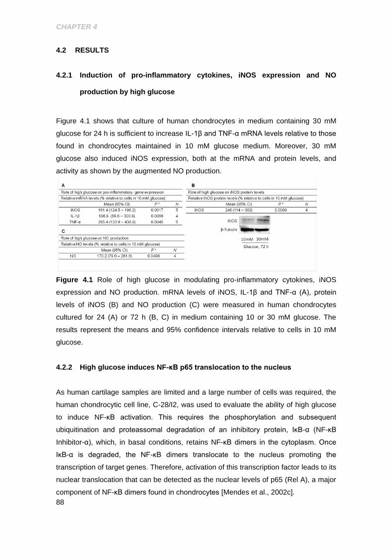
CHAPTER 4
88
4.2 RESULTS
4.2.1 Induction of pro-inflammatory cytokines, iNOS expression and NO
production by high glucose
Figure 4.1 shows that culture of human chondrocytes in medium containing 30 mM
glucose for 24 h is sufficient to increase IL-1β and TNF-α mRNA levels relative to those
found in chondrocytes maintained in 10 mM glucose medium. Moreover, 30 mM
glucose also induced iNOS expression, both at the mRNA and protein levels, and
activity as shown by the augmented NO production.
Figure 4.1 Role of high glucose in modulating pro-inflammatory cytokines, iNOS
expression and NO production. mRNA levels of iNOS, IL-1β and TNF-α (A), protein
levels of iNOS (B) and NO production (C) were measured in human chondrocytes
cultured for 24 (A) or 72 h (B, C) in medium containing 10 or 30 mM glucose. The
results represent the means and 95% confidence intervals relative to cells in 10 mM
glucose.
4.2.2 High glucose induces NF-κB p65 translocation to the nucleus
As human cartilage samples are limited and a large number of cells was required, the
human chondrocytic cell line, C-28/I2, was used to evaluate the ability of high glucose
to induce NF-κB activation. This requires the phosphorylation and subsequent
ubiquitination and proteassomal degradation of an inhibitory protein, IκB-α (NF-κB
Inhibitor-α), which, in basal conditions, retains NF-κB dimers in the cytoplasm. Once
IκB-α is degraded, the NF-κB dimers translocate to the nucleus promoting the
transcription of target genes. Therefore, activation of this transcription factor leads to its
nuclear translocation that can be detected as the nuclear levels of p65 (Rel A), a major
component of NF-κB dimers found in chondrocytes [Mendes et al., 2002c].

CHAPTER 4
89
Figure 2A shows that nuclear p65 levels gradually increased during culture in 30 mM
glucose, peaking at 1h. After 2h, nuclear levels of p65 were identical to those found in
cells maintained in 10 mM glucose and remained as such until up to, at least, 24h. Co-
treatment with the specific IκB-α phosphorylation inhibitor, Bay-11-7082, completely
reversed the high glucose-induced effect.
To rule out osmotic effects, 20 mM mannitol, a non-absorbable polysaccharide, were
added to the medium containing 10 mM glucose. No significant changes were
observed in cells cultured in this medium for 1h (mean=80.5, 95% CI: 61.0-100.0,
P=0.348) relative to those cultured in 10 mM glucose, indicating that the effects
detected with 30 mM glucose are not due to osmotic stimulation.
Figure 4.2 Role of glucose and insulin concentrations on nuclear NF-κB p65 levels. A)
C28/I2 cells were cultured in 10 or 30 mM glucose, with or without 5 μM Bay-11782, a
specific IκB-α phosphorylation inhibitor, for the indicated time periods. B) Insulin, 10 or
100 nM, was added to C28-I2 cultures in medium with 10 or 30 mM glucose for 1h. The
results represent the means and 95% confidence intervals relative to cells in 10 mM
glucose.
4.2.3 High Insulin modulates NF-κB translocation to the nucleus
To determine whether insulin modulates the inflammatory state induced by high
glucose, we evaluated the effect of physiologic (10 nM) and supraphysiologic (100 nM)
insulin concentrations on p65 nuclear levels in C28-I2 cells cultured in 10 or 30 mM
CHAPTER 4
90
glucose. Figure 4.2B shows that treatment of cells in 10 mM glucose medium with 100
nM insulin increased nuclear p65 levels to the same extent as 30 mM glucose.
Simultaneous treatment with high glucose (30 mM) and 100 nM insulin did not further
increase nuclear p65 levels. On the other hand, 10 mM insulin did not affect p65
nuclear levels, regardless of the glucose concentration in the medium.
CHAPTER 4
91
4.3 DISCUSSION
The present study aimed at determining whether high, hyperglycemia-like, glucose
concentrations induce inflammatory responses in human chondrocytes and thus,
contribute to unveil pathological mechanisms by which DM and impaired glucose
homeostasis can favor OA initiation and progression. For this purpose, we exposed
human chondrocytes to culture medium containing 10 or 30 mM glucose and compared
the expression of pro-inflammatory mediators known to be relevant in OA
pathogenesis, namely the pro-inflammatory and pro-catabolic cytokines, IL-1β and
TNF-α, and iNOS, the enzyme responsible for the production of large amounts of NO
[Stadler et al., 1991] To further elucidate the mechanisms involved, we also evaluated
the ability of high glucose (30 mM) to activate the transcription factor, Nuclear Factor-
κB (NF-κB), which plays a central role in inducing inflammatory and catabolic gene
expression, including IL-1β, TNF-α and iNOS, in chondrocytes [Rosa et al., 2008].
The results obtained show that culture under high glucose is sufficient to induce NF-κB
activation and the expression of IL-1β, TNF-α and iNOS. These observations, along
with the pro-catabolic and anti-anabolic effects that we reported previously [Rosa et al.,
2009; Rosa et al., 2011a], support the hypothesis that hyperglycemia can drive
cartilage damage and, thus, OA.
These results are consistent with several reports that demonstrated the induction of
exacerbated inflammatory responses by hyperglycemia in various human tissues and
cells [Donath and Shoelson, 2011; Jia et al., 2013].
iNOS mRNA and protein levels were similarly upregulated by high glucose, suggesting
that this effect occurs at the transcriptional level, since it is the major mechanism of
iNOS regulation. Nonetheless, upregulated iNOS mRNA levels were detected upon
culture in high glucose for 24h, whereas measurable amounts of the protein and NO
were detected after 72h and required culture periods longer than, at least, 48h.
Moreover, NF-κB activation was maximal after exposure to high glucose for 1h and by
2h the effect had returned to levels found in the control. This kinetics is similar to NF-κB
activation by other stimuli, like IL-1β, which on the contrary allows iNOS protein to be
detected much earlier than observed here with high glucose [Mendes et al., 2002c].
This suggests that other mechanisms may contribute to induce iNOS expression in
response to high glucose. One possibility is that high glucose-induced IL-1β and TNF-
α, which are potent iNOS inducers in chondrocytes [Stadler et al., 1991] are required to
mediate iNOS expression. On the other hand, since we did not evaluate the protein
levels of those cytokines, we cannot exclude the possibility that unknown mechanisms
CHAPTER 4
92
lead to a large gap between mRNA transcription of the three genes and synthesis of
the corresponding proteins.
The rapid nuclear translocation of p65 induced by high glucose, suggests a direct effect
on the NF-κB signalling pathway in chondrocytes. This is further supported by the
finding that the IκB-α phosphorylation inhibitor, Bay 11-7082, completely abolished high
glucose-induced nuclear p65 accumulation. This suggests that IκB-α phosphorylation
or another upstream event on the NF-κB activation pathway is sensitive to high
glucose.
Insulin, especially in supraphysiologic concentrations, was shown to induce anabolic
and inhibit catabolic responses in adult chondrocytes and cartilage explant cultures
from various species [Kellner et al., 2001; Rosa et al., 2011a]. Thus, we hypothesized
that insulin might counteract the deleterious pro-inflammatory effects of high glucose
that we observed in chondrocytes. Surprisingly, 100 nM insulin were sufficient to
induce nuclear p65 accumulation, either alone or in combination with high glucose,
whereas 10 nM insulin, which is at the upper physiologic limit had no effect. These
results are in agreement with a recent study showing that insulin, in concentrations
similar to those we used, induced NF-κB activation and synergized with TNF-α in
cardiac myoblasts [Madonna et al., 2014]. Moreover, accumulating evidence is
unravelling a role for hyperinsulinemia in driving or at least contributing to the low-
grade inflammation characteristic of metabolic disorders, from obesity and DM type 2 to
the metabolic syndrome and their complications [Guo, 2014]. The results presented
here also point to the possibility that hyperinsulinemia can induce inflammatory
responses in chondrocytes by activating NF-κB. Nonetheless, further studies are
required to confirm the proinflammatory effects of hyperinsulinemia in primary human
chondrocytes.
In summary, this study shows for the first time that hyperglycemia and
hyperinsulinemia can induce inflammatory responses in human chondrocytes. This can
be a relevant mechanism by which DM type 2, obesity, the metabolic syndrome and
other conditions associated with impaired glucose and insulin homeostasis contribute
to the development and progression of OA.
93
CHAPTER 5.
DIFFERENTIAL EFFECTS OF LAVANDULA LUISIERI AND ERYNGIUM DURAEI
SUBSP JURESIANUM ESSENTIAL OILS IN CELL MODELS OF TWO CHRONIC
INFLAMMATORY DISEASES
Ana T. Rufino, Isabel V. Ferreira*, Fernando Judas, Lígia Salgueiro, M. Celeste Lopes,
Carlos Cavaleiro and Alexandrina F. Mendes
Differential Effects of Lavandula Luisieri and Eryngium Duriaei Subsp.
Juresianum Essential Oils in Cell Models of Two Chronic Inflammatory Diseases
Submitted for publication to the Pharmaceutical Biology (July 2014)
CHAPTER 5
94
ABSTRACT
Context: Effective drugs to treat osteoarthritis (OA) and inflammatory bowel disease
(IBD) are needed.
Objective: To identify essential oils (EOs) with anti-inflammatory activity in cell models
of OA and IBD.
Material and methods: EOs from Eryngium duriaei subsp. juresianum (M. Laínz) M.
Laínz (Apiaceae), Laserpitium eliasii subsp. thalictrifolium Sennen & Pau (Apiaceae),
Lavandula luisieri (Rozeira) Rivas-Martínez (Lamiaceae), Othantus maritimus (L.) Hoff.
& Link (Asteraceae) and Thapsia villosa L. (Apiaceae) were analyzed by GC and
GC/MS. The anti-inflammatory activity of the EOs (5-200 μg/mL) was evaluated by
measuring iNOS and NF-κB activation (total and phosphorylated IκB-α), in primary
human chondrocytes and the intestinal cell line, C2BBe1, stimulated with IL-1β or IFN-
γ, IL-1β and TNF-α, respectively.
Results: The EO of L. luisieri significantly reduced iNOS (by 54.9 and 81.0%,
respectively) and phosphorylated IκB-α (by 87.4% and 62.3%, respectively), in both cell
models. The EO of E. duriaei subsp. juresianum caused similar effects in human
chondrocytes, but was inactive in intestinal cells, even at higher concentrations. The
EOs of L. eliasii subsp. thalictrifolium and O. maritimus decreased iNOS expression by
45.28.7% and 45.26.2%, respectively, in C2BBe1 cells and were inactive in
chondrocytes. The EO of T. villosa was inactive in both cell types.
Discussion and conclusion: This is the first study showing anti-inflammatory effects of
the EOs of L. luisieri and E. duriaei subsp. juresianum. These effects are cell type-
specific and may be valuable to develop new therapies or as sources of active
compounds with improved efficacy and selectivity towards OA and IBD.
CHAPTER 5
95
5.1 INTRODUCTION
An increasing number of reports describes anti-inflammatory properties of many natural
products and compounds. Nonetheless, very few studies have been dedicated to
evaluate their therapeutic potential on chronic inflammatory diseases. These remain
important therapeutic targets due to their high prevalence and lack of effective
therapies. Indeed, most studies concerning anti-inflammatory properties of natural
products have been performed in cell or animal models of acute inflammation. These
models do not entirely reflect pathological mechanisms and cells involved in chronic
inflammatory diseases.
Among natural products, essential oils (EOs) are particularly interesting to look for
compounds with pharmacological activities, since they are complex mixtures of a huge
diversity of low molecular weight (<300 Da) lipophilic compounds. These characteristics
may represent favorable pharmacokinetic properties [Miguel, 2010].
Recent reviews on the anti-inflammatory potential of EOs fully highlight that these plant
extracts and some of their components are useful therapeutic alternatives, modulating
several molecular targets of the acute inflammation cascades, namely in cells of the
immune system, like monocytes and macrophages [Adorjan and Buchbauer, 2010;
Miguel, 2010]. There is also an evident lack of information on the potential of EOs as
modulators of chronic inflammatory diseases, especially involving cells unrelated to the
immune system. Exceptions are two reports from our group [Neves et al., 2009; Rufino
et al., 2014] describing anti-inflammatory effects of the EO of Juniperus oxycedrus L.
subsp. oxycedrus (Cupressaceae) and -pinene in a cell model of osteoarthritis (OA).
Chronic inflammatory diseases, in general, lead to the up-regulation of a series of
enzymes and signalling molecules that bring about the characteristic inflammation and
tissue destruction. Likewise, OA and Inflammatory Bowel Disease (IBD) share many
features and mechanisms and are largely driven and perpetuated by pro-inflammatory
cytokines, such as Interleukin-1β (IL-1β), Tumor necrosis factor-α (TNF-α) and
Interferon- (IFN-) [Goldring and Otero, 2011; Goldring et al., 2008; Jobin and Sartor,
2000; Wielockx et al., 2004]. In chronic inflammatory diseases, the onset and
perpetuation of inflammatory responses and tissue destruction are primarily dependent
on the transcription factor, Nuclear Factor-κB (NF-κB) [Tak and Firestein, 2001]. This
promotes the expression of inflammation-related genes, including cytokines (IL-1β,
TNF-α, IL-6, etc.) and enzymes. One of these, the inducible nitric oxide synthase
(iNOS), produces large amounts of nitric oxide (NO), a potent and destructive
inflammatory mediator that plays an important role in the development and progression
CHAPTER 5
96
of OA and IBD. For instance, our previous study showed that iNOS expression, NO
production and NF-κB activity are spontaneously increased in chondrocytes isolated
from OA patients compared to those isolated from non-affected human cartilage [Rosa
et al., 2008]. Increased mucosal and plasma NO concentrations associated with
augmented iNOS activity have also been demonstrated in active IBD [Quenon et al.,
2012]. Moreover, NF-κB promotes the expression of specific proteases that degrade
the extracellular matrix, causing the characteristic tissue destruction of OA and IBD
[Goldring and Otero, 2011; Jobin and Sartor, 2000; Wielockx et al., 2004].
Therefore, this transcription factor constitutes an attractive target for anti-inflammatory
therapeutic interventions both in OA and IBD [Berenbaum, 2004; Goldring and Otero,
2011; Jobin and Sartor, 2000; Marcu et al., 2010]. Hence, we proposed to study the
ability of EOs to inhibit NF-κB activation in cell models of these diseases.
To address this purpose, EOs to be screened were selected considering two major
criteria: i) to collectively ensure the highest diversity of compounds from the chemical
classes usually found in EOs; and ii) availability of etnopharmacological information or
previous evidence of anti-inflammatory activity. For this, several EOs, isolated at
laboratory from native or endemic species of the Iberian flora, were first fully
characterized by identification and quantification of their components. Upon
composition elucidation, the EOs were assayed in cellular models relevant for the study
of OA and IBD [Csaki et al., 2009; Megias et al., 2007]. Since we found previously that
α-pinene inhibits inflammatory and catabolic responses in human chondrocytes,
namely IL-1β-induced NF-κB activation [Neves et al., 2009; Rufino et al., 2014], we
also tested this compound in the cellular model of IBD.
Primary human chondrocyte cultures stimulated with IL-1β were used as an in vitro
cartilage degradation model that emulates the damage seen in OA and cultures of the
human colorectal adenocarcinoma cell line, C2BBe1, stimulated with a mixture of pro-
inflammatory cytokines composed of IFN-γ, TNF-α and IL-1β, were used as a model of
IBD.
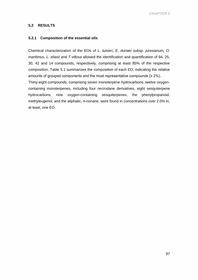
CHAPTER 5
97
5.2 RESULTS
5.2.1 Composition of the essential oils
Chemical characterization of the EOs of L. luisieri, E. duriaei subsp. juresianum, O.
maritimus, L. eliasii and T villosa allowed the identification and quantification of 94, 25,
30, 42 and 14 compounds, respectively, comprising at least 85% of the respective
composition. Table 5.1 summarizes the composition of each EO, indicating the relative
amounts of grouped components and the most representative compounds (≥ 2%).
Thirty-eight compounds, comprising seven monoterpene hydrocarbons, twelve oxygen-
containing monoterpenes, including four necrodane derivatives, eight sesquiterpene
hydrocarbons, nine oxygen-containing sesquiterpenes, the phenylpropanoid,
methyleugenol, and the aliphatic, n-nonane, were found in concentrations over 2.0% in,
at least, one EO.
CHAPTER 5
98
Table 5.1 Classes of compounds and major constituents of the essential oils tested
CHAPTER 5
99
5.2.2 Evaluation of cytotoxicity and selection of non cytotoxic concentrations
of the essential oils
Neither the EOs nor α-pinene were cytotoxic to C2BBe1 cells in concentrations up to
200 µg/mL, either in the presence or absence of the pro-inflammatory cytokine mixture
(Fig. 5.1C and D). On the contrary, in human chondrocytes, only the EO of T. villosa
had no significant cytotoxic effects at that concentration (Fig. 5.1A and B). In
concentrations up to 50 μg/mL for 24 h, the EOs of E. duriaei subsp. juresianum, L.
eliasii and L. luisieri did not affect chondrocyte viability either in the presence or
absence of IL-1β. The EO of O. maritimus showed no cytotoxicity at concentrations up
to 100 μg/mL (Fig. 5.1A and B). Therefore, subsequent experiments were performed
using the non-cytotoxic concentrations identified for each EO in each cell model, as
shown in Fig. 5.1.
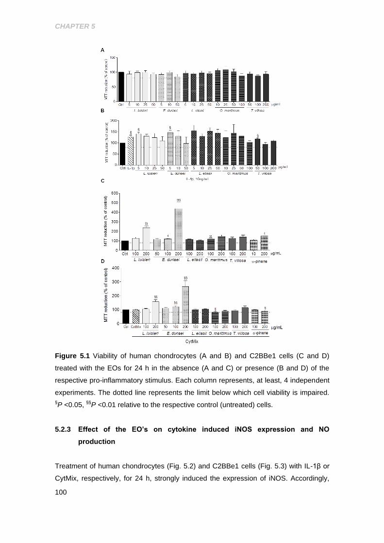
CHAPTER 5
100
Figure 5.1 Viability of human chondrocytes (A and B) and C2BBe1 cells (C and D)
treated with the EOs for 24 h in the absence (A and C) or presence (B and D) of the
respective pro-inflammatory stimulus. Each column represents, at least, 4 independent
experiments. The dotted line represents the limit below which cell viability is impaired.
§P <0.05, §§P <0.01 relative to the respective control (untreated) cells.
5.2.3 Effect of the EO’s on cytokine induced iNOS expression and NO
production
Treatment of human chondrocytes (Fig. 5.2) and C2BBe1 cells (Fig. 5.3) with IL-1β or
CytMix, respectively, for 24 h, strongly induced the expression of iNOS. Accordingly,
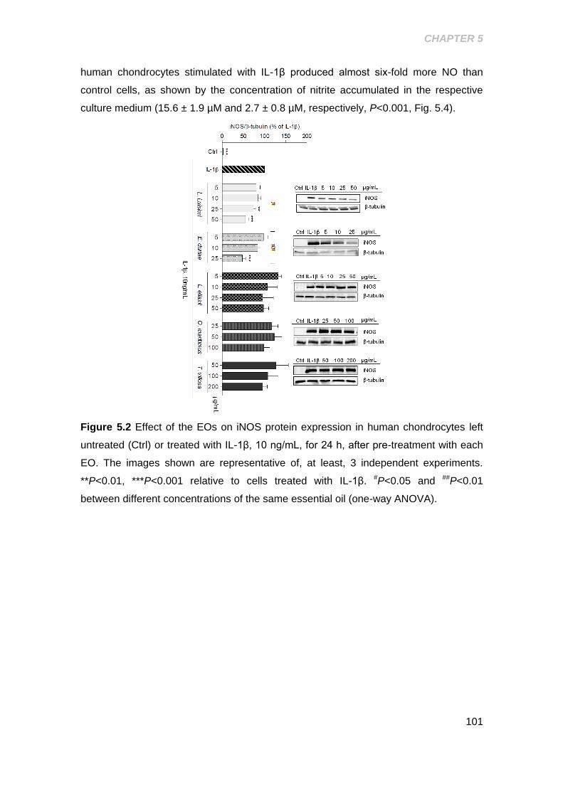
CHAPTER 5
101
human chondrocytes stimulated with IL-1β produced almost six-fold more NO than
control cells, as shown by the concentration of nitrite accumulated in the respective
culture medium (15.6 ± 1.9 µM and 2.7 ± 0.8 µM, respectively, P<0.001, Fig. 5.4).
Figure 5.2 Effect of the EOs on iNOS protein expression in human chondrocytes left
untreated (Ctrl) or treated with IL-1β, 10 ng/mL, for 24 h, after pre-treatment with each
EO. The images shown are representative of, at least, 3 independent experiments.
**P<0.01, ***P<0.001 relative to cells treated with IL-1β. #P<0.05 and ##P<0.01
between different concentrations of the same essential oil (one-way ANOVA).
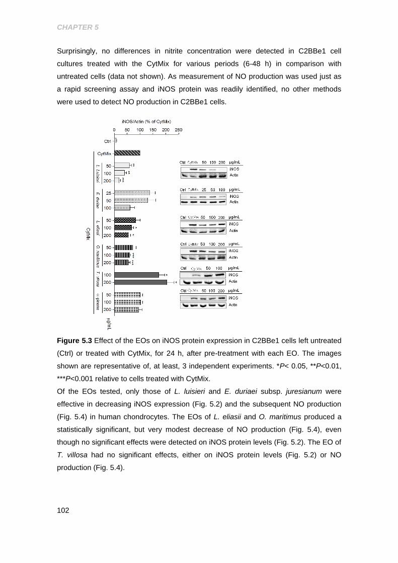
CHAPTER 5
102
Surprisingly, no differences in nitrite concentration were detected in C2BBe1 cell
cultures treated with the CytMix for various periods (6-48 h) in comparison with
untreated cells (data not shown). As measurement of NO production was used just as
a rapid screening assay and iNOS protein was readily identified, no other methods
were used to detect NO production in C2BBe1 cells.
Figure 5.3 Effect of the EOs on iNOS protein expression in C2BBe1 cells left untreated
(Ctrl) or treated with CytMix, for 24 h, after pre-treatment with each EO. The images
shown are representative of, at least, 3 independent experiments. *P< 0.05, **P<0.01,
***P<0.001 relative to cells treated with CytMix.
Of the EOs tested, only those of L. luisieri and E. duriaei subsp. juresianum were
effective in decreasing iNOS expression (Fig. 5.2) and the subsequent NO production
(Fig. 5.4) in human chondrocytes. The EOs of L. eliasii and O. maritimus produced a
statistically significant, but very modest decrease of NO production (Fig. 5.4), even
though no significant effects were detected on iNOS protein levels (Fig. 5.2). The EO of
T. villosa had no significant effects, either on iNOS protein levels (Fig. 5.2) or NO
production (Fig. 5.4).
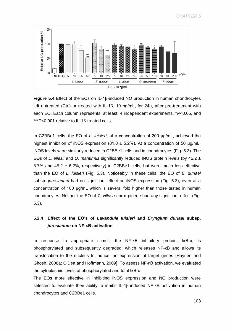
CHAPTER 5
103
Figure 5.4 Effect of the EOs on IL-1β-induced NO production in human chondrocytes
left untreated (Ctrl) or treated with IL-1β, 10 ng/mL, for 24h, after pre-treatment with
each EO. Each column represents, at least, 4 independent experiments. *P<0.05, and
***P<0.001 relative to IL-1β-treated cells.
In C2BBe1 cells, the EO of L. luisieri, at a concentration of 200 μg/mL, achieved the
highest inhibition of iNOS expression (81.0 ± 5.2%). At a concentration of 50 μg/mL,
iNOS levels were similarly reduced in C2BBe1 cells and in chondrocytes (Fig. 5.3). The
EOs of L. eliasii and O. maritimus significantly reduced iNOS protein levels (by 45.2 ±
8.7% and 45.2 ± 6.2%, respectively) in C2BBe1 cells, but were much less effective
than the EO of L. luisieri (Fig. 5.3). Noticeably in these cells, the EO of E. duriaei
subsp. juresianum had no significant effect on iNOS expression (Fig. 5.3), even at a
concentration of 100 μg/mL which is several fold higher than those tested in human
chondrocytes. Neither the EO of T. villosa nor α-pinene had any significant effect (Fig.
5.3).
5.2.4 Effect of the EO’s of Lavandula luisieri and Eryngium duriaei subsp.
juresianum on NF-κB activation
In response to appropriate stimuli, the NF-κB inhibitory protein, IκB-α, is
phosphorylated and subsequently degraded, which releases NF-κB and allows its
translocation to the nucleus to induce the expression of target genes [Hayden and
Ghosh, 2008a; O'Dea and Hoffmann, 2009]. To assess NF-κB activation, we evaluated
the cytoplasmic levels of phosphorylated and total IκB-α.
The EOs more effective in inhibiting iNOS expression and NO production were
selected to evaluate their ability to inhibit IL-1β-induced NF-κB activation in human
chondrocytes and C2BBe1 cells.
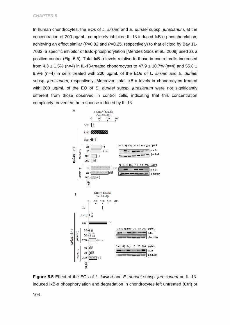
CHAPTER 5
104
In human chondrocytes, the EOs of L. luisieri and E. duriaei subsp. juresianum, at the
concentration of 200 µg/mL, completely inhibited IL-1β-induced IκB-α phosphorylation,
achieving an effect similar (P=0.82 and P=0.25, respectively) to that elicited by Bay 11-
7082, a specific inhibitor of IκBα-phosphorylation [Mendes Sdos et al., 2009] used as a
positive control (Fig. 5.5). Total IκB-α levels relative to those in control cells increased
from 4.3 ± 1.5% (n=4) in IL-1β-treated chondrocytes to 47.9 ± 10.7% (n=4) and 55.6 ±
9.9% (n=4) in cells treated with 200 μg/mL of the EOs of L. luisieri and E. duriaei
subsp. juresianum, respectively. Moreover, total IκB-α levels in chondrocytes treated
with 200 μg/mL of the EO of E. duriaei subsp. juresianum were not significantly
different from those observed in control cells, indicating that this concentration
completely prevented the response induced by IL-1β.
Figure 5.5 Effect of the EOs of L. luisieri and E. duriaei subsp. juresianum on IL-1β-
induced IκB-α phosphorylation and degradation in chondrocytes left untreated (Ctrl) or
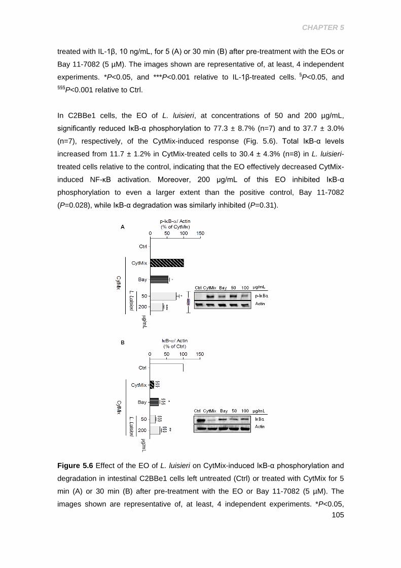
CHAPTER 5
105
treated with IL-1β, 10 ng/mL, for 5 (A) or 30 min (B) after pre-treatment with the EOs or
Bay 11-7082 (5 µM). The images shown are representative of, at least, 4 independent
experiments. *P<0.05, and ***P<0.001 relative to IL-1β-treated cells. §P<0.05, and
§§§P<0.001 relative to Ctrl.
In C2BBe1 cells, the EO of L. luisieri, at concentrations of 50 and 200 µg/mL,
significantly reduced IκB-α phosphorylation to 77.3 ± 8.7% (n=7) and to 37.7 ± 3.0%
(n=7), respectively, of the CytMix-induced response (Fig. 5.6). Total IκB-α levels
increased from 11.7 ± 1.2% in CytMix-treated cells to 30.4 ± 4.3% (n=8) in L. luisieri-
treated cells relative to the control, indicating that the EO effectively decreased CytMix-
induced NF-κB activation. Moreover, 200 μg/mL of this EO inhibited IκB-α
phosphorylation to even a larger extent than the positive control, Bay 11-7082
(P=0.028), while IκB-α degradation was similarly inhibited (P=0.31).
Figure 5.6 Effect of the EO of L. luisieri on CytMix-induced IκB-α phosphorylation and
degradation in intestinal C2BBe1 cells left untreated (Ctrl) or treated with CytMix for 5
min (A) or 30 min (B) after pre-treatment with the EO or Bay 11-7082 (5 µM). The
images shown are representative of, at least, 4 independent experiments. *P<0.05,
CHAPTER 5
106
**P<0.01 and ***P<0.001 relative to CytMix-treated cells. §§§P<0.001 relative to Ctrl and
###P<0.001 between different concentrations of the same essential oil (one-way
ANOVA)
CHAPTER 5
107
5.3 DISCUSSION
The results presented show clear differences in the ability of the EOs tested to inhibit
relevant mediators of inflammation in the two cell models of OA and IBD used. Such
differences are evident among distinct EOs, as well as comparing each one in the two
cell models. Indeed, only the EO of L. luisieri was capable of significantly inhibiting
inflammatory markers (iNOS expression and NF-κB activation) both in human
chondrocytes and intestinal C2BBe1 cells. These results confirm our previous
observation that this EO inhibits IL-1β-induced NO production in human chondrocytes
[Neves et al., 2009]. On the other hand, the EO of E. duriaei subsp. juresianum did not
significantly inhibit iNOS expression in intestinal cells over a wide range of
concentrations, but was effective in human chondrocytes, even at much lower
concentrations. Conversely, the EOs of O. maritimus and L. eliasii showed some
activity in intestinal cells, but were inactive in chondrocytes. These results indicate that
the EOs of L. luisieri and E. duriaei subsp. juresianum have cell type-specific anti-
inflammatory effects.
To our knowledge, this is the first study showing anti-inflammatory effects of the EOs of
L. luisieri and E. duriaei subsp. juresianum. Indeed, EOs from Lavandula ssp, namely
L. angustifolia L. stoechas, L. multifida and L. viridis, have demonstrated antibacterial,
antifungal, analgesic and anti-inflammatory effects [Amira et al., 2012; Ghelardini et al.,
1999; Kirmizibekmez et al., 2009; Zuzarte et al., 2011a; Zuzarte et al., 2011b], while
the EO of L. luisieri has only been reported to have antifungal effects [Zuzarte et al.,
2012]. On the other hand, EOs from Eryngium ssp, including E. duriaei subsp.
juresianum, have been shown to have anti-fungal and anti-bacterial properties
[Cavaleiro et al., 2011; Celik et al., 2011], whereas anti-inflammatory effects were only
reported for aqueous or alcoholic extracts from Eryngium species [Dawilai et al., 2013;
Kupeli et al., 2006] not including E. duriaei subsp. juresianum.
α-Pinene from a commercial source (purity ≥ 98%) was unable to decrease iNOS
expression in intestinal cells, while our previous studies showed that both commercial
α-pinene[Rufino et al., 2014] and the EO of J. oxycedrus subsp. oxycedrus (76.4% α-
pinene) and one of its fractions (93% α-pinene) [Neves et al., 2009], were effective in
human chondrocytes. Interestingly, the EOs of L. luisieri, O. maritimus and L. eliasii,
the only ones containing significant amounts of α-pinene (2.3%, 6.7% and 30.5%,
respectively), were all effective in intestinal cells, but only that of L. luisieri was also
effective in human chondrocytes, even though it presents the lowest α-pinene content.
Taken together, these results suggest that α-pinene is unlikely the compound
CHAPTER 5
108
responsible for the inhibitory activities of the EOs of L. luisieri, O. maritimus and L.
eliasii observed in this study.
Similar considerations can be made about other compounds present in two or more of
the EOs tested. Limonene, for instance, represents 58.8% of the EO of T. villosa, but
only 2.7% of the EO of L. eliasii. Nevertheless, this EO was effective in intestinal cells
while that of T. villosa was inactive in both cell types.
On the other hand, the EOs of L. luisieri and E. duriaei subsp. juresianum have distinct
compositions, the first being rich in oxygenated monoterpenes, while the second is
mainly composed of oxygenated and non-oxygenated sesquiterpenes. Nonetheless,
they were both effective in reducing markers of inflammation in human chondrocytes,
suggesting that distinct compounds are involved in the observed activities of these
EOs.
Moreover, we cannot discard the possibility that different components of the EOs act in
synergy or in antagonism to modulate their overall activity, contributing to the apparent
discrepancies described above. This possibility is even more plausible as some of
these compounds, like limonene [Chi et al., 2012], have been shown to exert anti-
inflammatory effects in various cell and animal models.
Taken together, the results presented indicate that two of the EOs studied, those of L.
luisieri and E. duriaei subsp. juresianum, have significant cell type-specific anti-
inflammatory effects that can be useful for the development of tissue-selective anti-
inflammatory therapies.
The results obtained also show that the EOs of L. luisieri and E. duriaei subsp.
juresianum which were the most effective in inhibiting iNOS expression in human
chondrocytes and/or intestinal epithelial cells, also decreased cytokine-induced NF-κB
activation. Since NF-κB is essential for iNOS expression [Taylor et al., 1998], these
results strongly suggest that the observed inhibition of iNOS is mediated, at least in
part, by the inhibitory effects of these EOs on NF-κB activation. The concentrations of
the EOs of L. luisieri and E. duriaei subsp. juresianum effective in inhibiting
IL-1β-induced NF-κB activation in human chondrocytes were substantially higher than
those that inhibited iNOS expression and NO production, suggesting that other
mechanisms may contribute to the inhibitory activity of these EOs, at least in human
chondrocytes. Since none of the EOs showed NO scavenging activity (data not
shown), this can be discarded as a potential contributing mechanism. Other
possibilities include direct inhibition of iNOS activity and inhibition of other signalling
pathways that are required for iNOS expression in human chondrocytes [Mendes et al.,
2002c]. More studies are required to identify the specific molecular targets of the EOs
CHAPTER 5
109
of L. luisieri and E. duriaei subsp. juresianum. On the other hand, since EOs are
complex mixtures of chemically diverse compounds, it is possible that distinct
components have different targets, so that the effects observed result from the
combined actions of individual compounds. Future work will be directed at fractionating
each of these EOs and elucidating the chemical composition and pharmacological
activity of each fraction in order to identify the active compound(s) and their specific
molecular targets, as well as potential pharmacological interactions.
In summary, this study shows for the first time that the EOs of L. luisieri and E. duriaei
subsp. juresianum efficiently inhibit NF-κB activation and the expression of its target
genes, namely iNOS, in cells unrelated to the immune system. Moreover, these EOs
display differential effects in relevant cell models of OA and IBD and in response to
distinct inflammatory stimuli. These properties may be of great value in the
development of new therapies with improved efficacy and selectivity towards distinct
chronic inflammatory diseases, namely OA and IBD.

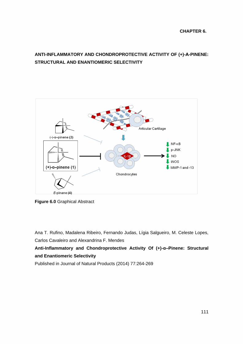
111
CHAPTER 6.
ANTI-INFLAMMATORY AND CHONDROPROTECTIVE ACTIVITY OF (+)-Α-PINENE:
STRUCTURAL AND ENANTIOMERIC SELECTIVITY
Figure 6.0 Graphical Abstract
Ana T. Rufino, Madalena Ribeiro, Fernando Judas, Lígia Salgueiro, M. Celeste Lopes,
Carlos Cavaleiro and Alexandrina F. Mendes
Anti-Inflammatory and Chondroprotective Activity Of (+)-α–Pinene: Structural
and Enantiomeric Selectivity
Published in Journal of Natural Products (2014) 77:264-269
CHAPTER 6
112
ABSTRACT
Previous studies have suggested that α-pinene, a common volatile plant metabolite,
may have anti-inflammatory effects in human chondrocytes, thus exhibiting potential
anti-osteoarthritic activity. The objective of this study was to further characterize the
potential anti-osteoarthritic activity of selected pinene derivatives by evaluating their
ability to modulate inflammation and extracellular matrix remodelling in human
chondrocytes and to correlate the biological and chemical properties by determining
whether the effects are isomer- and/or enantiomer-selective. To further elucidate
chemicopharmacological interactions, the activities of other naturally occurring
monoterpenes with the pinane nucleus were also investigated. At non-cytotoxic
concentrations, (+)-α-pinene (1) elicited the most potent inhibition of the IL-1β-induced
inflammatory and catabolic pathways, namely, NF-κB and JNK activation and the
expression of the inflammatory (iNOS) and catabolic (MMP-1 and -13) genes. (−)-α-
Pinene (2) was less active than the (+)-enantiomer (1), and β-pinene (3) was inactive.
E-Pinane (4) and oxygenated pinane-derived compounds, pinocarveol (5), myrtenal
(6), (E)-myrtanol (7), myrtenol (8), and (Z)-verbenol (9), were less effective or even
completely inactive and more cytotoxic than the pinenes tested (1−3). The data
obtained show isomer- and enantiomer-selective anti-inflammatory and anti-catabolic
effects of α-pinene in human chondrocytes, (+)-α-pinene (1) being the most promising
for further studies to determine its potential value as an anti-osteoarthritic drug.
CHAPTER 6
113
6.1 INTRODUCTION
Osteoarthritis (OA) is estimated to affect 10−15% of the world population, being the
leading cause worldwide of chronic disability in the elderly and also in those in the
working age range.[Kaplan et al., 2013; Zhang and Jordan, 2010] OA is a multifactorial
joint disease characterized by a progressive loss of the articular cartilage extracellular
matrix and local inflammation that lead to cartilage destruction, loss of joint function,
and pain.[Goldring and Otero, 2011] The huge socioeconomic burden of the disease
and the absence of effective disease-modifying OA drugs have led to its inclusion in a
group of priority diseases selected in the World Health Organization report “Priority
Medicines for Europe and the World”, as commissioned by the European
Commission.[Kaplan et al., 2013] In view of this, our previous study identified the
essential oil from the leaves of Juniperus oxycedrus L. subsp. oxycedrus
(Cupressaceae) as a promising source of compounds with potential anti-osteoarthritic
activity. In particular, it was found that two fractions of this essential oil, one rich in α-
pinene (93%) and the other containing oxygenated mono- and sesquiterpenes,
effectively inhibited nuclear factor-κB activation and the production of nitric oxide
(NO),[Neves et al., 2009] which play major roles in promoting the inflammatory and
catabolic responses in human articular chondrocytes that drive OA development and
progression.[Fernandes et al., 2002; Goldring and Otero, 2011; Goldring et al., 2008]
Several studies have attributed biological properties to α- pinene, including
antimicrobial, anti-inflammatory,[Zhou et al., 2004a] apoptotic, and anti-metastatic
effects,[Matsuo et al., 2011] but information concerning the activities of its enantiomers,
as well as of the β-isomer, is scarce. To our knowledge, this is restricted to
antibacterial, antifungal,[da Silva et al., 2012] and insecticide[Michaelakis et al., 2009]
activities. Therefore, this study aimed to confirm that the previously described inhibitory
effects of the essential oil of J. oxycedrus L. subsp. oxycedrus in human chondrocytes
are due primarily to α- pinene and especially to determine whether those effects are
enantio- and/or isomer-selective. For this purpose, (+)-α- pinene (1), (−)-α-pinene (2),
the racemic α-pinene (1 and 2) OA,[Akhtar and Haqqi, 2011; Csaki et al., 2009]
consisting of human articular chondrocytes treated with the pro-inflammatory and pro-
catabolic cytokine interleukin-1β (IL-1β).[Fernandes et al., 2002; Goldring and Otero,
2011; Goldring et al., 2008] To further elucidate possible relationships between
chemical structure and potential anti-osteoarthritic activity, other naturally occurring
pinane derivative compounds were also included in this study, namely, (E)-pinane (4),
pinocarveol (5), myrtenal (6), (E)-myrtanol (7), myrtenol (8), and (Z)-verbenol (9). Some
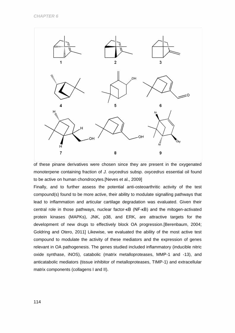
CHAPTER 6
114
of these pinane derivatives were chosen since they are present in the oxygenated
monoterpene containing fraction of J. oxycedrus subsp. oxycedrus essential oil found
to be active on human chondrocytes.[Neves et al., 2009]
Finally, and to further assess the potential anti-osteoarthritic activity of the test
compound(s) found to be more active, their ability to modulate signalling pathways that
lead to inflammation and articular cartilage degradation was evaluated. Given their
central role in those pathways, nuclear factor-κB (NF-κB) and the mitogen-activated
protein kinases (MAPKs), JNK, p38, and ERK, are attractive targets for the
development of new drugs to effectively block OA progression.[Berenbaum, 2004;
Goldring and Otero, 2011] Likewise, we evaluated the ability of the most active test
compound to modulate the activity of these mediators and the expression of genes
relevant in OA pathogenesis. The genes studied included inflammatory (inducible nitric
oxide synthase, iNOS), catabolic (matrix metalloproteases, MMP-1 and -13), and
anticatabolic mediators (tissue inhibitor of metalloproteases, TIMP-1) and extracellular
matrix components (collagens I and II).
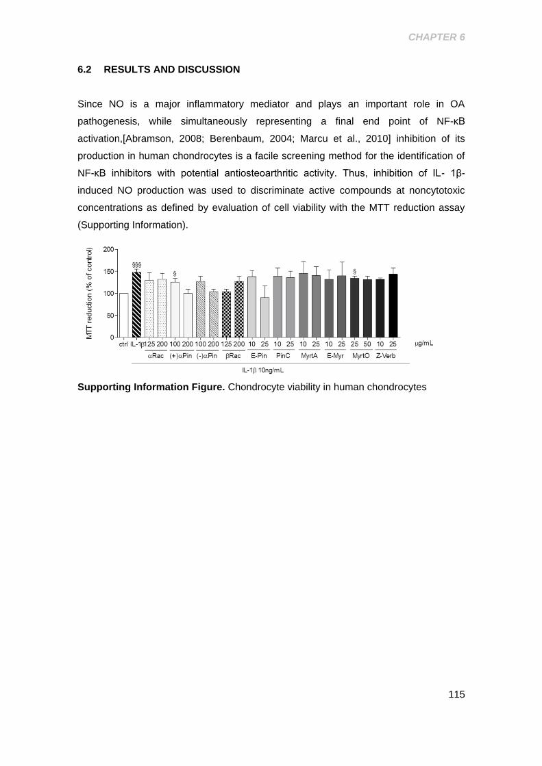
CHAPTER 6
115
6.2 RESULTS AND DISCUSSION
Since NO is a major inflammatory mediator and plays an important role in OA
pathogenesis, while simultaneously representing a final end point of NF-κB
activation,[Abramson, 2008; Berenbaum, 2004; Marcu et al., 2010] inhibition of its
production in human chondrocytes is a facile screening method for the identification of
NF-κB inhibitors with potential antiosteoarthritic activity. Thus, inhibition of IL- 1β-
induced NO production was used to discriminate active compounds at noncytotoxic
concentrations as defined by evaluation of cell viability with the MTT reduction assay
(Supporting Information).
Supporting Information Figure. Chondrocyte viability in human chondrocytes
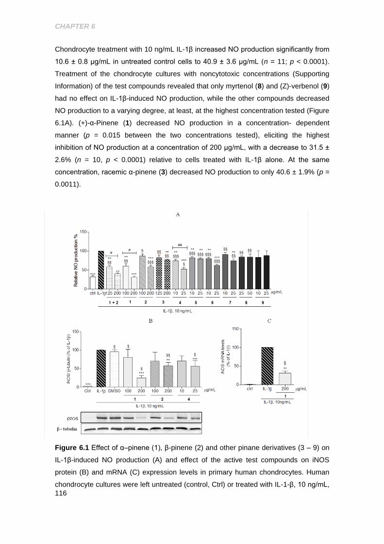
CHAPTER 6
116
Chondrocyte treatment with 10 ng/mL IL-1β increased NO production significantly from
10.6 ± 0.8 μg/mL in untreated control cells to 40.9 ± 3.6 μg/mL (n = 11; p < 0.0001).
Treatment of the chondrocyte cultures with noncytotoxic concentrations (Supporting
Information) of the test compounds revealed that only myrtenol (8) and (Z)-verbenol (9)
had no effect on IL-1β-induced NO production, while the other compounds decreased
NO production to a varying degree, at least, at the highest concentration tested (Figure
6.1A). (+)-α-Pinene (1) decreased NO production in a concentration- dependent
manner (p = 0.015 between the two concentrations tested), eliciting the highest
inhibition of NO production at a concentration of 200 μg/mL, with a decrease to 31.5 ±
2.6% (n = 10, p < 0.0001) relative to cells treated with IL-1β alone. At the same
concentration, racemic α-pinene (3) decreased NO production to only 40.6 ± 1.9% (p =
0.0011).
Figure 6.1 Effect of α–pinene (1), β-pinene (2) and other pinane derivatives (3 – 9) on
IL-1β-induced NO production (A) and effect of the active test compounds on iNOS
protein (B) and mRNA (C) expression levels in primary human chondrocytes. Human
chondrocyte cultures were left untreated (control, Ctrl) or treated with IL-1-β, 10 ng/mL,
CHAPTER 6
117
for 24 h (A and B) or 6h (C), following pre-treatment for 30 min with the indicated
concentrations of the test compounds or DMSO. Each column represents the mean ±
SEM of at least, four independent experiments. *p <0.05, **p <0.01, ***p <0.001 relative
to IL-1β-treated cells; §p <0.05, §§p <0.01, §§§p <0.001 relative to Ctrl; #p <0.05, ##p
<0.01 between the conditions indicated.
However, these results were not statistically different and are in the same range
reported for the α-pinene-rich fraction of the essential oil from the leaves of J.
oxycedrus subsp. oxycedrus. [Neves et al., 2009]
On the other hand, (−)-α-pinene (2) and the racemic β- pinene (3) were active only at
the highest concentrations tested (200 μg/mL), with much lower inhibitions (to 58.1 ±
4.9%, p = 0.0001 and 76.8 ± 2.3%, p = 0.0098) than those elicited by either racemic α-
pinene (1 and 2) or its (+) enantiomer (1) at the same concentration. In contrast, (E)-
pinane (4), pinocarveol (5), myrtenal (6), and (E)-myrtanol (7), at the highest non-
cytotoxic concentration, elicited only relatively small decreases of IL-1β-induced NO
production.
These results show clear differences in activity among the compounds tested (Figure
6.1A). In particular, it is noticeable that α-pinene is the most active, showing not only
selectivity concerning the β-isomer (3), almost devoid of activity, but also enantiomeric
selectivity, as (+)-α-pinene (1) was found to be more effective than the (−) enantiomer
(2).
To confirm these results and further elucidate the mechanism of action of the
compounds that more effectively reduced NO production, the α-pinene enantiomers (1
and 2) and (E) - pinane (4), effects on iNOS protein levels induced by IL-1β were
evaluated. Paralleling the results relative to NO production, Figure 6.1B shows that at
200 μg/mL (+)-α-pinene (1) elicited the greatest inhibition, decreasing iNOS protein
levels to 24.4 ± 14.9% (p < 0.0001) relative to those found in cells treated with IL-1β
alone. The highest concentrations tested of (−)-α-pinene (2) and (E)-pinane (4) elicited
a smaller, but statistically significant, decrease of iNOS protein levels. (+)-α-Pinene (1)
also extensively decreased IL-1β- induced iNOS mRNA levels (Figure 6.1C), indicating
that reduction of iNOS protein and enzymatic activity likely occur through effects at or
upstream of gene transcription.
As human cartilage samples are limited, both in number and quantity, and a large
number of cells is required, the human chondrocytic cell line, C-28/I2, was used to
evaluate the ability of α-pinene enantiomers to inhibit IL-1β-induced NF-κB activation.
This requires the phosphorylation and subsequent ubiquitination and proteassomal
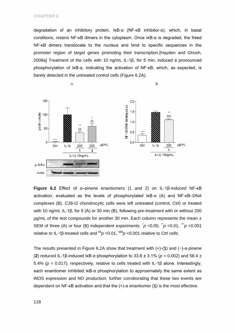
CHAPTER 6
118
degradation of an inhibitory protein, IκB-α (NF-κB inhibitor-α), which, in basal
conditions, retains NF-κB dimers in the cytoplasm. Once IκB-α is degraded, the freed
NF-κB dimers translocate to the nucleus and bind to specific sequences in the
promoter region of target genes promoting their transcription.[Hayden and Ghosh,
2008a] Treatment of the cells with 10 ng/mL IL-1β, for 5 min, induced a pronounced
phosphorylation of IκB-α, indicating the activation of NF-κB, which, as expected, is
barely detected in the untreated control cells (Figure 6.2A).
Figure 6.2 Effect of α–pinene enantiomers (1 and 2) on IL-1β-induced NF-κB
activation, evaluated as the levels of phosphorylated IκB-α (A) and NF-κB–DNA
complexes (B). C28-I2 chondrocytic cells were left untreated (control, Ctrl) or treated
with 10 ng/mL IL-1β, for 5 (A) or 30 min (B), following pre-treatment with or without 200
µg/mL of the test compounds for another 30 min. Each column represents the mean ±
SEM of three (A) or four (B) independent experiments. *p <0.05, **p <0.01, ***p <0.001
relative to IL-1β-treated cells and §§p <0.01, §§§p <0.001 relative to Ctrl cells.
The results presented in Figure 6.2A show that treatment with (+)-(1) and (−)-α-pinene
(2) reduced IL-1β-induced IκB-α phosphorylation to 33.6 ± 3.1% (p = 0.002) and 58.4 ±
5.4% (p = 0.017), respectively, relative to cells treated with IL-1β alone. Interestingly,
each enantiomer inhibited IκB-α phosphorylation to approximately the same extent as
iNOS expression and NO production, further corroborating that these two events are
dependent on NF-κB activation and that the (+)-α enantiomer (1) is the most effective.
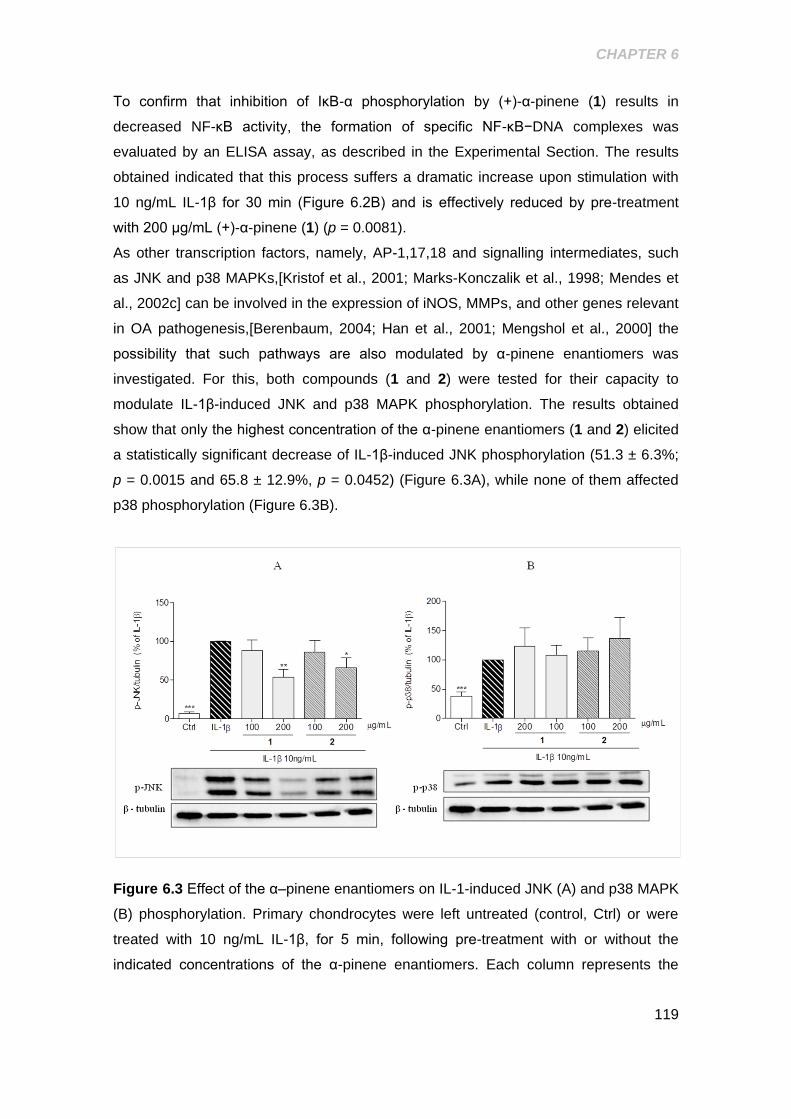
CHAPTER 6
119
To confirm that inhibition of IκB-α phosphorylation by (+)-α-pinene (1) results in
decreased NF-κB activity, the formation of specific NF-κB−DNA complexes was
evaluated by an ELISA assay, as described in the Experimental Section. The results
obtained indicated that this process suffers a dramatic increase upon stimulation with
10 ng/mL IL-1β for 30 min (Figure 6.2B) and is effectively reduced by pre-treatment
with 200 μg/mL (+)-α-pinene (1) (p = 0.0081).
As other transcription factors, namely, AP-1,17,18 and signalling intermediates, such
as JNK and p38 MAPKs,[Kristof et al., 2001; Marks-Konczalik et al., 1998; Mendes et
al., 2002c] can be involved in the expression of iNOS, MMPs, and other genes relevant
in OA pathogenesis,[Berenbaum, 2004; Han et al., 2001; Mengshol et al., 2000] the
possibility that such pathways are also modulated by α-pinene enantiomers was
investigated. For this, both compounds (1 and 2) were tested for their capacity to
modulate IL-1β-induced JNK and p38 MAPK phosphorylation. The results obtained
show that only the highest concentration of the α-pinene enantiomers (1 and 2) elicited
a statistically significant decrease of IL-1β-induced JNK phosphorylation (51.3 ± 6.3%;
p = 0.0015 and 65.8 ± 12.9%, p = 0.0452) (Figure 6.3A), while none of them affected
p38 phosphorylation (Figure 6.3B).
Figure 6.3 Effect of the α–pinene enantiomers on IL-1-induced JNK (A) and p38 MAPK
(B) phosphorylation. Primary chondrocytes were left untreated (control, Ctrl) or were
treated with 10 ng/mL IL-1β, for 5 min, following pre-treatment with or without the
indicated concentrations of the α-pinene enantiomers. Each column represents the
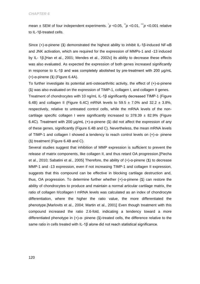
CHAPTER 6
120
mean ± SEM of four independent experiments. *p <0.05, **p <0.01, ***p <0.001 relative
to IL-1β-treated cells.
Since (+)-α-pinene (1) demonstrated the highest ability to inhibit IL-1β-induced NF-κB
and JNK activation, which are required for the expression of MMPs-1 and -13 induced
by IL- 1β,[Han et al., 2001; Mendes et al., 2002c] its ability to decrease these effects
was also evaluated. As expected the expression of both genes increased significantly
in response to IL-1β and was completely abolished by pre-treatment with 200 μg/mL
(+)-α-pinene (1) (Figure 6.4A).
To further investigate its potential anti-osteoarthritic activity, the effect of (+)-α-pinene
(1) was also evaluated on the expression of TIMP-1, collagen I, and collagen II genes.
Treatment of chondrocytes with 10 ng/mL IL-1β significantly decreased TIMP-1 (Figure
6.4B) and collagen II (Figure 6.4C) mRNA levels to 59.5 ± 7.0% and 32.2 ± 3.8%,
respectively, relative to untreated control cells, while the mRNA levels of the non-
cartilage specific collagen I were significantly increased to 378.39 ± 82.9% (Figure
6.4C). Treatment with 200 μg/mL (+)-α-pinene (1) did not affect the expression of any
of these genes, significantly (Figure 6.4B and C). Nevertheless, the mean mRNA levels
of TIMP-1 and collagen I showed a tendency to reach control levels on (+)-α- pinene
(1) treatment (Figure 6.4B and C).
Several studies suggest that inhibition of MMP expression is sufficient to prevent the
release of matrix components, like collagen II, and thus retard OA progression.[Piecha
et al., 2010; Sabatini et al., 2005] Therefore, the ability of (+)-α-pinene (1) to decrease
MMP-1 and -13 expression, even if not increasing TIMP-1 and collagen II expression,
suggests that this compound can be effective in blocking cartilage destruction and,
thus, OA progression. To determine further whether (+)-α-pinene (1) can restore the
ability of chondrocytes to produce and maintain a normal articular cartilage matrix, the
ratio of collagen II/collagen I mRNA levels was calculated as an index of chondrocyte
differentiation, where the higher the ratio value, the more differentiated the
phenotype.[Marlovits et al., 2004; Martin et al., 2001] Even though treatment with this
compound increased the ratio 2.6-fold, indicating a tendency toward a more
differentiated phenotype in (+)-α- pinene (1)-treated cells, the difference relative to the
same ratio in cells treated with IL-1β alone did not reach statistical significance.
CHAPTER 6
121
Figure 6.4 Effect of (+)-α-pinene (1) on IL-1β-induced changes in gene expression.
mRNA levels of (A) MMP-1 and -13, (B) TIMP-1 and (C) collagen I and collagen II were
evaluated by RT-PCR. Each column represents the mean ± SEM of, at least, four
independent experiments in which human chondrocytes were left untreated (control,
Ctrl) or treated for 12 h (A) or 24 h (B and C) with 10 ng/mL IL-1β, in the presence or
absence of 200 µg/mL (+)-α-pinene (1). *p <0.05, **p <0.01, ***p <0.001 relative to IL-1β-
treated cells; §§p <0.01 and §§§p <0.001 relative to Ctrl cells.
In summary, the results reported in this study showed enantiomeric-selective anti-
inflammatory and anticatabolic effects of (+)-α-pinene and derivatives in human
chondrocytes. Of the compounds tested, (+)-α-pinene (1) showed the greatest efficacy
to inhibit IL-1β-induced inflammatory and catabolic pathways, namely, NF-κB and JNK
activation and the expression of inflammatory (iNOS) and catabolic (MMP-1 and -13)
genes. Thus, (+)-α-pinene (1) deserves to be further investigated, namely, by
CHAPTER 6
122
evaluating its pharmacokinetic and pharmacodynamic in vivo properties, in order to
determine its potential as an antiosteoarthritic drug. Moreover, this study also shows
that oxygenated pinane-derived compounds, namely, pinocarveol (5), myrtenal (6), (E)-
myrtanol (7), myrtenol (8), and (Z)-verbenol (9), are less effective or even completely
inactive and more cytotoxic than the non-oxygenated counterparts tested.
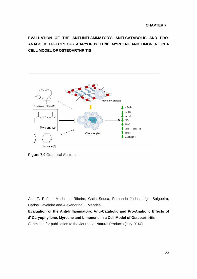
123
CHAPTER 7.
EVALUATION OF THE ANTI-INFLAMMATORY, ANTI-CATABOLIC AND PRO-
ANABOLIC EFFECTS OF E-CARYOPHYLLENE, MYRCENE AND LIMONENE IN A
CELL MODEL OF OSTEOARTHRITIS
Figure 7.0 Graphical Abstract
Ana T. Rufino, Madalena Ribeiro, Cátia Sousa, Fernando Judas, Lígia Salgueiro,
Carlos Cavaleiro and Alexandrina F. Mendes
Evaluation of the Anti-Inflammatory, Anti-Catabolic and Pro-Anabolic Effects of
E-Caryophyllene, Myrcene and Limonene in a Cell Model of Osteoarthritis
Submitted for publication to the Journal of Natural Products (July 2014)
CHAPTER 7
124
ABSTRACT
The essential oils of Eryngium duriaei subsp. juresianum and Lavandula luisieri
demonstrated anti-inflammatory properties in human chondrocytes. This study aimed at
identifying active compounds and characterizing their antiosteoarthritic properties. E-
caryophyllene (1), myrcene (2) and limonene (3), present in the active fractions of
these essential oils, were evaluated for their ability to modulate inflammatory, catabolic
and anabolic processes in human chondrocytes. Myrcene (2) (IC50=37.3 μg/mL) and
limonene (3) (IC50=85.3 µg/mL) inhibited IL-1β-induced nitric oxide production, but E-
caryophyllene was inactive. Myrcene, and limonene to a lesser extent, also decreased
IL-1β-induced NF-κB, JNK and p38 activation and the expression of inflammatory
(iNOS) and catabolic (MMP-1 and MMP-13) genes, while increasing the expression of
anti-catabolic genes [TIMP-1 and -3 by myrcene (2) and TIMP-1 by limonene (3)].
Neither myrcene (2) nor limonene (3) increased the expression of cartilage-specific
genes (collagen II and aggrecan), but both compounds prevented the increase in the
non-cartilage specific, collagen I. Limonene (3) increased ERK1/2 activation by 30%
which may be unfavorable, while myrcene (2) decreased it by 26%, relative to IL-1β-
treated cells.
These data show that myrcene (2) has significant anti-inflammatory and anti-catabolic
effects and, thus, may halt or, at least, slow down cartilage destruction and
osteoarthritis progression.
CHAPTER 7
125
7.1 INTRODUCTION
Osteoarthritis (OA) is a multifactorial degenerative joint disease characterized by
inflammation and progressive loss of the articular cartilage. It affects 10-15% of the
world population and is a major cause of disability in the elderly, as well as in the
workforce population.[Zhang and Jordan, 2010] Existing therapeutic approaches are
mainly symptomatic, thus novel drugs with disease-modifying and chondroprotective
properties are required to halt disease progression and decrease its huge socio-
economic impact.[Goldring and Goldring, 2007; Kaplan et al., 2013]
Plant-derived compounds show important biological properties that can be explored
in the context of OA for identification of compounds with potential anti-osteoarthritic
activity.[Calixto et al., 2004; Khalife and Zafarullah, 2011] Among compounds of plant
origin, those found in essential oils present favorable pharmacokinetic properties,
namely lipophilicity and low molecular weight.[Miguel, 2010] Our previous studies have
been focused in identifying essential oils with anti-inflammatory and anti-catabolic
properties in human chondrocytes to be used as sources of compounds with potential
anti-osteoarthritic activitya[Neves et al., 2009] In this context, we identified the essential
oils of Eryngium duriaei subsp. juresianum and Lavandula luisieri as possessing anti-
inflammatory properties in human chondrocytes.a The current study aims at identifying
the active compounds of these essential oils and further characterizing their
pharmacological properties in human chondrocytes.
For this, mechanisms relevant as pharmacological targets for the development of
anti-osteoarthritic drugs need to be addressed. Although OA etiology is not yet
completely understood, pro-inflammatory cytokines, like interleukin-1β (IL-1β), play a
central role in disease development and progression by inducing the expression of
cartilage matrix-degrading enzymes and impairing anabolic and anti-catabolic
responses.[Goldring et al., 2008] The consequent upregulated degradative process,
together with impaired reparative responses, results in progressive cartilage loss, the
hallmark of OA, accompanied by alterations in other joint tissues.
a Rufino, A. T.; Ferreira, I. V.; Judas, F.; Salgueiro, L.; Lopes, M. C.; Cavaleiro, C.; Mendes, A.
F. (submitted at Journal of Natural products _ Chapter 5).
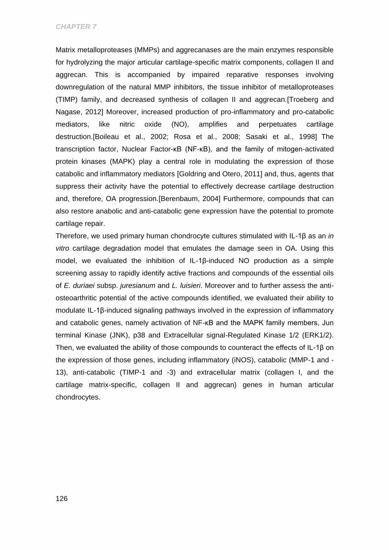
CHAPTER 7
126
Matrix metalloproteases (MMPs) and aggrecanases are the main enzymes responsible
for hydrolyzing the major articular cartilage-specific matrix components, collagen II and
aggrecan. This is accompanied by impaired reparative responses involving
downregulation of the natural MMP inhibitors, the tissue inhibitor of metalloproteases
(TIMP) family, and decreased synthesis of collagen II and aggrecan.[Troeberg and
Nagase, 2012] Moreover, increased production of pro-inflammatory and pro-catabolic
mediators, like nitric oxide (NO), amplifies and perpetuates cartilage
destruction.[Boileau et al., 2002; Rosa et al., 2008; Sasaki et al., 1998] The
transcription factor, Nuclear Factor-κB (NF-κB), and the family of mitogen-activated
protein kinases (MAPK) play a central role in modulating the expression of those
catabolic and inflammatory mediators [Goldring and Otero, 2011] and, thus, agents that
suppress their activity have the potential to effectively decrease cartilage destruction
and, therefore, OA progression.[Berenbaum, 2004] Furthermore, compounds that can
also restore anabolic and anti-catabolic gene expression have the potential to promote
cartilage repair.
Therefore, we used primary human chondrocyte cultures stimulated with IL-1β as an in
vitro cartilage degradation model that emulates the damage seen in OA. Using this
model, we evaluated the inhibition of IL-1β-induced NO production as a simple
screening assay to rapidly identify active fractions and compounds of the essential oils
of E. duriaei subsp. juresianum and L. luisieri. Moreover and to further assess the anti-
osteoarthritic potential of the active compounds identified, we evaluated their ability to
modulate IL-1β-induced signaling pathways involved in the expression of inflammatory
and catabolic genes, namely activation of NF-κB and the MAPK family members, Jun
terminal Kinase (JNK), p38 and Extracellular signal-Regulated Kinase 1/2 (ERK1/2).
Then, we evaluated the ability of those compounds to counteract the effects of IL-1β on
the expression of those genes, including inflammatory (iNOS), catabolic (MMP-1 and -
13), anti-catabolic (TIMP-1 and -3) and extracellular matrix (collagen I, and the
cartilage matrix-specific, collagen II and aggrecan) genes in human articular
chondrocytes.
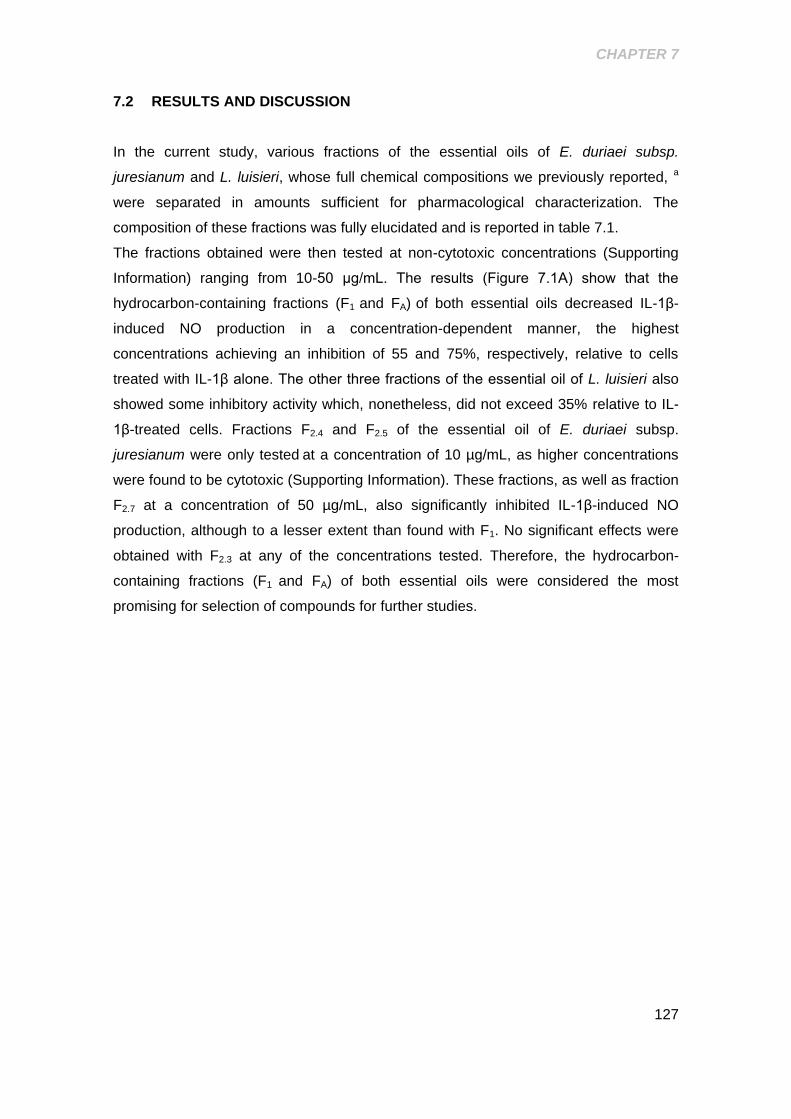
CHAPTER 7
127
7.2 RESULTS AND DISCUSSION
In the current study, various fractions of the essential oils of E. duriaei subsp.
juresianum and L. luisieri, whose full chemical compositions we previously reported, a
were separated in amounts sufficient for pharmacological characterization. The
composition of these fractions was fully elucidated and is reported in table 7.1.
The fractions obtained were then tested at non-cytotoxic concentrations (Supporting
Information) ranging from 10-50 μg/mL. The results (Figure 7.1A) show that the
hydrocarbon-containing fractions (F1 and FA) of both essential oils decreased IL-1β-
induced NO production in a concentration-dependent manner, the highest
concentrations achieving an inhibition of 55 and 75%, respectively, relative to cells
treated with IL-1β alone. The other three fractions of the essential oil of L. luisieri also
showed some inhibitory activity which, nonetheless, did not exceed 35% relative to IL-
1β-treated cells. Fractions F2.4 and F2.5 of the essential oil of E. duriaei subsp.
juresianum were only tested at a concentration of 10 µg/mL, as higher concentrations
were found to be cytotoxic (Supporting Information). These fractions, as well as fraction
F2.7 at a concentration of 50 µg/mL, also significantly inhibited IL-1β-induced NO
production, although to a lesser extent than found with F1. No significant effects were
obtained with F2.3 at any of the concentrations tested. Therefore, the hydrocarbon-
containing fractions (F1 and FA) of both essential oils were considered the most
promising for selection of compounds for further studies.
CHAPTER 7
128
Supporting Information Figure: Effect of the fractions isolated from the essential oils
of Eryngium duriaei subsp. juresianum and Lavandula luisieri (A) and of the isolated
compounds E-Caryophyllene (1), myrcene (2) and limonene (3) (B) on human
chondrocyte viability. Each column represents the mean ± SEM of at least four
independent experiments where chondrocytes were treated with the indicated
concentrations of the test compounds for 24 h together with IL-1β (10ng/mL) and cell
viability accessed as described above. §P<0.05, §§P<0.01 relative to untreated control
cells (Ctrl).
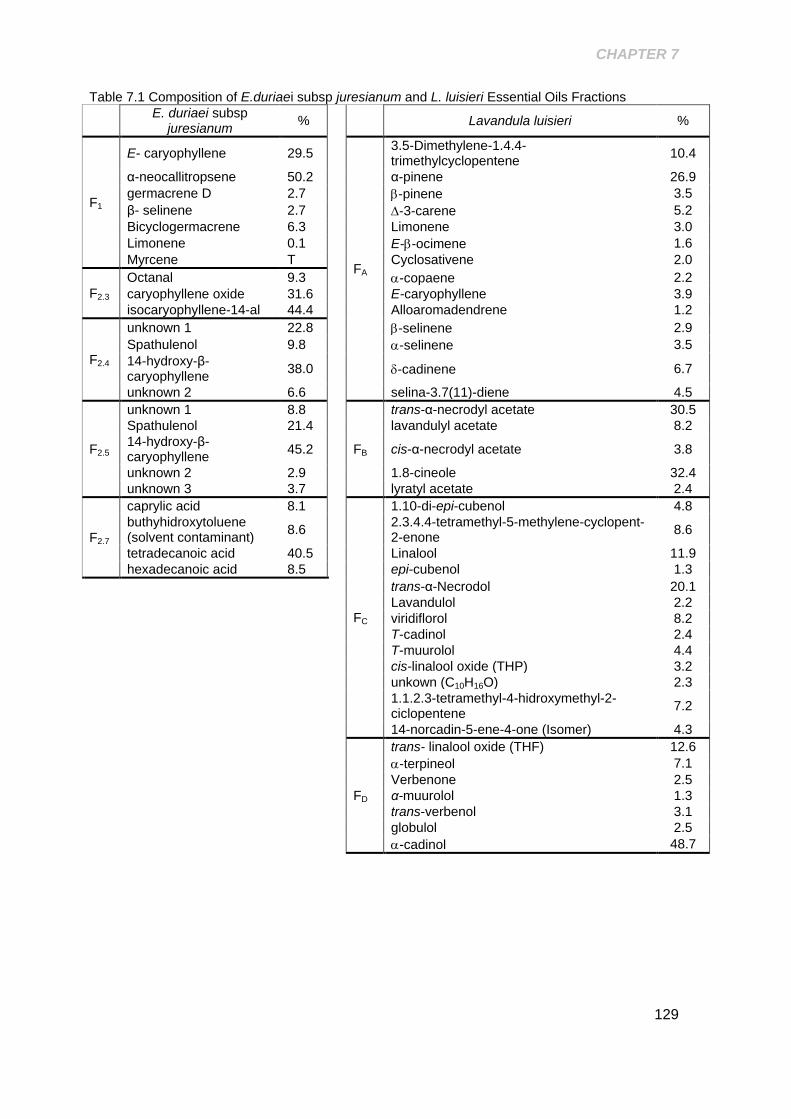
CHAPTER 7
129
Table 7.1 Composition of E.duriaei subsp juresianum and L. luisieri Essential Oils Fractions
E. duriaei subsp
juresianum %
Lavandula luisieri %
F1
E- caryophyllene 29.5
FA
3.5-Dimethylene-1.4.4-trimethylcyclopentene
10.4
α-neocallitropsene 50.2 α-pinene 26.9
germacrene D 2.7 -pinene 3.5
β- selinene 2.7 -3-carene 5.2
Bicyclogermacrene 6.3 Limonene 3.0
Limonene 0.1 E--ocimene 1.6
Myrcene T Cyclosativene 2.0
F2.3
Octanal 9.3 -copaene 2.2
caryophyllene oxide 31.6 E-caryophyllene 3.9
isocaryophyllene-14-al 44.4 Alloaromadendrene 1.2
F2.4
unknown 1 22.8 -selinene 2.9
Spathulenol 9.8 -selinene 3.5
14-hydroxy-β-caryophyllene
38.0 -cadinene 6.7
unknown 2 6.6 selina-3.7(11)-diene 4.5
F2.5
unknown 1 8.8
FB
trans-α-necrodyl acetate 30.5
Spathulenol 21.4 lavandulyl acetate 8.2
14-hydroxy-β-caryophyllene
45.2 cis-α-necrodyl acetate 3.8
unknown 2 2.9 1.8-cineole 32.4
unknown 3 3.7 lyratyl acetate 2.4
F2.7
caprylic acid 8.1
FC
1.10-di-epi-cubenol 4.8
buthyhidroxytoluene (solvent contaminant)
8.6
2.3.4.4-tetramethyl-5-methylene-cyclopent-2-enone
8.6
tetradecanoic acid 40.5 Linalool 11.9
hexadecanoic acid 8.5 epi-cubenol 1.3
trans-α-Necrodol 20.1
Lavandulol 2.2
viridiflorol 8.2
T-cadinol 2.4
T-muurolol 4.4
cis-linalool oxide (THP) 3.2
unkown (C10H16O) 2.3
1.1.2.3-tetramethyl-4-hidroxymethyl-2-ciclopentene
7.2
14-norcadin-5-ene-4-one (Isomer) 4.3
FD
trans- linalool oxide (THF) 12.6
-terpineol 7.1
Verbenone 2.5
α-muurolol 1.3
trans-verbenol 3.1
globulol 2.5
-cadinol 48.7
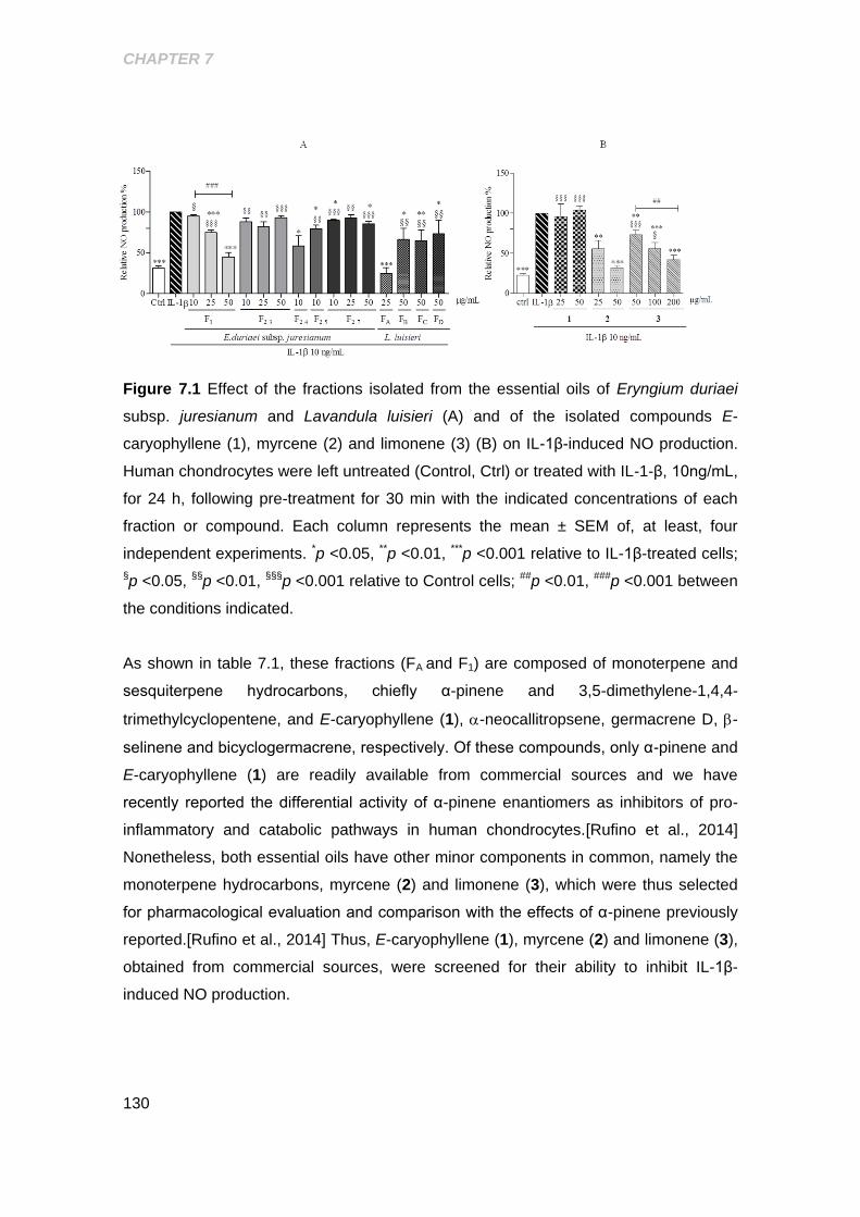
CHAPTER 7
130
Figure 7.1 Effect of the fractions isolated from the essential oils of Eryngium duriaei
subsp. juresianum and Lavandula luisieri (A) and of the isolated compounds E-
caryophyllene (1), myrcene (2) and limonene (3) (B) on IL-1β-induced NO production.
Human chondrocytes were left untreated (Control, Ctrl) or treated with IL-1-β, 10ng/mL,
for 24 h, following pre-treatment for 30 min with the indicated concentrations of each
fraction or compound. Each column represents the mean ± SEM of, at least, four
independent experiments. *p <0.05, **p <0.01, ***p <0.001 relative to IL-1β-treated cells;
§p <0.05, §§p <0.01, §§§p <0.001 relative to Control cells; ##p <0.01, ###p <0.001 between
the conditions indicated.
As shown in table 7.1, these fractions (FA and F1) are composed of monoterpene and
sesquiterpene hydrocarbons, chiefly α-pinene and 3,5-dimethylene-1,4,4-
trimethylcyclopentene, and E-caryophyllene (1), -neocallitropsene, germacrene D, -
selinene and bicyclogermacrene, respectively. Of these compounds, only α-pinene and
E-caryophyllene (1) are readily available from commercial sources and we have
recently reported the differential activity of α-pinene enantiomers as inhibitors of pro-
inflammatory and catabolic pathways in human chondrocytes.[Rufino et al., 2014]
Nonetheless, both essential oils have other minor components in common, namely the
monoterpene hydrocarbons, myrcene (2) and limonene (3), which were thus selected
for pharmacological evaluation and comparison with the effects of α-pinene previously
reported.[Rufino et al., 2014] Thus, E-caryophyllene (1), myrcene (2) and limonene (3),
obtained from commercial sources, were screened for their ability to inhibit IL-1β-
induced NO production.
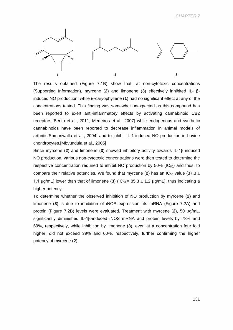
CHAPTER 7
131
The results obtained (Figure 7.1B) show that, at non-cytotoxic concentrations
(Supporting Information), myrcene (2) and limonene (3) effectively inhibited IL-1β-
induced NO production, while E-caryophyllene (1) had no significant effect at any of the
concentrations tested. This finding was somewhat unexpected as this compound has
been reported to exert anti-inflammatory effects by activating cannabinoid CB2
receptors,[Bento et al., 2011; Medeiros et al., 2007] while endogenous and synthetic
cannabinoids have been reported to decrease inflammation in animal models of
arthritis[Sumariwalla et al., 2004] and to inhibit IL-1-induced NO production in bovine
chondrocytes.[Mbvundula et al., 2005]
Since myrcene (2) and limonene (3) showed inhibitory activity towards IL-1β-induced
NO production, various non-cytotoxic concentrations were then tested to determine the
respective concentration required to inhibit NO production by 50% (IC50) and thus, to
compare their relative potencies. We found that myrcene (2) has an IC50 value (37.3
1.1 μg/mL) lower than that of limonene (3) (IC50 = 85.3 1.2 µg/mL), thus indicating a
higher potency.
To determine whether the observed inhibition of NO production by myrcene (2) and
limonene (3) is due to inhibition of iNOS expression, its mRNA (Figure 7.2A) and
protein (Figure 7.2B) levels were evaluated. Treatment with myrcene (2), 50 µg/mL,
significantly diminished IL-1β-induced iNOS mRNA and protein levels by 78% and
69%, respectively, while inhibition by limonene (3), even at a concentration four fold
higher, did not exceed 39% and 60%, respectively, further confirming the higher
potency of myrcene (2).
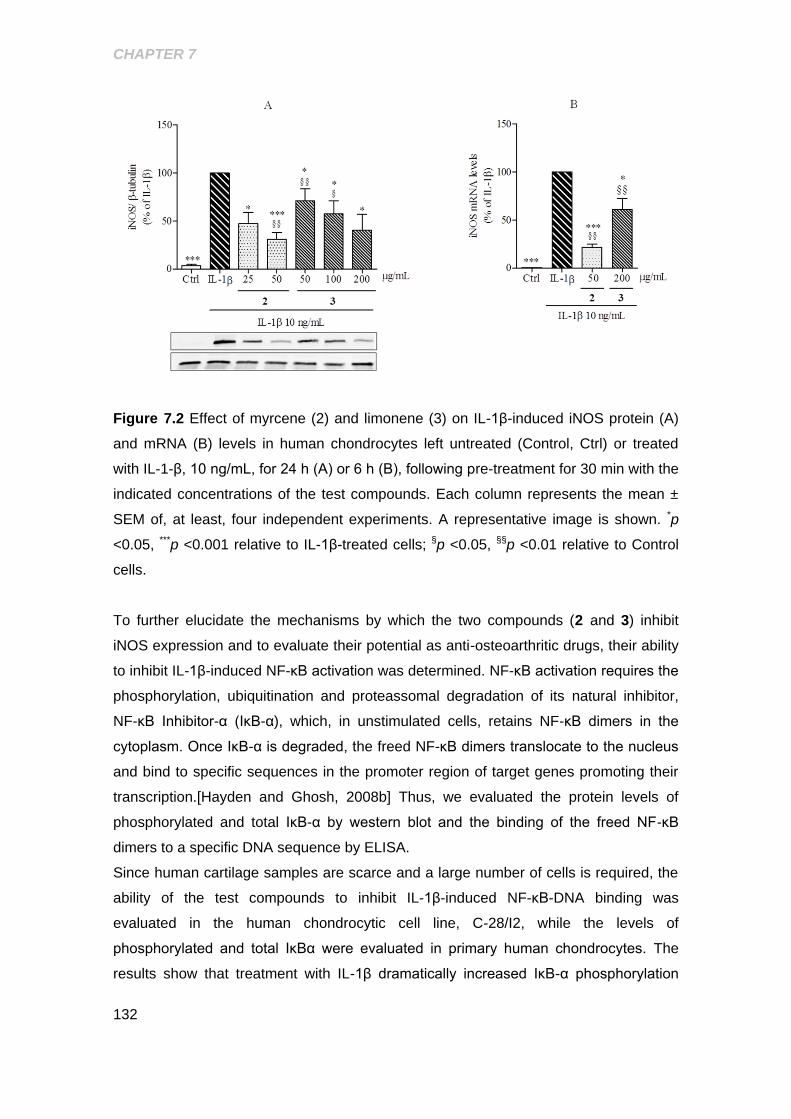
CHAPTER 7
132
Figure 7.2 Effect of myrcene (2) and limonene (3) on IL-1β-induced iNOS protein (A)
and mRNA (B) levels in human chondrocytes left untreated (Control, Ctrl) or treated
with IL-1-β, 10 ng/mL, for 24 h (A) or 6 h (B), following pre-treatment for 30 min with the
indicated concentrations of the test compounds. Each column represents the mean ±
SEM of, at least, four independent experiments. A representative image is shown. *p
<0.05, ***p <0.001 relative to IL-1β-treated cells; §p <0.05, §§p <0.01 relative to Control
cells.
To further elucidate the mechanisms by which the two compounds (2 and 3) inhibit
iNOS expression and to evaluate their potential as anti-osteoarthritic drugs, their ability
to inhibit IL-1β-induced NF-κB activation was determined. NF-κB activation requires the
phosphorylation, ubiquitination and proteassomal degradation of its natural inhibitor,
NF-κB Inhibitor-α (IκB-α), which, in unstimulated cells, retains NF-κB dimers in the
cytoplasm. Once IκB-α is degraded, the freed NF-κB dimers translocate to the nucleus
and bind to specific sequences in the promoter region of target genes promoting their
transcription.[Hayden and Ghosh, 2008b] Thus, we evaluated the protein levels of
phosphorylated and total IκB-α by western blot and the binding of the freed NF-κB
dimers to a specific DNA sequence by ELISA.
Since human cartilage samples are scarce and a large number of cells is required, the
ability of the test compounds to inhibit IL-1β-induced NF-κB-DNA binding was
evaluated in the human chondrocytic cell line, C-28/I2, while the levels of
phosphorylated and total IκBα were evaluated in primary human chondrocytes. The
results show that treatment with IL-1β dramatically increased IκB-α phosphorylation
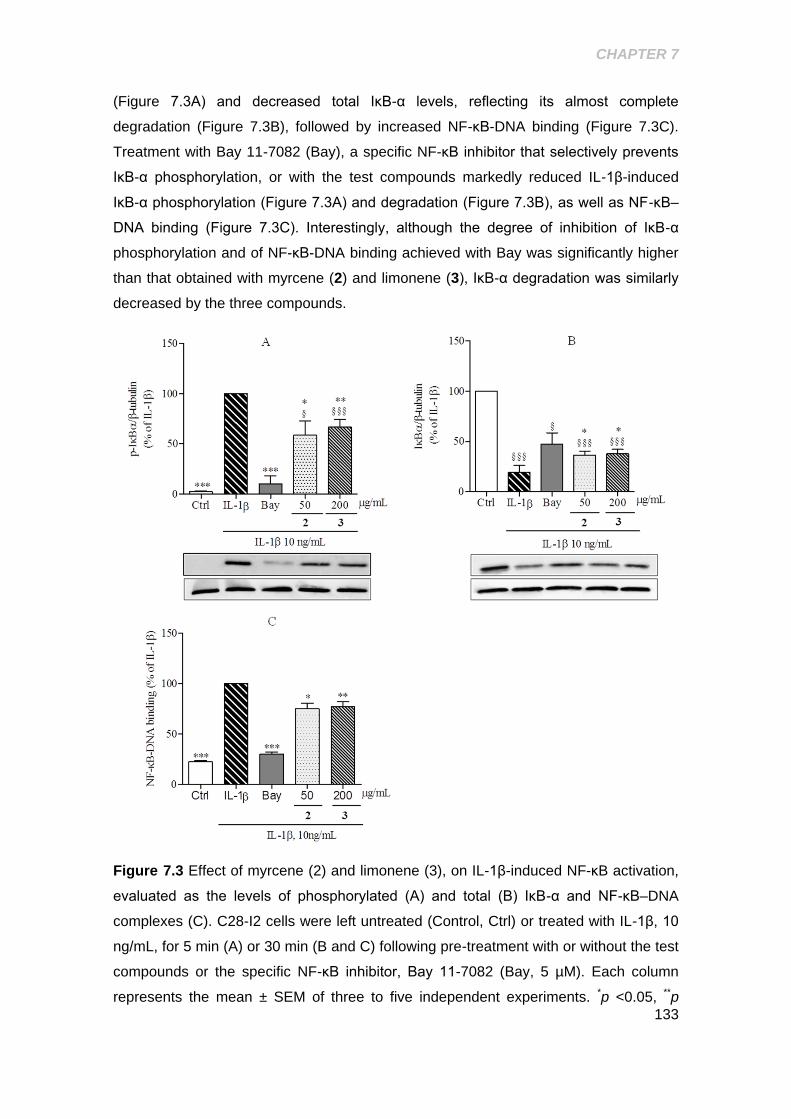
CHAPTER 7
133
(Figure 7.3A) and decreased total IκB-α levels, reflecting its almost complete
degradation (Figure 7.3B), followed by increased NF-κB-DNA binding (Figure 7.3C).
Treatment with Bay 11-7082 (Bay), a specific NF-κB inhibitor that selectively prevents
IκB-α phosphorylation, or with the test compounds markedly reduced IL-1β-induced
IκB-α phosphorylation (Figure 7.3A) and degradation (Figure 7.3B), as well as NF-κB–
DNA binding (Figure 7.3C). Interestingly, although the degree of inhibition of IκB-α
phosphorylation and of NF-κB-DNA binding achieved with Bay was significantly higher
than that obtained with myrcene (2) and limonene (3), IκB-α degradation was similarly
decreased by the three compounds.
Figure 7.3 Effect of myrcene (2) and limonene (3), on IL-1β-induced NF-κB activation,
evaluated as the levels of phosphorylated (A) and total (B) IκB-α and NF-κB–DNA
complexes (C). C28-I2 cells were left untreated (Control, Ctrl) or treated with IL-1β, 10
ng/mL, for 5 min (A) or 30 min (B and C) following pre-treatment with or without the test
compounds or the specific NF-κB inhibitor, Bay 11-7082 (Bay, 5 µM). Each column
represents the mean ± SEM of three to five independent experiments. *p <0.05, **p
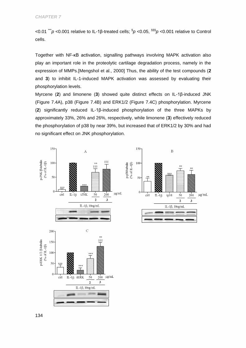
CHAPTER 7
134
<0.01 ***p <0.001 relative to IL-1β-treated cells; §p <0.05, §§§p <0.001 relative to Control
cells.
Together with NF-κB activation, signalling pathways involving MAPK activation also
play an important role in the proteolytic cartilage degradation process, namely in the
expression of MMPs.[Mengshol et al., 2000] Thus, the ability of the test compounds (2
and 3) to inhibit IL-1-induced MAPK activation was assessed by evaluating their
phosphorylation levels.
Myrcene (2) and limonene (3) showed quite distinct effects on IL-1β-induced JNK
(Figure 7.4A), p38 (Figure 7.4B) and ERK1/2 (Figure 7.4C) phosphorylation. Myrcene
(2) significantly reduced IL-1β-induced phosphorylation of the three MAPKs by
approximately 33%, 26% and 26%, respectively, while limonene (3) effectively reduced
the phosphorylation of p38 by near 39%, but increased that of ERK1/2 by 30% and had
no significant effect on JNK phosphorylation.
CHAPTER 7
135
Figure 7.4 Effects of myrcene (2) and limonene (3) on IL-1β- induced activation of
JNK, p38 and ERK1/2 in human chondrocytes. Phosphorylated levels of JNK (A), p38
(B) and ERK (1/2) (C) were analyzed in total cell extracts of human chondrocytes left
untreated (Control, Ctrl) or treated for 5 min with IL-1β, 10 ng/mL, following a pre-
treatment for 30 min with the indicated concentrations of myrcene, limonene or a
specific inhibitor of each MAPK. Each column represents the mean ± SEM of, at least,
four independent experiments. Representative images are shown. **p <0.01, ***p <0.001
relative to IL-1β-treated cells and §§§p <0.001 relative to Control cells. iJNK: SP600125
(20 µM); ip38: SB203580 (20 µM); iERK: U0126 (10 µM).
Reports on pharmacological properties of limonene (2) are scarce, but it has been
shown to have antimicrobial properties [Bevilacqua et al., 2010] and anti-inflammatory
effects in a mouse model of LPS-induced acute lung injury by suppressing MAPK and
NF-κB pathways[Chi et al., 2012]. The results presented here only partially agree with
this study, since limonene (3) inhibited NF-κB and p38 activation, but did not affect IL-
1β-induced JNK and actually potentiated ERK1/2 activation, suggesting that this
compound (3) has cell and/or stimulus specific effects. On the other hand, ERK1/2 is
required for a number of cellular processes, including activation of c-fos expression
which, among other functions, is involved in cell survival.[Karin et al., 1997; Shaulian
and Karin, 2002] Whether increased activation of ERK1/2 by limonene (3) contributes
to enhance chondrocyte survival was not addressed in this study, but is an interesting
possibility to study further, as increased chondrocyte death is a relevant feature of
OA.[Johnson et al., 2008] Nonetheless, since ERK1/2 has also been shown to inhibit
proteoglycan synthesis and promote inflammatory responses in chondrocytes,[Scherle
et al., 1997] the net effect resulting from its induction by limonene (3) is likely
undesirable, compromising its potential utility as a therapeutic agent in OA.
Then, we evaluated the ability of the compounds tested (2 and 3) to counteract the
effects of IL-1β on the expression of catabolic, anti-catabolic and extracellular matrix
genes, which, at least in part, are mediated by NF-κB and the MAPKs.[Goldring and
Otero, 2011] As expected, treatment of human chondrocytes with IL-1β (10 ng/mL)
increased MMP-1 and -13 mRNA levels by nearly 9- and 5-fold, respectively (Figure
7.5A), while decreasing TIMP-1 and -3 expression (Figure 7.5B). Myrcene (2)
decreased the mRNA levels of both MMPs by nearly 60%, relative to IL-1β, while
limonene (3) reduced MMP-1 and -13 levels by 51% and 39%, respectively.
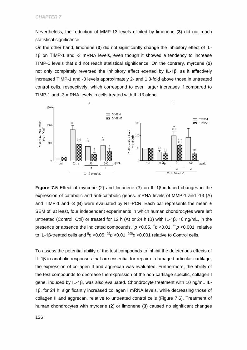
CHAPTER 7
136
Nevertheless, the reduction of MMP-13 levels elicited by limonene (3) did not reach
statistical significance.
On the other hand, limonene (3) did not significantly change the inhibitory effect of IL-
1β on TIMP-1 and -3 mRNA levels, even though it showed a tendency to increase
TIMP-1 levels that did not reach statistical significance. On the contrary, myrcene (2)
not only completely reversed the inhibitory effect exerted by IL-1β, as it effectively
increased TIMP-1 and -3 levels approximately 2- and 1.3-fold above those in untreated
control cells, respectively, which correspond to even larger increases if compared to
TIMP-1 and -3 mRNA levels in cells treated with IL-1β alone.
Figure 7.5 Effect of myrcene (2) and limonene (3) on IL-1β-induced changes in the
expression of catabolic and anti-catabolic genes. mRNA levels of MMP-1 and -13 (A)
and TIMP-1 and -3 (B) were evaluated by RT-PCR. Each bar represents the mean ±
SEM of, at least, four independent experiments in which human chondrocytes were left
untreated (Control, Ctrl) or treated for 12 h (A) or 24 h (B) with IL-1β, 10 ng/mL, in the
presence or absence the indicated compounds. *p <0.05, **p <0.01, ***p <0.001 relative
to IL-1β-treated cells and §p <0.05, §§p <0.01, §§§p <0.001 relative to Control cells.
To assess the potential ability of the test compounds to inhibit the deleterious effects of
IL-1β in anabolic responses that are essential for repair of damaged articular cartilage,
the expression of collagen II and aggrecan was evaluated. Furthermore, the ability of
the test compounds to decrease the expression of the non-cartilage specific, collagen I
gene, induced by IL-1β, was also evaluated. Chondrocyte treatment with 10 ng/mL IL-
1β, for 24 h, significantly increased collagen I mRNA levels, while decreasing those of
collagen II and aggrecan, relative to untreated control cells (Figure 7.6). Treatment of
human chondrocytes with myrcene (2) or limonene (3) caused no significant changes
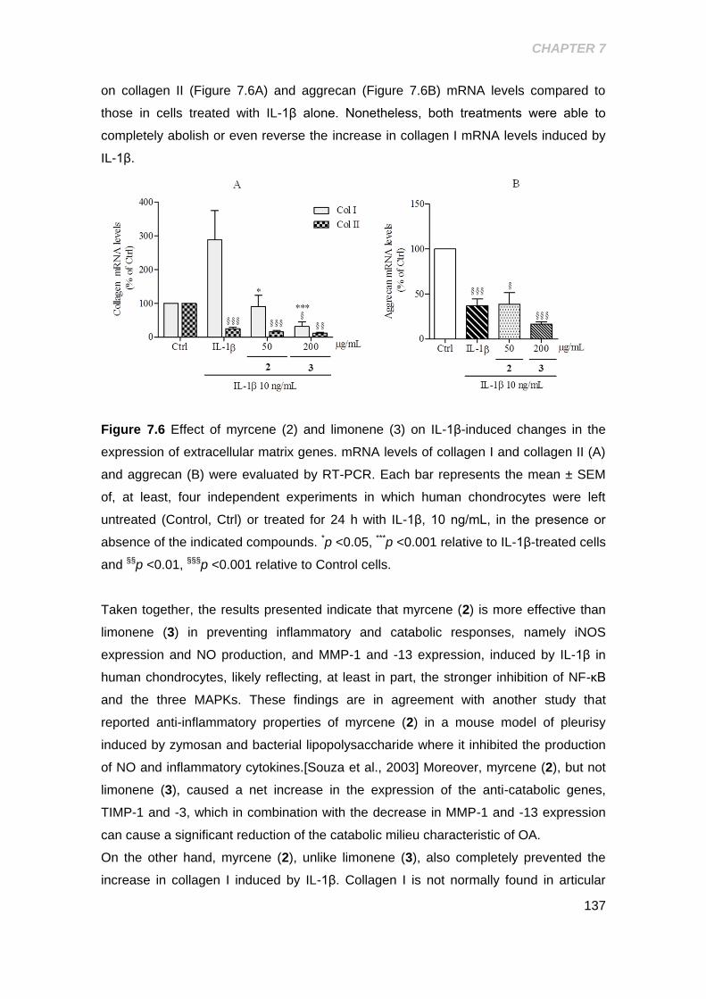
CHAPTER 7
137
on collagen II (Figure 7.6A) and aggrecan (Figure 7.6B) mRNA levels compared to
those in cells treated with IL-1β alone. Nonetheless, both treatments were able to
completely abolish or even reverse the increase in collagen I mRNA levels induced by
IL-1β.
Figure 7.6 Effect of myrcene (2) and limonene (3) on IL-1β-induced changes in the
expression of extracellular matrix genes. mRNA levels of collagen I and collagen II (A)
and aggrecan (B) were evaluated by RT-PCR. Each bar represents the mean ± SEM
of, at least, four independent experiments in which human chondrocytes were left
untreated (Control, Ctrl) or treated for 24 h with IL-1β, 10 ng/mL, in the presence or
absence of the indicated compounds. *p <0.05, ***p <0.001 relative to IL-1β-treated cells
and §§p <0.01, §§§p <0.001 relative to Control cells.
Taken together, the results presented indicate that myrcene (2) is more effective than
limonene (3) in preventing inflammatory and catabolic responses, namely iNOS
expression and NO production, and MMP-1 and -13 expression, induced by IL-1β in
human chondrocytes, likely reflecting, at least in part, the stronger inhibition of NF-κB
and the three MAPKs. These findings are in agreement with another study that
reported anti-inflammatory properties of myrcene (2) in a mouse model of pleurisy
induced by zymosan and bacterial lipopolysaccharide where it inhibited the production
of NO and inflammatory cytokines.[Souza et al., 2003] Moreover, myrcene (2), but not
limonene (3), caused a net increase in the expression of the anti-catabolic genes,
TIMP-1 and -3, which in combination with the decrease in MMP-1 and -13 expression
can cause a significant reduction of the catabolic milieu characteristic of OA.
On the other hand, myrcene (2), unlike limonene (3), also completely prevented the
increase in collagen I induced by IL-1β. Collagen I is not normally found in articular
CHAPTER 7
138
cartilage and its expression is associated with chondrocyte dedifferentiation, a process
that involves several alterations of chondrocyte gene expression and morphology and
leads to the formation of fibrocartilage, increasing in OA.[Martin et al., 2001] Therefore,
even though it did not increase the specific anabolic responses of human
chondrocytes, myrcene (2) may be effective in preventing chondrocyte dedifferentiation
associated with increased collagen I expression, while decreasing inflammatory and
catabolic processes directly involved in cartilage destruction.
In comparison with (+)-α-pinene,[Rufino et al., 2014] myrcene (2) shows potential
advantages as, besides inhibiting iNOS expression and activity and NF-κB and JNK
activation to a similar extent, this compound can further decrease ERK1/2 and p38
activation and increase anti-catabolic responses, namely TIMP-1 and -3 expression,
while promoting the maintenance of the differentiated chondrocyte phenotype, as it
also decreased collagen I expression. Nonetheless, none of the compounds tested (2
and 3), nor (+)-α-pinene, seem effective in promoting the expression of articular
cartilage matrix-specific genes.
In summary, the results presented suggest that myrcene (2) may be useful to halt or, at
least, slow down cartilage destruction and, thus, OA progression, although it is unlike to
promote the repair of areas already damaged.
.
139
CHAPTER 8.
DISCUSSION AND CONCLUSION
8.1 GENERAL DISCUSSION
Research on osteoarthritis in the last few years have been focused in particular
mechanisms underlying the course of the disease that can be related to particular risk
factors or events. This is essential for a better understanding of the intrinsic
mechanisms of specific phenotypes and, in that way, to enable the development of new
drugs targeted to the deregulated molecular processes responsible for disease onset
and progression that can prevent or halt OA and, consequently, reduce its individual
and socio-economic costs.
The association between Diabetes and the development and progression of OA is
increasingly clear. Independently of the glucose requirements for the plastic and
metabolic needs of chondrocytes, hyperglycemia has been shown to induce
deleterious effects in human chondrocytes. In diabetes, especially in uncontrolled type
2 diabetes, chronic hyperglycemia is the major hallmark and has been shown to
directly lead to diabetic complications, including those with inflammatory characteristics
as retinopathy, atherosclerosis or β-cell deterioration. The inflammatory process also
plays a central role in OA pathogenesis since the deregulated pro-inflammatory
mediators and, in particular, cytokine activities likely contribute to the disruption of the
balance between anabolic and catabolic processes of chondrocytes that lead to the
joint destruction characteristic of OA.
In this context, this work aimed to contribute to elucidate how extracellular glucose
levels are sensed by human chondrocytes, in particular by determining the role of
potassium channels in this process, and to further understand the mechanisms by
which hyperglycemia induces cartilage damage, unraveling in that way possible
pathological mechanisms and potential pharmacological targets of DM-associated OA.
Moreover, molecules, from plant origin were studied to assess their anti-inflammatory
and chondroprotective activities that can be explored for development of new drugs
with disease-modifying osteoarthritis properties.
In general, the results presented in chapters 3 and 4 show that:
Human chondrocytes of any OA grade present heterogeneous K(ATP)
channels, with Kir6.2 and SUR2B being the major subunits expressed.
The activity of K(ATP) channels influences the abundance of the major glucose
transporters, GLUT-1 and GLUT-3, in normal chondrocytes, but did not affected
CHAPTER 8
140
glucose transport, suggesting that these channels participate in a glucose
sensing apparatus that regulates GLUT levels which, by themselves, are not
sufficient to adjust glucose transport capacity.
Chondrocytes of increasing OA grade are less responsive to changes in K(ATP)
channel activity.
Exposure of human chondrocytes to high, hyperglycemia-like glucose
concentrations induces the expression of pro-inflammatory mediators, namely
IL-1β, TNF-α and iNOS, accompanied by increased NO production, in a
mechanism, at least partially, mediated by NF-κB.
Insulin, not only does not prevent the pro-inflammatory effects of hyperglycemia
in human chondrocytes, as, in supraphysiologic concentrations, it can induce
NF-κB activation by itself
Finally, the results presented in chapters 5, 6 and 7 show that:
The essential oils of Lavandula luisieri and Eryngium duriaei subsp. juresianum
display important anti-inflammatory properties, namely, significant inhibition of
IL-1β induced iNOS expression, NO production and NF-κB activation in human
chondrocytes. Likewise, these essential oils may be of great value in the
development of new therapies or as sources of active compounds for the
treatment of chronic inflammatory diseases, like OA.
(+)-α-pinene, showed the most potent chondroprotective and anti-inflammatory
activities among its isomers, enantiomers and other naturally occurring
compounds derived from the pinane nucleus, by significantly inhibiting IL-1β-
induced inflammatory and catabolic pathways, namely, NF-κB and JNK
activation and the expression of inflammatory (iNOS) and catabolic (MMP-1 and
-13) genes.
Limonene and especially Myrcene, natural compounds found in the essential
oils of Eryngium duriaei subsp juresianum and Lavandula luisieri, showed
interesting anti-inflammatory and anti-catabolic properties, namely inhibition of
IL-1β-induced expression of pro-inflammatory and catabolic mediators, namely
NO, iNOS, MMP-1 and -13, and pathways as NF-κB and p38 MAPK.
Myrcene further inhibits the phosphorylation of JNK and ERK MAPKs which
possibly contributes to the inhibition of pro-inflammatory, pro-catabolic and
chondrocyte dedifferentiation genes, namely the expression of collagen type I,
as well as to relieve the inhibition of TIMP-1 and -3 expression caused by IL-1β.
CHAPTER 8
141
Considering the objectives proposed, the findings that either hyperglycemia or
hyperinsulinemia, as well as the two combined, activate NF-κB, the major pro-
inflammatory signalling pathway, are by themselves main mechanisms that can shift
the chondrocyte homeostasis towards the catabolic program leading to OA
development and/or progression. Among the specific molecular events that may
underlie this response, the results obtained demonstrate that K(ATP) channels are
involved in glucose sensing, playing a role in the regulation of glucose transporter
availability in human chondrocytes which, nonetheless, is not sufficient to adjust
glucose transport in response to changes in its extracellular concentration. Thus, the
observed lack of response of chondrocytes of increasing age/OA grade to modulators
of K(ATP) channel activity, namely glibenclamide, a sulfonylurea drug used in DM 2
therapy as an insulin secretagogue, implies that in patients under this kind of therapy,
chondrocytes may still be susceptible to the deleterious effects caused not only by
hyperglycemia episodes, even occurring sporadically, as by hyperinsulinemia.
Nonetheless, glibenclamide has also been shown to decrease high glucose-induced IL-
1β production in pancreatic β cells [Zhou et al., 2010] which, if also occurring in
chondrocytes, may counteract the pro-inflammatory effects of hyperglycemia and
hyperinsulinemia. More studies are clearly required to elucidate the effects of anti-
diabetic drugs in human chondrocytes, which will contribute to fully assess their
risk/benefit and inform the rationale therapy of both conditions.
On the other hand and even though the specific molecular events were not elucidated
in this work, several other mechanisms may regulate glucose transport and metabolism
and mediate the effects of hyperglycemia in chondrocytes. Such mechanisms deserve
to be further investigated as they may disclose relevant targets both for prevention and
treatment of DM-associated OA. Among those potential mechanisms, AMP-activated
protein kinase (AMPK) and Silent Information Regulator 2 type 1 or sirtuin 1 (SirT1)
may play important roles.
AMPK is a metabolic sensor in a wide variety of cells that becomes activated during
metabolic stress. At least in pancreatic β cells, K(ATP) channels and AMPK cooperate
to regulate the energetic status [Lim et al., 2009]. Moreover, AMPK also seems to play
an important role in regulating the mechanisms involved in glucose transport in
response to hyper or hypoglycemic conditions [Lim et al., 2009; McCrimmon et al.,
2004; Ning et al., 2011]. Activation of AMPK has been shown to increase glucose
transport in various cells by promoting the recruitment of GLUT-4 [Li et al., 2004] or
GLUT-1 [Xi et al., 2001] to the plasma membrane. AMPK is expressed and
constitutively active in normal, but is decreased in human OA knee articular
CHAPTER 8
142
chondrocytes and cartilage [Terkeltaub et al., 2011]. So, it is possible that AMPK
participates in the glucose sensing apparatus in chondrocytes that also includes
K(ATP) channels, regulating, at least in part, GLUT trafficking to the plasma membrane
and glucose uptake. Further studies will be necessary to elucidate the role of AMPK in
the regulation of glucose transport in chondrocytes in normal and osteoarthritic
conditions and whether modulation of its activity can have beneficial effects on
chondrocyte functions under hyperglycemia.
SirT1 is a deacetylase that fine tunes many cellular biochemical processes through its
capacity to interact and modify various histone and non-histone proteins, playing
important roles in the coordination of the response to low calorie intake and in cellular
survival. On the other hand, under high glucose conditions, SirT1 expression and
activity are decreased in many cells, contributing to cell senescence and dysfunction
[Ceolotto et al., 2014; Servillo et al., 2013]. Accordingly, SirT1 has been associated
with metabolic and aging-related diseases, including DM 2 [Donath, 2014] and OA
[Dvir-Ginzberg and Steinmeyer, 2013]. SirT1 has been shown to promote cartilage
extracellular matrix synthesis [Dvir-Ginzberg et al., 2011] and chondrocyte survival
[Gabay et al., 2012], even under pro-inflammatory stress conditions. Furthermore,
SirT1 has been associated with anti-inflammatory effects induced by various stimuli,
including high glucose, in many cells through its ability to inhibit NF-κB [Kauppinen et
al., 2013; Liu and McCall, 2013]. Recent studies showed that the same mechanism is
also relevant in cell and animal models of OA [Lim et al., 2012] and inflammatory
arthritis [Wang et al., 2014]. Inhibition of NF-κB by SirT1 seems to occur through, at
least, two distinct mechanisms. The first is deacetylation of the p65 subunit of NF-kB
directly by active SirT1 while the second involves activation of other intermediates,
namely AMPK, PPAR-α and PGC-1α, which stimulate oxidative energy production and
also inhibit NF-κB activity [Kauppinen et al., 2013]. Thus, by impairing SirT1 expression
and activity, high glucose can induce NF-κB and the subsequent expression of pro-
inflammatory genes, like IL-1β and TNF-α, as we observed. Nonetheless, other
mechanisms are also likely involved as SirT1 impairment may not be sufficient to
explain the inability of OA/aged chondrocytes to downregulate glucose transport and
prevent its intracellular accumulation when exposed to hyperglycemia-like conditions.
Nonetheless, the role of SirT1 and its downstream effectors, namely AMPK, in the
adaptation of chondrocytes to high glucose deserves to be further investigated as it can
disclose an important therapeutic target for DM-associated OA.
From another point of view, our results show that the inflammatory response induced
by high glucose in human chondrocytes is not distinct from that induced by other
CHAPTER 8
143
stimuli, from ageing of chondrocytes to abnormal mechanical forces [Goldring and
Otero, 2011], that play significant roles in OA pathophysiology. These triggering factors
not only contribute to irreversible joint damage (progression), but importantly, also to
the initiation/onset phase, wherein the homeostatic balance between anabolic and
catabolic functions becomes disrupted. Our work thus points to pro-inflammatory
cytokines, namely IL-1β and TNF-α, as common mediators of DM-associated OA.
Given their central role in OA development and progression, these mediators and the
signalling pathways that they initiate are likely main targets for the development of new
drugs with disease-modifying properties for OA treatment. Therefore, we used IL-1β to
imitate those processes in vitro, so as to evaluate the anti-inflammatory, anti-catabolic
and pro-anabolic effects of compounds of plant origin.
The initial screening assays showed that essential oils have interesting anti-
inflammatory effects and thus can be important sources of compounds with anti-
osteoarthritic properties. Among the essential oils tested, those of Lavandula luisieri
and Eryngium duriaei subsp. juresianum presented the best properties, reason why
they were further studied. Screening of fractions of these essential oils containing
distinct families of compounds showed that in both cases, the most active fractions are
composed of mono- and sesquiterpene hydrocarbons, respectively, while fractions
containing oxygenated mono- and sesquiterpenes were less or completely inactive.
Finally, the screening of some commercially available compounds found in the active
fractions, allowed to identify myrcene and limonene as potential anti-inflammatory and
anti-catabolic compounds in human chondrocytes. Also (+)-α-pinene was shown to
have anti-inflammatory properties in human chondrocytes being the active enantiomer
of the α-pinene racemic mixture, previously presented by our group as showing anti-
inflammatory properties in this cells [Neves et al., 2009].
Among those compounds, myrcene showed to be the most promising as a lead
candidate for further development of new drugs that can halt OA progression. Even
though the 3 compounds showed ability to significantly reduce some of the major pro-
inflammatory and pro-catabolic mediators (NO, iNOS, MMP-1 and MMP-13) and
pathways (NF-κB) involved in the pro-inflammatory stress-related responses of
chondrocytes that contribute to OA onset and progression, myrcene was additionally
able to increase the anti-catabolic expression of the MMP inhibitors, TIMP-1 and -3,
and to further decrease the activation of the MAPK pathways (p38 and ERK1/2 in
relation to (+)α-pinene and JNK and ERK1/2 in relation to limonene)deeply involved in
the expression of catabolic mediators.
CHAPTER 8
144
Moreover, even though none of the compounds seemed to be effective in restoring the
synthesis of ECM components, myrcene may contribute to the maintenance of the
differentiated chondrocyte phenotype and to prevent the formation of fibrocartilage by
decreasing the expression of type I collagen. Further studies are needed to determine
if the effects observed result from the action of those compounds on a common single
molecular target upstream of all the signalling pathways evaluated or, on the other
hand, if they result from the combined action on various components of those signalling
pathways. Moreover, it will also be interesting to understand whether these
compounds, belonging to the same broad class of organic compounds, the terpenes,
act on the same molecular target or not. Future work can also involve the evaluation of
the observed myrcene properties in vivo using appropriate animal models of OA, as the
model induced by intra-articular injection of mono-iodoacetate in rats or the surgical
induction by transecting the anterior cruciate ligament [Bendele, 2001].
Furthermore, to establish a proof of concept for the efficacy of myrcene in Diabetes-
associated OA, it would also be interesting to induce OA in a model of non-obese type
2 diabetes, as the Goto-Kakizaki rat model which presents mild hyperglycemia, insulin
resistance and hyperinsulinemia [Akash et al., 2013], and evaluate the ability of the
compound to prevent OA development and/or to stop its progression.
In summary, a comprehensive view of the results suggests that if, on one hand, altered
glucose sensing mechanisms and high glucose exposure lead to an increased
expression of inflammatory mediators and pathways in chondrocytes, establishing a
new link between DM and OA and supporting the existence of a DM-induced OA
phenotype, on the other hand, those inflammatory and catabolic mediators and
signalling pathways are major targets for the development of new drugs with disease-
modifying OA properties, myrcene being an interesting lead compound for that
purpose.
145
CHAPTER 9.
REFERENCES
Abramson SB. 2008. Osteoarthritis and nitric oxide. Osteoarthritis Cartilage 16 Suppl
2:S15-20.
Acosta-Martinez M, Levine JE. 2007. Regulation of KATP channel subunit gene
expression by hyperglycemia in the mediobasal hypothalamus of female rats. Am J
Physiol Endocrinol Metab 292:E1801-7.
Adams RP. 1995. Identification of Essential Oil Components by Gas Chromatography /
Quadrupole Mass Spectroscopy. Allured Publishing Corporation, Carol Stream, Illinois,
USA.
Adorjan B, Buchbauer G. 2010. Biological properties of essential oils: an updated
review. Flavour and Fragance Journal
25:407-426.
Agarwal S, Deschner J, Long P, Verma A, Hofman C, Evans CH, Piesco N. 2004. Role
of NF-kappaB transcription factors in antiinflammatory and proinflammatory actions of
mechanical signals. Arthritis Rheum 50:3541-8.
Aguilar-Bryan L, Bryan J. 1999. Molecular biology of adenosine triphosphate-sensitive
potassium channels. Endocr Rev 20:101-35.
Akash MS, Rehman K, Chen S. 2013. Goto-Kakizaki rats: its suitability as non-obese
diabetic animal model for spontaneous type 2 diabetes mellitus. Curr Diabetes Rev
9:387-96.
Akhtar N, Haqqi TM. 2011. Epigallocatechin-3-gallate suppresses the global
interleukin-1beta-induced inflammatory response in human chondrocytes. Arthritis Res
Ther 13:R93.
Aktan F. 2004. iNOS-mediated nitric oxide production and its regulation. Life Sci
75:639-53.
Alcaraz MJ, Megias J, Garcia-Arnandis I, Clerigues V, Guillen MI. 2010. New molecular
targets for the treatment of osteoarthritis. Biochem Pharmacol 80:13-21.
Altman R, Asch E, Bloch D, Bole G, Borenstein D, Brandt K, Christy W, Cooke TD,
Greenwald R, Hochberg M, et al. 1986. Development of criteria for the classification
and reporting of osteoarthritis. Classification of osteoarthritis of the knee. Diagnostic
and Therapeutic Criteria Committee of the American Rheumatism Association. Arthritis
Rheum 29:1039-49.
REFERENCES
146
Amira S, Dade M, Schinella G, Rios JL. 2012. Anti-inflammatory, anti-oxidant, and
apoptotic activities of four plant species used in folk medicine in the Mediterranean
basin. Pak J Pharm Sci 25:65-72.
Amour A, Knight CG, Webster A, Slocombe PM, Stephens PE, Knauper V, Docherty
AJ, Murphy G. 2000. The in vitro activity of ADAM-10 is inhibited by TIMP-1 and TIMP-
3. FEBS Lett 473:275-9.
Anonimous. 2007. Naproxcinod: AZD 3582, HCT 3012, naproxen nitroxybutylester,
nitronaproxen, NO-naproxen. Drugs R D 8:255-8.
Arden N, Nevitt MC. 2006. Osteoarthritis: epidemiology. Best Pract Res Clin
Rheumatol 20:3-25.
Asakawa H, Miyagawa J, Hanafusa T, Kuwajima M, Matsuzawa Y. 1997. High glucose
and hyperosmolarity increase secretion of interleukin-1 beta in cultured human aortic
endothelial cells. J Diabetes Complications 11:176-9.
Askwith T, Zeng W, Eggo MC, Stevens MJ. 2011. Taurine reduces nitrosative stress
and nitric oxide synthase expression in high glucose-exposed human Schwann cells.
Exp Neurol 233:154-62.
Atayde SA, Yoshinari NH, Nascimento DP, Catanozi S, Andrade PC, Velosa AP, Parra
ER, Passarelli M, Nakandakare ER, Capelozzi VL, Teodoro WR. 2012. Experimental
diabetes modulates collagen remodelling of joints in rats. Histol Histopathol 27:1471-9.
Attur M, Samuels J, Krasnokutsky S, Abramson SB. 2010. Targeting the synovial
tissue for treating osteoarthritis (OA): where is the evidence? Best Pract Res Clin
Rheumatol 24:71-9.
Attur MG, Dave MN, Clancy RM, Patel IR, Abramson SB, Amin AR. 2000. Functional
genomic analysis in arthritis-affected cartilage: yin-yang regulation of inflammatory
mediators by alpha 5 beta 1 and alpha V beta 3 integrins. J Immunol 164:2684-91.
Ayral X. 2005. Arthroscopy and joint lavage. Best Pract Res Clin Rheumatol 19:401-15.
Babenko AP, Aguilar-Bryan L, Bryan J. 1998. A view of sur/KIR6.X, KATP channels.
Annu Rev Physiol 60:667-87.
Bannuru RR, Natov NS, Dasi UR, Schmid CH, McAlindon TE. 2011. Therapeutic
trajectory following intra-articular hyaluronic acid injection in knee osteoarthritis--meta-
analysis. Osteoarthritis Cartilage 19:611-9.
Barchowsky A, Frleta D, Vincenti MP. 2000. Integration of the NF-kappaB and mitogen-
activated protein kinase/AP-1 pathways at the collagenase-1 promoter: divergence of
IL-1 and TNF-dependent signal transduction in rabbit primary synovial fibroblasts.
Cytokine 12:1469-79.
REFERENCES
147
Barrett-Jolley R, Lewis R, Fallman R, Mobasheri A. 2010. The emerging chondrocyte
channelome. Front Physiol 1:135.
Bartels EM, Bliddal H, Schondorff PK, Altman RD, Zhang W, Christensen R. 2009.
Symptomatic efficacy and safety of diacerein in the treatment of osteoarthritis: a meta-
analysis of randomized placebo-controlled trials. Osteoarthritis Cartilage 18:289-96.
Bau B, Gebhard PM, Haag J, Knorr T, Bartnik E, Aigner T. 2002. Relative messenger
RNA expression profiling of collagenases and aggrecanases in human articular
chondrocytes in vivo and in vitro. Arthritis Rheum 46:2648-57.
Bendele AM. 2001. Animal models of osteoarthritis. J Musculoskelet Neuronal Interact
1:363-76.
Bento AF, Marcon R, Dutra RC, Claudino RF, Cola M, Leite DF, Calixto JB. 2011. beta-
Caryophyllene inhibits dextran sulfate sodium-induced colitis in mice through CB2
receptor activation and PPARgamma pathway. Am J Pathol 178:1153-66.
Berenbaum F. 2004. Signaling transduction: target in osteoarthritis. Curr Opin
Rheumatol 16:616-22.
Berenbaum F. 2008. New horizons and perspectives in the treatment of osteoarthritis.
Arthritis Res Ther 10 Suppl 2:S1.
Berenbaum F. 2011. Diabetes-induced osteoarthritis: from a new paradigm to a new
phenotype. Ann Rheum Dis 70:1354-6.
Berenbaum F. 2013. Osteoarthritis as an inflammatory disease (osteoarthritis is not
osteoarthrosis!). Osteoarthritis Cartilage 21:16-21.
Bevilacqua A, Corbo MR, Sinigaglia M. 2010. In vitro evaluation of the antimicrobial
activity of eugenol, limonene, and citrus extract against bacteria and yeasts,
representative of the spoiling microflora of fruit juices. J Food Prot 73:888-94.
Bissett D, O'Byrne KJ, von Pawel J, Gatzemeier U, Price A, Nicolson M, Mercier R,
Mazabel E, Penning C, Zhang MH, Collier MA, Shepherd FA. 2005. Phase III study of
matrix metalloproteinase inhibitor prinomastat in non-small-cell lung cancer. J Clin
Oncol 23:842-9.
Blanco FJ, Ochs RL, Schwarz H, Lotz M. 1995. Chondrocyte apoptosis induced by
nitric oxide. Am J Pathol 146:75-85.
Bobacz K, Sunk IG, Hofstaetter JG, Amoyo L, Toma CD, Akira S, Weichhart T,
Saemann M, Smolen JS. 2007. Toll-like receptors and chondrocytes: the
lipopolysaccharide-induced decrease in cartilage matrix synthesis is dependent on the
presence of toll-like receptor 4 and antagonized by bone morphogenetic protein 7.
Arthritis Rheum 56:1880-93.
REFERENCES
148
Boileau C, Martel-Pelletier J, Moldovan F, Jouzeau JY, Netter P, Manning PT, Pelletier
JP. 2002. The in situ up-regulation of chondrocyte interleukin-1-converting enzyme and
interleukin-18 levels in experimental osteoarthritis is mediated by nitric oxide. Arthritis
Rheum 46:2637-47.
Boos N, Nerlich AG, Wiest I, von der Mark K, Ganz R, Aebi M. 1999.
Immunohistochemical analysis of type-X-collagen expression in osteoarthritis of the hip
joint. J Orthop Res 17:495-502.
Brandt KD, Mazzuca SA, Katz BP, Lane KA, Buckwalter KA, Yocum DE, Wolfe F,
Schnitzer TJ, Moreland LW, Manzi S, Bradley JD, Sharma L, Oddis CV, Hugenberg
ST, Heck LW. 2005. Effects of doxycycline on progression of osteoarthritis: results of a
randomized, placebo-controlled, double-blind trial. Arthritis Rheum 52:2015-25.
Brandt KD, Radin EL, Dieppe PA, van de Putte L. 2006. Yet more evidence that
osteoarthritis is not a cartilage disease. Ann Rheum Dis 65:1261-4.
Brannan SR, Jerrard DA. 2006. Synovial fluid analysis. J Emerg Med 30:331-9.
Brownlee M. 2005. The pathobiology of diabetic complications: a unifying mechanism.
Diabetes 54:1615-25.
Buckwalter JA, Martin JA. 2006. Osteoarthritis. Adv Drug Deliv Rev 58:150-67.
Burke MA, Mutharasan RK, Ardehali H. 2008. The sulfonylurea receptor, an atypical
ATP-binding cassette protein, and its regulation of the KATP channel. Circ Res
102:164-76.
Burrage PS, Mix KS, Brinckerhoff CE. 2006. Matrix metalloproteinases: role in arthritis.
Front Biosci 11:529-43.
Busik JV, Mohr S, Grant MB. 2008. Hyperglycemia-induced reactive oxygen species
toxicity to endothelial cells is dependent on paracrine mediators. Diabetes 57:1952-65.
Cai L, Suboc P, Hogue DA, Fei DT, Filvaroff EH. 2002. Interleukin 17 induced nitric
oxide suppresses matrix synthesis and protects cartilage from matrix breakdown. J
Rheumatol 29:1725-36.
Calixto JB, Campos MM, Otuki MF, Santos AR. 2004. Anti-inflammatory compounds of
plant origin. Part II. modulation of pro-inflammatory cytokines, chemokines and
adhesion molecules. Planta Med 70:93-103.
Castaneda S, Roman-Blas JA, Largo R, Herrero-Beaumont G. 2013. Osteoarthritis: a
progressive disease with changing phenotypes. Rheumatology (Oxford) 53:1-3.
Cavaleiro C, Goncalves MJ, Serra D, Santoro G, Tomi F, Bighelli A, Salgueiro L,
Casanova J. 2011. Composition of a volatile extract of Eryngium duriaei subsp.
juresianum (M. Lainz) M. Lainz, signalised by the antifungal activity. J Pharm Biomed
Anal 54:619-22.
REFERENCES
149
Cawston TE, Wilson AJ. 2006. Understanding the role of tissue degrading enzymes
and their inhibitors in development and disease. Best Pract Res Clin Rheumatol
20:983-1002.
Celik A, Aydinlik N, Arslan I. 2011. Phytochemical constituents and inhibitory activity
towards methicillin-resistant Staphylococcus aureus strains of Eryngium species
(Apiaceae). Chem Biodivers 8:454-9.
Ceolotto G, De Kreutzenberg SV, Cattelan A, Fabricio AS, Squarcina E, Gion M,
Semplicini A, Fadini GP, Avogaro A. 2014. Sirtuin 1 stabilization by HuR represses
TNF-alpha- and glucose-induced E-selectin release and endothelial cell adhesiveness
in vitro: relevance to human metabolic syndrome. Clin Sci (Lond) 127:449-61.
Chadjichristos C, Ghayor C, Kypriotou M, Martin G, Renard E, Ala-Kokko L, Suske G,
de Crombrugghe B, Pujol JP, Galera P. 2003. Sp1 and Sp3 transcription factors
mediate interleukin-1 beta down-regulation of human type II collagen gene expression
in articular chondrocytes. J Biol Chem 278:39762-72.
Chan KW, Wheeler A, Csanady L. 2008. Sulfonylurea receptors type 1 and 2A
randomly assemble to form heteromeric KATP channels of mixed subunit composition.
J Gen Physiol 131:43-58.
Chard J, Dieppe P. 2001. Glucosamine for osteoarthritis: magic, hype, or confusion?
It's probably safe-but there's no good evidence that it works. BMJ 322:1439-40.
Chevalier X, Giraudeau B, Conrozier T, Marliere J, Kiefer P, Goupille P. 2005. Safety
study of intraarticular injection of interleukin 1 receptor antagonist in patients with
painful knee osteoarthritis: a multicenter study. J Rheumatol 32:1317-23.
Chi G, Wei M, Xie X, Soromou LW, Liu F, Zhao S. 2012. Suppression of MAPK and
NF-kappaB Pathways by Limonene Contributes to Attenuation of Lipopolysaccharide-
Induced Inflammatory Responses in Acute Lung Injury. Inflammation 36:501-11.
Chowdhury TT, Schulz RM, Rai SS, Thuemmler CB, Wuestneck N, Bader A,
Homandberg GA. 2010. Biomechanical modulation of collagen fragment-induced
anabolic and catabolic activities in chondrocyte/agarose constructs. Arthritis Res Ther
12:R82.
Clancy R, Rediske J, Koehne C, Stoyanovsky D, Amin A, Attur M, Iyama K, Abramson
SB. 2001. Activation of stress-activated protein kinase in osteoarthritic cartilage:
evidence for nitric oxide dependence. Osteoarthritis Cartilage 9:294-9.
Clancy RM, Gomez PF, Abramson SB. 2004. Nitric oxide sustains nuclear factor
kappaB activation in cytokine-stimulated chondrocytes. Osteoarthritis Cartilage 12:552-
8.
REFERENCES
150
Clutterbuck AL, Asplin KE, Harris P, Allaway D, Mobasheri A. 2009. Targeting matrix
metalloproteinases in inflammatory conditions. Curr Drug Targets 10:1245-54.
Colville CA, Seatter MJ, Gould GW. 1993. Analysis of the structural requirements of
sugar binding to the liver, brain and insulin-responsive glucose transporters expressed
in oocytes. Biochem J 294 ( Pt 3):753-60.
Coppe JP, Desprez PY, Krtolica A, Campisi J. 2010. The senescence-associated
secretory phenotype: the dark side of tumor suppression. Annu Rev Pathol 5:99-118.
Costa G, Francisco V, Lopes MC, Cruz MT, Batista MT. 2012. Intracellular signaling
pathways modulated by phenolic compounds: application for new anti-inflammatory
drugs discovery. Curr Med Chem 19:2876-900.
Csaki C, Mobasheri A, Shakibaei M. 2009. Synergistic chondroprotective effects of
curcumin and resveratrol in human articular chondrocytes: inhibition of IL-1beta-
induced NF-kappaB-mediated inflammation and apoptosis. Arthritis Res Ther 11:R165.
da Silva AC, Lopes PM, de Azevedo MM, Costa DC, Alviano CS, Alviano DS. 2012.
Biological activities of alpha-pinene and beta-pinene enantiomers. Molecules 17:6305-
16.
Das RH, van Osch GJ, Kreukniet M, Oostra J, Weinans H, Jahr H. 2010. Effects of
individual control of pH and hypoxia in chondrocyte culture. J Orthop Res 28:537-45.
Dawilai S, Muangnoi C, Praengamthanachoti P, Tuntipopipat S. 2013. Anti-
inflammatory activity of bioaccessible fraction from Eryngium foetidum leaves. Biomed
Res Int 2013:958567.
Dean DD, Martel-Pelletier J, Pelletier JP, Howell DS, Woessner JF, Jr. 1989. Evidence
for metalloproteinase and metalloproteinase inhibitor imbalance in human osteoarthritic
cartilage. J Clin Invest 84:678-85.
DeGroot J, Verzijl N, Bank RA, Lafeber FP, Bijlsma JW, TeKoppele JM. 1999. Age-
related decrease in proteoglycan synthesis of human articular chondrocytes: the role of
nonenzymatic glycation. Arthritis Rheum 42:1003-9.
Del Rosso A, Cerinic MM, De Giorgio F, Minari C, Rotella CM, Seghier G. 2006.
Rheumatological manifestations in diabetes mellitus. Curr Diabetes Rev 2:455-66.
Donath MY. 2014. Targeting inflammation in the treatment of type 2 diabetes: time to
start. Nat Rev Drug Discov 13:465-76.
Donath MY, Shoelson SE. 2011. Type 2 diabetes as an inflammatory disease. Nat Rev
Immunol 11:98-107.
Dvir-Ginzberg M, Gagarina V, Lee EJ, Booth R, Gabay O, Hall DJ. 2011. Tumor
necrosis factor alpha-mediated cleavage and inactivation of SirT1 in human
osteoarthritic chondrocytes. Arthritis Rheum 63:2363-73.
REFERENCES
151
Dvir-Ginzberg M, Steinmeyer J. 2013. Towards elucidating the role of SirT1 in
osteoarthritis. Front Biosci (Landmark Ed) 18:343-55.
Esposito K, Nappo F, Marfella R, Giugliano G, Giugliano F, Ciotola M, Quagliaro L,
Ceriello A, Giugliano D. 2002. Inflammatory cytokine concentrations are acutely
increased by hyperglycemia in humans: role of oxidative stress. Circulation 106:2067-
72.
Eyre D. 2002. Collagen of articular cartilage. Arthritis Res 4:30-5.
Eyre DR, Weis MA, Wu JJ. 2006. Articular cartilage collagen: an irreplaceable
framework? Eur Cell Mater 12:57-63.
Fan Z, Soder S, Oehler S, Fundel K, Aigner T. 2007. Activation of interleukin-1
signaling cascades in normal and osteoarthritic articular cartilage. Am J Pathol
171:938-46.
Fan Z, Yang H, Bau B, Soder S, Aigner T. 2006. Role of mitogen-activated protein
kinases and NFkappaB on IL-1beta-induced effects on collagen type II, MMP-1 and 13
mRNA expression in normal articular human chondrocytes. Rheumatol Int 26:900-3.
Felson DT. 2010. Identifying different osteoarthritis phenotypes through epidemiology.
Osteoarthritis Cartilage 18:601-4.
Fermor B, Weinberg JB, Pisetsky DS, Misukonis MA, Banes AJ, Guilak F. 2001. The
effects of static and intermittent compression on nitric oxide production in articular
cartilage explants. J Orthop Res 19:729-37.
Fernandes JC, Martel-Pelletier J, Pelletier JP. 2002. The role of cytokines in
osteoarthritis pathophysiology. Biorheology 39:237-46.
Ferretti M, Madhavan S, Deschner J, Rath-Deschner B, Wypasek E, Agarwal S. 2006.
Dynamic biophysical strain modulates proinflammatory gene induction in meniscal
fibrochondrocytes. Am J Physiol Cell Physiol 290:C1610-5.
Fichter M, Korner U, Schomburg J, Jennings L, Cole AA, Mollenhauer J. 2006.
Collagen degradation products modulate matrix metalloproteinase expression in
cultured articular chondrocytes. J Orthop Res 24:63-70.
Fidelix TS, Soares BG, Trevisani VF. 2006. Diacerein for osteoarthritis. Cochrane
Database Syst Rev:CD005117.
Fosang AJ, Beier F. 2011. Emerging Frontiers in cartilage and chondrocyte biology.
Best Pract Res Clin Rheumatol 25:751-66.
Frank S, Peters MA, Wehmeyer C, Strietholt S, Koers-Wunrau C, Bertrand J,
Heitzmann M, Hillmann A, Sherwood J, Seyfert C, Gay S, Pap T. 2013. Regulation of
matrixmetalloproteinase-3 and matrixmetalloproteinase-13 by SUMO-2/3 through the
transcription factor NF-kappaB. Ann Rheum Dis 72:1874-81.
REFERENCES
152
Fujisawa T, Hattori T, Takahashi K, Kuboki T, Yamashita A, Takigawa M. 1999. Cyclic
mechanical stress induces extracellular matrix degradation in cultured chondrocytes via
gene expression of matrix metalloproteinases and interleukin-1. J Biochem 125:966-75.
Gabay O, Oppenhiemer H, Meir H, Zaal K, Sanchez C, Dvir-Ginzberg M. 2012.
Increased apoptotic chondrocytes in articular cartilage from adult heterozygous SirT1
mice. Ann Rheum Dis 71:613-6.
Ge XP, Gan YH, Zhang CG, Zhou CY, Ma KT, Meng JH, Ma XC. 2011. Requirement of
the NF-kappaB pathway for induction of Wnt-5A by interleukin-1beta in condylar
chondrocytes of the temporomandibular joint: functional crosstalk between the Wnt-5A
and NF-kappaB signaling pathways. Osteoarthritis Cartilage 19:111-7.
Ghelardini C, Galeotti N, Salvatore G, Mazzanti G. 1999. Local anaesthetic activity of
the essential oil of Lavandula angustifolia. Planta Med 65:700-3.
Ghosh S, Karin M. 2002. Missing pieces in the NF-kappaB puzzle. Cell 109 Suppl:S81-
96.
Gillery P. 2006. [Oxidative stress and protein glycation in diabetes mellitus]. Ann Biol
Clin (Paris) 64:309-14.
Goldring MB. 2001. Anticytokine therapy for osteoarthritis. Expert Opin Biol Ther 1:817-
29.
Goldring MB. 2006. Update on the biology of the chondrocyte and new approaches to
treating cartilage diseases. Best Pract Res Clin Rheumatol 20:1003-25.
Goldring MB. 2012. Chondrogenesis, chondrocyte differentiation, and articular cartilage
metabolism in health and osteoarthritis. Ther Adv Musculoskelet Dis 4:269-85.
Goldring MB, Berenbaum F. 2004. The regulation of chondrocyte function by
proinflammatory mediators: prostaglandins and nitric oxide. Clin Orthop Relat Res:S37-
46.
Goldring MB, Birkhead J, Sandell LJ, Kimura T, Krane SM. 1988. Interleukin 1
suppresses expression of cartilage-specific types II and IX collagens and increases
types I and III collagens in human chondrocytes. J Clin Invest 82:2026-37.
Goldring MB, Goldring SR. 2007. Osteoarthritis. J Cell Physiol 213:626-34.
Goldring MB, Goldring SR. 2010. Articular cartilage and subchondral bone in the
pathogenesis of osteoarthritis. Ann N Y Acad Sci 1192:230-7.
Goldring MB, Marcu KB. 2009. Cartilage homeostasis in health and rheumatic
diseases. Arthritis Res Ther 11:224.
Goldring MB, Otero M. 2011. Inflammation in osteoarthritis. Curr Opin Rheumatol
23:471-8.
REFERENCES
153
Goldring MB, Otero M, Tsuchimochi K, Ijiri K, Li Y. 2008. Defining the roles of
inflammatory and anabolic cytokines in cartilage metabolism. Ann Rheum Dis 67 Suppl
3:iii75-82.
Green LC, Wagner DA, Glogowski J, Skipper PL, Wishnok JS, Tannenbaum SR. 1982.
Analysis of nitrate, nitrite, and [15N]nitrate in biological fluids. Anal Biochem 126:131-8.
Gribble FM, Ashcroft FM. 2000. Sulfonylurea sensitivity of adenosine triphosphate-
sensitive potassium channels from beta cells and extrapancreatic tissues. Metabolism
49:3-6.
Grimshaw MJ, Mason RM. 2000. Bovine articular chondrocyte function in vitro depends
upon oxygen tension. Osteoarthritis Cartilage 8:386-92.
Grimshaw MJ, Mason RM. 2001. Modulation of bovine articular chondrocyte gene
expression in vitro by oxygen tension. Osteoarthritis Cartilage 9:357-64.
Grishko VI, Ho R, Wilson GL, Pearsall AWt. 2009. Diminished mitochondrial DNA
integrity and repair capacity in OA chondrocytes. Osteoarthritis Cartilage 17:107-13.
Grunke M, Schulze-Koops H. 2006. Successful treatment of inflammatory knee
osteoarthritis with tumour necrosis factor blockade. Ann Rheum Dis 65:555-6.
Guo S. 2014. Insulin signaling, resistance, and the metabolic syndrome: insights from
mouse models into disease mechanisms. J Endocrinol 220:T1-T23.
Han Z, Boyle DL, Chang L, Bennett B, Karin M, Yang L, Manning AM, Firestein GS.
2001. c-Jun N-terminal kinase is required for metalloproteinase expression and joint
destruction in inflammatory arthritis. J Clin Invest 108:73-81.
Hardingham TE, Fosang AJ. 1995. The structure of aggrecan and its turnover in
cartilage. J Rheumatol Suppl 43:86-90.
Hart DJ, Doyle DV, Spector TD. 1995. Association between metabolic factors and knee
osteoarthritis in women: the Chingford Study. J Rheumatol 22:1118-23.
Hayden MS, Ghosh S. 2008a. Shared principles in NF-kappaB signaling. Cell 132:344-
62.
Hayden MS, Ghosh S. 2008b. Shared Principles in NF-kB Signaling. Cell 132:344-362.
Haywood L, McWilliams DF, Pearson CI, Gill SE, Ganesan A, Wilson D, Walsh DA.
2003. Inflammation and angiogenesis in osteoarthritis. Arthritis Rheum 48:2173-7.
Hedlund H, Hedbom E, Heineg rd D, Mengarelli-Widholm S, Reinholt FP, Svensson O.
1999. Association of the aggrecan keratan sulfate-rich region with collagen in bovine
articular cartilage. J Biol Chem 274:5777-81.
Heidari B. 2011. Knee osteoarthritis prevalence, risk factors, pathogenesis and
features: Part I. Caspian J Intern Med 2:205-212.
REFERENCES
154
Heinegard D. 2009. Proteoglycans and more--from molecules to biology. Int J Exp
Pathol 90:575-86.
Henrotin Y, Kurz B, Aigner T. 2005. Oxygen and reactive oxygen species in cartilage
degradation: friends or foes? Osteoarthritis Cartilage 13:643-54.
Henrotin Y, Priem F, Mobasheri A. 2013. Curcumin: a new paradigm and therapeutic
opportunity for the treatment of osteoarthritis: curcumin for osteoarthritis management.
Springerplus 2:56.
Henrotin YE, Zheng SX, Deby GP, Labasse AH, Crielaard JM, Reginster JY. 1998.
Nitric oxide downregulates interleukin 1beta (IL-1beta) stimulated IL-6, IL-8, and
prostaglandin E2 production by human chondrocytes. J Rheumatol 25:1595-601.
Hernvann A, Aussel C, Cynober L, Moatti N, Ekindjian OG. 1992. IL-1 beta, a strong
mediator for glucose uptake by rheumatoid and non-rheumatoid cultured human
synoviocytes. FEBS Lett 303:77-80.
Hibino H, Inanobe A, Furutani K, Murakami S, Findlay I, Kurachi Y. 2010. Inwardly
rectifying potassium channels: their structure, function, and physiological roles. Physiol
Rev 90:291-366.
Hiraiwa H, Sakai T, Mitsuyama H, Hamada T, Yamamoto R, Omachi T, Ohno Y,
Nakashima M, Ishiguro N. 2011. Inflammatory effect of advanced glycation end
products on human meniscal cells from osteoarthritic knees. Inflamm Res 60:1039-48.
Hochberg MC. 2005. COX-2 selective inhibitors in the treatment of arthritis: a
rheumatologist perspective. Curr Top Med Chem 5:443-8.
Hochberg MC, Altman RD, April KT, Benkhalti M, Guyatt G, McGowan J, Towheed T,
Welch V, Wells G, Tugwell P. 2012. American College of Rheumatology 2012
recommendations for the use of nonpharmacologic and pharmacologic therapies in
osteoarthritis of the hand, hip, and knee. Arthritis Care Res (Hoboken) 64:465-74.
Hochberg MC, Zhan M, Langenberg P. 2008. The rate of decline of joint space width in
patients with osteoarthritis of the knee: a systematic review and meta-analysis of
randomized placebo-controlled trials of chondroitin sulfate. Curr Med Res Opin
24:3029-35.
Hulejova H, Baresova V, Klezl Z, Polanska M, Adam M, Senolt L. 2007. Increased level
of cytokines and matrix metalloproteinases in osteoarthritic subchondral bone. Cytokine
38:151-6.
Hunter DJ. 2008. Are there promising biologic therapies for osteoarthritis? Curr
Rheumatol Rep 10:19-25.
Hunter DJ. 2011. Pharmacologic therapy for osteoarthritis--the era of disease
modification. Nat Rev Rheumatol 7:13-22.
REFERENCES
155
Igarashi M, Wakasaki H, Takahara N, Ishii H, Jiang ZY, Yamauchi T, Kuboki K, Meier
M, Rhodes CJ, King GL. 1999. Glucose or diabetes activates p38 mitogen-activated
protein kinase via different pathways. J Clin Invest 103:185-95.
Inagaki N, Seino S. 1998. ATP-sensitive potassium channels: structures, functions, and
pathophysiology. Jpn J Physiol 48:397-412.
Ishii H, Tanaka H, Katoh K, Nakamura H, Nagashima M, Yoshino S. 2002.
Characterization of infiltrating T cells and Th1/Th2-type cytokines in the synovium of
patients with osteoarthritis. Osteoarthritis Cartilage 10:277-81.
Issa RI, Griffin TM. 2012. Pathobiology of obesity and osteoarthritis: integrating
biomechanics and inflammation. Pathobiol Aging Age Relat Dis 2.
Jia J, Liu Y, Zhang X, Liu X, Qi J. 2013. Regulation of iNOS expression by NF-kappaB
in human lens epithelial cells treated with high levels of glucose. Invest Ophthalmol Vis
Sci 54:5070-7.
Jiang SY, Wei CC, Shang TT, Lian Q, Wu CX, Deng JY. 2012. High glucose induces
inflammatory cytokine through protein kinase C-induced toll-like receptor 2 pathway in
gingival fibroblasts. Biochem Biophys Res Commun 427:666-70.
Jobin C, Sartor RB. 2000. NF-kappaB signaling proteins as therapeutic targets for
inflammatory bowel diseases. Inflamm Bowel Dis 6:206-13.
Johnson EO, Charchandi A, Babis GC, Soucacos PN. 2008. Apoptosis in osteoarthritis:
morphology, mechanisms, and potential means for therapeutic intervention. J Surg
Orthop Adv 17:147-52.
Johnson GL, Lapadat R. 2002. Mitogen-activated protein kinase pathways mediated by
ERK, JNK, and p38 protein kinases. Science 298:1911-2.
Johnson GL, Nakamura K. 2007. The c-jun kinase/stress-activated pathway:
regulation, function and role in human disease. Biochim Biophys Acta 1773:1341-8.
Joulain D. 1998. The Atlas of Spectral Data of Sesquiterpene Hydrocarbons. Hamburg
Verlag, Hamburg.
Kaplan W, Wirtz VJ, Mantel-Teeuwisse A, Stolk P, Duthey B, Laing R. 2013. Priority
Medicines for Europe and the World - 2013 Update. WHO Library Cataloguing-in-
Publication Data.
Kapoor M, Martel-Pelletier J, Lajeunesse D, Pelletier JP, Fahmi H. 2011. Role of
proinflammatory cytokines in the pathophysiology of osteoarthritis. Nat Rev Rheumatol
7:33-42.
Karin M, Liu Z, Zandi E. 1997. AP-1 function and regulation. Curr Opin Cell Biol 9:240-
6.
REFERENCES
156
Kashiwagi M, Enghild JJ, Gendron C, Hughes C, Caterson B, Itoh Y, Nagase H. 2004.
Altered proteolytic activities of ADAMTS-4 expressed by C-terminal processing. J Biol
Chem 279:10109-19.
Katz JD, Agrawal S, Velasquez M. 2010. Getting to the heart of the matter:
osteoarthritis takes its place as part of the metabolic syndrome. Curr Opin Rheumatol
22:512-9.
Kauppinen A, Suuronen T, Ojala J, Kaarniranta K, Salminen A. 2013. Antagonistic
crosstalk between NF-kappaB and SIRT1 in the regulation of inflammation and
metabolic disorders. Cell Signal 25:1939-48.
Kay A, Richardson J, Forsyth NR. 2011. Physiological normoxia and chondrogenic
potential of chondrocytes. Front Biosci (Elite Ed) 3:1365-74.
Kelley KM, Johnson TR, Ilan J, Moskowitz RW. 1999. Glucose regulation of the IGF
response system in chondrocytes: induction of an IGF-I-resistant state. Am J Physiol
276:R1164-71.
Kellner K, Schulz MB, Gopferich A, Blunk T. 2001. Insulin in tissue engineering of
cartilage: a potential model system for growth factor application. J Drug Target 9:439-
48.
Kerkhof HJ, Meulenbelt I, Akune T, Arden NK, Aromaa A, Bierma-Zeinstra SM, Carr A,
Cooper C, Dai J, Doherty M, Doherty SA, Felson D, Gonzalez A, Gordon A, Harilainen
A, Hart DJ, Hauksson VB, Heliovaara M, Hofman A, Ikegawa S, Ingvarsson T, Jiang Q,
Jonsson H, Jonsdottir I, Kawaguchi H, Kloppenburg M, Kujala UM, Lane NE, Leino-
Arjas P, Lohmander LS, Luyten FP, Malizos KN, Nakajima M, Nevitt MC, Pols HA,
Rivadeneira F, Shi D, Slagboom E, Spector TD, Stefansson K, Sudo A, Tamm A,
Tamm AE, Tsezou A, Uchida A, Uitterlinden AG, Wilkinson JM, Yoshimura N, Valdes
AM, van Meurs JB. 2010. Recommendations for standardization and phenotype
definitions in genetic studies of osteoarthritis: the TREAT-OA consortium. Osteoarthritis
Cartilage 19:254-64.
Khalife S, Zafarullah M. 2011. Molecular targets of natural health products in arthritis.
Arthritis Res Ther 13:102.
Kiani C, Chen L, Wu YJ, Yee AJ, Yang BB. 2002. Structure and function of aggrecan.
Cell Res 12:19-32.
King J, Zhao J, Clingan P, Morris D. 2003. Randomised double blind placebo control
study of adjuvant treatment with the metalloproteinase inhibitor, Marimastat in patients
with inoperable colorectal hepatic metastases: significant survival advantage in patients
with musculoskeletal side-effects. Anticancer Res 23:639-45.
REFERENCES
157
King KB, Findley TW, Williams AE, Bucknell AL. 2013. Veterans with diabetes receive
arthroplasty more frequently and at a younger age. Clin Orthop Relat Res 471:3049-54.
Kirmizibekmez H, Demirci B, Yesilada E, Baser KH, Demirci F. 2009. Chemical
composition and antimicrobial activity of the essential oils of Lavandula stoechas L.
ssp. stoechas growing wild in Turkey. Nat Prod Commun 4:1001-6.
Knobloch TJ, Madhavan S, Nam J, Agarwal S, Jr., Agarwal S. 2008. Regulation of
chondrocytic gene expression by biomechanical signals. Crit Rev Eukaryot Gene Expr
18:139-50.
Kobayashi M, Squires GR, Mousa A, Tanzer M, Zukor DJ, Antoniou J, Feige U, Poole
AR. 2005. Role of interleukin-1 and tumor necrosis factor alpha in matrix degradation of
human osteoarthritic cartilage. Arthritis Rheum 52:128-35.
Kojima F, Naraba H, Miyamoto S, Beppu M, Aoki H, Kawai S. 2004. Membrane-
associated prostaglandin E synthase-1 is upregulated by proinflammatory cytokines in
chondrocytes from patients with osteoarthritis. Arthritis Res Ther 6:R355-65.
Konttinen YT, Ainola M, Valleala H, Ma J, Ida H, Mandelin J, Kinne RW, Santavirta S,
Sorsa T, Lopez-Otin C, Takagi M. 1999. Analysis of 16 different matrix
metalloproteinases (MMP-1 to MMP-20) in the synovial membrane: different profiles in
trauma and rheumatoid arthritis. Ann Rheum Dis 58:691-7.
Kristof AS, Marks-Konczalik J, Moss J. 2001. Mitogen-activated protein kinases
mediate activator protein-1-dependent human inducible nitric-oxide synthase promoter
activation. J Biol Chem 276:8445-52.
Krzeski P, Buckland-Wright C, Balint G, Cline GA, Stoner K, Lyon R, Beary J,
Aronstein WS, Spector TD. 2007. Development of musculoskeletal toxicity without
clear benefit after administration of PG-116800, a matrix metalloproteinase inhibitor, to
patients with knee osteoarthritis: a randomized, 12-month, double-blind, placebo-
controlled study. Arthritis Res Ther 9:R109.
Kupeli E, Kartal M, Aslan S, Yesilada E. 2006. Comparative evaluation of the anti-
inflammatory and antinociceptive activity of Turkish Eryngium species. J
Ethnopharmacol 107:32-7.
Lahm A, Mrosek E, Spank H, Erggelet C, Kasch R, Esser J, Merk H. 2010. Changes in
content and synthesis of collagen types and proteoglycans in osteoarthritis of the knee
joint and comparison of quantitative analysis with Photoshop-based image analysis.
Arch Orthop Trauma Surg 130:557-64.
Lane NE, Brandt K, Hawker G, Peeva E, Schreyer E, Tsuji W, Hochberg MC. 2011.
OARSI-FDA initiative: defining the disease state of osteoarthritis. Osteoarthritis
Cartilage 19:478-82.
REFERENCES
158
Lee MS, Trindade MC, Ikenoue T, Goodman SB, Schurman DJ, Smith RL. 2003.
Regulation of nitric oxide and bcl-2 expression by shear stress in human osteoarthritic
chondrocytes in vitro. J Cell Biochem 90:80-6.
Lee RB, Urban JP. 1997. Evidence for a negative Pasteur effect in articular cartilage.
Biochem J 321 ( Pt 1):95-102.
Lefebvre V, Peeters-Joris C, Vaes G. 1990. Modulation by interleukin 1 and tumor
necrosis factor alpha of production of collagenase, tissue inhibitor of
metalloproteinases and collagen types in differentiated and dedifferentiated articular
chondrocytes. Biochim Biophys Acta 1052:366-78.
Leff RL, Elias I, Ionescu M, Reiner A, Poole AR. 2003. Molecular changes in human
osteoarthritic cartilage after 3 weeks of oral administration of BAY 12-9566, a matrix
metalloproteinase inhibitor. J Rheumatol 30:544-9.
Leong DJ, Gu XI, Li Y, Lee JY, Laudier DM, Majeska RJ, Schaffler MB, Cardoso L, Sun
HB. 2010a. Matrix metalloproteinase-3 in articular cartilage is upregulated by joint
immobilization and suppressed by passive joint motion. Matrix Biol 29:420-6.
Leong DJ, Li YH, Gu XI, Sun L, Zhou Z, Nasser P, Laudier DM, Iqbal J, Majeska RJ,
Schaffler MB, Goldring MB, Cardoso L, Zaidi M, Sun HB. 2010b. Physiological loading
of joints prevents cartilage degradation through CITED2. FASEB J 25:182-91.
Levick JR, McDonald JN. 1995. Fluid movement across synovium in healthy joints: role
of synovial fluid macromolecules. Ann Rheum Dis 54:417-23.
Li H, Li L, Min J, Yang H, Xu X, Yuan Y, Wang D. 2012. Levels of metalloproteinase
(MMP-3, MMP-9), NF-kappaB ligand (RANKL), and nitric oxide (NO) in peripheral
blood of osteoarthritis (OA) patients. Clin Lab 58:755-62.
Li J, Hu X, Selvakumar P, Russell RR, 3rd, Cushman SW, Holman GD, Young LH.
2004. Role of the nitric oxide pathway in AMPK-mediated glucose uptake and GLUT4
translocation in heart muscle. Am J Physiol Endocrinol Metab 287:E834-41.
Liacini A, Sylvester J, Li WQ, Huang W, Dehnade F, Ahmad M, Zafarullah M. 2003.
Induction of matrix metalloproteinase-13 gene expression by TNF-alpha is mediated by
MAP kinases, AP-1, and NF-kappaB transcription factors in articular chondrocytes. Exp
Cell Res 288:208-17.
Liacini A, Sylvester J, Li WQ, Zafarullah M. 2002. Inhibition of interleukin-1-stimulated
MAP kinases, activating protein-1 (AP-1) and nuclear factor kappa B (NF-kappa B)
transcription factors down-regulates matrix metalloproteinase gene expression in
articular chondrocytes. Matrix Biol 21:251-62.
REFERENCES
159
Liacini A, Sylvester J, Zafarullah M. 2005. Triptolide suppresses proinflammatory
cytokine-induced matrix metalloproteinase and aggrecanase-1 gene expression in
chondrocytes. Biochem Biophys Res Commun 327:320-7.
Lim A, Park SH, Sohn JW, Jeon JH, Park JH, Song DK, Lee SH, Ho WK. 2009.
Glucose deprivation regulates KATP channel trafficking via AMP-activated protein
kinase in pancreatic beta-cells. Diabetes 58:2813-9.
Lim HD, Kim YS, Ko SH, Yoon IJ, Cho SG, Chun YH, Choi BJ, Kim EC. 2012.
Cytoprotective and anti-inflammatory effects of melatonin in hydrogen peroxide-
stimulated CHON-001 human chondrocyte cell line and rabbit model of osteoarthritis
via the SIRT1 pathway. J Pineal Res 53:225-37.
Liu TF, McCall CE. 2013. Deacetylation by SIRT1 Reprograms Inflammation and
Cancer. Genes Cancer 4:135-47.
Loeser RF. 2006. Molecular mechanisms of cartilage destruction: mechanics,
inflammatory mediators, and aging collide. Arthritis Rheum 54:1357-60.
Loeser RF. 2013. The effects of aging on the development of osteoarthritis. HSS J
8:18-9.
Loeser RF, Erickson EA, Long DL. 2008. Mitogen-activated protein kinases as
therapeutic targets in osteoarthritis. Curr Opin Rheumatol 20:581-6.
Loeser RF, Forsyth CB, Samarel AM, Im HJ. 2003. Fibronectin fragment activation of
proline-rich tyrosine kinase PYK2 mediates integrin signals regulating collagenase-3
expression by human chondrocytes through a protein kinase C-dependent pathway. J
Biol Chem 278:24577-85.
Loeser RF, Goldring SR, Scanzello CR, Goldring MB. 2012. Osteoarthritis: a disease of
the joint as an organ. Arthritis Rheum 64:1697-707.
Lorenzo P, Bayliss MT, Heinegard D. 2004. Altered patterns and synthesis of
extracellular matrix macromolecules in early osteoarthritis. Matrix Biol 23:381-91.
Lotz M, Terkeltaub R, Villiger PM. 1992. Cartilage and joint inflammation. Regulation of
IL-8 expression by human articular chondrocytes. J Immunol 148:466-73.
Lotz MK, Carames B. 2011. Autophagy and cartilage homeostasis mechanisms in joint
health, aging and OA. Nat Rev Rheumatol 7:579-87.
Lotz MK, Otsuki S, Grogan SP, Sah R, Terkeltaub R, D'Lima D. 2010. Cartilage cell
clusters. Arthritis Rheum 62:2206-18.
Madonna R, Geng YJ, Bolli R, Rokosh G, Ferdinandy P, Patterson C, De Caterina R.
2014. Co-Activation of Nuclear Factor-kappaB and Myocardin/Serum Response Factor
Conveys the Hypertrophy Signal of High Insulin Levels in Cardiac Myoblasts. J Biol
Chem:DOI: jbc.M113.540559.
REFERENCES
160
Magnano MD, Chakravarty EF, Broudy C, Chung L, Kelman A, Hillygus J, Genovese
MC. 2007. A pilot study of tumor necrosis factor inhibition in erosive/inflammatory
osteoarthritis of the hands. J Rheumatol 34:1323-7.
Maher MC, Schnabel LV, Cross JA, Papich MG, Divers TJ, Fortier LA. 2013. Plasma
and synovial fluid concentration of doxycycline following low-dose, low-frequency
administration, and resultant inhibition of matrix metalloproteinase-13 from interleukin-
stimulated equine synoviocytes. Equine Vet J.
Majumdar MK, Askew R, Schelling S, Stedman N, Blanchet T, Hopkins B, Morris EA,
Glasson SS. 2007. Double-knockout of ADAMTS-4 and ADAMTS-5 in mice results in
physiologically normal animals and prevents the progression of osteoarthritis. Arthritis
Rheum 56:3670-4.
Maldonado M, Nam J. 2013. The role of changes in extracellular matrix of cartilage in
the presence of inflammation on the pathology of osteoarthritis. Biomed Res Int
2013:284873.
Marcu KB, Otero M, Olivotto E, Borzi RM, Goldring MB. 2010. NF-kappaB signaling:
multiple angles to target OA. Curr Drug Targets 11:599-613.
Marks-Konczalik J, Chu SC, Moss J. 1998. Cytokine-mediated transcriptional induction
of the human inducible nitric oxide synthase gene requires both activator protein 1 and
nuclear factor kappaB-binding sites. J Biol Chem 273:22201-8.
Marlovits S, Hombauer M, Truppe M, Vecsei V, Schlegel W. 2004. Changes in the ratio
of type-I and type-II collagen expression during monolayer culture of human
chondrocytes. J Bone Joint Surg Br 86:286-95.
Maroudas A. 1970. Distribution and diffusion of solutes in articular cartilage. Biophys J
10:365-79.
Maroudas A, Bayliss MT, Uchitel-Kaushansky N, Schneiderman R, Gilav E. 1998.
Aggrecan turnover in human articular cartilage: use of aspartic acid racemization as a
marker of molecular age. Arch Biochem Biophys 350:61-71.
Martel-Pelletier J, Boileau C, Pelletier JP, Roughley PJ. 2008. Cartilage in normal and
osteoarthritis conditions. Best Pract Res Clin Rheumatol 22:351-84.
Martel-Pelletier J, McCollum R, Fujimoto N, Obata K, Cloutier JM, Pelletier JP. 1994.
Excess of metalloproteases over tissue inhibitor of metalloprotease may contribute to
cartilage degradation in osteoarthritis and rheumatoid arthritis. Lab Invest 70:807-15.
Martin I, Jakob M, Schafer D, Dick W, Spagnoli G, Heberer M. 2001. Quantitative
analysis of gene expression in human articular cartilage from normal and osteoarthritic
joints. Osteoarthritis Cartilage 9:112-8.
REFERENCES
161
Martinez-Huedo MA, Villanueva M, de Andres AL, Hernandez-Barrera V, Carrasco-
Garrido P, Gil A, Martinez D, Jimenez-Garcia R. 2013. Trends 2001 to 2008 in
incidence and immediate postoperative outcomes for major joint replacement among
Spanish adults suffering diabetes. Eur J Orthop Surg Traumatol 23:53-9.
Masuko K, Murata M, Nakamura H, Yudoh K, Nishioka K, Kato T. 2007. Sphingosine-
1-phosphate attenuates proteoglycan aggrecan expression via production of
prostaglandin E2 from human articular chondrocytes. BMC Musculoskelet Disord 8:29.
Matsuo AL, Figueiredo CR, Arruda DC, Pereira FV, Scutti JA, Massaoka MH,
Travassos LR, Sartorelli P, Lago JH. 2011. alpha-Pinene isolated from Schinus
terebinthifolius Raddi (Anacardiaceae) induces apoptosis and confers antimetastatic
protection in a melanoma model. Biochem Biophys Res Commun 411:449-54.
Matsuo M, Tanabe K, Kioka N, Amachi T, Ueda K. 2000. Different binding properties
and affinities for ATP and ADP among sulfonylurea receptor subtypes, SUR1, SUR2A,
and SUR2B. J Biol Chem 275:28757-63.
Mbvundula EC, Bunning RA, Rainsford KD. 2005. Effects of cannabinoids on nitric
oxide production by chondrocytes and proteoglycan degradation in cartilage. Biochem
Pharmacol 69:635-40.
McAlindon TE, Bannuru RR, Sullivan MC, Arden NK, Berenbaum F, Bierma-Zeinstra
SM, Hawker GA, Henrotin Y, Hunter DJ, Kawaguchi H, Kwoh K, Lohmander S, Rannou
F, Roos EM, Underwood M. 2014. OARSI guidelines for the non-surgical management
of knee osteoarthritis. Osteoarthritis Cartilage 22:363-88.
McCrimmon RJ, Fan X, Ding Y, Zhu W, Jacob RJ, Sherwin RS. 2004. Potential role for
AMP-activated protein kinase in hypoglycemia sensing in the ventromedial
hypothalamus. Diabetes 53:1953-8.
McInnes IB, Leung B, Wei XQ, Gemmell CC, Liew FY. 1998. Septic arthritis following
Staphylococcus aureus infection in mice lacking inducible nitric oxide synthase. J
Immunol 160:308-15.
McLafferty W. 2009. Wiley Registry of Mass Spectral Data 9th / NIST 08. Mass
Spectral Library.
McNulty AL, Stabler TV, Vail TP, McDaniel GE, Kraus VB. 2005. Dehydroascorbate
transport in human chondrocytes is regulated by hypoxia and is a physiologically
relevant source of ascorbic acid in the joint. Arthritis Rheum 52:2676-85.
McTaggart JS, Clark RH, Ashcroft FM. 2010. The role of the KATP channel in glucose
homeostasis in health and disease: more than meets the islet. J Physiol 588:3201-9.
REFERENCES
162
Meats JE, McGuire MB, Russell RG. 1980. Human synovium releases a factor which
stimulates chondrocyte production of PGE and plasminogen activator. Nature 286:891-
2.
Medeiros R, Passos GF, Vitor CE, Koepp J, Mazzuco TL, Pianowski LF, Campos MM,
Calixto JB. 2007. Effect of two active compounds obtained from the essential oil of
Cordia verbenacea on the acute inflammatory responses elicited by LPS in the rat paw.
Br J Pharmacol 151:618-27.
Megias J, Busserolles J, Alcaraz MJ. 2007. The carbon monoxide-releasing molecule
CORM-2 inhibits the inflammatory response induced by cytokines in Caco-2 cells. Br J
Pharmacol 150:977-86.
Melas IN, Chairakaki AD, Chatzopoulou EI, Messinis DE, Katopodi T, Pliaka V, Samara
S, Mitsos A, Dailiana Z, Kollia P, Alexopoulos LG. 2014. Modeling of signaling
pathways in chondrocytes based on phosphoproteomic and cytokine release data.
Osteoarthritis Cartilage.
Melchiorri C, Meliconi R, Frizziero L, Silvestri T, Pulsatelli L, Mazzetti I, Borzi RM,
Uguccioni M, Facchini A. 1998. Enhanced and coordinated in vivo expression of
inflammatory cytokines and nitric oxide synthase by chondrocytes from patients with
osteoarthritis. Arthritis Rheum 41:2165-74.
Mendes AF, Caramona MM, Carvalho AP, Lopes MC. 2002a. Role of mitogen-
activated protein kinases and tyrosine kinases on IL-1-Induced NF-kappaB activation
and iNOS expression in bovine articular chondrocytes. Nitric Oxide 6:35-44.
Mendes AF, Caramona MM, Carvalho AP, Lopes MC. 2003a. Differential roles of
hydrogen peroxide and superoxide in mediating IL-1-induced NF-kappa B activation
and iNOS expression in bovine articular chondrocytes. J Cell Biochem 88:783-93.
Mendes AF, Caramona MM, Carvalho AP, Lopes MC. 2003b. Hydrogen peroxide
mediates interleukin-1beta-induced AP-1 activation in articular chondrocytes:
implications for the regulation of iNOS expression. Cell Biol Toxicol 19:203-14.
Mendes AF, Caramona MM, de Carvalho AP, Lopes MC. 2002b. Diacerhein and rhein
prevent interleukin-1beta-induced nuclear factor-kappaB activation by inhibiting the
degradation of inhibitor kappaB-alpha. Pharmacol Toxicol 91:22-8.
Mendes AF, Carvalho AP, Caramona MM, Lopes MC. 2002c. Role of nitric oxide in the
activation of NF-kappaB, AP-1 and NOS II expression in articular chondrocytes.
Inflamm Res 51:369-75.
Mendes Sdos S, Candi A, Vansteenbrugge M, Pignon MR, Bult H, Boudjeltia KZ,
Munaut C, Raes M. 2009. Microarray analyses of the effects of NF-kappaB or PI3K
pathway inhibitors on the LPS-induced gene expression profile in RAW264.7 cells:
REFERENCES
163
synergistic effects of rapamycin on LPS-induced MMP9-overexpression. Cell Signal
21:1109-22.
Mengshol JA, Vincenti MP, Coon CI, Barchowsky A, Brinckerhoff CE. 2000. Interleukin-
1 induction of collagenase 3 (matrix metalloproteinase 13) gene expression in
chondrocytes requires p38, c-Jun N-terminal kinase, and nuclear factor kappaB:
differential regulation of collagenase 1 and collagenase 3. Arthritis Rheum 43:801-11.
Michael JW, Schluter-Brust KU, Eysel P. 2010. The epidemiology, etiology, diagnosis,
and treatment of osteoarthritis of the knee. Dtsch Arztebl Int 107:152-62.
Michaelakis A, Papachristos D, Kimbaris A, Koliopoulos G, Giatropoulos A, Polissiou
MG. 2009. Citrus essential oils and four enantiomeric pinenes against Culex pipiens
(Diptera: Culicidae). Parasitol Res 105:769-73.
Miguel MG. 2010. Antioxidant and anti-inflammatory activities of essential oils: a short
review. Molecules 15:9252-87.
Miki T, Minami K, Zhang L, Morita M, Gonoi T, Shiuchi T, Minokoshi Y, Renaud JM,
Seino S. 2002. ATP-sensitive potassium channels participate in glucose uptake in
skeletal muscle and adipose tissue. Am J Physiol Endocrinol Metab 283:E1178-84.
Minami K, Miki T, Kadowaki T, Seino S. 2004. Roles of ATP-sensitive K+ channels as
metabolic sensors: studies of Kir6.x null mice. Diabetes 53 Suppl 3:S176-80.
Miosge N, Hartmann M, Maelicke C, Herken R. 2004. Expression of collagen type I and
type II in consecutive stages of human osteoarthritis. Histochem Cell Biol 122:229-36.
Mobasheri A. 1998. Correlation between [Na+], [glycosaminoglycan] and Na+/K+ pump
density in the extracellular matrix of bovine articular cartilage. Physiol Res 47:47-52.
Mobasheri A. 2012. Intersection of inflammation and herbal medicine in the treatment
of osteoarthritis. Curr Rheumatol Rep 14:604-16.
Mobasheri A. 2013. The future of osteoarthritis therapeutics: targeted pharmacological
therapy. Curr Rheumatol Rep 15:364.
Mobasheri A, Bondy CA, Moley K, Mendes AF, Rosa SC, Richardson SM, Hoyland JA,
Barrett-Jolley R, Shakibaei M. 2008. Facilitative glucose transporters in articular
chondrocytes. Expression, distribution and functional regulation of GLUT isoforms by
hypoxia, hypoxia mimetics, growth factors and pro-inflammatory cytokines. Adv Anat
Embryol Cell Biol 200:1 p following vi, 1-84.
Mobasheri A, Gent TC, Nash AI, Womack MD, Moskaluk CA, Barrett-Jolley R. 2007.
Evidence for functional ATP-sensitive (K(ATP)) potassium channels in human and
equine articular chondrocytes. Osteoarthritis Cartilage 15:1-8.
Mobasheri A, Lewis R, Ferreira-Mendes A, Rufino A, Dart C, Barrett-Jolley R. 2012.
Potassium channels in articular chondrocytes. Channels (Austin) 6:416-25.
REFERENCES
164
Mobasheri A, Neama G, Bell S, Richardson S, Carter SD. 2002a. Human articular
chondrocytes express three facilitative glucose transporter isoforms: GLUT1, GLUT3
and GLUT9. Cell Biol Int 26:297-300.
Mobasheri A, Richardson S, Mobasheri R, Shakibaei M, Hoyland JA. 2005. Hypoxia
inducible factor-1 and facilitative glucose transporters GLUT1 and GLUT3: putative
molecular components of the oxygen and glucose sensing apparatus in articular
chondrocytes. Histol Histopathol 20:1327-38.
Mobasheri A, Vannucci SJ, Bondy CA, Carter SD, Innes JF, Arteaga MF, Trujillo E,
Ferraz I, Shakibaei M, Martin-Vasallo P. 2002b. Glucose transport and metabolism in
chondrocytes: a key to understanding chondrogenesis, skeletal development and
cartilage degradation in osteoarthritis. Histol Histopathol 17:1239-67.
Mohamed-Ali H. 1995. Influence of interleukin-1 beta, tumour necrosis factor alpha and
prostaglandin E2 on chondrogenesis and cartilage matrix breakdown in vitro.
Rheumatol Int 14:191-9.
Moritz W, Leech CA, Ferrer J, Habener JF. 2001. Regulated expression of adenosine
triphosphate-sensitive potassium channel subunits in pancreatic beta-cells.
Endocrinology 142:129-38.
Mosmann T. 1983. Rapid colorimetric assay for cellular growth and survival: application
to proliferation and cytotoxicity assays. J Immunol Methods 65:55-63.
Muir H. 1995. The chondrocyte, architect of cartilage. Biomechanics, structure, function
and molecular biology of cartilage matrix macromolecules. Bioessays 17:1039-48.
Murphy G, Lee MH. 2005. What are the roles of metalloproteinases in cartilage and
bone damage? Ann Rheum Dis 64 Suppl 4:iv44-7.
Nah SS, Choi IY, Lee CK, Oh JS, Kim YG, Moon HB, Yoo B. 2008. Effects of advanced
glycation end products on the expression of COX-2, PGE2 and NO in human
osteoarthritic chondrocytes. Rheumatology (Oxford) 47:425-31.
Nah SS, Choi IY, Yoo B, Kim YG, Moon HB, Lee CK. 2007. Advanced glycation end
products increases matrix metalloproteinase-1, -3, and -13, and TNF-alpha in human
osteoarthritic chondrocytes. FEBS Lett 581:1928-32.
Nam J, Perera P, Liu J, Rath B, Deschner J, Gassner R, Butterfield TA, Agarwal S.
2011. Sequential alterations in catabolic and anabolic gene expression parallel
pathological changes during progression of monoiodoacetate-induced arthritis. PLoS
One 6:e24320.
Neogi T, Zhang Y. 2013. Epidemiology of osteoarthritis. Rheum Dis Clin North Am
39:1-19.
REFERENCES
165
Neves A, Rosa S, Goncalves J, Rufino A, Judas F, Salgueiro L, Lopes MC, Cavaleiro
C, Mendes AF. 2009. Screening of five essential oils for identification of potential
inhibitors of IL-1-induced Nf-kappaB activation and NO production in human
chondrocytes: characterization of the inhibitory activity of alpha-pinene. Planta Med
76:303-8.
Newman DJ, Cragg GM. 2007. Natural products as sources of new drugs over the last
25 years. J Nat Prod 70:461-77.
Nielsen LB, Ploug KB, Swift P, Orskov C, Jansen-Olesen I, Chiarelli F, Holst JJ,
Hougaard P, Porksen S, Holl R, de Beaufort C, Gammeltoft S, Rorsman P, Mortensen
HB, Hansen L. 2007. Co-localisation of the Kir6.2/SUR1 channel complex with
glucagon-like peptide-1 and glucose-dependent insulinotrophic polypeptide expression
in human ileal cells and implications for glycaemic control in new onset type 1 diabetes.
Eur J Endocrinol 156:663-71.
Nietfeld JJ, Wilbrink B, Den Otter W, Huber J, Huber-Bruning O. 1990a. The effect of
human interleukin 1 on proteoglycan metabolism in human and porcine cartilage
explants. J Rheumatol 17:818-26.
Nietfeld JJ, Wilbrink B, Helle M, van Roy JL, den Otter W, Swaak AJ, Huber-Bruning O.
1990b. Interleukin-1-induced interleukin-6 is required for the inhibition of proteoglycan
synthesis by interleukin-1 in human articular cartilage. Arthritis Rheum 33:1695-701.
Nieves-Plaza M, Castro-Santana LE, Font YM, Mayor AM, Vila LM. 2013. Association
of hand or knee osteoarthritis with diabetes mellitus in a population of Hispanics from
Puerto Rico. J Clin Rheumatol 19:1-6.
Ning J, Xi G, Clemmons DR. 2011. Suppression of AMPK activation via S485
phosphorylation by IGF-I during hyperglycemia is mediated by AKT activation in
vascular smooth muscle cells. Endocrinology 152:3143-54.
Nolan T, Hands RE, Bustin SA. 2006. Quantification of mRNA using real-time RT-PCR.
Nat Protoc 1:1559-82.
Nuesch E, Rutjes AW, Husni E, Welch V, Juni P. 2009. Oral or transdermal opioids for
osteoarthritis of the knee or hip. Cochrane Database Syst Rev:CD003115.
O'Dea E, Hoffmann A. 2009. NF-kappaB signaling. Wiley Interdiscip Rev Syst Biol Med
1:107-15.
Oeckinghaus A, Ghosh S. 2009. The NF-kappaB family of transcription factors and its
regulation. Cold Spring Harb Perspect Biol 1:a000034.
Okada Y, Shinmei M, Tanaka O, Naka K, Kimura A, Nakanishi I, Bayliss MT, Iwata K,
Nagase H. 1992. Localization of matrix metalloproteinase 3 (stromelysin) in
osteoarthritic cartilage and synovium. Lab Invest 66:680-90.
REFERENCES
166
Ortiz PA, Honkanen RA, Klingman DE, Haspel HC. 1992. Regulation of the functional
expression of hexose transporter GLUT-1 by glucose in murine fibroblasts: role of
lysosomal degradation. Biochemistry 31:5386-93.
Otte P. 1991. Basic cell metabolism of articular cartilage. Manometric studies. Z
Rheumatol 50:304-12.
Pacquelet S, Presle N, Boileau C, Dumond H, Netter P, Martel-Pelletier J, Pelletier JP,
Terlain B, Jouzeau JY. 2002. Interleukin 17, a nitric oxide-producing cytokine with a
peroxynitrite-independent inhibitory effect on proteoglycan synthesis. J Rheumatol
29:2602-10.
Palmer RM, Hickery MS, Charles IG, Moncada S, Bayliss MT. 1993. Induction of nitric
oxide synthase in human chondrocytes. Biochem Biophys Res Commun 193:398-405.
Patwari P, Gao G, Lee JH, Grodzinsky AJ, Sandy JD. 2005. Analysis of ADAMTS4 and
MT4-MMP indicates that both are involved in aggrecanolysis in interleukin-1-treated
bovine cartilage. Osteoarthritis Cartilage 13:269-77.
Peansukmanee S, Vaughan-Thomas A, Carter SD, Clegg PD, Taylor S, Redmond C,
Mobasheri A. 2009. Effects of hypoxia on glucose transport in primary equine
chondrocytes in vitro and evidence of reduced GLUT1 gene expression in pathologic
cartilage in vivo. J Orthop Res 27:529-35.
Pelletier JP. 2004. Rationale for the use of structure-modifying drugs and agents in the
treatment of osteoarthritis. Osteoarthritis Cartilage 12 Suppl A:S63-8.
Pelletier JP, McCollum R, Cloutier JM, Martel-Pelletier J. 1995. Synthesis of
metalloproteases and interleukin 6 (IL-6) in human osteoarthritic synovial membrane is
an IL-1 mediated process. J Rheumatol Suppl 43:109-14.
Pelletier JP, Mineau F, Fernandes JC, Duval N, Martel-Pelletier J. 1998. Diacerhein
and rhein reduce the interleukin 1beta stimulated inducible nitric oxide synthesis level
and activity while stimulating cyclooxygenase-2 synthesis in human osteoarthritic
chondrocytes. J Rheumatol 25:2417-24.
Petrella RJ, Petrella M. 2006. A prospective, randomized, double-blind, placebo
controlled study to evaluate the efficacy of intraarticular hyaluronic acid for
osteoarthritis of the knee. J Rheumatol 33:951-6.
Phillips T, Ferraz I, Bell S, Clegg PD, Carter SD, Mobasheri A. 2005. Differential
regulation of the GLUT1 and GLUT3 glucose transporters by growth factors and pro-
inflammatory cytokines in equine articular chondrocytes. Vet J 169:216-22.
Piecha D, Weik J, Kheil H, Becher G, Timmermann A, Jaworski A, Burger M, Hofmann
MW. 2010. Novel selective MMP-13 inhibitors reduce collagen degradation in bovine
articular and human osteoarthritis cartilage explants. Inflamm Res 59:379-89.
REFERENCES
167
Poole AR. 2003. Biochemical/immunochemical biomarkers of osteoarthritis: utility for
prediction of incident or progressive osteoarthritis. Rheum Dis Clin North Am 29:803-
18.
Pulai JI, Chen H, Im HJ, Kumar S, Hanning C, Hegde PS, Loeser RF. 2005. NF-kappa
B mediates the stimulation of cytokine and chemokine expression by human articular
chondrocytes in response to fibronectin fragments. J Immunol 174:5781-8.
Quenon L, Hindryckx P, De Vos M, De Looze D, Joos G, Brusselle G, Peeters H. 2012.
Hand-held fractional exhaled nitric oxide measurements as a non-invasive indicator of
systemic inflammation in Crohn's disease. J Crohns Colitis S1873-9946:422-9.
Qvist P, Bay-Jensen AC, Christiansen C, Dam EB, Pastoureau P, Karsdal MA. 2008.
The disease modifying osteoarthritis drug (DMOAD): Is it in the horizon? Pharmacol
Res 58:1-7.
Rajpurohit R, Koch CJ, Tao Z, Teixeira CM, Shapiro IM. 1996. Adaptation of
chondrocytes to low oxygen tension: relationship between hypoxia and cellular
metabolism. J Cell Physiol 168:424-32.
Reboul P, Pelletier JP, Tardif G, Cloutier JM, Martel-Pelletier J. 1996. The new
collagenase, collagenase-3, is expressed and synthesized by human chondrocytes but
not by synoviocytes. A role in osteoarthritis. J Clin Invest 97:2011-9.
Richardson S, Neama G, Phillips T, Bell S, Carter SD, Moley KH, Moley JF, Vannucci
SJ, Mobasheri A. 2003. Molecular characterization and partial cDNA cloning of
facilitative glucose transporters expressed in human articular chondrocytes; stimulation
of 2-deoxyglucose uptake by IGF-I and elevated MMP-2 secretion by glucose
deprivation. Osteoarthritis Cartilage 11:92-101.
Richette P, Dumontier MF, Tahiri K, Widerak M, Torre A, Benallaoua M, Rannou F,
Corvol MT, Savouret JF. 2007. Oestrogens inhibit interleukin 1beta-mediated nitric
oxide synthase expression in articular chondrocytes through nuclear factor-kappa B
impairment. Ann Rheum Dis 66:345-50.
Rosa SC, Goncalves J, Judas F, Mobasheri A, Lopes C, Mendes AF. 2009. Impaired
glucose transporter-1 degradation and increased glucose transport and oxidative stress
in response to high glucose in chondrocytes from osteoarthritic versus normal human
cartilage. Arthritis Res Ther 11:R80.
Rosa SC, Judas F, Lopes MC, Mendes AF. 2008. Nitric oxide synthase isoforms and
NF-kappaB activity in normal and osteoarthritic human chondrocytes: regulation by
inducible nitric oxide. Nitric Oxide 19:276-83.
Rosa SC, Rufino AT, Judas F, Tenreiro C, Lopes MC, Mendes AF. 2011a. Expression
and function of the insulin receptor in normal and osteoarthritic human chondrocytes:
REFERENCES
168
modulation of anabolic gene expression, glucose transport and GLUT-1 content by
insulin. Osteoarthritis Cartilage 19:719-27.
Rosa SC, Rufino AT, Judas FM, Tenreiro CM, Lopes MC, Mendes AF. 2011b. Role of
glucose as a modulator of anabolic and catabolic gene expression in normal and
osteoarthritic human chondrocytes. J Cell Biochem 112:2813-24.
Rousseau J, Garnero P. 2012. Biological markers in osteoarthritis. Bone 51:265-77.
Rudolphi K, Gerwin N, Verzijl N, van der Kraan P, van den Berg W. 2003. Pralnacasan,
an inhibitor of interleukin-1beta converting enzyme, reduces joint damage in two
murine models of osteoarthritis. Osteoarthritis Cartilage 11:738-46.
Rufino AT, Ribeiro M, Judas F, Salgueiro L, Lopes MC, Cavaleiro C, Mendes AF. 2014.
Anti-inflammatory and Chondroprotective Activity of (+)-alpha-Pinene: Structural and
Enantiomeric Selectivity. J Nat Prod 77:264-9.
Rutjes AW, Juni P, da Costa BR, Trelle S, Nuesch E, Reichenbach S. 2012.
Viscosupplementation for osteoarthritis of the knee: a systematic review and meta-
analysis. Ann Intern Med 157:180-91.
Sabatini M, Lesur C, Thomas M, Chomel A, Anract P, de Nanteuil G, Pastoureau P.
2005. Effect of inhibition of matrix metalloproteinases on cartilage loss in vitro and in a
guinea pig model of osteoarthritis. Arthritis Rheum 52:171-80.
Saklatvala J. 1986. Tumour necrosis factor alpha stimulates resorption and inhibits
synthesis of proteoglycan in cartilage. Nature 322:547-9.
Sasaki K, Hattori T, Fujisawa T, Takahashi K, Inoue H, Takigawa M. 1998. Nitric oxide
mediates interleukin-1-induced gene expression of matrix metalloproteinases and basic
fibroblast growth factor in cultured rabbit articular chondrocytes. J Biochem 123:431-9.
Scherle PA, Pratta MA, Feeser WS, Tancula EJ, Arner EC. 1997. The effects of IL-1 on
mitogen-activated protein kinases in rabbit articular chondrocytes. Biochem Biophys
Res Commun 230:573-7.
Schett G, Kleyer A, Perricone C, Sahinbegovic E, Iagnocco A, Zwerina J, Lorenzini R,
Aschenbrenner F, Berenbaum F, D'Agostino MA, Willeit J, Kiechl S. 2012a. Diabetes Is
an Independent Predictor for Severe Osteoarthritis: Results from a longitudinal cohort
study. Diabetes Care:403-409.
Schett G, Kleyer A, Perricone C, Sahinbegovic E, Iagnocco A, Zwerina J, Lorenzini R,
Aschenbrenner F, Berenbaum F, D'Agostino MA, Willeit J, Kiechl S. 2012b. Diabetes Is
an Independent Predictor for Severe Osteoarthritis: Results from a longitudinal cohort
study. Diabetes Care.
Schneider N, Mouithys-Mickalad A, Lejeune JP, Duyckaerts C, Sluse F, Deby-Dupont
G, Serteyn D. 2007. Oxygen consumption of equine articular chondrocytes: Influence
REFERENCES
169
of applied oxygen tension and glucose concentration during culture. Cell Biol Int
31:878-86.
Seguin CA, Bernier SM. 2003. TNFalpha suppresses link protein and type II collagen
expression in chondrocytes: Role of MEK1/2 and NF-kappaB signaling pathways. J
Cell Physiol 197:356-69.
Seidl PR. 2002. Pharmaceuticals from natural products: current trends. An Acad Bras
Cienc 74:145-50.
Servillo L, D'Onofrio N, Longobardi L, Sirangelo I, Giovane A, Cautela D, Castaldo D,
Giordano A, Balestrieri ML. 2013. Stachydrine ameliorates high-glucose induced
endothelial cell senescence and SIRT1 downregulation. J Cell Biochem 114:2522-30.
Shakibaei M, Allaway D, Nebrich S, Mobasheri A. 2012. Botanical Extracts from
Rosehip (Rosa canina), Willow Bark (Salix alba), and Nettle Leaf (Urtica dioica)
Suppress IL-1beta-Induced NF-kappaB Activation in Canine Articular Chondrocytes.
Evid Based Complement Alternat Med 2012:509383.
Shakibaei M, Csaki C, Mobasheri A. 2008. Diverse roles of integrin receptors in
articular cartilage. Adv Anat Embryol Cell Biol 197:1-60.
Shakibaei M, Mobasheri A, Buhrmann C. 2011. Curcumin synergizes with resveratrol
to stimulate the MAPK signaling pathway in human articular chondrocytes in vitro.
Genes Nutr 6:171-9.
Shakibaei M, Schulze-Tanzil G, John T, Mobasheri A. 2005. Curcumin protects human
chondrocytes from IL-l1beta-induced inhibition of collagen type II and beta1-integrin
expression and activation of caspase-3: an immunomorphological study. Ann Anat
187:487-97.
Shaulian E, Karin M. 2002. AP-1 as a regulator of cell life and death. Nat Cell Biol
4:E131-6.
Shi NQ, Ye B, Makielski JC. 2005. Function and distribution of the SUR isoforms and
splice variants. J Mol Cell Cardiol 39:51-60.
Shikhman AR, Brinson DC, Lotz M. 2000. Profile of glycosaminoglycan-degrading
glycosidases and glycoside sulfatases secreted by human articular chondrocytes in
homeostasis and inflammation. Arthritis Rheum 43:1307-14.
Shikhman AR, Brinson DC, Lotz MK. 2004. Distinct pathways regulate facilitated
glucose transport in human articular chondrocytes during anabolic and catabolic
responses. Am J Physiol Endocrinol Metab 286:E980-5.
Shikhman AR, Brinson DC, Valbracht J, Lotz MK. 2001. Cytokine regulation of
facilitated glucose transport in human articular chondrocytes. J Immunol 167:7001-8.
REFERENCES
170
Shinmei M, Okada Y, Masuda K, Naramatsu M, Kikuchi T, Harigai M, Shimomura Y.
1990. The mechanism of cartilage degradation in osteoarthritic joints. Semin Arthritis
Rheum 19:16-20.
Soder S, Hambach L, Lissner R, Kirchner T, Aigner T. 2002. Ultrastructural localization
of type VI collagen in normal adult and osteoarthritic human articular cartilage.
Osteoarthritis Cartilage 10:464-70.
Sokolove J, Lepus CM. 2013. Role of inflammation in the pathogenesis of
osteoarthritis: latest findings and interpretations. Ther Adv Musculoskelet Dis 5:77-94.
Sondergaard BC, Schultz N, Madsen SH, Bay-Jensen AC, Kassem M, Karsdal MA.
MAPKs are essential upstream signaling pathways in proteolytic cartilage degradation--
divergence in pathways leading to aggrecanase and MMP-mediated articular cartilage
degradation. Osteoarthritis Cartilage 18:279-88.
Song RH, Tortorella MD, Malfait AM, Alston JT, Yang Z, Arner EC, Griggs DW. 2007.
Aggrecan degradation in human articular cartilage explants is mediated by both
ADAMTS-4 and ADAMTS-5. Arthritis Rheum 56:575-85.
Soubrier M, Rosenbaum D, Tatar Z, Lahaye C, Dubost JJ, Mathieu S. 2013. Vascular
effects of nonsteroidal antiinflammatory drugs. Joint Bone Spine 80:358-62.
Souza MC, Siani AC, Ramos MF, Menezes-de-Lima OJ, Henriques MG. 2003.
Evaluation of anti-inflammatory activity of essential oils from two Asteraceae species.
Pharmazie 58:582-6.
Sowers MR, Karvonen-Gutierrez CA. 2010. The evolving role of obesity in knee
osteoarthritis. Curr Opin Rheumatol 22:533-7.
Spector TD, Cicuttini F, Baker J, Loughlin J, Hart D. 1996. Genetic influences on
osteoarthritis in women: a twin study. BMJ 312:940-3.
Stadler J, Stefanovic-Racic M, Billiar TR, Curran RD, McIntyre LA, Georgescu HI,
Simmons RL, Evans CH. 1991. Articular chondrocytes synthesize nitric oxide in
response to cytokines and lipopolysaccharide. J Immunol 147:3915-20.
Stone AV, Loeser RF, Vanderman KS, Long DL, Clark SC, Ferguson CM. 2013. Pro-
inflammatory stimulation of meniscus cells increases production of matrix
metalloproteinases and additional catabolic factors involved in osteoarthritis
pathogenesis. Osteoarthritis Cartilage.
Stove J, Huch K, Gunther KP, Scharf HP. 2000. Interleukin-1beta induces different
gene expression of stromelysin, aggrecan and tumor-necrosis-factor-stimulated gene 6
in human osteoarthritic chondrocytes in vitro. Pathobiology 68:144-9.
REFERENCES
171
Sturmer T, Brenner H, Brenner RE, Gunther KP. 2001. Non-insulin dependent diabetes
mellitus (NIDDM) and patterns of osteoarthritis. The Ulm osteoarthritis study. Scand J
Rheumatol 30:169-71.
Sumariwalla PF, Gallily R, Tchilibon S, Fride E, Mechoulam R, Feldmann M. 2004. A
novel synthetic, nonpsychoactive cannabinoid acid (HU-320) with antiinflammatory
properties in murine collagen-induced arthritis. Arthritis Rheum 50:985-98.
Sylvester J, El Mabrouk M, Ahmad R, Chaudry A, Zafarullah M. 2012. Interleukin-1
induction of aggrecanase gene expression in human articular chondrocytes is mediated
by mitogen-activated protein kinases. Cell Physiol Biochem 30:563-74.
Tak PP, Firestein GS. 2001. NF-kappaB: a key role in inflammatory diseases. J Clin
Invest 107:7-11.
Taskiran D, Stefanovic-Racic M, Georgescu H, Evans C. 1994. Nitric oxide mediates
suppression of cartilage proteoglycan synthesis by interleukin-1. Biochem Biophys Res
Commun 200:142-8.
Taylor BS, de Vera ME, Ganster RW, Wang Q, Shapiro RA, Morris SM, Jr., Billiar TR,
Geller DA. 1998. Multiple NF-kappaB enhancer elements regulate cytokine induction of
the human inducible nitric oxide synthase gene. J Biol Chem 273:15148-56.
Terkeltaub R, Yang B, Lotz M, Liu-Bryan R. 2011. Chondrocyte AMP-activated protein
kinase activity suppresses matrix degradation responses to proinflammatory cytokines
interleukin-1beta and tumor necrosis factor alpha. Arthritis Rheum 63:1928-37.
Tetlow LC, Adlam DJ, Woolley DE. 2001. Matrix metalloproteinase and
proinflammatory cytokine production by chondrocytes of human osteoarthritic cartilage:
associations with degenerative changes. Arthritis Rheum 44:585-94.
Thalhamer T, McGrath MA, Harnett MM. 2008. MAPKs and their relevance to arthritis
and inflammation. Rheumatology (Oxford) 47:409-14.
Towheed TE, Anastassiades T. 2007. Glucosamine therapy for osteoarthritis: an
update. J Rheumatol 34:1787-90.
Towheed TE, Maxwell L, Anastassiades TP, Shea B, Houpt J, Robinson V, Hochberg
MC, Wells G. 2005. Glucosamine therapy for treating osteoarthritis. Cochrane
Database Syst Rev:CD002946.
Towheed TE, Maxwell L, Judd MG, Catton M, Hochberg MC, Wells G. 2006.
Acetaminophen for osteoarthritis. Cochrane Database Syst Rev:CD004257.
Troeberg L, Nagase H. 2012. Proteases involved in cartilage matrix degradation in
osteoarthritis. Biochim Biophys Acta 1824:133-45.
Uwe S. 2008. Anti-inflammatory interventions of NF-kappaB signaling: potential
applications and risks. Biochem Pharmacol 75:1567-79.
REFERENCES
172
van der Bijl AE, Breedveld FC, Antoni CE, Kalden JR, Kary S, Burmester GR,
Beckmann C, Unnebrink K, Kupper H. 2008. An open-label pilot study of the
effectiveness of adalimumab in patients with rheumatoid arthritis and previous
infliximab treatment: relationship to reasons for failure and anti-infliximab antibody
status. Clin Rheumatol 27:1021-8.
van der Kraan PM, Buma P, van Kuppevelt T, van den Berg WB. 2002. Interaction of
chondrocytes, extracellular matrix and growth factors: relevance for articular cartilage
tissue engineering. Osteoarthritis Cartilage 10:631-7.
van Meurs J, van Lent P, Stoop R, Holthuysen A, Singer I, Bayne E, Mudgett J, Poole
R, Billinghurst C, van der Kraan P, Buma P, van den Berg W. 1999. Cleavage of
aggrecan at the Asn341-Phe342 site coincides with the initiation of collagen damage in
murine antigen-induced arthritis: a pivotal role for stromelysin 1 in matrix
metalloproteinase activity. Arthritis Rheum 42:2074-84.
Veihelmann A, Landes J, Hofbauer A, Dorger M, Refior HJ, Messmer K, Krombach F.
2001. Exacerbation of antigen-induced arthritis in inducible nitric oxide synthase-
deficient mice. Arthritis Rheum 44:1420-7.
Velasquez MT, Katz JD. 2010. Osteoarthritis: another component of metabolic
syndrome? Metab Syndr Relat Disord 8:295-305.
Verzijl N, Bank RA, TeKoppele JM, DeGroot J. 2003. AGEing and osteoarthritis: a
different perspective. Curr Opin Rheumatol 15:616-22.
Verzijl N, DeGroot J, Bank RA, Bayliss MT, Bijlsma JW, Lafeber FP, Maroudas A,
TeKoppele JM. 2001. Age-related accumulation of the advanced glycation endproduct
pentosidine in human articular cartilage aggrecan: the use of pentosidine levels as a
quantitative measure of protein turnover. Matrix Biol 20:409-17.
Verzijl N, DeGroot J, Oldehinkel E, Bank RA, Thorpe SR, Baynes JW, Bayliss MT,
Bijlsma JW, Lafeber FP, Tekoppele JM. 2000. Age-related accumulation of Maillard
reaction products in human articular cartilage collagen. Biochem J 350 Pt 2:381-7.
Videira R, Castanheira P, Grãos M, Salgueiro L, Faro C, Cavaleiro C. 2013. A
necrodane monoterpenoid from Lavandula luisieri essential oil as a cell-permeable
inhibitor of BACE-1, the ß-secretase in Alzheimer's disease. Flavour and Fragrance
Journal.
Volpi N. 2004. The pathobiology of osteoarthritis and the rationale for using the
chondroitin sulfate for its treatment. Curr Drug Targets Immune Endocr Metabol Disord
4:119-27.
REFERENCES
173
Volpi N. 2006. Advances in chondroitin sulfate analysis: application in physiological and
pathological States of connective tissue and during pharmacological treatment of
osteoarthritis. Curr Pharm Des 12:639-58.
Wandel S, Juni P, Tendal B, Nuesch E, Villiger PM, Welton NJ, Reichenbach S, Trelle
S. 2010. Effects of glucosamine, chondroitin, or placebo in patients with osteoarthritis
of hip or knee: network meta-analysis. BMJ 341:c4675.
Wang H, Huang S, Zhao H, Cao Y, Wang G, Wang D, Wang Z, Liu Y. 2014. Inhibitory
effect of the Baicalin on collagen-induced arthritis in rats through nuclear factor kappa
B pathway. J Pharmacol Exp Ther.
Wenham CY, Conaghan PG. 2013. New horizons in osteoarthritis. Age Ageing 42:272-
8.
Wheeler A, Wang C, Yang K, Fang K, Davis K, Styer AM, Mirshahi U, Moreau C,
Revilloud J, Vivaudou M, Liu S, Mirshahi T, Chan KW. 2008. Coassembly of different
sulfonylurea receptor subtypes extends the phenotypic diversity of ATP-sensitive
potassium (KATP) channels. Mol Pharmacol 74:1333-44.
Wieland HA, Michaelis M, Kirschbaum BJ, Rudolphi KA. 2005. Osteoarthritis - an
untreatable disease? Nat Rev Drug Discov 4:331-44.
Wielockx B, Libert C, Wilson C. 2004. Matrilysin (matrix metalloproteinase-7): a new
promising drug target in cancer and inflammation? Cytokine Growth Factor Rev
15:111-5.
Xi X, Han J, Zhang JZ. 2001. Stimulation of glucose transport by AMP-activated protein
kinase via activation of p38 mitogen-activated protein kinase. J Biol Chem 276:41029-
34.
Xia W, Szomor Z, Wang Y, Murrell GA. 2006. Nitric oxide enhances collagen synthesis
in cultured human tendon cells. J Orthop Res 24:159-72.
Xie QW, Kashiwabara Y, Nathan C. 1994. Role of transcription factor NF-kappa B/Rel
in induction of nitric oxide synthase. J Biol Chem 269:4705-8.
Xu L, Peng H, Glasson S, Lee PL, Hu K, Ijiri K, Olsen BR, Goldring MB, Li Y. 2007.
Increased expression of the collagen receptor discoidin domain receptor 2 in articular
cartilage as a key event in the pathogenesis of osteoarthritis. Arthritis Rheum 56:2663-
73.
Yan W, Li X. 2013. Impact of diabetes and its treatments on skeletal diseases. Front
Med 7:81-90.
Yang KG, Raijmakers NJ, van Arkel ER, Caron JJ, Rijk PC, Willems WJ, Zijl JA,
Verbout AJ, Dhert WJ, Saris DB. 2008. Autologous interleukin-1 receptor antagonist
REFERENCES
174
improves function and symptoms in osteoarthritis when compared to placebo in a
prospective randomized controlled trial. Osteoarthritis Cartilage 16:498-505.
Yang L, Carlson SG, McBurney D, Horton WE, Jr. 2005. Multiple signals induce
endoplasmic reticulum stress in both primary and immortalized chondrocytes resulting
in loss of differentiation, impaired cell growth, and apoptosis. J Biol Chem 280:31156-
65.
Yao D, Brownlee M. 2010. Hyperglycemia-induced reactive oxygen species increase
expression of the receptor for advanced glycation end products (RAGE) and RAGE
ligands. Diabetes 59:249-55.
Yasuda T. 2011. Activation of Akt leading to NF-kappaB up-regulation in chondrocytes
stimulated with fibronectin fragment. Biomed Res 32:209-15.
Yasuda T, Kakinuma T, Julovi SM, Yoshida M, Hiramitsu T, Akiyoshi M, Nakamura T.
2004. COOH-terminal heparin-binding fibronectin fragment induces nitric oxide
production in rheumatoid cartilage through CD44. Rheumatology (Oxford) 43:1116-20.
Yerneni KK, Bai W, Khan BV, Medford RM, Natarajan R. 1999. Hyperglycemia-induced
activation of nuclear transcription factor kappaB in vascular smooth muscle cells.
Diabetes 48:855-64.
Yoshihara Y, Nakamura H, Obata K, Yamada H, Hayakawa T, Fujikawa K, Okada Y.
2000. Matrix metalloproteinases and tissue inhibitors of metalloproteinases in synovial
fluids from patients with rheumatoid arthritis or osteoarthritis. Ann Rheum Dis 59:455-
61.
Zhang W, Doherty M. 2006. EULAR recommendations for knee and hip osteoarthritis:
a critique of the methodology. Br J Sports Med 40:664-9.
Zhang W, Moskowitz RW, Nuki G, Abramson S, Altman RD, Arden N, Bierma-Zeinstra
S, Brandt KD, Croft P, Doherty M, Dougados M, Hochberg M, Hunter DJ, Kwoh K,
Lohmander LS, Tugwell P. 2008. OARSI recommendations for the management of hip
and knee osteoarthritis, Part II: OARSI evidence-based, expert consensus guidelines.
Osteoarthritis Cartilage 16:137-62.
Zhang Y, Jordan JM. 2010. Epidemiology of osteoarthritis. Clin Geriatr Med 26:355-69.
Zhou JY, Tang FD, Mao GG, Bian RL. 2004a. Effect of alpha-pinene on nuclear
translocation of NF-kappa B in THP-1 cells. Acta Pharmacol Sin 25:480-4.
Zhou R, Tardivel A, Thorens B, Choi I, Tschopp J. 2010. Thioredoxin-interacting
protein links oxidative stress to inflammasome activation. Nat Immunol 11:136-40.
Zhou S, Cui Z, Urban JP. 2004b. Factors influencing the oxygen concentration gradient
from the synovial surface of articular cartilage to the cartilage-bone interface: a
modeling study. Arthritis Rheum 50:3915-24.
REFERENCES
175
Zhuo Q, Yang W, Chen J, Wang Y. 2012. Metabolic syndrome meets osteoarthritis.
Nat Rev Rheumatol 8:729-37.
Zuzarte M, Goncalves MJ, Cavaleiro C, Canhoto J, Vale-Silva L, Silva MJ, Pinto E,
Salgueiro L. 2011a. Chemical composition and antifungal activity of the essential oils of
Lavandula viridis L'Her. J Med Microbiol 60:612-8.
Zuzarte M, Goncalves MJ, Cruz MT, Cavaleiro C, Canhoto J, Vaz S, Pinto E, Salgueiro
L. 2012. Lavandula luisieri essential oil as a source of antifungal drugs. Food Chem
135:1505-10.
Zuzarte M, Vale-Silva L, Goncalves MJ, Cavaleiro C, Vaz S, Canhoto J, Pinto E,
Salgueiro L. 2011b. Antifungal activity of phenolic-rich Lavandula multifida L. essential
oil. Eur J Clin Microbiol Infect Dis 31:1359-66.


![Modulation of myo-[3H]inositol uptake by glucose and sorbitol in](https://img.pdfslide.us/doc/110x75/586a9e901a28abf52b8bff27/modulation-of-myo-3hinositol-uptake-by-glucose-and-sorbitol-in-.jpg)