Embed Size (px)
Citation preview
University of Manchester - Chondrocyte Protocol (Susan Kimber Lab)
Title University of Manchester - Chondrocyte protocol Date Submitted Jan 2012Submitted by ‐ Sunita DSouzaAdapted from ‐ Directed differentiation of human embryonic stem cells
toward chondrocytes Rachel A Oldershaw, Melissa A Baxter, Emma T Lowe, Nicola Bates, Lisa M Grady, Francesca Soncin, Daniel R Brison, Timothy E Hardingham & Susan J Kimber Nature Biotechnology 28, 1187–1194 (2010) Published online, 22 October 2010
Contributors ‐ Rachel A Oldershaw, Melissa A Baxter, Emma T Lowe,
Nicola Bates, Lisa M Grady, Francesca Soncin, Daniel R Brison, Timothy E Hardingham & Susan J Kimber
Niversity of University of Manchester, UK Manchester
*This next section will be on the side and is only relevant for website programmers* Score =Likes/(Likes+Dislikes)+Likes/Downloads
or some other algorithm…Suggestions??? I can also come up with
something better in due time. Downloads #“Likes” #“Dislikes” # Status [Validated (>=3, In Progress (=1‐2), Not
Validated (none)] Validated Core 1 Name / Institute Validated Core 2 Name / Institute Validated Core 3 Name / Institute
Table of Contents 1. Introduction 2. FlowChart 3. Materials a) Reagents
b) Media Preparation 4. Protocol
a) Feeder – free culture b) Directed Differentiation Protocol
5. Acknowledgements 6. Contributors
INTRODUCTION

We have developed for hESc a new 3-Stage directed differentiation protocol (DDP) to generate chondrocytes, the specialized cells that form cartilage tissue. The protocol is segmented into stages that mimic the developmental processes that occur in cell lineage specification during embryogenesis. Pluripotent hESc, are established as feeder-free cultures4 prior to DDP being carried out in monolayer culture on a sequence of matrix protein-coated surfaces with a chemicallydefined medium sequentially supplemented with growth factors to directdifferentiation through developmental intermediate cell populations of primitive streak/mesendoderm (Stage 1), mesoderm (Stage 2) and then chondrocytes (Stage 3) (Figure 1). This unique protocol is highly efficient, scalable and completely chemically-defined, thus making it appropriate for translation towards clinical applications of chondrocytes, as well as providing a defined system for characterizing the molecular mechanisms regulating hESc differentiation.
FLOWCHART:
FIGURES
Reagents

Cell culture plastics:
Cat #Company35mm Culture Dishes BD BioCoat™ Poly‐L‐Lysine BD
Biosciences 354518
6 Well Costar® Clear TC‐Treated Multiple Well Plates Corning 3516 5ml Costar® Stripette® polystyrene serological pipettes Corning 448710ml Costar® Stripette® polystyrene serological pipettes Corning 448825ml Costar® Stripette® polystyrene serological pipettes Corning 448920μl Bevelled Filter Tip Starlab S1120‐1810 200μl Graduated Filter Tips Starlab S1120‐88101000μl Filter Tip Starlab S1126‐7810 1.5ml Microcentrifuge Tube Starlab E1415‐150015ml polypropylene centrifuge tubes Corning CLS430790 50ml polypropylene centrifuge tubes Corning CLS430790 0.22μm syringe‐driven Millex‐GP Filter Unit Millipore SLGP033RB 0.22μm vacuum‐driven Steritop‐GP Filter Unit Millipore SCGPT01RE Stericup Receiver Flask, 45 mm thread Millipore SC00B05RE 20ml syringe VWR 613‐3921Knockout™ DMEM Invitrogen 10829‐018Knockout™ Serum Replacement Invitrogen 10828‐028DMEM:F12 high glucose with 2mM L‐glutamine and 15mM HEPES buffer
Invitrogen 11330‐032
DMEM high glucose Invitrogen 31053‐028Fetal Bovine Serum Invitrogen 10082‐147Insulin Transferrin Selenium (ITS) Supplement (100X) Invitrogen 51300‐044MEM Non‐Essential Amino Acids (NEAA) 10mM (100X) Invitrogen 11140‐035 L‐Glutamine 200mM (100X) (To prevent freeze‐thaw cycles the L‐Glutamine is thawed, aliquoted in 5ml volumes and stored at ‐20 ºC)
Invitrogen 25030‐024
Penicillin‐Streptomycin (100X) contains 5,000 units of penicillin (base) and 5,000 µg of streptomycin (base)/ml utilizing penicillin G (sodium salt) and streptomycin sulfate in 0.85% saline(To prevent freeze‐thaw cycles the Penicillin‐Streptomycin is thawed, aliquoted in 5ml volumes and stored at ‐20 ºC)
Invitrogen
β‐Mercaptoethanol 55mM in DPBS Invitrogen 31350010N2 Liquid Supplement (100X) Invitrogen 17502‐048B27 Liquid Supplement (50X) Invitrogen 17504‐044Dulbecco’s Phosphate Buffered Saline (DPBS) (1X) Invitrogen 14040‐091 Trypsin:EDTA (To prevent freeze‐thaw cycles the Trypsin:EDTA is thawed, aliquoted in 10ml volumes and stored at ‐20 ºC)
L11‐660
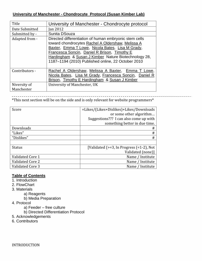
Distilled H2O (dH2O) is sterilized by autoclaving ‐ ‐Dimethyl Sulfoxide Sigma D2650Porcine gelatin Sigma G1890 Bovine Serum Albumin (BSA) Sigma A4161Human Plasma Fibronectin Millipore FC010 Human recombinant FGF2 Peprotech AA10‐155
Peprotech Human recombinant Neurotrophin‐4 Peprotech 450‐04Human recombinant BMP4 Peprotech 120‐05Human recombinant GDF5 Peprotech BMP14/CDMP1 Human recombinant Follistatin 300 Peprotech F1175‐25UG Mouse recombinant Wnt3a Peprotech 1324‐WN
120‐14
Tip: Due to the decrease in the biological activity of supplementary growth factors we store all culture media at 4ºC and discard 14 days after preparation. In order to prevent waste from discarding expired media we frequently prepare 500ml volumes of hESc FeederFree Medium and Directed Differentiation Basal Medium without the growth factors and store 40ml aliquots at 20ºC. The required growth factor combinations are then added to thawed aliquots. We have not detected any adverse effects in cell cultures when using media that has been stored at 20ºC for up to 2 months from preparation.
Growth factors:
Growth factors are purchased as lyophilized powders which are reconstituted to a stock concentration with 0.1% (vol/vol) BSA in DPBS. To prepare 0.1% (vol/vol) BSA add 0.01g of BSA to 10ml DPBS, vortex well and sterilize through a 0.22μm syringe driven filter.
Human recombinant FGF2 (AA10‐155) (Biosource™, Invitrogen cat. no. PHG0021) – Dissolve 1mg of lyophilized powder in 10ml of 0.1% (vol/vol) BSA in DPBS to make a stock concentration of 100μg/ml (wt/vol). Aliquot the stock growth factor solution in 50μl volumes.
Human recombinant ActivinA (Peprotech cat. no. 120‐14) – dissolve 5μg of lyophilized powder in500μl of 0.1% (vol/vol) BSA in DPBS to make a stock concentration of 10μg/ml (wt/vol). Aliquot the stock growth factor solution in 50μl volumes.
Human recombinant Neurotrophin4 (NT4) (Peprotech cat. no. 450‐04) – Dissolve 10μg of lyophilized powder in 1ml of 0.1% (vol/vol) BSA in DPBS to make a stock concentration of 10μg/ml (wt/vol). Aliquot the stock growth factor solution in 20μl volumes.
Human recombinant BMP4 (Peprotech cat. no. 120‐05) – Dissolve 5μg of lyophilized powder in 500μlof 0.1% (vol/vol) BSA in DPBS to make a stock concentration of 10μg/ml (wt/vol). Aliquot the stock growth factor solution in 50μl volumes.
Human recombinant GDF5 (BMP14/CDMP1) (Peprotech cat. no. 120‐01) – Dissolve 50μg of lyophilizedpowder in 5ml of 0.1% (vol/vol) BSA in DPBS to make a stock concentration of 10μg/ml (wt/vol). Aliquot the stock growth factor solution in 50μl volumes.
Human recombinant Follistatin 300 (Sigma cat. no. F1175‐25UG) – Dissolve 25μg of lyophilizedpowder in 2.5ml of 0.1% (vol/vol) BSA in DPBS to make a stock concentration of 10μg/ml (wt/vol).Aliquot the stock growth factor solution in 50μl volumes.
Mouse recombinant Wnt3a (R&D Systems cat. no. 1324‐WN) – Dissolve 2μg of lyophilized powder is dissolved in 200μl of 0.1% (vol/vol) BSA in DPBS to make a stock concentration of 10μg/ml (wt/vol).Aliquot the stock growth factor solution in 25μl volumes.
Human recombinant Activin‐A
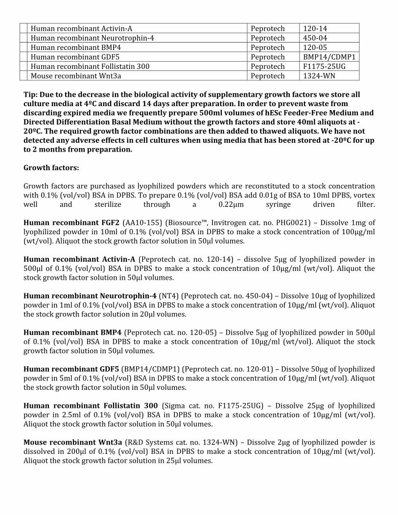
Critical: Growth factor polypeptides are highly unstable and biological activity decreases rapidly. Because of this lyophilized growth factors are assumed to be sterile on opening and reconstitution is carried out in a microbiological safety cabinet using aseptic technique. Growth factor solutions are never passed through 0.22μm filters and cell culture media are sterilized prior to the addition of growth factors. Once reconstituted the growth factor aliquots are transferred on ice for storage at 80ºC. Thawed aliquots are never refrozen but are stored at 4ºC and discarded 14 days postthaw. Cryopreservation medium: Add a 1ml volume of Dimethyl Sulfoxide (Sigma cat. no. D2650) to 9ml volume of FBS. Pipette up and down to mix well. Porcine gelatin (Sigma cat. no. G1890) – A 1% stock gelatin solution is prepared by adding 4g of lyophilized gelatin powder to 400ml dH2O. The solution is autoclaved to sterilise, aliquotted in 20ml volumes and stored at ‐20ºC. Working gelatin solutions of 0.1% (vol/vol) are prepared in sterile T‐75 cell culture flasks by adding 20ml of thawed 1% (vol/vol) gelatin to 180ml of sterile dH2O. The 0.1% (vol/vol) gelatin solution is stored at 4ºC and discarded after 28 days Bovine Serum Albumin (BSA) (Sigma cat. no. A4161) Human Plasma Fibronectin (Millipore cat. no.FC010) – Fibronectin is supplied as a liquid at a concentration of 1mg/ml (wt/vol) to be stored at 4ºC. Working solutions of 50μg/ml (wt/vol) are prepared by diluting 1ml of stock fibronectin in 19ml of DPBS and stored at 4ºC. Table 1: Formulation of MEF Medium
Table 2: Formulation of hESc Medium
hESc culture on Inactivated Mouse Embryonic Fibroblast (iMEF) Feeder Cell Layer: 1. Transfer 1ml of 0.1% (vol/vol) gelatin to 1×35mm culture dish and incubate in a cell culture incubator at 37ºC for 1 hour.

2. Remove one cryovial of iMEF cells from the liquid nitrogen cell storage dewar and place on ice fortransfer to the cell culture suite. 3. Hold the cryovial by the lid and submerge three quarters of the cryovial in the 37ºC water bath for 2‐3 minutes to thaw the cell suspension. Tip: Swirl the cryovial in the water to facilitate heat transfer. Thaw the iMEF cells until approximately 10% of ice remains, the iMEF cells will continue to thaw during transfer to the microbiological safety cabinet. 4. Draw up 4ml of MEF medium in a 5ml serological pipette and then the 1ml volume of iMEF cell suspension from the cryovial. Transfer the 5ml volume to a sterile 15ml polystyrene centrifuge tube. 5. Add 90μl of the iMEF cell suspension and 10μl of trypan blue solution to a 1.5ml microfuge tube and mix well by pipetting up and down. Transfer 10μl to each chamber of haemocytometer and perform a viable cell count. MEF cells are seeded at a cell density of 3×10⁴ cells/cm2 which for a 35mm culture dish is 2.92×10⁵ cells; calculate the volume of iMEF cell suspension required: 2.92×10⁵ cells / viable cells/ml = Xml of MEF cell suspension. Transfer the calculated volume to a sterile 15ml polystyrene centrifuge tube and centrifuge at 720 x g for 3 minutes at room temperature. Remove the supernatant and resuspend the cell pellet in 2ml of MEF medium (Table 1). 6. Take the 35mm culture dish with gelatin from the incubator, remove the gelatin and add the 2ml iMEF cell suspension. Transfer to the cell culture incubator. 7. Twenty‐four hours post‐seeding check the iMEF cells under the microscope to ensure that they have adhered to the cell culture surface and formed a homogeneous culture of approximately 80‐85% confluence. 8. Thaw hESc and seed onto the iMEF layer. Remove a vial of hESc from liquid nitrogen storage and thaw as for the iMEF cells in steps 3‐5 but substitute hESc medium (Table 2) for MEF medium. Centrifuge the hESc at 720 x g for 3 minutes at room temperature and resuspend the cell pellet in 2ml of hESc Medium. 9. Remove the iMEF medium from the iMEF cell layer and add the hESc suspension. Transfer the cell culture dish to the cell culture incubator. 10. Change the hESc medium every 2 days and check the hESc for growth under the microscope. Colonies of hESc can be observed growing between iMEF cells after approximately 2‐4 days. 11. Passage the hESc at a ratio of 1:6 when the culture has grown to approximately 90‐95% confluence. Prepare 6 wells of a 6 well culture dish with a iMEF feeder cell layer as described in steps 1‐7. 12. Pre‐warm a 10ml aliquot of Trypsin:EDTA and hESc medium by placing in the 37ºC water bath for 30 minutes. 13. Remove and discard the hESc medium from the hESc culture and add 1ml of Trypsin:EDTA. Incubatein the cell culture incubator at 37ºC and check under the microscope every 2‐3 minutes for cell dissociation. iMEF cells are seen to round up whereas hESc can be seen to separate from each other. Critical: Cell:cell contact is crucial in maintaining the hESc phenotype and so it is important that the hESc are not overexposed to the Trypsin:EDTA. The cells should remain in clumps of approximately 510 cells to permit the formation of new hESc colonies.
14. Transfer the hESc suspension to a sterile 15ml polystyrene centrifuge tube and centrifuge at 720 x g for 3 minutes at room temperature. Resuspend the hESc in 12ml of hESc medium.
15. Remove the MEF medium from the wells of the 6 well culture dish and add 2ml of the hESc cellsuspension to each well. Transfer the culture dish to the cell culture incubator. Tip: We frequently keep 1×6 well cell culture dish of hESc in continuous culture on an iMEF feeder layer. When confluent 1 x well is passaged to another 6 well culture dish with iMEFs, 2 x wells are taken to establish feederfree cultures and 3 wells are cryopreserved in order to maintain low passage stocks of hESc.
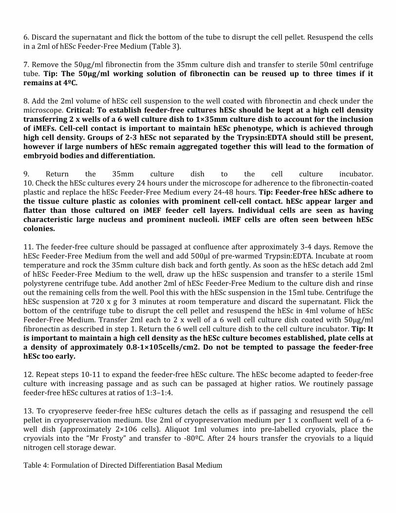
Table 3: Formulation of hESc Feeder-Free Medium
FeederFree hESc Culture:
1. Prepare 35mm and 6 well culture dishes by transferring 1ml of 50μg/ml fibronectin to each well and incubating for at least 16 hours at 4ºC.
2. Remove and discard the hESc medium from 2 x wells of hESc iMEF cultures and rinse twice with DPBS.
3. Add 500μl of Trypsin:EDTA to each of the 2 wells and transfer to the cell culture incubator.
4. Check the hESc cultures every 2‐3 minutes as if passaging the hESc cultures on to iMEF feeder cell layers. The Trypsin:EDTA can be left longer to permit more thorough dissociation of the hESc.
5. Transfer the cell suspension to a sterile 15ml polystyrene centrifuge tube and centrifuge at 720 x g for 3 minutes at room temperature.
6. Discard the supernatant and flick the bottom of the tube to disrupt the cell pellet. Resuspend the cells in a 2ml of hESc Feeder‐Free Medium (Table 3).
7. Remove the 50μg/ml fibronectin from the 35mm culture dish and transfer to sterile 50ml centrifuge tube. Tip: The 50μg/ml working solution of fibronectin can be reused up to three times if it remains at 4ºC.
8. Add the 2ml volume of hESc cell suspension to the well coated with fibronectin and check under the microscope. Critical: To establish feederfree cultures hESc should be kept at a high cell density transferring 2 x wells of a 6 well culture dish to 1×35mm culture dish to account for the inclusion of iMEFs. Cellcell contact is important to maintain hESc phenotype, which is achieved through high cell density. Groups of 23 hESc not separated by the Trypsin:EDTA should still be present, however if large numbers of hESc remain aggregated together this will lead to the formation of embryoid bodies and differentiation.
9. Return the 35mm culture dish to the cell culture incubator.10. Check the hESc cultures every 24 hours under the microscope for adherence to the fibronectin‐coatedplastic and replace the hESc Feeder‐Free Medium every 24‐48 hours. Tip: Feederfree hESc adhere to the tissue culture plastic as colonies with prominent cellcell contact. hESc appear larger and flatter than those cultured on iMEF feeder cell layers. Individual cells are seen as having characteristic large nucleus and prominent nucleoli. iMEF cells are often seen between hESc colonies.
11. The feeder‐free culture should be passaged at confluence after approximately 3‐4 days. Remove the hESc Feeder‐Free Medium from the well and add 500μl of pre‐warmed Trypsin:EDTA. Incubate at roomtemperature and rock the 35mm culture dish back and forth gently. As soon as the hESc detach add 2ml of hESc Feeder‐Free Medium to the well, draw up the hESc suspension and transfer to a sterile 15mlpolystyrene centrifuge tube. Add another 2ml of hESc Feeder‐Free Medium to the culture dish and rinseout the remaining cells from the well. Pool this with the hESc suspension in the 15ml tube. Centrifuge the hESc suspension at 720 x g for 3 minutes at room temperature and discard the supernatant. Flick the bottom of the centrifuge tube to disrupt the cell pellet and resuspend the hESc in 4ml volume of hEScFeeder‐Free Medium. Transfer 2ml each to 2 x well of a 6 well cell culture dish coated with 50μg/ml fibronectin as described in step 1. Return the 6 well cell culture dish to the cell culture incubator. Tip: It is important to maintain a high cell density as the hESc culture becomes established, plate cells at a density of approximately 0.81×10⁵cells/cm2. Do not be tempted to passage the feederfree hESc too early.
12. Repeat steps 10‐11 to expand the feeder‐free hESc culture. The hESc become adapted to feeder‐free culture with increasing passage and as such can be passaged at higher ratios. We routinely passage feeder‐free hESc cultures at ratios of 1:3–1:4.
13. To cryopreserve feeder‐free hESc cultures detach the cells as if passaging and resuspend the cellpellet in cryopreservation medium. Use 2ml of cryopreservation medium per 1 x confluent well of a 6‐well dish (approximately 2×10⁶ cells). Aliquot 1ml volumes into pre‐labelled cryovials, place the cryovials into the “Mr Frosty” and transfer to ‐80ºC. After 24 hours transfer the cryovials to a liquidnitrogen cell storage dewar.
Table 4: Formulation of Directed Differentiation Basal Medium

Directed Differentiation Protocol The directed differentiation protocol is carried out on feeder‐free hESc which have been cultured off feeders for at least 3 passages to exclude contaminating iMEFs. 1. Coat 1×35mm culture dish by adding 1ml of 50μg/ml fibronectin and incubating overnight at 4ºC. 2. Detach feeder‐free hESc from the cell culture plastic using pre‐warmed Trypsin:EDTA, centrifuge at 720 x g for 3 minutes at room temperature and resuspend to a cell density of 5×10⁵ cells/ml in hESc Feeder‐Free Medium. 3. Remove the fibronectin solution from the well of the 35mm culture dish and add a 2ml volume of feeder‐free hESc suspension. 4. Return the culture dish to the cell culture incubator. 5. After 24 hours check the hESc culture under the microscope. DDP should be initiated when the cultures are at approximately 80% confluence. Denote this as Day 1. 6. Prepare Day 1 medium by adding 6.25μl of 10μg/ml mouse recombinant Wnt3a and 12.5μl of 10μg/ml human recombinant Activin‐A to 2.5ml of Directed Differentiation Basal Medium (Table 4). 7. Remove the hESc Feeder‐Free Medium, rinse the feeder‐free hESc culture three times with DPBS and add 2.5ml of Day 1 medium. Return to the cell culture incubator. 8. Day 2: add 6.25μl of 10μg/ml mouse recombinant Wnt3a; 6.25μl of 10μg/ml human recombinant Activin‐A and 0.5μl of 100μg/ml human recombinant FGF2 to 2.5ml of Directed Differentiation Basal Medium. 9. Remove the Day 1 medium from the cell culture and replace with 2.5ml of Day 2 medium. Return to the cell culture incubator. 10. Day 3: add 6.25μl of 10μg/ml mouse recombinant Wnt3a; 2.5μl of 10μl/ml human recombinant Activin‐A; 0.5μl of 100μg/ml human recombinant FGF2 and 10μl of 10μg/ml human recombinant BMP4 to 2.5ml of Directed Differentiation Basal Medium. 11. Remove the Day 2 medium from the cell culture and replace with 2.5ml of Day 3 medium. Return to the cell culture incubator. 12. Day 4: Analyse Stage 1 cultures if appropriate.

13. Add 0.5μl of 100μg/ml human recombinant FGF2; 10μl of 10μg/ml human recombinant BMP4; 25μl of 10μg/ml human recombinant follistatin and 0.5μl of 10μg/ml human recombinant NT4 to 2.5ml ofDirected Differentiation Basal Medium.
14. Remove the Day 3 medium from the cell culture and replace with 2.5ml of Day 4 medium. Return to the cell culture incubator.
15. Transfer 1ml of 50μg/ml fibronectin to each of 5 x wells of a 6 well culture dish and place at 4ºC.
16. Day 5: add 2μl of 100μg/ml human recombinant FGF2; 40μl of 10μg/ml human recombinant BMP4;100μl of 10μg/ml human recombinant follistatin and 2μl of 10μg/ml human recombinant NT4 to 10ml ofDirected Differentiation Basal Medium.
17. Remove the Day 4 medium from the cell culture and add 500μl of prewarmed Trypsin:EDTA.Incubate at room temperature and rock the 35mm culture dish back and forth gently. As soon as the cells detach add 2ml of Directed Differentiation Basal Medium to the well, draw up the cell suspension and transfer to a sterile 15ml polystyrene centrifuge tube. Add another 2ml volume of Directed Differentiation Basal Medium to the culture dish and rinse out the remaining cells from the well. Pool this with the cell suspension. Centrifuge the cell suspension at 720 x g for 3 minutes at room temperature anddiscard the supernatant. Flick the bottom of the centrifuge tube to disrupt the cell pellet and resuspendthe cells in 10ml of Day 5 Medium. Remove the 50μg/ml fibronectin solution and transfer 2ml of the cell suspension to each of the 5 wells. Return the culture dishes to the cell culture incubator.
18. Day 6: add 2μl of 100μg/ml human recombinant FGF2; 40μl of 10μg/ml human recombinant BMP4;100μl of 10μg/ml human recombinant follistatin and 2μl of 10μg/ml human recombinant NT4 to 10ml ofDirected Differentiation Basal Medium.
19. Remove the Day 5 medium from the 5 cell culture dishes and replace each with 2ml of Day 6 medium. Return to the cell culture incubator.
20. Day 7: add 2μl of 100μg/ml human recombinant FGF2; 40μl of 10μg/ml human recombinant BMP4;100μl of 10μg/ml human recombinant follistatin and 2μl of 10μg/ml human recombinant NT4 to 10ml ofDirected Differentiation Basal Medium.
21. Remove the Day 6 medium from the 5 cell culture dishes and replace each with 2ml of Day 7 medium. Return to the cell culture incubator.
22. Transfer 10ml of 50μg/ml fibronectin and 10ml of 0.1% (vol/vol) gelatin to a sterile 50mlpolystyrene centrifuge tube and mix well by pipetting up and down.
23. Transfer 1ml of the mixed fibronectin:gelatin solution to 20 x wells of 6 well culture dishes and place at 4ºC.
24. Day 8: add 8μl of 100μg/ml human recombinant FGF2; 160μl of 10μg/ml human recombinant BMP4and 8μl of 10μg/ml human recombinant NT4 to 40ml of Directed Differentiation Basal Medium.
25. Remove the Day 7 medium from the cell cultures and add 500μl of prewarmed Trypsin:EDTA to each of the 5 wells. Incubate at room temperature; rocking the cell culture dish back and forth gently. As soon as the cells detach add 2ml of Directed Differentiation Basal Medium to each cell culture dish, draw up the cell suspension and transfer to a sterile 15ml polystyrene centrifuge tube. Add another 2ml
of Directed Differentiation Basal Medium to the culture dishes and rinse out the remaining cells from the well. Pool this with the cell suspension. Centrifuge the cell suspension at 720 x g for 3 minutes at room temperature and discard the supernatant. Flick the bottom of the centrifuge tube to disrupt the cell pellet and resuspend the cells in 40ml of Day 8 Medium. Remove the fibronectin:gelatin solution and transfer2ml of the cell suspension to each of the 20 wells of the culture dishes. Return the culture dishes to the cell culture incubator.
26. Day 9: Analyse Stage 2 cultures if appropriate.
27. Add 8μl of 100μg/ml human recombinant FGF2; 80μl of 10μg/ml human recombinant BMP4; 80μl of10μg/ml human recombinant GDF5 and 8μl of 10μg/ml human recombinant NT4 to 40ml of Directed Differentiation Basal Medium.
28. Remove the Day 8 medium from the cell cultures and replace with 2ml of Day 9 medium to each well.Return to the cell culture incubator.
29. Day 10: add 8μl of 100μg/ml human recombinant FGF2; 80μl of 10μg/ml human recombinant BMP4;80μl of 10μg/ml human recombinant GDF5 and 8μl of 10μg/ml human recombinant NT4 to 40ml ofDirected Differentiation Basal Medium.
30. Remove the Day 9 medium from the cell cultures and replace with 2ml of Day 10 medium to each ofthe wells. Return to the cell culture incubator.
31. Day 11: add 8μl of 100μg/ml human recombinant FGF2; 160μl of 10μg/ml human recombinant GDF5and 8μl of 10μg/ml human recombinant NT4 to 40ml of Directed Differentiation Basal Medium.
32. Remove the Day 10 medium from the cell cultures and replace with 2ml of Day 11 medium to each well. Return to the cell culture incubator.
33. Transfer 1ml of 0.1% (vol/vol) gelatin solution to 30 x wells of 6 well cell culture dishes and place at 4ºC.
34. Day 12: add 12μl of 100μg/ml human recombinant FGF2; 240μl of 10μg/ml human recombinant GDF5 and 12μl of 10μg/ml human recombinant NT4 to 30ml of Directed Differentiation Basal Medium. Note: This makes a 2 X concentration of Day 12 medium.
35. Remove the Day 11 medium from the cell cultures and add 500μl of pre‐warmed Trypsin:EDTA to each well. Incubate at room temperature; rocking the cell culture dishes back and forth gently. As soon as the cells detach add a 2ml volume of Directed Differentiation Basal Medium to each cell culture dish, draw up the cell suspension and transfer in equal volumes to 2 x sterile 50ml polystyrene centrifuge tubes. Add another 2ml volume of Directed Differentiation Basal Medium to the culture dishes and rinse out the remaining cells from the well. Transfer this cell suspension in equal volumes to the 2×50mlcentrifuge tubes. Centrifuge the cell suspension at 720 x g for 3 minutes at room temperature and discard the supernatant. Flick the bottom of the centrifuge tube to disrupt the cell pellet and resuspend the cells in 30ml of Directed Differentiation Basal Medium. Remove the 0.1%(vol/vol) gelatin solution and transfer 1ml of the cell suspension to each of the 30 wells of the 6 well culture dishes.
36. Add 1ml of the 2 X Day 12 medium to each of the 30 wells. Return the culture dishes to the cell culture incubator.
37. Day 13: add 12μl of 100μg/ml human recombinant FGF2; 240μl of 10μg/ml human recombinant
GDF5 and 12μl of 10μg/ml human recombinant NT4 to 30ml of Directed Differentiation Basal Medium. Note: This makes a 2 X concentration of Day 13 medium.
38. Remove the Day 12 medium from the cell cultures and replace with 1ml each of Directed Differentiation Basal Medium to each of the 30 wells of the 6 well cell culture dishes
39. Add 1ml each of the 2 X Day 13 medium to each of the 30 wells. Return to the cell culture incubator.
40. Day 14 terminate the cultures and carry out Stage 3 (end point) analyses.
REFERENCES
A chemically-defined protocol for generating chondrocytes from human embryonic stem cells Rachel Oldershaw , Melissa Baxter , Emma Lowe , Nicola Bates , Lisa Grady , Francesca Soncin , Daniel Brison Timothy Hardingham & Susan Kimber Protocol Exchange (2010) Published online 30 November 2010