Embed Size (px)
Citation preview

Arthroscopic Treatment ofChondrocyte Transplantatioji
Pi
Andrea Fontana, M.D., AlessanFederica Rosso, M.D.
Purpose: To compare the effectiveness o
We carried out a controlled retrospectivechondropathy of the third or fourth degree,
lip Chondral Defects: AutologousVersus Simple Debridement—A
ot Study
ro Bistolfi, M.D., Maurizio Crova, M.D.,and Giuseppe Massazza, M.D.
simple arthroscopic debridement versus arthroscopicautologous chondrocyte transplantation (AC T) for the treatment of hip chondral lesions. Methods:
study of 30 patients affected by a post-traumatic hipccording to the Outerbridge classification, measuring 2
cm2 in area or more. Of these patients, 1:5 underwent arthroscopic ACT, whereas the other 15underwent arthroscopic debridement. The 2 groups were similar in age, sex, degree, and location ofthe pathology. All the patients were assessed before and after the procedure with the Harris Hip Score(HHS). Results: In both groups the mean fcllow-up was approximately 74 months (range, 72 to 76months). The mean size of the defect was 1.6 cm. The patients who underwent ACT (group A)improved after the procedure compared with the group that underwent debridement alone (group B).The mean HHS preoperatively was 48.3 (95% confidence interval [CI], 45.4 to 51.2) in group A and46 (95% CI, 42.7 to 49.3) in group B (P = .^-28 [no significant difference]). The final HHS was 87.4(95% CI, 84.3 to 90.5) in group A and 56.3 (95% CI, 54.4 to 58.7) in group B (P < .001 [significantdifference]). Conclusions: This study indicates that an ACT procedure can be used in the hip foracetabular chondral defects. Level of Evidence: Level m, retrospective comparative study.
A'ter the initial experiences with arthrotomy tech-niques, several arthroscopic techniques were de-
veloped for the treatment of chondral lesion in theknee joint.1-4 Among these, autologous chondrocytetransplantation (ACT) with a 2-step technique has ikdto good clinical results.5'6 New techniques based on
From the Hip and Knee Unit (A.F.) and Biotechnologies De-partment (A.F.), Clinica Capitanio, Milan; Department of Ortho-paedics, Traumatology and Health Medicine, CTO/M. Adela deHospital (A.B., M.C., G.M.), Turin; and Department of Orthopae-dics, Traumatology and Health Medicine, University of Tu -in(M.C., F.R., G.M.), Turin, Italy.
The authors report no conflict of interest.Received January 9, 2011; accepted August 22, 2011.Address correspondence to Alessandro Bistolfi, M.D., CTO/M.
Adelaide Hospital, via Zuretti 29, 10126 Turin, Italy. E-mail:alessandro. bistolfi @ cto. to. it
Crown Copyright © 2012 Published by Elsevier Inc. on beh ilfof the Arthroscopy Association of North America. All rightsreserved.
0749-8063/1125/$36.00doi:10.1016/j.arthro.2011.08.304
322
biodegradable scaffolds have been developed to avoidthe periostea! flap.7-10 Several materials have beenproposed as matrices to deliver the chondrocytes tothe cartilage defect and to provide mechanical supportand nutrition to the cells: protein-based polymers,carbohydrate polymers, and artificial polymers.11-14
Cartilage lesions and chondral defects are also com-mon in the hip after several traumatic and congenitalpathologies.15"18 Numerous studies have shown thedirect association between acetabular labrum lesionsand chondral lesions of the femoral head and acetab-ulum.19-24
Currently, there is growing interest in non-arthroplastysurgical treatment of chondropathies of the acetab-ulum and the femoral head because they are afrequent cause of pain and functional limitation.Usually, the treatment of hip cartilage lesions isbased exclusively on arthroscopic debridement ormicrofractures,25-30 and new different treatmentshave been proposed only recently, such as ar-
Arthroscopy: The Journal of Arthroscopic and Related Surgery, Vol 28, No 3 (March), 2012: pp 322-329

SUPRASCAPULAR NERVE INJURY DURING LATARJET 3.21
3. Walch G. Recurrent anterior shoulder instability. Rev ChirOrthop Reparatrice Appar Mot 1991;77:177-191 (Suppl 1) (in
' • 'French], •- .4. Latarjet 1C' Jteatment of recurrent dislocation of the shoulder.
Lyon Chir 1954;49:994-997 (in French).5. Allain J, GauiaHler.D, .Glprion C. Long-term results of the
Latarjet procedure;, fa? the treatment of anterior instability ofthe shoulder. / Bone Joint Surg Am 1998;80:841-852.
6. Benammar MN, Saragaglia D, Legrand JJ, Faure C, Butel J.Latarjet's surgery in recurrent anterior dislocations of theshoulder. 117 cases with an 8-year follow-up. Rev Chir OrthopReparatrice Appar Mot 1986;72:447-454 (in French).
7. Huguet D, Pietu G, Bresson C, Potaux F, Letenneur J. Anteriorinstability of the shoulder in athletes: Apropos of 51 cases ofstabilization using the Latarjet-Patte intervention. Acta OrthopBelg 1996;62:200-206 (in French).
8. Matton D, Van Looy F, Geens S. Recurrent anterior disloca-tions of the shoulder joint treated by the Bristow-Latarjetprocedure. Historical review, operative technique and results.Acta Orthop Belg 1992;58:16-22 (in French).
9. Nielson AB, Nielsen K. The modified Bristow procedure forrecurrent anterior dislocation of the shoulder. Results andcomplications. Acta Orthop Scand 1982;53:229-232.
10. Bigliani LU, Dalsey RM, McCann PD, April BW. An anatomicalstudy of the suprascapular nerve. Arthroscopy 1990;6:301-3D5.
11. Shishido H, Krkuchi S. Injury of the suprascapular nerve'inshoulder surgery: An anatomic study. / Shoulder Elbow Surg2001;10:372-376.
12. Maquieira GJ, Gerber C, Schneeberger AG. Suprascapularnerve palsy after the Latarjet procedure. J Shoulder ElbowSurg 2007;16:el3-el5.
13. Burkhart SS, De Beer JF. Traumatic glenohumeral bone de-fects and their relationship to failure of arthroscopic Bankartrepairs: Significance of the inverted-pear glenoid and the hu-meral engaging Hill-Sachs lesion. Arthroscopy 2000;16:677-694.
14. De Mulder K, Marynissen H, Van Laere C, Lagae K, DeclercqG. Arthroscopic transglenoid suture of Bankart lesions. ActaOrthop Belg 1998;64:160-166.
15. Mologne TS, Lapoint JM, Morin WD, Zilberfarb J, O'BrienTJ. Arthroscopic anterior labral reconstruction using a.transglenoid suture technique. Results in active-duty militarypatients. Am J Sports Med 1996;24:268-274. ' : •
16. Bowden RE. The factors influencing functional recovery ofperipheral nerve injuries in man. Ann R Coll Surg Engl 1951;8:366-376.

HIP CHONDRAL DEFECTS 323
throscopic repair of acetabular chondral delamina-tion with fibrin adhesive.31
This technique is based on previous studies of theknee,7-14 which have led to the development of scaf-folds as implants, used for the delivery of culturedcells. Arthroscopic treatment of chondral lesions inthe hip may reduce the risks of avascular necrosiscorrelated to surgical dislocation. In addition, thistechnique makes it possible to avoid the periostealcover, which—in our opinion—would be very diffi-cult to introduce in the hip.
The purpose of this study was to compare ar-throscopic ACT with simple arthroscopic debride-ment. The hypotheses were that arthroscopic ACT isbetter than debridement and that arthroscopic ACT ispractical for chondral lesions in the hip joint.
METHODS
Patient Selection
From 1996 to 2004, we carried out 274 hip arthros-copies for treatment of chondral defects or hip pathol-ogies. Before surgery, patients were examined withstandard radiographs and magnetic resonance imaging(MRI). All 274 hips presented with pain clinically,reduction of range of motion, and signs of fernoro-acetabular conflict and/or labral lesions, as well assome pathology on radiographs or MRI, and weretherefore subjected to hip arthroscopy after informedconsent was obtained. Inclusion criteria for this studywere signs of arthritis of the hip, including slightlyreduced articular space on radiographs (Tonnis grade2 [moderate], with small cysts, moderate narrowing ofthe joint space, and moderate loss of head sphericity),but we excluded cases with severely reduced articularspace on radiographs (Tonnis grade 3 [severe], withlarge cysts, severe narrowing or obliteration of thejoint space, and severe deformity of the head) andmassive chondral lesions on MRI. We unilaterallydecided to exclude grade 3 lesions because in thesecases the arthritis is too advanced to yield benefitsfrom arthroscopic treatment. Therefore the decision toinclude or exclude patients in the study was based onradiographs, MRI, and arthroscopy as the final step.
A total of 181 patients met the inclusion criteria; theothers underwent different surgical treatments (syno-vectomy, hip replacement, simple removal of loosebodies, and so on). During arthroscopy, the cartilagedamage was assessed according to the Outerbridgeclassification and localized on both the acetabulumand the femoral head in 3 areas: anterior, superior, and
posterior. The area of the cartilage lesion was alsomeasured in square centimeters. The size of the lesionwas evaluated arthroscopically by optical estimationbased on our experience. In 37 of 181 patients with asimilar chondral defect of the third or fourth degree,which extended 2 cm2 or more, arthroscopic ACT wasperformed (representing 12.4% of the cases). Theremaining 144 patients were treated by hip arthros-copy and simple debridement for chondral defects.Fifteen patients who completed follow-up of at least 6years (range, 72 to 76 months) were chosen from thegroup that underwent ACT (group A). Among the 144patients treated with the debridement, a second group of15 patients was chosen to serve as the control group(group B). In determining the criteria used to select thepatients in group B, our objective was to obtain 2 homo-geneous groups in terms of age, sex, body mass index,and degree and area of the chondral lesion. Thereforegroup B included cases where the third- to fourth-degreechondral lesion, which extended 2 cm2 or more, hadbeen exclusively debrided, and the patients were similarto those hi group A, who were treated with arthroscopicACT. In both groups the hip arthroscopy procedure wasindicated for persistent pain, reduced range of motion,and signs of acetabular conflict hi association with ra-diographic signs of initial arthritis of the hip. Both groupswere clinically assessed preoperatively and postopera-tively with the Harris Hip Score (HHS). This study wasapproved by the local institutional review board.
Surgical Technique
All arthroscopies were carried out with the patient hia lateral decubitus position and with combined longitu-dinal and inguinal traction applied. The hip was accessedby the superior trochanteric, anterior trochanteric, andposterior trochanteric portals for evaluation, biopsy,treatment, and implantation of the scaffold.
Group A: In those cases treated by ACT (groupA), the surgical treatment was always carried out in 2steps. The first step, diagnostic arthroscopy, was usedto evaluate the chondral damage and to take a cartilagebiopsy specimen from the area surrounding the pulv-inar. Then, the transplant was implanted during thesecond step, operative arthroscopy, after approxi-mately 30 days. First, a chondrectomy was alwaysperformed in the area affected by the chondropathy,by use of angled curettes or motorized shavers andexposure of the subchondral bone, to create clear mar-gins between the healthy cartilage and the degeneratedarea. The chondrocyte culture was carried out on 1 ofthe bioresorbable two-component gel-polymer scaffolds

324 A. FONTANA ET AL.
used at our institution: the BioSeed-C (BioTissue Tech-nologies GmbH, Freiburg, Germany). The BioSeed is areabsorbable composite material of a polymer-basedscaffold (2 X 3 cm and 0.2 cm in height) of polyglycolic/polylactic acid (polyglactin, vicryl) and polydioxanone.It is used for the 3-dimensional growth of culturedchondrocytes and their implantation in transplant pro-cedures. After the biopsy, the cells can be stored forup to 2 years. The cells were cultured at the cellprocessing facility of the manufacturer according tothe state-of-the-art technique. Over a period of 3 to 5weeks, they were cultured on a monolayer to increaseto approximately 12 million cells. Then, the cells wereincubated over the membrane for the 3-dimensionalgrowth and stored before surgery for an approximatetime of 2 to 6 weeks. Every step of the manufacturingprocess was monitored to ensure high quality andsafety. The scaffold membrane with the chondrocyteswas cut to exactly fit the chondral defect after itspreparation and then rolled to pass along the cannula(Fig 1). Then, the transplant was inserted directly into thearticular cavity through an arthroscopic cannula and wasadapted to the chondral defect (Fig 2). After the im-plant had been positioned on the cartilage defect,traction was released and the articulation was sub-jected to a series of 5 extension and rotation move-ments. Afterward, traction was reapplied and the posi-tion of the transplant was controlled arthroscopically, toverify the fact that the transplant had remained in itsposition. The implantation and the arthroscopic evalua-tion that followed were performed without intra-articularfluid.
Group B: Patients in group B underwent a singlesurgery: once the chondral defect was identified, achondrectomy was performed in the area affected by
FIGURE 1. The membrane is rolled for insertion into the joint.
FIGURE 2. Arthroscopic view of membrane covering acetabularchondral defect. The fluid was stopped.
the chondropathy, again with angled curettes or mo-torized shavers. In this case an intra-articular debride-ment was associated and the chondrectomy was lim-ited to the clearly damaged or detached cartilage,leaving in situ as much of the cartilage as possible. Forthis reason, the debridement may ultimately result in asmaller defect than occurs with preparation for ACT.The exposure of the subchondral bone was reduced toonly the regions where it was strictly necessary toavoid conflicts or potential loose bodies.
Postoperative and Rehabilitation Protocol
Postoperatively, the patients in group A and groupB followed a similar standard rehabilitation program.Exercises began from the first postoperative day. Pa-tients were discharged from the hospital on the secondday and underwent both active and passive physio-therapy to regain complete range of motion withoutputting any weight on the articulation for 4 weeks.Partial load was allowed after 4 weeks in group A andafter 2 weeks in group B. Exercises on a gym bike andswimming were recommended after 4 weeks in bothgroups. After 7 weeks, crutches were no longer re-quired, and the patients were allowed to return tonormal work activity. Jogging was allowed only after6 months, whereas a complete return to sports activi-ties was recommended only 1 year after the surgicalprocedure.
Statistical Analysis
The analysis of variance test was used to test thestatistical significance of the observed differences be-
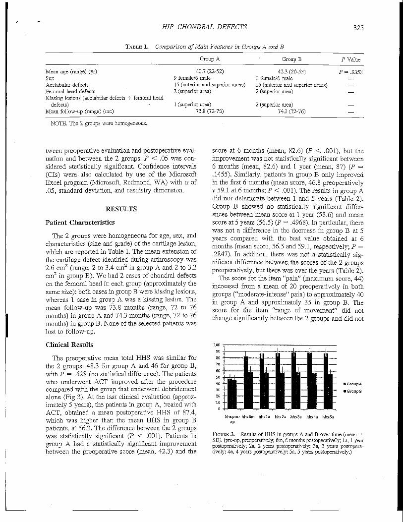
HIP CHONDRAL DEFECTS 325
TABLE 1. Comparison of Main Features in Groups A and B
Group A Group B P Value
Mean age (range) (yr)SexAcetabular defectsFemoral head defectsKissing lesions (acetabular defects + femoral head
defects)Mean follow-up (range) (mo)
40.7 (22-52)9 female/6 male15 (anterior and superior areas)2 (superior area)
1 (superior area)73.8 (72-76)
42.3 (20-53)9 female/6 male15 (anterior and superior areas)2 (superior area)
2 (superior area)74.3 (72-76)
P = .9353
NOTE. The 2 groups were homogeneous.
tween preoperative evaluation and postoperative eval-uation and between the 2 groups. P < .05 was con-sidered statistically significant. Confidence intervals(CIs) were also calculated by use of the MicrosoftExcel program (Microsoft, Redmond, WA) with a of.05, standard deviation, and casuistry dimension.
RESULTS
Patient Characteristics
The 2 groups were homogeneous for age, sex, andcharacteristics (size and grade) of the cartilage lesion,which are reported in Table 1. The mean extension ofthe cartilage defect identified during arthroscopy was2.6 cm2 (range, 2 to 3.4 cm2 in group A and 2 to 3.2cm2 in group B). We had 2 cases of chondral defectson the femoral head in each group (approximately thesame size): both cases in group B were kissing lesions,whereas 1 case in group A was a kissing lesion. Themean follow-up was 73.8 months (range, 72 to 76months) in group A and 74.3 months (range, 72 to 76months) in group B. None of the selected patients waslost to follow-up.
Clinical Results
The preoperative mean total HHS was similar forthe 2 groups: 48.3 for group A and 46 for group B,with P = .428 (no statistical difference). The patientswho underwent ACT improved after the procedurecompared with the group that underwent debridementalone (Fig 3). At the last clinical evaluation (approx-imately 5 years), the patients in group A, treated withACT, obtained a mean postoperative HHS of 87.4,which was higher than the mean HHS in group Bpatients, at 56.3. The difference between the 2 groupswas statistically significant (P < .001). Patients ingroup A had a statistically significant improvementbetween the preoperative score (mean, 42.3) and the
score at 6 months (mean, 82.6) (P < .001), but theimprovement was not statistically significant between6 months (mean, 82.6) and 1 year (mean, 87) (P =.1455). Similarly, patients in group B only improvedin the first 6 months (mean score, 46.8 preoperativelyv 59.1 at 6 months; P < .001). The results in group Adid not deteriorate between 1 and 5 years (Table 2).Group B showed no statistically significant differ-ences between mean score at 1 year (58.6) and meanscore at 5 years (56.5) (P = .4968). In particular, therewas not a difference in the decrease in group B at 5years compared with the best value obtained at 6months (mean score, 56.5 and 59.1, respectively; P =.2847). In addition, there was not a statistically sig-nificant difference between the scores of the 2 groupspreoperatively, but there was over the years (Table 2).
The score for the item "pain" (maximum score, 44)increased from a mean of 20 preoperatively in bothgroups ("moderate-intense" pain) to approximately 40in group A and approximately 35 in group B. Thescore for the item "range of movement" did notchange significantly between the 2 groups and did not
• Group A
• Groups
hhspre- hhs6m hhsla hhs2a hhsSa hhs4a hhsSaop
FIGURE 3. Results of HHS in groups A and B over time (mean ±SD). (pre-op, preoperatively; 6m, 6 months postoperatively; la, 1 yearpostoperatively; 2a, 2 years postoperatively; 3a, 3 years postopera-tively; 4a, 4 years postoperatively; 5a, 5 years postoperatively.)

326 A. FONTANA ET AL.
TABLE 2. HHS for Groups A and B During First 5 Years
Postoperative
Preoperative 6 mo lyr 2yr 3 yr 4 yr Syr
Group AGroup BP valueStatistical
significance
48.3(45.4-51.2) 82.6(79.7-85.4) 87(84.1-89.8) 87.7(84.8-90.6) 88.4(85.5-91.3) 88.6(85.7-91.4) 87.7(84.5-90.3)46.4 (43.5-49.3) 58.7 (55.8-61.6) 57.8 (54.9-60.7) 57.7 (54.8-60.6) 58.4 (55.5-61.3) 57.7 (54.8-60.6) 56.3 (53.4-59.1)
.428 <.001 <.001 <.001 <.001 <.001 <.001
No Yes Yes Yes Yes Yes Yes
NOTE. Analysis was performed with the analysis of variance test. Mean values (95% CIs) are reported.
significantly increase from before surgery comparedwith after surgery. The score associated with the item"walking distance" (maximum score, 11) increasedfrom a mean of 7 preoperatively to a mean of 10 ingroup A and 8.5 in group B. Table 3 reports the scoresfor pain and walking distance, with 95% CIs and theP values between the 2 groups.
Complications
Unsatisfactory results were obtained in 3 cases ingroup A, with a mean HHS of 74.8 (95% CI, 73.3to 76.5). In 1 case the cartilage defect was locatedon the superior area of the femoral head, and in 2cases the defect was located both on the superiorarea of the femoral head and on the superior andanterior area of the acetabulum with joint space nar-rowing on standard radiographs. The worst results ingroup B were found in 4 patients with a cartilagelesion of more than 3 cm2 located on the acetabulum.In these 4 patients the mean postoperative HHS was51.5 (95% CI, 49.7 to 53.2). Calf-vein thrombosisdeveloped at 2 weeks in 1 patient in group A and wastreated by anticoagulants. A transient neurapraxia ofthe pudendal nerve developed in 1 patient in group B.
DISCUSSION
Our results suggest several comments. First, thehigher HHS at the final clinical evaluation of the
patients treated with ACT indicated a better outcomeof this technique compared with simple debridement.This suggests the effectiveness of the technique interms of pain relief. The role of ACT is unclear inpostponing the progression of the arthritic process, aswell as in terms of the lack of radiologic progressionand the generation of cartilage. In addition, it must benoted that the postponement of the progression of thearthritic process is true only for the selected samples,as underlined later among the limitations. In addition,the results are based on clinical data, and there is noproof that the scaffold turned into cartilage. Post-operative radiographs, which we did not obtain,could have been helpful in clarifying these aspects.Second-look arthroscopy and MRI would have im-proved the value of the results. However, the formeris not possible for ethical reasons, whereas the lattershould be taken into consideration for further stud-ies and further follow-up.
We do not think that the difference in the resultscould have been somehow related to the difference inweight bearing, which was allowed 2 weeks earlier inthe debridement group. This protocol was decided onwith the sole intent of protecting the scaffold andpreventing its mobilization. The lower the startingHHS, the more unsatisfactory was the result, regard-less of the technique used. As far as the treatment ofhip chondropathies by use of arthroscopic debride-ment is concerned, this method shows scarce effec-
TABLE 3. Scores for Main Items of HHS (Pain and Walking Distance)
Preoperative Postoperative
Group A Group B Group A Group B
Pain score (maximum, 44)P value for pain scoreWalking distance score (maximum, 11)P value for walking distance score
20 (18.7-21.38) 20 (18.9-21.1)>.9999
7 (5.8-8.2) 7 (5.7-8.1)X9999
40 (38.6-41.4) 35 (33.2-36.8.0002
10 (9.4-10.5) 8.5 (7.6-9.4).1115
NOTE. Mean values (95% CIs) are reported, along with P values comparing the 2 groups.
HIP CHONDRAL DEFECTS 327
tiveness. The worst results were recorded in caseswith a chondral defect equal to or greater than 3 cm2,as already indicated.15-32 The clinical result of de-bridement is inversely proportional to the extension ofthe cartilage defect.32 For these reasons, we believethat in those cases where radiographic signs ofosteoarthritis are present, along with a reduction inthe joint space, the ACT technique is not indicated.The lack of surrounding cartilage makes the cre-ation of stable, clear margins impossible, with the"shoulder" clearly delineated, which is fundamentalfor greater stability. In addition, the geometric de-formity of the femoral head, caused by the arthriticdegeneration, does not guarantee the articular con-gruity required for implant stability. Moreover, thisarticular congraity in cases of arthritis is even morecompromised by the acetabular labrum degenera-tion, which represents a fundamental element forboth marntaining the negative intra-articular pressure andstabilizing the coxofemoral articulation.19'22
Hip arthroscopy, though considerably less commonthan knee arthroscopy, allows for chondropathies inthis joint to be detected33; however, the therapeuticapproach is different from the knee, because the hip isa deep articulation surrounded by large muscularmasses that make surgical access difficult. Neverthe-less, hip arthritis is highly limiting for patients, and asurgical procedure that has the potential .to postponeits progression is of great interest. Hip arthrotomyexposes the articulation to the serious risk of asepticnecrosis of the femoral head, along with being asignificantly invasive procedure. The arthroscopic ap-proach to treating hip chondropathies therefore solvesthe serious problem associated with arthrotomy.
A critical point regarding the ACT performed byhip arthroscopy is implant stability. The various tech-niques used in the knee, both by arthrotomy and byarthroscopy,2-6 might be difficult to use in the hip.Recently, a minimally invasive technique for ACT ofthe knee that could be applied to the hip was sug-gested.34 The efficacy of ACT techniques is well sup-ported in the literature,35-36 and the application to thehip is attractive. Some points of the surgical techniqueshould be pointed out: Performing a chondrectomywith wide exposure of the subchondral bone and thecreation of clear margins allows for greater stability ofthe implant on the acetabulum. This stability is rein-forced by the geometric and physical properties fea-tured by the hip.
The scaffold used in this study as a support for thecellular transplant features intrinsic rigidity that main-tains a structural "memory." This polymer,-rolled and
inserted through the arthroscopic cannula, then un-folds and returns to its original 3-dimensional shapeonce it has reached the articular cavity. This allows forit to easily adapt to the concave surface of the acetab-ulum that needs to be covered. As shown in Fig 2,which shows the membrane in a gas environment, weturned off the fluid inflow during graft delivery andplacement. We believe this is helpful for positioningthe graft at its site.
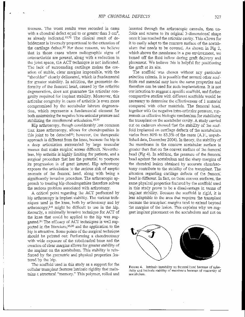
The scaffold was chosen without any particularselection criteria. It is possible that several other scaf-folds and material may have the same properties andtherefore can be used for such implantations. It is notour intention to suggest a specific scaffold, and furthercomparative studies with different scaffolds would benecessary to determine the effectiveness of 1 materialcompared with other materials. The femoral head,together with the negative intra-articular pressure, rep-resents an effective biologic mechanism for stabilizingthe transplant on the acetabular cavity. A study carriedout on cadavers showed that the stability of the scaf-fold implanted on cartilage defects of the acetabulumvaries from 80% to 83.3% of the cases (A.F., unpub-lished data, December 2004). In theory, the stability ofthe membrane in the concave acetabular surface isgreater than that on the convex surface of the femoralhead (Fig 4). In addition, the pressure of the femoralhead against the acetabulum and the sharp margins ofthe chondral lesion obtained by accurate chondrec-tomy contribute to the stability of the transplant. Thesituation regarding cartilage defects of the femoralhead is different. In fact, on these convex surfaces, thesame physical properties featured by the scaffold usedin this study prove to be a disadvantage in terms ofimplant stability. Because the scaffold is rigid, it isless adaptable to the area that requires the transplantbecause the transplant margins tend to extend beyondthe margins of the lesion. This explains why we sug-gest implant placement on the acetabulum and not on
FIGURE 4. Intrinsic instability on femoral head because of sphe-ricity and intrinsic stability of membrane because of concavity ofacetabulum.

328 A. FONTANA ET AL
the femoral head. The use of a device to fix theimplant in place, such as bioabsorbable nails or pins,might also be considered in further studies. In addi-tion, the use of microfracture technique must be con-sidered in further studies for creating the controlgroup. Moreover, microfractures can be used in com-bination with a scaffold without the cultivation ofchondrocytes. In this case the scaffold would act as asupport and a growth environment for the mesenchy-mal cells provided by the microfractures. This workillustrates the feasibility of using a scaffold intraoper-atively, but whether the addition of chondrocytes isnecessary and the success of seeding the scaffold withautologous cells remain unclear and require furtherspecific studies. In addition, this work focuses on thetreatment of chondral lesions localized in the acetab-ulum, whereas femoral lesions are not suitable forsuch treatment.
This study was limited by the reduced number ofpatients, the lack of an objective method for the eval-uation of the results (i.e., radiographic data, second-look arthroscopy, or MRI), and the lack of poweranalysis. Other limitations are the criteria for patientinclusion, selection bias in the randomization process,and the lack of comparison to microfracture tech-nique. Another limitation is the availability of thepatch in the United States and other markets. How-ever, we think that other materials could be used.
CONCLUSIONS
The main aim of this study was to suggest that anACT procedure can be used in the hip for acetabularchondral defects. The findings of the study suggestthat ACT may be an option for cartilage defects of thehip joint.
Acknowledgment: The first author (A.F.) thanks Mr.Villar for training and assistance in performing the hiparthroscopy surgical technique.
REFERENCES
1. Steadman JR, Rodkey WG, Rodrigo JJ. Microfracture: Surgi-cal technique and rehabilitation to treat chondral defects. ClinOrthop Relat Res 2001:362-369 (Suppl).
2. Gooding CR, Bartlett W, Bentley G, Skinner JA, Carrington R,Flanagan A. A prospective, randomised study comparing twotechniques of autologous chondrocyte implantation for osteo-chondral defects in the knee: Periosteum covered versus typeI/HI collagen covered. Knee 2006;13:203-210.
3. Briggs TWR, Mahroof S, David LA, Flannelly J, Pringle J,Bayliss M. Histological evaluation of chondral defects after
autologous chondrocyte implantation of the knee. JSone JointSurg Br 2003;85:1077-1083.
4. Erggelet C, Sittinger M, Lahm A. The arthroscopic implanta-tion of autologous chondrocytes for the treatment of full-thickness cartilage defects of the knee j oint. Arthroscopy 2003;19:108-110.
5. Kreuz PC, Mffller S, Ossendorf C, Kaps C, Erggelet C. Treat-ment of focal degenerative cartilage defects with polymer-based autologous chondrocyte grafts: Four-year clinical re-snlts. Arthritis Res Ther 2009;! 1 :R33.
6. Brittberg M, Lindahl A, Nilsson A, Ohlsson C, Isaksson 0,Peterson L. Treatment of deep cartilage defects in the kneewith autologous chondrocyte transplantation. N Engl J Med1994;331:889-895.
7. Gomoll AH, Farr K. Future developments in cartilage repair.In: Cole BJ, Gomoll AH, eds. Biologic joint reconstruction,alternative to arthroplasty. Thorofare: Slack, 2009;163-168.
8. Chang KY, Cheng LW, Ho GH, Huang YP, Lee YD. Fabri-cation and characterization of poly(gamma-glutamic acid)-graft-chondroitin sulfate/polycaprolactone porous scaffolds forcartilage tissue engineering. Acta Biomater 2009;5:1937-1947.
9. Knecht S, Erggelet C, Endres M, Sittinger M, Kaps C, StussiE. Mechanical testing of fixation techniques for scaffold-basedtissue-engineered grafts. J Biomed Mater Res B Appl Biomater2007:83:50-57.
10. Steinwachs MR, Guggi T, Kreuz PC. Marrow stimulationtechniques. Injury 2008:39:26-31.
11. Gigante A, Bevilacqua C, Ricevuto A, Mattioli-Belmonte M,Greco F. Membrane-seeded autologous chondrocytes: Cellviability and characterization at surgery. Knee Surg SportsTraumatol Arthrosc 2007;15:88-92.
12. Steinwachs M, Kreuz PC. Autologous chondrocyte implanta-tion in chondral defects of the knee with a type I/HI collagenmembrane: A prospective study with a 3-year follow-up. Ar-throscopy 2007;23:381-387.
13. Gobbi A, Bathan L. Minimally invasive second generationautologous chondrocyte implantation. In: Cole BJ, GomollAH, eds. Biologic joint reconstruction, alternative to arthro-plasty, Thorofare: Slack, 2009:155-161.
14. Iwasa J, Engebretsen L, Shima Y, Ochi M. Clinical applicationof scaffolds for cartilage tissue engineering. Knee Surg SportsTraumatol Arthrosc 2009;17:561-577.
15. McCarthy JC, Lee J. Hip arthroscopy: Indications and techni-cal pearls. Clin Orthop Relat Res 2005:441:180-187.
16. Tibor LM, Sekiya IK. Differential diagnosis of pain around thehip joint. Arthroscopy 2008;24:1407-1421.
17. Ganz R, Parvizi J, Beck M, Leunig M, Notzli H, SiebenrockKA. Femoroacetabular impingement: A cause for osteoarthri-tis of the hip. Clin Orthop Relat Res 2003:112-120.
18. Beck M, Kalhor M, Leunig M, Ganz R. Hip morphologyinfluences the pattern of damage to the acetabular cartilage:Femoroacetabular impingement as a cause of early osteo-arthritis of the hip. J Bone Joint Surg Br 2005:87:1012-1018.
19. Konrath GA, Hamel AJ, Olson SA, Bay B, Sharkey NA. Therole of the acetabular labrum and the transverse acetabularligament in load transmission in the hip. J Bone Joint Surg Am1998:80:1781-1788.
20. Byrd JW. Labral lesions: An elusive source of hip pain casereports and literature review. Arthroscopy 1996:12:603-612.
21. Cartridge IJ, Scott JH. The interned acetabular labrum inosteoarthrosis of the hip. J R Coll Surg Edinb 1982;27:339-344.
22. Harris WH, Bourne RB, Oh J. Intra-articular acetabularlabrum: A possible etiological factor in certain cases of osteo-arthritis of the hip. / Bone Joint Surg Am 1979:61:510-514.
23. McCarthy JC, Noble PC, Schuck MR, Wright J, Lee J. TheOtto E. Aufranc Award: The role of labral lesions to develop-

HIP CHONDRAL DEFECTS 329
ment of early degenerative hip disease. Clin Orthop Relat Res2001:25-37.
24. McCarthy JC, Noble PC, Schuok MR, Aluisio FV, Wright J,Lee J. Acetabular and labral pathology. In: McCarthy JC, ed.Early hip disorders. Chapter 12. New York: Springer-Verlag,2003:113-134.
25. Yen YM, Kocher MS. Chondral lesions of the hip: Microfrac-ture and chondroplasty. Sports Med Arthrosc 2010;18:83-89.
26. Haviv B, Singh PJ, Takla AK, O'Donnell J, Kulkarni MG,Nayak P. Arthroscopic femoral osteochondroplasty for camlesions with isolated acetabular chondral damage. J Bone JointSurg Br 2010;92:629-633.
27. Wright TM, Maher SA. Current and novel approaches totreating chondral lesions. / Bone Joint Surg Am 2009:91:120-5 (Suppl 1).
28. Philippon MJ, Schenker ML, Briggs KK, Maxwell RB. Canmicrofracture produce repair tissue in acetabular chondral de-fects? Arthroscopy 2008;24:46-50.
29. Crawford K, Philippon MJ, Sekiya JK, et al. Microfracture of thehip in athletes. Clin Sports Med 2006;25:327-335.
30. Barber FA, Iwasko NG. Treatment of grade HI femoral chon-dral lesions: Mechanical chondroplasty versus monopolar ra-diofrequency probe. Arthroscopy 2006;22:1312-1317.
31. Tzaveas AP, Villar RN. Arthroscopic repair of acetabularchondral delamination with fibrin adhesive. Hip Int 2010-20'115-119.
32. Glick JM. Hip arthroscopy using the lateral approach. InstrCourse Lect 1988;37:223-231.
33. Santori N, Villar RN. Arthroscopic findings in the initial stagesof hip osteoarthritis. Orthopedics 1999:22:405-409.
34. Anders S, Schaumburger J, Schubert T, Grifka J, Behrens P.Matrix-associated autologous chondrocyte transplantation (MACT).Minimally invasive technique in the knee. Oper Orthop Trau-matol 2008;20:208-219 (in German).
35. Batty L, Dance S, Bajaj S, Cole BJ. Autologous chondrocyteimplantation: An overview of technique and outcomes. ANZ•/Surg 2011:81:18-25.
36. Strauss EJ, Fonseca LE, Shah MR, Yorum T. Management offocal cartilage defects in the knee—Is ACI the answer? BullNYU Hasp Jt Dis 2011;69:63-72.
ABSTRACTS IN SPANISH!
RESUMENES AL ESPANOL!
are now available at www.arthroscopyjournal.org
Subscribers can access from the Table of Contents by clicking "SupplementalMaterials." Nonsubscribers can access the Spanish Abstracts by issue for
free from the home page.

Meniscal Morphologic Changes on Magnetic ResonanceImaging Are Associated With Symptomatic Discoid Lateral
Meniscal Tear in Children
Won Joon Yoo, M.D., Kang Lee, M.D., Hyuk Ju Moon, M.D., Chang Ho Shin, M.D.,Tae-Joon Cho, MIX, In Ho Choi, M.D., and Jung-Eun Cheon, M.D.
Purpose: To determine whether meniscal deformation and displacement on magnetic resonanceimaging (MRI) are associated with tears by use of arthroscopic findings as a standard of reference inchildren with discoid lateral meniscus (DLM). Methods: We reviewed MRI scans and intraoperativevideos of 69 consecutive patients (79 knees) treated arthroscopically for suspicious DLM tears. Themean age at surgery was 10.9 years (range, 4.3 to 17.6 years). Signal changes and morphologicchanges (deformation or displacement) of DLM on magnetic resonance (MR) images were graded by2 independent observers using our modifications of previously described classification schemes, andthen the grades were determined by consensus of the observers. Meniscal tears were assessed by anobserver, blinded to the MRI studies, based on arthroscopic findings. Signal changes and morpho-logic changes of DLM on MR images were correlated with tears. Results: Tears were found morefrequently in menisci showing morphologic changes on MR images (P = .001). Of the 25 menisciwith a grade 3 signal change (linear or band-like signal intensity extending to the superior or inferiormeniscal surface), 24 had tears, and a horizontal cleavage was the most commonly associated teartype. Of the 50 menisci with a grade 1 (dot-like intrameniscal signal change), grade 2 (linear orband-like intrameniscal signal change), or diffuse signal change, 34 were morphologically changedon MR images, and 29 of these (85%) were torn, whereas 9 of the 16 menisci (56%) notmorphologically changed were torn (P = .036). Conclusions: Preoperative MRI evaluations basedon signal intensities do not accurately predict the presence of a DLM tear in children, except whena DLM shows a grade 3 signal change. Meniscal deformation or displacement observed onpreoperative MR images suggests a higher risk of meniscal tears, even in menisci with signal changesother than grade 3 changes. Level of Evidence: Level IV, therapeutic case series.
The discoid lateral meniscus (DLM) is a morpholog-ically enlarged anomaly with a reported prevalence
from 0.4% to 20%.1~5 Most patients are asymptomatic inearly childhood except for some children with "snapping
From the Departments of Orthopaedic Surgery and Radiology(J-E.C.), Seoul National University Children's Hospital, Seoul,South Korea.
The authors report no conflict of interest.Received January 3, 2011; accepted August 18, 2011.Address correspondence to Jung-Eun Cheon, M.D., Department
of Radiology, Seoul National University Children's Hospital, 28Yeongon-dong, Jongro-gu, 110-744 Seoul, South Korea. E-mail:cheonje @snu. ac.kr
© 2012 by the Arthroscopy Association of North America0749-8063/0018/$3 6.00doi:10.1016/j.arthro.2011.08.300
knee syndrome," but pain and limitation of knee exten-sion may develop after an unstable meniscal tear, usuallyat 8 to 9 years of age.2-4-6-8 Magnetic resonance imaging(MRI) is known to be an accurate noninvasive diagnosticmodality for nondiscoid meniscal tears,9'14 but its rolefor DLM tears is still unclear. AraM et al.15 reported thatMRI has a high diagnostic accuracy (81% to 97%) forDLM tears, whereas Ryu et al.16 reported a low posi-tive predictive value (57%). Furthermore, MRI-based evaluations can be challenging in growingchildren and adolescents because of age-relatedchanges in vascularity and collagen-fiber arrange-ments in normal meniscal tissue.12
Signal changes of menisci on magnetic resonance(MR) images have been used to determine the pres-
330 Arthroscopy: The Journal of Arthroscopic and Related Surgery, Vol 28, No 3 (March), 2012: pp 330-336