Embed Size (px)
Citation preview
Genetically Determined Epilepsy Syndromes Gregory Neal Barnes MD/PhD Assistant Professor of Neurology and Pediatrics Director, Pediatric Epilepsy Monitoring Unit Vanderbilt University Medical Center
Disclosure
Investigator: NIH, Autism Speaks, HRSA, SFARI
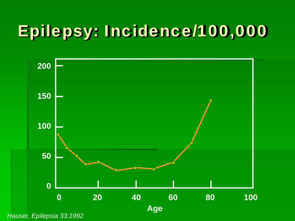
Epilepsy: Incidence/100,000
200
150
100
50
0 0 20 40 60 80 100
Age Hauser, Epilepsia 33:1992
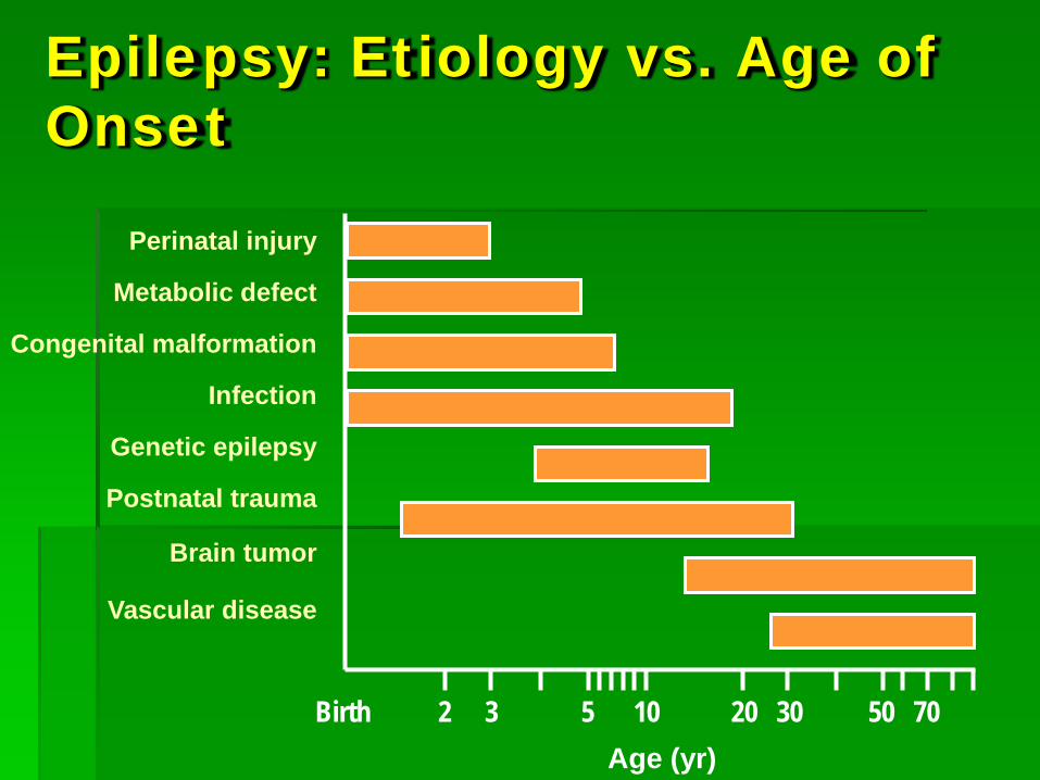
Epilepsy: Etiology vs. Age of Onset
Perinatal injury
Metabolic defect
Congenital malformation
Infection
Genetic epilepsy
Postnatal trauma
Brain tumor
Vascular disease
Birth 2 3 5 10 20 30 50 70 Age (yr)
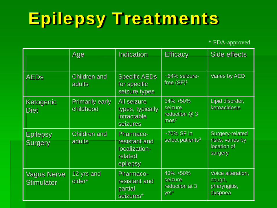
Epilepsy Treatments
Age Indication Efficacy Side effects
AEDs Children and adults
Specific AEDs for specific seizure types
~64% seizure-free (SF)1
Varies by AED
Ketogenic Diet
Primarily early childhood
All seizure types, typically intractable seizures
54% >50% seizure reduction @ 3 mos2
Lipid disorder, ketoacidosis
Epilepsy Surgery
Children and adults
Pharmaco-resistant and localization-related epilepsy
~70% SF in select patients3
Surgery-related risks; varies by location of surgery
Vagus Nerve Stimulator
12 yrs and older*
Pharmaco-resistant and partial seizures*
43% >50% seizure reduction at 3 yrs4
Voice alteration, cough, pharyngitis, dyspnea
* FDA-approved
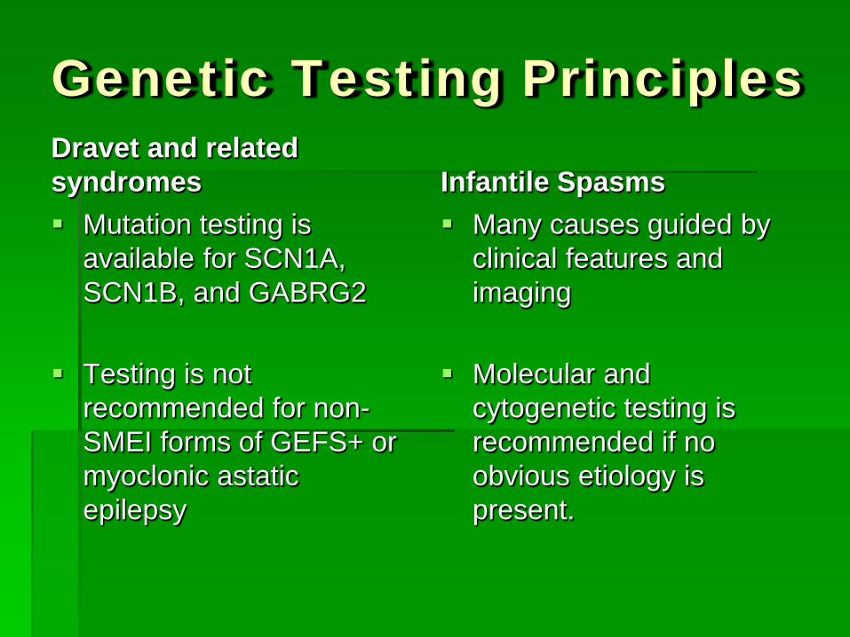
Genetic Testing Principles Dravet and related syndromes Mutation testing is
available for SCN1A, SCN1B, and GABRG2
Testing is not
recommended for non-SMEI forms of GEFS+ or myoclonic astatic epilepsy
Infantile Spasms Many causes guided by
clinical features and imaging
Molecular and
cytogenetic testing is recommended if no obvious etiology is present.
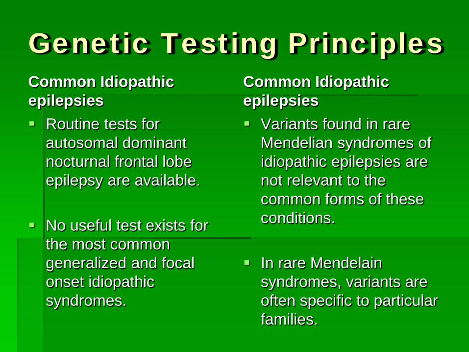
Genetic Testing Principles Common Idiopathic epilepsies Routine tests for
autosomal dominant nocturnal frontal lobe epilepsy are available.
No useful test exists for
the most common generalized and focal onset idiopathic syndromes.
Common Idiopathic epilepsies Variants found in rare
Mendelian syndromes of idiopathic epilepsies are not relevant to the common forms of these conditions.
In rare Mendelain syndromes, variants are often specific to particular families.
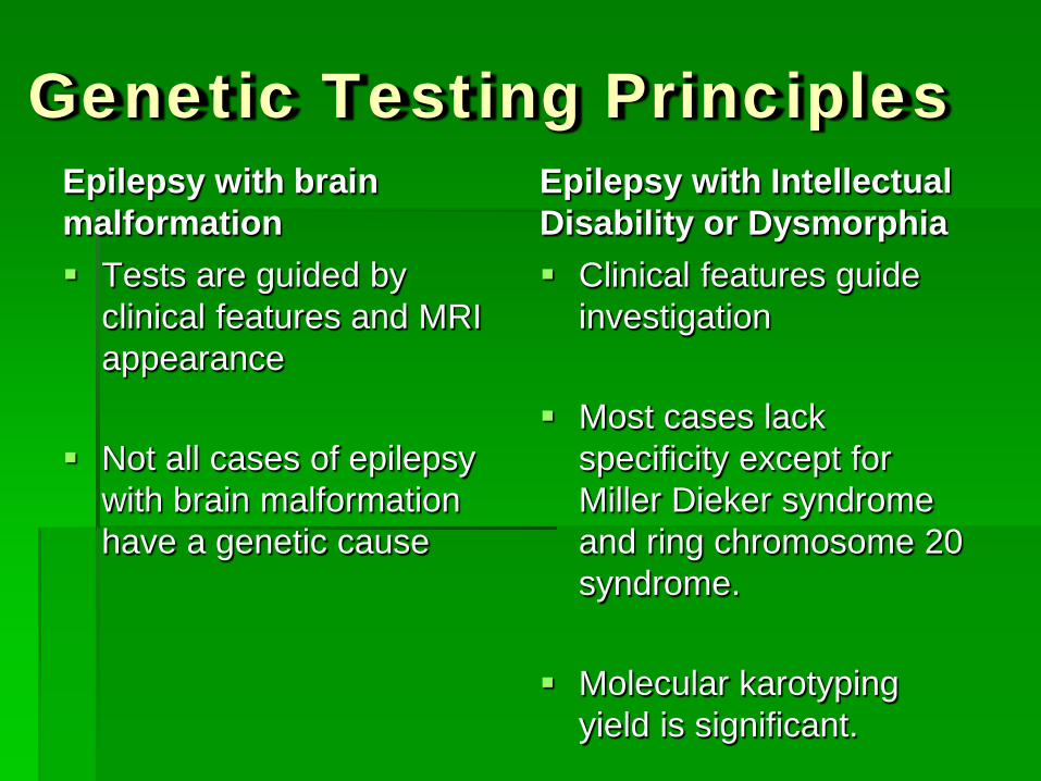
Genetic Testing Principles Epilepsy with brain malformation Tests are guided by
clinical features and MRI appearance
Not all cases of epilepsy
with brain malformation have a genetic cause
Epilepsy with Intellectual Disability or Dysmorphia Clinical features guide
investigation Most cases lack
specificity except for Miller Dieker syndrome and ring chromosome 20 syndrome.
Molecular karotyping
yield is significant.
The Good: Genetic Epilepsy Syndromes
Generalized Epilepsy Syndromes
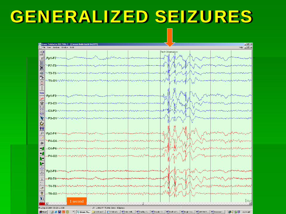
GENERALIZED SEIZURES
1 second
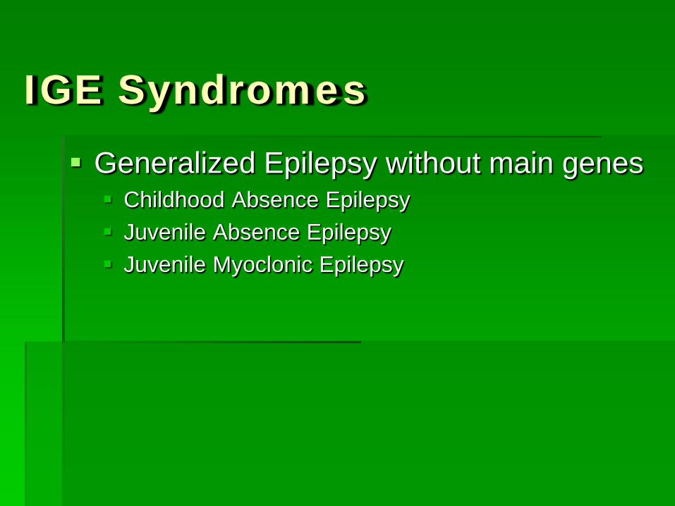
IGE Syndromes Generalized Epilepsy without main genes Childhood Absence Epilepsy Juvenile Absence Epilepsy Juvenile Myoclonic Epilepsy
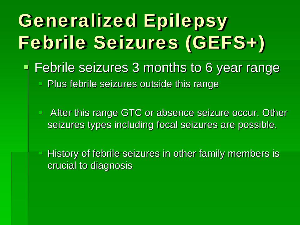
Generalized Epilepsy Febrile Seizures (GEFS+) Febrile seizures 3 months to 6 year range Plus febrile seizures outside this range
After this range GTC or absence seizure occur. Other
seizures types including focal seizures are possible. History of febrile seizures in other family members is
crucial to diagnosis

Generalized Epilepsy Febrile Seizures (GEFS+) Neuro exam is normal but can have
cognitive issues EEG is either normal, irregular
generalized SW or can have focal epileptiform discharges Genetics: SCN1A, SCN1B, SCN2A,
GABRG2 Prognosis: Likely remission by puberty

AD Nocturnal Frontal Lobe Epilepsy 17 different nAChR genes. Mutations in CHRNA4,
CHRNB2, CHRNA2, (CHRNA7)
Onset in late childhood
Presynaptic receptors that regulate GABA and glutamate release
EEG- normal or bifrontal slowing +/- SW
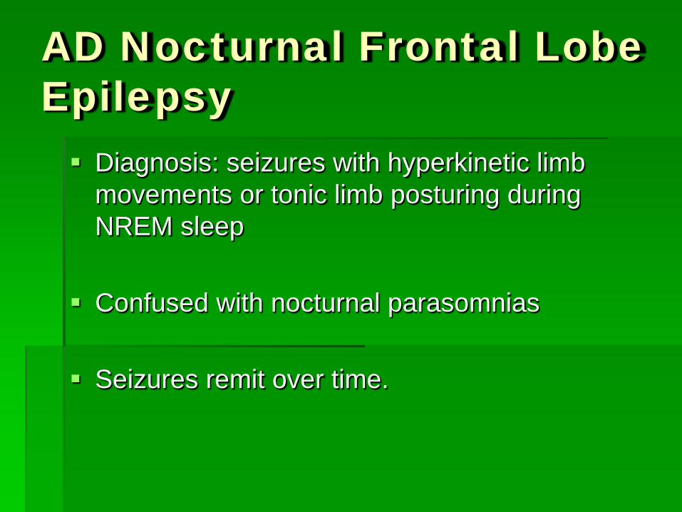
AD Nocturnal Frontal Lobe Epilepsy Diagnosis: seizures with hyperkinetic limb
movements or tonic limb posturing during NREM sleep
Confused with nocturnal parasomnias Seizures remit over time.
Familial TLE AD Lateral Temporal Lobe Epilepsy
Chromosome 10q24 containing LGI1 gene
50% have this mutation
Auditory hallucinations (ringing, humming, whistling, buzzing) plus ictal aphasia. All symptoms are often followed by 2ndarily GTC seizure.
Onset: late adolescence
Familial TLE Familial MTLE Pure form- not associated
with febrile seizure or hippocampal sclerosis
Pure form-linked to chromosome 4q13.2-21.3 in AD incomplete penetrance fashion
Familial forms –MTLE + MTS or MTLE + FS
Linked to more than a dozen genes including interleukins, GRIN1, SCN3A, SCN3B

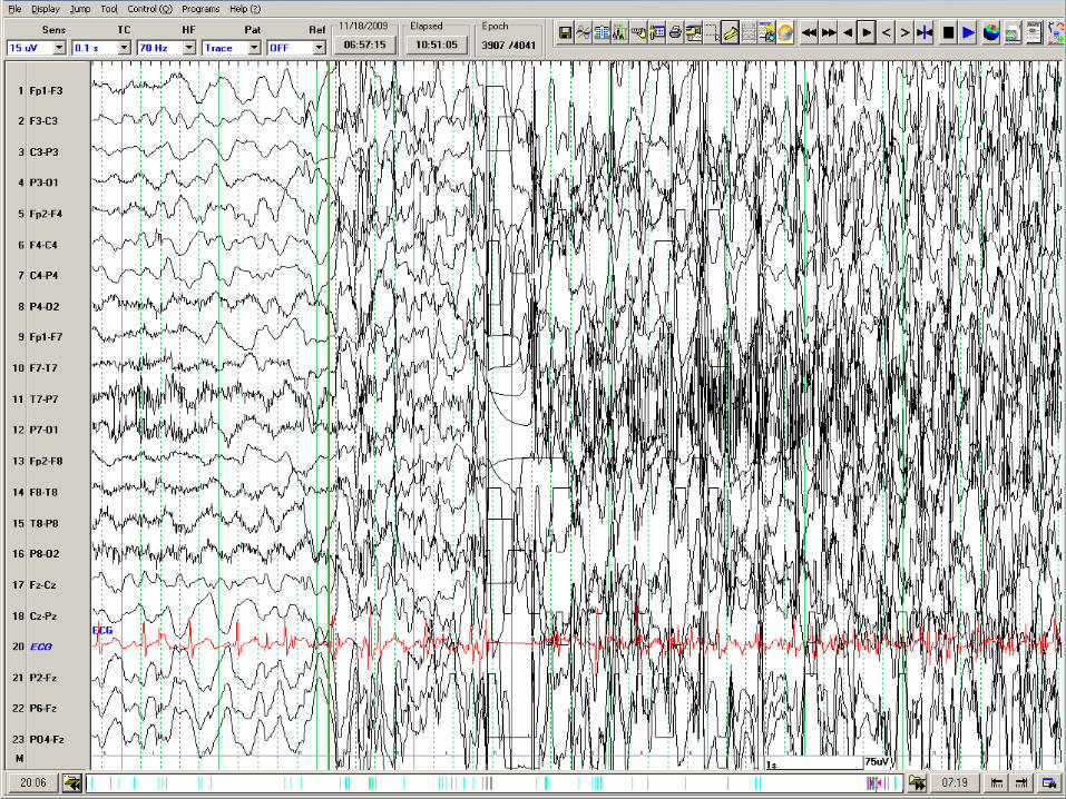
The Bad: Dravet Syndrome
Dravet’s Syndrome
Severe Myoclonic Epilepsy of Infancy First described by Dravet in 1982 Genetics:
Missense and truncation mutations in SCN1A Voltage gated Na channel subunit gene Present in >70% of patients
Dravet’s Syndrome
Severe Myoclonic Epilepsy of Infancy First described by Dravet in 1982 Genetics:
Missense and truncation mutations in SCN1A Voltage gated Na channel subunit gene Present in >70% of patients
Progressive Course: Developmentally normal or mildly delayed Febrile status epilepticus Afebrile generalized and unilateral clonic seizures Development of myoclonus, atypical absence, partial
seizures Significant cognitive and developmental deterioration,
eventually nonverbal and nonambulatory
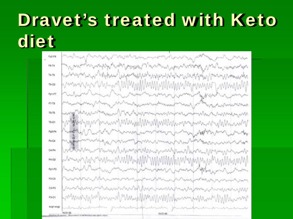
Dravet’s treated with Keto diet
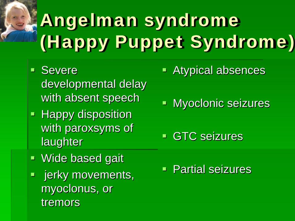
Angelman syndrome (Happy Puppet Syndrome) Severe
developmental delay with absent speech
Happy disposition with paroxsyms of laughter
Wide based gait jerky movements,
myoclonus, or tremors
Atypical absences
Myoclonic seizures
GTC seizures
Partial seizures
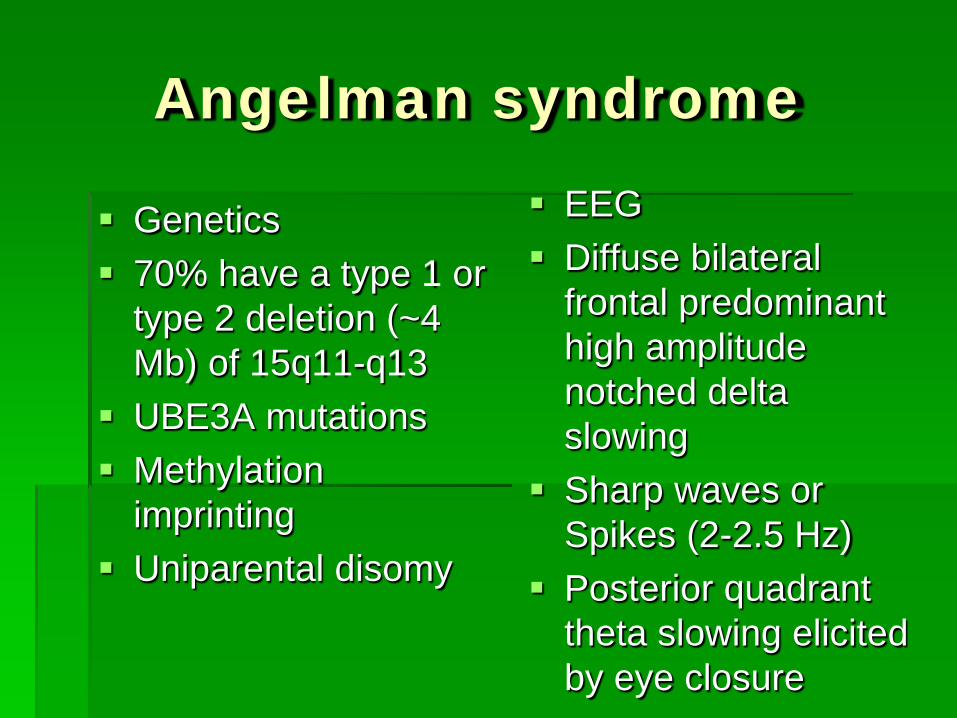
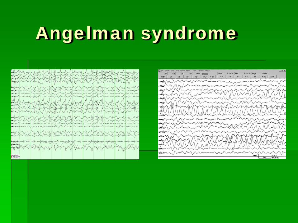
Angelman syndrome
Genetics 70% have a type 1 or
type 2 deletion (~4 Mb) of 15q11-q13
UBE3A mutations Methylation
imprinting Uniparental disomy
EEG Diffuse bilateral
frontal predominant high amplitude notched delta slowing
Sharp waves or Spikes (2-2.5 Hz)
Posterior quadrant theta slowing elicited by eye closure
Angelman syndrome
Rett Syndrome Deletions or
duplications of MECP2 gene
FoxG1 gene Normal development
until between 6-18 months
Regression characterized by ataxia and hand wringing movement
Cognitive decline Autistic features Acquired
microcephaly
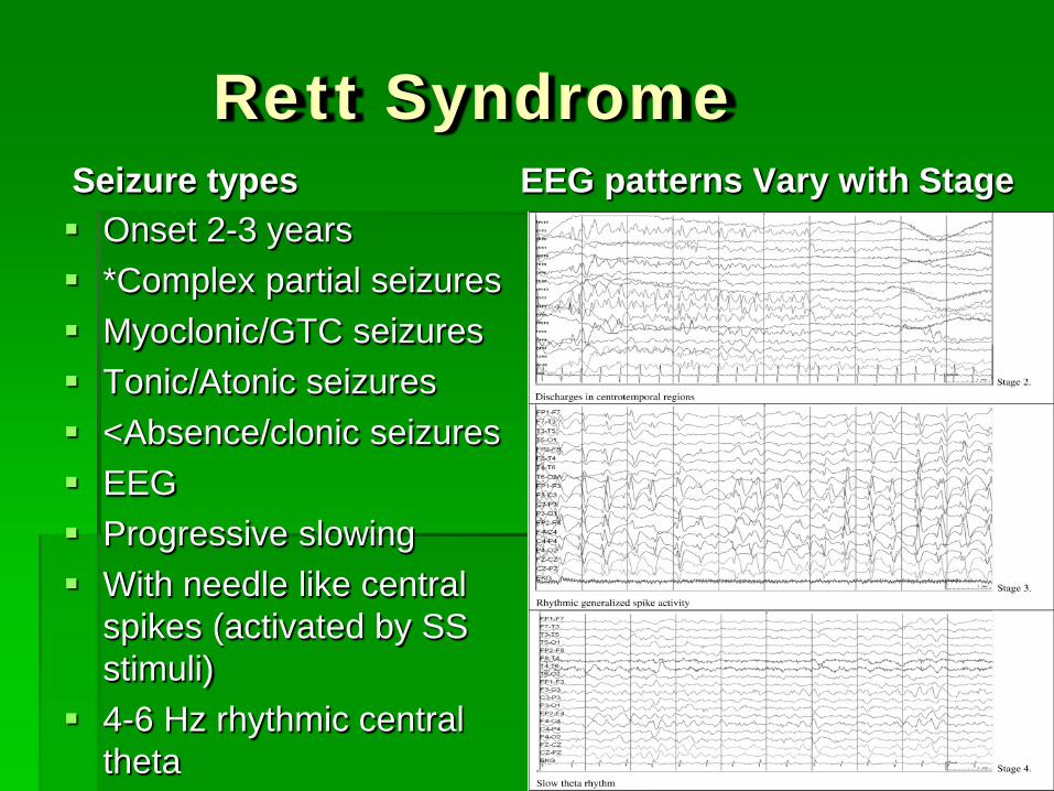
Rett Syndrome Seizure types Onset 2-3 years *Complex partial seizures Myoclonic/GTC seizures Tonic/Atonic seizures <Absence/clonic seizures EEG Progressive slowing With needle like central
spikes (activated by SS stimuli)
4-6 Hz rhythmic central theta
EEG patterns Vary with Stage
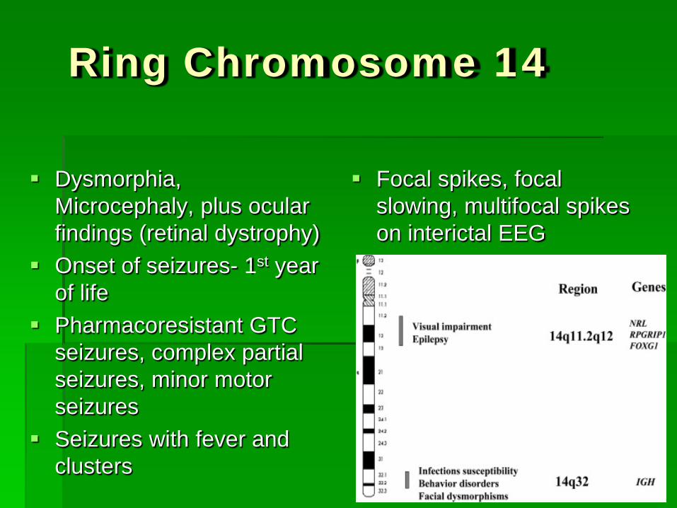
Ring Chromosome 14
Dysmorphia, Microcephaly, plus ocular findings (retinal dystrophy)
Onset of seizures- 1st year of life
Pharmacoresistant GTC seizures, complex partial seizures, minor motor seizures
Seizures with fever and clusters
Focal spikes, focal slowing, multifocal spikes on interictal EEG
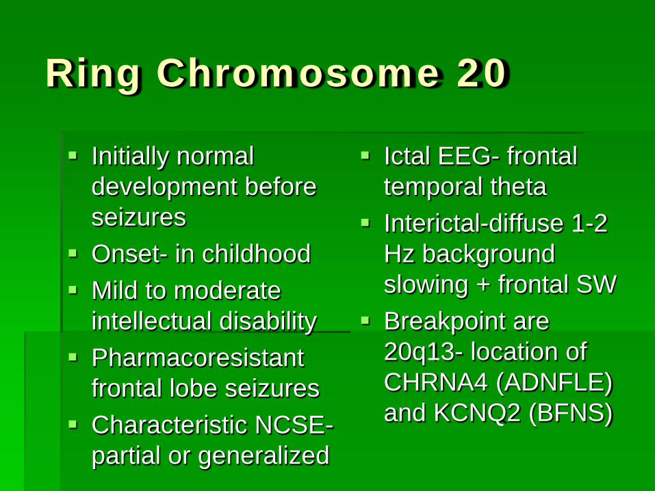
Ring Chromosome 20
Initially normal development before seizures
Onset- in childhood Mild to moderate
intellectual disability Pharmacoresistant
frontal lobe seizures Characteristic NCSE-
partial or generalized
Ictal EEG- frontal temporal theta
Interictal-diffuse 1-2 Hz background slowing + frontal SW
Breakpoint are 20q13- location of CHRNA4 (ADNFLE) and KCNQ2 (BFNS)
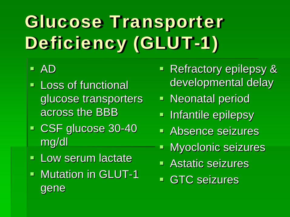
Glucose Transporter Deficiency (GLUT-1) AD Loss of functional
glucose transporters across the BBB
CSF glucose 30-40 mg/dl
Low serum lactate Mutation in GLUT-1
gene
Refractory epilepsy & developmental delay
Neonatal period Infantile epilepsy Absence seizures Myoclonic seizures Astatic seizures GTC seizures
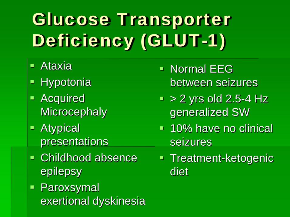
Glucose Transporter Deficiency (GLUT-1) Ataxia Hypotonia Acquired
Microcephaly Atypical
presentations Childhood absence
epilepsy Paroxsymal
exertional dyskinesia
Normal EEG between seizures
> 2 yrs old 2.5-4 Hz generalized SW
10% have no clinical seizures
Treatment-ketogenic diet

The UGLY: Mitochondrial Disorders & Progressive Myoclonic Epilepsies
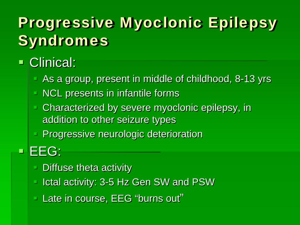
Progressive Myoclonic Epilepsy Syndromes Clinical: As a group, present in middle of childhood, 8-13 yrs NCL presents in infantile forms Characterized by severe myoclonic epilepsy, in
addition to other seizure types Progressive neurologic deterioration
EEG: Diffuse theta activity Ictal activity: 3-5 Hz Gen SW and PSW Late in course, EEG “burns out”
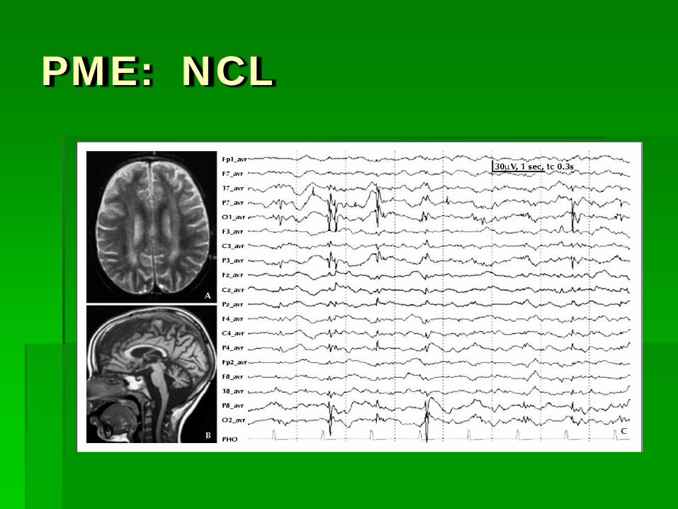
PME: NCL
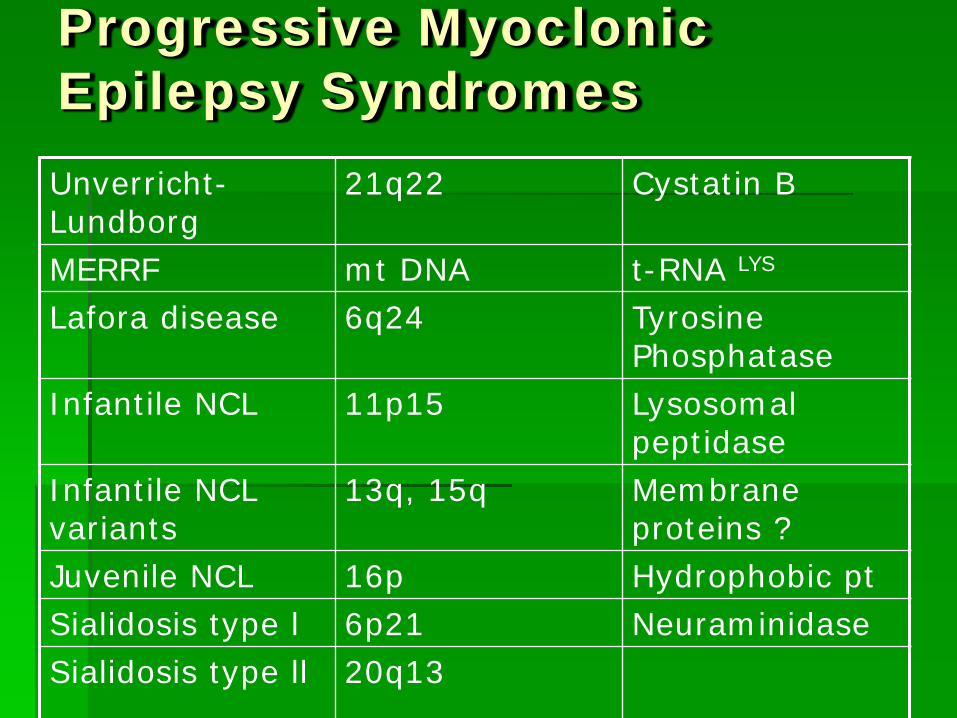
Progressive Myoclonic Epilepsy Syndromes Unverricht-Lundborg
21q22 Cystatin B
MERRF mt DNA t-RNA LYS
Lafora disease 6q24 Tyrosine Phosphatase
Infantile NCL 11p15 Lysosomal peptidase
Infantile NCL variants
13q, 15q Membrane proteins ?
Juvenile NCL 16p Hydrophobic pt Sialidosis type l 6p21 Neuraminidase Sialidosis type ll 20q13
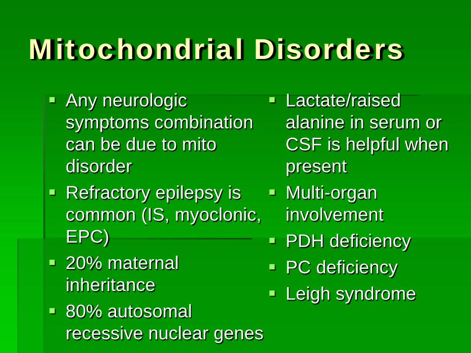
Mitochondrial Disorders Any neurologic
symptoms combination can be due to mito disorder
Refractory epilepsy is common (IS, myoclonic, EPC)
20% maternal inheritance
80% autosomal recessive nuclear genes
Lactate/raised alanine in serum or CSF is helpful when present
Multi-organ involvement
PDH deficiency PC deficiency Leigh syndrome
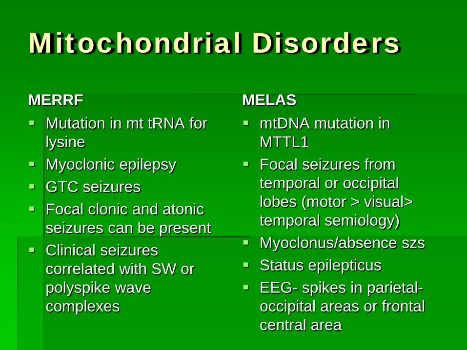
Mitochondrial Disorders
MERRF Mutation in mt tRNA for
lysine Myoclonic epilepsy GTC seizures Focal clonic and atonic
seizures can be present Clinical seizures
correlated with SW or polyspike wave complexes
MELAS mtDNA mutation in
MTTL1 Focal seizures from
temporal or occipital lobes (motor > visual> temporal semiology)
Myoclonus/absence szs Status epilepticus EEG- spikes in parietal-
occipital areas or frontal central area
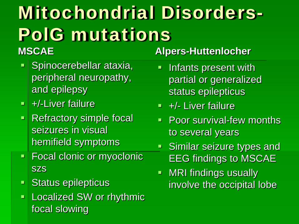
Mitochondrial Disorders-PolG mutations
Alpers-Huttenlocher Infants present with
partial or generalized status epilepticus
+/- Liver failure Poor survival-few months
to several years Similar seizure types and
EEG findings to MSCAE MRI findings usually
involve the occipital lobe
MSCAE Spinocerebellar ataxia,
peripheral neuropathy, and epilepsy
+/-Liver failure Refractory simple focal
seizures in visual hemifield symptoms
Focal clonic or myoclonic szs
Status epilepticus Localized SW or rhythmic
focal slowing
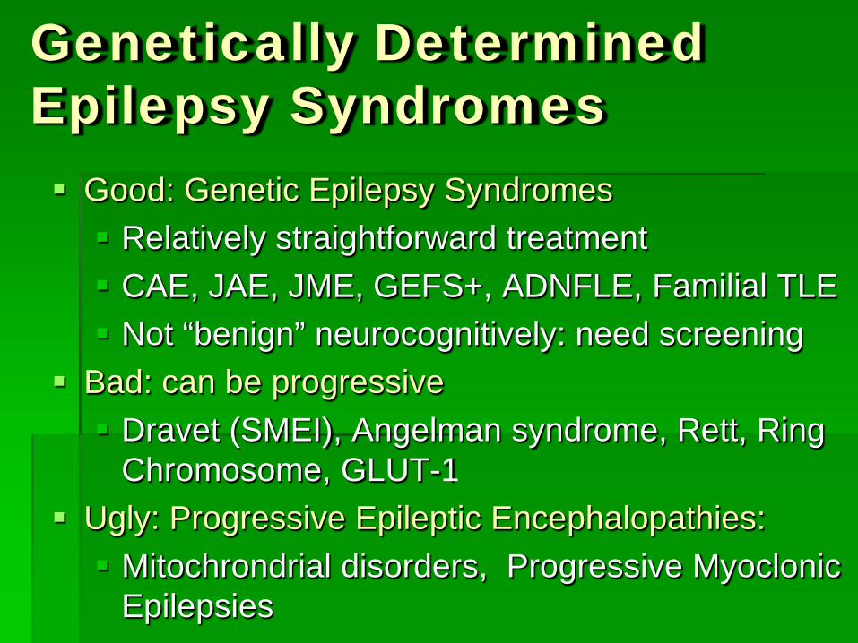
Genetically Determined Epilepsy Syndromes Good: Genetic Epilepsy Syndromes Relatively straightforward treatment CAE, JAE, JME, GEFS+, ADNFLE, Familial TLE Not “benign” neurocognitively: need screening
Bad: can be progressive Dravet (SMEI), Angelman syndrome, Rett, Ring
Chromosome, GLUT-1 Ugly: Progressive Epileptic Encephalopathies: Mitochrondrial disorders, Progressive Myoclonic
Epilepsies

Quiz

Questions?