Embed Size (px)
Citation preview
Genetic and Epigenetic Characterization of
Colorectal and Endometrial Cancer
Shannon A. Kuismanen
Department of Medical Genetics Biomedicum
University of Helsinki Finland
To be publicly discussed, with the permission of the Medical Faculty of the University of Helsinki, in the lecture hall 2, Biomedicum Helsinki,
Haartmaninkatu 8, on 9 June 2006, at 12 noon.
2
Supervised by: Professor Päivi Peltomäki, MD, PhD Department of Medical Genetics University of Helsinki Helsinki, Finland Reviewed by: Maija Wessman, PhD, Doc Academy Research Fellow Folkhälsän Research Center and Finnish Genome Center Helsinki University Hospital Helsinki, Finland Ari Ristimäki, MD, PhD, Doc Pathology/HUSLAB Helsinki University Central Hospital Molecular and Cancer Biology Research Program Biomedicum Helsinki University of Helsinki Helsinki, Finland Official opponent: Anne Kallioniemi, MD, PhD Professor of Cancer Genetics Institute of Medical Technology University of Tampere Tampere, Finland ISBN 952-92-0382-9 (paperback) ISBN 952-10-3169-7 (PDF) http://ethesis.helsinki.fi Helsinki University Press 2006

3
THE ROAD NOT TAKEN
Two roads diverged in a yellow wood,
And sorry I could not travel both
And be one traveler, long I stood
And looked down one as far as I could
To where it bent in the undergrowth;
Then took the other, as just as fair,
And having perhaps the better claim,
Because it was grassy and wanted wear;
Though as for that the passing there
Had worn them really about the same,
And both that morning equally lay
In leaves no step had trodden black.
Oh, I kept the first for another day!
Yet knowing how way leads on to way,
I doubted if I should ever come back.
I shall be telling this with a sigh
Somewhere ages and ages hence:
Two roads diverged in a wood, and I–
I took the one less traveled by,
And that has made all the difference.
– Robert Frost, 1916
This thesis is dedicated to the one who took the road less traveled with me.
4
TABLE OF CONTENTS
LIST OF ORIGINAL PUBLICATIONS..............................................................................................6 1. REVIEW OF THE LITERATURE...........................................................................................11
1.1. CANCER................................................................................................................................11 1.1.1. Cancer and Cancer-Associated Genes ...........................................................................11 1.1.2. Colorectal Cancer ..........................................................................................................12 1.1.3. Endometrial Cancer........................................................................................................15
1.2. HEREDITARY NON-POLYPOSIS COLORECTAL CANCER (HNPCC) AS A CANCER MODEL ........15 1.2.1. History and Definitions...................................................................................................15 1.2.2. Genetic Instability...........................................................................................................17 1.2.3. Mechanisms of Microsatellite Instability........................................................................19 1.2.4. Tumor Spectrum .............................................................................................................19
1.3. GENETIC BASIS OF ENDOMETRIAL CANCER...........................................................................20 1.4. DNA METHYLATION IN NORMAL AND CANCER DEVELOPMENT .............................................21
1.4.1. Vertebrate Methylation...................................................................................................21 1.4.2. DNA Methyltransferases.................................................................................................22 1.4.3. Role of Methylation in Normal Development .................................................................23 1.4.4. DNA Methylation and Evolution.....................................................................................24 1.4.5. DNA Methylation and Cancer ........................................................................................25 1.4.5.1. Genetic System...........................................................................................................25 1.4.5.2. Epigenetic System ......................................................................................................26 1.4.6. Methods to Study DNA Methylation ...............................................................................27
2. AIMS OF STUDY.......................................................................................................................29 3. MATERIALS AND METHODS ...............................................................................................30
3.1. PATIENT SAMPLES (I-IV) ......................................................................................................30 3.2. DNA METHYLATION (I AND II) ..............................................................................................31 3.3. IMMUNOHISTOCHEMISTRY (I-III) .........................................................................................31 3.4. MUTATION ANALYSIS (II-III) ................................................................................................32
3.4.1. MLH1 and MSH2 Somatic Mutations (II) ......................................................................32 3.4.2. PTEN Mutations (III)......................................................................................................32
3.5. LOSS OF HETEROZYGOSITY (LOH) ANALYSIS (II) .................................................................32 3.6. MICROSATELLITE INSTABILITY ANALYSIS (IV) .....................................................................33 3.7. STATISTICAL ANALYSIS (I-IV) ..............................................................................................33
4. RESULTS ....................................................................................................................................34 4.1. DESCRIPTION OF TWO MAIN EPIGENETIC PHENOTYPES FOR COLORECTAL CANCER (I) ..........34
4.1.1. DNA Methylation Patterns and Correlation with Microsatellite Instability...................34 4.1.2. Methylation Specificity ...................................................................................................36 4.1.3. Correlation with MLH1 Protein Expression...................................................................37
4.2. BASIS OF MMR GENE INACTIVATION IN SPORADIC AND HEREDITARY COLORECTAL CANCERS (I, II) 38
4.2.1. Sporadic Colorectal Cancer ...........................................................................................38 4.2.2. Colorectal Cancer with Inherited MMR Deficiency (HNPCC) ......................................39
4.3. PTEN IN SPORADIC AND HEREDITARY ENDOMETRIAL CANCERS (III, IV) .............................40 4.4. PATTERNS OF MICROSATELLITE INSTABILITY IN ENDOMETRIAL AND COLORECTAL TUMORS FROM INDIVIDUALS WITH IDENTICAL PREDISPOSING MUTATIONS IN MMR GENES (IV)........................41 4.5. CLINICOPATHOLOGICAL CORRELATIONS (I, IV) ...................................................................42
5. DISCUSSION ..............................................................................................................................44 5.1. ROLE OF DNA METYLATION IN COLORECTAL TUMORIGENESIS (I).........................................44 5.2. DIFFERENCES AND SIMILARITIES OF TUMORIGENIC MECHANISMS IN SPORADIC VS HEREDITARY TUMORS WITH MICROSATELLITE INSTABILITY (II-IV)....................................................47 5.3. INSIGHTS INTO THE GENETIC BASIS OF THE HNPCC TUMOR SPECTRUM (I-IV) .......................50
6. CONCLUSIONS .........................................................................................................................52

5
7. FUTURE PROSPECTS..............................................................................................................53 7.1. COLORECTAL CARCINOMA (I-II) ...........................................................................................53 7.2. ENDOMETRIAL CARCINOMA (III-IV) ....................................................................................53
8. ACKNOWLEDGEMENTS........................................................................................................54 9. REFERENCES............................................................................................................................56
6
LIST OF ORIGINAL PUBLICATIONS
I. Kuismanen S, Holmberg M, Salovaara R, Aaltonen L, de la Chapelle A, Nyström-Lahti M, Peltomäki P. Epigenetic phenotypes distinguish microsatellite-stable and –unstable colorectal cancers. PNAS 1999; 96(22): 12661-12666
II. Kuismanen S, Holmberg M, Salovaara R, de la Chapelle A, Peltomäki P. Genetic and epigenetic modification of MLH1 accounts for a major share of microsatellite-unstable colorectal cancers. Am J Pathol 2000; 156(5): 1773-1779
III. Zhou XP, Kuismanen S, Nyström-Lahti M, Peltomäki P, Eng C. Distinct
PTEN mutational spectra in hereditary non-polyposis colon cancer syndrome-related endometrial carcinomas compared to sporadic microsatellite unstable tumors. Hum Mol Genet 2002; 11(4): 445-450
IV. Kuismanen S, Moisio AL, Schweizer P, Truninger K, Salovaara R, Arola
J, Butzow R, Jiricny J, Nyström-Lahti M, Peltomäki P. Endometrial and colorectal tumors from patients with hereditary nonpolyposis colon cancer display different patterns of microsatellite instability. Am J Pathol 2002; 160(6): 1953-1958
7
ABBREVIATIONS 5’mC 5-methylcytosine
5-aza-dC 5’-aza-2’ deoxycytidine
APC adenomatous polyposis coli
bp base pairs
CFS cancer family syndrome
CIN chromosomal instability
CpA cytosine-adenine dinucleotide
CpG cytosine-guanine dinucleotide
COBRA combined bisulphite restriction analysis
CRC colorectal carcinoma
C-terminal carboxyl terminal
DNA deoxyribonucleic acid
DNA-mtase DNA-methyltransferase
DNMT1 DNA- methyltransferase 1
DNMT2 DNA-methyltransferase 2
DNMT3 DNA-methyltransferase 3
dsDNA double strand DNA
EC endometrial carcinoma
FAP familial adenomatous polyposis
HNPCC hereditary non-polyposis colorectal cancer
IHC immunohistochemical analysis
LOH loss of heterozygosity
MBD methyl-CpG binding domain
MeCP1 methyl-CpG binding protein 1
MeCP2 methyl-CpG binding protein 2
MIN microsatellite instabilty
MLH1 mut L homologue 1
MLH1/1 MLH1 fragment 1 (methylation site -567)
MLH1/2 MLH1 fragment 2 (methylation site -527)
MLH1/3 MLH1 fragment 3 (methylation sites -347 and -341)
MMR mismatch repair
MSH2 mut S homologue 2
8
MSI microsatellite instability
MSI–H microsatellite instability–high
MSP methylation-specific PCR
MSS microsatellite stable
MSS microsatellite stable
NS not significant
N-terminal amino terminal
PCR polymerase chain reaction
PTEN phosphatase and tensin homolog
RB retinoblastoma
RLGS restriction landmark genomic scanning
TpG thymine-guanine dinucleotide
vs versus
9
INTRODUCTION
Three major types of cancer exist: sarcoma, lymphoma and leukemia, and
carcinoma. Sarcoma is cancer originating in the connective tissue, lymphoma
and leukemia in the lymphatic and blood systems, and carcinoma in the
epithelium. All three types of cancer are considered genetic diseases. A series of
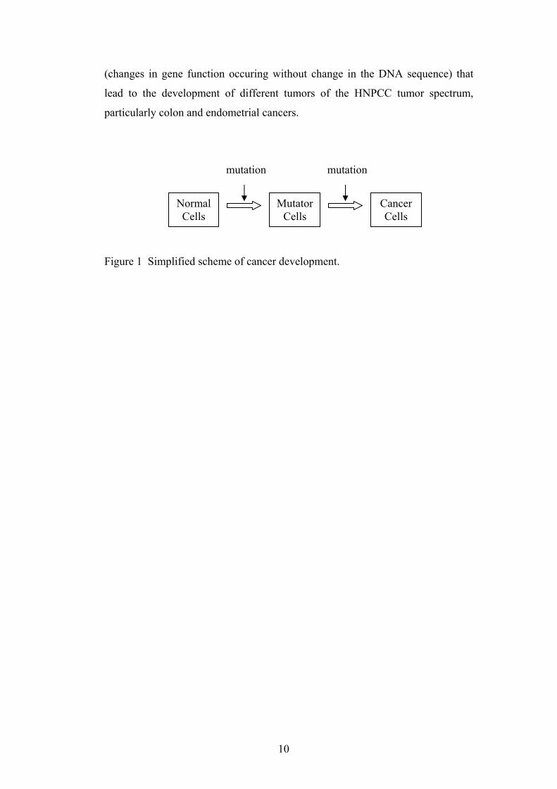
mistakes, called mutations, occur in the DNA of genes causing cancer to develop
(fig. 1). These mutations can be inherited, passed down from parent to offspring,
or acquired during an organism’s lifetime. An inherited mutation is found in all
the cells of an organism, including the germline cells and the disease is
considered heritable. An acquired mutation only occurs in isolated somatic cells,
and is not transmitted to the next generation. Acquired mutations are a result of
different internal or external factors, including, but not limited to, environmental
pollutants, unhealthy lifestyles, and increasing longevity.
Cancer is among the leading causes of death in the western world today, with
colorectal cancer being one of the three most common types of cancer in both
men and women (Finnish Cancer Registry; NAACCR). An estimated 5-15% of
all cancers that occur each year are accounted for by hereditary forms. Of the
hereditary forms of colorectal cancer, hereditary non-polyposis colorectal cancer
(HNPCC) is the most common and accounts for an estimated 0.5-13% (Lynch
and de la Chapelle 2003). This is an autosomal dominant disorder and
susceptibility is associated with germline mutations in four genes with DNA
mismatch repair (MMR) function, a further two genes have been proposed but
have not so far shown any clinical significance (Peltomäki 2005). In addition to
gastrointestinal carcinomas, HNPCC is characterized by familial accumulation of
endometrial, gastric, urological, and ovarian tumors. Individuals with HNPCC
have a 70% chance of developing colon cancer during their lifetime, with 45
being the average age of diagnosis. In the general population, colon cancer
occurs 20 to 30 years later and the lifetime incidence is approximately 5%. Even
though 75% of HNPCC cases are known to be caused by germline mutations in
the MMR genes, the remaining cases are still molecularly unexplained.
The objective of this work was to clarify the mechanisms that are associated
with tumor development in HNPCC vs sporadic cases. Of particular interest
were the genetic events (changes in the DNA sequence) and epigenetic events
10
(changes in gene function occuring without change in the DNA sequence) that
lead to the development of different tumors of the HNPCC tumor spectrum,
particularly colon and endometrial cancers.
Figure 1 Simplified scheme of cancer development.
Normal Cells
Mutator Cells
Cancer Cells
mutation mutation
11
1. REVIEW OF THE LITERATURE
1.1. CANCER
1.1.1. Cancer and Cancer-Associated Genes
Cancer is a genetic disease in which the progression from normal
cells to malignant ones is a multi-step process. In cancer, cells lose their
ability for normal, regulated cell growth and apoptosis, and without these
regulations cells continue to grow and mutate into invasive tumors.
Different types of genes play a role in cell regulation, including
oncogenes, tumor suppressors, and mismatch repair genes.
Oncogenes are modified, mutated forms of proto-oncogenes and can
increase the chance of normal cells developing into tumor cells. Proto-
oncogenes code for proteins that regulate cell growth and differentiation.
Tumor suppressor genes, as their name implies, lessen the chance of cells
becoming malignant and forming tumors. These genes code for proteins
that typically either dampen cell cycle progression or promote apoptosis.
Unlike oncogenes, which are activated even by a small change, tumor
suppressor genes follow a “two-hit hypothesis” in which both alleles
must be affected for the deactivation (Knudson 1971). Mismatch repair
(MMR) genes also follow this “two-hit hypothesis” and can be
considered to be a type of tumor suppressor. As the name suggests, these
genes repair DNA base pair mismatches and help maintain normal base
pair matches.
A minor fraction of cancer, about 5% to 15%, is hereditary but the
vast majority occurs spontaneously. In hereditary cancer, typically one
of the two alleles of a given gene is mutated in all cells from birth leading
to the early onset of cancer. In sporadic cancer, the mutations are
acquired over time causing this form of cancer to usually occur later in
life, through various external factors. These factors include, but are not
limited to, diet, environment, UV-radiation, and viral infections
(Zuckerman 1979; Marx 1986; Marwick 1990; Graham 2000).
12
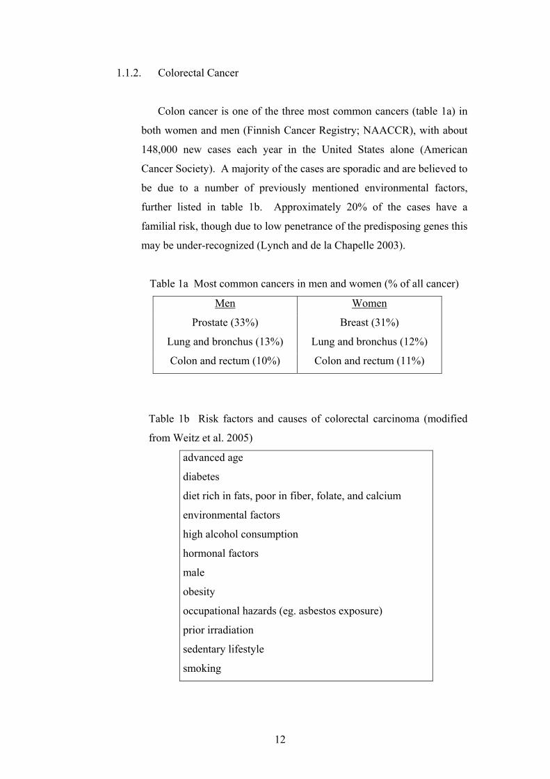
1.1.2. Colorectal Cancer
Colon cancer is one of the three most common cancers (table 1a) in
both women and men (Finnish Cancer Registry; NAACCR), with about
148,000 new cases each year in the United States alone (American
Cancer Society). A majority of the cases are sporadic and are believed to
be due to a number of previously mentioned environmental factors,
further listed in table 1b. Approximately 20% of the cases have a
familial risk, though due to low penetrance of the predisposing genes this
may be under-recognized (Lynch and de la Chapelle 2003).
Table 1a Most common cancers in men and women (% of all cancer)
Men Women
Prostate (33%) Breast (31%)
Lung and bronchus (13%) Lung and bronchus (12%)
Colon and rectum (10%) Colon and rectum (11%)
Table 1b Risk factors and causes of colorectal carcinoma (modified
from Weitz et al. 2005)
advanced age
diabetes
diet rich in fats, poor in fiber, folate, and calcium
environmental factors
high alcohol consumption
hormonal factors
male
obesity
occupational hazards (eg. asbestos exposure)
prior irradiation
sedentary lifestyle
smoking
13
The progression of normal cells into metastic tumor cells is a multi-
stepped process occurring over a long period of time, usually 10-15 years
(Vogelstein and Kinzler 1993). Renan estimated in 1993 that a total of
four to seven mutations, providing a clonal advantage, are needed for
malignancy development. This progression process is called the
adenoma-carcinoma sequence (Fearon and Vogelstein 1990), which
appears to be initiated by mutations in the tumor suppressor APC gene.
This gene acts as a form of gatekeeper in colorectal cancer development.
Additional mutations in the tumor suppressors, DCC, DPC4/Smad4, p53,
nm32, and TGFβRII, as well as in the oncogenes, K-ras, c-myc,
HER2/neu (erbB-2), c-src, are involved in this process. Although
mutations of these genes occur in a favored sequence, it is their total
accumulation rather than chronological occurrence that determines the
biological properties of tumors (Cho and Vogelstein 1992).
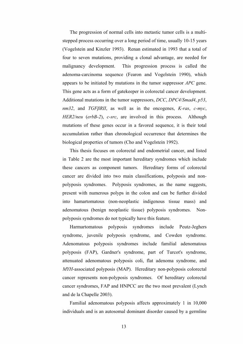
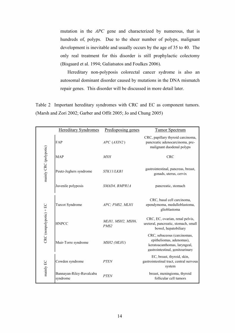
This thesis focuses on colorectal and endometrial cancer, and listed
in Table 2 are the most important hereditary syndromes which include
these cancers as component tumors. Hereditary forms of colorectal
cancer are divided into two main classifications, polyposis and non-
polyposis syndromes. Polyposis syndromes, as the name suggests,
present with numerous polyps in the colon and can be further divided
into hamartomatous (non-neoplastic indigenous tissue mass) and
adenomatous (benign neoplastic tissue) polyposis syndromes. Non-
polyposis syndromes do not typically have this feature.
Harmartomatous polyposis syndromes include Peutz-Jeghers
syndrome, juvenile polyposis syndrome, and Cowden syndrome.
Adenomatous polyposis syndromes include familial adenomatous
polyposis (FAP), Gardner's syndrome, part of Turcot's syndrome,
attenuated adenomatous polyposis coli, flat adenoma syndrome, and
MYH-associated polyposis (MAP). Hereditary non-polyposis colorectal
cancer represents non-polyposis syndromes. Of hereditary colorectal
cancer syndromes, FAP and HNPCC are the two most prevalent (Lynch
and de la Chapelle 2003).
Familial adenomatous polyposis affects approximately 1 in 10,000
individuals and is an autosomal dominant disorder caused by a germline
14
mutation in the APC gene and characterized by numerous, that is
hundreds of, polyps. Due to the sheer number of polyps, malignant
development is inevitable and usually occurs by the age of 35 to 40. The
only real treatment for this disorder is still prophylactic colectomy
(Bisgaard et al. 1994; Galiatsatos and Foulkes 2006).
Hereditary non-polyposis colorectal cancer sydrome is also an
autosomal dominant disorder caused by mutations in the DNA mismatch
repair genes. This disorder will be discussed in more detail later.
Table 2 Important hereditary syndromes with CRC and EC as component tumors.
(Marsh and Zori 2002; Garber and Offit 2005; Jo and Chung 2005)
Hereditary Syndromes Predisposing genes Tumor Spectrum
FAP APC (AXIN2 )CRC, papillary thyroid carcinoma, pancreatic adenocarcinoma, pre-
malignant duodenal polyps
MAP MYH CRC
Peutz-Jeghers syndrome STK11/LKB1 gastrointestinal, pancreas, breast, gonads, uterus, cervix
Juvenile polyposis SMAD4, BMPR1A pancreatic, stomach
Turcot Syndrome APC; PMS2, MLH1CRC, basal cell carcinoma,
ependymoma, medulloblastoma, glioblastoma
HNPCC MLH1, MSH2, MSH6, PMS2
CRC, EC, ovarian, renal pelvis, ureteral, pancreatic, stomach, small
bowel, hepatobiliary
Muir-Torre syndrome MSH2 (MLH1)
CRC, sebaceous (carcinomas, epitheliomas, adenomas),
keratoacanthomas, laryngeal, gastrointestinal, genitourinary
Cowden syndrome PTENEC, breast, thyroid, skin,
gastrointestinal tract, central nervous system
Bannayan-Riley-Ruvalcaba syndrome PTEN breast, meningioma, thyroid
follicular cell tumors
mai
nly
CR
C (p
olyp
otic
)C
RC
(non
poly
potic
) + E
Cm
ainl
y EC
15
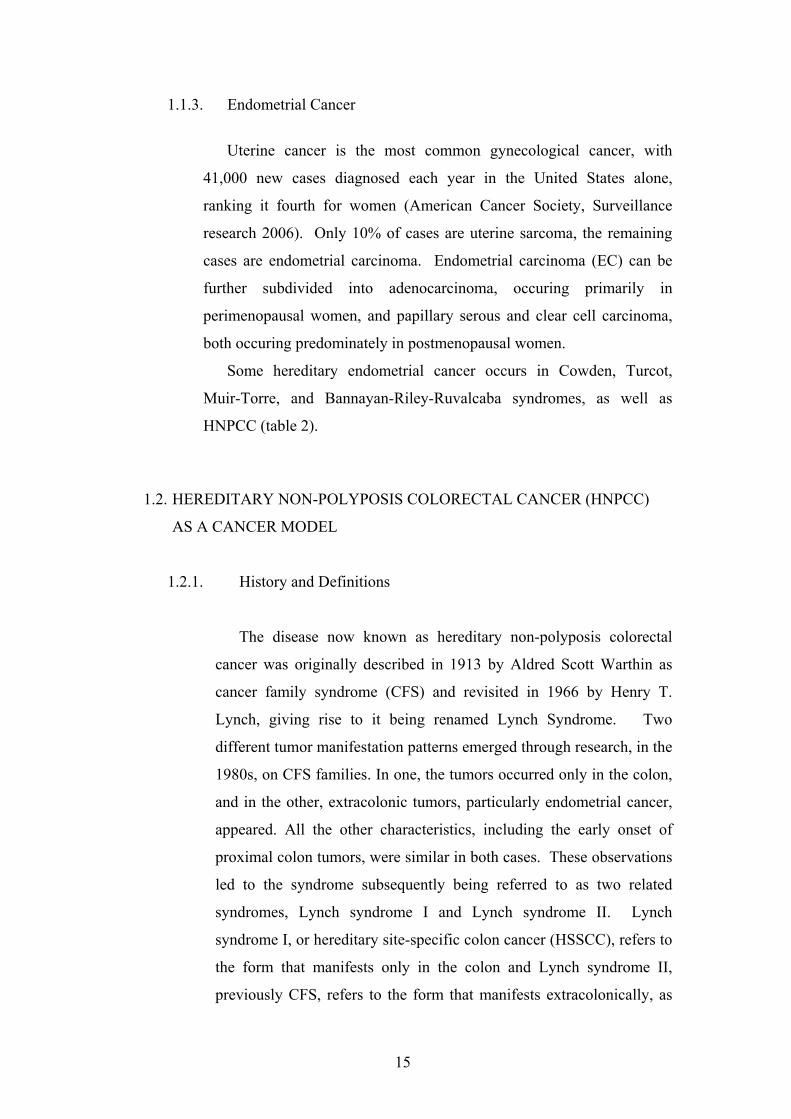
1.1.3. Endometrial Cancer
Uterine cancer is the most common gynecological cancer, with
41,000 new cases diagnosed each year in the United States alone,
ranking it fourth for women (American Cancer Society, Surveillance
research 2006). Only 10% of cases are uterine sarcoma, the remaining
cases are endometrial carcinoma. Endometrial carcinoma (EC) can be
further subdivided into adenocarcinoma, occuring primarily in
perimenopausal women, and papillary serous and clear cell carcinoma,
both occuring predominately in postmenopausal women.
Some hereditary endometrial cancer occurs in Cowden, Turcot,
Muir-Torre, and Bannayan-Riley-Ruvalcaba syndromes, as well as
HNPCC (table 2).
1.2. HEREDITARY NON-POLYPOSIS COLORECTAL CANCER (HNPCC)
AS A CANCER MODEL
1.2.1. History and Definitions
The disease now known as hereditary non-polyposis colorectal
cancer was originally described in 1913 by Aldred Scott Warthin as
cancer family syndrome (CFS) and revisited in 1966 by Henry T.
Lynch, giving rise to it being renamed Lynch Syndrome. Two
different tumor manifestation patterns emerged through research, in the
1980s, on CFS families. In one, the tumors occurred only in the colon,
and in the other, extracolonic tumors, particularly endometrial cancer,
appeared. All the other characteristics, including the early onset of
proximal colon tumors, were similar in both cases. These observations
led to the syndrome subsequently being referred to as two related
syndromes, Lynch syndrome I and Lynch syndrome II. Lynch
syndrome I, or hereditary site-specific colon cancer (HSSCC), refers to
the form that manifests only in the colon and Lynch syndrome II,
previously CFS, refers to the form that manifests extracolonically, as
16
well as in the colon (Lynch et al. 1985). Unlike other cancer
syndromes, Lynch syndrome (I and II) presented physicians with a
dilemma in diagnosis, due to the wide range of symptoms, as well as
the need for a well-documented family history. In 1991, Vasen et al.
put forth the Amsterdam criteria as an aid for diagnosis of Lynch
syndrome I, further modifying it in 1999 to include Lynch syndrome II
(tables 3a and 3b).
The term hereditary non-polyposis colorectal cancer was first
coined in 1985 by Lynch himself; however, the work he did focused
mainly on colon cancer and ignored the fact that many families have an
increase in endometrial cancer occurrence as well. Many feel that this
name is misleading, due to its exclusion of endometrial cancer and its
inference that there are never polyps in this syndrome. Subsequently, a
consensus was reached at the last two Bethesda conferences that the
name HNPCC is no longer the preferred one. The name suggested
now is Lynch Syndrome (Boland 2005; Lynch 2005).
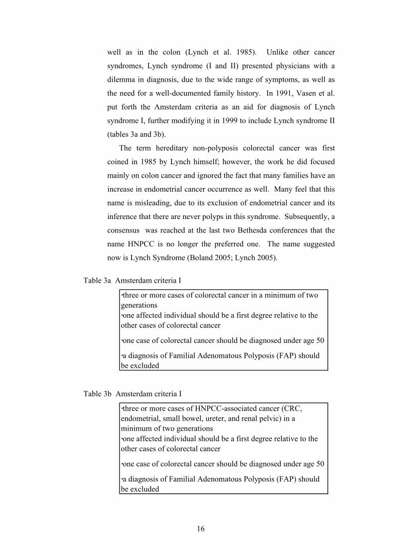
Table 3a Amsterdam criteria I
·three or more cases of colorectal cancer in a minimum of two generations·one affected individual should be a first degree relative to the other cases of colorectal cancer
·one case of colorectal cancer should be diagnosed under age 50
·a diagnosis of Familial Adenomatous Polyposis (FAP) should be excluded
Table 3b Amsterdam criteria I
·three or more cases of HNPCC-associated cancer (CRC, endometrial, small bowel, ureter, and renal pelvic) in a minimum of two generations·one affected individual should be a first degree relative to the other cases of colorectal cancer
·one case of colorectal cancer should be diagnosed under age 50
·a diagnosis of Familial Adenomatous Polyposis (FAP) should be excluded
17
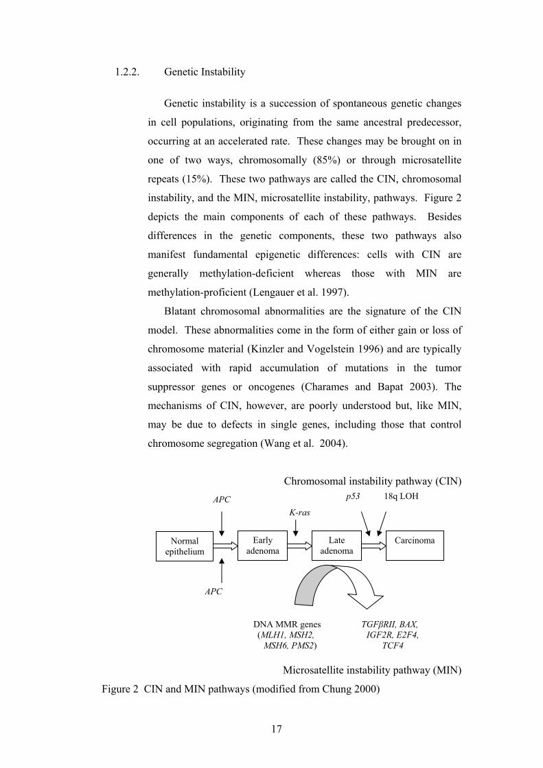
1.2.2. Genetic Instability
Genetic instability is a succession of spontaneous genetic changes
in cell populations, originating from the same ancestral predecessor,
occurring at an accelerated rate. These changes may be brought on in
one of two ways, chromosomally (85%) or through microsatellite
repeats (15%). These two pathways are called the CIN, chromosomal
instability, and the MIN, microsatellite instability, pathways. Figure 2
depicts the main components of each of these pathways. Besides
differences in the genetic components, these two pathways also
manifest fundamental epigenetic differences: cells with CIN are
generally methylation-deficient whereas those with MIN are
methylation-proficient (Lengauer et al. 1997).
Blatant chromosomal abnormalities are the signature of the CIN
model. These abnormalities come in the form of either gain or loss of
chromosome material (Kinzler and Vogelstein 1996) and are typically
associated with rapid accumulation of mutations in the tumor
suppressor genes or oncogenes (Charames and Bapat 2003). The
mechanisms of CIN, however, are poorly understood but, like MIN,
may be due to defects in single genes, including those that control
chromosome segregation (Wang et al. 2004).
Chromosomal instability pathway (CIN)
Microsatellite instability pathway (MIN)
Figure 2 CIN and MIN pathways (modified from Chung 2000)
Normal epithelium
APCK-ras
p53 18q LOH
DNA MMR genes (MLH1, MSH2,
MSH6, PMS2)
TGFβRII, BAX, IGF2R, E2F4,
TCF4
Early adenoma
Late adenoma
Carcinoma
APC
18
The MIN model is associated with a mutator phenotype and entails
three intercellular mechanisms. These mechanisms are: nucleotide
excision repair (NER), base excision repair (BER), and mismatch
repair. A hallmark of the MIN pathway is the instability of the
microsatellites (Aaltonen et al. 1993; Ionov et al. 1993; Thibodeau et
al. 1993), which will be described later.
HNPCC features are clinically similar to those of microsatellite
instable (MSI) tumors. These are: the proximal location of the tumors,
mucinous/undifferentiated tumors, infiltration of lymphocytes, and
improved prognosis. An unsurprising 85-95% of HNPCC tumors are
microsatellite instable, whereas only 15% of sporadic tumors are.
Microsatellites, also called short-tandem repeats, are often
polymorphic DNA loci that contain a repeated nucleotide sequence,
such as CAn and An. The MSI phenomenon is characterized by
numerous extra alleles in the microsatellite markers. It indicates a
malfunction in the mismatch repair system and occurs more commonly
in proximal tumors.
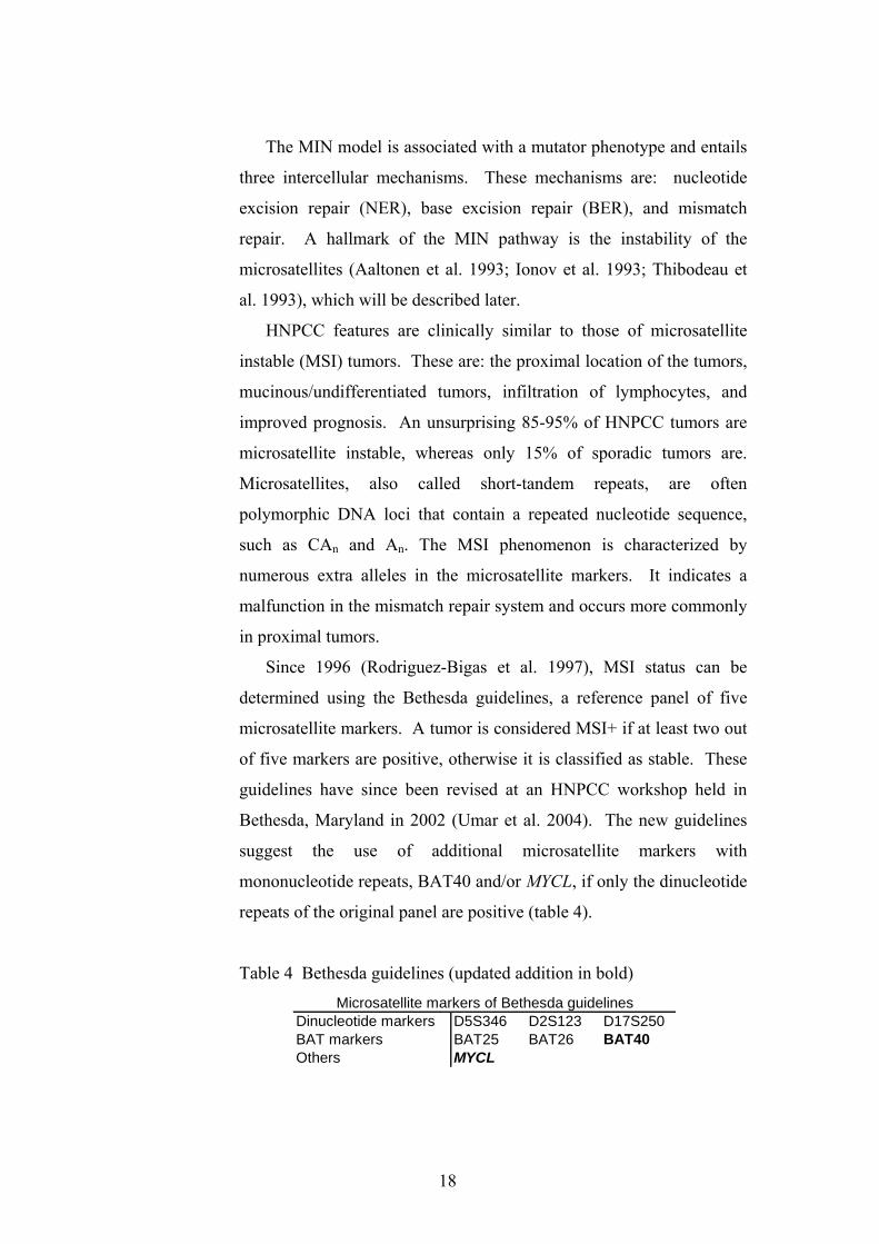
Since 1996 (Rodriguez-Bigas et al. 1997), MSI status can be
determined using the Bethesda guidelines, a reference panel of five
microsatellite markers. A tumor is considered MSI+ if at least two out
of five markers are positive, otherwise it is classified as stable. These
guidelines have since been revised at an HNPCC workshop held in
Bethesda, Maryland in 2002 (Umar et al. 2004). The new guidelines
suggest the use of additional microsatellite markers with
mononucleotide repeats, BAT40 and/or MYCL, if only the dinucleotide
repeats of the original panel are positive (table 4).
Table 4 Bethesda guidelines (updated addition in bold)
Dinucleotide markers D5S346 D2S123 D17S250BAT markers BAT25 BAT26 BAT40Others MYCL
Microsatellite markers of Bethesda guidelines
19
1.2.3. Mechanisms of Microsatellite Instability
Microsatellite instability occurs when nucleotide repeats expand
past a critical number. The underlying mechanism behind this is the
inactivation of the DNA mismatch repair system. In 1971, Knudson et
al. postulated that inactivation of a tumor suppression gene could be
accounted for by a two-hit mechanism, in which two separate
mutations occur in the same gene, rendering it incapable of
functioning. This seems to hold true for the DNA mismatch repair
gene inactivation as well (Hemminki et al. 1994). In hereditary
cancers, like HNPCC, a germline mutation in either MLH1 or MSH2 is
the first hit leading to disease. This may then be accompanied by
either the loss of the wild-type allele, known as loss of heterozygosity
(LOH), or its mutation (Hemminki et al. 1994; Konishi et al. 1996;
Tannergård et al. 1997; Chung et al. 2003). In sporadic MSI cases the
two hits are not so obvious, these cancers display a lower frequency of
LOH as well as somatic MLH1 and MSH2 mutations. Sporadic MSI
tumors, however, frequently have hypermethylation of the MLH1
promoter region (Kane et al. 1997; Cunningham et al. 1998; Herman et
al. 1998). This may possibly account for MLH1 inactivation in these
tumors. The MSH2 gene promoter, however, does not show
hypermethylation (Cunningham et al. 1998; Herman et al. 1998).
1.2.4. Tumor Spectrum
HNPCC is a multi-organ cancer syndrome with tumors mainly
occurring in the colon or the endometrium. In some families
endometrial cancer may even be more common than colon cancer
(Aarnio et al. 1999). The tumor spectrum, however, also includes
gastric, small bowel, hepatobiliary tract, upper urologic tract, ovarian,
and brain cancer (Watson and Riley 2005). Since all cells have
inherited a copy of a defective DNA mismatch repair gene, resulting in
hypermutability, why then are only some tissues involved.
Endometrial cancer observations suggest that there may be a
20
differential role of different predisposing mutations. A study by Duval
et al. (2002) showed that gastric and colorectal cancers had similar
mutation profiles but that they differed from those of endometrial
cancer. The occurrence of endometrial carcinoma in HNPCC appears
to be connected predominantly with mutations in MSH2 and MSH6
(Wijnen et al. 1999).
1.3. GENETIC BASIS OF ENDOMETRIAL CANCER
The most common gynecological cancer in the United States is endometrial
carcinoma. Although, like colon cancer, endometrial cacner mostly arises as a
sporadic disease, it may also be a part of certain hereditary syndromes. This
cancer is likely a constituent tumor of Cowden syndrome, an autosomal dominant
disorder distinguished by multiple hamartomas, as well as thyroid and breast
cancer risk (Eng 2000). An estimated 80% of CS is associated with PTEN
germline mutations (Marsh et al. 1998), as well as an additional 60% with
Bannayan-Riley-Ruvalcaba (BRR) syndrome, up to 20% with Proteus syndrome
(PS), and roughly 50% with Proteus-like syndrome (Eng 2003). The phosphatase
PTEN, classified as a tumor suppressor, mediates cell arrest and apoptosis by
signaling down the phosphoinositol-3-kinase/Akt pathway (Furnari et al. 1998; Li
et al. 1998; Maehama and Dixon 1998; Stambolic et al. 1998; Weng et al. 1999,
Vivanco and Sawyers 2002). Somatic mutations and deletions, affecting
expression of this gene, occur in as much as 93% (Mutter et al. 2000) of
endometrial carcinomas with endometrioid histology, and evidence suggests it
may even be a gatekeeper for endometrial carcinoma since PTEN function loss
occurs in the very early stages of tumor development (Levine et al. 1998).
DNA mismatch repair defects, leading to microsatellite instability, are found
in 20-30% of sporadic endometrial carcinomas. Endometrial carcinomas are the
most common malignancy in female HNPCC patients and MSI is a common
feature of HNPCC. Though MSI tumors were originally supposed to harbor a
higher frequency of somatic PTEN mutations than MSS tumors, studies have now
shown that there are similar frequencies for both (Nassif et al. 2004). About 19%
of sporadic MSI colorectal carcinomas have somatic PTEN frameshift mutations
21
almost solely in the two 6(A) tracts of exons 7 and 8, whereas no more than 5%
of MSI unknown or MSS colorectal carcinomas do, though none in the poly(N)
tracts (Guanti et al. 2000).
1.4. DNA METHYLATION IN NORMAL AND CANCER DEVELOPMENT
1.4.1. Vertebrate Methylation
In vertebrates, DNA methylation, that is the addition of a methyl
group to the internal cytosine of a CCGG sequence of CpG islands,
occurs in promoter regions which are located 5' to many genes that
are rich in CG content. A number of important genome functions are
affected by DNA methylation, including the control of gene
expression during development and differentiation, as well as
provision of a transcriptional backnoise reduction mechanism (Bird
1986; Razin and Kafri 1994). DNA methylation provides a post-
replicative addition of a methyl group, and is therefore epigenetic in
nature providing for both fidelity, due to symmetry of the CpG target
sequence, and flexibility. The semi-conservative nature of DNA
replication ensures flexibility, allowing for hemi-methylated CpG
sites to persist or be methylated (Laird and Jaenisch 1996). If no
methylation occurs, however, then an acquired altered methylation
pattern will arise in one of the two daughter strands upon the next
replication.
Promoter DNA methylation causes gene expression to be
repressed. Two proposed mechanisms may explain this repression.
The first, though not all transcription factors fit the necessary
structure, suggests a sequence-based direct inhibition of transcription
factors (Tate and Bird 1993). The second entails the non-sequence-
specific binding to methylated DNA by proteins from the MBD
family, that is MeCP1 and MeCP2 (Boyes and Bird 1992). This
protein family also contains a transcriptional repression domain
which, when bound to the DNA, forms a complex with core
22
repression molecules and histone deacetylase proteins; thereby,
compressing chromatin structure making it less accessible to active
transcription (Nan et al. 1997; Bestor et al. 1998; Jones et al. 1998).
A combination of these two mechanisms probably occurs in vivo.
1.4.2. DNA Methyltransferases
DNA methyltransferases are responsible for two distinct patterns
of methylation. These are maintenance activity, which is sequence
specific and restores full methylation of hemi-methylated CpG sites,
and de novo methylation activity, wherein a previously unmethylated
target sequence is methylated.
Originally only one DNA methyltransferase enzyme was
identified and cloned in both mice and humans, DNMT1. This is a
large enzyme containing a C-terminal catalytic domain, akin to that
found in prokaryotes, and a large N-terminal regulatory domain,
which targets replication foci in addition to other functions
(Leonhardt et al. 1992). Highly conserved in eukaryotes (Yen et al.
1992), DNMT1 is extremely affinitive to hemi-methylated DNA,
permitting parental DNA pattern duplication in daughter strands
through its N-terminus domain targeting capability. Removal of this
domain results in de novo methylation (Bestor 1992), and was
originally considered as the de novo methylation mechanism.
The second possible responsible enzyme for de novo methylation
appeared to be another methyltransferase enzyme, suggested in a
study by Lei et al. (1996). In their study, embryonic stem cells
exhibited de novo methylation despite being null for DNMT1. This,
in addition to the inexplicability of the role DNMT1 plays in tumor-
specific methylation abnormalities, drove the search for additional
DNMT’s. Two groups (Okano et al. 1998a; Yonder and Bestor 1998)
isolated a prospective second enzyme, DNMT2, but it did not exhibit
any methylation ability. Shortly after this, a third potential enzyme
group including DNMT3a and b, however, was isolated by Okano et
23
al. (1998b) in a database search. Another member of this group is
DNMT3L, a nonenzymatic protein, originally found by Aapola et al.
(2000), and further characterized by Aapola et al. (2001), Hata et al.
(2002), and Deplus et al. (2002). These proteins, though down
regulated in differentiated embryonic stem cells and adult murine
tissue, are expressed in undifferentiated embryonic stem cells at
elevated levels. Moreover, they proved equally proficient in
methylation of both hemi-methylated DNA and unmethylated DNA.
Further studies confirmed that DNMT3a and b are essential for de
novo methylation and development (Okano et al. 1999; Kaneda et al.
2004), but their methylation activity is stimulated by DNMT3L
(Suetake et al. 2004).
Ramchandani et al. 1999 described putative demethylase activity
but it has been studied far less extensively than its counterpart. The
proposed demethylase enzyme seems to bind specifically to the CpG
dinucleotide, regardless of whether the DNA is fully or hemi-
methylated.
1.4.3. Role of Methylation in Normal Development
DNA methylation is necessary for normal mammalian
development. During development and gametogenesis three distinct
phases are manifested: demethylation, de novo methylation, and
maintenance methylation. Preimplantation embryos undergo a wave of
genome-wide demethylation during cleavage. Widespread de novo
methylation, establishing overall methylation patterns, then occurs
prior to gastrulation. This pattern is preserved in the somatic cells,
throughout life (Monk et al. 1987, Kafri et al. 1992), while the
embryonic lineage maintains lower methylation levels (Jaenisch 1997).
Li et al. (1992) demonstrated that methylation is undeniably
essential for development. Despite mutated homozygous Dnmt1
embryonic stem cells being viable, even with only trace levels of DNA
methyltransferase activity, introduction of the same mutation into the
germline of mice caused a recessive lethal phenotype with successive
24
homozygous embryos not developing past mid-gestation. These
observations are supported by previous studies which showed that both
preimplantation and undifferentiated embryonic stem cells have high
de novo methylation activity levels (Jaenisch 1997). No evidence of
this de novo methylation is discernible after gastrulation and
differentiation; low levels, however, can be detected in somatic cells.
Although this process is slow and inefficient in targeting genes for
silencing, it could provide a selective advantage for tumor growth
(Laird et al. 1995).
1.4.4. DNA Methylation and Evolution
Genome size of free-living organisms has increased from a few
thousand genes in prokaryotes, to 7,000-25,000 genes in invertebrates,
up to 30,000-100,000 genes in vertebrates (Olivier et al. 2001; Venter
et al. 2001). This increase in gene number and complexity is the
foundation for the first of two hypotheses concerning the evolutionary
role of DNA methylation, which suggests that since tissue-specific
genes have increased, a high efficiency of repression of those genes
must also exist. In eukaryotes, repression and transcriptional noise
reduction are foremostly attributed to the nuclear envelope and
histones. Since vertebrate eukaryotes have even more genes than
invertebrate eukaryotes there must be additional repression
mechanisms, one of which could be DNA methylation. Invertebrates,
indeed, have only a few methylated CpG dinucleotides whereas,
excluding CpG islands, most, 60-90%, are methylated in vertebrates
(Kress et al. 2001). These methylated CpG dinucleotides may function
as a global repression and transcriptional noise reduction layer (Bird
1995). The second theory views DNA methylation as a nuclear host-
defense system. Since cytosine methylation may cause promoter
inactivation of most viruses and transposons, including retroviruses
and Alu elements (Yoder et al. 1997), it is useful in the fight against
possible threats by endogenous parasitic mobile genetic elements in
mammals.
25
1.4.5. DNA Methylation and Cancer
DNA methylation is an ideal participant in the multi-step process
by which normal cells develop into malignant cells. Two basic
methylation patterns are observable in tumor cells, namely, global
genome hypomethylation, which apparently begins early on in tumor
development (Christman et al. 1993; Pogribny et al. 1997), and
targeted hypermethylation of specific regions. Tumorigenesis
transpires by either a genetic or epigenetic system of DNA
methylation, as described below.
1.4.5.1. Genetic System
The 5’methylcytosine residues within CpG pairs are highly
mutable. This is possibly a consequence of the increased
deamination rate of 5’mC to T; three factors influence repair of
this lesion.
First, cytosine deamination forms the base uracil, but
deamination of 5’mC forms the base thymine. Uracil does not
naturally occur in DNA and is therefore more easily recognized
for correction by uracil DNA glycosylase than other DNA
glycosylases recognize errant thymine (Jiricny 1996). This
results in an increase of CpG to TpG mutations or, on the
opposite strand, CpG to CpA mutations.
The second and third factors are less complicated.
Deamination rate is reasonably constant and thereby sufficient to
account for observed dsDNA mutations (Shen et al. 1994).
Finally, compared to unmethylated cytosine, 5’mC has an
enhanced mutability in cell division thereby making it more
vulnerable to mutation in cancer, in which an increase in cell
division occurs (Lieb and Reihmat 1997).
26
1.4.5.2. Epigenetic System
The effect of epigenetic mechanisms of DNA methylation on
gene activity is difficult to study. DNA methylation allegedly
affects chromatin structure. Whether the structure is affected by
increased affinity of methylated DNA for Histone H1, as
proposed by Campoy et al. (1995), or by physical binding of
Histone H1 determining the methylation levels is, however,
debatable.
Several pieces of evidence demonstrate that transcription
repression of hypermethylated promoter sequences is directly
caused by DNA methylation. This evidence includes the Min
mouse model (Laird et al. 1995), retinoblastoma (RB) gene
promoter region methylation effects (Stirzaker 1997), and the
high promoter hypermethylation frequency in microsatellite
unstable sporadic colon cancers of the MMR gene MLH1.
Using a combination of methods, Laird et al. (2003)
determined the effect of DNA methyltransferases in ApcMin mice
with induced intestinal neoplasia. Reducing the methylation level
of these mice through reduction of methyltransferase activity
reduced the amount of polyps in the test mice compared to the
control mice.
Second, the RB gene promoter methylation, the first identified
with methylation silencing, is documented in familial cases of
unilateral retinoblastoma. In addition, in vitro tests show that the
methylation of this promoter blocks the promoter activity of the
RB gene.
Third, in sporadic microsatellite unstable colon cancers the
promoter region of the MLH1 mismatch repair gene is frequently
methylated (Herman et al. 1998). Plumb et al. (2000) and
Herman et al. (1998) found that MLH1 methylation is a reversible
phenomenon. Treating cell lines with the demethylating agent 5’-
aza-2’deoxycytidine (5-aza-dC) substantially increases the MLH1
expression after five days of treatment. This observation has
27
important implications. Treatment of cell lines, derived from
MLH1 deficient tumors, with the DNA methyltransferase
inhibitor 5-aza-dC results in demethylation of the MLH1 promoter
and ultimate restoration of MMR activity, indicative of the
primary nature of the inactivation of MLH1 by CpG island
hypermethylation. Since MMR deficient tumors are resistant to
alkylating agents used for chemotherapy, this may also be
clinically important. Plumb et al. (2000) showed that after 5-aza-
dC treatment these tumors become resensitized to such drugs.
Finally, when studying p16INK4a, Myöhänen et al. (1998)
observed that if one copy of a tumor suppressor gene is mutated
or lost, then the remaining wild-type gene copy is more readily
hypermethylated, resulting in inactivation. All these observations
provide evidence that a primary contributing factor in
tumorigenesis is DNA methylation.
The mechanism by which DNA methylation occurs in the
above-described events is still unclear, though the methylation
machinery itself may contribute to this process. As demonstrated
in the Min mice experiments, the DNA-Mtase activity level has a
direct effect on the number of polyps in neoplastic intestinal cells.
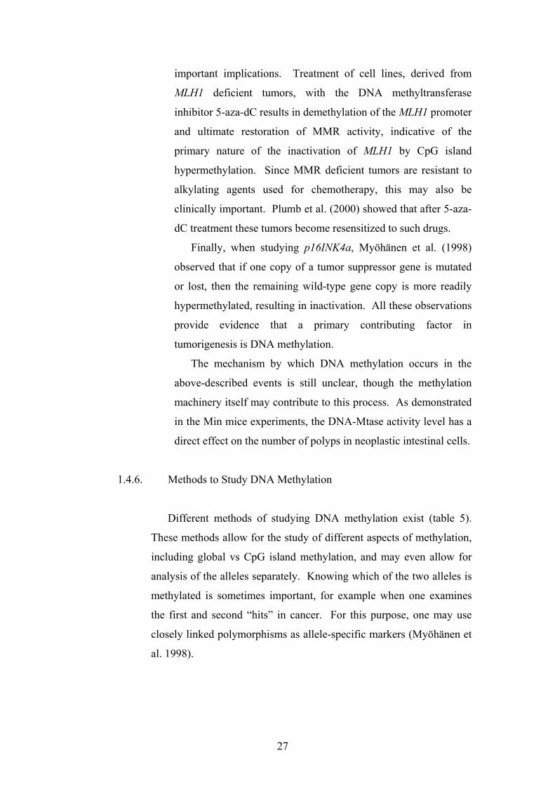
1.4.6. Methods to Study DNA Methylation
Different methods of studying DNA methylation exist (table 5).
These methods allow for the study of different aspects of methylation,
including global vs CpG island methylation, and may even allow for
analysis of the alleles separately. Knowing which of the two alleles is
methylated is sometimes important, for example when one examines
the first and second “hits” in cancer. For this purpose, one may use
closely linked polymorphisms as allele-specific markers (Myöhänen et
al. 1998).
28
Table 5 Most commonly used laboratory methods to study DNA methylation
(adapted from Laird 2003).
Technology Methylation discrimination principle Application
Southern blot restriction digestion quantitative analysis
HpaIIPCR restriction digestion qualitative analysis
COBRA bisulphite conversion quantitative analysis
MSP bisulphite conversion sensitive detection
Technology Methylation discrimination principle Application
SssI methyl acceptance assay DNA methyltransferase substrate global methylation analysis
RLGS restriction digestion marker discoveryEpigenomics microarray bisulphite conversion disease stratification
I Methylation of CpG sites in specific genes
II Global DNA methylation
29
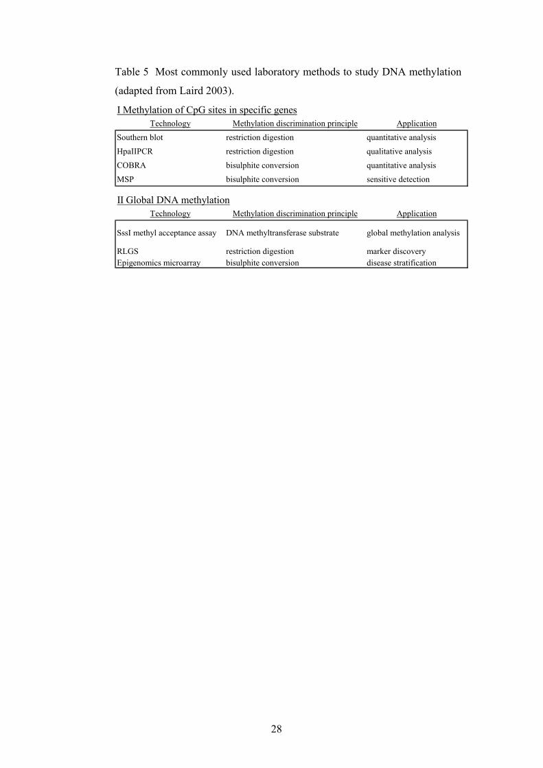
2. AIMS OF STUDY
• Characterization of hereditary nonpolyposis colon cancer (HNPCC)
versus sporadic colon cancer in relation to tumorigenic mechanisms.
o What mechanisms underlie microsatellite instability (MSI) in
sporadic colorectal cancers?
o What is the prevalence of DNA methylation changes in colorectal
cancer and how do these changes correlate with MSI?
• Comparison of endometrial carcinoma and colorectal carcinoma with
similar underlying inherited mutations, in relation to microsatellite status
and target genes.
o To which extent do somatic alterations in target tissue explain the
HNPCC tumor spectrum?
30
3. MATERIALS AND METHODS
3.1. PATIENT SAMPLES (I-IV)
Patient samples used in studies I and II were primarily derived from a
prospective collection of 509 unselected colorectal adenocarcinomas harvested at
nine large regional hospitals in Southern Finland between May 1994 and April
1996 (Aaltonen et al. 1998). The samples represented fresh-frozen or paraffin-
derived normal colonic mucosa or tumor tissue. To ensure the sections used for
DNA extraction had the highest possible tumor cell percentage all specimens
were examined histologically.
We included all tumors (n=51) demonstrating high MSI and no germline
mutations (MLH1 or MSH2) by direct sequencing (I and II). Additionally, we
analyzed 38 MSS tumors selected randomly, except for equal numbers of
proximal and distal tumors (I), and 21 HNPCC patients with known germline
mutations, MLH1 n=20 and MSH2 n=1 (II).
Study III and IV involved samples derived from known MLH1 and MSH2
germline mutation carriers. In study IV we analyzed a total of 44 colon cancers
and 57 endometrial cancers, including cancer samples of both tissues in eight
patients. Both groups had similar germline mutation distributions. Study III
included only endometrial cancer patients. Samples consisted of paraffin-
embedded tissue or blood, with known MLH1 or MSH2 germline mutations
(n=41), as well as aberrant PTEN-expressing tumors (n=20), (see table 6 for the
list of germline mutations).
All studies were approved by the local ethics committees.
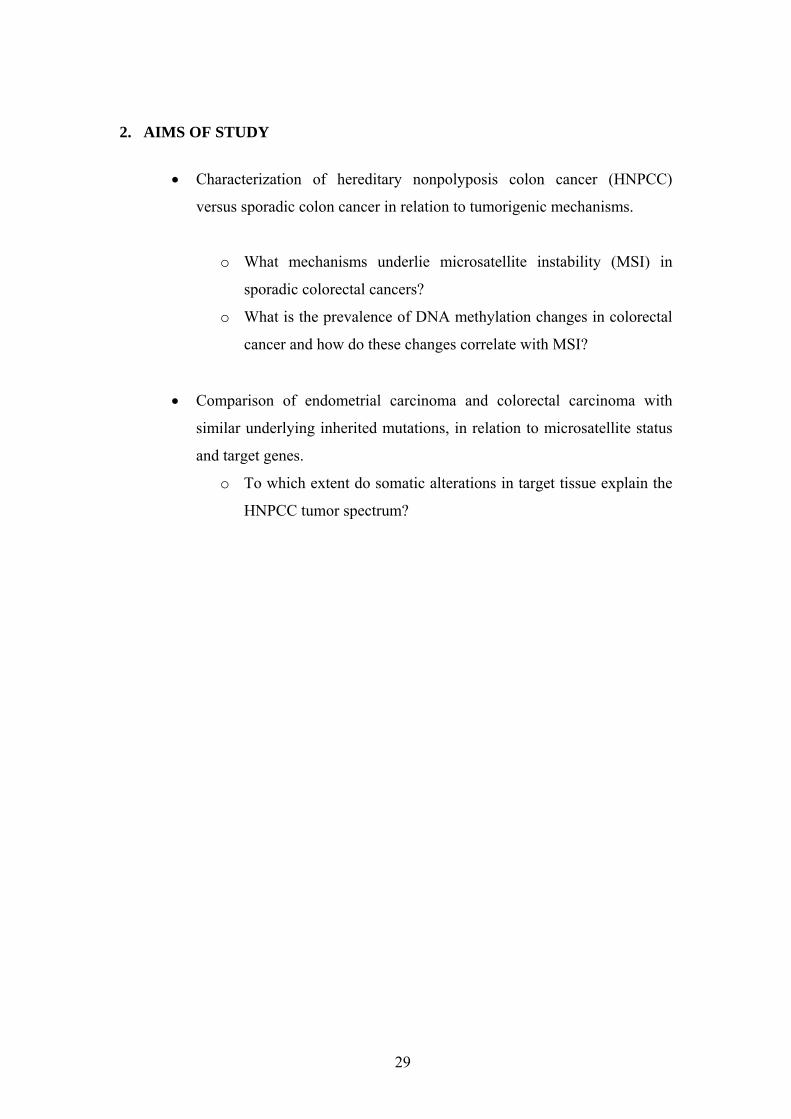
Table 6 MLH1 and MSH2 germline mutations included in present study.
Gene Mutation # Type of mutation1 3.5 kb genomic deletion affecting codons 578–632 of exon 162 G>A at 454–1 at splice acceptor of exon 63 G>C at 1976 (codon 659) of exon 174 G>C at 1409 + 1 at splice donor of exon 125 T>G at 320 (codon 107) of exon 46 G>A at 1039–1 at splice acceptor of exon 127 G>T at 1559–1 at splice acceptor of exon 148 C>T at 1975 (codon 659) in exon 17
MSH2 9 CA deletion at 1550 (codon 518) of exon 10
MLH
1
31
3.2. DNA METHYLATION (I AND II)
We applied a PCR-based assay relying on the inability of the HpaII restriction
enzyme to cleave CCGG sequences with an internal methylated cytosine. At the
MLH1 promoter region there are four HpaII sites located at 567, 527, 347, and
341 relative to the initiation codon. Using flanking primers, we studied each site
individually, with the exception of the latter two sites, which, due to their close
proximity were a single reaction. In addition to an HpaII digestion of each
sample, we included both an undigested DNA control, to verify amplification,
and an MspI digestion, an isoschizomer to HpaII insensitive to methylation, used
to verify the presence of the methylation site. We determined the optimal number
of PCR cycles to be the amount at which a detectable band was visible from the
undigested template, but no bands from DNA digested with MspI. For
comparison we studied a functionally neutral gene, calcitonin, analyzing region V
of the gene promoter as described in Heiskanen et al. (1994). The protocols for
restriction digestions and PCR analyses can be found in the original articles.
3.3. IMMUNOHISTOCHEMISTRY (I-III)
Using mouse monoclonal antibodies against the full-length human MLH1
protein (clone G168-728 from PharMingen, San Diego, CA), we studied the
expression of the MLH1 protein in the sporadic colon cancer samples (I, II).
Additionally, we used a monoclonal antibody against the full-length MSH2
protein (clone G219-1129 from PharMingen) in study II. The method for the
staining of these different slide sets varied in relation to what lab they were
analyzed in. In general, however, we used paraffin-embedded tissue which was
first stained with a primary antibody, followed by a secondary antibody before
counterstaining and photographing. See original articles for specific protocols.
To study the expression of the PTEN protein in the endometrial cancers in
study III we used the specific monoclonal antibody 6H2.1 raised against the last
100 C-terminal amino acids of PTEN (Ziebold and Lees, unpublished). We
conducted immunohistochemical staining, using paraffin-embedded tissue and a
32
two step primary and secondary antibody staining method previously described in
Mutter et al. (2000) and Perren et al. (1999).
3.4. MUTATION ANALYSIS (II-III)
3.4.1. MLH1 and MSH2 Somatic Mutations (II)
Using two-dimensional DNA electrophoresis methods, described
by Wu et al. (1997), we screened 31 sporadic colorectal cancers and
two HNPCC cases for MLH1 and MSH2 somatic mutations.
3.4.2. PTEN Mutations (III)
On DNA from 20 HNPCC-related endometrial carcinomas with
either absent or weak PTEN expression we preformed mutation
analysis, using PCR-based denaturing gradient gel electrophoresis
(DGGE) and semi-automated sequencing described by Mutter et al.
(2000), of all nine exons, exon-intron junctions, and flanking intronic
sequences.
3.5. LOSS OF HETEROZYGOSITY (LOH) ANALYSIS (II)
We used radioactive PCR amplification of microsatellite markers to
determine LOH (Peltomäki et al. 1993) and scored all cases with either an absent
or greatly reduced allele in tumor vs normal DNA as LOH. We considered all
cases exhibiting homozygosity in the normal tissue, for a given marker, as well as
those with instability in the tumor DNA to be uninformative.
For MLH1 we used three microsatellite markers previously shown to have the
highest deletions rates. Two of the markers, D3S1029 and D3S1283, flank the
MLH1 gene at 5cM distance on either side (Hemminki et al. 1994), while marker
D3S1611 is located in intron 12 (unpublished data).
For MSH2 we used the markers D2S2259 and D2S123 which encompass the
33
region of 9cM around MSH2. Additionally, we also used a marker flanked by the
previously mentioned markers, D2S391.
3.6. MICROSATELLITE INSTABILITY ANALYSIS (IV)
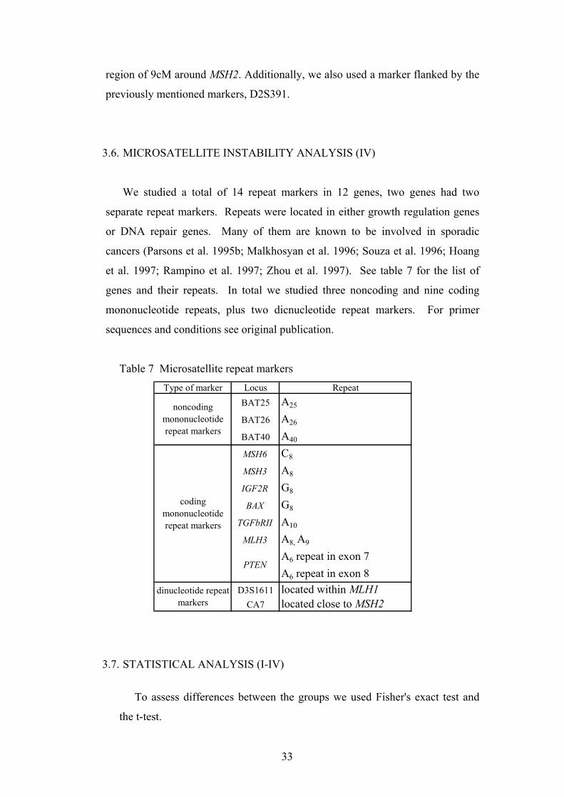
We studied a total of 14 repeat markers in 12 genes, two genes had two
separate repeat markers. Repeats were located in either growth regulation genes
or DNA repair genes. Many of them are known to be involved in sporadic
cancers (Parsons et al. 1995b; Malkhosyan et al. 1996; Souza et al. 1996; Hoang
et al. 1997; Rampino et al. 1997; Zhou et al. 1997). See table 7 for the list of
genes and their repeats. In total we studied three noncoding and nine coding
mononucleotide repeats, plus two dicnucleotide repeat markers. For primer
sequences and conditions see original publication.
Table 7 Microsatellite repeat markers Type of marker Locus Repeat
BAT25 A25
BAT26 A26
BAT40 A40
MSH6 C8
MSH3 A8
IGF2R G8
BAX G8
TGFbRII A10
MLH3 A8, A9
A6 repeat in exon 7A6 repeat in exon 8
D3S1611 located within MLH1CA7 located close to MSH2
noncoding mononucleotide repeat markers
coding mononucleotide repeat markers
PTEN
dinucleotide repeat markers
3.7. STATISTICAL ANALYSIS (I-IV)
To assess differences between the groups we used Fisher's exact test and
the t-test.
34
4. RESULTS
4.1. DESCRIPTION OF TWO MAIN EPIGENETIC PHENOTYPES FOR
COLORECTAL CANCER (I)
4.1.1. DNA Methylation Patterns and Correlation with Microsatellite
Instability
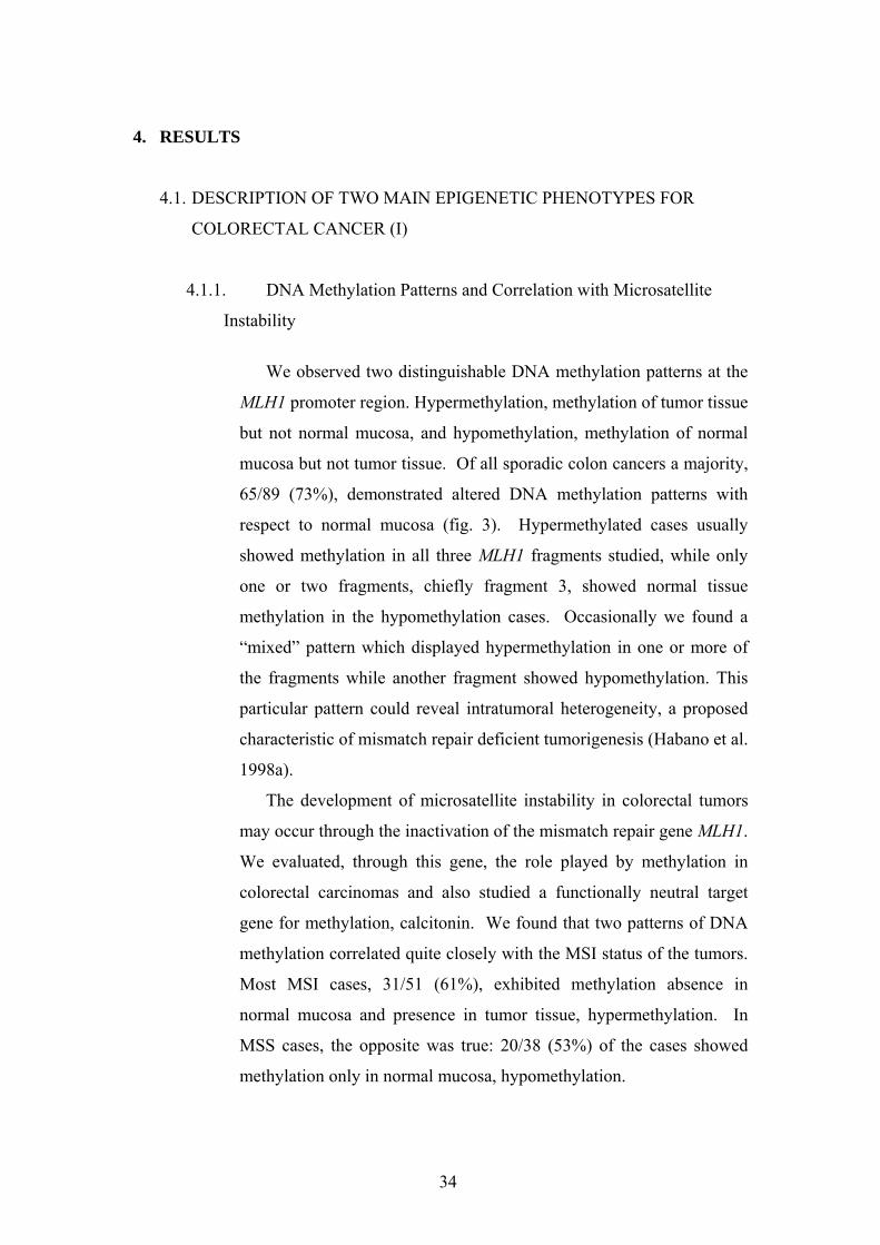
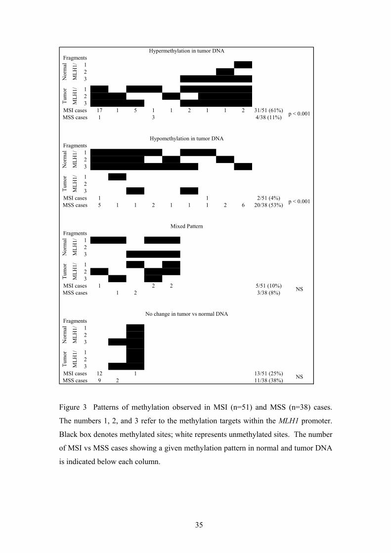
We observed two distinguishable DNA methylation patterns at the
MLH1 promoter region. Hypermethylation, methylation of tumor tissue
but not normal mucosa, and hypomethylation, methylation of normal
mucosa but not tumor tissue. Of all sporadic colon cancers a majority,
65/89 (73%), demonstrated altered DNA methylation patterns with
respect to normal mucosa (fig. 3). Hypermethylated cases usually
showed methylation in all three MLH1 fragments studied, while only
one or two fragments, chiefly fragment 3, showed normal tissue
methylation in the hypomethylation cases. Occasionally we found a
“mixed” pattern which displayed hypermethylation in one or more of
the fragments while another fragment showed hypomethylation. This
particular pattern could reveal intratumoral heterogeneity, a proposed
characteristic of mismatch repair deficient tumorigenesis (Habano et al.
1998a).
The development of microsatellite instability in colorectal tumors
may occur through the inactivation of the mismatch repair gene MLH1.
We evaluated, through this gene, the role played by methylation in
colorectal carcinomas and also studied a functionally neutral target
gene for methylation, calcitonin. We found that two patterns of DNA
methylation correlated quite closely with the MSI status of the tumors.
Most MSI cases, 31/51 (61%), exhibited methylation absence in
normal mucosa and presence in tumor tissue, hypermethylation. In
MSS cases, the opposite was true: 20/38 (53%) of the cases showed
methylation only in normal mucosa, hypomethylation.
35
1 X2 X3 X X X X
1 X X X X X X X2 X X X X X X3 X X X X X X X
17 1 5 1 1 2 1 1 2 31/51 (61%)1 3 4/38 (11%)
1 X X X X X X2 X X X X X3 X X X X X X X
1 X23 X X
1 1 2/51 (4%)5 1 1 2 1 1 1 2 6 20/38 (53%)
1 X X X X23 X X X
1 X X2 X X X3 X X
1 2 2 5/51 (10%)1 2 3/38 (8%)
1 X2 X3 X X
1 X2 X3 X X
12 1 13/51 (25%)9 2 11/38 (38%) NS
Tum
or
MLH
1/
MSI casesMSS cases
No change in tumor vs normal DNAFragments
Nor
mal
MLH
1/
Tum
or
MLH
1/M
LH1/
NS
Tum
or
MLH
1/
MSI casesMSS cases p < 0.001
Mixed Pattern
Hypermethylation in tumor DNA
Hypomethylation in tumor DNAFragments
Nor
mal
MLH
1/
MSI casesMSS cases
Fragments
p < 0.001
Nor
mal
MSI casesMSS cases
Fragments
Nor
mal
MLH
1/
Tum
or
MLH
1/
Figure 3 Patterns of methylation observed in MSI (n=51) and MSS (n=38) cases.
The numbers 1, 2, and 3 refer to the methylation targets within the MLH1 promoter.
Black box denotes methylated sites; white represents unmethylated sites. The number
of MSI vs MSS cases showing a given methylation pattern in normal and tumor DNA
is indicated below each column.
36
A known silencing method is methylation triggered by DNA
repetition (Selker 1999), and consequently microsatellite length and/or
configuration alterations caused by DNA mismatch repair deficiency
might induce hypermethylation. If true, and microsatellite instability is
a primary inducing factor, then all MSI tumors regardless of etiology
would be expected to be hypermethylated.
In order to achieve additional insight into the relationship between
DNA methylation and MSI we evaluated methylation changes in a
cohort (n=26) of HNPCC patients with germline MLH1 mutations and
profound MSI phenotype in tumor DNA. The frequencies for the
different patterns (table 8) did not support a primary role of MSI in
methylation changes. Instead they suggested that promoter
hypermethylation was a special characteristic of truly sporadic MSI
colorectal carcinomas.
Table 8 Frequencies of DNA methylation patterns observed in
colorectal cancers (n=26) from HNPCC cases
Pattern Frequency
hypermethylation 23%
hypomethylation 19%
mixed 23%
no change 35%
4.1.2. Methylation Specificity
To further understand whether or not the patterns observed in
MLH1 were restricted to this gene or more widespread, we looked at
methylation patterns of the calcitonin gene. We chose this gene for
comparison because it is a well-characterized methylation target in a
number of malignancies, including colon cancer, but unlike MLH1 its
alterations have not been associated with any selective advantage
37
(Baylin et al. 1987; Silverman et al. 1989; Heiskanen et al. 1994;
Baylin et al. 1998).
The calcitonin gene was hypermethylated in all MSI cases except
one. It was also methylated in cases with no MLH1 promoter
hypermethylation, which is consistent with it being a methylation
target. More importantly, a significant fraction, 5/19 (26%) of the
MLH1 hypomethylated MSS tumors was also hypomethylated in the
calcitonin gene.
Our combined findings of MLH1 and calcitonin gene methylation
support the idea of distinct patterns for MSI and MSS tumors in
addition to surrounding normal mucosa, though genome-wide
screening of CpG islands for methylation would need to be performed
for confirmation.
4.1.3. Correlation with MLH1 Protein Expression
The MLH1 promoter contains at least 23 CpG sites (Deng et al.
1999) including the four HpaII restriction sites contained in the three
fragments we studied. Only some regulate MLH1 protein expression
through methylation, making it of interest to study whether
methylation of these sites may have an effect on gene expression in the
present tumors, and more importantly, normal mucosae. We analyzed
available paraffin-embedded samples of MSI tumors for the expression
of the MLH1 protein by immunohistochemistry. We considered cases
in which there were fewer than 25% cancer cells with visible staining
to have reduced or lost expression of MLH1 protein. A majority of the
MSI tumors in which we observed promoter hypermethylation in one
or more of the three MLH1 sites fell into this category, 30/36 (83%).
Our results were compatible with those reported by others
(Cunningham et al. 1998; Herman et al. 1998).
Of particular interest were the six cases in which we found
hypomethylation at all three sites (fig. 3, pg. 35). Of these cases we
only had four MSS cases available for examination by
immunohistochemistry. Severe reduction or loss of protein expression
38
was visible in all of the normal mucosal tissues, while the adjacent
tumor tissue, which lacked methylation, showed an intense staining
(see fig. 3, in the original article I). These observations emphasize the
pathogenic relevance of methylation changes by showing that the loss
of MLH1 protein expression accompanies MLH1 promoter
hypermethylation in both normal mucosa and tumor tissue.
4.2. BASIS OF MMR GENE INACTIVATION IN SPORADIC AND
HEREDITARY COLORECTAL CANCERS (I, II)
4.2.1. Sporadic Colorectal Cancer
As described above, we observed two main types of methylation
patterns, hypermethylation and hypomethylation, in sporadic colorectal
cancer. Of the 51 MSI+ cases we analyzed for methylation changes, 31
cases were hypermethylated (61%), two were hypomethylated (4%),
five had a mixed pattern (10%), and 13 displayed no change (25%).
The distribution of the patterns observed in the MSS cases were four
hypermethylated (11%), 20 hypomethylated (53%), three mixed
pattern (8%), and 11 no change (38%).
We obtained an interpretable immunohistochemical analysis (IHC)
result for a total of 46/51 MSI+ cases. Of these, 80% showed an
involvement of the MMR genes MLH1, MSH2, or both. Loss of or
reduction of MLH1 expression occurred in 36 of the cases and MSH2
loss or reduction in seven.
An overwhelming majority of the cases, 30/36, demonstrated loss
of MLH1 protein expression in the IHC analysis presented with DNA
hypermethylation at one or more of the studied CpG islands.
Methylation of MSH2 was not addressed because we have previously
found that, unlike MLH1, it is not a methylation target (unpublished
data). In addition to methylation changes we also found eight MLH1-
linked tumors and two MSH2-linked tumors with loss of
39
heterozygosity in the respective regions, as well as two somatic
mutations each for both the MLH1 and MSH2 cases.
4.2.2. Colorectal Cancer with Inherited MMR Deficiency (HNPCC)
Hereditary cases did not display as many methylation changes as
sporadic cases. Out of 26 MLH1 germline HNPCC cases only six were
hypermethylated (23%), five were hypomethylated (19%), six had a
mixed pattern (23%), while nine displayed no change (35%). All the
cases, 26 MLH1 and one MSH2 with germline mutations as their first
hits (see table 6 in Materials and Methods for the list), exhibited
expression loss of their relevant gene, however.
In contrast to the frequency of hypermethylation in sporadic cases
(34/51), only 12/26 MLH1 germline mutation cases tested positive for
DNA hypermethylation (p=0.003). We found LOH, however, to occur
in hereditary cases at a similar rate compared to sporadic cases, 33%
compared to 24%.
When we looked at the combined data for both LOH and
hypermethylation in the two types of tumors we saw that LOH and
hypermethylation were mutually exclusive, possibly because they have
similar MLH1 inactivation functions. All HNPCC tumors already
carrying a MLH1 germline mutation as their first hit showed, with one
exception, no hypermethylation in the presence of LOH. Our one
MSH2 case had LOH accompanying its germline mutation. See table 2
in the original article II for a summary. While our methylation
analyses were not allele-specific, the mutually exclusive occurrence of
LOH and hypermethylation at MLH1 suggests that the wild-type allele,
rather than the mutated allele, was a favored hypermethylation target.
40
4.3. PTEN IN SPORADIC AND HEREDITARY ENDOMETRIAL CANCERS
(III, IV)
We used immunohistochemical analysis to assess the PTEN protein
expression in 41 MMR gene mutation positive endometrial carcinomas from
HNPCC families. All cases had either stroma and/or normal endometrial
epithelium which presented with strong PTEN immunostaining in both the
nucleus and the cytoplasm. We graded these ++ and used them as internal
controls for our samples.
We found a weak (+) or no (-) cytoplasmic PTEN staining in 28/41 (68%), of
the MMR gene mutation positive endometrial carcinomas from HNPCC families.
Of these, twelve (29%) had weak PTEN immunostaining and the remaining 16
(39%) had no immunoreactivity.
We only had adequate material from 20 of these 28 endometrial carcinomas
for mutational analysis of the entire PTEN gene. In 17/20 (85%) of the tumors
we found 18 frameshift mutations. Of these, 12 (67%) were in the poly(A) tracts,
normally consisting of either four, five, or six A-repeats. Notably, somatic
insertions or deletions involving one of the two 6(A) tracts in exon 7 or 8
occurred in ten (56%) cases. With possibly one exception, each tumor showed
only one mutant PTEN allele, PTEN mutations were monoallelic. A total of
seven cases harboring a monoallelic mutation also showed decreased PTEN
expression while nine exhibited none. The remaining negative-expression case
carried two different mutations.
When we compared the mutational frequency of PTEN in sporadic versus
HNPCC-related endometrial carcinomas, we found a significant difference
between the two groups, P=0.006. Additionally, the spectral difference between
sporadic and HNPCC-related endometrial cancers with PTEN mutations was also
significant, p=0.0009. Sixty of 118 (51%) sporadic MSI+ endometrial cancers
had somatic mutations, which included 64 truncating mutations. Of these, 39
were frameshift mutations, only eight of which (21%) occurred in the 6(A) tracts
in exons 7 and 8. Notably, compared to sporadic MSI+ tumors we also found a
significant over-representation of frameshift mutations in 6(A) tracts of exons 7
and 8 in HNPCC-related endometrial cancers, p=0.01.
41
In study IV, we compared 57 endometrial cancers and 44 colorectal cancers
from germline carriers of eight MLH1 mutations and one MSH2 mutation. We
found an association with PTEN instability and endometrial cancer. These
endometrial cancers, as opposed to colorectal cancers with the same underlying
germline mutations, were associated with instability in 6(A) tracts of exons 7 and
8 of PTEN, as discussed below (4.4).
4.4. PATTERNS OF MICROSATELLITE INSTABILITY IN ENDOMETRIAL
AND COLORECTAL TUMORS FROM INDIVIDUALS WITH
IDENTICAL PREDISPOSING MUTATIONS IN MMR GENES (IV)
We observed distinct MSI profiles for colorectal cancers (n=44) and
endometrial cancers (n=57) despite the origin of these tumors from carriers of
identical predisposing mutations. Most analyzed cases were from carriers of
either mutation 1 or 2, both common founder mutations affecting MLH1 in the
Finnish population.
The predominant pattern exhibited by the colorectal cancers consisted of
instability in at least one of the noncoding BAT repeats in 89% of the tumors,
TGFβRII in 73%, at least one dinucleotide repeat in 70%, MSH3 in 43%, and
BAX in 30%. Endometrial cancers, however, showed a more heterogeneous
pattern of instability typically affecting different coding repeats in different
tumors. Notably, TGFβRII and PTEN often displayed mutually exclusive
instability. See figures 1A and 1B in the original article for a pictorial summary
of these data.
When we compared the individual marker loci instability frequencies against
tumor type TGFβRII turned out to be a “target” gene for colorectal cancers,
instable in 73% of the colorectal cancer tumors and only 18% of the endometrial
cancer tumors, p=2.2 X 10⁻⁸. Conversely, PTEN instability occurred in only 5%
of the colorectal cancers and 20% of endometrial cancers, p=0.04. We also
observed a significantly lower proportion of marker instability, average fractions
of instable markers per tumor, in endometrial cancers, 0.27, than colorectal
cancers, 0.45, p<0.001. Though not statistically significant, complete stability
42
occurred in 23% of endometrial cancers while in only 11% of the colorectal
cancers.
In addition, endometrial cancer had shorter allelic shifts in the BAT markers
than colorectal cancers. The mean basepair deviation in endometrial cancers was
4.1 for BAT25 (range = 1-7), 8.5 for BAT26 (range = 4-13), and 6.1 for BAT40
(range = 3-9). Colorectal cancers had shifts of 6.7 (range = 4-11) for BAT25,
shifts of 13.5 (range = 9-17), and for BAT26, shifts of 9.6 (range = 3-13). The
difference between endometrial cancer and colorectal cancer in all cases was
significant, p<0.001. Each tumor showed close correlation of the size shifts of
the individual BAT markers, with the average size shift in basepair for all
markers being 5.1 (range = 1-12) in endometrial cancers compared to 9.3 (range
= 3-16) in colorectal cancers, p<0.001. See figure 2 in the original article for a
graphic summary.
A double diagnosis of both endometrial cacner and colorectal cancer occurred
for eight of our patients; together they represented five MLH1 germline
mutations and afforded us supreme circumstances for the comparative evaluation
of these two cancer types. These cases combined substantiated our findings for
the larger group which were: more frequent TGFβRII mutations in colorectal than
endometrial cancers, 88% compared to 25%, PTEN mutations more common in
endometrial than colorectal cancers, 25% compared to 13%, instable marker
proportion per tumor lower in endometrial cancers than in colorectal cancers,
0.42 vs 0.58, and smaller allelic BAT marker shifts in endometrial cancers vs
colorectal cancers, 6.1 vs 10.5.
4.5. CLINICOPATHOLOGICAL CORRELATIONS (I, IV)
In order to examine their potential relevance to the observed DNA
methylation phenotypes we evaluated age of diagnosis as well as tumor location.
We observed a higher incidence of MLH1 promoter hypermethylation with
respect to age; Miyakura et al. (2001) described a similar phenomenon.
Methylation rate at the MLH1 promoter region increased as a function of age in
MSI tumors (mean age of diagnosis, 72 yrs) and also to some degree in the
normal mucosa. Examining both categories, age and location together, normal
43
mucosa remained mostly unmethylated, while most tumors were methylated in
accordance with hypermethylation in tumors relative to normal mucosa. In MSS
cases (mean age of onset, 69 yrs) the normal mucosa mostly displayed
methylation whereas most tumors were unmethylated. The rate of methylation
increased with age in a similar, yet reversed, way as it did in MSI cases (fig. 2B,
in original article I).
We also examined if developmental and biological differences associated
with the location of tumors, proximal compared to distal, might influence the
susceptibility to neoplastic transformation (Bufill 1990). We more commonly
found MLH1 promoter region methylation in MSI cases among the proximal
tumors than distal tumors (79% compared to 44% respectively, p=0.05), while,
regardless of location, the normal mucosa methylation was approximately 30%
(table 2, in original article I). In MSS cases, the proportion of promoter
methylation in the normal mucosa increased from distal (55%) to proximal
(78%), though this was not statistically significant. The methylation, however,
was about 30% regardless of the location of the tumor. On the basis of these
observations it would seem that the hypermethylation phenotype is associated
with proximal location. The comprable rate of the normal mucosa methylation in
both proximal and distal locations of MSI cases, however, argues against a simple
physiological basis but implies that methylation tendency, MSI status, and
proximal location all have a mutual etiological denominator.
In study IV, performed on HNPCC tumors, we looked at the correlation
between small allelic shifts at BAT loci and tumor stages. We found no
distinction of colorectal and endometrial cancers based on clinical stage,
according to the Dukes and International Federation of Obstetrics and
Gynecology (FIGO) classification, since most were diagnosed at local stages,
typical of HNPCC. According to their histological grade, however, the two
tumor sets differed greatly. Colon cancers were mostly, 44%, poorly
differentiated, grade 3, while only 22% of endometrial cancers presented as grade
3 and most were either moderately or well-differentiated, grades 1 to 2. An
increase in tumor grade was directly correlated to the average basepair size shift.
Colon cancers with grades 1 to 2 had on average a 9 bp shift while those with
grade 3 had an 11 bp. Endometrial cancers had a 5 bp shift with grades 1 to 2 and
a 7 bp with grade 3.
44
5. DISCUSSION
Our aim was to characterize hereditary nonpolyposis colorectal carcinoma vs
sporadic colorectal carcinoma, as well as HNPCC cases with different inherited
mutations in relation to methylation status, somatic mutation, loss of
heterozygosity, and protein expression loss. In addition, endometrial carcinoma
and HNPCC cases with the same underlying genetic mutations were compared to
determine similarities and differences in relation to the microsatellite status of
their target genes, in particular PTEN mutations instable vs stable tumors.
5.1. ROLE OF DNA METYLATION IN COLORECTAL TUMORIGENESIS (I)
We found evidence, in sporadic colorectal cancer tumors and the neighboring
normal mucosa, for the existence of two distinct epigenetic phenotypes. First, a
“hypermethylator phenotype” (methylation in tumor tissue and lack of
methylation in normal mucosa) was apparent predominantly in MSI tumors as
outlined by Ahuja et al. (1997) and Toyota et al. (1999) and further supported by
Xiong et al. (2001). Second, and more importantly, we identified a previously
uncharacterized phenotype in which normal mucosa exhibits methylation but
tumor tissue is free of methylation. This phenotype occurred primarily in
sporadic MSS colorectal carcinomas. The latter phenotype may apply to most
colorectal cancers because, of all cancers of the large bowel and rectum, 85% are
MSS.
Although our discovery of methylated CpG islands of autosomal genes in the
normal mucosa was unexpected, it is not unheard of. Using similar methods,
Gonzalez-Zulueta et al. (1995), reported the phenomenon of methylated normal
mucosa and corresponding expression loss for the tumor suppressor gene
p16/CDKN2 in a high proportion of colon cancer cases. Unlike in our study, the
microsatellite status of the tumors was not noted, however. Another investigation
(Toyota et al. 1999) appears to disagree. They reported an absence of
methylation in normal mucosa at MLH1 and p16 regardless of microsatellite
status. This discrepancy may be due to differences in methodology (restriction
enzyme assay vs bisulphate modification), analysis of different CpG sites (Deng
45
et al. 1999), or regional differences in the methylation status of the normal
colonic mucosa. All of our four normal specimens showing hypermethylation
and gene silencing were adjacent to the respective tumors and we made no
attempt to determine whether the methylation changes in the colon were limited
to the immediate vicinity of the tumors or were more widespread. The
methylation of the MLH1 promoter may affect an extended portion of the colon,
but with a patchy pattern (Nakagawa et al. 2001).
Differing developmental pathways may exist for MSI and MSS tumors. In
MSI tumors, one of the key targets is likely to be the mismatch repair gene
MLH1. Hypermethylation of this gene may be followed by a cascade of
mutations in various growth-regulatory genes (Malkosyan et al. 1996 and
Percesepe et al. 1998), which allow for the proliferation of mismatch repair-
deficient clones. As for MSS tumors, other genes, like p16 (see above) or APC
(Hiltunen et al 1997), may be among the main targets for hypermethylation in the
normal mucosa surrounding the tumors. The loss of the MLH1 protein in the
normal mucosa adjacent to MSS tumors suggests that MMR deficiency may have
played a role in the development of even these tumors. If so, the different
microsatellite instability phenotypes may have been selected for at a later stage in
development. This cannot, however, be presently determined for sure, since the
timing of the methylation events remains unknown.
A close association of microsatellite instability and MLH1 promoter
hypermethylation emerges from studies including Herman et al. (1998), Kane et
al. (1997), Cunningham et al. (1998), Veigl et al. (1998), and Shannon and
Iacopetta (2001). According to the results of these studies and our study, MSI
tumors appear to arise from the silencing of the mismatch repair gene MLH1 via
promoter hypermethylation. In light of the similar methylation patterns in the
functionally neutral calcitonin gene and the occurrence of methylation in tumors,
as well as in normal mucosa, one may hypothesize that methylation precedes
other genetic and epigenetic events and clonal selection. Further support for the
notion of methylation as an early event in tumorigenesis is provided in a number
of recent studies including Costello et al. (2000) and Toyota et al. (2000).
We observed a higher incidence of MLH1 promoter hypermethylation with
respect to age. The importance of MLH1 promoter region methylation as a
function of age is further supported by Nakagawa et al. (2001), who showed that
46
patients over 80, compared to patients under 60, had a significantly elevated rate
of MLH1 promoter hypermethylation. This elevated rate with respect to age may
represent a cellular response to environmental carcinogens, from dietary sources
in this case, as first suggested by Issa et al. (1996) and further substantiated by
Kim et al. (2001).
The basis of the biological mechanisms for the epigenetic phenotypes we
describe are unknown at present. While we did not conduct any experiments
addressing this question, other investigators have attempted to explore the basis
of the altered DNA methylation. Singal and Ginder (1999) showed that global
increases and decreases in DNA methylation occur during embryonic
development and tissue- or gene-specific differentiation. During these processes,
two opposing systems are at work. For increases to occur, a methyl group,
directed by DNA methyltransferases, is added to a cytosine residue of a CpG
island. Decreases in DNA methylation occur when a methyl group is removed,
through possible demethylase activity (Ramchandani et al. 1999). A
phenomenon similar to this may be occurring in tumorigenesis. Deregulation of
methyltransferases, which are responsible for the methylation of DNA, may cause
the methylation changes arising in cancer. Observations about this matter are,
however, conflicting. Eads et al. (1999) found no evidence to support the idea of
deregulation of methyltransferases, while Kanai et al. (2001) found evidence for
the involvement of the over-expression of not only DNMT1 but also the recently
identified de novo methyltransferase, DNMT3b. Rhee et al. (2002) has since
showed that the major de novo methyltransferase is DNMT1, despite their
previous findings that it has little effect on human cancer cells (Rhee et al. 2000).
They did find, however, that DNMT1 works in cooperation with DMNT3b to
maintain CpG island methylation. A novel measuring method of de novo CpG
methylation activity, described in Jair et al. (2006), has since not only confirmed
these findings but has additionally shown that DNMT1 may indeed be involved in
the initiation of promoter CpG island hypermethylation in cancer cells. Apart
from possible deregulation of methyltransferases, the mechanisms normally
guarding CpG islands against access of the methyltransferases may be disrupted
in cancer (Macleod et al. 1994). A probable explanation for these different
observations is that not only a single system is involved, but rather an imbalance
between several systems.
47
We have, however, studied only the CpG island methylation, which does not
necessarily correlate with global levels of DNA methylation. Indeed, by studying
colorectal mucosa and different colorectal neoplasia for methyl-accepting
capacity of DNA (Bariol et al. 2003) found that there is no relationship between
the level of global hypomethylation and CpG island methylation in colorectal
tumors.
5.2. DIFFERENCES AND SIMILARITIES OF TUMORIGENIC
MECHANISMS IN SPORADIC VS HEREDITARY TUMORS WITH
MICROSATELLITE INSTABILITY (II-IV)
We found that MSI sporadic colorectal tumors show a predominant
involvement of MLH1, since most of our cases, 78%, presented with either a loss
or reduction of MLH1 protein expression while only 15% displayed a reduction
in MSH2 protein expression. Supporting our results, Thibodeau et al. (1998) also
found MLH1 expression reduction in 91% of their MSI-H unselected colorectal
carcinomas.
All but three MLH1-linked cases, 92%, had either a genetic or epigenetic
defect as a possible explanation for expression alteration. In a small number of
cases, six (17%), a combination of LOH and/or somatic mutations occurred along
with MLH1 promoter hypermethylation, which itself occurred in 30 cases, 83%.
MLH1 promoter hypermethylation is an established effective MLH1 gene silencer
(Herman et al. 1998; Veigl et al. 1998) and though our analyses were not
designed to determine if both alleles were affected by hypermethylation, as
described in previous studies (Veigl et al. 1998), we can conclude that they likely
are. If both alleles are definitely affected, then expression changes could be
explained in most cases by methylation alone, with the minority of cases having
LOH and/or somatic mutations as second hits.
Hypermethylation was not addressed as a cause of MSH2 inactivation since
both Herman et al. (1998) and Cunnigham et al. (1998) found no tendency for it
in the MSH2 promoter. We did, however, detect a somatic flaw in two of the
seven MSH2-associated cases, with one having both a mutated allele as well as a
lost allele accounting for MSH2 inactivation.
48
Though most sporadic colorectal cases fell into these categories, MLH1 or
MSH2 associated, some, 13%, showed decreased expression of both. Half of
these six cases presented with somatic hits in both MLH1 and MSH2. In nine
cases, 20%, however, MLH1 and MSH2 expression remained unaltered despite
the MSI phenotype. This could be accounted for in these tumors by one of two
ways. First, other genes may account for the MSI, including other MMR genes,
such as MSH3 or MSH6 (Risinger et al. 1996), the genes encoding DNA
polymerase delta (da Costa et al. 1995), or exonuclease I (Jäger et al. 2001).
Second, a mutated nonfunctional protein may be expressed. Our findings also
suggest that promoter hypermethylation in sporadic colorectal cancers may be
viewed as a part of a more widespread hypermethylation predisposition that
characterizes MSI tumors and is rare in MSS tumors.
Colorectal cancers from HNPCC patients already have the predisposing
mutation as the first “hit”. Worldwide, of all known HNPCC-associated germline
mutations, MLH1 accounts for 50%, MSH2 for 40%, and MSH6 for 10%
(Peltomäki and Vasen 2004). In Finland, due to founder effects, over two-thirds
of all HNPCC families segregate MLH1 mutations (Holmberg et al. 1998).
Although in the literature the possibility of a germline “epimutation”, epigenetic
silencing as a germline event, in MLH1 has been raised (Gazzoli et al. 2002;
Suter et al. 2004), more data are necessary to confirm these findings. An
epigenetic change, however, may serve as a second “hit” in HNPCC–associated
colorectal tumors, as shown by our study II and the observations by others
(Esteller et al. 2001). Yet, in the hereditary MLH1-linked tumors examined by us
the occurrence of promoter hypermethylation was significantly less than in
sporadic MSI tumors, displaying reduced MLH1 protein expression.
Accordingly, the relative share of other types of second “hits”, LOH and somatic
mutation, may be higher in HNPCC compared to sporadic MSI colorectal
cancers.
Despite similar MSI phenotypes in both sporadic and hereditary colorectal
cancrs, the different hypermethylation prevalence and its possible regulation
consequences may manifest in pathogenic differences. A higher proportion of
sporadic MSI tumors, 83% to 94%, are located proximally compared to HNPCC
tumors, 70%. One possible explanation is that preferential tumor formation in the
proximal colon is connected with a more universal methylation phenomenon
49
(Kim et al. 1994). For example, the APC gene displays promoter
hypermethylation particularly in proximal tumors (Hiltunen et al. 1997).
In sporadic MSI colorectal cancers MLH1 region LOH was associated with
distal tumors; however, no such correlation emerged in HNPCC. In published
studies on sporadic colorectal tumors LOH has been found to be characteristic of
distal tumors (Delattre et al. 1989). In HNPCC, the inherent lack of DNA
mismatch repair may strongly determine tumor location, whereas sporadic distal
tumors may develop as a result of LOH regardless of microsatellite instability.
In endometrial cancer, we examined in detail the role played by PTEN, a
common mutation target in this tumor type. Previous studies on sporadic
endometrial cancers have shown that PTEN mutation frequency does not
essentially differ in MSI vs MSS tumors (Kong et al. 1997; Tashiro et al. 1997;
Simpkins et al. 1998; Gurin et al. 1999; Cohn et al. 2000; Mutter et al. 2000).
According to our results PTEN inactivation is important in both sporadic and
HNPCC endometrial tumors, and that the PTEN somatic mutational range and
incidence in sporadic MSI endometrial cancers differs from that in the HNPCC-
related endometrial cancers.
Furthermore, we were able to establish the likely chronological sequence of
somatic PTEN mutations and MMR deficiency. Frameshift mutations, in PTEN
occurred in 85% of the HNPCC endometrial cases, with more than half in the two
6(A) tracts of exons 7 and 8. Of these tumors, 59% displayed acquired loss of
MLH1, MSH2, and/or MSH6 protein expression in addition to the loss of MMR
protein corresponding to the germline mutation, whereas only 33% of tumors
without somatic PTEN frameshift mutations showed similar results. Schweizer et
al. (2001) found that endometrial HNPCC tumors are selectively associated with
acquired deficiency in the MSH2/MSH6 protein complex. Our data suggest that
PTEN frameshift mutations of the 6(A) tracts in exons 7 and 8 are a result of
serious lack of DNA MMR activity.
The occurrence of frameshift mutations in sporadic MSI endometrial cancers,
however, was far less common, showing up in only half of the tumors with a
mere 20% in the 6(A) tracts. Schwartz et al. (1999) and Duval et al. (2001)
showed that mononucleotide frameshift mutations are a phenotypic mark of
MMR deficiency. Our observations together suggest that the order of events in
HNPCC endometrial tumors is: first, germline mutation of one MMR gene,
50
followed by somatic inactivation of other MMR genes, and, finally, somatic
mutations of other mononucleotide repeats in other genes, for instance PTEN.
Sporadic MSI tumors may arise in a very different fashion. Based on our
observations, they may first exhibit somatic PTEN mutations, which are followed
by MMR deficiency. This idea is supported by Mutter et al. (2000 and 2001).
5.3. INSIGHTS INTO THE GENETIC BASIS OF THE HNPCC TUMOR
SPECTRUM (I-IV)
The basis of the tumor spectrum in HNPCC is incompletely understood. We
found that both general microsatellite instability patterns and target gene selection
may influence where a tumor develops in MMR gene germline mutation carriers.
In regard to the general frequency of MSI, we found a lower percentage of
instability in endometrial than colorectal cancers from HNPCC patients, 77% vs
89%, respectively. Despite the occasional emergence of MMR gene-mutated
tumors without microsatellite instability, both in mice (de Wind et al. 1998) and
in humans (Fujiwara et al. 1998), the relativly frequent lack of MSI in our
endometrial tumors was unexpected because immunohistochemical analysis
showed MMR protein inactivation corresponding to the germline mutation (see
figure 1 in article IV; Schweizer et al. 2001). It is possible, but unlikely, that we
had normal tissue contamination based on hematoxylin and eosin-stained slides
that showed tumor percentages of 40% and 50% for stable and unstable
endometrial tumors and 60% and 50% for stable and unstable colorectal tumors.
Intertumoral heterogeneity might offer another explanation. Despite high tumor
percentages, microsatellite stable subcolonies may occur in addition to unstable
ones (de Wind et al. 1998; Habano et al. 1998b; Barnetson et al. 2000), and MSI
results could depend on which population succeeds. Tumor “age” may provide
additional insights. Janin (2000) and Loeb (2001) proposed that MSI
carcinogenesis occurs in two phases: first, normal cells lose MMR function
counterselectively, then progression to malignancy proceeds rapidly provided that
mutations block apoptosis and senescence. In line with Tsao et al. (2000), we
suggest that differences in MSI profiles between endometrial and colorectal
cancers reflect the duration of tumor development, with endometrial cancer
51
showing a relatively “young” tumor age and colorectal tumors developing over a
longer period of time.
In our investigation, TGFβRII and PTEN emerged as “target” genes for MSI
in HNPCC colorectal and endometrial cancers, respectively. The tumor
suppressor effects of both are well established in both stable and unstable tumors
(Markowitz et al. 1995; Grady et al. 1999; Mutter et al. 2000). Additionally, MSI
and HNPCC tumors show comparable frequencies of mutation of these genes
(Myeroff et al. 1995; Kong et al. 1997; Fujiwara et al. 1998; Guanti et al. 2000;
Percesepe et al. 2000; Cohn et al. 2000; Shin et al. 2001). In accordance with our
findings, Duval et al. (2002) also found tissue specific target gene mutations
between colorectal cancers and endometrial cancers.
Other studies have also focused on the issue of HNPCC tumor spectrum,
among them are: Schulmann et al. (2005) who addressed small bowel carcinoma,
Malander et al. (2005) who dealt with ovarian carcinoma, and Soravia et al.
(2003) who proposed that prostate cancer be included in the spectrum. Finally, a
review by Watson and Riley (2005) predicts that the tumor spectrum of HNPCC
may expand as even more studies are carried out.
52
6. CONCLUSIONS
• Development of MSI and MSS tumors is distinguished by different
epigenetic phenotypes in colonic mucosa of individuals, possibly
underlying the differential developmental pathways of the corresponding
tumors.
• Epigenetic modification, methylation, is a major mechanism of MLH1
inactivation in sporadic MSI colorectal cancers. Hereditary nonpolyposis
colorectal cancer tumors with MLH1 germline mutations exhibit a
significantly lower prevalence for MLH1 promoter hypermethylation,
explaining some differences in clinicopathological characteristics and
tumorigenic pathways between sporadic and hereditary MSI colorectal
cancers.
• Unlike colorectal cancer scenarios, a significant pathogenic role in both
HNPCC and sporadic endometrial carcinogenesis is played by PTEN. In
HNPCC-related endometrial carcinomas, somatic PTEN mutation,
especially frameshift, is a consequence of profound MMR deficiency,
contrary to sporadic MSI endometrial cancers in which PTEN mutations
may precede MMR deficiency.
• Important determinants of the HNPCC tumor spectrum may, despite
identical genetic predisposition for endometrial and colorectal cancers, be
different instability profiles and organ-specific target genes.
53
7. FUTURE PROSPECTS
7.1. COLORECTAL CARCINOMA (I-II)
Most sporadic MSI tumors have an MLH1-associated etiology influenced
primarily by epigenetic modification causing MLH1 inactivation. Clarification of
the cause and mechanisms of such hypermethylation and of its tumorigenic
targets, besides the MLH1 gene, as well as identification of the basis of MSI in
the minority (20%) of sporadic MSI colorectal cancers not associated with
decreased expression of the MSH2 or MLH1 proteins, should become clear with
further studies.
Since by scrutinizing only some genes and CpG sites, we found evidence of
clear parallels between normal mucosa methylation and clinical and biochemical
properties of tumors, future elemental analyses of additional genes and
methylation sites should help illuminate whether such distinct epigenetic changes
are present on a more global scale.
7.2. ENDOMETRIAL CARCINOMA (III-IV)
We found tumor-specific patterns of MSI and target gene involvement for
colorectal and endometrial cancer in HNPCC. Similar comparative studies of
other tumors from MMR gene mutation carriers would be useful in offering a
better understanding of factors influencing different organ susceptibility to cancer
development. Collectively, such findings may one day abet the design of suitable
surveillance and prevention strategies in HNPCC.
PTEN inactivation plays an important pathogenic role in both HNPCC-related
endometrial cancers and sporadic tumors but the somatic PTEN mutational
spectra and frequency differ between HNPCC-related endometrial cancers and
sporadic MSI endometrial cancers. Further studies may help to confirm whether
frameshift somatic PTEN mutations are a later consequence of profound MMR
deficiency in HNPCC-related endometrial cancers, and whether somatic PTEN
mutation or possible epigenetic inactivation may be one of the earliest, if not the
earliest, events preceding MMR deficiency in sporadic endometrial cancers.
54
8. ACKNOWLEDGEMENTS
The research for this study was carried out at the Medical Genetics
Department in Haartman Institute and Biomedicum at the University of Helsinki
between May 1998 and June 2002. The Academy of Finland, Sigrid Juselius
Foundation, Finnish Cancer Society, Maud Kuistila Foundation, and Helsinki
University provided financial support.
I wish to express my gratitude to the numerous people involved in this
undertaking (all of whom I am unfortunately not able to list here without writing a
second book!):
Päivi Peltomäki, my supervisor, for her trust, patience, and guidance as I
developed from an eager undergraduate to a more seasoned researcher.
Professors Pertti Aula, Anna-Elina Lehejoki, Leena Palotie, Kristiina
Aittomäki, and Päivi Peltomäki, the past and present Department of Medical
Genetics heads, for excellent work facilities.
Docents Ari Ristimäki and Maija Wessman, my pre-examiners, for their
excellent work in reviewing my thesis as well as their valuable comments.
Terry Forster and Charlie Schick, two of my good friends, for taking time out
of their busy schedules to revise my text for me.
Lauri Aaltonen, for taking me under the wing of his group, for the first year
and a half of my work, while Päivi was in the States. As well as the past members
of his group including Stina Roth, Egle Avizienyte, and especially Anu Loukola
for her encouragement and friendship.
All the people who have been involved in the HNPCC group here in Finland.
In particular the following: Siv Lindroos for putting up with all my questions,
Reijo Salovaara for his expertise and enjoyable discussions, Anu-Liisa Moisio for
her enthusiasm and professionalism, Dr. Heikki Järvinen and Dr. Jukka-Pekka
Mecklin for their surgical expertise and commitment, Paula Kristo and Elvi Karila

56
9. REFERENCES
Aaltonen LA, Peltomäki P, Leach FS, Sistonen P, Pylkkänen L, Mecklin JP, Järvinen,
H, Powell SM, Jen J, Hamilton SR, Petersen GM, Kinzler KW, Vogelstein B, de la
Chapelle A. (1993) Clues to the pathogenesis of familial colorectal cancer. Science
260 (5109): 812-816.
Aaltonen LA, Salovaara R, Kristo P, Canzian F, Hemminki A, Peltomäki P,
Chadwick RB, Kaarinen H, Eskelinen M, Järvinen H, Meckilin JP, de la Chapelle A.
(1998) Incidence of hereditary nonpolyposis colorectal cancer and the feasibility of
molecular screening for the disease. N Engl J Med 21: 1481-1487.
Aapola U, Lyle R, Krohn K, Antonarakis SE, Peterson P. (2001) Isolation and initial
characterization of the mouse Dnmt3l gene. Cytogenet Cell Genet 92 (1-2): 122-126.
Aapola U, Kawasaki K, Scott HS, Ollila J, Vihinen M, Shintani A, Kawasaki K,
Minoshima S, Krohn K, Antomarakis SE, Shimizu N, Kudoh J, Peterson P. (2000)
Isolation and initial characterization of a novel zinc finger gene, DNMT3L, on
21q22.3, related to the cytosine-5-methyltransferase 3 gene family. Genomics 65 (3):
293-298.
Aarnio M, Sankila R, Pukkala E, Salovaara R, Aaltonen LA, de la Chapelle A,
Peltomäki P, Mecklin J-P, Järvinen H. (1999) Cancer risk in mutation carriers of
DNA mismatch repair genes. Int J Cancer 81: 214-218.
Ahuja N, Mohan AL, Li Q, Stolker JM, Herman JG, Hamilton SR, Baylin SB, Issa JP.
(1997) Association betweeen CpG island methylation and microsatellite instability in
colorectal cancer. Cancer Res 57: 3370-3374.
American Cancer Society. (2006) Surveillance research.
Antequera F, Bird A. (1993) Number of CpG islands and genes in human and mouse.
Proc Natl Acad Sci U.S.A. 90: 11995-11999.
57
Bariol C, Suter C, Cheong K, Ku SL, Meagher A, Hawkins N, Ward R. (2003) The
relationship between hypomethylation and CpG island methylation in colorectal
neoplasia. Am J Pathol 162 (4): 1361-1371.
Barnetson R, Jass J, Tse R, Eckstein R, Robinson B, Schnitzler M. (2000) Mutations
associated with microsatellite-unstable colorectal carcinomas exhibit widespread
intratumoral heterogeneity. Genes Chromosomes Cancer 29: 130-136.
Baylin SB. (1997) Tying it all together: epigenetics, genetics, cell cycle, and cancer.
Science 277: 1948-1949.
Baylin SB, Fearon ER, Vogelstein B, de Bustros A, Sharkis SJ, Burke PJ, Staal SP,
Nelkin BD. (1987) Hypermethylation of the 5’ region of calcitonin gene is a property
of human lymphoid and acute myeloid malignancies. Blood 70: 412-417.
Baylin SB, Herman JG, Graff JR, Vertino PM, Issa JP. (1998) Alterations in DNA
methylation: a fundamental aspect of neoplasia. Adv Cancer Res 72: 141-196.
Bestor TH. (1992) Activation of mammalian DNA methylatransferase by cleavage og
a Zn binding regulatory domain. EMBO J 11: 2611-2617.
Bestor T, Laudano A, Mattaliano R, Ingram V. (1998) Cloning of sequencing of a
cDNA encoding DNA methyltransferase of mouse cells. The carboxyl-terminal
domain of the mammalian enzymes is related to bacterial restriction
methylatranferases. J Mol Biol 203: 971-983.
Bird AP. (1986) Cpg-rich islands and the function of DNA methylation. Nature 321:
209-213.
Bird AP. (1995) Gene number, noise reduction and biological complexity. Trends
Genet 11: 94.
58
Bisgaard ML, Fenger K, Bulow S, Niebuhr E, Mohr J. (1994) Familial adenomatous
polyposis (FAP): Frequency, penetrance, and mutation rate. Hum Mutat 3 (2): 121-
125.
Boland CR. (2005) Guest editorial. Familial Cancer 4: 207.
Boyes J, Bird A. (1992) Repression of genes by DNA methylation depends on CpG
density and promoter strength: evidence for involvment of a methyl-CpG binding
protein. EMBO J 11: 327-333.
Bufill JA. (1990) Colorectal cancer: evidence for distinct genetic categories based on
proxomal or distal tumor location. Ann Intern Med 113: 779-788.
Campoy FJ, Meehan RR, McKay S, Nixon J, Bird A. (1995) Binding of histone H1
to DNA is indifferent to methylation at CpG sequences. J Biol Chem 270: 26473-
26481.
Charames GS, Bapat B. (2003) Genomic Instability and Cancer. Current Molecular
Medicine 3: 589-596.
Cho KR, Vogelstein B. (1992) Genetic alterations in the adenoma--carcinoma
sequence. Cancer 70 (6 Suppl): 1727-1731.
Christman JK, Sheikhnejad G, Dizik M, Abileah S, Wainfan E. (1993) Reversibility
of changes in nucleic acid methylation and gene expression induced in rat liver by
severe dietary methyl deficiency. Carcinogenesis 14: 551.
Chung DC. (2000) The genetic basis of colorectal cancer: insights into critical
pathways of tumorigenesis. Gastroenterol 119(3): 854-865.
Chung DC, Rustgi AK. (2003) The hereditary nonpolyposis colorectal cancer
syndrome: genetics and clinical implications. Ann Intern Med 138: 560-570.
59
Cohn DE, Basil JB, Venegoni AR, Mutch DG, Rader JS, Herzog TJ, Gersell DJ,
Goodfellow PJ. (2000) Absence of PTEN repeat tract mutation in endometrial
cancers with microsatellite instability. Gynecol Oncol 79: 101-106.
Cooper DN, Krawczak M. (1989) Cytosine methylation and the fate of CpG
dinucleotides in vertebrate genomes. Hum Genet 83: 181-188.
Costello JF, Fruhwald MC, Smiragalia DJ, Rush LJ, Robertson GP, Gao X, Wright
FA, Feramisco JD, Peltomäki P, Lang JC, Schuller DE, Yu L, Bloomfield CD,
Caligiuri MA, Yates A, Nishikawa R, Su Huang H, Petrelli NJ, Zhang X, O’Dorisio
MS, Held WA, Cavenee WK, Plass C. (2000) Abherrant CpG-island methylation has
non-random and tumor-type-specific patterns. Nat Genet 24: 132-138.
Cunningham JM, Christensen ER, Tester DJ, Kim CY, Roche PC, Burgart LJ,
Thibodeau SN. (1998) Hypermethylation of the hMLH1 promoter in colon cancer
with microsatellite instability. Cancer Res 58(15): 3455-3460.
da Costa LT, Liu B, El Deiry WS, Hamilton SR, Kinzler KW, Vogelstein B,
Markowitz S, Willson JKV, de la Chapelle A, Downey KM, So AG. (1995)
Polymerase variants in RER colorectal tumours. Nat Genet 9: 10-11.
Delattre O, Olschwang S, Law DJ, Melot T, Remvikos Y, Salmon RJ, Sastre X,
Validire P, Feinberg AP, Thomas G. (1989) Multiple genetic alterations in distal and
proximal colorectal cancer. Lancet 2(8659): 353-356.
Deng G, Chen A, Hong J, Chae H S, Kim Y S. (1999) Methylation of CpG in a small
region of the hMLH1 romoter invariably correlates with the absence of gene
expression. Cancer Res 59: 2029–2033.
Deplus R, Brenner C, Burgers WA, Putmans P, Kouzarides T, de Launoit Y, Fuks F.
(2002) Dnmt3L is a transcriptional repressor that recruits histone deacetylase.
Nucleic Acids Res 30(17): 3831-3838.
de Wind N, Dekker M, van Rossum A, van der Valk M, te Riele H. (1998) Mouse
models for hereditary nonpolyposis colorectal cancer. Cancer Res 58: 248-255.
60
Dietmaier W, Wallinger S, Bocker T, Kullman F, Fishel R, Rüschoff J. (1997)
Diagnostic microsatellite instability: definition and correlation with mismatch repair
expression. Cancer Res 57: 4749-4756.
Duval A, Reperant M, Compoint A, Seruca R, Ranzani GN, Iacopetta B, Hamelin R.
(2002) Target gene mutation profile differs between gastrointestinal and endometrial
tumors with mismatch repair deficiency. Cancer Res 62(6): 1609-1612.
Duval A, Rolland S, Compoint A, Tubacher E, Iacopetta B, Thomas G, Hamelin R.
(2001) Evolution of instability at coding and non-coding repeat sequences in human
MSI-H colorectal cancers. Hum Mol Genet 10: 513-518.
Eads CA, Danenberg KD, Kawakami K, Saltz LB, Danenberg PV, Laird PW. (1999)
CpG island hypermethylation in human colorectal tumors is not associated with DNA
methyltransferase overexpression. Cancer Res 59: 2302.
Eng C. (2003) PTEN: one gene, many syndromes. Human mutation 22: 183-198.
Eng C. (2000) Will the real Cowden syndrome please stand up: revised diagnostic
criteria. J. Med. Genet 37: 828–830.
Esteller M, Fraga MF, Guo M, Garcia-Foncillas J, Hedenfalk I, Godwin AK, Trojan J,
Vaurs-Barriere C, Bignon YJ, Ramus S, Benitez J, Caldes T, Akiyama Y, Yuasa Y,
Launonen V, Canal MJ, Rodriguez R, Capella G, Peinado MA, Borg A, Aaltonen LA,
Ponder BA, Baylin SB, Herman JG. (2001) DNA methylation patterns in hereditary
human cancers mimic sporadic tumorigenesis. Hum Mol Genet 10(26): 3001-3007.
Fearon ER, Vogelstein B. (1990) A genetic model for colorectal tumorigenesis. Cell
61: 759-767.
Finnish Cancer Registry. (2005) Cancer in Finland 2002 and 2003.
Fujiwara T, Stolker JM, Watanabe T, Rashid A, Longo P, Eshleman JR, Booker S,
Lynch HT, Jass JR, Green JS, Kim H, Jen J, Vogelstein B, Hamilton SR. (1998)
61
Accumulated clonal genetic alterations in familial and sporadic colorectal carcinomas
with widespread instability in microsatellite sequences. Am J Pathol 153: 1063-1078.
Furnari FB, Huang HJ, Cavenee WK. (1998) The phosphoinositol phosphatase
activity of PTEN mediates a serum-sensitive G1 growth arrest in glioma cells. Cancer
Res 58(22): 5002-5008.
Galiatsatos P, Foulkes WD. (2006) Familial Adenomatous Poyposis. Am J
Gastroenterol 101(2): 385-398.
Garber JE, Offit K. (2005) Hereditary cancer predisposition syndromes. J Clin Oncol
23(2): 276-292.
Gazzoli I, Loda M, Garber J, Syngal S, Koldner RD. (2002) A hereditary
nonpolyposis colorectal carcinoma case associated with hypermethylation of the
MLH1 gene in normal tissue and loss of heterozygosity of the unmethylated allele in
the resulting microsatellite instability-high tumor. Cancer Res 62(14): 3925-3928.
Gonzalez-Zulueta M, Bender CM, Yang AS, Nguyen T, Beart RW, Van Tornout JM,
Jones PA. (1995) Methylation of the 5’ CpG island of the p16/CDKN2 tumor
suppressor gene in normal and tranformed human tissues correlates with gene
silencing. Cancer Res 55: 4531-4535.
Grady WM, Myeroff LL, Swinler SE, Rajput A, Thiagalingam S, Lutterbaugh JD,
Neumann A, Brattain MG, Chang J, Kim S-J, Kinzler KW, Vogelstein B, Willson
JKW, Markowitz S. (1999) Mutational inactivation of transforming growth factor-ß
receptor type II in microsatellite-stable colon cancers. Cancer Res 59: 320-324.
Graham DY. (2000) Helicobacter pylori infection is the primary cause of gastric
cancer. J Gastroenterol 35 Suppl 12: 90-97.
Greenles RT, Murray T, Bolden S, Wingo PA. (2000) Cancer statistics. CA Cancer J
Clin 50: 7-33.
62
Guanti G, Resta N, Simone C, Cariola F, Demma I., Fiorente P, Gentile M. (2000)
Involvement of PTEN mutations in the genetic pathways of colorectal cancerogenesis.
Hum Mol Genet 9: 283-287.
Gurin CC, Federici MG, Kang L, Boyd J. (1999) Causes and consequences of
microsatellite instability in endometrial carcinoma. Cancer Res 59: 462-466.
Habano W, Nakamura S, Sugai T. (1998a) Microsatellite instability in the
mitochondrial DNA of colorectal carcinomas: evidence for mismatch repair systems
in mitochondrial genome. Oncogene 17: 1931-1937.
Habano W, Sugai T, Nakamura S. (1998b) Mismatch repair deficiency leads to a
unique mode of colorectal tumorigenesis characterized by intratumoral heterogeneity.
Oncogene 16: 1259-1265.
Hata K, Okano M, Lei H, Li E. (2002) Dnmt3L cooperates with the Dnmt3 family of
de novo DNA methyltransferases to establish maternal imprints in mice. 129(8):
1983-1993.
Heiskanen M, Syvänen AC, Siitari H, Laine S, Palotie A. (1994) A novel method to
quantitate methylation of specific genomic regions. PCR Methods Appl 4: 26-30.
Hemminki A, Peltomäki P, Mecklin J-P, Järvinen H, Salovaara R, Nyström-Lahti M,
de la Chapelle A, Aaltonen LA. (1994) Loss of the wild type MLH1 gene is a feature
of hereditary nonpolyposis colorectal cancer. Nat Genet 8: 405-410.
Herman JG, Umar A, Polyak K, Graff JR, Ahuja N, Issa J-P, Markowitz S, Willson
JKV, Hamilton SR, Kinzler KW, Kane MF, Kolodner RD, Vogelstein B, Kunkel TA,
Baylin SB. (1998) Incidence and functional consequences of hMLH1 promoter
hypermethylation in colorectal carcinoma. Proc Natl Acad Sci U.S.A. 95: 6870-6875.
Hiltunen MO, Alhonen L, Kuistinaho J, Myöhänen S, Paakonen M, Marin S, Kosma
VM, Jänne J. (1997) Hypermethylation of the APC (adenomatous polyposis coli)
gene promoter region in human colorectal carcinoma. Int J Cancer 70: 644-648.
63
Hoang J-M, Cottu PH, Thuille B, Salmon RJ, Thomas G, Hamelin R. (1997) BAT-
26, an indicator of the replication error phenotype in colorectal cancers and cell lines.
Cancer Res 57: 300-303.
Holmberg M, Kristo P, Chadwicks RB, Mecklin JP, Järvinen H, de la Chapelle A,
Nyström-Lahti M, Peltomäki P. (1998) Mutation sharing, predominant involvement
of the MLH1 gene and description of four novel mutations in hereditary nonpolyposis
colorectal cancer. Hum Mutat 11(6): 482.
Ionov Y, Peinado MA, Malkhosyan S, Shibata D, Perucho M. (1993) Ubiquitous
somatic mutations in simple repeated sequences reveal a new mechanism for colonic
carcinogenesis. Nature 363(6429): 558-561.
Issa JP, Baylin SB, Belinsky SA. (1996) Methylation of the estrogen receptor CpG
island in lung tumors is related to the specific type of carcinogen exposure. Cancer
Res 56: 3655-3658.
Jaenisch R. (1997) DNA methylation and imprinting: why bother? Trends Genet 13:
323.
Jair KW, Bachman KE, Suzuki H, ting AH, Rhee I, Yen RW, Baylin SB, Schuebel
KE. (2006) De novo CpG island methylation in human cancer cells. Cancer Res
66(2): 682-692.
Janin N. (2000) A simple model for carcinogenesis of colorectal cancers with
microsatellite instability. Adv Cancer Res 77: 189-221.
Jiricny J. (1996) Mismatch repair and cancer. Cancer Surv 28: 47-68.
Jo WS, Chung DC. (2005) Genetics of hereditary colorectal cancer. Semin Oncol
32(1): 11-23.
64
Jones PL, Veenstra GJ, Wade PA, Vermaak D, Kass SU, Landsberger N, Strouboulis
J, Wolffe AP. (1998) Methylated DNA and MeCP2 recruit histone deacetylase to
repress transcription. Nat Genet 19: 187-191.
Jäger AC, Rasmussen M, Bisgaard HC, Singh KK, Nielsen FC, Rasmussen LJ.
(2001) HNPCC mutations in the human DNA mismatch repair gene hMLH1 influence
assembly of hMutLα and hMLH1−hEXO1 complexes. Oncogene 20(27): 3590-3595.
Kafri T, Ariel M, Brandeis M, Shemer R, Urven L, McCarrey J, Cedar H, Razin A.
(1992) Developmental pattern of gene-specific DNA methylation in the mouse
embryo and germ line. Genes Dev 6: 705.
Kafri T, Gao X, Razin A. (1993) Mechanistic aspects of genome-wide demethylation
in the preimplantation mouse embryo. Proc Natl Acad Sci U.S.A. 90(22): 10558-
10562.
Kanai Y, Ushijima S, Kondo Y, Nakanishi Y, Hirohashi S. (2001) DNA
methylatransferase expression and DNA methylation of CpG islands and peri-
centromeric satellite regions in human colorectal and stomach cancers. Int J Cancer
91: 205-212.
Kane MF, Loda M, Gaida GM, Lipman J, Mishra R, Goldman H, Jessup JM,
Kolodner R. (1997) Methylation of the hMLH1 promoter correlates with lack of
expression of hMLH1 in sporadic colon tumors and mismatch repair-defective human
tumor cells. Cancer Res 57: 808-811.
Kaneda M, Okano M, Hata K, Sado T, Tsujimoto N, Li E, Sasaki H. (2004) Essential
role for de novo DNA methyltransferase Dnmt3a in paternal and maternal imprinting.
Nature 429(6994): 900-903.
Kim DF, Nelson HH, Wiencke JK, Zheng S, Christiani DC, Wain JC, Mark EJ,
Kelsey KT. (2001) p16(INK4a) and histology-specific methylation of CpG islands by
exposure to tobacco smoke in non-small cell lung cancer. Cancer Res 61: 3419-3424.
65
Kim H, Jen J, Vogelstein B, Hamilton SR. (1994) Clinical and pathological
characteristics of sporadic colorectal carcinomas with DNA replication errors in
microsatellite sequences. Am J Pathol 145: 148-156.
Kinzler KW, Vogelstein B. (1996) Lessons from hereditary colorectal cancer. Cell
87(2): 159-170.
Kong D, Suzuki A, Zou TT, Sakurada A, Kemp LW, Wakatsuki S, Yokoyama T,
Yamakawa H, Furukawa T, Sato M, Wand S, Abraham JM, Souza RF, Smolinski KN,
Metzler SJ, Horii A. (1997) PTEN1 is frequently mutated in primary endometrial
carcinomas. Nat Genet 17: 143–144.
Konishi M, Kikuchi-Yanoshita R, Tanaka K, Muraoka M, Onda A, Okumura Y, Kishi
N, Iwama T, Mori T, Koike M, Ushio K, Chiba M, Nomizu S, Konishi F, Utsunomiya
J, Miyaki M. (1996) Molecular nature of colon tumor in hereditary nonpolyposis
colon cancer, familial polyposis, and sporadic colon cancer. Gastroenterol 111: 307-
331.
Knudson AG Jr. (1971) Mutation and cancer: statistical study of retinoblastoma.
Proc Natl Acad Sci U.S.A. 68(4): 820-823.
Kress C, Thomassin H, Grange T. (2001) Local DNA demethylation in vertebrates:
how could it be performed and targeted? FEBS Lett 494(3): 135-140.
Laird PW. (2003) The power and the promise of DNA methylation markers. Nat Rev
Cancer 3(4): 253-266.
Laird PW, Jackson-Grusby L, Fazeli A, Dickinson SL, Jung WE, Li E, Weinberg RA,
Jaenisch R. (1995) Suppression of intestinal neoplasia by DNA hypomethylation.
Cell 81: 197-205.
66
Laird PW, Jaenisch R. (1996) The role of DNA methylation in cancer genetic and
epigenetics. Annu Rev Genet 30: 441-464.
Lei H, Oh SP, Okanao M, Jutterman R, Goss KA, Jaenisch R, Li E. (1996) De novo
DNA cytosine methylatransferase activities in mouse embryonic stem cells.
Development 122: 3195.
Lengauer C, Kinzler KW, Vogelstein B. (1997) DNA methylation and genetic
instability in colorectal cancer cells. Proc Natl Acad Sci U.S.A. 94(6): 2545-2550.
Leonardt H, Page AW, Welser H, Bestor TH. (1992) A targeting sequence directs
DNA methylatransferase to sites of DNA replication in mammalian nuclei. Cell 71:
865-873.
Levine RL, Cargile CB, Blazes MS, van Rees B, Kurman RJ, Ellenson LH. (1998)
PTEN mutations and microsatellite instability in complex atypical hyperplasia, a
precursor lesion to uterine endometrioid carcinoma. Cancer Res 58: 3254–3258.
Li E, Bestor TH, Jaenisch R. (1992) Targetedmutation of the DNA methyltransferase
gene result in embryonic lethality. Cell 69: 915.
Li J, Simpson L, Takahashi M, Miliaresis C, Myers MP, Tonks N, Parsons R. (1998)
The PTEN/MMAC1 tumor suppressor induces cell death that is rescued by the
AKT/protein kinase B oncogene. Cancer Res 58: 5667–5672.
Lieb M, Rehmat S. (1997) 5-Methylacytosine is not a mutation hot spot in
nondividing Escherichia coli. Proc Natl Acad Sci U.S.A. 94: 940-945.
Loeb LA. (2001) A mutator phenotype in cancer. Cancer Res 61: 3230-3239.
Lynch HT. (2005) Evolution of the nomenclature for the hereditary colorectal cancer
syndromes. Familial Cancer 4: 211–218.
Lynch HT. (2005) Introduction. Familial Cancer 4: 209-210.
67
Lynch, HT, de la Chapelle A. (2003) Hereditary colorectal cancer. N Engl J Med
348: 919-932.
Lynch HT, Drouhard TJ, Schuelke GS, Biscone KA, Lynch JF, Danes BS. (1985)
Hereditary nonpolyposis colorectal cancer in a Navajo Indian family. Cancer Genet
Cytogenet 15: 209–213.
Lynch HT, Lanspa SJ, Smyrk TC, Boman B, Watson P, Lynch J. (1991) Hereditary
nonpolyposis colorectal cancer (Lynch syndrome I & II). Genetics, pathology, natural
history, and cancer control, part 1. Cancer Genet Cytogenet 53: 146.
Lynch HT, Shaw MW, Magnuson CW, Larsen AL, Krush AJ. (1966) Hereditary
factors in cancer. Study of two large midwestern kindreds. Arch Intern Med 117(2):
206-212.
Lynch HT, Smyrk TC, Lynch JF. (1996) Overview of natural history, pathology,
molecular genetics, and management HNPCC (Lynch syndrome). Int J Cancer 69:
38.
Macleod D, Charlton J, Mullins J, Bird AP. (1994) Sp1 sites in the mouse aprt gene
promoter are required to prevent methylation of the CpG island. Genes Dev 8: 2282-
2292.
Maehama T, Dixon JE. (1998) The tumor suppressor, PTEN/MMAC1,
dephosphorylates the lipid second messenger, phosphatidylinositol 3,4,5-
trisphosphate. J Biol Chem 273(22): 13375-13378.
Malander S, RambechE, Kritoffersson U, Halvarsson B, Ridderheim M, Borg A,
Nilbert M. (2005) The contribution of the hereditary nonpolyposis colorectal cancer
syndrome to the development of ovarian cancer. Gynecol Oncol Article in press;
Epub ahead of print.
68
Malkhosyan S, Rampino N, Yamamoto H, Perucho M. (1996) Frameshift mutator
mutations. Nature 382(6591): 499-500.
Markowitz S, Wang J, Myeroff L, Parsons R, Sun L, Lutterbaugh J, Fan RS,
Zborowska E, Kinzler KW, Vogelstein B, Brattain M, Willson JKW. (1995)
Inactivation of the type II TGF-ß receptor in colon cancer cells with microsatellite
instability. Science 268: 1336-1338.
Marsh D, Zori R. (2002) Genetic insights into familial cancers-- update and recent
discoveries. Cancer Lett 181(2): 125-164.
Marsh DJ, Dahia PL, Caron S, Kum JB, Frayling IM, Tomlinson I.P, Hughes KS,
Eeles RA, Hodgson SV, Murday VA, Houlston R, Eng C. (1998) Germline PTEN
mutations in Cowden syndrome-like families. J Med Genet 35: 881–885.
Marwick C. (1990) Helicobacter: new name, new hypothesis involving type of
gastric cancer. JAMA 264(21): 2724-2727.
Marx JL. (1986) Human papilloma virus and cervical cancer. Science 231: 920.
Mikayura Y, Sugano K, Konishi F, Ichikawa A, Maekawa M, Shitoh K, Igarashi S,
Kotake K, Koyama Y, Nagai H. (2001) Extensive methylation of hMLH1 promoter
region predominates in proximal colon cancer with microsatellite instability.
Gastroenterol 121: 1300-1309.
Monk M, Boubelik M, Lehnert S. (1987) Temporal and regional changes in DNA
methylation in the embryonic, extraembryonic and germ cell lineages during mouse
embryo development. Development 99: 371.
Mutter GL, Lin MC, Fitzgerald JT, Kum JB, Baak JP, Lees JA, Weng LP, Eng C.
(2000) Altered PTEN expression as a diagnostic marker for the earliest endometrial
precancers. J Natl Cancer Inst 92: 924–930.
69
Mutter GL, Ince TA, Baak JP, Kust GA, Zhou XP, Eng C. (2001) Molecular
identification of latent precancers in histologically normal endometrium. Cancer Res
61: 4311-4314.
Myeroff LL, Parsons R, Kim S-J, Hedrick L, Cho KR, Orth K, Mathis M, Kinzler
KW, Lutterbaugh J, Park K, Bang Y-J, Lee HY, Park JG, Lynch HT, Roberts AB,
Vogelstein B, Markowitz SD. (1995) A transforming growth factor-ß receptor type II
gene mutation common in colon and gastric but rare in endometrial cancers. Cancer
Res 55: 5545-5547.
Myöhänen SK, Baylin SB, Herman JG. (1998) Hypermethylation can selectively
silence individual p16ink4A alleles in neoplasia. 58(4): 591-593.
Nassif NT, Lobo GP, Wu X, Henderson CJA, Morrison CD, Eng C, Jalaludin B,
Segelov E. (2004) PTEN mutations are common in sporadic microsatellite stable
colorectal cancer. Oncogene 23(2): 617-628.
Nakagawa H, Nuovo GJ, Zervos EE, Martin EW Jr, Salovaara R, Aaltonen LA, de la
Chapelle A. (2001) Age-related hypermethylationof the 5’ region of MLH1 in normal
colonic mucosa is associated with microsatellite-unstable colorectal cancer
development. Cancer Res 61: 6991-6995.
Nan X, Campoy FJ, Bird A. (1997) MeCP2 is a transcriptional repressor with
abundant binding sites in genomic chromation. Cell 88: 471-481.
North American Association of Central Cancer Registries (NAACCR), The. (2002)
New Cancer Data: Top five most commonly diagnosed cancers in the U.S.
Okano M, Bell DW, Haber DA, Li E. (1999) DNA methyltransferases Dnmt3a and
Dnmt3b are essential for de novo methylation and mammalian development. Cell
99(3): 247-257.
70
Okano M, Xie S, Li E. (1998a) Dnmt2 is not required for de novo and maintenance
methylation of viral DNA in embryonic stem cells. Nucleic Acids Res 26: 2536-
2540.
Okano M, Xie S, Li E. (1998b) Cloning and characterization of a family of novel
mammalian DNA (cytosine-5) methylatransferases. Nature Genet 19: 219-220.
Okano M, Bell DW, Haber DA, Li E. (1999) DNA methyltransferases Dnmt3a and
Dnmt3b are essential for de novo methylation and mammalian development. Cell 99:
247-257.
Olivier M, Aggarwal A, Allen J, Almendras AA, Bajorek ES, Beasley EM, Brady SD,
Bushard JM, Bustos VI, Chu A, Chung TR, De Witte A, Denys ME, Dominguez R,
Fang NY, Foster BD, Freudenberg RW, Hadley D, Hamilton LR, Jeffrey TJ, Kelly L,
Lazzeroni L, Levy MR, Lewis SC, Liu X, Lopez FJ, Louie B, Marquis JP, Martinez
RA, Matsuura MK, Misherghi NS, Norton JA, Olshen A, Perkins SM, Perou AJ,
Piercy C, Piercy M, Qin F, Reif T, Sheppard K, Shokoohi V, Smick GA, Sun WL,
Stewart EA, Fernando J, Tejeda, Tran NM, Trejo T, Vo NT, Yan SC, Zierten DL,
Zhao S, Sachidanandam R, Trask BJ, Myers RM, Cox DR. (2001) A high-resolution
radiation hybrid map of the human genome draft sequence. Science 291(5507):1298-
1302.
Parkin DM. (2001) Global cancer statistics in the year 2000. Lancet Oncol 2: 533-
543.
Parsons R, Li G-M, Longley M, Modrich P, Liu B, Berk T, Hamilton SR, Kinzler
KW, Vogelstein B. (1995a) Mismatch repair deficiency in phenotypically normal
human cells. Science 268: 738-740.
Parsons R, Myeroff L, Liu B, Willson J, Markowitz S, Kinzler K, Vogelstein B.
(1995b) Microsatellite instability and mutations in the transforming growth factor-ß
type II receptor gene in colorectal cancer. Cancer Res 55: 5548-5550.
Peltomäki P. (2005) Lynch syndrome genes. Familial Cancer 4: 227–232.
71
Peltomäki P, Aaltonen, LA, Sistonen P, Pylkkänen, L, Mecklin JP, Järvinen H,
Green JS, Jass JR, Weber JL, Leach FS, Petersen GM, Hamilton SR, de la Chapelle
A, Vogelstein B. (1993) Genetic Mapping of a Locus Predisposing to Human
Colorectal Cancer. Science 260(5109): 810-812.
Peltomäki P, Vasen H. (2004) Mutations associated with HNPCC predisposition --
Update of ICG-HNPCC/INSiGHT mutation database. Dis Markers 20(4-5): 269-276.
Percesepe A, Kristo P, Aaltonen LA, Ponz de Leon M, de la Chapelle A, Peltomäki P.
(1998) Mismatch repair genes and mononeucleotide tracts as mutation targets in
colorectal tumors with different degrees of microsatellite instability. Oncogene 17(2):
157-163.
Percesepe A, Pedroni M, Sala E, Menigatti M, Borghi F, Losi L, Viel A, Genuardi M,
Benatti P, Roncucci L, Peltomäki P, Ponz de Leon M. (2000) Genomic instability and
target gene mutations in colon cancers with different degrees of allelic shifts. Genes
Chromosomes Cancer 27: 424-429.
Perren A, Weng LP, Boag AH, Ziebold U, Thakore K, Dahia PL, Komminoth P, Lees
JA, Mulligan LM, Mutter GL, Eng C. (1999) Immunohistochemical evidence of loss
of PTEN expression in primary ductal adenocarcinomas of the breast. Am J Pathol
155(4): 1253-1260.
Plumb JA, Strathdee G, Sludden J, Kaye SB, Brown R. (2000) Reversal of drug
reistance in human tumor xenografts by 2’-deoxy-5-azacytidine-induced
demethylation of the hMLH1 gene promoter. Cancer Res 60: 6039-6040.
Pogribny IP, Miller BJ, James SJ. (1997) Alterations in hepatic p53 gene methylation
patterns during tumor progression with folate/methyl deficiency in the rat. Cancer
Lett 115: 31.
Rachandani S, Bhattacharya SK, Cervoni N, Szyf M. (1999) DNA methylation is a
reversible biological signal. Proc Acad Natl Sci U.S.A. 96: 6107.
72
Rampino N, Yamamoto H, Ionov Y, Li Y, Sawai H, Reed JC, Perucho M. (1997)
Somatic frameshift mutations in the BAX gene in colon cancers of the microsatellite
mutator phenotype. Science 275: 967-969.
Razin A, Kafri T. (1994) DNA methylation from embryo to adult. Prog Nucleic Acid
Res Mol Biol 48: 53-81.
Renan MJ. (1993) How many mutations are required for tumorigenesis? Implications
from human cancer data. Mol Carcinog 7(3): 139-146.
Rhee I, Jair KW, Chiu Yen RW, Lengauer C, Herman JG, Kinzler KW, Vogelstein B,
Baylin SB, Schuebel KE. (2000) CpG methylation is maintained in human cancer
cells lacking DNMT1. Nature 404: 1003-1007.
Rhee I, Bachman KE, Park BH, Jair KW, Chiu Yen RW, Schuebel KE, Cui H,
Feinberg AP, Lengauer C, Kinzler KW, Baylin SB, Vogelstein B. (2002) DNMT1
and DNMT3b cooperate to silence genes in human cancer cells. Nature 416: 552-556.
Risinger JI, Umar A, Boyd J, Berchuck A, Kunkel TA, Barrett JC. (1996) Mutation
of MSH3 in endometrial cancer and evidence for its functional role in heteroduplex
repair. Nat Genet 14: 102-105.
Robertson KD, Jones PA. (2000) DNA methylation: past, present and future
directions. Carcinogenesis 21: 461-467.
Rodriguez-Bigas MA, Boland CR, Hamilton SR, Henson DE, Jass JR, Khan PM,
Lynch H, Perucho M, Smyrk T, Sobin L, Srivastava S. (1997) A National Cancer
Institute Workshop on Hereditary Nonpolyposis Colorectal Cancer Syndrome:
meeting highlights and Bethesda Guidelines. J Natl Cancer Inst 89(23): 1758–1762.
Schulmann K, Brasch FE, Kunstmann E, Engel C, Pagenstecher C, Vogelsang H,
Kruger S, Vogel T, Knaebel HP, Ruschoff J, Hahn SA, Knebel-Doeberitz MV,
Moeslein G, Meltzer SJ, Schackert HK, Tympner C, Mangold E, Schmiegel W; The
73
German HNPCC Consortium. (2005) HNPCC-associated small bowel cancer:
Clinical and molecular characteristics. Gastroenterol 128(3): 590-599.
Schwartz S Jr, Yamamoto H, Navarro M, Maestro M, Reventos J, Perucho M. (1999)
Frameshift mutations at mononucleotide repeats in caspase-5 and other target genes in
endometrial and gastrointestinal cancer of the microsatellite mutator phenotype.
Cancer Res 59: 2995-3002.
Schweizer P, Moisio A-L, Kuismanen SA, Truninger K, Vierumäki R, Salovaara R,
Arola J, Bützow R, Jiricny J, Peltomäki P, Nyström-Lahti M. (2001) Lack of MSH2
and MSH6 characterizes endometrial but not colon carcinomas in hereditary
nonpolyposis colorectal cancer. Cancer Res 61: 2813-2815.
Selker EU. (1999) Gene silencing: repeats that count. Cell 97: 157-160.
Shannon BA, Iacopetta BJ. (2001) Methylation of the hMLH1, p16, and MDR1
genes in colorectal carcinoma: associations with clinicopathological features. Cancer
Lett 167: 91-97.
Shen JC, Rideout WM 3rd, Jones PA. (1994) The rate of hydrolytic deamination of 5-
methylcytosine in double-stranded DNA. Nucleic Acids Res 22: 972-976.
Shin K-H, Park YJ, Park J-G. (2001) PTEN gene mutations in colorectal cancers
displaying microsatellite instability. Cancer Lett 174: 189-194.
Silverman Al, Park JG, Hamilton SR, Gazdar AF, Luk GD, Baylin SB. (1989)
Abormal methylation of the calcitonin gene in human colonic neoplasms. Cancer Res
49: 3468-3473.
Simpkins SB, Peiffer-Schneider S, Mutch DG, Gersell D, Goodfellow PJ. (1998)
PTEN mutations in endometrial cancers with 10q LOH: additional evidence for the
involvement of multiple tumor suppressors. Gynecol Oncol 71: 391–395.
Signal R, Ginder GD. (1999) DNA Methylation. Blood 93: 4059.
74
Soravia C, van der Klift H, Brundler MA, Blouin JL, Wijinen J, Hutter P, Fodde R,
Delozier-Blanchet C. (2003) Prostate cancer is part of the hereditary non-polyposis
colorectal cancer (HNPCC) tumor spectrum. Am J Med Genet A 121(2): 159-162.
Souza RF, Appel R, Yin J, Wang S, Smolinski KN, Abraham JM, Zou TT, Shi Y-Q,
Lei J, Cottrell J, Cymes K, Biden K, Simms L, Leggett B, Lynch PM, Frazier M,
Powell SM, Harpaz N, Sugimura H, Young J, Meltzer SJ (1996) The insulin growth
factor II receptor gene in colon cancers of the microsatellite mutator phenotype. Nat
Genet 14: 255-257.
Stambolic V, Suzuki A, de la Pompa JL, Brothers GM, Mirtsos C, Sasaki T, Ruland J,
Penninger JM, Siderovski DP, Mak TW. (1998) Negative regulation of PKB/Akt-
dependent cell survival by the tumor suppressor PTEN. Cell 95: 29–39.
Stirzaker C, Millar DS, Paul CL, Warnecke PM, Harrison J, Vincent PC, Frommer M,
Clark SJ. (1997) Extensive DNA methylation spannin the Rb promoter in
retinoblastoma tumors. Cancer Res 57: 2229-2237.
Suetake I, Shinozaki F, Miyagawa J, Takeshima H, Tajima S. (2004) DNMT3L
stimulates the DNA methylation activity of Dnmt3a and Dnmt3b through a direct
interaction. J Biol Chem 279(26): 27816-27823.
Suter CM, Martin DI, Ward RL. (2004) Germline epimutation of MLH1 in
individuals with multiple cancers. Nat Genet 36(5): 497-501.
Tannergård P, Liu T, Weger A, Nordenskjöld M, Lindblom A. (1997) Tumorigenesis
in colorectal tumors from patients with hereditary non-polyposis colorectal cancer.
Hum Genet 101: 56-60.
Tashiro H, Blazes MS, Wu R, Cho KR, Bose S, WangSI, Li J, Parsons R, Ellenson
LH. (1997) Mutations in PTEN are frequent in endometrial carcinoma but rare in
other common gynecological malignancies. Cancer Res 57: 3935-3940.
75
Tate PH, Bird AP. (1993) Effects of DNA methylation on DNA-binding proteins and
gene expression. Curr Opin Genet Dev 3: 226-231.
Thibodeau SN, French AJ, Cunningham JM, Tester D, Burgart LJ, Roche PC,
McDonnell SK, Schaid DJ, Vockley CW, Michels VV, Farr GH, Jr, O’Connell MJ.
(1998) Microsatellite instability in colorectal cancer: different mutator phenotypes and
the principal involvement of hMLH1. Cancer Res 58: 1713-1718.
Thibodeau SN, Bren G, Schaid D. (1993) Microsatellite instability in cancer of the
proximal colon. Science 260(5109): 816-819.
Toyota M, Issa JP. (1999) CpG island metylator phenotypes in aging and cancer.
Semin Cancer Biol 9: 349-357.
Toyota M, Ohe-Toyota M, Ahuja N, Issa JP. (2000) Distinct genetic profiles in
colorectal tumors with or without the CpG island methylator phenotype. Proc Natl
Acad Sci U.S.A. 97: 710-715.
Tsao J-L, Yatabe Y, Salovaara R, Järvinen HJ, Mecklin J-P, Aaltonen LA, Tavare S,
Shibata D. (2000) Genetic reconstruction of individual colorectal tumor histories.
Proc Natl Acad Sci U.S.A. 97: 1236-1241.
Umar A, Boland CR, Terdiman JP, Syngal S, de la Chapelle A, Ruschoff J, Fishel R,
Lindor NM, Burgart LJ, Hamelin R, Hamilton SR, Hiatt RA, Jass J, Lindblom A,
Lynch HT, Peltomäki P, Ramsey SD, Rodriquez-Bigas MA, Vasen HF, Hawk ET,
Barrett JC, Freedman AN, Srivastava S. (2004) Revised Bethesda Guidelines for
hereditary nonpolyposis colorectal cancer (Lynch syndrome) and microsatellite
instability. J Natl Cancer Inst 96(4): 261-268.
Vasen HF, Mecklin JP, Khan PM, Lynch HT. (1991) The International Collaborative
Group on Hereditary Non-Polyposis Colorectal Cancer (ICG-HNPCC). Dis Colon
Rectum 34(5): 424-425.
76
Vasen HF, Watson P, Mecklin JP, Lynch HT. (1999) New clinical criteria for
hereditary nonpolyposis colorectal cancer (HNPCC, Lynch syndrome) proposed by
the International Collaborative group on HNPCC. Gastroenterol 116(6): 1453-1456.
Veigl ML, Kasturi L, Olechnowicz J, Ma A, Lutterbaugh JD, Periyasamy S, Li G-M,
Drummond J, Modrich PL, Sedwick WD, Markowitz SD. (1998) Biallelic
inactivation of hMLH1 by epigenetic gene silencing, a novel mechanism causing
human MSI cancers. Proc Natl Acad Sci U.S.A. 95: 8698-8702.
Venter JC, Adams MD, Myers EW, Li PW, Mural RJ, Sutton GG, Smith HO, Yandell
M, Evans CA, Holt RA, Gocayne JD, Amanatides P, Ballew RM, Huson DH,
Wortman JR, Zhang Q, Kodira CD, Zheng XH, Chen L, Skupski M, Subramanian G,
Thomas PD, Zhang J, Gabor Miklos GL, Nelson C, Broder S, Clark AG, Nadeau J,
McKusick VA, Zinder N, Levine AJ, Roberts RJ, Simon M, Slayman C, Hunkapiller
M, Bolanos R, Delcher A, Dew I, Fasulo D, Flanigan M, Florea L, Halpern A,
Hannenhalli S, Kravitz S, Levy S, Mobarry C, Reinert K, Remington K,
Abu-Threideh J, Beasley E, Biddick K, Bonazzi V, Brandon R, Cargill M,
Chandramouliswaran I, Charlab R, Chaturvedi K, Deng Z, Di Francesco V, Dunn P,
Eilbeck K, Evangelista C, Gabrielian AE, Gan W, Ge W, Gong F, Gu Z, Guan P,
Heiman TJ, Higgins ME, Ji RR, Ke Z, Ketchum KA, Lai Z, Lei Y, Li Z, Li J, Liang
Y, Lin X, Lu F, Merkulov GV, Milshina N, Moore HM, Naik AK, Narayan VA,
Neelam B, Nusskern D, Rusch DB, Salzberg S, Shao W, Shue B, Sun J, Wang Z,
Wang A, Wang X, Wang J, Wei M, Wides R, Xiao C, Yan C, Yao A, Ye J, Zhan M,
Zhang W, Zhang H, Zhao Q, Zheng L, Zhong F, Zhong W, Zhu S, Zhao S, Gilbert D,
Baumhueter S, Spier G, Carter C, Cravchik A, Woodage T, Ali F, An H, Awe A,
Baldwin D, Baden H, Barnstead M, Barrow I, Beeson K, Busam D, Carver A, Center
A, Cheng ML, Curry L, Danaher S, Davenport L, Desilets R, Dietz S, Dodson K,
Doup L, Ferriera S, Garg N, Gluecksmann A, Hart B, Haynes J, Haynes C, Heiner C,
Hladun S, Hostin D, Houck J, Howland T, Ibegwam C, Johnson J, Kalush F, Kline L,
Koduru S, Love A, Mann F, May D, McCawley S, McIntosh T, McMullen I, Moy M,
Moy L, Murphy B, Nelson K, Pfannkoch C, Pratts E, Puri V, Qureshi H, Reardon M,
Rodriguez R, Rogers YH, Romblad D, Ruhfel B, Scott R, Sitter C, Smallwood M,
Stewart E, Strong R, Suh E, Thomas R, Tint NN, Tse S, Vech C, Wang G, Wetter J,
Williams S, Williams M, Windsor S, Winn-Deen E, Wolfe K, Zaveri J, Zaveri K,
77
Abril JF, Guigo R, Campbell MJ, Sjolander KV, Karlak B, Kejariwal A, Mi H,
Lazareva B, Hatton T, Narechania A, Diemer K, Muruganujan A, Guo N, Sato S,
Bafna V, Istrail S, Lippert R, Schwartz R, Walenz B, Yooseph S, Allen D, Basu A,
Baxendale J, Blick L, Caminha M, Carnes-Stine J, Caulk P, Chiang YH, Coyne M,
Dahlke C, Mays A, Dombroski M, Donnelly M, Ely D, Esparham S, Fosler C, Gire H,
Glanowski S, Glasser K, Glodek A, Gorokhov M, Graham K, Gropman B, Harris M,
Heil J, Henderson S, Hoover J, Jennings D, Jordan C, Jordan J, Kasha J, Kagan L,
Kraft C, Levitsky A, Lewis M, Liu X, Lopez J, Ma D, Majoros W, McDaniel J,
Murphy S, Newman M, Nguyen T, Nguyen N, Nodell M, Pan S, Peck J, Peterson M,
Rowe W, Sanders R, Scott J, Simpson M, Smith T, Sprague A, Stockwell T, Turner
R, Venter E, Wang M, Wen M, Wu D, Wu M, Xia A, Zandieh A, Zhu X. (2001) The
sequence of the human genome. Science 291(5507): 1304-1351.
Vivanco I, Sawyers CL. (2002) The phosphatidylinositol 3-kinase-AKT pathway in
human cancer. Nat Rev Cancer 2: 489-501.
Vogelstein B, Kinzler KW. (1993) The multistep nature of cancer. Trends Genet
9(4): 138-141.
Wang Z, Cummins JM, Shen D, Cahill DP, Jallepalli PV, Wang T-L, Parsons DW,
Traverso G, Awad M, Silliman N, Ptak J, Szabo S, Willson JKV, Markowitz SD,
Goldberg ML, Karess R, Kinzler KW, Vogelstein B, Velculescu VE, Lengauer C.
(2004) Three Classes of Genes Mutated In Colorectal Cancers with Chromosomal
Instability. Cancer Res 64: 2998-3001.
Warthin AS. (1913) Heredity with reference to carcinoma. Arch Intern Med 12: 546-
555.
Watson JD, Crick FH. (1953) Genetic implications of the structure of
deoxyribonucleic acid. Nature 171: 964-967.
Watson P, Riley B. (2005) The tumor spectrum in Lynch syndrome. Familial Cancer
4: 245-248.
78
Weitz J, Koch M, Debus J, Hohler T, Galle PR, Buchler MW. (2005) Colorectal
cancer. Lancet 365(9454): 153-165.
Weng LP, Smith WM, Dahia PL, Ziebold U, Gil E, Lees JA, Eng C. (1999) PTEN
suppresses breast cancer cell growth by phosphatase activity-dependent G1 arrest
followed by cell death. Cancer Res 59: 5808-5814.
Wijnen J, de Leeuw W, Vasen H, van der Klift H, Møller P, Stormorken A, Meijers-
Heijboer H, Lindhout D, Menko F, Vossen S, Möslein G, Tops C, Bröcker-Vriends A,
Wu Y, Hofstra R, Sijmons R, Cornelisse C, Morreau H, Fodde R. (1999) Familial
endometrial cancer in female carriers of MSH6 germline mutations. Nat Genet 23:
142-144.
Wu Y, Nyström-Lahti M, Osinga J, Looman MWG, Peltomäki P, Aaltonen L, de la
Chapelle A, Hofstra RMW, Buys CHCM. (1997) MSH2 and MLH1 mutations in
sporadic replication-error positive colorectal carcinoma as assessed by two-
dimensional DNA electrophoresis. Genes Chromosomes Cancer 18: 269-278.
Xiong Z, Wu AH, Bender CM, Tsao JL, Blake C, Shibata D, Jones PA, Yu MC, Ross
RK, Laird PW. (2001) Mismatch repair deficiency and CpG island hypermethylation
in sporadic colon adenocarcinomas. Cancer Epidemiol Biomarkers Prev 10: 799-803.
Yen RW, Vertino PM, Nelkin BD, Yu JJ, el-Deiry W, Cumaraswamy A, Lennon GG,
Trask BJ, Celano P, Baylin SB. (1992) Isolation and characterization of the cDNA
encoding human DNA methyltransferase. Nucleic Acids Res 20: 2287.
Yoder JA, Walsh CP, Bestor TH. (1997) Cytosine methylation and the ecology of
intragenomic parasites. Trends Genet 13: 335.
Yoder JA, Bestor TH. (1998) A candidate mammalian DNA methyltransferase
related to pmt1 from fission yeast. Hum Mol Genet 7: 279-284.

79
Zhou X-P, Hoang J-M, Cottu P, Thomas G, Hamelin R. (1997) Allelic profiles of
mononucleotide repeat microsatellites in control individuals and in colorectal tumors
with and without replication errors. Oncogene 15:1713-1718.
Zuckerman AJ. (1979) Role of the hepatitis B virus in primary liver cancer. J
Toxicol Environ Health 5: 275-280.