Embed Size (px)
Citation preview

ARTICLEdoi:10.1038/nature13835
Genetic and epigenetic fine mapping ofcausal autoimmune disease variantsKyle Kai-How Farh1,2*, Alexander Marson3*, Jiang Zhu1,4,5,6, Markus Kleinewietfeld1,7{, William J. Housley7, Samantha Beik1,Noam Shoresh1, Holly Whitton1, Russell J. H. Ryan1,5, Alexander A. Shishkin1,8, Meital Hatan1, Marlene J. Carrasco-Alfonso9,Dita Mayer9, C. John Luckey9, Nikolaos A. Patsopoulos1,10,11, Philip L. De Jager1,10,11, Vijay K. Kuchroo12, Charles B. Epstein1,Mark J. Daly1,2, David A. Hafler1,71 & Bradley E. Bernstein1,4,5,61
Genome-wide association studies have identified loci underlying human diseases, but the causal nucleotide changes andmechanisms remain largely unknown. Here we developed a fine-mapping algorithm to identify candidate causal variantsfor 21 autoimmune diseases from genotyping data. We integrated these predictions with transcription and cis-regulatoryelement annotations, derived by mapping RNA and chromatin in primary immune cells, including resting and stimu-lated CD41 T-cell subsets, regulatory T cells, CD81 T cells, B cells, and monocytes. We find that 90% of causal variants arenon-coding, with 60% mapping to immune-cell enhancers, many of which gain histone acetylation and transcribeenhancer-associated RNA upon immune stimulation. Causal variants tend to occur near binding sites for master regula-tors of immune differentiation and stimulus-dependent gene activation, but only 10–20% directly alter recognizabletranscription factor binding motifs. Rather, most non-coding risk variants, including those that alter gene expression, affectnon-canonical sequence determinants not well-explained by current gene regulatory models.
Genome-wide association studies (GWAS) have revolutionized thestudy of complex human traits by identifying thousands of genetic locithat contribute susceptibility for a diverse set of diseases1,2. However,progress towards understanding disease mechanisms has been limitedby difficulty in assigning molecular function to the vast majority ofGWAS hits that do not affect protein-coding sequence. Efforts to deci-pher biological consequences of non-coding variation face two majorchallenges. First, due to haplotype structure, GWAS tend to nominatelarge clusters of single nucleotide polymorphisms (SNPs) in linkagedisequilibrium (LD), making it difficult to distinguish causal SNPs fromneutral variants in linkage. Second, even assuming the causal variantcan be identified, interpretation is limited by incomplete knowledge ofnon-coding regulatory elements, their mechanisms of action, and thecellular states and processes in which they function.
Inflammatory autoimmune diseases, which reflect complex interac-tions between genetic variation and environment, are important systemsfor genetic investigation of human disease3. They share a substantialdegree of immunopathology, with increased activity of auto-reactiveCD41 T cells secreting inflammatory cytokines and loss of regulatoryT-cell (Treg) function4. A critical role for B cells in certain diseases hasalso been revealed with the therapeutic efficacy of anti-CD20 antibodies5.Immune homeostasis depends on a balance of CD41 pro-inflammatory(TH1, TH2, TH17) cells and FOXP31 suppressive Tregs, each of whichexpresses distinct cytokines and surface molecules6. Each cell type iscontrolled by a unique set of master transcription factors (TFs) thatdirectly shape cell-type-specific gene expression programs, which includegenes implicated in autoimmune diseases7–9. Immune subsets also have
characteristic cis-regulatory landscapes, including distinct sets of enhancersthat may be distinguished by their chromatin states9–13 and associatedenhancer RNAs (eRNA)14. Familial clustering of different autoimmunediseases suggests that heritable factors underlie common disease path-ways, although disparate clinical presentations and paradoxical effectsof drugs in different diseases support key distinctions15.
GWAS have identified hundreds of risk loci for autoimmunity15.Although most risk variants have subtle effects on disease susceptibil-ity, they provide unbiased support for possible aetiological pathways,including antigen presentation, cytokine signalling, and NF-kB tran-scriptional regulation15. The associated loci are enriched for immunecell-specific enhancers10,16,17 and expansive enhancer clusters18,19, termed‘super-enhancers’, implicating gene regulatory processes in disease aeti-ology. However, as is typical of GWAS, the implicated loci comprisemultiple variants in LD and rarely alter protein-coding sequence, whichcomplicates their interpretation.
Here, we integrated genetic and epigenetic fine mapping to identifycausal variants in autoimmune disease-associated loci and explore theirfunctions. Based on dense genotyping data20, we developed a novel algo-rithm to predict for each individual variant associated with 21 auto-immune diseases, the likelihood that it represents a causal variant. Inparallel, we generated cis-regulatory element maps for a spectrum ofimmune cell types. Remarkably, ,60% of likely causal variants map toenhancer-like elements, with preferential correspondence to stimulus-dependent CD41 T-cell enhancers that respond to immune activationby increasing histone acetylation and transcribing non-coding RNAs.Although these enhancers frequently reside within extended clusters,
*These authors contributed equally to this work.1These authors jointly supervised this work.
1Broad Institute of MIT and Harvard, Cambridge, Massachusetts 02142, USA. 2Analytical and Translational Genetics Unit, Massachusetts General Hospital, Boston, Massachusetts 02114, USA. 3DiabetesCenter and Division of Infectious Diseases, Department of Medicine, University of California, San Francisco, California 94143, USA. 4Howard Hughes Medical Institute, Chevy Chase, Maryland 20815, USA.5Department of Pathology, Massachusetts General Hospital and Harvard Medical School, Boston, Massachusetts 02114, USA. 6Center for Systems Biology and Center for Cancer Research, MassachusettsGeneral Hospital, Boston, Massachusetts 02114, USA. 7Departments of Neurology and Immunobiology, Yale School of Medicine, New Haven, Connecticut 06511, USA. 8California Institute of Technology,1200 E California Boulevard, Pasadena, California 91125, USA. 9Department of Pathology, Brigham and Women’s Hospital and Harvard Medical School, Boston, Massachusetts 02115, USA. 10Program inTranslational NeuroPsychiatric Genomics, Institute for the Neurosciences, Department of Neurology, Brigham and Women’s Hospital and Harvard Medical School, Boston, Massachusetts 02142, USA.11Division of Genetics, Department of Medicine, Brigham and Women’s Hospital, Harvard Medical School, Boston, Massachusetts 02142, USA. 12Center for Neurologic Diseases, Brigham and Women’sHospital, Harvard Medical School, Boston, Massachusetts 02142, USA. {Present address: Translational Immunology, Medical Faculty Carl Gustav Carus, TU Dresden, 01307 Dresden, Germany.
0 0 M O N T H 2 0 1 4 | V O L 0 0 0 | N A T U R E | 1
Macmillan Publishers Limited. All rights reserved©2014

their distinct regulatory patterns and phenotypic associations suggestthey represent independent functional units. Causal SNPs are enrichednear binding sites for immune-related transcription factors, but rarelyalter their cognate motifs. Our study provides a unique resource for thestudy of autoimmunity, links causal disease variants with high proba-bility to context-specific immune enhancers, and suggests that most non-coding causal variants act by altering non-canonical regulatory sequencerather than recognizable consensus transcription factor motifs.
Fine-mapped genetic architecture of diseaseTo explore the genetic architecture underlying common diseases, wecollected 39 well-powered GWAS studies (Methods). Clustering of dis-eases and traits based on their shared genetic loci revealed groups ofphenotypes with related clinical features (Fig. 1a). This highlighted alarge cluster of immune-mediated diseases forming a complex networkof shared genetic loci; on average, 69% of the associated loci for eachdisease were shared with other autoimmune diseases, although no twodiseases shared more than 38% of their loci.
We focused subsequent analysis on autoimmune diseases, reasoningthat recent dense genotyping data combined with emerging approachesfor profiling epigenomes of specialized immune cells would provide anopportunity to identify and characterize the specific causal SNPs. Priorstudies that have integrated GWAS with epigenomic features focusedon lead SNPs or multiple associated SNPs within a locus, of which onlya small minority reflects causal variants10,16–19,21. Although these studiesdemonstrated enrichments within enhancer-like regulatory elements,they could not with any degree of certainty pinpoint the specific elementsor processes affected by the causal variants. To overcome this limitation,we leveraged dense genotyping data to refine a statistical model for pre-dicting causal SNPs from genetic data alone. Rare recombination eventswithin haplotypes can provide information on the identity of the causalSNP, provided sufficient genotyping density and sample size. We there-fore examined a cohort of 14,277 cases with multiple sclerosis and 23,605healthy controls genotyped using the Immunochip, which comprehen-sively covers 1000 Genomes Project SNPs22 within 186 loci associatedwith autoimmunity20. We developed an algorithm, Probabilistic Iden-tification of Causal SNPs (PICS), that estimates the probability that an
individual SNP is a causal variant given the haplotype structure andobserved pattern of association at the locus (Methods, Extended DataFigs 1–4).
The IFI30 locus (Fig. 1b, c) presents an illustrative example of theLD problem and the PICS strategy. The most strongly associated SNPat the locus is rs11554159 (R76Q, G.A; minor allele is protective), amissense variant in IFI30, which encodes a lysosomal enzyme that pro-cesses antigens for MHC presentation23. Although dozens of SNPs atthe locus are significantly associated with disease, the association foreach additional SNP follows a linear relationship with its linkage tors11554159/R76Q, suggesting they owe their association solely to link-age with this causal variant. We used permutation to estimate the pos-terior probability for each SNP in the locus to be the causal variant, giventhe observed patterns of association. Interestingly, prior GWAS studies24
had attributed the signal at this locus to a missense variant in a neigh-bouring gene, MPV17L2 (rs874628, r2 5 0.9 to R76Q), with no knownimmune function. However, we find that the R76Q variant is approxi-mately ten times more likely than rs874628 to be the causal SNP andthree times more likely than the next closest SNP (a non-coding variant),providing compelling evidence that the IFI30 missense variant is thecausal variant in the locus.
We next generalized PICS to analyse 21 autoimmune diseases, usingImmunochip data when they were available or imputation to the 1000Genomes Project22 when they were not (Methods; Supplementary Table 1).We mapped 636 autoimmune GWAS signals to 4,950 candidate causalSNPs (mean probability of representing the causal variant responsiblefor the GWAS signal: ,10%). PICS indicates that index SNPs reportedin the GWAS catalogue have on average only a 5% chance of represent-ing a causal SNP. Rather, GWAS catalogue index SNPs are typically somedistance from the PICS lead SNP (median 14 kb), and many are not intight LD (Fig. 1d and Extended Data Fig. 5). PICS identified a single mostlikely causal SNP (.75% probability) at 12% of loci linked to autoim-munity. However, most GWAS signals could not be fully resolved dueto LD and thus contain several candidate causal SNPs (Fig. 1e).
To confirm the functional significance of fine-mapped SNPs, we com-pared PICS SNPs against a strict background of random SNPs drawnfrom the same loci. Candidate causal SNPs derived by PICS were strongly
IFI30 missense rs11554159MPV17L2 missense rs874628
0
5
10
15
0.0 0.2 0.4 0.6 0.8 1.0
r2 to causal SNP rs11554159
IFI30 missense rs11554159MPV17L2 missense rs874628
0
5
10
15
18200000 18300000 18400000
–Lo
g10(P
valu
e)
Chromosome 19
IFI30 MPV17L2
1 10 100 103 104 105 106
20
40
60
f
NH
GR
I cata
log
ue S
NP
s
Distance (bp) to lead PICS SNPsM
isse
nse
Non
sens
e
Fram
eshift
Synon
ymou
s
3′ UTR
d
cb
Splice
PICS SNPRandom control SNPs (same locus)
SN
Ps
–Lo
g10(P
valu
e)
a Restless legs syndrom
e
Progressive supranuclear palsy
Migraine
Asthma
Atopic dermatitis
AllergyPrim
ary scelrosing cholangitis
Alopecia areata
Type 1 diabetes
Juvenile idiopathic arthritis
Systemic sclerosis
VitiligoGraves disease
Rheum
atoid arthritis
Kawasaki disease
Multiple sclerosis
Celiac disease
Primary biliary cirrhosis
Systemic lupus erythem
atosus
Behcet’s disease
Psoriasis
Ankylosing spondylitis
Crohn’s disease
Ulcerative colitis
Alzheimer’s com
bined
Triglycerides
HDL cholesterol
LDL cholesterol
C-reactive protein
Fasting glucose
Type 2 diabetes
Liver enzyme G
GT
Platelet counts
Red blood cell traits
Chronic kidney disease
Creatinine levels
Renal function B
UN levels
Urate levels
Bone m
ineral density
1 2–5 6–10 11–20 >200
10
20
30
0
20
40
60
80
100
Candidate causal SNPs
Auto
imm
une a
sso
cia
tio
ns (%
)
0
1
e
Co
rrela
tio
n c
oeffi
cie
nt
Figure 1 | Genetic fine mapping of human disease. a, GWAS catalogue lociwere clustered to reveal shared genetic features of common human diseasesand phenotypes. Colour scale indicates correlation between phenotypes(red 5 high, blue 5 low). b, Association signal to multiple sclerosis for SNPs atthe IFI30 locus. c, Scatter plot of SNPs at the IFI30 locus demonstrates the linearrelationship between LD distance (r2) to rs11554159 (red) and associationsignal. d, Candidate causal SNPs were predicted for 21 autoimmune diseases
using PICS. Histogram indicates genomic distance (bp) between PICSImmunochip lead SNPs and GWAS catalogue index SNPs. e, Histogramindicates number of candidate causal SNPs per GWAS signal needed toaccount for 75% of the total PICS probability for that locus. f, Plot showscorrespondence of PICS SNPs to indicated functional elements, compared torandom SNPs from the same loci (error bars indicate standard deviation from1,000 iterations using locus-matched control SNPs).
RESEARCH ARTICLE
2 | N A T U R E | V O L 0 0 0 | 0 0 M O N T H 2 0 1 4
Macmillan Publishers Limited. All rights reserved©2014

enriched for protein-coding (missense, nonsense, frameshift) changes,which account for 14% of the predicted causal variants compared to just4% of the random SNPs. Modest enrichments over the locus backgroundwere also observed for synonymous substitutions (5%), 39 UTRs (3%),and splice junctions (0.2%) (Fig. 1f). Although these results support theefficacy of PICS for identifying causal variants, ,90% of GWAS hits forautoimmune diseases remain unexplained by protein-coding variants.Candidate causal SNPs and the PICS algorithm are available throughan accompanying online portal (http://www.broadinstitute.org/pubs/finemapping).
Causal SNPs map to immune enhancersTo investigate the functions of predicted causal non-coding variants,we generated a resource of epigenomic maps for specialized immunesubsets (Extended Data Fig. 6). We examined primary human CD41
T-cell populations from pooled healthy donor blood, including FOXP31
CD25hiCD127lo/2 regulatory (Tregs), CD252CD45RA1CD45RO2 naive(Tnaive) and CD252CD45RA2CD45RO1 memory (Tmem) T cells, andex vivo phorbol myristate acetate (PMA)/ionomycin stimulated CD41
T cells separated into IL-17-positive (CD252IL17A1; TH17) and IL-17-negative (CD252IL17A2; THstim) subsets. We also examined naiveand memory CD81 T cells, B cell centroblasts from paediatric tonsils(CD201CD101CXCR41CD442), and peripheral blood B cells (CD201)and monocytes (CD141). We mapped six histone modifications by chro-matin immunoprecipitation followed by sequencing (ChIP-seq) for allten populations, and performed RNA sequencing (RNA-seq) for eachCD41 T-cell population. We also incorporated data for B lymphoblas-toid cells17, TH0, TH1 and TH2 stimulated T cells10, and non-immune cellsfrom the NIH Epigenomics Project25 and ENCODE26, for a total of 56cell types.
For each cell type, we computed a genome-wide map of cis-regulatoryelements based on H3 lysine 27 acetylation (H3K27ac), a marker ofactive promoters and enhancers12. We then clustered cell types basedon these cis-regulatory element patterns (Extended Data Fig. 7). Finedistinctions could be drawn between CD41 T-cell subsets based on quan-titative differences in H3K27ac at thousands of putative enhancers(Fig. 2a). These cell-type-specific H3K27ac patterns correlate with theexpression of proximal genes. In contrast, H3 lysine 4 mono-methylation(H3K4me1) was more uniform across subsets, consistent with its asso-ciation to open or ‘poised’ sites shared between related cell types12.
Mapping of autoimmune disease PICS SNPs to these regulatory anno-tations revealed enrichment in B-cell and T-cell enhancers (Fig. 2a). Adisproportionate correspondence to enhancers activated upon T-cellstimulation prompted us to examine such elements more closely. Sub-stantial subsets of immune-specific enhancers markedly increase theirH3K27ac signals upon ex vivo stimulation, often in conjunction with non-coding eRNA transcription, and induction of proximal genes (Fig. 2a, b).Compared to naive T cells, enhancers in stimulated T cells are stronglyenriched for consensus motifs recognized by AP-1 transcription fac-tors, master regulators of cellular responses to stimuli. PICS SNPs arestrongly enriched within stimulus-dependent enhancers (P , 10220
for combined PMA/ionomycin; P , 10211 for combined CD3/CD28),whereas enhancers preferentially marked in unstimulated T cells showno enrichment for causal variants. Candidate causal SNPs were furtherenriched in T-cell enhancers that produce non-coding RNAs upon stim-ulation (1.6-fold; P , 0.01).
The association of candidate causal SNPs to immune enhancers increaseswith PICS probability score (Fig. 2c). We estimate that immune enhancersoverall account for ,60% of candidate causal SNPs, whereas promotersaccount for another ,8% of these variants (Extended Data Fig. 7). When
Macro
phag
es
Bnaiv
e
Bg
erm
inalc
entr
e
Tnaiv
e
Tm
em
Tre
g
TH
stim
TH17
TH1
TH2
Cluster 11
Cluster 10
Cluster 9
Cluster 8
Cluster 7
Cluster 6
Cluster 5
Cluster 4
Cluster 3
Cluster 2
Cluster 1
TH1
TH2
TH17
TH
stim
Tre
g
Tnaiv
e
Tm
em
B c
entr
ob
last
B c
ell
Mo
no
cyte
s
H3K
27ac p
eaks (n
= 1
2,0
00)
H3K4me1 RNA-seqH3K27ac
TH0
TH0
Cluster 12
SNP
enrichment
-log (P value)
14
16
9
16
22
11
11
5
5
15
8
3
a
P < 10–20
b Multiple sclerosisCrohn’s disease
AllergyUlcerative colitis 100 kb
PTGER4TTC33
Tnaive
THstim
TH17
Tnaive
THstim
TH17
H3K
27ac
RN
A-s
eq
Lym
pho
bla
sto
id
TH1
TH2
TH17
TH
stim
Tre
g
Tnaiv
e
Tm
em
B c
entr
ob
last
B c
ell
Mo
no
cyte
s
TH0
Lym
pho
bla
sto
id
Lym
pho
bla
sto
id
Unstimulated CD3/28P/I Unstimulated CD3/28P/I Unstimulated CD3/28P/I
P < 10–33
P < 10–27
P < 10–21
P < 10–23
All SN
Ps
Locu
s Ctrl
0
10
20
30
Overlap
pin
g f
unctio
nal
ele
ments
(%
)
CodingImmune enhancers
c
0
10
20
SN
Ps o
verlap
pin
g T
−cell
sp
ecifi
c H
3K
27ac p
eaks (%
)
All
PIC
S
All
r2 =
1
All
r2 >
0.8
Lo
cus C
trl
r2 =
1 n
ot
PIC
S
r2 >
0.8
no
t P
ICS
PIC
S o
nly
< 0.0
5
~ 0.0
5 – 0.
1
~ 0.1
– 0
.25
> 0.2
5
PICS probability
d
e
11,580 3,117144
2,396
1,599
PICSr2 > 0.8
r2 = 1
Figure 2 | Epigenetic fine mapping of enhancers.a, Heatmaps show H3K27ac and H3K4me1 signalsfor 1,000 candidate enhancers (rows) in 12immune cell types (columns). Enhancers areclustered by the cell type-specificity of theirH3K27ac signals. Adjacent heatmap shows averageRNA-seq expression for the genes nearest to theenhancers in each cluster. Greyscale (right) depictsthe enrichment of PICS autoimmunity SNPs ineach enhancer cluster (hypergeometric P valuescalculated based on the number of PICS SNPsoverlapping enhancers from each cluster, relativeto random SNPs from the same loci). The AP-1motif is over-represented in enhancerspreferentially marked in stimulated T cells,compared to naive T cells. b, Candidate causalSNPs displayed along with H3K27ac and RNA-seqsignals at the PTGER4 locus. A subset of enhancerswith disease variants (shaded) shows evidenceof stimulus-dependent eRNA transcription.c, Stacked bar graph indicates percentage overlapwith immune enhancers and coding sequence forPICS SNPs at different probability thresholds,compared to control SNPs drawn from the entiregenome (all SNPs) or the same loci (locus Ctrl).d, Venn diagram compares PICS SNPs to GWAScatalogue SNPs with indicated r2 thresholds.e, Bar graph indicates percentage overlap withannotated T-cell enhancers for PICS SNPs, GWAScatalogue SNPs at indicated r2 thresholds, locuscontrol SNPs, and three subsets of SNPs definedand shaded as in panel d.
ARTICLE RESEARCH
0 0 M O N T H 2 0 1 4 | V O L 0 0 0 | N A T U R E | 3
Macmillan Publishers Limited. All rights reserved©2014

we compared these statistics against GWAS catalogue SNPs, which werethe focus of prior studies linking GWAS to regulatory annotations10,16–19,21,we found that the subset of associated SNPs that do not correspond to aPICS SNP fail to show any enrichment for T-cell enhancers, relative tolocus controls (Fig. 2d, e). These data support the efficacy of PICS andlink probable causal autoimmune disease variants to specific enhancersactivated upon immune stimulation.
Cell-type signatures of complex diseasesAlong with the 21 autoimmune diseases, we predicted causal SNPs for18 other traits and diseases (Methods). Comparing SNP locations withchromatin maps for 56 cell types revealed the cell-type specificities ofcis-regulatory elements that coincide with PICS SNPs, thus predictingcell types contributing to each phenotype (Fig. 3). The patterns are moreinformative than the expression patterns of genes targeted by codingGWAS hits (Extended Data Fig. 8). Notable examples include SNPs asso-ciated with Alzheimer’s disease and migraine, which map to enhancersand promoters active in brain tissues, and SNPs associated with fasting
blood glucose, which map to elements active in pancreatic islets. Nearlyall of the autoimmune diseases preferentially mapped to enhancers andpromoters active in CD41 T-cell subpopulations. However, a few dis-eases, such as systemic lupus erythematosus, Kawasaki disease, and pri-mary biliary cirrhosis, preferentially mapped to B-cell elements. Notably,ulcerative colitis also mapped to gastrointestinal tract elements, consis-tent with its bowel pathology. Although the primary signature of type 1diabetes SNPs is in T-cell enhancers, there is also enrichment in pan-creatic islet enhancers (P , 1027). Thus, although immune cell effectsmay be shared among autoimmune diseases, genetic variants affectingtarget organs such as bowel and pancreatic islets may shape disease-specific pathology.
Discrete functional units in super-enhancersGenomic loci that encode cellular identity genes frequently contain largeregions with clustered or contiguous enhancers bound by transcriptionalco-activators and marked by H3K27ac. Recent studies showed that such‘super-enhancer’ regions are enriched for GWAS catalogue SNPs, includ-ing those related to autoimmunity18,19. Consistently, we find that PICSSNPs are 7.5-fold enriched in CD41 T-cell super-enhancers, relative torandom SNPs from the genome. We therefore parsed the topography ofsuper-enhancers in immune cells using our genetic and epigenetic data.
The IL2RA locus exemplifies the complex landscape of enhancer reg-ulation. IL2RA encodes a receptor with key roles in T-cell stimulationand Treg function15. The super-enhancer in this locus comprises a clusterof elements recognizable as distinct H3K27ac peaks (Fig. 4a). Althoughthe region meets the super-enhancer definition in multiple CD41 T-celltypes18, sub-elements are preferentially acetylated in Treg, TH17 and/orTHstim T-cells, consistent with differential regulation. Some sub-elementsappear bound by T-cell master regulators, including FOXP3 in Tregs,T-bet (also known as TBX21) in TH1 cells, and GATA3 in TH2 cells. Asystematic analysis indicates PICS SNPs are most enriched at distinctstimulus-dependent H3K27ac peaks within super-enhancer regions(Extended Data Fig. 7).
PICS SNPs for eight autoimmune diseases map to distinct segmentsof the IL2RA super-enhancer. For example, Immunochip data identifya candidate causal SNP for multiple sclerosis that has no effect on auto-immune thyroiditis disease risk. Conversely, a candidate causal SNP forautoimmune thyroiditis has no effect on multiple sclerosis risk, despitethe proximity of the two SNPs within the super-enhancer (Fig. 4b). Fur-thermore, index SNPs for multiple other diseases are not in LD, suggestingthat multiple sites of nucleotide variation in the locus have separable dis-ease associations (Fig. 4c). The distribution of PICS SNPs and the par-tially discordant regulation of sub-regions suggest that super-enhancers
TnaiveTmemTregTHstimTH17TH0TH1TH2CD8+ naive
CD8+ mem
B cell
K562
AdiposeHepG2LiverPancreatic IsletsKidneyHSMM NH osteoblast
Bo
ne m
inera
l d
en
sity
Ura
te levels
Ren
al fu
nctio
n B
UN
Cre
atin
ine levels
Ch
ron
ic k
idn
ey d
isease
Typ
e 2
dia
bete
sF
astin
g g
luco
se
C-r
eactive p
rote
inL
iver
en
zym
e G
GT
LD
L c
ho
leste
rol
HD
L c
ho
leste
rol
Trig
lycerid
es
Red
blo
od
cell
traits
Pla
tele
t co
un
tsU
lcera
tive c
olit
isC
roh
n’s
dis
ease
An
kylo
sin
g s
po
nd
ylit
isP
so
riasis
Beh
cet’
s d
isease
Kaw
asaki d
isease
Syste
mic
lu
pu
s e
ryth
em
ato
su
sP
rim
ary
bili
ary
cirrh
osis
Celia
c d
isease
Mu
ltip
le s
cle
rosis
Rh
eu
mato
id a
rth
ritis
Au
toim
mu
ne t
hyro
iditis
Typ
e 1
dia
bete
sV
itili
go
Syste
mic
scle
rosis
Ju
ven
ile id
iop
ath
ic a
rth
ritis
Alo
pecia
are
ata
Prim
ary
scle
rosin
g c
ho
lan
gitis
Alle
rgy
Ato
pic
derm
atitis
Asth
ma
Mig
rain
eP
rog
ressiv
e s
up
ran
ucle
ar
pals
yR
estless leg
syn
dro
me
Alz
heim
er’
s d
isease
P < 10–30
P = 10–20
P = 10–10
P = 1
Monocytes
Lymphoblastoid
Chondrogenic diff
Mid-frontal lobeCingulate gyrus
Angular gyrusInferior temporal lobe
Hippocampus middleAnterior caudateSubstantia nigra
B centroblastCD34+(PB)
Colonic mucosaDuodenum mucosa
Figure 3 | Cell-type specificity of human diseases. Heatmap depictsenrichment (red 5 high; blue 5 low) of PICS SNPs for 39 diseases/traits inacetylated cis-regulatory elements of 33 different cell types.
ba
c
rs706779
rs2104286
0
1
2
3
4
5
6
0 5 10 15 20
–Log10 (P value) multiple sclerosis risk
–Lo
g1
0 (P
valu
e) auto
imm
une
thyro
iditis
ris
k
Primary sclerosing cholangitisType 1 diabetesVitiligoMultiple sclerosisCrohn’s diseaseAutoimmune thyroiditisAlopecia areataJuvenile idiopathic arthritis
IL2RA RBM17
20 kb
Tnaive
Tmem
Treg
THstim
TH17
Tnaive
Tmem
Treg
THstim
TH17
FOXP3 (Treg)
T-bet (TH1)
GATA3 (TH2)
H3
K2
7ac
RN
A-s
eq
PIC
S S
NP
s
Vitiligo 1.00 1.00 0.45 0.25 0.15 0.00 0.00 0.00
AITD 1.00 1.00 0.45 0.25 0.15 0.00 0.00 0.00
Alopecia 0.45 0.45 1.00 0.66 0.00 0.14 0.00 0.00
PSC 0.25 0.25 0.66 1.00 0.00 0.11 0.00 0.00
Crohn’s 0.15 0.15 0.00 0.00 1.00 0.56 0.00 0.00
MS 0.00 0.00 0.14 0.11 0.56 1.00 0.29 0.14
JIA 0.00 0.00 0.00 0.00 0.00 0.29 1.00 0.42
T1D 0.00 0.00 0.00 0.00 0.00 0.14 0.42 1.00
Vitili
go
AIT
D
Alo
pecia
PS
C
Cro
hn
’s
MS
JIA
T1
D
Figure 4 | Disease variants map to discreteelements in super-enhancers. a, Candidate causalSNPs for autoimmune diseases are displayed alongwith H3K27ac, RNA-seq and transcriptionfactor binding profiles for the IL2RA locus, whichcontains a super-enhancer (pink shade). b, For allSNPs in the IL2RA locus, scatter plot comparesstrength of association with multiple sclerosisversus autoimmune thyroiditis. Immunochip dataresolve rs706779 (red) as the lead SNP forautoimmune thyroiditis and rs2104286 (blue) asthe lead SNP for multiple sclerosis. c, LD matrixdisplaying r2 between lead SNPs for differentdiseases at the IL2RA locus confirms distinct andindependent genetic associations within thesuper-enhancer. AITD, autoimmune thyroiditis;JIA, juvenile idiopathic arthritis; MS, multiplesclerosis; PSC, primary sclerosing cholangitis; T1D,type 1 diabetes.
RESEARCH ARTICLE
4 | N A T U R E | V O L 0 0 0 | 0 0 M O N T H 2 0 1 4
Macmillan Publishers Limited. All rights reserved©2014

may comprise multiple discrete units with distinct regulatory signals,functions, and phenotypic associations.
Disease SNPs fall near consensus motifsThe enrichment of candidate causal variants within enhancers suggeststhat they affect disease risk by altering gene regulation, but does not dis-tinguish the underlying mechanisms. Enhancer activity is dependenton complex interplay between transcription factors, chromatin, non-coding RNAs and tertiary interactions of DNA loci27. A straightforwardhypothesis is that disease SNPs alter transcription factor binding. Indeed,PICS SNPs tend to coincide with nucleosome-depleted regions, char-acterized by DNase hypersensitivity and localized (,150 bp) dips inH3K27ac signal26, which are indicative of transcription factor occupancy(Fig. 5a).
We therefore overlapped PICS SNPs with 31 transcription factorbinding maps generated by ENCODE26 (Fig. 5b). Candidate causal SNPsare strongly enriched within binding sites for immune-related transcrip-tion factors, including NF-kB, PU1 (also known as SPI1), IRF4, andBATF. Variants associated with different diseases correlate to differentcombinations of transcription factors that control immune cell iden-tity and response to stimulation. For example, multiple sclerosis SNPspreferentially coincide with NF-kB, EBF1 and MEF2A-bound regions,whereas rheumatoid arthritis and coeliac disease SNPs preferentiallycoincide with IRF4 regions.
Next, we examined whether causal variants disrupt or create cognatesequence motifs recognized by these transcription factors. We focusedon 823 of the highest-likelihood non-coding PICS SNPs, an estimated30% of which represent true causal variants. We identified PICS SNPsthat alter motifs for NF-kB (n 5 2), AP-1 (n 5 8), or ETS/ELF1 (n 5 5).Overall, we identified 7 known transcription factor motifs and 6 conserved
sequence motifs28,29 with a significant tendency to overlap causal vari-ants likely to alter binding affinity. Of the highest-likelihood SNPs, 7%affected one of these over-represented motifs, with a roughly equal dis-tribution between motif creation and disruption (Extended Data Fig. 9).
A notable motif-disrupting PICS SNP is the Crohn’s disease-associatedvariant rs17293632 (C . T, minor allele increases disease risk; PICS prob-ability ,54%), which resides in an intron of SMAD3 (Fig. 5c). SMAD3encodes a transcription factor downstream of transforming growth fac-torb (TGF-b) with pleiotropic roles in immune homeostasis30. The SNPdisrupts a conserved AP-1 consensus site. ChIP-seq data for AP-1 tran-scription factors (Jun, Fos) in a heterozygous cell line reveal robust bind-ing to the reference sequence, but not to the variant sequence created bythe SNP. As described above, a prominent AP-1 signature is associatedwith enhancers activated upon immune stimulation (Fig. 2a). This suggeststhat rs17293632 may increase Crohn’s disease risk by directly disrupt-ing AP-1 regulation of the TGF-b–SMAD3 pathway.
Despite this and other compelling examples, only ,7% of the highest-likelihood non-coding PICS SNPs alter an over-represented transcrip-tion factor motif. Scanning a large database of transcription factor motifs,we found that ,13% of high-likelihood causal SNPs create or disruptsome known consensus sequence derived by in vitro selection28, whereas,27% create or disrupt a putative consensus sequence derived fromphylogenetic analysis29. However, these proportions are similar to therate for background SNPs (Fig. 5d). Even extrapolating for uncertaintyin causal SNP assignments, our data suggest that at most 10–20% of non-coding GWAS hits act by altering a recognizable transcription factor motif.
Notwithstanding their infrequent coincidence to the precise tran-scription factor motifs, non-coding PICS SNPs have a strong tendencyto reside in close proximity to such sequences. Candidate causal vari-ants are most significantly enriched in the vicinity of NF-kB, RUNX1,
PU
1IR
F4
NF
KB
BC
L1
1A
EB
F1
PA
X5
BA
TF
PO
U2
F2
ME
F2
AO
CT
2T
CF
12
SP
1M
EF
2C
PO
L2
EP
30
0E
LF
1S
RF
MY
CU
SF
2R
XR
AS
TA
T3
YY
1M
AX
AT
F3
JU
NH
NF
4A
MA
FK
RE
ST
ER
RA
ST
AT
1F
OS
Ulcerative colitisCrohn’s disease
Ankylosing spondylitisPsoriasis
Behcet’s diseaseKawasaki disease
Systemic lupus erythematosusPrimary biliary cirrhosis
Celiac diseaseMultiple sclerosis
Rheumatoid arthritisAutoimmune thyroiditis
Type 1 diabetesVitiligo
Systemic sclerosisJuvenile idiopathic arthritis
Alopecia areataPrimary sclerosing cholangitis
AllergyAtopic dermatitis
Asthma
0
10
20
–Lo
g1
0 (P
valu
e)
c
10
20
30
40
Nu
mb
er
of
AP
-1 r
ead
s
n = 31
n = 1
(C) A
llele
d
b
5 kb
rs17293632
POLR2ASTAT1
E2F1MYC
EP300FOSJUN
GATA2JUNDMAFK
CD14+ H3K27ac
CD14+ DNaseI
100 vert. cons
rs17293632
SMAD3 intron
a
HumanSquirrelMouse
Guinea_pigPig
DolphinDog
ElephantArmadillo
G C C G G T G A C T
G C C G G T G A C T C A T A A T T A A GG C C A G T G A C T C A T A A - - - - GG C T T C T G A C T C A T A A C T A T GC C C A G T G A G T C A T A A T C C A GG C C G G T G A C T C A T A A T G A A GG C C A G T G A C T C A T A A T T A A GT C C A G T G A C T C A T A A T T A A GG C C A G T G A C T C A T A A T T A A GG C C A G T G A C T C A T A A T T G A G
100 vert. cons
AP-1
A T A A T T A A GCT
P < 10–5
P < 10–4
P < 10–2
P < 10–3
P < 10–1
P < 1
–5 –4 –3 –2 –1 0 1 2 3 4 50
2
4
6
8
10
12
Position relative to PICS SNP (kb)
Avera
ge n
orm
aliz
ed
read
s
H3K27acDNase
Top motifs Selexmotifs
ConservedK-mers
0
10
20
30
SN
Ps c
reatin
g o
r
destr
oyin
g m
otif
(%)
PICS SNP
Locus control SNPs
(T) A
llele
Figure 5 | Causal variants map to regions of transcription factor binding.a, Plot depicts composite H3K27ac and DNase signals26 in immune cells overPICS autoimmunity SNPs. Overall PICS SNPs coincide with nucleosome-depleted, hypersensitive sites, indicative of transcription factor binding. b, Barplot indicates transcription factors whose binding is enriched near PICS SNPsfor all 21 autoimmune diseases26. Heatmap depicts enrichment of thesetranscription factors near variants associated with specific diseases (red 5 high;blue 5 low). c, H3K27ac, DNaseI26 and conservation signals, and selectedtranscription factor binding intervals are shown in a SMAD3 intronic locus.rs17293632, a non-coding candidate causal SNP for Crohn’s disease, disrupts a
conserved AP-1 binding motif in an enhancer marked by H3K27ac in CD141
monocytes. Summing of ChIP-seq reads overlapping the SNP in theheterozygous HeLa cell line shows that only the intact motif binds AP-1transcription factors, Jun and Fos. d, Bar graph shows the fraction of PICSSNPs (black) versus random SNPs from the same locus (white) that create ordisrupt one of the significantly enriched motifs, any SELEX (systematicevolution of ligands by exponential enrichment) motif, or any conservedK-mer. Error bars indicate standard deviation from 1,000 iterations usinglocus-matched control SNPs.
ARTICLE RESEARCH
0 0 M O N T H 2 0 1 4 | V O L 0 0 0 | N A T U R E | 5
Macmillan Publishers Limited. All rights reserved©2014

AP-1, ELF1, and PU1 motifs (Extended Data Fig. 9), with 26% residingwithin 100 bp of such a motif. These findings parallel recent studies ofgenetic variation in mice, where DNA variants affecting NF-kB bind-ing are dispersed in the vicinity of the actual binding sites31. Our resultssuggest that many causal non-coding SNPs modulate transcription factordependent enhancer activity (and confer disease risk) by altering adja-cent DNA bases whose mechanistic roles are not readily explained byexisting gene regulatory models.
Gene regulatory effects of disease SNPsTo assess the effects of autoimmunity-associated genetic variation ongene regulation, we incorporated a recent study that mapped variantsassociated with heritable differences in peripheral blood gene expression32.We used PICS to predict causal expression quantitative locus (eQTL)SNPs, which we compared against random SNPs from the same loci.These eQTL SNPs are strongly enriched in promoters (9%) and 39 UTRs(25%), but show relatively modest preference for immune enhancers(14%), compared to GWAS SNPs (Fig. 6a). Overall, ,12% of causal non-coding autoimmune disease variants also score as eQTL SNPs (ExtendedData Fig. 10). Disease SNPs that did not score as eQTLs in peripheralblood may score in more precise immune subsets in relevant regula-tory contexts. Nonetheless, their modest overlap with eQTLs and theirstriking correspondence to enhancers suggest that most disease variantsexert subtle and highly context-specific effects on gene regulation.
Incorporation of eQTL SNPs allowed us to link causal non-codingdisease variants to specific genes. For example, PICS fine mapping iden-tified two SNPs in the IKZF3 locus with independent effects on IKZF3expression, rs12946510 and rs907091. IKZF3 encodes an IKAROS familytranscription factor with key roles in lymphocyte differentiation andfunction33. Interestingly, the minor allele of rs12946510 is associatedwith decreased IKZF3 expression and increased multiple sclerosis risk(Fig. 6b, c), whereas the minor allele of rs907091 is associated withincreased IKZF3 expression, but does not affect disease risk. This sug-gests that disease risk is dependent on the specific mode and context inwhich a variant influences gene expression.
Despite strong evidence from fine mapping that rs12946510 is thecausal SNP affecting multiple sclerosis risk and IKZF3 expression, theunderlying sequence does not reveal a clear mechanism of action. Thedisease SNP resides within a conserved element with enhancer-like chro-matin in immune cells. It coincides with a nucleosome-depleted, DNasehypersensitive site bound by multiple transcription factors, includingimmune-related factors RUNX3, RELA (NF-kB family member), EBF1,POU2F2 and MEF2 (Fig. 6d). The C/T variation at this site does not createor disrupt a readily recognizable consensus DNA motif, but overlaps ahighly degenerate MEF2 motif and might thus modulate transcriptionfactor binding despite incomplete sequence specificity. This exampleillustrates the value of integrative functional genomic analysis for inves-tigating the complex mechanisms by which non-coding variants modu-late gene expression and disease risk.
DiscussionInterpretation of non-coding disease variants, which comprise the vastmajority of GWAS hits, remains a momentous challenge due to hap-lotype structure and our limited understanding of the mechanisms andphysiological contexts of non-coding elements. Here we addressed theseissues through combination of high-density genotyping and epigenomicdata. Focusing on autoimmune diseases, we triaged causal variants basedsolely on genetic evidence and integrated chromatin and transcriptionfactor binding maps to distinguish their probable functions and physi-ologic contexts. We found that most causal variants map to enhancers andfrequently coincide with nucleosome-depleted sites bound by immune-related transcription factors. The resulting resource highlights specifictranscription factors, target loci and pathways with disease-specific orgeneral roles in autoimmunity.
Yet despite their close proximity to immune transcription factor bind-ing sites, only a fraction of causal non-coding variants alter recognizable
transcription factor sequence motifs. Moreover, disease variants havea distinct functional distribution and infrequently overlap peripheralblood eQTLs, which suggests that they exert highly contextual regulatoryeffects. Although these features of non-coding disease variants furtherchallenge GWAS interpretation, they might not be unexpected. Bio-chemical and genetic manipulations have established the potential ofmotif-adjacent sequences to influence transcription factor activity34. Rolesfor such non-canonical sequences are also supported by the extended
cb
a
Coding 3.8%Synonymous 6.4%
3′ UTR 24.9%
Splice 0.7%
Promoter 9.0%Enhancer 13.9%
Other 41.3%
eQTL SNPs
Coding 10.1%Synonymous 5.4%
3′ UTR 3.2%Splice 0.2%
Promoter 7.6%
Enhancer 59.5%
Other 14.0%
GWAS SNPs
d 5 kb
IKZF3 3′ UTR
POLR2A
RUNX3
IKZF1
STAT3
RELANFIC
MEF2C
FOXM1PAX5
BHLHE40BCL11A
EBF1POU2F2NFATC1
SP1
CD20+ H3K27ac
CD20+ DNaseI
100 vert. cons
HumanSquirrelMouse
Guinea_pig
DolphinDog
ElephantArmadillo
G A G T T A A A A A C A A A A C C A CG A G T T A A A A A C A A A A C C A CG A G T T A A A A A C A A A A C C G CG G G T T A A A A A C A A A A C C G C
G A G T T A A A A A C A A A A C C G CG A G T T - - - A A A A A A A C C G C
G A G T T A A A - A C A A A A C C G C
G A G T T A A A A A C A A A A C C G C
rs12946510
100 vert. cons
G A G T T A A A A A A A A A C C A CCT
rs12946510
Pig G A G T T A A A A A C A A A A C C G C
GWAS + eQTL rs12946510
eQTL rs907091
0
2
4
6
8
37400000 37800000 38200000
Chromosome 17
GWAS + eQTL rs12946510
eQTL rs907091
5
10
15
20
25
37400000 37800000 38200000
Chromosome 17
IKZF3IKZF3
MS
GW
AS
–lo
g1
0 (P
valu
e)
IKZ
F3 e
QT
L –
log
10
(P v
alu
e)
Figure 6 | Functional effects of disease variants on gene expression.a, Pie charts show the fraction of PICS autoimmunity SNPs (left) or peripheralblood eQTLs (right) explained by the indicated genomic features. b, GWASsignal for multiple sclerosis risk at the IKZF3 locus. The minor allele ofrs12946510 (red) is associated with both disease risk and eQTL effect(decreased IKZF3 expression), while the minor allele of rs907091 (blue) scoredas an eQTL only (increased IKZF3 expression). c, eQTL association signalfor IKZF3 shown for the same regions as in b. d, H3K27ac, DNaseI andconservation signals, and selected transcription factor binding intervals areshown in the vicinity of rs12946510, which occurs in a conserved site markedby H3K27ac in multiple cell types, including CD201 B cells, and bound bymultiple transcription factors. The C/T variation at this SNP does not disruptany clearly defined DNA motif, but coincides with a degenerate MEF2 motif.
RESEARCH ARTICLE
6 | N A T U R E | V O L 0 0 0 | 0 0 M O N T H 2 0 1 4
Macmillan Publishers Limited. All rights reserved©2014

nucleotide conservation at many enhancers, most of which lies outsideof known motifs, and the complex structural interactions and loopingevents that underlie gene regulation27. Furthermore, common variantscontributing to polygenic autoimmunity are expected to have modest,context-restricted effects, given that strongly deleterious mutations wouldbe eliminated from the population1. Compared to mutations that disrupttranscription factor motifs, alterations to non-canonical determinantsmay produce subtle but pivotal alterations to the immune response,without reaching a level of disruption that would result in strong neg-ative selection.
Systematic integration of fine-mapped genetic and epigenetic dataimplies a nuanced complexity to disease variant function that will con-tinue to push the limits of experimental and computational approaches.Much work remains to be done to characterize SNPs whose causalitycan be firmly established through genotyping and to facilitate effortsto resolve GWAS signals that remain refractory to fine mapping due tohaplotype structure. Understanding their regulatory mechanisms couldhave broad implications for autoimmune disease biology and treatment,given genetic links to immune regulators, such as NF-kB, IL2RA andIKZF3 (also known as AIOLOS), and implied transcriptional and epige-netic aberrations, all of which are candidates for therapeutic intervention.
Online Content Methods, along with any additional Extended Data display itemsandSourceData, are available in the online version of the paper; references uniqueto these sections appear only in the online paper.
Received 11 February; accepted 4 September 2014.
Published online 29 October 2014.
1. Altshuler, D., Daly, M. J. & Lander, E. S. Genetic mapping in human disease. Science322, 881–888 (2008).
2. Hindorff, L. A. et al. Potential etiologic and functional implications of genome-wideassociation loci for human diseases and traits. Proc. Natl Acad. Sci. USA 106,9362–9367 (2009).
3. Vyse, T. J. & Todd, J. A. Genetic analysis of autoimmune disease. Cell 85, 311–318(1996).
4. Buckner, J. H. Mechanisms of impaired regulation by CD41CD251FOXP31
regulatory T cells in human autoimmune diseases. Nature Rev. Immunol. 10,849–859 (2010).
5. Browning, J. L. B cells move to centre stage: novel opportunities for autoimmunedisease treatment. Nature Rev. Drug Discov. 5, 564–576 (2006).
6. Zhou, L., Chong, M. M. & Littman, D. R. Plasticity of CD41 T cell lineagedifferentiation. Immunity 30, 646–655 (2009).
7. Ciofani, M.et al.A validated regulatorynetwork for TH17cell specification.Cell 151,289–303 (2012).
8. Marson, A. et al. Foxp3 occupancy and regulation of key target genes during T-cellstimulation. Nature 445, 931–935 (2007).
9. Samstein, R. M. et al. Foxp3 exploits a pre-existent enhancer landscape forregulatory T cell lineage specification. Cell 151, 153–166 (2012).
10. Hawkins, R. D. et al. Global chromatin state analysis reveals lineage-specificenhancers during the initiation of human T helper 1 and T helper 2 cellpolarization. Immunity 38, 1271–1284 (2013).
11. Vahedi, G. et al. STATs shape the active enhancer landscape of T cell populations.Cell 151, 981–993 (2012).
12. Rivera, C. M. & Ren, B. Mapping human epigenomes. Cell 155, 39–55 (2013).13. Ostuni, R. et al. Latent enhancers activated by stimulation in differentiated cells.
Cell 152, 157–171 (2013).14. Lam, M. T. et al. Rev-Erbs repress macrophage gene expression by inhibiting
enhancer-directed transcription. Nature 498, 511–515 (2013).15. Parkes, M., Cortes, A., van Heel, D. A. & Brown, M. A. Genetic insights into common
pathways and complex relationships among immune-mediated diseases. NatureRev. Genet. 14, 661–673 (2013).
16. Maurano, M. T. et al. Systematic localization of common disease-associatedvariation in regulatory DNA. Science 337, 1190–1195 (2012).
17. Ernst, J. et al. Mapping and analysis of chromatin state dynamics in nine humancell types. Nature 473, 43–49 (2011).
18. Hnisz, D. et al. Super-enhancers in the control of cell identity and disease. Cell 155,934–947 (2013).
19. Parker, S. C. et al. Chromatin stretch enhancer states drive cell-specific generegulation and harbor human disease risk variants. Proc. Natl Acad. Sci. USA 110,17921–17926 (2013).
20. International Multiple Sclerosis Genetics Consortium et al. Analysis of immune-related loci identifies 48 new susceptibility variants for multiple sclerosis. NatureGenet. 45, 1353–1360 (2013).
21. Trynka, G. et al. Chromatin marks identify critical cell types for fine mappingcomplex trait variants. Nature Genet. 45, 124–130 (2013).
22. 1000 Genomes Project Consortium. An integrated map of genetic variation from1,092 human genomes. Nature 491, 56–65 (2012).
23. West, L. C. & Cresswell, P. Expanding roles for GILT in immunity. Curr. Opin.Immunol. 25, 103–108 (2013).
24. International Multiple Sclerosis Genetics Consortium & The Wellcome Trust CaseConsortium 2. Genetic risk and a primary role for cell-mediated immunemechanisms in multiple sclerosis. Nature 476, 214–219 (2011).
25. Bernstein, B. E. et al. The NIH roadmap epigenomics mapping consortium. NatureBiotechnol. 28, 1045–1048 (2010).
26. The ENCODE Project Consortium. An integrated encyclopedia of DNA elements inthe human genome. Nature 489, 57–74 (2012).
27. Bulger, M. & Groudine, M. Functional and mechanistic diversity of distaltranscription enhancers. Cell 144, 327–339 (2011).
28. Jolma, A. et al. DNA-binding specificities of human transcription factors. Cell 152,327–339 (2013).
29. Xie, X. et al. Systematic discovery of regulatory motifs in human promoters and 39
UTRs by comparison of several mammals. Nature 434, 338–345 (2005).30. Li, M. O. & Flavell, R. A. TGF-b: a master of all T cell trades. Cell 134, 392–404
(2008).31. Heinz, S. et al. Effect of natural genetic variation on enhancer selection and
function. Nature 503, 487–492 (2013).32. Wright, F. A. et al. Heritability and genomics of gene expression in peripheral blood.
Nature Genet. 46, 430–437 (2014).33. Quintana, F. J. et al. Aiolos promotes TH17 differentiation by directly silencing Il2
expression. Nature Immunol. 13, 770–777 (2012).34. Gordan, R. et al. Genomic regions flanking E-box binding sites influence DNA
binding specificity of bHLH transcription factors through DNA shape. Cell Reports3, 1093–1104 (2013).
Supplementary Information is available in the online version of the paper.
Acknowledgements We thank members of the NIH Epigenomics Consortium,M. Greenberg, H. Chang and G. Haliburton for constructive comments. We also thankIIBDGC and P. Sullivan for sharing data pre-publication, and G. Cvetanovich, S. Bhela,C. Hartnick, F. Preffer, D. Dombkowski and the Brigham and Women’s HospitalPhenoGenetic Project for assistance with data collection. This research was supportedby the NIH Common Fund (ES017155), the National Human Genome ResearchInstitute (HG004570), the National Institute of Allergy and Infectious Disease(AI045757, AI046130, AI070352, AI039671), the National Institute of NeurologicalDisorders and Stroke (NS24247, NS067305), the National Institute of General MedicalSciences (GM093080), the National Multiple Sclerosis Society (CA1061-A-18), theUCSF Sandler Fellowship, a gift from Jake Aronov, the Penates Foundation, the NancyTaylor Foundation, and the Howard Hughes Medical Institute.
Author Contributions A.M., D.A.H. and B.E.B. designed the study. K.K.F. performedgenetic analysis, PICSdevelopment and integration.M.J.D. supervisedgenetic analysis.J.Z., M.K., W.J.H., S.B., N.S., H.W., R.J.H.R., A.A.S., M.H., M.J.C.-A., D.M., C.J.L., V.K.K. andC.B.E. contributed to data collection and analysis. N.A.P. and P.L.D.J. contributedmultiple sclerosis genotyping data. K.K.F., A.M., D.A.H. and B.E.B. wrote the manuscript.
Author Information Reprints and permissions information is available atwww.nature.com/reprints. The authors declare competing financial interests: detailsare available in the online version of the paper. Readers are welcome to comment onthe online version of the paper. Correspondence and requests for materials should beaddressed to A.M. ([email protected]).
ARTICLE RESEARCH
0 0 M O N T H 2 0 1 4 | V O L 0 0 0 | N A T U R E | 7
Macmillan Publishers Limited. All rights reserved©2014

METHODSCell isolation and culturePurification and culture of human CD41 T-cell subsets. Cells were obtainedfrom the peripheral blood of pooled healthy subjects in compliance with Institu-tional Review Board (Yale University and Partners Human Research Committee)protocols. Untouched CD41 T cells were isolated by gradient centrifugation (Ficoll-Hypaque; GE Healthcare) using the RosetteSep Human CD41 T-cell Enrichmentkit (StemCell Technologies). CD41 T cells were next subjected to anti-CD25 mag-netic bead labelling (Miltenyi Biotech), to allow magnetic cell separation (MACS) ofCD251 and CD252 cells. Subsequently CD251 cells were stained with fluorescence-labelled monoclonal antibodies to CD4, CD25 and CD127 (BD Pharmingen), andsorted using a FACS ARIA (BD Biosciences ) for CD25hiCD127lo/2 Treg cells, whichexpress FOXP3 (Biolegend) as confirmed by intracellular post-sort analysis by FACS(Extended Data Fig. 6). Dead cells were excluded by propidium iodide (BD). Analiquot of CD252 cells was labelled with fluorescence-labelled monoclonal anti-bodies to CD4, CD45RA and CD45RO (BD Pharmingen), and sorted on a FACSARIA to isolate CD45RO1CD45RA2 memory (Tmem) and CD45RO2CD45RA1
naive (Tnaive) CD41 T-cell populations. Dead cells were excluded by propidiumiodide. Highly pure human TH17 cells were isolated with modifications as previouslydescribed35. In brief, CD252 cells were stimulated in serum-free X-VIVO15 medium(BioWhittaker) with PMA (50 ng ml21) and ionomycin (250 ng ml21; both fromSigma-Aldrich) for 8 h and sorted by a combined MACS and FACS cell sortingstrategy based on surface expression of IL-17A. Stimulated cells were stained withanti-IL-17A-PE (Miltenyi) and labelled with anti-PE microbeads (Miltenyi) andsubsequently pre-enriched over an LS column (Miltenyi). The IL-17A negative frac-tion was used as control population (THstim). MACS-enriched TH17 cells were fur-ther sorted on a FACS ARIA (BD) for highly pure IL-17A1 cells (TH17).Purification of human naive and memory CD81 T cells. Leukocyte-enrichedfractions of peripheral blood (byproduct of Trima platelet collection) from anon-ymous healthy donors were obtained from the Kraft Family Blood Donor Center(DFCI, Boston, MA) in compliance with the institutional Investigational ReviewBoard (Partners Human Research Committee) protocol. For two independent puri-fications of each cell subset, blood fractions from 7 and 8 donors were pooled. TotalT cells were isolated by immunodensity negative selection using the RosetteSepHuman T-cell Enrichment Cocktail (STEMCELL Technologies, Vancouver, Canada)and gradient centrifugation on Ficoll-Paque PLUS (GE Healthcare, Pittsburgh, PA),according to the manufacturer’s instructions. Subsequently, T cells were stained at4 uC for 30 min using fluorescently labelled monoclonal anti-human CD8 (FITC,2.5mg ml21, clone RPA-T8, Biolegend, San Diego, CA), CD4 (PE, 1.25mg ml21, cloneRPA-T4, Biolegend), CD45RA (PerCP-Cy5.5, 2.4mg ml21, clone HI100, eBioscience,San Diego, CA) and CD45RO (APC, 0.6mg ml21, clone UCHL1, eBioscience) anti-bodies diluted in staining buffer (PBS supplemented with 2% fetal bovine serum,FBS). 49,6-diamidino-2-phenylindole (DAPI, 2.5mg ml21, Life Technologies, GrandIsland, NY) was also included to stain for dead cells. After washing with stainingbuffer, naive (CD45RA1CD45RO2) and memory (CD45RA2CD45RO1) CD81
or CD41 were isolated using a BD FACSAria 4-way cell sorter (BD Biosciences,San Jose, CA). Cell subsets were identified using a BD FACSDiva Software (BDBiosciences) after gating on lymphocytes (by plotting forward versus side scatters)and excluding aggregated (by plotting forward scatter pulse height versus pulse area),dead (DAPI1), and CD8/CD4 double positive cells (Extended Data Fig. 6). Cell puritywas 90–94% CD81 or 97–99% CD41, and . 99% naive or memory.Purification of human B centroblasts. Cells were obtained in compliance withInstitutional Review Board (Partners Human Research Committee) protocols. Forpurification of human centroblasts, bulk mononuclear cells were isolated fromfresh paediatric tonsillectomy specimens by mechanical disaggregation and Ficoll-Paque centrifugation36. MACS enrichment of germinal centre cells was performedusing anti-CD10-PE-Cy7 (BD Biosciences), and anti-PE microbeads (Miltenyi Biotec).Centroblasts37 (CD191CD101CXCR41CD442CD32) were purified from the enrichedgerminal centre cells by FACS antibodies for CD19 (APC, clone SJ25C1, BD), CD3(BV606, clone OKT3, Biolegend), CD10 (PE-Cy7, clone HI10A, BD), CD44 (FITC,clone L178, BD) and CXCR4 (PE, clone 12G5, eBioscience) (Extended Data Fig. 6).Purification of adult human peripheral blood B cells and monocytes. Humanperipheral B cells and monocytes were provided by the S. Heimfeld laboratory atthe Fred Hutchinson Cancer Research Center. The cells were obtained from humanleukapheresis product using standard procedures. Briefly, peripheral B cells (CD201
CD191) and monocytes (CD141) were isolated by immunomagnetic separationusing the CliniMACS affinity-based technology (Miltenyi Biotec GmbH, BergischGladbach, Germany) according to the manufacturer’s recommendation. Reagents,tubing sets, and buffers were purchased from Miltenyi Biotec.ChIP-seq. Following isolation (6 ex vivo stimulation), cells were crosslinked in 1%formaldehyde at room temperature or 37 uC for 10 min in preparation for ChIP. Chro-matin immunoprecipitation and sequencing were performed as previously described38.Data sets were publicly released upon verification at (http://epigenomeatlas.org).
RNA-seq. RNA was extracted from CD41 T-cell subsets with TRIzol. Briefly, poly-adenylated RNA was isolated using oligo dT beads (Invitrogen) and fragmented to200–600 base pairs and then ligated to RNA adaptors using T4 RNA ligase (NEB),preserving strand of origin information as previously described39,40.Enhancer annotation and clustering. ChIP-seq data were processed as previouslydescribed38. Briefly, ChIP-seq reads of 36 bp were aligned to the reference genome(hg19) using the Burroughs–Wheeler Alignment tool (BWA)41. Reads aligned tothe same position and strand were only counted once. Aligned reads were extendedby 250 bp to approximate fragment sizes and then a 25-bp resolution chromatinmap was derived by counting the number of fragments overlapping each position.H3K27ac and H3K4me1 peaks were identified by scanning the genome for enriched1 kb windows and then merging all enriched windows within 1 kb, using as a thresh-old 4 genome-normalized reads per base pair38. Adjacent windows separated bygaps less than 500 bp in size were joined. H3K27ac peaks that do not overlap a6 2.5 kb region of an annotated transcriptional start site (TSS) were defined as can-didate distal regulatory elements. In order to define the cell-specific H3K27ac peaks,we calculated the mean signal in 5 kb regions centred at distal H3K27ac peaks andsorted the peaks by the ratio of signal in one cell type to all remaining cell types. Foreach immune cell type, the top 1,000 distal H3K27ac peaks with highest ratio werecatalogued as the cell-specific distal H3K27ac peaks (Fig. 2). The heatmaps for H3K27acand H3K4me1 signal were plotted over 10 kb regions surrounding all distal cell-specific H3K27ac peaks.
The distal H3K27ac peaks were assigned to their potential target genes if theylocate in the gene body or within 100 kb regions upstream the TSS. Expressionlevels of the target genes were derived from RNA-seq data. Paired-end RNA-seqreads were aligned to RefSeq transcripts using Bowtie2 (ref. 42). RNA-seq data forB cells, B centroblast, macrophages, TH1, TH2 and TH0 were retrieved from NCBIGEO and SRA database (Bnaive: GSE45982; Bgerminalcenter: GSE45982 (ref. 43); Mac-rophages: GSE36952 (ref. 44); TH0, TH1 and TH2: SRA082670 (ref. 10)). RNA-seqdata for lymphoblastoid (GM12878) was retrieved from ENCODE project26. Thenumber of reads per kilobase per million reads (RPKM) was calculated for eachgene locus. Heatmap of RNA-seq data shows the average relative expression of allpotential target genes for each cluster of cell type-specific regulatory elements.Shared genetic loci for common human diseases. Publicly available GWAS cat-alogue data were obtained from the NHGRI website, (http://www.genome.gov/gwastudies/), current as of July 2013 (refs 45, 46). Studies were included based onthe criteria that they had at least 6 hits at the genome-wide significant level ofP # 5 3 1028. From a set of 21 autoimmune diseases and 18 representative non-autoimmune diseases/traits, we included index SNPs with significance P # 1026
for downstream analysis.In some cases, the same disease had multiple index SNPs mapping to the same
locus (defined as within 500 kb of each other), due to independently conductedGWAS studies identifying different lead SNPs within the same region. For theseloci, only the most significant GWAS index SNP was kept for downstream ana-lysis, resulting in 1,170 GWAS index SNPs for 39 diseases/traits. For each pair ofdiseases/traits, we compared their respective lists of index SNPs to find instancesof common genetic loci (defined as the two diseases sharing index SNPs within500 kb of each other). The number of overlapping loci was calculated for each dis-ease pair. To measure the genetic similarity between two diseases/traits, a disease-by-disease correlation matrix was calculated based on the number of overlappingloci for each disease/trait with each of the other diseases, and the results are shownin Fig. 1a.Sources of Immunochip and Non-Immunochip GWAS data. Summary statisticsfor published Immunochip studies of coeliac disease47, autoimmune thyroiditis48,primary biliary cirrhosis49, and rheumatoid arthritis50 were downloaded from theImmunobase website, (http://www.immunobase.org/). Full genotype data and PCAanalysis for the multiple sclerosis Immunochip GWAS study20 was provided by theInternational Multiple Sclerosis Genetics Consortium. For ankylosing spondylitis51,atopic dermatitis52, primary sclerosing cholangitis53, juvenile idiopathic arthritis54,and psoriasis55, Immunochip studies had been previously been published, but onlythe lead SNPs from associated Immunochip regions were available. We also includedGWAS of autoimmune diseases that had not been studied using Immunochip,including asthma, allergy, Kawasaki disease, Behcet’s disease, vitiligo, alopecia arreata,systemic lupus erythematosis, systemic sclerosis, type 1 diabetes, Crohn’s disease,and ulcerative colitis. For these diseases and the 18 representative non-immunediseases, index SNPs from the GWAS catalogue were used46. In addition, full geno-type data and PCA analysis for the inflammatory bowel disease Immunochip GWASstudy were provided by the International Inflammatory Bowel Diseases GeneticsConsortium for purposes of calculating the statistical models used in PICS. Becausethe results for the IBD Immunochip analysis are unpublished, we used the previ-ously published index SNP results for inflammatory bowel disease from the GWAScatalogue.
RESEARCH ARTICLE
Macmillan Publishers Limited. All rights reserved©2014

Probabilistic identification of causal SNPs (PICS). We developed a fine-mappingalgorithm, which we call probabilistic identification of causal SNPs (PICS), that makesuse of densely-mapped genotyping data to estimate each SNP’s probability of beinga causal variant, given the observed pattern of association at the locus. We devel-oped PICS on large multiple sclerosis (MS) (14,277 cases, 23,605 controls20) andinflammatory bowel disease (IBD) cohorts (34,594 cases, 28,999 controls; unpub-lished data) that were genotyped using the Immunochip, a targeted ultra-dense geno-typing array with comprehensive coverage of 1000 Genomes Project SNPs22 within186 autoimmune disease-associated loci.
Analysis of IBD risk associated with SNPS at the IL23R locus presents an illus-trative example of the LD problem and the potential for PICS to overcome thischallenge (Extended Data Fig. 1). The most strongly associated SNP is rs11209026,a loss of function missense variant that changes a conserved arginine to glutamineat amino acid position 381 (R381Q) and decreases downstream signalling throughthe STAT3 pathway56,57. Association with IBD decreases with physical distance alongthe chromosome, due to rare recombination events that break up the haplotype anddistinguish the causal missense mutation from other tightly linked neutral variants.These rare informative recombination events would be missed by standard genotyp-ing arrays with probes spread thinly across the entire genome.
For neutral SNPs whose association signal is only due to being in LD with acausal SNP, the strength of association, as measured by chi-square (or log P value,since chi-square and log P value are asymptotically linear) scales linearly with theirr2 to the causal SNP. This is because strength of association is linear with r2 by theformula for the Armitage trend test58:
x2~ n{1ð Þr2
wherex2 is the chi-square association test statistic, n is the sample size, and r2 is thesquare of the correlation coefficient.
This linear trend is observed at the IL23R locus, consistent with a model whereR381Q is the causal variant, and neutral SNPs demonstrate association signal inproportion to their LD to the causal variant (Extended Data Fig. 1). SNPs in linkageto R381Q do not perfectly fall on the expected line, due to statistical fluctuations.Independent association studies for the same disease tend to nominate different SNPswithin a given locus as their best association, due to statistical fluctuation pushinga different SNP to the forefront in each subsequent study59–62. Note that a group ofSNPs that are strongly associated to disease but are not in linkage with rs11209026(R381Q) represent independent association signals at the locus.
Although we know from functional studies that R381Q is the likely causal variant,we sought additional statistical evidence to support R381Q as the causal variant, andto refute the null hypothesis that the prominent association of R381Q (compared toother SNPs in the haplotype) is due to chance. We simulated 1,000 permutationsby fixing the association signal at R381Q, but with all other SNPs being neutral,while preserving the LD relationships between SNPs in the locus. An odds ratio of1.2 was used rather than the approximately twofold odds ratio naturally observedat R381Q, because this was more representative of the modest association signalstrengths observed at other GWAS loci. For each round of permutation, we obtainedthe association signal at all SNPs in the locus. Because only the association signal atR381Q is fixed, the signal at the remaining neutral SNPs in the locus are free to varydue to statistical fluctuations; four typical examples of simulated association resultsat the R381Q locus are shown (Extended Data Fig. 1), including two examples wherethe causal variant is not the most strongly associated SNP in the locus. From these1,000 iterations, we calculated the standard deviation in the association signal foreach of the SNPs in the IL23R locus (Extended Data Fig. 2). We show that the dis-tribution of association signals for each SNP approximates a normal distribution,centred at the expected value based on that SNP’s r2 to the causal variant (ExtendedData Fig. 2).
These permutations demonstrate that the causal variant need not be the moststrongly associated SNP within the locus, due to statistical fluctuations. Rather, giventhe observed pattern of association at a locus, we are interested in knowing theprobability of each SNP within the locus to be the causal variant. We can use Bayes’theorem to infer the probability of each SNP being the causal variant, by using infor-mation derived from the permutations. As the prior probability of each SNP to bethe causal variant is equal, the SNP most likely to be the causal variant is thereforethe SNP whose simulated signal most closely approximates the observed associationat the locus. By performing permutations of a simulated association signal at eachSNP within the locus, we can estimate the probability that the SNP could lead to theobserved association at the locus.
For example, consider a two SNP example where SNP A and SNP B are in LD,and SNP A is the lead SNP in the locus (Extended Data Fig. 2). If we are interestedin knowing P(BcausaljAlead), that is, the probability that SNP B is the causal variantgiven that SNP A is the top signal in the locus, then by Bayes’ theorem:
P BcausaljAlead� �
~P AleadjBcausal� �
|P Alead� �
=P Bcausal� �
Where P(AleadjBcausal) is the probability of SNP A being the top signal in the locus,given that SNP B is the causal variant. P(AleadjBcausal) is straightforward to calcu-late by performing permutations with a simulated signal at SNP B, and measuringthe number of permutations where SNP A emerges as the top signal in the locusdespite SNP B being the actual causal variant. We have assumed that the priorprobability of each SNP to be the causal variant or the lead SNP is equal, althoughthis could be adjusted based on external information, such as functional annota-tion of the SNP to be a coding variant.
Using the formula above, we calculate both P(BcausaljAlead) and P(AcausaljAlead), andthen normalize both of these probabilities so that P(BcausaljAlead) 1 P(AcausaljAlead) 5 1.In cases where there are more than two SNPs to consider, we similarly normalizethe probabilities so that they sum to 1. Probabilities were calculated for all SNPswith r2 . 0.5 to the lead SNP.
Because the calculation of thousands of permutations is computationally expensiveand requires full genotype data, we sought to generalize the results of the permutation-based method in order to extend it to the analysis of autoimmune diseases for whichImmunochip data were not available, or only the identity of the lead index SNPswas reported, such as from the GWAS catalogue. We developed a general model,where PICS was able to calculate P(BcausaljAlead), where B is a SNP within a locus,and A is the lead SNP in the locus, by using LD relationships from the Immunochipwhere these were available, and from the 1000 Genomes Project otherwise. As thedistribution of association signal at neutral SNPs in the locus approximates a normaldistribution, given the lead SNP in the locus, we need to be able to estimate the meanexpected association for a neutral SNP in LD with the lead SNP, and the standarddeviation for that SNP.
The expected mean association signal for SNPs in the locus scales linearly withr2 to the causal SNP in the locus. We derived an approximation for the standarddeviation for each SNP in the locus based on the results of empiric testing. We picked30,000 random SNPs from densely-mapped Immunochip loci, with half comingfrom the MS Immunochip data, and half coming from the IBD Immunochip data.For each SNP, we simulated 100 permutations with that SNP being the causal var-iant. SNPs selected had minor allele frequency above 0.05, and the odds ratio usedvaried from 1.1-fold to 2.0-fold. The number of cases and controls and total samplesize were also allowed to randomly vary from 1–100% of the total number of sam-ples in the original studies. These results indicated that the standard deviation forthe association signal at a SNP in LD (with r2 . 0.5) to a causal variant in the locuswas approximately:
s~sqrt 1{rk� �
|sqrt indexpvalð Þ=2
m~r2|indexpval
where s is the standard deviation of the association signal at the SNP, m is theexpected mean of the association signal at the SNP, indexpval is the –log10(P value)of the causal SNP in the locus, r2 is the square of the correlation coefficient (a mea-sure of LD) between the SNP and the causal SNP in the locus, and k is an empiricconstant that can be adjusted to fit the curve; in practice, we found that choosing kfrom a wide range of values between 6 and 8 had little measurable effect on thecandidate causal SNPs selected, and we used a value of k 5 6.4. The results of the30,000 simulated iterations and the empiric curve fitted using the above equation isshown in Extended Data Fig. 3. To verify that our method was applicable to a widerange of case-control ratios and effect sizes, we performed six additional simula-tions, with the percentage of case samples fixed at 10%, 20%, and 50%, and the effectsizes of causal SNPs fixed at 1.2-fold, 1.5-fold, and 2.0-fold, which cover a broadrange of parameters likely to be encountered in practical GWAS studies (ExtendedData Fig. 3). We found that for all six scenarios, the relationship between r2 to thecausal SNP and standard deviation similarly followed the empirically fitted curve.
For each SNP in the locus, we used the estimated mean and standard deviationof the association signal at each neutral SNP in LD (r2 . 0.5) to the lead SNP in thelocus to calculate the probability of each SNP to be the causal variant relative to thelead SNP. We then normalized the probabilities so that the total of their probabil-ities summed to 1.
For diseases where summary SNP information was available, but the r2 rela-tionships between SNPs was unknown, the r2 relationship was estimated based onthe ratio between the association signal at the lead SNP versus the SNP in ques-tion. For diseases where only the lead SNP was known, r2 values were drawn fromthe LD relationships from the MS Immunochip study if the SNP was from anImmunochip, or from the 1000 Genomes Project otherwise. 1000 Genomes Euro-pean LD relationships were used for diseases, except for Kawasaki disease, for which1000 Genomes East Asian LD relationships were used. For diseases that had bothGWAS catalogue results and Immunochip results, we used Immunochip resultswhenever possible, and GWAS catalogue results in regions outside Immunochipdense-mapping coverage.
ARTICLE RESEARCH
Macmillan Publishers Limited. All rights reserved©2014

Multiple independent association signals. For the MS data, we were able to usefull genotyping information to distinguish multiple independent signals. We usedstepwise regression to condition away SNPs one at a time until no associations remainat the P , 1026 level, which is an effective method for separating independent sig-nals, when LD between the independent causal variants is low. We then treated eachindependent signal separately for the purpose of using PICS to derive the likelycausal variants.Missing Immunochip data. For the minority of SNPs that were missing from theImmunochip, we used 1000 Genomes SNPs LD relations to the index SNP to esti-mate their probability of being the causal SNP. For the diseases with only Immunochipsummary statistic data, we could not be certain of the LD relationships, and there-fore we estimated the LD to the index SNP from the difference between the associ-ation at the lead SNP and the SNP in question, as these follow a linear relationship.For the diseases that only had Immunochip index SNP data, we used ImmunochipLD relationships where available from the MS data, and 1000 Genomes SNPs LDrelations to the index SNP where these were not available.Distance between GWAS catalogue SNPs and lead SNPs. For Immunochip regionsthat were previously studied by non-Immunochip studies, we examined the per-formance of prior non-fine-mapped studies at correctly determining the lead SNP.GWAS catalogue SNPs within 200 kb of Immunochip regions were considered,and the LD and genomic distance between the catalogue SNP and any Immunochiplead SNPs for that disease in the Immunochip region were measured and reportedin the histograms in Fig. 1d and Extended Data Fig. 5. PICS was also used to calcu-late the probability of GWAS catalogue SNPs to be causal variants; the probabilitywas 5.5% on average.Number of candidate causal SNPs per GWAS signal. For each GWAS signal, weobtained a set of candidate causal SNPs, each with a probability of being the causalvariant. For each signal, we asked what was the minimum number of candidatecausal SNPs required to cover at least 75% of the probability (Fig. 1e).Distribution of GWAS signals in functional genomic elements: signal to back-ground. For downstream analyses, we considered the set of 4,905 candidate causalSNPs which had mean probability of greater than 10% of being the candidate causalSNP (the cutoff was probability . 0.0275). We performed 1,000 iterations, picking4,905 minor-allele-frequency-matched random SNPs from the same loci (fromgenomic regions within 50 kb of the candidate causal SNPs and excluding the actualcausal SNPs). It was necessary to match for minor-allele-frequency because lowerMAF SNPs are far more likely to be coding variants. Furthermore, it was necessaryto match for locus, because GWAS SNPs are greatly enriched at gene bodies, andusing a background of random 1,000 genome SNPs for comparison results in mas-sive non-specific enrichment of all functional elements. Because we are comparingthe candidate causal SNPs to a background set of control SNPs from the same regions,the observed enrichments at functional elements strongly argues that PICS effectivelypredicts causal variants within the loci. For each functional category (missense, non-sense, and frameshift were merged), we calculated the number of actual candidatecausal SNPs above mean background (mean of 1,000 random iterations), dividedby the total number of GWAS signals represented (635), and used these results topopulate the pie chart indicating the approximate percentage of GWAS signals thatcan be attributed to each assessed functional category (Fig. 6).Analysis of ex vivo stimulation-dependent enhancers. We searched for motifsenriched in cell type-specific enhancers in the five stimulated T-cell subsets (PMA/ionomycin stimulated THstim and TH17 T cells, anti-CD3/CD28 stimulated TH0, TH1,and TH2 T cells) compared to enhancers in naive T cells, using the motif findingprogram HOMER (http://homer.salk.edu/homer/)63. AP-1 was the most stronglyenriched motif in enhancers that gained H3K27ac in the stimulated T cells (Fig. 2),whereas this enrichment was absent when comparing naive T cells with memory orregulatory T cells. Additional motifs that were enriched in the stimulation-dependentenhancers included NFAT for the PMA/ionomycin stimulation conditions andSTAT for the anti-CD3/CD28 stimulation conditions.Enhancer signal-to-noise analysis. We focused on 14 immune cell types (8 CD41
T-cell subsets, 2 CD81 T-cell subsets, CD141 monocytes, and 3 B-cell subsets) and19 representative non-immune cell/tissue types from the Roadmap Epigenomicsproject. Enhancers were broken up into 1 kb segments and immune specific enhancerswere identified based on the following criteria: (1) number of normalized meanH3K27ac ChIP-seq extended reads/base . 4, and (2) mean H3K27ac in the topfifteenth percentile when comparing immune cells to non-immune cells/tissues.We measured the percentage of PICS SNPs (with different probability cutoffs) thateither map to an immune enhancer or cause an amino acid coding change (Fig. 2).We next considered the 4,300 candidate causal SNPs that were not associated withprotein-coding changes, and compared them against 1,000 iterations of frequencyand locus matched controls (picked from genomic regions within 50 kb of the can-didate causal SNPs and excluding the actual candidate causal SNPs; see discussionof background calculations above). Enhancers were enriched approximately 2:1above background. We also measured the signal-to-backround ratio for GWAS
signals that had been attributed to coding variants; these produced a much lowersignal to background ratio for immune enhancers, as would be anticipated by thefact that most of these are acting on coding regions rather than enhancers (ExtendedData Fig. 7). The mean signal above background was shown in a pie chart (Fig. 6).Comparison to other methods for determining candidate causal variants. Wecompared the efficacy of PICS versus previously published methods used to deter-mine candidate causal variants (Fig. 2d, e). We first considered studies that hadused cutoffs of r2 5 1.0 and r2 . 0.8 to determine likely causal SNPs. Because priorstudies had not made use of dense genotyping data, we used only the GWAS cata-logue results for this comparison, and applied PICS, and the two r2-cutoff criteria.In practice these were much more stringent than prior analyses, because we limitedthe GWAS catalogue studies to those that produced 6 or more genome-wide signi-ficant hits, thereby pruning underpowered studies. We also required a significanceof P , 1026 for index SNPs, and merged index SNPs at the same locus to use thestrongest and most accurate lead SNP. We found that PICS autoimmunity SNPswere much more likely to map to immune enhancers than SNPs identified by theother statistics. In addition, when the PICS SNPs which overlapped the r2 . 0.8and r2 . 1.0 sets were removed, the remaining SNPs did not show any enrichmentabove background. In contrast, the candidate causal SNPs identified by PICS, butmissed by both of the other methodologies, were significantly enriched for immuneenhancers. Background was calculated based on random SNPs drawn from thesame loci (within 50 kb, frequency-matched controls) as the candidate causal SNPs.
We also compared PICS with a recently reported Bayesian approach64, using arecently published study of MS20 that employed this methodology to call candidatecausal SNPs. Because this published method required full genotypes to be available,this comparison was limited to only the MS dataset. Both PICS and the publishedmethod are Bayesian approaches, where each SNP within the locus is given equalprior consideration to be the causal variant, and the algorithm then weighs each SNPbased on the likelihood of each model given the data. However, the PICS methodprovides two advantages. First, the probabilities assigned to each SNP by PICS aredetermined empirically using permutation, rather than using a theoretical estimatefor the weight of each SNP. Second, PICS can be generalized to all GWAS data withpublicly available summary statistic data and does not rely on genotype data.
For the same MS Immunochip data set, PICS called 434 candidate causal SNPs,whereas the prior method called 4,070 candidate causal SNPs; 177 SNPs were sharedbetween the two analyses. Of the 434 PICS candidate causal SNPs, 26.5% overlappedimmune enhancers, whereas 9.5% of the SNPs from the other method overlappedimmune enhancers; the background rate of random SNPs from the same loci over-lapping immune enhancers was 8% (Extended Data Fig. 4). Because the method64
is clearly less stringent than PICS, we also tried using a high confidence set of SNPsderived by that method, by selecting the top SNPs such that their average prob-ability of being a causal variant was 10% (the same cutoff used for the PICS SNPs).There were 165 SNPs in this high confidence set, compared to 434 for PICS, withan overlap of 65 SNPs. 20.3% of the candidate causal SNPs in the high confidenceset64 overlapped immune enhancers. Although anecdotal, these results suggest thatPICS performs at least as well as the prior method.Tissue-specificity of diseases. We used PICS fine mapping to determine the set ofcandidate causal SNPs for each of 39 different diseases, and examined whether theywere enriched within the enhancers most specific to each cell type (defined as beingin the top fifteenth percentile of H3K27ac signal compared to other cell types, andwith . 1 normalized mean H3K27ac ChIP-seq extended reads/base). To compareenhancer regions across different cell types, we first subdivided regions of the genomethat were marked as enhancers into enhancer segments ,1 kb in size. Next, H3K27acread density at each enhancer segment in the genome was compared across all 33 celltypes to determine the cell types in the top fifteenth percentile (H3K27ac signal wasquantile normalized across the cell types before comparison). The heatmap (Fig. 3)depicts P values for the enrichment of PICS SNPs for each disease in H3K27acelements for each cell type, as calculated by the chi square test. For this comparativeanalysis, enrichment of PICS SNPs was measured against a background of all com-mon 1000 Genomes SNPs. We used this approach because the goal was to high-light cell-type-specificity of the diseases, which would have been normalized out bythe rigorous locus controls used above, and given that the specificity of PICS SNPsfor enhancers within the loci was already established. We also mapped the express-ion patterns of genes with PICS candidate causal coding SNPs associated withCrohn’s disease, MS and rheumatoid arthritis (Extended Data Fig. 8).Super-enhancer enrichment. The full set of loci called as super-enhancers18 in CD41
T-cell subsets (Tnaive, Tmem, TH17, THstim) were merged and identified as CD41 T-cellsuper-enhancer regions. These regions often contain clusters of discrete enhancersmarked with H3K27ac, separated by non-acetylated regions. We assessed if PICSSNPs mapping to super-enhancers were more likely to occur in H3K27ac-markedenhancer regions than in intervening regions. Within CD41 T-cell super-enhancerregions, we compared overlap of PICS candidate causal SNPs with CD41 T-cellH3K27ac regions, compared to frequency-matched background SNPs drawn from
RESEARCH ARTICLE
Macmillan Publishers Limited. All rights reserved©2014

these same regions (Extended Data Fig. 7). H3K27ac intervals in CD41 T-cellsuper-enhancers were called based on being in the top fifteenth percentile in meanH3K27ac in T cells compared to the other 25 cell types. In addition, we assessedoverlap between PICS SNPs and H3K27ac elements preferential to either stimulatedor unstimulated CD41 T cells. Stimulated CD41 T-cell elements were defined asthose with a mean increase of . 25% in H3K27ac in the (average of) TH17, THstim,TH0, TH1, TH2 cells, compared to the Tnaive, Tmem, Treg; the remainder of the CD41
T-cell set were defined as unstimulated elements.Figure 4 shows that some sub-elements within IL2RA super-enhancer locus appear
bound by T-cell master regulators based on published ChIP-seq data, includingFOXP3 in Tregs, T-bet in TH1 cells, and GATA3 in TH2 cells65,66.Non-coding RNA analysis. We next examined the set of disease-associated enhancers,that is, immune enhancers containing PICS autoimmunity SNPs, and their asso-ciation with non-coding RNAs. Non-coding RNA transcripts were called basedon a RNA-seq read density of 0.5 genome-normalized reads per bp over a windowsize of at least 2 kb, excluding RNA transcripts overlapping annotated exons orgene bodies. We found that enhancers containing PICS autoimmunity SNPs wereenriched for non-coding transcript production, primarily consistent with unsplicedenhancer-associated RNAs. Candidate causal SNPs were enriched 1.6-fold withinT-cell enhancers that transcribed non-coding RNAs, compared to T-cell enhancersoverall (P , 0.01).H3K27ac and DNase profiles. We measured H3K27ac profiles and DNase hyper-sensitivity profiles in a 12 kb window centred around candidate causal SNPs, takingthe average signal for the 14 immune cell types for which H3K27ac was available,and immune cell types from ENCODE26 for which DNase was available (CD141,GM12878, CD201, TH17, TH1, TH2). Average normalized reads for H3K27ac andDNase centred at PICS SNPs are displayed in Fig. 5a.Transcription factor ChIP-seq binding site analysis. We compared the enrich-ment of PICS autoimmunity SNPs at transcription factor binding sites identifiedby ENCODE ChIP-seq67, relative to random SNPs drawn from the same loci (50 kbwindow around the candidate causal SNPs, frequency matched). We show the resultsfor the 31 transcription factors whose binding sites are most significantly enrichedfor PICS SNPs (Fig. 5b).Motif creation / disruption analysis. We downloaded consensus motifs fromSELEX28 and Xie et al.29 (represented as degenerate nucleotide codes). We used the853 highest probability non-coding PICS SNPs (mean probability 5 0.30, cutoff. 0.1187), representing 403 different GWAS signals. For each candidate causal SNP,we examined whether it created or disrupted a known motif from SELEX or Xieet al.29 For comparison, we ran 1,000 iterations using frequency-matched randomSNPs drawn from the same loci (within 50 kb of the PICS SNPs). We found severalknown motifs (Extended Data Fig. 9) to be significantly enriched, including AP1,ETS, NF-KB, SOX, PITX, as well as several unknown conserved motifs (ExtendedData Fig. 9). Subtracting the number of motifs found to be disrupted against thatexpected by background, and dividing by the total number of GWAS signals, weestimate that approximately 11% of non-coding GWAS hits can be attributed todirect disruption or creation of transcription factor binding motifs.Neighbouring motif analysis. We compared the sequence within 100 nt of high-likelihood PICS SNPs (cutoff . 0.1187) against random flanking sequence (10 kbaway on either side from the causal SNPs) and looked for enriched motifs usingHOMER (http://homer.salk.edu/homer/)63. We found significant enrichments forNF-KB, RUNX, AP1, ELF1, and PU1 (Extended Data Fig. 9). Interestingly, therewas a palindromic unknown motif TGGCWNNNWGCCA (P , 1024) previouslydefined by phylogenetic conservation that was significant both in this method andin the motif disruption/creation analysis. This motif resembles the consensus motiffor Nuclear Factor I (NFI) transcription factors , suggesting a role for at least onemember of this transcription factor family in autoimmunity.Expression quantitative trait loci (eQTL) analysis. We used PICS to predict causalSNPs from a peripheral blood eQTL data set with 1000 Genomes summary statisticdata available for all cis-eQTLs. We required a gene to have a cis-eQTL with a P value, 1026 for this analysis, giving us 4,136 genes. For each gene we applied PICS. Weconsidered a autoimmunity GWAS hit to score as an eQTL if any autoimmunityPICS SNP in the locus coincided with an eQTL PICS SNP with average probability. 0.01%. We found that 11.6% (74/636) of autoimmunity GWAS hits were alsoeQTL hits. In addition, 18.5% (15/81) of coding GWAS hits also showed eQTLeffects, suggesting that they may actually operate at the transcriptional level, in addi-tion to any coding effects they may have.
To quantify overlap of candidate causal eQTL SNPs with functional elements,we compared PICS eQTL SNPs against frequency-matched background SNPs drawnfrom the same loci (within 50 kb) in 1,000 iterations. These comparisons are shownin signal-to-background bar graphs for both coding/transcript-related functionalelements and for enhancers and promoters (Extended Data Fig. 10). The signal abovemean background was calculated for each functional category, and these results were
compared against the results for autoimmunity GWAS hits in the pie charts shownin Fig. 6a.
We further examined whether the magnitudes of disease-associated eQTLsdiffered, compared to the space of all eQTLs (Extended Data Fig. 10). Disease-associated variants had modestly larger effects on gene expression (P , 1026 byrank-sum test), but did not necessarily correspond to the strongest eQTLs.
35. Brucklacher-Waldert, V. et al. Phenotypical characterization of human Th17 cellsunambiguously identified by surface IL-17A expression. J. Immunol. 183,5494–5501 (2009).
36. Johnston, A., Sigurdardottir, S. L. & Ryon, J. J. Isolation of mononuclear cells fromtonsillar tissue. Current Protoc. Immunol. http://dx.doi.org/10.1002/0471142735.im0708s86 (2009).
37. Caron, G., Le Gallou, S., Lamy, T., Tarte, K. & Fest, T. CXCR4 expression functionallydiscriminates centroblasts versus centrocytes within human germinal center Bcells. J. Immunol. 182, 7595–7602 (2009).
38. Zhu, J. et al. Genome-wide chromatin state transitions associated withdevelopmental and environmental cues. Cell 152, 642–654 (2013).
39. Gifford, C. A. et al. Transcriptional and epigenetic dynamics during specification ofhuman embryonic stem cells. Cell 153, 1149–1163 (2013).
40. Engreitz, J. M. et al. The Xist lncRNA exploits three-dimensional genomearchitecture to spread across the X chromosome. Science 341, 1237973 (2013).
41. Li, H. & Durbin, R. Fast and accurate short read alignment with Burrows-Wheelertransform. Bioinformatics 25, 1754–1760 (2009).
42. Langmead, B. & Salzberg, S. L. Fast gapped-read alignment with Bowtie 2. NatureMethods 9, 357–359 (2012).
43. Beguelin, W. et al. EZH2 is required for germinal center formation and somaticEZH2mutationspromote lymphoid transformation.CancerCell23,677–692(2013).
44. Beyer, M. et al. High-resolution transcriptome of human macrophages. PLoS ONE7, e45466 (2012).
45. Welter, D. et al. The NHGRI GWAS Catalog, a curated resource of SNP-traitassociations. Nucleic Acids Res. 42, D1001–D1006 (2014).
46. Hindorff, L. A. et al. A catalog of published genome-wide association studies.(http://www.genome.gov/gwastudies accessed July 2013).
47. Trynka, G. et al. Dense genotyping identifies and localizes multiple common andrare variant association signals in celiac disease. Nature Genet. 43, 1193–1201(2011).
48. Cooper, J. D. et al. Seven newly identified loci for autoimmune thyroid disease.Hum. Mol. Genet. 21, 5202–5208 (2012).
49. Liu, J. Z. et al. Dense fine-mapping study identifies new susceptibility loci forprimary biliary cirrhosis. Nature Genet. 44, 1137–1141 (2012).
50. Eyre, S. et al. High-density genetic mapping identifies new susceptibility loci forrheumatoid arthritis. Nature Genet. 44, 1336–1340 (2012).
51. International Genetics of Ankylosing Spondylitis Consortium. Identification ofmultiple risk variants for ankylosing spondylitis through high-density genotypingof immune-related loci. Nature Genet. 45, 730–738 (2013).
52. Ellinghaus, D. et al. High-density genotyping study identifies four newsusceptibility loci for atopic dermatitis. Nature Genet. 45, 808–812 (2013).
53. Liu, J. Z. et al. Dense genotyping of immune-related disease regions identifies ninenew risk loci for primary sclerosingcholangitis. NatureGenet. 45, 670–675 (2013).
54. Hinks, A. et al. Dense genotyping of immune-related disease regions identifies 14new susceptibility loci for juvenile idiopathic arthritis. Nature Genet. 45, 664–669(2013).
55. Tsoi, L. C. et al. Identification of 15 new psoriasis susceptibility loci highlights therole of innate immunity. Nature Genet. 44, 1341–1348 (2012).
56. Pidasheva, S. et al. Functional studies on the IBD susceptibility gene IL23Rimplicate reduced receptor function in the protective genetic variant R381Q. PLoSONE 6, e25038 (2011).
57. Duerr, R. H. et al. A genome-wide association study identifies IL23R as aninflammatory bowel disease gene. Science 314, 1461–1463 (2006).
58. Armitage, P. Tests for linear trends in proportions and frequencies. Biometrics 11,375–386 (1955).
59. Franke, A. et al. Genome-wide meta-analysis increases to 71 the number ofconfirmedCrohn’sdiseasesusceptibility loci.NatureGenet. 42,1118–1125 (2010).
60. The UK IBD Genetics Consortium and The Wellcome Trust Case Consortium 2.Genome-wide association study of ulcerative colitis identifies three newsusceptibility loci, including theHNF4A region.NatureGenet.41,1330–1334(2009).
61. Barrett, J. C. et al. Genome-wide association defines more than 30 distinctsusceptibility loci for Crohn’s disease. Nature Genet. 40, 955–962 (2008).
62. Anderson, C. A. et al. Meta-analysis identifies 29 additional ulcerative colitis riskloci, increasing the number of confirmed associations to 47. Nature Genet. 43,246–252 (2011).
63. Heinz, S. et al. Simple combinations of lineage-determining transcription factorsprime cis-regulatory elements required for macrophage and B cell identities. Mol.Cell 38, 576–589 (2010).
64. Wellcome Trust Case Control Consortium. Bayesian refinement of associationsignals for 14 loci in 3 common diseases. Nature Genet. 44, 1294–1301 (2012).
65. Birzele, F. et al. Next-generation insights into regulatory T cells: expressionprofiling and FoxP3 occupancy in human. Nucleic Acids Res. 39, 7946–7960(2011).
66. Kanhere, A.et al.T-bet andGATA3orchestrateTh1andTh2differentiation throughlineage-specific targeting of distal regulatory elements. Nature Commun. 3, 1268(2012).
67. Gerstein, M. B. et al. Architecture of the human regulatory network derived fromENCODE data. Nature 489, 91–100 (2012).
ARTICLE RESEARCH
Macmillan Publishers Limited. All rights reserved©2014

Extended Data Figure 1 | GWAS result for IBD Immunochip data at IL23Rlocus. a, Each of the 500 SNPs in the IL23R densely genotyped locus is plottedaccording to its association signal and position along the chromosome. TheR381Q missense variant is circled in red. b, Each of the 500 SNPs in the IL23Rdensely genotyped locus is plotted according to its association signal and r2
linkage to R381Q. c, Same as b, but showing the association signal on the y axisin x2 units. Over the range of values typically encountered in GWAS analyses,x2 units and log P value are asymptotically linear. d, Simulated permutation
analysis of signal at IL23R locus. The 1.2-fold odds ratio signal was simulatedat the R381Q SNP by fixing the association signal at R381Q, but permutingcases and controls such that all other SNPs are neutral and vary only withstatistical noise. Four representative results from the simulations are shown,with the panels on the left showing the association signal in genomic space, andthe panels on the right (e) showing the association signal for each SNP inrelation to r2. Actual data is shown in a–c, simulated permutation is shownin d, e.
RESEARCH ARTICLE
Macmillan Publishers Limited. All rights reserved©2014

Extended Data Figure 2 | Calculating the relative likelihood of being thecausal SNP from standard deviation in association signal. a, b, For each SNPin the IL23R locus, the mean association signal and the standard deviation,calculated across 1,000 permutations (using a 1.2-fold odds ratio at the R381QSNP), are shown in genomic space (a) and in terms of each SNP’s r2 linkagedisequilibrium to the causal R381Q variant (b). c, The distribution ofassociation signals at rs77319898 (r2 5 0.71 to the causal variant) for 1,000permutations is shown. The distribution of association signal values at eachSNP approximated a normal distribution. d, PICS analysis of a two SNP case todetermine the relative likelihood of each to explain the pattern of association at
the locus. The SNPs represented here are R381Q (SNP A) and rs77319898(SNP B), which has an r2 5 0.71 to R381Q. The signal at SNP B is well-explained by LD to SNP A, in a model where SNP A is treated as the putativecausal variant. The error bars indicate the standard deviation in the associationsignal expected for SNP B, under the assumption that SNP A is causal.e, The signal at SNP A is poorly explained by LD to SNP B, in a model whereSNP B is treated as the putative causal variant. The error bars indicate thestandard deviation in the association signal expected for SNP A, under theassumption that SNP B is causal.
ARTICLE RESEARCH
Macmillan Publishers Limited. All rights reserved©2014

Extended Data Figure 3 | Simulated permutations and empiric curve fittingfor 30,000 GWAS signals at Immunochip loci. a, We simulated 30,000 causalSNPs in densely mapped Immunochip regions. Plot shows the relationshipbetween standard deviation in the association signal of neutral SNPs and theirr2 to the causal SNP (neutral SNPs within r2 . 0.5 of the simulated causalvariant are shown). The red line indicates the expected values derived from theempiric equation for the standard deviation of the association signal at neutralSNPs in LD with the causal SNP. b, Plot shows the relationship betweenstandard deviation in the association signals of neutral SNPs and theassociation signal of the causal SNP. Each panel represents the set of neutralSNPs with the indicated r2 to the causal variant. c, Simulated permutations over
a range of case-control ratios. We plotted the relationship between standarddeviation at neutral SNPs and their r2 to the causal SNP. Plots are shown forthree series of simulations, with the percentage of cases fixed at 10%, 20%, and50% of the total sample size, and a causal SNP P value of 10220. Red lineindicates the expected values derived from the empiric equation for thestandard deviation of the association signal at neutral SNPs in LD with thecausal SNP in the locus. d, Simulated permutations over a range of effect sizes.Plots are shown for three series of simulations, with the effect size fixed at1.2-fold, 1.5-fold, and 2.0-fold, and the corresponding lead SNP P values fixedat 10220, 10270, and 102150, respectively.
RESEARCH ARTICLE
Macmillan Publishers Limited. All rights reserved©2014
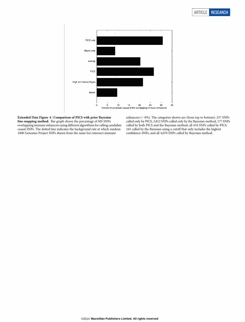
Extended Data Figure 4 | Comparison of PICS with prior Bayesianfine-mapping method. Bar graph shows the percentage of MS SNPsoverlapping immune enhancers using different algorithms for calling candidatecausal SNPs. The dotted line indicates the background rate at which random1000 Genomes Project SNPs drawn from the same loci intersect immune
enhancers (,8%). The categories shown are (from top to bottom): 257 SNPscalled only by PICS, 3,812 SNPs called only by the Bayesian method, 177 SNPscalled by both PICS and the Bayesian method, all 434 SNPs called by PICS,165 called by the Bayesian using a cutoff that only includes the highestconfidence SNPs, and all 4,070 SNPs called by Bayesian method.
ARTICLE RESEARCH
Macmillan Publishers Limited. All rights reserved©2014

Extended Data Figure 5 | LD distance between PICS lead SNPs and GWAScatalogue index SNPs. Histogram indicates LD distance (in r2) between PICSfine-mapped Immunochip lead SNPs and previously reported GWAScatalogue index SNPs from the same loci.
RESEARCH ARTICLE
Macmillan Publishers Limited. All rights reserved©2014

Extended Data Figure 6 | Purification of human immune cell subsets.a, Immune populations subjected to epigenomic profiling in this study (redlabels) or prior publications. b, CD41 cells were enriched based on CD25expression (MACS) and subsequently sorted based on CD25hiCD127lo/2 toisolate Treg cells; confirmed with FOXP3 intracellular staining. c, CD41CD252
cells were sorted to isolate Tmem (CD45RO1CD45RA2) and Tnaive
(CD45RO2CD45RA1) cells. d, CD41CD252 cells were PMA/ionomycin
stimulated and separated based on IL17 surface expression (MACS andFACS) to isolate TH17 cells (IL171) and THstim cells (IL172). e, Naive(CD45RA1CD45RO2) and memory (CD45RA2CD45RO1) CD81 T cellswere isolated using a BD FACSAria 4-way cell sorter. Results are shownfrom one of two large-scale sorts. f, Mononuclear cells were isolated frompaediatric tonsils. Following CD10 enrichment (MACS), B centroblasts(CD191CD101CXCR41CD442CD32) were purified by FACS.
ARTICLE RESEARCH
Macmillan Publishers Limited. All rights reserved©2014

Extended Data Figure 7 | PICS SNPs localize to immune enhancers andstimulus-dependent H3K27ac peaks in super-enhancers. a, Correlationmatrix of 56 cell types, clustered by similarity of H3K27ac profiles (high 5 red,low 5 blue). b, Enrichment of non-coding autoimmune disease candidatecausal SNPs within immune enhancers and promoters compared tobackground. The background expectation is based on frequency-matchedcontrol SNPs drawn from within 50 kb of the candidate causal SNPs. Candidatecausal SNPs that produced coding changes or were in LD with a coding variant
(paired bars on the right) showed a smaller degree of enrichment in immuneenhancers and promoters compared to background. c, Overlap of PICS SNPswith H3K27ac peaks within T-cell super-enhancers. Bar plot shows overlapof PICS SNPs with H3K27ac peaks in super-enhancers in CD41 T-cells,compared to random SNPs drawn from within the same super-enhancers(all CD41; left bar graph). Adjacent bars show overlap to H3K27ac peakswithin CD41 T-cell super-enhancers that do (Stim) or do not (Unstim)increase their acetylation upon stimulation.
RESEARCH ARTICLE
Macmillan Publishers Limited. All rights reserved©2014

Extended Data Figure 8 | Expression pattern of genes with PICS autoimmunity coding SNPs. Heatmap shows the relative expression levels of genes withcoding SNPs associated with Crohn’s disease, multiple sclerosis, and rheumatoid arthritis.
ARTICLE RESEARCH
Macmillan Publishers Limited. All rights reserved©2014

Extended Data Figure 9 | Motifs directly altered by or adjacent to candidatecausal SNPs. a, Known motifs (identified by conservation or SELEX) createdor disrupted by candidate causal SNPs at a higher frequency than expectedby chance when compared to control SNPs drawn from the same loci.
b, Additional motifs, identified by conservation, created or disrupted bycandidate causal SNPs more frequently than by chance. c, Known motifssignificantly enriched within 100 bp of candidate causal SNPs, compared tobackground control SNPs drawn from the same loci.
RESEARCH ARTICLE
Macmillan Publishers Limited. All rights reserved©2014

Extended Data Figure 10 | Enrichment of candidate causal eQTL SNPs infunctional elements. a, PICS was used to identify candidate causal SNPs for4,136 eQTL signals in peripheral blood. Bar plot show their overlap withindicated functional genic annotations. Background expectation was calculatedbased on frequency-matched control SNPs drawn from within 50 kb of the
candidate causal SNPs. b, Overlap of candidate causal eQTL SNPs withimmune enhancers and promoters, versus background. c, Magnitudes ofdisease-associated eQTLs compared to the space of all eQTLs. Histogramcompares the magnitudes of PICS eQTL SNPs that overlap PICS autoimmunitySNPs against the full set of PICS eQTL SNPs.
ARTICLE RESEARCH
Macmillan Publishers Limited. All rights reserved©2014