Embed Size (px)
Citation preview
CentralBringing Excellence in Open Access
Journal of Cancer Biology & Research
Cite this article: Shamma A, Muranaka H (2015) The Genetic-Epigenetic Interplay in Cancer. J Cancer Biol Res 3(4): 1072.
*Corresponding author
Awad Shamma, Division of Oncology and Molecular Biology; Cancer Research Institute, Kanazawa University, Kanazawa, Ishikawa 920-1192, Japan, Email:
Submitted: 19 November 2015
Accepted: 09 December 2015
Published: 11 December 2015
Copyright© 2015 Shamma et al.
OPEN ACCESS
Keywords•RB•ATM•DNA damage response•DNA methylation•Histonemodifications•Epigenetic control•Tumor evolution
Review Article
The Genetic-Epigenetic Interplay in CancerAwad Shamma* and Hayato MuranakaDivision of Oncology and Molecular Biology, Cancer Research Institute, Japan
Abstract
Genetic and epigenetic alterations have important implications inhuman cancer. Genetic aberrations are associated with wide spread variations in deregulation of cellular functions that lead to cancer. However, there has been clear evidence that epigenetic alterations have profound influence on malignant progression. The genetic and epigenetic alterations have been long considered as two independent mechanisms and the interplay between the two systems in cancer is poorly understood. In this review, we summarize the molecular links between the genetic and epigenetic events that lead to cancer and discuss the Rb-ATM-DNMT1 nexus as a novel pathway linking genetic aberration of two genes commonly inactivated in human cancer into epigenetic events indispensable for tumor evolution.
INTRODUCTION
Epigenetics and cellular identity
The term “epigenetics” was originally introduced by Conrad Waddington to describe heritable changes in the cellular phenotype independent of alterations in the DNA sequence [1]. Epigenetics is also considered as chromatin-based events that affect local transcriptional profile during cellular proliferation or differentiation. These events include modifications of the DNA and the histone proteins in a dynamic and highly regulated manner. The basic unit of the chromatin is the nucleosome, which contains 145–147 base pairs of DNA wrapped around an octamer of the four core histones H2A, H2B, H3 and H4. The nucleosome unit is repeated throughout the genome connected by linker DNA, and the chromatin is further compacted by association with the linker histone H1. This provides proper packaging of the entire genome that contains the heritable material of the eukaryotic cells, and limits DNA accessibility to its binding partner proteins. There are at least four known DNA modifications and 16 different histone modifications [2]. These modifications are the docking sites for specialized proteins known as chromatin readers that specifically recognize and bind to these modifications through specific domains. Then, the chromatin-bound readers recruit additional modifiers and remodeling enzymes to convey information of the chromatin modifications to regulate critical cellular functions such as transcription, DNA replication, DNA repair, cellular identity and phenotype.
DNA modifications
Epigenetic modifications of the mammalian DNA at the 5-carbon position of the cytosine residues include methylation (5mC), hydroxyl methylation (5hmC), formylation (5fC) and carboxylation (5caC). The 5mC is the most extensively studied
DNA modification, it constitutes ~1% of all DNA bases and is usually located in the promoter regions known as CpG islands that exist in ~70% of mammalian promoters. The 5hmC is a further modification of the 5mC by enzymatic oxidation, catalyzed by Ten-11 translocation (Tet) proteins [3]. However, recent genome-wide studies in mouse and human embryonic stem cells (ESCs)identified the5hmC modifications mainly localized in the intragenic regions of active genes as well as in the binding sites of the pluripotency regulators [4,5]. In addition, it has been reported that the methyl CpG binding protein 2 (MeCP2) and DNA methyltransferase-1 (DNMT1) have a poor binding affinity to the 5hmC, which leads passive DNA demethylation due to improper maintenance of the DNA methylation code during DNA replication [6,7].
DNA methyltransferases and DNA methylation
Methylation of the mammalian DNA is initiated and maintained by the DNA methyltransferases (DNMTs) that are responsible for transfer of methyl group from the universal methyl donor, S-adenosyl-L-methionine (SAM), to the 5-carbon position of cytosine residues. Previous reports demonstrated that DNA methylation is essential for the mammalian development [8,9]. There are at least 5 known mammalian DNMTs including DNMT1, DNMT2, DNMT3A, DNMT3B, and DNMT3L. DNMT3A and DNMT3B encode the de novo DNMTs that establish the DNA methylation code shortly after implantation of the fertilized egg [9]. DNMT1 encodes the maintenance methyltransferases responsible for copying the DNA methylation code on the parental strand to the daughter strand during DNA replication owing to its high affinity and specificity to the hemi-methylated cytosines [10]. DNMT2 is a highly conserved human tRNA methyltransferases [11]. DNMT3L does not possess an inherent enzymatic activity but it induces de novo DNA methylation
CentralBringing Excellence in Open Access
Shamma et al. (2015)Email:
J Cancer Biol Res 3(4): 1072 (2015) 2/13
by docking DNMT3A to the nucleosome [12]. The N-terminal cysteine-rich domain of DNMT3L specifically interacts with the amino terminus of histone H3 only when H3K4 is not modified suggesting that DNMT3L acts as a sensor for H3K4 methylation [13].
DNA methyl lysine readers
The methyl-CpG binding domain proteins MBD1, MBD2, MBD3, MBD4, MeCP2, and Kaiso are responsible for recognition and interpretation of the DNA methylation mark in mammals. All MBDs except MBD3 have high binding affinity to methylated cytosine [14]. MBD2 exist in the MeCP1 complex together with the chromatin remodeling protein NuRD/Mi2, HDAC1/2 and RbAp46/48where the MeCP1 complex is preferentially targeted to methylated DNA by MBD2 [15]. MBD4 has endonuclease activity and is involved in repair of methylated DNA damage, andits deficiency is associated with accumulation of mCpG sites mutations that lead to tumorigenesis [16]. MeCP2 can be targeted only to mCpG flanked by A/T bases and it is a target molecule for the DNA methylation maintenance DNMT1 [17,18]. Kaiso binds to mCpG preferably within the sequence 5’-CGCG-3’ and has an essential role as a global repressor during early development [19,20].
DNA demethylases
DNA methylation is regulated by coordination of the DNMTs and DNA demethylases (DDMs).Passive DNA demethylation can result from improper maintenance of the DNA methylation due to low DNMT1 specificity to 5hmC or DNMT1 protein destabilization. The 5mC marks are also subjected to active demethylation mediated by the thymine DNA glycosylase (TDG) and the TET family proteins (Figure 2) [3,4]. Previous studies indicated that the lysine demethylases (KDMs) KDM1A and KDM1B are essential for the maintenance of global DNA methylation and genomic imprinting as well as coordination of histone methylation [21,22]. Furthermore, loss of KDM1Ainduces early embryonic lethality in mice due to DNMT1 protein destabilization and consequent global DNA hypomethylation [21]
Posttranslational modifications of the histone
The four core histones share similar structure with globular hydrophobic core regions and flexible N‐terminal regions that protrudes from the nucleosome and known as histone tails. Posttranslational modifications (PTMs) of these N‐terminal tails are major epigenetic events. PTMs of the histones influence chromatin modifications depending on the site, the degree, and the type of modification with high diversity and complexity in their functional outcomes [23]. The most extensively studied PTMs of the histone are acetylation, methylation, phosphorylation, ubiquitination and sumoylation.
Histone acetylation-deacetylation dynamics
Acetylation of lysine residues on the histone tail is a dynamic process under the control of lysine acetyl transferases (KATs) and histone deacetylases (HDACs) [24]. It is widely known that acetylation on lysine residues results in reduction of the positive charge of the histones, and this weakens their interaction with the negatively charged DNA leading to chromatin relaxation. The
HDACs family of enzymes includes at least 18 members (HDAC1-18) that reverse lysine acetylation restoring the positive charge of the histones and strengthen their interaction with the DNA resulting in DNA compaction with less accessibility by the DNA interacting proteins. It was shown that KATs and HDACs could also target non-histone proteins such as c-MYC, p53, STAT3, HIF1α, Smad7, NF-KB, E2F1-3 and pRB [25].
Histone methylation diversity
Histones are methylated on the side chains of the basic residues lysine (K), arginine (R) and histidine (His)with variable degree of methylation such as mono-methylation (Kme1, Rme1 and Hisme1), di-methylation (Kme2, Rme2), tri-methylation (Kme3). The most extensively studied histone methylation includes methylation of histone H3 at lysine 4 (K4), lysine 9 (K9), lysine 27 (K27), lysine 36 (K36) and lysine 79 (K79) and histone H4at lysine 20(K20).Histone arginine residues can be also di-methylated asymmetrically (H3R2me2a, H3R17me2a, H3R26me2a and H3R42me2a), a modification that was found to be associated with transcriptionally active regions or symmetrically(H3R8me2s and H4R3me2s), a modification that was found to be associated with heterochromatin formation and gene repression [26]. However, deregulation of the arginine residues was reported to be rare and their functions are complex [27]. Histone methylation marks were originally believed to be irreversible, however as mentioned earlier, discovery of the lysine methyltransferases (KMTs) and recently the KDMs suggested that histone methylation is a reversible and dynamic process [28]. In contrast to KATs, KMTs are highly specific in targeting certain lysine residues and they contain a conserved SET domain with methyl transferase activity, and they are divided into 9 groups (KDM1-8 and PADI). List of histone modifiers are listed in Table 1. The functional outcome of the histone tail methylation is context-dependent being influenced by location of the methylated lysine residue, degree of methylation either mono-methylated (me1) or di-methylated (me2) or tri-methylated (me3), combinations of methylated lysine residues at different locations, type of the reader proteins, and the functional coordination between KMTs and KDMs.H3K4me3 is generally considered as an active mark, and frequently associated with genes poised for transcriptional activation whereas H3K27me3 is widely known as a repressive mark. H3K4me1 is associated with gene enhancers whereas H3K4me3 is linked to promoter activation [29]. H3K79me2 is important for cell cycle regulation, whereas H3K79me3 is linked to the wnt signaling pathway [30,31]. The H3K4me2 and H3K4me3 are generally associated with transcriptional activation, but in some instances they can be interpreted as repressive marks probably due to interpretation by different reader proteins such as when bound to the PHD domain containing co-repressor protein inhibitor of growth family member 2 (ING2) [32]. Although H3K4me3 and H3K27me3 are marks associated with active and repressive transcription, respectively, when they are present together, they appear to have a role in poising the gene transcriptional activity [33], probably due to interpretation by methyl lysine readers (MLRs)containing multiple domains that are capable of recognizing different histone marks at the same time for initiating specific transcriptional outcome. In other situation, maximum transcriptional activation requires addition of an active mark and removal of repression mark. For example,
CentralBringing Excellence in Open Access
Shamma et al. (2015)Email:
J Cancer Biol Res 3(4): 1072 (2015) 3/13
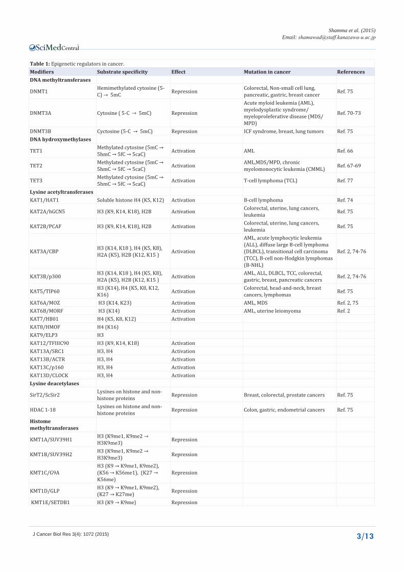
Table 1: Epigenetic regulators in cancer.Modifiers Substrate specificity Effect Mutation in cancer ReferencesDNA methyltransferases
DNMT1 Hemimethylated cytosine (5-C) → 5mC Repression Colorectal, Non-small cell lung,
pancreatic, gastric, breast cancer Ref. 75
DNMT3A Cytosine ( 5-C → 5mC) Repression
Acute myloid leukemia (AML), myelodysplastic syndrome/myeloproleferative disease (MDS/MPD)
Ref. 70-73
DNMT3B Cyctosine (5-C → 5mC) Repression ICF syndrome, breast, lung tumors Ref. 75DNA hydroxymethylases
TET1 Methylated cytosine (5mC → 5hmC → 5fC → 5caC) Activation AML Ref. 66
TET2 Methylated cytosine (5mC → 5hmC → 5fC → 5caC) Activation AML,MDS/MPD, chronic
myelomonocytic leukemia (CMML) Ref. 67-69
TET3 Methylated cytosine (5mC → 5hmC → 5fC → 5caC) Activation T-cell lymphoma (TCL) Ref. 77
Lysine acetyltransferasesKAT1/HAT1 Soluble histone H4 (K5, K12) Activation B-cell lymphoma Ref. 74
KAT2A/hGCN5 H3 (K9, K14, K18), H2B Activation Colorectal, uterine, lung cancers, leukemia Ref. 75
KAT2B/PCAF H3 (K9, K14, K18), H2B Activation Colorectal, uterine, lung cancers, leukemia Ref. 75
KAT3A/CBP H3 (K14, K18 ), H4 (K5, K8), H2A (K5), H2B (K12, K15 ) Activation
AML, acute lymphocytic leukemia (ALL), diffuse large B-cell lymphoma (DLBCL), transitional cell carcinoma (TCC), B-cell non-Hodgkin lymphomas (B-NHL)
Ref. 2, 74-76
KAT3B/p300 H3 (K14, K18 ), H4 (K5, K8), H2A (K5), H2B (K12, K15 ) Activation AML, ALL, DLBCL, TCC, colorectal,
gastric, breast, pancreatic cancers Ref. 2, 74-76
KAT5/TIP60 H3 (K14), H4 (K5, K8, K12, K16) Activation Colorectal, head-and-neck, breast
cancers, lymphomas Ref. 75
KAT6A/MOZ H3 (K14, K23) Activation AML, MDS Ref. 2, 75KAT6B/MORF H3 (K14) Activation AML, uterine leiomyoma Ref. 2KAT7/HB01 H4 (K5, K8, K12) ActivationKAT8/HMOF H4 (K16)KAT9/ELP3 H3KAT12/TFIIIC90 H3 (K9, K14, K18) ActivationKAT13A/SRC1 H3, H4 ActivationKAT13B/ACTR H3, H4 ActivationKAT13C/p160 H3, H4 ActivationKAT13D/CLOCK H3, H4 ActivationLysine deacetylases
SirT2/ScSir2 Lysines on histone and non-histone proteins Repression Breast, colorectal, prostate cancers Ref. 75
HDAC 1-18 Lysines on histone and non-histone proteins Repression Colon, gastric, endometrial cancers Ref. 75
Histome methyltransferases
KMT1A/SUV39H1 H3 (K9me1, K9me2 → H3K9me3) Repression
KMT1B/SUV39H2 H3 (K9me1, K9me2 → H3K9me3) Repression
KMT1C/G9AH3 (K9 → K9me1, K9me2), (K56 → K56me1), (K27 → K56me)
Repression
KMT1D/GLP H3 (K9 → K9me1, K9me2), (K27 → K27me) Repression
KMT1E/SETDB1 H3 (K9 → K9me) Repression
CentralBringing Excellence in Open Access
Shamma et al. (2015)Email:
J Cancer Biol Res 3(4): 1072 (2015) 4/13
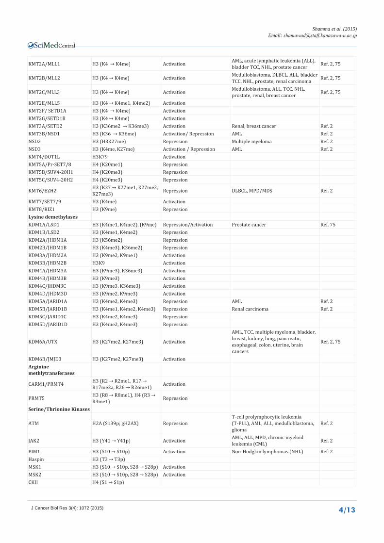
KMT2A/MLL1 H3 (K4 → K4me) Activation AML, acute lymphatic leukemia (ALL), bladder TCC, NHL, prostate cancer Ref. 2, 75
KMT2B/MLL2 H3 (K4 → K4me) Activation Medulloblastoma, DLBCL, ALL, bladder TCC, NHL, prostate, renal carcinoma Ref. 2, 75
KMT2C/MLL3 H3 (K4 → K4me) Activation Medulloblastoma, ALL, TCC, NHL, prostate, renal, breast cancer Ref. 2, 75
KMT2E/MLL5 H3 (K4 → K4me1, K4me2) ActivationKMT2F/ SETD1A H3 (K4 → K4me) ActivationKMT2G/SETD1B H3 (K4 → K4me) ActivationKMT3A/SETD2 H3 (K36me2 → K36me3) Activation Renal, breast cancer Ref. 2KMT3B/NSD1 H3 (K36 → K36me) Activation/ Repression AML Ref. 2NSD2 H3 (H3K27me) Repression Multiple myeloma Ref. 2NSD3 H3 (K4me, K27me) Activation / Repression AML Ref. 2KMT4/DOT1L H3K79 ActivationKMT5A/Pr-SET7/8 H4 (K20me1) RepressionKMT5B/SUV4-20H1 H4 (K20me3) RepressionKMT5C/SUV4-20H2 H4 (K20me3) Repression
KMT6/EZH2 H3 (K27 → K27me1, K27me2, K27me3) Repression DLBCL, MPD/MDS Ref. 2
KMT7/SET7/9 H3 (K4me) ActivationKMT8/RIZ1 H3 (K9me) RepressionLysine demethylasesKDM1A/LSD1 H3 (K4me1, K4me2), (K9me) Repression/Activation Prostate cancer Ref. 75KDM1B/LSD2 H3 (K4me1, K4me2) RepressionKDM2A/JHDM1A H3 (K56me2) RepressionKDM2B/JHDM1B H3 (K4me3), K36me2) RepressionKDM3A/JHDM2A H3 (K9me2, K9me1) ActivationKDM3B/JHDM2B H3K9 ActivationKDM4A/JHDM3A H3 (K9me3), K36me3) ActivationKDM4B/JHDM3B H3 (K9me3) ActivationKDM4C/JHDM3C H3 (K9me3, K36me3) ActivationKDM4D/JHDM3D H3 (K9me2, K9me3) ActivationKDM5A/JARID1A H3 (K4me2, K4me3) Repression AML Ref. 2KDM5B/JARID1B H3 (K4me1, K4me2, K4me3) Repression Renal carcinoma Ref. 2KDM5C/JARID1C H3 (K4me2, K4me3) RepressionKDM5D/JARID1D H3 (K4me2, K4me3) Repression
KDM6A/UTX H3 (K27me2, K27me3) Activation
AML, TCC, multiple myeloma, bladder, breast, kidney, lung, pancreatic, esophageal, colon, uterine, brain cancers
Ref. 2, 75
KDM6B/JMJD3 H3 (K27me2, K27me3) Activation Arginine methlytransferases
CARM1/PRMT4 H3 (R2 → R2me1, R17 → R17me2a, R26 → R26me1) Activation
PRMT5 H3 (R8 → R8me1), H4 (R3 → R3me1) Repression
Serine/Thrionine Kinases
ATM H2A (S139p; gH2AX) RepressionT-cell prolymphocytic leukemia (T-PLL), AML, ALL, medulloblastoma, glioma
Ref. 2
JAK2 H3 (Y41 → Y41p) Activation AML, ALL, MPD, chronic myeloid leukemia (CML) Ref. 2
PIM1 H3 (S10 → S10p) Activation Non-Hodgkin lymphomas (NHL) Ref. 2Haspin H3 (T3 → T3p)MSK1 H3 (S10 → S10p, S28 → S28p) ActivationMSK2 H3 (S10 → S10p, S28 → S28p) ActivationCKII H4 (S1 → S1p)
CentralBringing Excellence in Open Access
Shamma et al. (2015)Email:
J Cancer Biol Res 3(4): 1072 (2015) 5/13
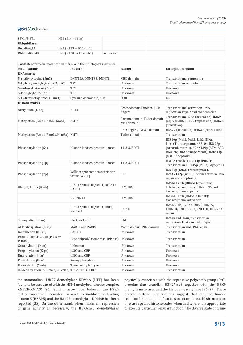
STK4/MST1 H2B (S14→ S14p)UbiquitilasesBmi/Ring1A H2A (K119 → K119ub1)RNF20/RNF40 H2B (K120 → K120ub1) Activation
Table 2: Chromatin modification marks and their biological relevance.Modifications Inducer Reader Biological functionDNA marks5-methylcytosine (5mC) DNMT3A, DNMT3B, DNMT1 MBD domain Transcriptional repression5-hydroxymethylcytosine (5hmC) TET Unknown Transcription activation5-carboxylcytosine (5caC) TET Unknown Unknown5-formylcytosine (5fC) TET Unknown Unknown5-hydroxmethyluracil (5hmU) Cytosine deaminase, AID DDR BERHistone marks
Acetylation (K-ac) HATs BromodomainTandem, PHD fingers
Transcriptional activation, DNA replication, repair and condensation
Methylation (Kme1, Kme2, Kme3) KMTs Chromodomain, Tudor domain, MBT domain,
Transcription: H3K4 (activation), H3K9 (repression), H3K27 (repression), H3K36 (activation),
PHD fingers, PWWP domain H3K79 (activation), H4K20 (repression) Methylation (Rme1, Rme2s, Kme3a) KMTs Tudor domain Transcription
Phosphorylation (Sp) Histone kinases, protein kinases 14-3-3, BRCT
H3S10p (Msk1, Msk2, Rsk2, IKKa, Pim1; Transcription), H3S10p, H3S28p (AuroraB;mitosis), H2AX139p (ATM, ATR, DNA-PK; DNA damage repair), H2BS14p (Mst1; Apoptosis)
Phosphorylation (Tp) Histone kinases, protein kinases 14-3-3, BRCT H3T6p (PKCb1) H3T11p (PRK1); Transcription, H3T45p (PKCd); Apoptosis
Phosphorylation (Yp) William syndrome transcription factor (WSTF) SH3
H3Y41p (JAK2; Transcription), H2AXY142p (WSTF; Switch between DNA repair and apoptosis)
Ubiquitylation (K-ub) RING1A/RING1B/BMI1, BRCA1/BARD1 UIM, IUM
H2AK119-ub (BRCA1); maintains heterochromatin at satellite DNA and transcriptional repression
RNF20/40 UIM, IUM H2BK120-ub (RNF20/RNF40); transcriptional activation
RING1A/RING1B/BMI1, RNF8, RNF168 RAP80
H2AK63ub, H2AXK63ub (RING1A/RING1B/BMI1, RNF8, RNF168) DDR and repair
Sumoylation (K-su) ubc9, siz1,siz2 SIM H2Asu and H4su; transcription repression, H2A.Zsu; DSBs repair
ADP ribosylation (E-ar) MARTs and PARPs Macro domain, PBZ domain Transcription and DNA repairDeimination (R→cit) PAD1-4 Unknown TranscriptionProline isomerisation (P-cis ⇔ P-trans) Peptidylprolyl isomerase (PPIase) Unknown Transcription
Crotonylation (K-cr) Unknown Unknown TranscriptionPropionylation (K-pr) p300 and CBP Unknown UnknownButyrylation K-bu) p300 and CBP Unknown UnknownFormylation (K-fo) Formylphosphate Unknown UnknownHyroxylation (Y-oh) Tyrosine Hydroxylase Unknown UnknownO-GlcNAcylation (S-GlcNac, -GlcNac) TET2, TET3 → OGT Unknown Transcription
the mammalian H3K27 demethylase KDM6A (UTX) has been found to be associated with the H3K4 methyltransferase complex KMT2B-KMT2C [34]. Similar association between the H3K4 methyltransferase complex subunit retinoblastoma-binding protein 5 (RBBP5) and the H3K27 demethylase KDM6B has been reported [35]. On the other hand, when maximum repression of gene activity is necessary, the H3K4me3 demethylases
physically associates with the repressive polycomb group (PcG) proteins that establish H3K27me3 together with the H3K9 methyltransferases and the histone deacetylases [36, 37]. These diverse histone modifications suggest that the coordinated reciprocal histone modifications function to establish, maintain or erase specific histone codes when and where it is appropriate to execute particular cellular function. The diverse state of lysine
CentralBringing Excellence in Open Access
Shamma et al. (2015)Email:
J Cancer Biol Res 3(4): 1072 (2015) 6/13
modifications are read and interpreted by the MLRs containing specialized recognition domains. The MLRs include the Royal family of Tudor domains, malignant brain tumor domains (MBT) and chromo domains and the plant home domain (PHD) family members. The MLRs recruit various proteins necessary for the execution of the PTMs and communications with other cellular systems.
Histone phosphorylation
Histone phosphorylation is a dynamic process, in which phosphate group is added to specific serine, thymine or tyrosine residues. Histone H1, H2A, H2B, H3, and H4 can be phosphorylated at multiple phosphorylation sites. Histone phosphorylation is catalyzed by specific kinases and it has been linked to many cellular functions including transcriptional activation, mitosis, DNA damage repair, and apoptosis (Table 2). Histone H3 phosphorylation at serine 10 (H3S10p) is the most extensively investigated histone phosphorylation mark. H3S10p is considered as hallmark of mitosis and has been directly linked to chromatin condensation, faithful chromosomal segregation and post-mitotic dissociation of the HP1 protein [38,39]. Phosphorylation of histone H3 on threonine 6(H3T6p) prevents demethylation of H3K4 by KDM1A and KDM5B, which contributes to transcriptional activation [40]. Phosphorylation of H2AX on serine 139 (H2AX) is key component of DNA damage response (DDR).This modification is rapid and spread over megabases from the break site and mediated by the phosphatidylinositol-3 kinase–like kinases (PIKK)including ATM, which is a central molecule in the DDR pathway. The recently identified constitutive phosphorylation of histone H2A at tyrosine 142 (H2ApY142) is catalyzed by william syndrome transcription factor (WSTF) and dephosphorylated by eyes absent homolog (EYA) phosphatases following DNA damage [41]. The reciprocal correlation between H2ApY142and γH2AX has been suggested as switch mechanism between DNA damage repair and apoptosis following DNA damage [42]. Future elucidation of the regulatory mechanisms of the H2ApY142in determining cell fate after DNA damage would be of great interest. Chromatin modification marks and their biological interpretation are listed in Table 2.
DNA methyltransferases deregulation in cancer
The mammalian DNA is subjected to wave of demethylation to erase the whole genome DNA methylation prior to implantation of the fertilized egg, followed by de novo DNA methylation organized by DNMT3A and DNMT3B.The DNA methylation code is established in coordination with the histone code to determine specific transcriptional output essential for the development and embryogenesis. DNMT3A and DNMT3B are highly expressed in embryonic tissues and ESCs but expressed at low level in differentiated cells. On the other hand, DNMT1 is highly expressed in proliferating cells but down regulated in non-proliferating cells [43]. DNMT1 is mainly regulated by posttranslational modification mechanisms including acetylation, methylation, phosphorylation and ubiquitination [44].The histone H3K4-specific KMT7 (SET7) directly methylates DNMT1 at K142 during S and G2 phases of the cell cycle and promotes DNMT1 degradation [45]. In addition, the interplay between DNMT1 methylation at lysine 142 and DNMT1 phosphorylation at serine
143 coordinates DNMT1 protein stability [46]. Furthermore, the lysine demethylase LSD1 (KDM1A) demethylates and stabilizes DNMT1 in vivo [21]. On the other hand, the acetyltransferase Tip60 triggers DNMT1 acetylation and subsequent ubiquitination by the PHD and ring finger domains 1 (UHRF1) resulting in DNMT1 destabilization [47]. Consistently, we recently demonstrated that the genetic interaction of the retinoblastoma (Rb) and the ataxia telangiectasia mutated (ATM) coordinates the Tip60 and UHRF1 functions inDNMT1destabilization that result in DNA hypomethylation-induced cellular senescence that serve to restrict malignant transformation [48]. Furthermore, recent data showed that Tip60 couples ATM signaling to chromatin modifications [49]. Taken together, these data revealed previously unknown molecular network coordinating protein posttranslational modifications, epigenetic alterations, genomic instability and malignant transformation. DNMT1 knockout mice have extensive genome-wide hypomethylation and die shortly after gastrulation [50]. In addition, DNMT1 inactivating mutations lead to loss of imprinting and defective X chromosome inactivation [51,52]. DNMT1-deficient ESCs are viable but they show genome-wide DNA hypomethylation, genomic instability and they die when induced to differentiate [50,53]. Furthermore, DNMT1 knockout MEFs are not viable due to immediate apoptosis butDNMT1conditional knockout in MEFs induces apoptosis within few passages [54,55]. We also reported a critical role of DNMT1 in malignant transformation of the thyroid C cells in mice model as well as in MEFs immortalization [48]. We discovered that inactivation of Rb and ATM induces DNMT1 protein stabilization that result in aberrant DNA hypermethylation patterns localized to cancer related gene promoters. Importantly, DNMT1 depletion by specific shRNA or by DNA methyl transferase inhibitors 5-Aza-C or Trichostatin A in these cells reversed the aberrant DNA methylation patterns and signs of cellular transformation. These numerous genetic studies indicated that DNMT1 is indispensable for embryonic development, cell proliferation and tumorigenesis. DNMT3B knockout mice die at embryonic day E9.5 and show multiple developmental defects, whereas the DNMT3A knockout mice die shortly after birth [9].Therefore, disruption of any of the catalytically active DMTs is lethalin mice indicating that DNA methylation plays a central role in mammalian development. DNMT3B but not DNMT3A knockout embryos and ESCs showed significant hypomethylation of the pericentromeric satellite repeats implicating that these genomic regions are specific targets of DNMT3B for methylation maintenance in vivo [9]. Human DNMT3B gene mutations cause a rare autosomal syndrome characterized by immunodeficiency, centromere instability and facial abnormalities (ICF) [56]. ICF patients have centromeric instability of chromosomes 1, 9, and 16 associated with abnormal hypomethylation at their pericentromeric satellite regions. Indeed, many human tumors have similar losses of DNA methylation and chromosomal structural changes in these regions [57]. DNMT3L knockout male mice are viable but sterile due to defective meiosis and germ cells loss but pups from DNMT3L knockout female mice are not viable due to neural tubes defects due to hypomethylation of the maternally imprinted genes [58].
Aberration of the DNA methylation in cancer
Deregulation of the cellular mechanisms that maintain DNA

CentralBringing Excellence in Open Access
Shamma et al. (2015)Email:
J Cancer Biol Res 3(4): 1072 (2015) 7/13
methylation results in aberrant DNA methylation patterns frequently detected in cancer [59], (Figure 1 and Figure 2). In general, promoter methylation of tumor suppressor genes is a common cause of cancer and predicts poor prognosis in cancer patients [60-62]. Hypermethylation of the CpG island methylator phenotype (CIMP) genes is associated with methylation of the DNA mismatch repair gene MLH1 and predicts a favorable prognosis in colon cancers [63]. Although DNA methylation of CpG islands is an important part of cancer epigenetics, this perspective has been recently changed after the discovery of the CpG island shores and asymmetric nucleosome modifications during DNA replication [64]. In addition, studies of whole genome DNA methylation analysis revealed abnormal DNA methylation pattern in previously unexpected genomic regions such as gene body, around transcription start site and the far upstream promoters. Although the functional relevance of these regions is unknown methylation of non-CG sequence is enriched in the body of transcriptionally active genes [5,65]. DNA methylation is not a stable modification as previously thought because they are subjected to active demethylation mediated by TDG and passive demethylation by DNA hydroxylases. The mammalian DNA hydroxylases TET1 is a translocation partner of MLL-translocated in a subset of AML patients [66]. In addition, several reports indicated recurrent inactivating mutations in TET2 in hematological malignancies and these mutations are associated with poor prognosis of these patients [67-69]. Although mutations in DNA methyl transferases are associated with developmental abnormalities, somatic mutations of DNMT3A have been recently detected in human malignancies, and these mutations are associated with poor prognosis of AML patients [70-73].
Histone acetyltransferases in cancer
Frequent inactivating mutations in histone acetyl transferases (HATs) have been reported in various types of human cancer [74-76]. In addition, recurrent chromosomal translocation or coding mutations in various KATs were detected in solid and hematological cancers [2]. Although HDACs somatic mutations are not prevalent in cancers, altered expression level of various HDACs is detected in many types of cancer [2,75].There are currently two HDAC inhibitors specifically approved by the Food and Drug Administration (FDA) for the treatment of hematological malignancies. Vorinostat was approved for the treatment of cutaneous T cell lymphoma (CTCL) patients in 2006, followed by Romidepsin in 2009 and several HDAC inhibitors are currently in the phase of clinical trial for treatment of several types of human cancers.
Lysine methyltransferases in cancer
Recent data from human cancer genome mutational analysis identified driver mutations in several epigenetic regulators. For example, 89% of follicular lymphomas and 32% of diffuse large B-cell lymphomas have recurrent mutations in KMT2B (MLL2) [2,74]. Another study identified inactivating somatic mutations in KDM6A (UTX) in various types of human cancers, and introduction of KDM6A mutant resulted in low proliferation and marked transcriptional changes [2,75,77,78]. In addition, recurrent coding mutations have been identified in KDM5A, and KDM5C [2,75]. Although initial studies suggested that EZH2 (catalytic component of the PRC2 complex) functions as an oncogene, next generation sequencing (NGS) and targeted resequencing of cancer genome have recently identified coding mutations in EZH2 in various lymphoid and myeloid neoplasms.
Figure 1 DNA methylation patterns in development and cancer. Inactivation of the RB-ATM pathway induces epigenetic events that allow cells proliferation despite of unrepaired DNA damage resulting in genomic instability and malignant transformation.
CentralBringing Excellence in Open Access
Shamma et al. (2015)Email:
J Cancer Biol Res 3(4): 1072 (2015) 8/13
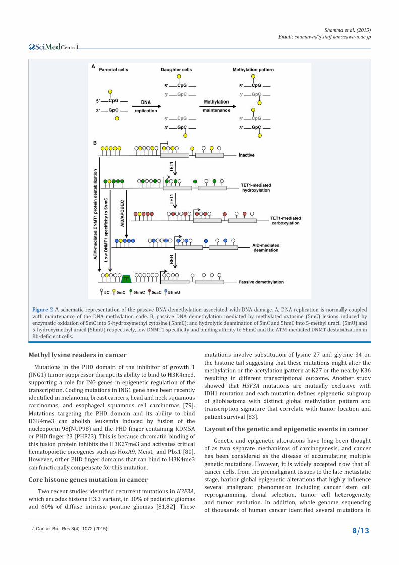
Figure 2 A schematic representation of the passive DNA demethylation associated with DNA damage. A, DNA replication is normally coupled with maintenance of the DNA methylation code. B, passive DNA demethylation mediated by methylated cytosine (5mC) lesions induced by enzymatic oxidation of 5mC into 5-hydroxymethyl cytosine (5hmC); and hydrolytic deamination of 5mC and 5hmC into 5-methyl uracil (5mU) and 5-hydroxymethyl uracil (5hmU) respectively, low DNMT1 specificity and binding affinity to 5hmC and the ATM-mediated DNMT destabilization in Rb-deficient cells.
Methyl lysine readers in cancer
Mutations in the PHD domain of the inhibitor of growth 1 (ING1) tumor suppressor disrupt its ability to bind to H3K4me3, supporting a role for ING genes in epigenetic regulation of the transcription. Coding mutations in ING1 gene have been recently identified in melanoma, breast cancers, head and neck squamous carcinomas, and esophageal squamous cell carcinomas [79]. Mutations targeting the PHD domain and its ability to bind H3K4me3 can abolish leukemia induced by fusion of the nucleoporin 98(NUP98) and the PHD finger containing KDM5A or PHD finger 23 (PHF23). This is because chromatin binding of this fusion protein inhibits the H3K27me3 and activates critical hematopoietic oncogenes such as HoxA9, Meis1, and Pbx1 [80]. However, other PHD finger domains that can bind to H3K4me3 can functionally compensate for this mutation.
Core histone genes mutation in cancer
Two recent studies identified recurrent mutations in H3F3A, which encodes histone H3.3 variant, in 30% of pediatric gliomas and 60% of diffuse intrinsic pontine gliomas [81,82]. These
mutations involve substitution of lysine 27 and glycine 34 on the histone tail suggesting that these mutations might alter the methylation or the acetylation pattern at K27 or the nearby K36 resulting in different transcriptional outcome. Another study showed that H3F3A mutations are mutually exclusive with IDH1 mutation and each mutation defines epigenetic subgroup of glioblastoma with distinct global methylation pattern and transcription signature that correlate with tumor location and patient survival [83].
Layout of the genetic and epigenetic events in cancer
Genetic and epigenetic alterations have long been thought of as two separate mechanisms of carcinogenesis, and cancer has been considered as the disease of accumulating multiple genetic mutations. However, it is widely accepted now that all cancer cells, from the premalignant tissues to the late metastatic stage, harbor global epigenetic alterations that highly influence several malignant phenomenon including cancer stem cell reprogramming, clonal selection, tumor cell heterogeneity and tumor evolution. In addition, whole genome sequencing of thousands of human cancer identified several mutations in
CentralBringing Excellence in Open Access
Shamma et al. (2015)Email:
J Cancer Biol Res 3(4): 1072 (2015) 9/13
the epigenetic regulatory genes such asTET1, TET2, DNMT3A, H3F3A, IDH1 and IDH2, KDM6A, KDM1, HDAC2. Furthermore, loss of functions of tumor suppressor genes and DNA damage repair genes due to promoter methylation is a well-documented mechanism of carcinogenesis. For example RB, BRACA1/2 and PTEN were reported to be hypermethylated, mutated or deleted in cancer [84]. Another group of genes known to be protective against cancer such as O6-methylguanine-DNA methyltransferase (MGMT), cyclin-dependent kinase inhibitor 2B (CDKN2B) and RASSF1Aare inactivated in cancer predominantly by promoter hypermethylation [85]. The MGMT is involved in methylated DNA damage repair by removing carcinogen-induced O6-methylguanine adducts that result inG to A transition mutation.Inactivation ofMGMT by epigenetic mechanism results in genetic mutations in critical genes such as p53 and KRAS resulting in cancer [85]. These observations indicated that epigenetic events could influence the genetic output through epigenetic control of the transcriptional activity of certain genes or even by inducing mutations of critical cancer-driver genes. However, there is little information about how genetic aberrations induce specific epigenetic events that would be enough for tumor evolution it out the requirement of multiple genetic mutations. While lots of efforts have been made to study the epigenetic-genetic layout in cancer, it is of interest to investigate the genetic-epigenetic layout as a mechanism of cancer, and to determine how genetic aberrations impact certain epigenetic events indispensable for malignant transformation. This is important simply because clonal selection and tumor evolution is mainly driven by epigenetic events, and the majority of these epigenetic events are chemical modifications that could be easily targeted and reversed in vivo.
The Rb epigenetic functions
The retinoblastoma (RB) tumor suppressor gene was the first tumor suppressor gene to be discovered more than three decades ago. Numerous studies have characterized the appreciated role of pRB-E2F interaction in transcription and cell cycle control. Later studies delineated the role of pRB interaction with tissue-specific transcription factors in regulation of terminal differentiation. We also reported novel functions of Rb in DDR and cellular senescence [86]. Recently, pRB has been shown to physically interact with several epigenetic modifiers including histone deacetylases (HDAC1, HDAC2), histone demethylases (RBP2), DNA methyl transferases (DNMT1), helicases (Brg1, Brm), histone methyl transferases (Suv39h1, RIZ and Suv4–20h1/h2) and histone binding proteins such as HP1[87]. These finding urged us to speculate that pRB functions as double-sided adhesive tape interacting with E2Fs transcription factors on one side and the epigenetic modifiers on the other side for dual transcriptional repression [88]. Recent reports suggested genome-wide functions of pRB in the regulation of heterochromatin domains including pericentric heterochromatin, telomeres and senescence-associated heterochromatic foci (SAHFs). There is also evidence that pRB interacts with Suv4–20h1/h2 histone methyltransferases that regulate histone H4K20me3 at the pericentric heterochromatin [87]. Furthermore, genome-wide analysis demonstrated that primary human retinoblastomas harbor mutation only in the RB gene, and this mutation was associated with promoter localized epigenetic alterations rather
than chromosomal instability (CIN) as a mechanism underlies the malignant transformation of these tumors [89]. These findings indicated that pRB repress the transcription of genomic regions much broader than the E2F consensus sequence as we previously thought. Accordingly, we recently demonstrated that inactivation of Rb allows ATM to physically bind to DNMT1 in a complex with Tip60 (acetyltransferase) and UHRF1(E3 ligase), and this confirmation results in ubiquitination-mediated degradation of DNMT1 causing global DNA hypomethylation and induction of DDR and cellular senescence regulatory genes to protect the cells against malignant transformation. Whereas simultaneous inactivation of Rb and ATM stabilizes the DNMT1 protein,which is recruited to the unrepaired DNA breaks leading to aberrant DNA hypermethylation pattern localized to promoter of genes that protect against cancer, and this allow the cells to continue growing despite of the unrepaired DNA damage resulting in genomic instability and cellular transformation [48]. These findings explain the pRb global epigenetic functions, and directly link genetic inactivation of the Rb-ATM pathway to DNA methylation through DNMT1 protein stabilization (Figure 3).
Epigenetic functions of ATM
ATM is a serine/threonine kinase, a member of the PI3K-like protein kinase (PIKK) family and is functionally implicated in DNA damage response and repair signaling. Matsuoka et al. previously identified wide range of ATM targets [90] however most of these targets are obviously unrelated to DNA repair suggesting that ATM exerts functions other than those related to the DNA damage and repair signaling. The first epigenetic signal in response to DNA damage is initiated by the ATM through induction ofγH2AX, a key epigenetic component of the DDR. Indeed, dephosphorylation of H2ApY142 and the induction of γH2AX constitute a cell fate switch mechanism between DDR and apoptosis following DNA damage [42]. The ATM protein has been also shown to regulate histone acetylation by negatively regulating the recruitment of HDAC4 [92]. Therefore, the full 370 KD protein leads someone to think about the functions of regions other than the PI3K-like kinase domain functions, which is extensively investigated. This is because the PI3K-like kinase domain constitutes only 10% of the ATM amino acid sequence. One possible explanation of ATM functions in regulating large diversity of targets is that ATM might serve as adaptor protein to bring interacting proteins in close proximity of the designated protein-protein interactions. Our recent data assigned similar role of ATM in assembling the UHRF1-DNMT1-Tip60 proteins interaction in macromolecule complex that facilitates ubiquitination and subsequent degradation of the DNMT1protein resulting DNA hypomethylation and induction of cellular senescence genes to restrict malignant transformation of the Rb-deficient cells (Figure 3).
The Rb-ATM pathway is a novel genetic-epigenetic layout in cancer
Several studies demonstrated various posttranslational modifications critical for the DNMT1 protein stability [44-47]. However, linking genetic aberration of tumor suppressor genes to DNMT1 stability in cancer was unknown. We recently demonstrated that the Rb-ATM genetic interaction is critical for
CentralBringing Excellence in Open Access
Shamma et al. (2015)Email:
J Cancer Biol Res 3(4): 1072 (2015) 10/13
DNMT1 protein stability and the malignant phenotype induced by aberrant DNA methylation pattern [48]. Importantly, blocking of this genetic-epigenetic axis through DNMT1 inhibition by specific shRNA or methyl transferase inhibitors markedly attenuated the signs of cellular transformation induced by genetic inactivation of the Rb-ATM pathway. These observations directly link somatic inactivation of the Rb gene by loss of hetrozygosity (LOH) to the ATM-induced DNMT1 destabilization
and the aberrant DNA hypomethylation pattern that antagonize transformation of the Rb-deficient cells. These observations also indicated that ATM exerts critical tumor suppressor functions by mediating epigenetic modifications to protect against malignant transformation of the Rb-deficient cells. Taken together, these data indicated that the genetic and epigenetic alterations are intertwined during cellular transformation, and highlighted the genetic aberration of the Rb-ATM-DNMT1pathway as a novel
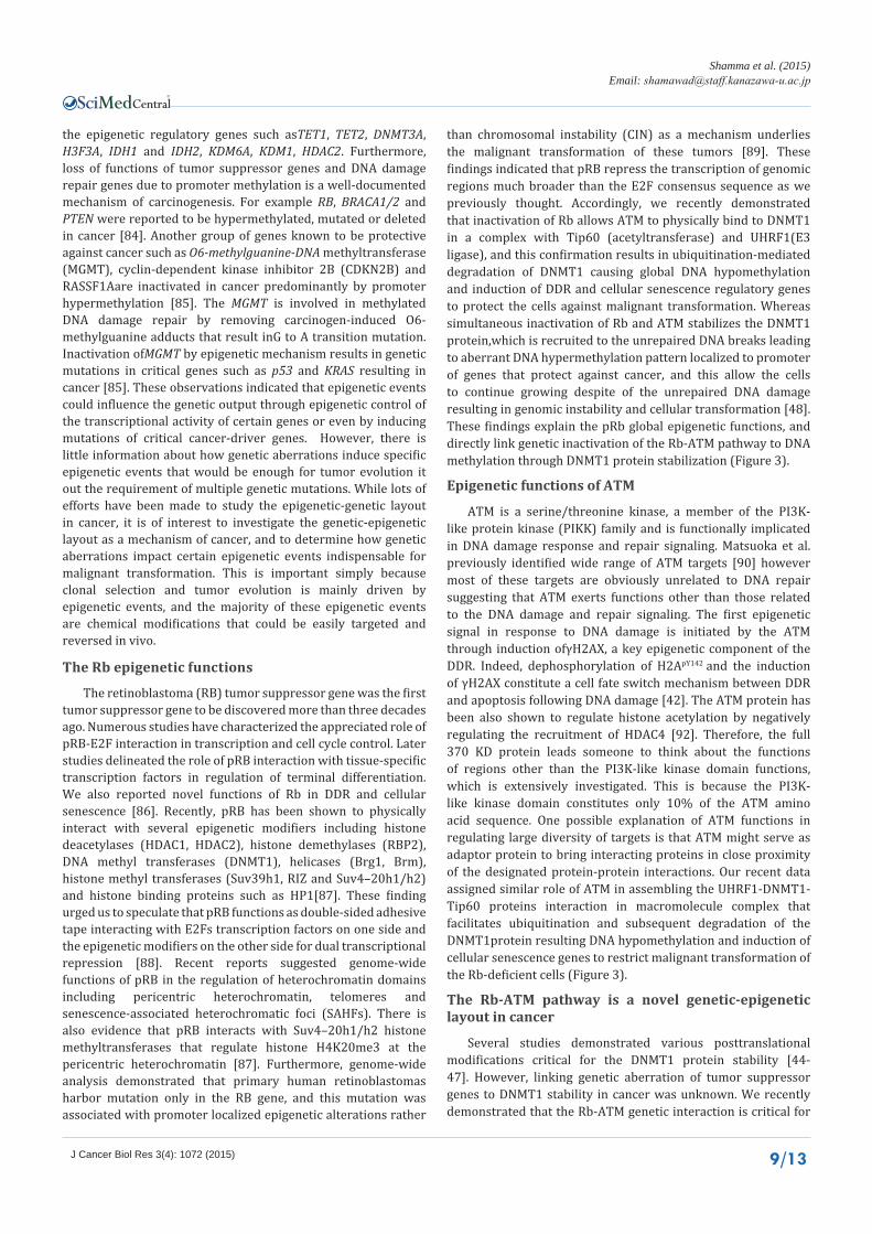
Figure 3 A schematic representation of the interplay between the genetic and epigenetic systems in cancer. A, the epigenetic output influenced by protein-protein interaction and posttranslational modifications of the �epigenetic regulators. B, the Rb-ATM-DNMT1 pathway is a novel mechanism of the genetic-epigenetic interplay in cancer; ATM functions as adaptor bringing DNMT1, Tip60 and UHRF1 into proximity for specific protein-protein interaction resulting in DNMT1 degradation, DNA hypomethylation, DDR and cellular senescence which antagonize transformation of the Rb-deficient cells. C, simultaneous inactivation of Rb and ATM stabilizes and recruits DNMT1 into the nucleosome resulting in DNA hypermethylation, which repress DDR and cellular senescence genes and allows the cells to escape the DDR and senescence barriers with unrepaired DNA damage leading to genomic instability and cancer.
CentralBringing Excellence in Open Access
Shamma et al. (2015)Email:
J Cancer Biol Res 3(4): 1072 (2015) 11/13
mechanism of the genetic-epigenetic interplay in cancer. Further investigation of the Rb-ATM-DNMT1 pathway would enable us to identify epigenetic alterations indispensible for clonal selection and tumor evolution.
CONCLUSIONS AND PERSPECTIVESSince the discovery of altered DNA methylation in cancer
gene promoters, epigenetic alterations were considered as a reflection of the genetic aberrations. In addition, cancer is widely recognized as disease resulting from accumulation of multiple mutations of critical genes. However, epigenetic mechanisms including aberrant DNA promoter methylation and several different histone modifications were linked to the development of cancer, and the literature indicates that the genetic and epigenetic events are obviously linked in cancer. However, there is little information about coordination of the interplay between the two systems. There are several reports about the epigenetic control of the transcriptional activity of cancer genes however; there is no information about whether these epigenetic events candrive cancer without gene mutations. Some human tumors such as retinoblastomas are driven by single genetic hit in the RB gene and the associated epigenetic modifications. Whether the epigenetic events associated with the RB gene mutation function as cancer drivers remains to be determined. Our recent studies demonstrated some of the Rb epigenetic functions in the malignant transformation mediated by aberrant DNA methylation in mice model. The tremendous progress in DNA sequencing brought by the next generation sequencing technology has made DNA methylation profiling at single base resolution along with comparative analyses of mRNA and several histone modifications at the whole genome scale possible. We believe that further analysis of the epigenetic functions of the Rb-ATM pathway at whole genome scale will enable us to determine unique epigenetic events indispensable for malignant transformation followingin activation of critical tumor suppressor genes. These efforts might endue us in the future with novel rationale of cancer therapy. Finally, although cancer will continue to evolve, our efforts to conquer the disease with wisdom and effective therapy will also continue to be fruitful.
AUTHORS CONTRIBUTIONS AS wrote the manuscript and created the figures. HM helped
in editing the manuscript. The authors read and approved the manuscript.
ACKNOWLEDGMENTSThe authors are grateful to the members of Takahashi
laboratory for helpful comments on the manuscript. The authors apologize to colleagues whose work could not be cited due to space limitation. This review was written with the supported of a grant from the Funding Program for Next Generation Grant-In-Aid for Scientific Research (MEXT) to AS.
REFERENCES1. Waddington CH. The Strategy of the Genes. London: Geo Allen &
Unwin. 1957.
2. Dawson MA, Kouzarides T. Cancer epigenetics: from mechanism to therapy. Cell. 2012; 150: 12-27.
3. Wu H, Zhang Y. Mechanisms and functions of Tet protein-mediated 5-methylcytosine oxidation. Genes Dev. 2011; 25: 2436-2452.
4. Ficz G, Branco MR, Seisenberger S, Santos F, Krueger F, Hore TA, et al. Dynamic regulation of 5-hydroxy- methylcytosine in mouse ES cells and during differentiation. Nature. 2011; 473: 398-402.
5. Lister R, Pelizzola M, Dowen RH, Hawkins RD, Hon G, Tonti-Filippini J, et al. Human DNA methylomes at base resolution show widespread epigenomic differences. Nature. 2009; 462: 315-322.
6. Valinluck V, Tsai HH, Rogstad DK, Burdzy A, Bird A, Sowers LC. Oxidative damage to methyl-CpG sequences inhibits the binding of the methyl-CpG binding domain (MBD) of methyl-CpG binding protein 2 (MeCP2). Nucleic Acids Res. 2004; 32: 4100-4108.
7. Valinluck V, Sowers LC. Endogenous cytosine damage products alter the site selectivity of human DNA maintenance methyltransferase DNMT1. Cancer Res. 2007; 67: 946-950.
8. Li E, Bestor TH, Jaenisch R. Targeted mutation of the DNA methyltransferase gene results in embryonic lethality. Cell. 1992; 69: 915-926.
9. Okano M, Bell DW, Haber DA, Li E. DNA methyltransferases Dnmt3a and Dnmt3b are essential for de novo methylation and mammalian development. Cell. 1999; 99: 247-257.
10. Bashtrykov P, Jankevicius G, Smarandache A, Jurkowska RZ, Ragozin S, Jeltsch A. Specificity of Dnmt1 for methylation of hemimethylated CpG sites resides in its catalytic domain. Chem Biol. 2012; 19: 572-578.
11. Goll MG, Kirpekar F, Maggert KA, Yoder JA, Hsieh CL, Zhang X, et al. Methylation of tRNAAsp by the DNA methyltransferase homolog Dnmt2. Science. 2006; 311: 395-398.
12. Kareta MS, Botello ZM, Ennis JJ, Chou C, Chédin F. Reconstitution and mechanism of the stimulation of de novo methylation by human DNMT3L. J Biol Chem. 2006; 281: 25893-25902.
13. Ooi SK, Qiu C, Bernstein E, Li K, Jia D, Yang Z, et al. DNMT3L connects unmethylated lysine 4 of histone H3 to de novo methylation of DNA. Nature. 2007; 448: 714-717.
14. Sakai H, Urano T, Ookata K, Kim MH, Hirai Y, Saito M, et al. MBD3 and HDAC, two components of the NuRD complex, are localized at Aurora-A-positive centrosomes in M phase. J Biol Chem. 2002; 277: 48714-48723.
15. Feng Q, Zhang Y. The MeCP1 complex represses transcription through preferential binding, remodeling, and deacetylating methylated nucleosomes. Genes Dev. 2001; 15: 827-832.
16. Millar CB, Guy J, Sansom OJ, Selfridge J, MacDougall E, Hendrich B, et al. Enhanced CpG mutability and tumorigenesis in MBD4-deficient mice. Science. 2002; 297: 403-405.
17. Klose RJ, Sarraf SA, Schmiedeberg L, McDermott SM, Stancheva I, Bird AP. DNA binding selectivity of MeCP2 due to a requirement for A/T sequences adjacent to methyl-CpG. Mol Cell. 2005; 19: 667-678.
18. Kimura H, Shiota K. Methyl-CpG-binding protein, MeCP2, is a target molecule for maintenance DNA methyltransferase, Dnmt1. J Biol Chem. 2003; 278: 4806-4812.
19. Prokhortchouk A, Hendrich B, Jørgensen H, Ruzov A, Wilm M, Georgiev G, et al. The p120 catenin partner Kaiso is a DNA methylation-dependent transcriptional repressor. Genes Dev. 2001; 15: 1613-1618.
20. Ruzov A, Dunican DS, Prokhortchouk A, Pennings S, Stancheva I, Prokhortchouk E, et al. Kaiso is a genome-wide repressor of transcription that is essential for amphibian development. Development. 2004; 131: 6185-6194.
CentralBringing Excellence in Open Access
Shamma et al. (2015)Email:
J Cancer Biol Res 3(4): 1072 (2015) 12/13
21. Wang J, Hevi S, Kurash JK, Lei H, Gay F, Bajko J, et al. The lysine demethylase LSD1 (KDM1) is required for maintenance of global DNA methylation. Nat Genet. 2009; 41: 125-129.
22. Ciccone DN, Su H, Hevi S, Gay F, Lei H, Bajko J, et al. KDM1B is a histone H3K4 demethylase required to establish maternal genomic imprints. Nature. 2009; 461: 415-418.
23. Berger SL. The complex language of chromatin regulation during transcription. Nature. 2007; 447: 407-412.
24. Khan SN, Khan AU. Role of histone acetylation in cell physiology and diseases: An update. Clin Chim Acta. 2010; 411: 1401-1411.
25. Glozak MA, Sengupta N, Zhang X, Seto E. Acetylation and deacetylation of non-histone proteins. Gene. 2005; 363: 15-23.
26. Di Lorenzo A, Bedford MT. Histone arginine methylation. FEBS Lett. 2011; 585: 2024-2031.
27. Greer EL, Shi Y. Histone methylation: a dynamic mark in health, disease and inheritance. Nat Rev Genet. 2012; 13: 343-357.
28. Mosammaparast N, Shi Y. Reversal of histone methylation: biochemical and molecular mechanisms of histone demethylases. Annu Rev Biochem. 2010; 79: 155-179.
29. Heintzman ND, Stuart RK, Hon G, Fu Y, Ching CW, Hawkins RD, et al. Distinct and predictive chromatin signatures of transcriptional promoters and enhancers in the human genome. Nat Genet. 2007; 39: 311-318.
30. Schulze JM, Jackson J, Nakanishi S, Gardner JM, Hentrich T, Haug J, et al. Linking cell cycle to histone modifications: SBF and H2B monoubiquitination machinery and cell-cycle regulation of H3K79 dimethylation. Mol Cell. 2009; 35: 626-641.
31. Mohan M, Herz HM, Takahashi YH, Lin C, Lai KC, Zhang Y, et al. Linking H3K79 trimethylation to Wnt signalling through a novel Dot1-containing complex (DotCom). Genes Dev. 2010; 24: 574-589.
32. Shi X, Hong T, Walter KL, Ewalt M, Michishita E, Hung T, et al. ING2 PHD domain links histone H3 lysine 4 methylation to active gene repression. Nature. 2006; 442: 96-99.
33. Bernstein BE, Mikkelsen TS, Xie X, Kamal M, Huebert DJ, Cuff J, et al. A bivalent chromatin structure marks key developmental genes in embryonic stem cells. Cell. 2006; 125: 315-326.
34. Lee MG, Villa R, Trojer P, Norman J, Yan KP, Reinberg D, et al. Demethylation of H3K27 regulates polycomb recruitment and H2A ubiquitination. Science. 2007; 318: 447-450.
35. Miller SA, Huang AC, Miazgowicz MM, Brassil MM, Weinmann AS. Coordinated but physically separable interaction with H3K27-demethylase and H3K4-methyltransferase activities are required for T-box protein-mediated activation of developmental gene expression. Genes Dev. 2008; 22: 2980-2993.
36. Lee MG, Norman J, Shilatifard A, Shiekhattar R. Physical and functional association of a trimethyl H3K4 demethylase and Ring6a/MBLR, a polycomb-like protein. Cell. 2007; 128: 877-887.
37. Tahiliani M, Mei P, Fang R, Leonor T, Rutenberg M, Shimizu F, et al. The histone H3K4 demethylase SMCX links REST target genes to X-linked mental retardation. Nature. 2007; 447: 601-605.
38. Dou Y, Gorovsky MA. Regulation of transcription by H1 phosphorylation in Tetrahymena is position independent and requires clustered sites. Proc Natl Acad Sci U S A. 2002; 99: 6142-6146.
39. Fischle W, Tseng BS, Dormann HL, Ueberheide BM, Garcia BA, Shabanowitz J, et al. Regulation of HP1-chromatin binding by histone H3 methylation and phosphorylation. Nature. 2005; 438: 1116-1122.
40. Metzger E, Imhof A, Patel D, Kahl P, Hoffmeyer K, Friedrichs N,
et al. Phosphorylation of histone H3T6 by PKCbeta(I) controls demethylation at histone H3K4. Nature. 2010; 464: 792-796.
41. Xiao A, Li H, Shechter D, Ahn SH, Fabrizio LA, Erdjument-Bromage H, et al. WSTF regulates the H2A.X DNA damage response via a novel tyrosine kinase activity. Nature. 2009; 457: 57-62.
42. Cook PJ, Ju BG, Telese F, Wang X, Glass CK, Rosenfeld MG. Tyrosine dephosphorylation of H2AX modulates apoptosis and survival decisions. Nature. 2009; 458: 591-596.
43. Robertson KD, Uzvolgyi E, Liang G, Talmadge C, Sumegi J, Gonzales FA, et al. The human DNA methyltransferases (DNMTs), 3a and 3b: coordinate mRNA expression in normal tissues and overexpression in tumors. Nucleic Acids Res. 1999; 27: 2291-2298.
44. Qin W, Leonhardt H, Pichler G. Regulation of DNA methyltransferase 1 by interactions and modifications. Nucleus. 2011; 2: 392-402.
45. Estève PO, Chin HG, Benner J, Feehery GR, Samaranayake M, Horwitz GA, et al. Regulation of DNMT1 stability through SET7-mediated lysine methylation in mammalian cells. Proc Natl Acad Sci U S A. 2009; 106: 5076-5081.
46. Estève PO, Chang Y, Samaranayake M, Upadhyay AK, Horton JR, Feehery GR, et al. A methylation and phosphorylation switch between an adjacent lysine and serine determines human DNMT1 stability. Nat Struct Mol Biol. 2011; 18: 42-48.
47. Du Z, Song J, Wang Y, Zhao Y, Guda K, Yang S, et al. DNMT1 stability is regulated by proteins coordinating deubiquitination and acetylation-driven ubiquitination. Sci Signal. 2010; 3.
48. Shamma A, Suzuki M, Hayashi N, Kobayashi M, Sasaki N, Nishiuchi T, et al. ATM mediates pRB function to control DNMT1 protein stability and DNA methylation. Mol Cell Biol. 2013; 33: 3113-3124.
49. Kaidi A, Jackson SP. KAT5 tyrosine phosphorylation couples chromatin sensing to ATM signalling. Nature. 2013; 498: 70-74.
50. Li E, Bestor TH, Jaenisch R. Targeted mutation of the DNA methyltransferase gene results in embryonic lethality. Cell. 1992; 69: 915-926.
51. Howell CY, Bestor TH, Ding F, Latham KE, Mertineit C, Trasler JM, et al. Genomic imprinting disrupted by a maternal effect mutation in the Dnmt1 gene. Cell. 2001; 104: 829-838.
52. Sado T, Fenner MH, Tan SS, Tam P, Shioda T, Li E. X inactivation in the mouse embryo deficient for Dnmt1: distinct effect of hypomethylation on imprinted and random X inactivation. Dev Biol. 2000; 225: 294-303.
53. Chen RZ, Pettersson U, Beard C, Jackson-Grusby L, Jaenisch R. DNA hypomethylation leads to elevated mutation rates. Nature. 1998; 395: 89-93.
54. Takashima S, Takehashi M, Lee J, Chuma S, Okano M, Hata K, et al. Abnormal DNA methyltransferase expression in mouse germline stem cells results in spermatogenic defects. Biol Reprod. 2009; 81: 155-164.
55. Jackson-Grusby L, Beard C, Possemato R, Tudor M, Fambrough D, Csankovszki G, et al. Loss of genomic methylation causes p53-dependent apoptosis and epigenetic deregulation. Nat Genet. 2001; 27: 31-39.
56. Xu GL, Bestor TH, Bourc’his D, Hsieh CL, Tommerup N, Bugge M, et al. Chromosome instability and immunodeficiency syndrome caused by mutations in a DNA methyltransferase gene. Nature. 1999; 402: 187-191.
57. Qu GZ, Grundy PE, Narayan A, Ehrlich M. Frequent hypomethylation in Wilms tumors of pericentromeric DNA in chromosomes 1 and 16. Cancer Genet Cytogenet. 1999; 109: 34-39.
CentralBringing Excellence in Open Access
Shamma et al. (2015)Email:
J Cancer Biol Res 3(4): 1072 (2015) 13/13
Shamma A, Muranaka H (2015) The Genetic-Epigenetic Interplay in Cancer. J Cancer Biol Res 3(4): 1072.
Cite this article
58. Webster KE, O’Bryan MK, Fletcher S, Crewther PE, Aapola U, Craig J, et al. Meiotic and epigenetic defects in Dnmt3L-knockout mouse spermatogenesis. Proc Natl Acad Sci U S A. 2005; 102: 4068-4073.
59. Baylin SB, Ohm JE. Epigenetic gene silencing in cancer - a mechanism for early oncogenic pathway addiction? Nat Rev Cancer. 2006; 6: 107-116.
60. Laird PW. The power and the promise of DNA methylation markers. Nat Rev Cancer. 2003; 3: 253-266.
61. Brock MV, Hooker CM, Ota-Machida E, Han Y, Guo M, Ames S, et al. DNA methylation markers and early recurrence in stage I lung cancer. N Engl J Med. 2008; 358: 1118-1128.
62. Shen L, Kantarjian H, Guo Y, Lin E, Shan J, Huang X, et al. DNA methylation predicts survival and response to therapy in patients with myelodysplastic syndromes. J Clin Oncol. 2010; 28: 605-613.
63. Toyota M, Ahuja N, Ohe-Toyota M, Herman JG, Baylin SB, Issa JP. CpG island methylator phenotype in colorectal cancer. Proc Natl Acad Sci U S A. 1999; 96: 8681-8686.
64. Timp W, Feinberg AP. Cancer as a dysregulated epigenome allowing cellular growth advantage at the expense of the host. Nat Rev Cancer. 2013; 13: 497-510.
65. Jones PA. Functions of DNA methylation: islands, start sites, gene bodies and beyond. Nat Rev Genet. 2012; 13: 484-492.
66. Lorsbach RB, Moore J, Mathew S, Raimondi SC, Mukatira ST, Downing JR. TET, a member of a novel protein family, is fused to MLL in acute myeloid leukemia containing the t(10;11)(q22;q23). Leukemia. 2003; 17: 637-641.
67. Delhommeau F, Dupont S, Della Valle V, James C, Trannoy S, Massé A, et al. Mutation in TET2 in myeloid cancers. N Engl J Med. 2009; 360: 2289-2301.
68. Langemeijer SM, Kuiper RP, Berends M, Knops R, Aslanyan MG, Massop M, et al. Acquired mutations in TET2 are common in myelodysplastic syndromes. Nat Genet. 2009; 41: 838-842.
69. Patel JP, Gönen M, Figueroa ME, Fernandez H, Sun Z, Racevskis J, et al. Prognostic relevance of integrated genetic profiling in acute myeloid leukemia. N Engl J Med. 2012; 366: 1079-1089.
70. Ley TJ, Ding L, Walter MJ, McLellan MD, Lamprecht T, Larson DE, et al. DNMT3A mutations in acute myeloid leukemia. N Engl J Med. 2010; 363: 2424-2433.
71. Yan XJ, Xu J, Gu ZH, Pan CM, Lu G, Shen Y, et al. Exome sequencing identifies somatic mutations of DNA methyltransferase gene DNMT3A in acute monocytic leukemia. Nat Genet. 2011; 43: 309-315.
72. Shivarov V, Gueorguieva R, Stoimenov A, Tiu R. DNMT3A mutation is a poor prognosis biomarker in AML: results of a meta-analysis of 4500 AML patients. Leuk Res. 2013; 37: 1445-1450.
73. Walter MJ, Ding L, Shen D, Shao J, Grillot M, McLellan M, et al. Recurrent DNMT3A mutations in patients with myelodysplastic syndromes. Leukemia. 2011; 25: 1153-1158.
74. Pasqualucci L, Dominguez-Sola D, Chiarenza A, Fabbri G, Grunn A, Trifonov V, et al. Inactivating mutations of acetyltransferase genes in B-cell lymphoma. Nature. 2011; 471: 189-195.
75. You JS, Jones PA. Cancer genetics and epigenetics: two sides of the same coin? Cancer Cell. 2012; 22: 9-20.
76. Morin RD, Mendez-Lago M, Mungall AJ, Goya R, Mungall KL, Corbett RD, et al. Frequent mutation of histone-modifying genes in non-Hodgkin lymphoma. Nature. 2011; 476: 298-303.
77. Palomero T, Couronné L, Khiabanian H, Kim MY, Ambesi-Impiombato A, Perez-Garcia A, et al. Recurrent mutations in epigenetic regulators, RHOA and FYN kinase in peripheral T cell lymphomas. Nat Genet. 2014; 46: 166-170.
78. van Haaften G, Dalgliesh GL, Davies H, Chen L, Bignell G, Greenman C, et al. Somatic mutations of the histone H3K27 demethylase gene UTX in human cancer. Nat Genet. 2009; 41: 521-523.
79. Coles AH, Jones SN. The ING gene family in the regulation of cell growth and tumorigenesis. J Cell Physiol. 2009; 218: 45-57.
80. Wang GG, Song J, Wang Z, Dormann HL, Casadio F, Li H, et al. Haematopoietic malignancies caused by dysregulation of a chromatin-binding PHD finger. Nature. 2009; 459: 847-851.
81. Schwartzentruber J, Korshunov A, Liu XY, Jones DT, Pfaff E, Jacob K, et al. Driver mutations in histone H3.3 and chromatin remodelling genes in paediatric glioblastoma. Nature. 2012; 482: 226-231.
82. Wu G, Broniscer A, McEachron TA, Lu C, Paugh BS, Becksfort J, et al. Somatic histone H3 alterations in pediatric diffuse intrinsic pontine gliomas and non-brainstem glioblastomas. Nat Genet. 2012; 44: 251-253.
83. Sturm D, Witt H, Hovestadt V, Khuong-Quang DA, Jones DT, Konermann C, et al. Hotspot mutations in H3F3A and IDH1 define distinct epigenetic and biological subgroups of glioblastoma. Cancer Cell. 2012; 22: 425-437.
84. Hatziapostolou M, Iliopoulos D. Epigenetic aberrations during oncogenesis. Cell Mol Life Sci. 2011; 68: 1681-1702.
85. Baylin SB, Jones PA. A decade of exploring the cancer epigenome - biological and translational implications. Nat Rev Cancer. 2011; 11: 726-734.
86. Shamma A, Takegami Y, Miki T, Kitajima S, Noda M, Obara T, et al. Rb Regulates DNA damage response and cellular senescence through E2F-dependent suppression of N-ras isoprenylation. Cancer Cell. 2009; 15: 255-269.
87. Talluri S, Dick FA. Regulation of transcription and chromatin structure by pRB: here, there and everywhere. Cell Cycle. 2012; 11: 3189-3198.
88. Shamma A. The RB epigenetic functions in cancer progression control. J Cancer Biol Res. 2013; 1: 2.
89. Zhang J, Benavente CA, McEvoy J, Flores-Otero J, Ding L, Chen X, et al. A novel retinoblastoma therapy from genomic and epigenetic analyses. Nature. 2012; 481: 329-334.
90. Matsuoka S, Ballif BA, Smogorzewska A, McDonald ER 3rd, Hurov KE, Luo J, et al. ATM and ATR substrate analysis reveals extensive protein networks responsive to DNA damage. Science. 2007; 316: 1160-1166.
91. Herrup K. ATM and the epigenetics of the neuronal genome. Mech Ageing Dev. 2013; 134: 434-439.