Embed Size (px)
Citation preview

of June 20, 2018.This information is current as
Severity of Lyme ArthritisPathways Associated with Differential Gene Expression Profiling Reveals Unique
and Janis J. WeisWooten, James F. Zachary, John H. Weis, Robert B. Weiss Hillary Crandall, Diane M. Dunn, Ying Ma, R. Mark
http://www.jimmunol.org/content/177/11/7930doi: 10.4049/jimmunol.177.11.7930
2006; 177:7930-7942; ;J Immunol
MaterialSupplementary http://www.jimmunol.org/content/suppl/2006/11/16/177.11.7930.DC1
Referenceshttp://www.jimmunol.org/content/177/11/7930.full#ref-list-1
, 29 of which you can access for free at: cites 58 articlesThis article
average*
4 weeks from acceptance to publicationFast Publication! •
Every submission reviewed by practicing scientistsNo Triage! •
from submission to initial decisionRapid Reviews! 30 days* •
Submit online. ?The JIWhy
Subscriptionhttp://jimmunol.org/subscription
is online at: The Journal of ImmunologyInformation about subscribing to
Permissionshttp://www.aai.org/About/Publications/JI/copyright.htmlSubmit copyright permission requests at:
Email Alertshttp://jimmunol.org/alertsReceive free email-alerts when new articles cite this article. Sign up at:
Print ISSN: 0022-1767 Online ISSN: 1550-6606. Immunologists All rights reserved.Copyright © 2006 by The American Association of1451 Rockville Pike, Suite 650, Rockville, MD 20852The American Association of Immunologists, Inc.,
is published twice each month byThe Journal of Immunology
by guest on June 20, 2018http://w
ww
.jimm
unol.org/D
ownloaded from
by guest on June 20, 2018
http://ww
w.jim
munol.org/
Dow
nloaded from

Gene Expression Profiling Reveals Unique PathwaysAssociated with Differential Severity of Lyme Arthritis1
Hillary Crandall,* Diane M. Dunn,† Ying Ma,* R. Mark Wooten,‡ James F. Zachary,§
John H. Weis,* Robert B. Weiss,† and Janis J. Weis2*
The murine model of Lyme disease provides a unique opportunity to study the localized host response to similar stimulus, Borreliaburgdorferi, in the joints of mice destined to develop severe arthritis (C3H) or mild disease (C57BL/6). Pathways associated withthe response to infection and the development of Lyme arthritis were identified by global gene expression patterns using oligo-nucleotide microarrays. A robust induction of IFN-responsive genes was observed in severely arthritic C3H mice at 1 wk ofinfection, which was absent from mildly arthritic C57BL/6 mice. In contrast, infected C57BL/6 mice displayed a novel expressionprofile characterized by genes involved in epidermal differentiation and wound repair, which were decreased in the joints of C3Hmice. These expression patterns were associated with disease state rather than inherent differences between C3H and C57BL/6mice, because C57BL/6-IL-10�/� mice infected with B. burgdorferi develop more severe arthritis than C57BL/6 mice and displayedan early gene expression profile similar to C3H mice. Gene expression profiles at 2 and 4 wk postinfection revealed a commonresponse of all strains that was likely to be important for the host defense to B. burgdorferi and mediated by NF-�B-dependentsignaling. The gene expression profiles identified in this study add to the current understanding of the host response to B.burgdorferi and identify two novel pathways that may be involved in regulating the severity of Lyme arthritis. The Journal ofImmunology, 2006, 177: 7930–7942.
L yme disease is a multisystem disorder caused by infectionwith the tick-borne spirochete Borrelia burgdorferi (1).Signs of infection include a bull’s-eye rash at the site of
the tick bite, termed erythema migrans, followed by disseminationof bacteria to various tissues resulting in neurological abnormali-ties, myocarditis, and arthritis (2). Lyme arthritis occurs in �60%of individuals not treated with antibiotics at the time of the tickbite, is associated with the presence of B. burgdorferi in joint tis-sue, and resolves with successful antibiotic treatment (3, 4). Asmall percentage of individuals with subacute arthritis progress toa chronic treatment-resistant arthritis that is no longer associatedwith bacteria in joint tissue and is postulated to be autoimmune-mediated (5, 6).
Infection-associated Lyme arthritis has been studied in themouse, where arthritis develops 3–4 wk following intradermal in-oculation and is histopathologically similar to Lyme arthritis inhumans (7, 8). The severity of arthritis is genetically regulated,with C3H mice developing severe arthritis whereas C57BL/6 mice
develop mild to moderate disease (8, 9). Although this differenceis not dependent on MHC alleles, it has been linked to quantitativetrait loci (QTL)3 on chromosomes 1, 4, 5, 11, and 12 (10, 11).Interestingly, infected C3H and C57BL/6 mice harbor similarnumbers of bacteria in joint tissues, indicating that differences inarthritis severity are not due to differences in host defense, butrather reflect different abilities to regulate the localized inflamma-tory response (9). B. burgdorferi lipoprotein interaction with TLR2results in the production of proinflammatory cytokines and che-mokines, several of which have been implicated in modulating thedevelopment of arthritis (12–15). Furthermore, C57BL/6 micelacking the potent anti-inflammatory cytokine IL-10 (C57BL/6 IL-10�/�) develop more severe arthritis than wild-type C57BL/6mice while more effectively controlling bacterial growth (16–18).
The mouse model of Lyme arthritis provides a unique opportu-nity to study contrasting responses to similar bacterial stimuli inmice developing severe or mild arthritis (8, 9, 19). Localized re-sponses to B. burgdorferi were assessed by global gene expressionanalysis in whole joint tissue from C3H, C57BL/6, and C57BL/6-IL-10�/� mice during the progression of disease development.This analysis revealed the activation of two unexpected and diver-gent pathways in response to infection in mice destined to developarthritis of different severities, and suggested that an early com-mitment to a gene expression phenotype in infected joint tissuecould determine the severity of subsequent Lyme arthritis.
Materials and MethodsMice
Female C3H/HeNCr (C3H) and C57BL/6NCr (C57BL/6) mice were ob-tained from the National Cancer Institute, whereas female B6.129P2-IL-10tm1Cgn/J (C57BL/6-IL-10�/�) mice on the closely related C57BL/6Jmouse were obtained from The Jackson Laboratory. Mice were housed in
*Department of Pathology, and †Department of Human Genetics, University of Utah,Salt Lake City, Utah 84112; ‡Department of Medical Microbiology and Immunology,Medical University of Ohio, Toledo, Ohio 43614; and §Department of Pathobiology,University of Illinois at Urbana-Champaign, Urbana, Illinois 61802
Received for publication June 13, 2006. Accepted for publication September12, 2006.
The costs of publication of this article were defrayed in part by the payment of pagecharges. This article must therefore be hereby marked advertisement in accordancewith 18 U.S.C. Section 1734 solely to indicate this fact.1 This work was supported by National Institutes of Health Grants (AI-32223 toJ.J.W. and J.F.Z.; AR-43521 to J.J.W.; AI-24158 to J.H.W.; and HL-072903 toR.B.W.), National Institutes of Health/National Institute of Diabetes and Digestiveand Kidney Diseases Training Grant (DK07115 to H.C.), the American Heart Asso-ciation (Grant 0335148N to R.M.W.), and by funds from Associated Regional Uni-versity Pathologists.2 Address correspondence and reprint requests to Dr. Janis J. Weis, Department ofPathology, University of Utah School of Medicine, 15 North Medical Drive East,Room 2100, Salt Lake City, Utah 84112-5650. E-mail address: [email protected]
3 Abbreviations used in this paper: QTL, quantitative trait loci; SAM, significanceanalysis of microarray; F, forward; R, reverse; KO, knockout; NC, not changed;MMP, matrix metalloproteinase; TIMP, tissue inhibitor of MMP.
The Journal of Immunology
Copyright © 2006 by The American Association of Immunologists, Inc. 0022-1767/06/$02.00
by guest on June 20, 2018http://w
ww
.jimm
unol.org/D
ownloaded from

the Animal Resource Center at the University of Utah Health Science Cen-ter according to the guidelines of the National Institutes of Health for thecare and use of laboratory animals.
B. burgdorferi culture and infection
Mice were infected by intradermal injection at 6 wk of age with 2 � 103
B. burgdorferi clone N40 (provided by S. Barthold, University of Califor-nia, Davis, CA) that had been cultured for 4 days in Barbour-Stoenner-Kelly II medium containing 6% rabbit serum (Sigma-Aldrich).
Assessment of infection status and arthritis severity
Infection of mice was confirmed by culture of spirochetes from bladders,production of B. burgdorferi-specific Abs, and detection of B. burgdorferirecA in ear tissues by quantitative PCR (7, 20). Ankle swelling was usedas a relative indicator of arthritis development in the actual tissue collectedfor microarray analysis and was determined from measurements made ofthe rear ankle joints with a metric caliper. Increases in ankle measurementwere similar to those in previous studies, where complete histological as-sessment of arthritis severity was performed (9, 16).
Isolation of RNA
Total RNA was isolated from tissues and cells using acid guanidine ex-traction (21). Skin was removed from the rear ankles, and tissue extending�5 mm in each direction was collected from infected and control mice atthe indicated times. Joint tissues were flash frozen and homogenized incold acid guanidine using an Ultra-Turrax disperser (IKA Works), andRNA was separated by cesium chloride cushion centrifugation. RNA wasrecovered by ethanol precipitation and applied to a RNeasy kit (Qiagen).
Gene expression analysis
Equal amounts of total RNA from the more swollen ankle of five individualmice of each genotype from each time point were pooled into a singlesample that was prepared for Affymetrix array hybridization according tothe manufacturer’s instructions (Affymetrix) (22). cDNA was synthesizedfrom 8 �g of total RNA and biotin-labeled using the One-Cycle TargetLabeling Kit (Affymetrix). Each sample was hybridized to triplicateGeneChip arrays; either GeneChip Mouse Expression Array 430A (C3H/HeN) or GeneChip Mouse Genome 430 2.0 Array (C57BL/6 and C57BL/6IL-10�/�) (Affymetrix) depending on availability, and then stained andwashed in the Affymetrix Fluidics Station 450 with program EukGE-WS2v4. Arrays were scanned with either the GeneArray 2500 Scanner(C3H/HeN) or GeneChip Scanner 3000 (C57BL/6 and C57BL/6 IL-10�/�)laser confocal slide scanner. Data from all GeneChips were preprocessedusing the affy and gcRMA (Robust Multiarray Average) packages in R(23–25). Statistical analysis was performed using significance analysis ofmicroarrays (SAM), and data were filtered based on a significant p value( p � 0.05 as determined by SAM) (26). Transcripts with changes meetingsignificant p values were filtered based on fold change, and those with achange of 2-fold or greater were considered differentially expressed,whereas other transcripts were considered not changed (NC). The functionof gene products was primarily determined with the National CancerInstitute/Center for Information Technology microarray database (NCI/CIT mAdb, �http://www.cit.nih.gov/home.asp�) and with Gene Ontologyannotations according to Mouse Genome Informatics (27). In some cases,gene function was inferred based on the function of orthologous genesand/or phenotype of other gene family members as determined from publicdatabases such as Ensembl and Mouse Genome Informatics (27, 28). Mi-croarray data from this manuscript may be accessed at the GEO database�http://www.ncbi.nlm.nih.gov/geo/� under accession no. GSE6055.
Real-time quantitative RT-PCR
RT-RCR on 5 �g of total RNA was performed using random primers andM-MLV Reverse Transcriptase (Invitrogen Life Technologies). QuantitativePCR was performed using LightCycler SYBRPlus MasterMix on the Light-Cycler (Roche Applied Science). The oligonucleotide primers used to detect�-actin were bactin.F (forward) (5�-GTAACAATGCCATGTTCAAT-3�) andbactin.R (reverse) (5�-CTCCATCGTGGGCCGCTCTAG-3�); Cxcl13 werecxcl13.F (5�- -3�) and cxcl13.R (5�- -3�); Elovl4 were elovl4.F (5�-TACTATGGGCTGACTGCGTTCG-3�) and elovl4.R (5�-GACTGCTTCGGCTCATTGTATGTC-3�); Flg were flg.F (5�-CAATGAAGACTGGGAGGCAAGC-3�) and flg.R (5�-TGACTGGAGATGGTTTGGAGTGG-3�); Hrnrwere hrnr.F (5�-GCAACAAGATGCCTAAACTCCTGG-3�) and hrnr.R(5�-GCTGGTGACTGTGATTTTTCTGC-3�); Igtp were igtp.F (5�-TAGAGCAGACCCACAGAGTTCAGG-3�) and igtp.R (5�-CAGCAGTCATAGATTTAGACCACGG-3�); Iigp1 were iigp1.F (5�-GTAGTGTGCTCAATGTTGCT
GTCAC-3�) and iigp1.R (5�-TACCTCCACCACCCCAGTTTTAGC-3�); Il1bwere il1b.F (5�-TCCCAAGCAATACCCAAAGAGAA-3�) and il1b.R (5�-TGGGGAAGGCATTAGAAACAGTC-3�); Irf7 were irf7.F (5�-TGTGACCCTCAACACCCTAATACC-3�) and irf7.R (5�-CAATAGCCAGTCTCCAAACAGCAC-3�); Mmp3 were mmp3.F (5�-TTGTGTGCTCATCCTACCCATTG-3�) and mmp3.R (5�-TTCCTCCATTTTGGCGAACC-3�); and Stat1 werestat1.F (5�-CGTGGGAACGGAAGCATTTG-3�) and stat1.R (5�-ACGAGACATCATAGGCAGCGTG-3�). Primers for B. burgdorferi 16S rRNA were5�GGTCAAGACTGACGCTGAGTCA and 5�GGCGGTCCACTTAACACGTTAG, as described previously (29).
FIGURE 1. Gene expression profiles as they relate to arthritis develop-ment. A, Ankle swelling was determined for each infected and uninfectedmouse as tissues were collected for expression analysis, as described inMaterials and Methods. B, RT-PCR with primers for Borrelia 16S rRNAwas performed on the individual RNA samples used for microarray toestimate relative levels of B. burgdorferi in tissues. Values for C3H andC57BL/6 mice were not significantly different at 1 and 2 wk of infection,whereas the difference at 4 wk was significant (p � 0.05, Student’s t test).Differences between C57BL/6 and IL-10�/� mice were significant at 2 and4 wk of infection. C, Lines representative of gene expression reflect theaverage fold change of five genes selected from the IFN-responsive profile(Igtp, Iigp2, Iigp1, Tgtp, and Ifi1) and the epidermal differentiation profile(D) (Flg, Krt2–1, Hrnr, Sprr1b, and Lor.
7931The Journal of Immunology
by guest on June 20, 2018http://w
ww
.jimm
unol.org/D
ownloaded from

Design of microarray experiment
Gene expression profiling using Affymetrix GeneChip microarrays wasperformed during the development of arthritis of differing severities inC3H, C57BL/6, and C57BL/6-IL-10�/� mice infected with B. burgdorferi.Expression profiles in joint tissues were determined at 1, 2, and 4 wk ofinfection, and compared with mock-infected mice, age matched with the1-wk time point. Previous studies indicated that by 1 wk of infection, B.burgdorferi had spread to the joint tissue, but that inflammatory cell infil-trate was not yet detectable, thus providing an opportunity to capture theearly response of endogenous cells of the joint tissue to invading bacteria(4, 7). By 2 wk of infection, bacterial number was at its greatest and therewas robust inflammatory cell infiltrate, whereas the 4-wk time point wasdesigned to coincide with the peak of arthritis development in C3H mice(4, 7). Expression profiles were performed on C57BL/6-IL-10�/� mice,which develop more severe arthritis than C57BL/6 mice, to help iden-tify changes in gene expression that are common to the development ofarthritis (16).
Because technical and biological variation were expected, RNA wasisolated from the joint tissue of five mice at each time point and pooledbefore microarray analysis, which was performed in triplicate (30). Resultswere confirmed for selected transcripts by quantitative RT-PCR with theindividual samples used to generate the pool, as well as confirmationof selected transcripts from individual animals infected in a secondexperiment.
ResultsOverview of gene expression profiles
B. burgdorferi infection of C3H, C57BL/6, and C57BL/6-IL-10�/� mice was followed over time, with measurements of rearankle swelling taken as an approximation of the progression ofarthritis in the actual joint tissues pooled for microarray analysis(Fig. 1A). Although ankle measurements provide a limited assess-ment of arthritis, the results reported in Fig 1A are similar to thosein our previous report that included histological assessment of ar-thritis development in C57BL/6-IL-10�/� mice (16). To gain anapproximation of B. burgdorferi levels in the individual ankle tis-sues collected for microarray analysis, we performed RT-PCR us-ing primers for the flaB gene and for 16S rRNA of Borrelia (29,31). Results were similar with the two primer sets and are shownfor the more sensitive 16S rRNA (Fig. 1B). Similar levels of 16SrRNA were present in ankle tissues from C3H and C57BL/6 miceat 1 and 2 wk of infection, whereas at 4 wk of infection 16S rRNAlevels were greater in C57BL/6 than C3H joint tissues. 16S rRNAwas not detected in samples from uninfected mice, and levels of16S rRNA were much lower at all time points in C57BL/6-IL-10�/� mice than in either wild type, consistent with previous re-ports using quantitative PCR detection of DNA in tissues (16).
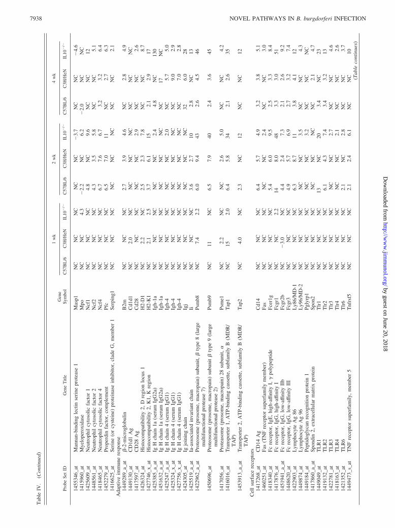
Affymetrix GeneChip array analysis revealed alterations in ex-pression of discrete subsets of genes in RNA collected from thejoint tissue of infected mice. At 1 wk of infection, transcripts from156 genes were increased by �2-fold in C3H mice, whereas inC57BL/6 mice 119 transcripts were increased; this result is basedon the 22,000 unique transcripts included on GeneChip 430A ofthe mouse (Table I). Interestingly, there were only three transcripts
changed by �2-fold that were common to C3H and C57BL/6 miceat 1 wk of infection (Figs. 2, 3A). Tissue from infected C57BL/6-IL-10�/� mice revealed 419 gene transcripts increased at 1 wk ofinfection and displayed clear overlap with those increased in C3Hmice (Fig. 3A). At 2 wk postinfection, significant recruitment ofleukocytes to joint tissues is evident by histological analysis (7, 9),which was supported by the increase in a large number of genes inall three strains of mice associated with host defense and inflam-matory cell infiltrate (Fig. 3B). The 4-wk postinfection time pointrepresents the peak of arthritis as well as full development of theadaptive immune response, consistent with the identification of agroup of genes induced only at this late time point. The number ofgenes changed at 4 wk was less than at 2 wk in both C3H andC57BL/6 mice, possibly reflecting resolution of infection and inflam-matory response (Table I and Fig. 3C). In contrast, C57BL/6 IL-10�/� mice displayed an exaggerated and uncontrolled response toinfection, with the number of genes changed �2-fold still increasingat the 4-wk time point. This is consistent with the potent ability ofIL-10 to regulate acute inflammatory processes (Table I) (32).
B. burgdorferi infection induces an early proinflammatoryresponse in arthritis-susceptible C3H mice
At 1 wk of infection, C3H mice displayed a robust induction ofgenes indicative of a strong proinflammatory response by endog-enous cells. In particular, the most highly increased genes werethose known to be inducible by type I (IFN-� and IFN-�) and/ortype II (IFN-�) IFN (Table II). In fact, 36% (56 of 156) of thegenes induced �2-fold in C3H mice were clearly annotated as IFNresponsive, and 67 of the 100 most highly induced genes wereeither IFN responsive or could be linked to the induction and reg-ulation of IFN responses (Fig. 2 and Supplement 1)4 (27). Addi-tional genes increased in C3H mice could be indirectly linked toIFN by their role in regulation and development of the immuneresponse, such as Parp14, Bst2, and Nfil3 (Table II). The genes ofthe C3H profile were highly induced, up to 120-fold comparedwith uninfected joint tissue. It is difficult to assign responsibilityfor this robust IFN response because changes were not detected ineither type I or type II IFN transcripts in C3H or C57BL/6 mice atany time, and most of the genes listed in Table II can be inducedby either type I or type II IFN (including the highly induced Iigp1,Gbp1, and Tap1 (27, 33)). The increased transcript levels for thesignaling molecules Stat1, Irf1, Irf7, and Irf8 also did not providestrong evidence for the selective presence of type I or type II IFN(Table II) (33).
In striking contrast, none of the IFN-inducible gene transcriptswas increased �2-fold at 1 wk postinfection in the ankle joints ofC57BL/6 mice (Table II and Fig. 2). Although some IFN-inducibletranscripts were increased by 2 wk postinfection, the magnitude of
4 The on-line version of this article contains supplemental material.
Table I. Number of unique gene transcripts changed �2-fold in the joints of C3H, C57BL/6, and C57BL/6IL-10�/� mice at 1, 2, and 4 wk postinfectiona
ArthritisPhenotypeb
Borreila no.in Jointc 1 wk 2 wk 4 wk
C3H/HeN Severe High 1156 2401 1695 2647 1456 2222C57BL/6 Mild High 1119 26 1594 223 1340 221IL-10 KO Intermediate Low 1419 1231 11332 2769 11423 2870
a Gene expression in ankle joint tissue at 1, 2, and 4 wk postinfection was compared to uninfected ankle joint tissue.Affymetrix probe sets identifying the same gene transcript were counted as a single gene transcript.
b As described in Ref. 16 and Fig 1A.c As described in Ref. 16 and Fig 1B.
7932 NOVEL PATHWAYS IN B. burgdorferi INFECTION
by guest on June 20, 2018http://w
ww
.jimm
unol.org/D
ownloaded from
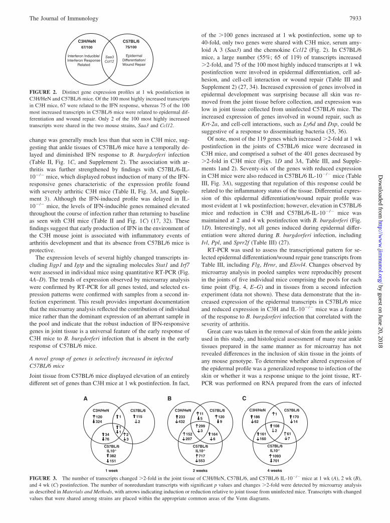
change was generally much less than that seen in C3H mice, sug-gesting that ankle tissues of C57BL/6 mice have a temporally de-layed and diminished IFN response to B. burgdorferi infection(Table II, Fig. 1C, and Supplement 2). The association with ar-thritis was further strengthened by findings with C57BL/6-IL-10�/� mice, which displayed robust induction of many of the IFN-responsive genes characteristic of the expression profile foundwith severely arthritic C3H mice (Table II, Fig. 3A, and Supple-ment 3). Although the IFN-induced profile was delayed in IL-10�/� mice, the levels of IFN-inducible genes remained elevatedthroughout the course of infection rather than returning to baselineas seen with C3H mice (Table II and Fig. 1C) (17, 32). Thesefindings suggest that early production of IFN in the environment ofthe C3H mouse joint is associated with inflammatory events ofarthritis development and that its absence from C57BL/6 mice isprotective.
The expression levels of several highly changed transcripts in-cluding Iigp1 and Igtp and the signaling molecules Stat1 and Irf7were assessed in individual mice using quantitative RT-PCR (Fig.4A–D). The trends of expression observed by microarray analysiswere confirmed by RT-PCR for all genes tested, and selected ex-pression patterns were confirmed with samples from a second in-fection experiment. This result provides important documentationthat the microarray analysis reflected the contribution of individualmice rather than the dominant expression of an aberrant sample inthe pool and indicate that the robust induction of IFN-responsivegenes in joint tissue is a universal feature of the early response ofC3H mice to B. burgdorferi infection that is absent in the earlyresponse of C57BL/6 mice.
A novel group of genes is selectively increased in infectedC57BL/6 mice
Joint tissue from C57BL/6 mice displayed elevation of an entirelydifferent set of genes than C3H mice at 1 wk postinfection. In fact,
of the �100 genes increased at 1 wk postinfection, some up to40-fold, only two genes were shared with C3H mice, serum amy-loid A 3 (Saa3) and the chemokine Ccl12 (Fig. 2). In C57BL/6mice, a large number (55%; 65 of 119) of transcripts increased�2-fold, and 75 of the 100 most highly induced transcripts at 1 wkpostinfection were involved in epidermal differentiation, cell ad-hesion, and cell-cell interaction or wound repair (Table III andSupplement 2) (27, 34). Increased expression of genes involved inepidermal development was surprising because all skin was re-moved from the joint tissue before collection, and expression waslow in joint tissue collected from uninfected C57BL/6 mice. Theincreased expression of genes involved in wound repair, such asKrt-2a, and cell-cell interactions, such as Ly6d and Dsp, could besuggestive of a response to disseminating bacteria (35, 36).
Of note, most of the 119 genes which increased �2-fold at 1 wkpostinfection in the joints of C57BL/6 mice were decreased inC3H mice, and comprised a subset of the 401 genes decreased by�2-fold in C3H mice (Figs. 1D and 3A, Table III, and Supple-ments 1and 2). Seventy-six of the genes with reduced expressionin C3H mice were also reduced in C57BL/6 IL-10�/� mice (TableIII, Fig. 3A), suggesting that regulation of this response could berelated to the inflammatory status of the tissue. Differential expres-sion of this epidermal differentiation/wound repair profile wasmost evident at 1 wk postinfection; however, elevation in C57BL/6mice and reduction in C3H and C57BL/6-IL-10�/� mice wasmaintained at 2 and 4 wk postinfection with B. burgdorferi (Fig.1D). Interestingly, not all genes induced during epidermal differ-entiation were altered during B. burgdorferi infection, includingIvl, Ppl, and Sprr2f (Table III) (27).
RT-PCR was used to assess the transcriptional pattern for se-lected epidermal differentiation/wound repair gene transcripts fromTable III, including Flg, Hrnr, and Elovl4. Changes observed bymicroarray analysis in pooled samples were reproducibly presentin the joints of five individual mice comprising the pools for eachtime point (Fig. 4, E–G) and in tissues from a second infectionexperiment (data not shown). These data demonstrate that the in-creased expression of the epidermal transcripts in C57BL/6 miceand reduced expression in C3H and IL-10�/� mice was a featureof the response to B. burgdorferi infection that correlated with theseverity of arthritis.
Great care was taken in the removal of skin from the ankle jointsused in this study, and histological assessment of many rear ankletissues prepared in the same manner as for microarray has notrevealed differences in the inclusion of skin tissue in the joints ofany mouse genotype. To determine whether altered expression ofthe epidermal profile was a generalized response to infection of theskin or whether it was a response unique to the joint tissue, RT-PCR was performed on RNA prepared from the ears of infected
FIGURE 2. Distinct gene expression profiles at 1 wk postinfection inC3H/HeN and C57BL/6 mice. Of the 100 most highly increased transcriptsin C3H mice, 67 were related to the IFN response, whereas 75 of the 100most increased transcripts in C57BL/6 mice were related to epidermal dif-ferentiation and wound repair. Only 2 of the 100 most highly increasedtranscripts were shared in the two mouse strains, Saa3 and Ccl12.
FIGURE 3. The number of transcripts changed �2-fold in the joint tissue of C3H/HeN, C57BL/6, and C57BL/6 IL-10�/� mice at 1 wk (A), 2 wk (B),and 4 wk (C) postinfection. The number of nonredundant transcripts with significant p values and changes �2-fold were detected by microarray analysisas described in Materials and Methods, with arrows indicating induction or reduction relative to joint tissue from uninfected mice. Transcripts with changedvalues that were shared among strains are placed within the appropriate common areas of the Venn diagrams.
7933The Journal of Immunology
by guest on June 20, 2018http://w
ww
.jimm
unol.org/D
ownloaded from

Tab
leII
.E
xpre
ssio
nle
vels
ofse
lect
edIF
N-r
espo
nsiv
ege
netr
ansc
ript
sin
crea
sed
at1
wk
post
infe
ctio
nin
join
tsti
ssue
ofC
3Hm
icea
Prob
eSe
tID
Gen
eT
itle
Gen
eSy
mbo
l
1w
k2
wk
4w
k
C57
BL
/6C
3H/H
eNIL
-10
KO
C57
BL
/6C
3H/H
eNIL
-10
KO
C57
BL
/6C
3H/H
eNIL
-10
KO
IFN
-ind
ucib
lege
nes
1417
141_
atIF
N-�
-ind
uced
GT
Pase
Igtp
NC
128
NC
7.1
1649
NC
3.6
4914
1779
3_at
IFN
-ind
ucib
leG
TPa
se2
Iigp
2N
C12
32.
44.
812
35N
C2.
638
1419
042_
atIF
N-i
nduc
ible
GT
Pase
1Ii
gp1
NC
113
2.5
1216
622.
45.
386
1449
009_
atT
cell-
spec
ific
GT
Pase
Tgt
pN
C42
2.5
7.5
1472
2.1
5.3
7714
3867
6_at
Mac
roph
age
activ
atio
n2
like
Mpa
21N
C36
NC
4.1
3.6
31N
CN
C36
1418
825_
atIF
N-i
nduc
ible
prot
ein
1Ifi
1N
C35
NC
3.6
3.3
32N
CN
C34
1419
714_
atC
D27
4A
gC
d274
NC
33N
C5.
35.
153
NC
NC
5714
1601
6_at
Tra
nspo
rter
1,A
TP-
bind
ing
cass
ette
,su
bfam
ilyB
(MD
R/T
AP)
Tap
1N
C15
2.0
6.4
5.8
342.
12.
635
1418
240_
atG
uany
late
nucl
eotid
e-bi
ndin
gpr
otei
n2
Gbp
2N
C12
2.8
124.
172
2.6
2.1
7114
5069
6_at
Prot
eoso
me
(pro
som
e,m
acro
pain
)su
buni
t,�
type
9(l
arge
mul
tifun
ctio
nal
prot
ease
2)Ps
mb9
NC
11N
C6.
57.
940
2.4
3.6
45
1453
196_
a_at
2�–5
�ol
igoa
deny
late
synt
heta
se-l
ike
2O
asl2
NC
4.4
4.2
2.8
NC
29N
CN
C33
Sign
alin
gm
olec
ules
1450
034_
atSi
gnal
tran
sduc
eran
dac
tivat
orof
tran
scri
ptio
n1
Stat
1N
C23
2.1
3.0
5.2
24N
CN
C39
1448
436_
a_at
IFN
regu
lato
ryfa
ctor
1Ir
f1N
C4.
5N
C2.
72.
515
NC
NC
1614
1724
4_a_
atIF
Nre
gula
tory
fact
or7
Irf7
NC
4.0
2.1
2.2
NC
18N
CN
C16
1416
714_
atIF
Nre
gula
tory
fact
or8
Irf8
NC
3.8
NC
4.9
4.0
152.
5N
C15
1418
131_
atSA
Mdo
mai
nan
dH
Ddo
mai
n,1
Sam
hd1
NC
4.7
NC
4.0
6.0
24N
C2.
521
1418
265_
s_at
IFN
regu
lato
ryfa
ctor
2Ir
f2N
CN
CN
CN
CN
CN
CN
CN
C2.
1O
ther 1451
564_
atPo
ly(A
DP-
ribo
se)
poly
mer
ase
fam
ily,
mem
ber
14Pa
rp14
NC
7.3
NC
2.9
3.2
15N
CN
C16
1429
947_
a_at
Z-D
NA
-bin
ding
prot
ein
1Z
bp1
NC
5.3
NC
4.9
3.4
40N
C2.
147
1438
855_
x_at
TN
F,�
-ind
uced
prot
ein
2T
nfai
p2N
C4.
5N
CN
C2.
05.
1N
CN
C6.
214
2492
1_at
Bon
em
arro
wst
rom
alce
llA
g2
Bst
2N
C3.
8N
C2.
72.
111
NC
NC
13
aG
ene
expr
essi
onin
ankl
ejo
int
tissu
eat
1,2,
and
4w
kpo
stin
fect
ion
was
com
pare
dto
unin
fect
edan
kle
join
ttis
sue.
Num
bers
indi
cate
fold
chan
ge.
Cha
nges
�2-
fold
are
desi
gnat
ed“N
C.”
7934 NOVEL PATHWAYS IN B. burgdorferi INFECTION
by guest on June 20, 2018http://w
ww
.jimm
unol.org/D
ownloaded from

and uninfected mice. Transcript levels for filaggrin were notchanged in ear tissue from infected C3H or C57BL/6 mice, sug-gesting that this differential response to infection was specific forthe ankle tissue (data not shown).
B. burgdorferi infection results in expression of genes involvedin host defense
In addition to the very distinct gene expression profiles seen earlyduring infection that correlate with arthritis severity, all threestrains of mice shared a common response to B. burgdorferi thatwas evident at 2 and 4 wk postinfection (Fig. 3, B and C). Therecruitment of PMNs is a hallmark of Lyme arthritis (8, 15), andby 2 wk of infection modest increases (5-fold) in the PMN-recruit-ing chemokines Cxcl1, Cxcl2, and Cxcl5 were seen in both C3Hand C57BL/6 mice as well as increased levels of PMN gene prod-ucts such as Mpo, Ncf1, and Ncf4 (Table IV and Supplements 1–3).Additionally, increased transcription of several mononuclear cell-recruiting chemokines, such as Ccl2, Ccl3, Ccl7, and Ccl12, wasobserved (37). The IFN-responsive T cell-recruiting chemokineCxcl9 was increased in both C3H and C57BL/6 mice over thecourse of infection, whereas Cxcl10 was also highly induced injoints of C57BL/6 and C57BL/6-IL-10�/� mice (Table IV) (37).Additional T cell-recruiting chemokines were increased during in-fection in both C3H and C57BL/6 mice, including Cxcl16, Ccl2,and Ccl7, (37). Transcript levels for the B cell chemokine Cxcl13were dramatically increased in the joints of both C57BL/6 andC3H mice by 2 wk infection, whereas joints of C57BL/6-IL-10�/�
mice displayed a less robust induction of Cxcl13 (Table IV). Infact, Cxcl13 was the only chemokine with greater induction inC57BL/6 mice than in C57BL/6 IL-10�/� mice, an interestingfinding in light of the clinical association between Cxcl13 expres-sion and spirochete load in neuroborreliosis (38) and the reducedpresence of B. burgdorferi in the joints of IL-10�/� mice (Fig. 1B).Expression profiles for many of the chemokines were confirmed byRT-PCR in individual mice, with the unique profile for Cxcl13shown in Fig. 4H.
Genes whose expression is associated with host defense were alsoincreased at 2 wk of infection, including additional markers for neu-trophils and macrophages, Mpo, Cd14, Tlrs, Fc receptors, and argi-nase. Complement component C1qa, C1qb, C1qg, C1r, C2, and C3transcripts were modestly increased in the joints of both C3H andC57BL/6 mice, further suggesting the presence of activated mac-rophages (Table IV). Transcripts for markers of APCs and Ag-processing machinery, including the MHC, Cd1, Tap1/2, andproteosome subunits indicated the presence of an active ac-quired host defense in joint tissues at 2 and 4 wk of infection,as did the increased expression of Ig genes (Table IV) (27).
Borrelia and its lipoproteins are known to activate NF-�B andinduce the production of many cytokines including IL-6, IL-1�,TNF-�, and IL-12 in vitro (39, 40). Surprisingly, IL-1� was theonly hallmark inflammatory cytokine to be increased in either C3Hor C57BL/6 ankle tissue (Table IV and Fig. 4I). In contrast, tran-scripts for many cytokines were robustly induced at 2 and 4 wk ofinfection in the IL-10�/� mice (including IL-1b, IFN-�, IL-6, IL-7,
FIGURE 4. Quantification of selected genetranscripts in the joints of individual mice. Expres-sion level of IFN-related transcripts Iigp1 (A), Igtp(B), Stat1 (C), and Irf7 (D); epidermal differenti-ation-related transcripts Flg (E), Hrnr (F), andElovl4 (G); transcripts associated with the host de-fense Cxcl13 (H) and Il1b (I); and a transcript as-sociated with chondrocyte activation Mmp3 (J)were determined using quantitative RT-PCR. Foldchange was determined based on the differencebetween the five individual mice used to generatepooled samples and the average of uninfected con-trols. In all cases, trends observed by microarraywere confirmed by RT-PCR analysis.
7935The Journal of Immunology
by guest on June 20, 2018http://w
ww
.jimm
unol.org/D
ownloaded from

Tab
leII
I.E
xpre
ssio
nle
vels
ofse
lect
edep
ider
mal
diffe
rent
iati
on/w
ound
repa
ir-a
ssoc
iate
dge
netr
ansc
ript
sin
crea
sed
at1
wk
post
infe
ctio
nin
join
tti
ssue
ofC
57B
L/6
mic
ea
Prob
eSe
tID
Gen
eT
itle
Gen
eSy
mbo
l
1w
k2
wk
4w
k
C57
BL
/6C
3H/H
eNIL
10�
/�C
57B
L/6
C3H
/HeN
IL10
�/�
C57
BL
/6C
3H/H
eNIL
10�
/�
Stru
ctur
alm
olec
ules
1427
268_
atFi
lagg
rin
Flg
47�
12�
2324
�7.
2N
C66
�16
�17
1422
481_
atK
erat
inco
mpl
ex2,
basi
c,ge
ne1
Krt
2-1
38�
124
�41
16�
3.7
NC
67�
131
�23
1451
613_
atH
orne
rin
Hm
r36
�8.
6�
1831
�5.
7�
5.3
90�
10�
1714
2715
4_at
Ker
atin
com
plex
2,ba
sic,
gene
17K
rt2-
1724
�86
�6.
817
�6.
7N
C40
�90
�6.
814
2267
2_at
Smal
lpr
olin
e-ri
chpr
otei
n1B
Sprr
1b20
�7.
0�
1314
�5.
9�
2.1
74�
8.9
�13
1420
183_
atL
oric
rin
Lor
8.5
�18
�2.
75.
6�
5.0
NC
10�
24�
6.3
1422
667_
atK
erat
inco
mpl
ex1,
acid
ic,
gene
15K
rt1-
156.
7N
CN
C3.
7N
CN
C5.
9�
2.1
NC
1422
222_
atIn
volu
crin
Iv1
NC
NC
NC
NC
NC
NC
NC
NC
NC
1460
732_
a_at
Peri
plak
inPp
1N
CN
CN
CN
CN
CN
CN
CN
CN
C14
4983
3_at
Smal
lpr
olin
e-ri
chpr
otei
n2F
Sprr
2fN
CN
CN
CN
CN
CN
CN
CN
CN
CC
ell
adhe
sion
/cel
l-ce
llin
tera
ctio
ns14
1693
0_at
Lym
phoc
yte
Ag
6co
mpl
ex,
locu
sD
Ly6
d15
�7.
8�
3.1
8.5
�6.
5N
C25
�9.
2�
2.9
1435
493_
atD
esm
opla
kin
Dsp
15�
7.3
�5.
38.
4�
4.5
NC
24�
9.4
�5.
014
3519
1_at
Cor
neod
esm
osin
Cds
n14
�4.
9�
5.0
7.0
�3.
3N
C24
�5.
9�
7.5
1418
799_
a_at
Proc
olla
gen,
type
XV
II,
alph
a1
Col
17a1
7.3
�3.
6�
2.0
3.4
�3.
2N
C7.
0�
4.9
�3.
5W
ound
resp
onse
rela
ted
1421
752_
a_at
Seri
ne(o
rcy
stei
ne)
prot
eina
sein
hibi
tor,
clad
eB
,m
embe
r5
Serp
inb5
11�
4.4
�4.
17.
1�
2.2
NC
23�
3.4
�3.
6
1421
092_
atSe
rine
(or
cyst
eine
)pr
otei
nase
inhi
bito
r,cl
ade
A(�
-1A
ntip
rote
inas
e,an
titry
psin
),m
embe
r12
Serp
ina1
24.
7�
3.1
�4.
83.
1�
2.7
NC
10�
3.4
�5.
5
1422
784_
atK
erat
inco
mpl
ex2,
basi
c,ge
ne6a
Krt
2-6a
3.7
�2.
2N
C3.
9�
2.1
6.6
8.2
�2.
2N
CO
ther 1450
633_
atC
alm
odul
in4
Cal
m4
29�
86�
1016
�7.
7N
C49
�13
4�
1314
2430
6_at
Elo
ngat
ion
ofve
rylo
ngch
ain
fatty
acid
s(F
EN
1/E
lo2,
SUR
4/E
lo3,
yeas
t)-l
ike
4E
lov1
414
�4.
5N
C7.
0�
4.1
NC
15�
5.7
�3.
0
1450
645_
atM
etal
loth
ione
in4
Mt4
20�
22�
126.
5�
4.4
NC
36�
30�
9.0
aG
ene
expr
essi
onin
ankl
ejo
int
tissu
eat
1,2,
and
4w
kpo
stin
fect
ion
was
com
pare
dto
unin
fect
edan
kle
join
ttis
sue.
Num
bers
indi
cate
fold
chan
ge.
Num
bers
prec
eded
bya
“_”
are
redu
ced
com
pare
dto
unin
fect
edan
kle
join
ttis
sue.
Cha
nges
�2-
fold
are
desi
gnat
ed“N
C.”
7936 NOVEL PATHWAYS IN B. burgdorferi INFECTION
by guest on June 20, 2018http://w
ww
.jimm
unol.org/D
ownloaded from

Tab
leIV
.E
xpre
ssio
nle
vels
ofge
nes
invo
lved
inth
eim
mun
ere
spon
seth
atar
ech
ange
d�
2-fo
lda
Prob
eSe
tID
Gen
eT
itle
Gen
eSy
mbo
l
1w
k2
wk
4w
k
C57
BL
/6C
3H/H
eNIL
10�
/�C
57B
L/6
C3H
/HeN
IL10
�/�
C57
BL
/6C
3H/H
eNIL
10�
/�
Che
mok
ines
1420
380_
atC
hem
okin
e(C
-Cm
otif
)lig
and
2C
cl2
NC
NC
2.1
226.
742
3.7
2.8
6614
1956
1_at
Che
mok
ine
(C-C
mot
if)
ligan
d3
Ccl
3N
CN
CN
C14
NC
7.5
2.4
NC
1114
1726
6_at
Che
mok
ine
(C-C
mot
if)
ligan
d6
Ccl
6N
CN
CN
C2.
9N
C7.
0N
CN
C5.
214
2122
8_at
Che
mok
ine
(C-C
mot
if)
ligan
d7
Ccl
7N
CN
CN
C13
1128
3.4
4.6
4914
1968
4_at
Che
mok
ine
(C-C
mot
if)
ligan
d8
Ccl
8N
C2.
9N
C7.
615
484.
99.
784
1417
936_
atC
hem
okin
e(C
-Cm
otif
)lig
and
9C
cl9
NC
NC
NC
4.0
3.3
3.4
2.0
NC
3.0
1419
282_
atC
hem
okin
e(C
-Cm
otif
)lig
and
12C
cl12
2.5
2.6
NC
412.
017
3.5
NC
3214
1920
9_at
Che
mok
ine
(C-X
-Cm
otif
)lig
and
1C
xcl1
NC
NC
3.4
5.9
5.6
343.
45.
779
1449
984_
atC
hem
okin
e(C
-X-C
mot
if)
ligan
d2
Cxc
l2N
CN
C2.
25.
7N
C17
NC
NC
2014
1972
8_at
Che
mok
ine
(C-X
-Cm
otif
)lig
and
5C
xcl5
NC
NC
NC
NC
6.7
13N
C5.
627
1418
480_
atC
hem
okin
e(C
-X-C
mot
if)
ligan
d7
Cxc
l7N
CN
C2.
4N
CN
CN
CN
CN
CN
C14
1865
2_at
Che
mok
ine
(C-X
-Cm
otif
)lig
and
9C
xcl9
NC
173.
160
1059
612
3.6
811
1418
930_
atC
hem
okin
e(C
-X-C
mot
if)
ligan
d10
Cxc
l10
NC
2.6
2.7
11N
C14
12.
3N
C18
914
4882
3_at
Che
mok
ine
(C-X
-Cm
otif
)lig
and
12C
xcl1
2N
C�
2.0
NC
NC
NC
2.1
NC
2.2
2.6
1417
851_
atC
hem
okin
e(C
-X-C
mot
if)
ligan
d13
Cxc
l13
2.3
NC
NC
401
377.
812
145
2014
1845
7_at
Che
mok
ine
(C-X
-Cm
otif
)lig
and
14C
xcl1
4N
CN
CN
CN
C5.
4N
CN
C2.
8N
C14
4919
5_s_
atC
hem
okin
e(C
-X-C
mot
if)
ligan
d16
Cxc
l16
NC
NC
NC
6.4
7.9
154.
33.
322
1415
803_
atC
hem
okin
e(C
-X3-
Cm
otif
)lig
and
1C
x3cl
1N
CN
CN
CN
CN
CN
CN
CN
C3.
0C
hem
okin
ere
cept
ors
1419
609_
atC
hem
okin
e(C
-Cm
otif
)re
cept
or1
Ccr
1N
CN
CN
C2.
5N
C3.
1N
CN
C2.
414
2118
7_at
Che
mok
ine
(C-C
mot
if)
rece
ptor
2C
cr2
NC
NC
2.4
4.0
NC
8.8
2.1
NC
18C
ytok
ines
1425
947_
atIF
N-�
Ifng
NC
NC
NC
NC
NC
16N
CN
C22
1450
330_
atIL
-10
Il10
NC
NC
NC
2.2
NC
6.2
NC
NC
6.4
1418
219_
atIL
-15
Il15
NC
NC
NC
NC
NC
2.8
NC
NC
3.4
1449
399_
a_at
IL-1
�Il
1bN
CN
CN
C4.
03.
113
2.9
2.5
4214
2137
0_a_
atIL
-1fa
mily
,m
embe
r5
(�)
Il1f
52.
6N
CN
CN
CN
CN
C6.
5�
2.1
NC
1451
798_
atIL
-1re
cept
oran
tago
nist
Il1m
NC
NC
NC
9.4
NC
143.
0N
C22
1421
034_
a_at
IL-4
rece
ptor
,�
Il4r
aN
CN
CN
CN
C3.
34.
8N
CN
C5.
514
5029
7_at
IL-6
Il6
NC
NC
NC
NC
NC
13N
CN
C72
1436
861_
atIL
-7Il
7N
CN
CN
CN
CN
CN
CN
CN
C2.
114
1913
5_at
Lym
phot
oxin
BL
tbN
CN
CN
CN
CN
C4.
3N
CN
C4.
214
1834
5_at
TN
F(l
igan
d)su
perf
amily
,m
embe
r13
Tnf
sf13
NC
NC
NC
NC
NC
NC
NC
2.1
2.3
1460
255_
atT
NF
(lig
and)
supe
rfam
ily,
mem
ber
13b
Tnf
sf13
bN
CN
CN
C4.
3N
C6.
63.
2N
C10
Inna
teim
mun
ere
spon
se14
1954
9_at
Arg
inas
e1,
liver
com
plem
ent
com
pone
nt1,
qsu
bcom
pone
nt,
�A
rg1
2.1
�4.
2N
C63
NC
4716
�2.
832
1417
381_
atPo
lype
ptid
eco
mpl
emen
tco
mpo
nent
1,q
subc
ompo
nent
,�
C1q
aN
CN
CN
C2.
92.
34.
82.
32.
95.
2
1417
063_
atPo
lype
ptid
eco
mpl
emen
tco
mpo
nent
1,q
subc
ompo
nent
,�
C1q
bN
CN
CN
C4.
23.
36.
72.
93.
57.
7
1449
401_
atPo
lype
ptid
eC
1qg
NC
NC
NC
3.2
3.1
5.5
2.6
2.6
5.9
1417
009_
atC
ompl
emen
tco
mpo
nent
1,r
subc
ompo
nent
C1r
NC
NC
NC
NC
2.6
6.8
NC
2.5
7.6
1416
051_
atC
ompl
emen
tco
mpo
nent
2(w
ithin
H-2
S)C
2N
CN
CN
CN
C2.
26.
2N
CN
C8.
814
2395
4_at
Com
plem
ent
com
pone
nt3
C3
NC
NC
NC
3.4
3.9
102.
52.
111
1460
334_
atD
rebr
in-l
ike
Dbn
1N
CN
CN
CN
CN
C3.
5N
CN
C3.
814
2554
8_a_
atL
euko
cyte
spec
ific
tran
scri
pt1
Lst
1N
CN
CN
C7.
416
162.
54.
512
(Tab
leco
ntin
ues)
7937The Journal of Immunology
by guest on June 20, 2018http://w
ww
.jimm
unol.org/D
ownloaded from
Tab
leIV
.(C
onti
nued
)
Prob
eSe
tID
Gen
eT
itle
Gen
eSy
mbo
l
1w
k2
wk
4w
k
C57
BL
/6C
3H/H
eNIL
10�
/�C
57B
L/6
C3H
/HeN
IL10
�/�
C57
BL
/6C
3H/H
eNIL
10�
/�
1455
346_
atM
anna
n-bi
ndin
gle
ctin
seri
nepr
otea
se1
Mas
p1N
CN
CN
CN
CN
C�
3.7
NC
NC
�4.
614
1596
0_at
Mye
lope
roxi
dase
Mpo
NC
NC
4.3
�2.
2N
C6.
2�
2.0
NC
NC
1425
609_
atN
eutr
ophi
lcy
toso
licfa
ctor
1N
cf1
NC
NC
NC
4.8
NC
9.6
NC
NC
1214
4856
1_at
Neu
trop
hil
cyto
solic
fact
or2
Ncf
2N
CN
CN
C4.
33.
55.
8N
CN
C5.
114
1846
5_at
Neu
trop
hil
cyto
solic
fact
or4
Ncf
4N
CN
CN
C6.
77.
66.
73.
23.
26.
414
5227
9_at
Prop
erdi
nfa
ctor
,co
mpl
emen
tPf
cN
CN
CN
C6.
57.
011
NC
2.7
6.3
1416
625_
atSe
rine
(or
cyst
eine
)pr
otei
nase
inhi
bito
r,cl
ade
G,
mem
ber
1Se
rpin
g1N
CN
CN
CN
CN
CN
CN
CN
C2.
1A
dapt
ive
imm
une
resp
onse
1449
289_
a_at
�-2
-mic
rogl
obul
inB
2mN
CN
CN
C2.
73.
94.
6N
C2.
84.
914
4913
0_at
CD
1d1
Ag
Cd1
d1N
C2.
0N
CN
CN
CN
CN
CN
CN
C14
1759
7_at
CD
28A
gC
d28
NC
NC
NC
NC
NC
2.9
NC
NC
2.6
1426
324_
atH
isto
com
patib
ility
2,D
regi
onlo
cus
1H
2-D
1N
C2.
2N
C2.
52.
37.
8N
CN
C8.
714
2774
6_x_
atH
isto
com
patib
ility
2,K
1,K
regi
onH
2-K
1N
C2.
12.
53.
76.
115
2.1
2.9
1714
2538
5_a_
atIg
Hch
ain
1a(s
erum
IgG
2a)
Igh-
1aN
CN
CN
CN
CN
C2.
44.
8N
C13
014
5163
2_a_
atIg
Hch
ain
1a(s
erum
IgG
2a)
Igh-
1aN
CN
CN
CN
CN
CN
CN
C17
NC
1425
247_
a_at
IgH
chai
n4
(ser
umIg
G1)
Igh-
4N
CN
CN
CN
CN
C2.
0N
C5.
75.
014
2532
4_x_
atIg
Hch
ain
4(s
erum
IgG
1)Ig
h-4
NC
NC
NC
NC
NC
NC
NC
9.0
2.9
1427
756_
x_at
IgH
chai
n4
(ser
umIg
G1)
Igh-
4N
CN
CN
CN
CN
CN
CN
C7.
02.
814
2430
5_at
Igjo
inin
gch
ain
Igj
NC
NC
NC
NC
NC
NC
326.
028
1425
519_
a_at
Ia-a
ssoc
iate
din
vari
ant
chai
nIi
NC
NC
NC
3.6
2.7
102.
8N
C13
1422
962_
a_at
Prot
eoso
me
(pro
som
e,m
acro
pain
)su
buni
t,�
type
8(l
arge
mul
tifun
ctio
nal
prot
ease
7)Ps
mb8
NC
7.4
2.2
6.0
9.4
432.
64.
546
1450
696_
atPr
oteo
som
e(p
roso
me,
mac
ropa
in)
subu
nit
�ty
pe9
(lar
gem
ultif
unct
iona
lpr
otea
se2)
Psm
b9N
C11
NC
6.5
7.9
402.
43.
645
1417
056_
atPr
otea
som
e(p
roso
me,
mac
ropa
in)
28su
buni
t,�
Psm
e1N
C2.
2N
CN
C2.
65.
0N
CN
C4.
214
1601
6_at
Tra
nspo
rter
1,A
TP-
bind
ing
cass
ette
,su
bfam
ilyB
(MD
R/
TA
P)T
ap1
NC
152.
06.
45.
834
2.1
2.6
35
1453
913_
a_at
Tra
nspo
rter
2,A
TP-
bind
ing
cass
ette
,su
bfam
ilyB
(MD
R/
TA
P)T
ap2
NC
4.0
NC
2.3
NC
12N
CN
C12
Cel
lsu
rfac
ere
cept
ors
1417
268_
atC
D14
Ag
Cd1
4N
CN
CN
C6.
45.
74.
93.
23.
85.
114
6025
1_at
Fas
(TN
Fre
cept
orsu
perf
amily
mem
ber)
Fas
NC
NC
NC
NC
NC
2.4
NC
NC
3.0
1418
340_
atFc
rece
ptor
,Ig
E,
high
-affi
nity
I,�
poly
pept
ide
Fcer
1gN
CN
CN
C5.
46.
09.
52.
53.
38.
414
1787
6_at
Fcre
cept
or,
IgG
,hi
gh-a
ffini
tyI
Fcgr
1N
CN
C2.
214
8.0
483.
33.
051
1451
941_
a_at
Fcre
cept
or,
IgG
,lo
w-a
ffini
tyII
bFc
gr2b
NC
�3.
0N
C4.
42.
47.
32.
12.
69.
214
4862
0_at
Fcre
cept
or,
IgG
,lo
w-a
ffini
tyII
IFc
gr3
NC
NC
NC
4.9
5.7
6.9
2.7
3.2
7.4
1422
903_
atL
ymph
ocyt
eA
g86
Ly8
6/M
D-1
NC
NC
NC
6.3
8.7
113.
84.
112
1449
874_
atL
ymph
ocyt
eA
g96
Ly9
6/M
D-2
NC
NC
NC
NC
NC
3.5
NC
NC
4.3
1449
184_
atPe
ptid
ogly
can
reco
gniti
onpr
otei
n1
Pgly
rp1
NC
NC
NC
NC
NC
3.2
NC
NC
NC
1417
860_
a_at
Spon
din
2,ex
trac
ellu
lar
mat
rix
prot
ein
Spon
2N
CN
CN
CN
CN
CN
CN
C2.
14.
714
4904
9_at
TL
R1
Tlr
1N
CN
CN
C13
NC
203.
4N
C23
1419
132_
atT
LR
2T
lr2
NC
NC
NC
6.1
4.3
7.4
3.4
3.2
1314
2278
1_at
TL
R3
Tlr
3N
CN
CN
CN
CN
C2.
7N
CN
C4.
614
1816
3_at
TL
R4
Tlr
4N
CN
CN
CN
CN
C2.
1N
CN
C2.
614
2135
2_at
TL
R6
Tlr
6N
CN
CN
C2.
1N
C2.
8N
CN
C3.
714
4947
3_s_
atT
NF
rece
ptor
supe
rfam
ily,
mem
ber
5T
nfrs
f5N
CN
CN
C2.
12.
46.
1N
CN
C10
(Tab
leco
ntin
ues)
7938 NOVEL PATHWAYS IN B. burgdorferi INFECTION
by guest on June 20, 2018http://w
ww
.jimm
unol.org/D
ownloaded from

IL15, and TNF family members), indicating that B. burgdorferidoes induce these cytokines in the joint but that in the presence ofIL-10 their levels are quite low (Table IV and Supplement 3). Infact, RT-PCR, which is much more sensitive than microarray anal-ysis, did reveal the presence of extremely low levels of transcriptsfor IL-6, TNF, IFN-�, and IFN-� in the joint tissues of infectedC3H and C57BL/6 mice, with levels below detection in uninfectedtissues (data not shown). These results may explain the failure todetect significant changes by microarray. Taken together, they sug-gest that whereas the contribution of proinflammatory cytokinesappears to be tightly controlled in wild-type mice, it is not a dom-inant feature of the localized response to infection in joint tissue.Interestingly, RT-PCR analysis of spleen samples from infectedmice also revealed very low levels of these proinflammatory cy-tokines and of the IFN-inducible genes Igtp and Iipt (data notshown).
B. burgdorferi infection activates chondrocytes and triggersreactive responses in C3H mice
The most severe manifestation of arthritis in C3H mice includesevidence of reactive processes such as the formation of foci of newchondrocytes and new bone formation at 4 wk of infection (4, 9).Microarray revealed modest increases in gene products associatedwith chondrocytes from the joints of 4 wk postinfection C3Hmice, including several collagen genes, such as Ctsk and Dspg3(Table V). Quantification of Col1�2 by RT-PCR demonstrated arange of expression, and in this case only a portion of the individ-ual samples (3 of 5) showed increased expression of this gene latein disease. Future studies will be required to determine whetherassociations between gene induction and clinical markers of dis-ease can be made. Interesting, many of these genes associated withend-stage arthritis are also induced in rodent models of rheumatoidarthritis (41).
In agreement with findings reported by others (42, 43), tran-scripts for Mmp3, Mmp9, Mmp13, and Mmp23, as well as tissueinhibitor of matrix metalloproteinase 1 (Timp1) were induced inthe joints of C3H mice, whereas transcripts for Mmp3, Mmp8, andTimp1 were increased in C57BL/6-IL-10�/� mice (Table V).Mmp3 expression was also increased in joints of C57BL/6 mice,but to a lesser extent than in C3H or C57BL/6-IL-10�/� mice.Expression profiles of several of the metalloproteinases were con-firmed by RT-PCR with individual mice, with results for Mmp3shown in Fig. 4J.
DiscussionThe murine model of Lyme disease initially developed by Bartholdet al. (8) has provided a unique opportunity to study the localizedresponse to a similar infectious challenge in tissues that will ulti-mately develop distinct severities of arthritis (9, 19). The responseto invading bacteria at 1 wk of infection precedes the influx ofinflammatory cells and revealed a dramatic difference in the local-ized response. Although C3H mice displayed a robust responsedominated by IFN-inducible transcripts, these transcripts were ab-sent at 1 wk from infected C57BL/6 mice (Table II and Figs. 1 and2). Instead, the C57BL/6 mouse displayed an increase in an en-tirely distinct and unexpected group of genes previously associatedwith epidermal differentiation and wound repair (Table III). TheC57BL/6-IL-10�/� mouse shared expression profiles with theC3H mouse, implicating the IFN response in arthritis developmentrather than in a strain-specific response to infection, and suggestingthat suppression of the IFN response in C57BL/6 mice was crucialto suppression of arthritis (Figs. 1 and 3).
A second surprising feature of the early C3H profile was thedown-regulation of a large number of genes, many of which wereT
able
IV.
(Con
tinu
ed)
Prob
eSe
tID
Gen
eT
itle
Gen
eSy
mbo
l
1w
k2
wk
4w
k
C57
BL
/6C
3H/H
eNIL
10�
/�C
57B
L/6
C3H
/HeN
IL10
�/�
C57
BL
/6C
3H/H
eNIL
10�
/�
Sign
alin
gm
olec
ules
1423
048_
a_at
Tol
l-in
tera
ctin
gpr
otei
nT
ollip
NC
NC
3.3
NC
NC
NC
NC
NC
2.0
1419
272_
atM
yelo
iddi
ffer
entia
tion
prim
ary
resp
onse
gene
88M
yd88
NC
NC
NC
NC
NC
4.5
NC
NC
4.9
1417
856_
atA
vian
retic
uloe
ndot
helio
sis
vira
l(v
-rel
)on
coge
ne-r
elat
edB
Rel
bN
CN
CN
CN
CN
C3.
2N
CN
C3.
814
1811
0_a_
atIn
osito
lpo
lyph
osph
ate-
5-ph
osph
atas
eD
Inpp
5dN
CN
CN
C3.
43.
65.
5N
CN
C4.
614
2293
2_a_
atV
av1
onco
gene
Vav
1N
CN
CN
C7.
42.
45.
92.
2N
C6.
3C
ell-
cell
inte
ract
ions
/lect
ins
1422
013_
atC
-typ
ele
ctin
dom
ain
fam
ily4,
mem
ber
a2C
lec4
a2N
CN
CN
C15
1412
4.2
4.3
9.5
1420
804_
s_at
C-t
ype
lect
indo
mai
nfa
mily
4,m
embe
rd
Cle
c4d
NC
NC
NC
1316
798.
08.
486
1420
330_
atC
-typ
ele
ctin
dom
ain
fam
ily4,
mem
ber
eC
lec4
eN
CN
CN
C3.
72.
265
NC
NC
6614
2595
1_a_
atC
-typ
ele
ctin
dom
ain
fam
ily4,
mem
ber
nC
lec4
nN
CN
CN
CN
CN
C12
2.1
2.8
1614
1962
7_s_
atC
-typ
ele
ctin
dom
ain
fam
ily4,
mem
ber
nC
lec4
nN
CN
CN
CN
C4.
77.
02.
05.
210
1421
366_
atC
-typ
ele
ctin
dom
ain
fam
ily5,
mem
ber
aC
lec5
aN
CN
CN
C2.
1N
C4.
2N
CN
C5.
714
1969
3_at
Col
lect
insu
bfam
ilym
embe
r12
Col
ec12
NC
NC
NC
NC
NC
�2.
3N
CN
CN
C14
2213
3_at
Sial
opho
rin
Spn
NC
NC
NC
2.1
NC
3.6
NC
NC
4.6
Imm
une
deve
lopm
ent
1417
976_
atA
deno
sine
deam
inas
eA
daN
CN
CN
CN
C4.
24.
2N
C2.
54.
014
2492
3_at
Seri
ne(o
rcy
stei
ne)
prot
eina
sein
hibi
tor,
clad
eA
,m
embe
r3G
Serp
ina3
gN
C6.
62.
18.
42.
111
9N
CN
C11
414
4936
1_at
T-b
ox21
Tbx
21/T
bet
NC
NC
NC
NC
NC
2.7
NC
NC
2.4
aG
ene
expr
essi
onin
ankl
ejo
int
tissu
eat
1,2,
and
4w
kpo
stin
fect
ion
was
com
pare
dto
unin
fect
edan
kle
join
ttis
sue.
Num
bers
indi
cate
fold
chan
ge.
Num
bers
prec
eded
bya
“_”
are
redu
ced
com
pare
dto
unin
fect
edan
kle
join
ttis
sue.
Cha
nges
�2-
fold
are
desi
gnat
ed“N
C.”
7939The Journal of Immunology
by guest on June 20, 2018http://w
ww
.jimm
unol.org/D
ownloaded from

in the same group of epidermal genes that were increased inC57BL/6 mice. More striking was the fact that expression of thesegenes was also reduced in C57BL/6-IL-10�/� mice, similar toC3H mice, and again arguing that regulation of expression of theepidermal profile was related to the development of inflammatoryarthritis, not due to strain-specific differences between C3H andC57BL/6 mice (Table III and Figs. 1 and 3). Expression of severalof the epidermal profile genes, including filaggrin, loricrin, andcytokeratins, can be down-regulated by endogenous peptide ligandsfor nicotinic acetylcholine receptors, providing precedent for reducedexpression following signaling pathway activation (44).
It cannot be determined from our results which IFN is respon-sible for the intense induction of this group of transcripts in C3Hmice, or even the possible role of another pleotropic cytokine suchas TNF-� (45). Studies with mice lacking IFN-� indicate that it isnot required for arthritis development (46); however, it is possiblethat there are compensatory pathways acting in the gene ablationmodel. Based on the known inflammatory potential of B. burgdor-feri, the production of either type I IFN or IFN-� by the milieu ofthe joint tissue could certainly occur by 1 wk of infection (17, 47,48). Alternatively, the early presence the dendritic cell-specifictranscript Oasl2 (Table II) in C3H mice may be highly relevantdue to the importance of dendritic cells as a sources of type I IFN(49, 50). In clinical trials, treatment with type I IFN has resulted intransient arthritis in patients with multiple sclerosis and hepatitis Cinfection, providing precedent for involvement of IFN in inflam-matory arthritis development (51, 52). Additional experiments willbe required to identify the cellular source and identity of the cy-tokine responsible for the IFN-inducible profile.
Additional experiments will also be required to detail the inter-play between the epidermal profile and inflammatory responses.Several published reports provide some insight as to a connectionbetween altered epidermal gene expression profiles in the joints ofB. burgdorferi-infected mice and regulation of the inflammatoryresponse. Normal differentiation of the epidermis during develop-ment requires the I�B kinase IKK1 (53), a key component of theclassical and alternative NF-�B-signaling pathways, implicatingIKK1 as a common link between regulation of epidermal differ-entiation genes and inflammation. Additionally, mice with a kera-tinocyte-specific deletion of the inflammation-associated transcrip-tion factor AP-1 displayed altered expression of epidermal
differentiation genes and the development of both localized andsystemic inflammatory lesions including psoriasis and psoriatic ar-thritis (54). Because B. burgdorferi achieve high levels in the skinof infected mice, it is also possible that dissemination through theskin promotes increased trafficking of cells that are not normallypresent in the joint tissue, and that this trafficking influences theoverall inflammatory status of the joint (55, 56). Finally, in rheu-matoid arthritis, citrullinated proteins such as filaggrin have beenidentified as targets for autoantibody; however, it is only the cit-rullinated species that are recognized by rheumatoid arthritis se-rum, and unmodified filaggrin is actually not expressed in jointtissue (57).
The 2-wk profile revealed unexpected similarities in genes in-creased in C3H and C57BL/6 mice (Table IV and Fig. 3). Numer-ous chemokines were induced in both wild-type strains of mice,without correlation with the greater inflammatory cell infiltrateseen in C3H mice (8, 15). In contrast, the robust production ofNF-�B-dependent inflammatory cytokines seen with cultured cellsin vitro and during infection of animals and patients was absentfrom localized response in joint tissues of C3H and C57BL/6 mice(2, 12, 48). The lack of differentially regulated genes downstreamof the TLR2, MyD88, and NF-�B-driven response to B. burgdor-feri was unexpected and suggests that activation of this pathwayoccurs independently from the development and regulation ofLyme arthritis. The possibility that the NF-�B pathway is a betterindicator of the host response to infection rather than a determinantof arthritis severity is consistent with the heightened expression ofcytokines and more effective host defense in IL-10�/� mice (Fig.1B and Table IV). These findings should also be interpreted in lightof previous studies with TLR2-deficient mice: these mice harborhigh levels of spirochetes in tissues and develop severe arthritiseven though isolated cells are defective in production of cytokinesin response to B. burgdorferi (58). The lack of differential expres-sion of the NF-�B-dependent cytokines in B. burgdorferi-infectedC3H and C57BL/6 mice is consistent with the observed develop-ment of Lyme arthritis in TLR2�/� C3H. The possible implicationof type I IFN in arthritic responses is also consistent with Lymearthritis development in the TLR2�/� mouse, because type I IFNis not a target of TLR2 signaling and its production should proceednormally in the absence of TLR2 (50, 58, 59).
Table V. Expression levels of gene transcripts indicative of reactive processes and/or activation of chondrocytes by B. burgdorferia
Probe Set ID Gene TitleGene
Symbol
2 wk 4 wk
C57BL/6 C3H/HeN IL10�/� C57BL/6 C3H/HeN IL10�/�
MMPs and TIMPs1416136_at MMP 2 Mmp2 NC NC NC NC 3.3 NC1418945_at MMP 3 Mmp3 7.7 18 34 5.3 13 451449366_at MMP 8 Mmp8 NC NC 4.9 NC NC 2.11448291_at MMP 9 Mmp9 NC 3.9 NC NC NC NC1417256_at MMP 13 Mmp13 NC 10 NC NC 13 3.01417282_at MMP 23 Mmp23 NC 2.4 NC NC 2.6 NC1460227_at TIMP 1 Timp1 NC 10 4.0 NC 5.8 3.8
Chondrocyte related1423606_at Periostin, osteoblast-specific factor Postn NC 6.3 NC NC 6.0 NC1418365_at Cathepsin H Ctsh 2.5 6.2 5.7 2.1 3.7 5.91450652_at Cathepsin K Ctsk NC 2.7 NC NC 3.8 NC1421114_a_at Dermatan sulphate proteoglycan 3 Dspg3 NC 2.8 NC NC 3.7 NC1423607_at Lumican Lum NC 3.7 NC NC 6.6 NC1423110_at Procollagen, type I, � 2 Col1a2 NC 2.1 NC NC 2.3 NC1427884_at Procollagen, type III, � 1 Col3a1 NC 8.8 NC NC 7.9 NC1450625_at Procollagen, type V, � 2 Col5a2 NC 3.7 NC NC 4.6 NC
a Gene expression in ankle joint tissue at 2 and 4 wk postinfection was compared to uninfected ankle joint tissue. Numbers indicate fold change. Changes �2-fold aredesignated “NC.”
7940 NOVEL PATHWAYS IN B. burgdorferi INFECTION
by guest on June 20, 2018http://w
ww
.jimm
unol.org/D
ownloaded from

Global gene expression analysis led to the identification of pre-viously unrecognized differences in expression profiles in thejoints of C3H and C57BL/6 mice in response to the same stimulus:invading B. burgdorferi. This justifies our strategy of assessingexpression in the complex joint tissue and provides new insightinto the development of this inflammatory pathology. The differ-ences in gene expression between the arthritis-susceptible (C3Hand IL-10�/�) and arthritis-resistant (C57BL/6) strains was evi-dent by 1 wk of infection, suggesting that a very early discrimi-nating event occurs in each strain that determines the outcome ofinfection. One interpretation of these results is that in the presenceof high levels of chemokines, the IFN response of C3H mice pro-motes leukocytic infiltration resulting in inflammation and arthri-tis; whereas in C57BL/6 mice, some aspect of the epidermal/wound profile prevents leukocytic infiltration and inflammation,thus limiting arthritis. These early initial events determine the out-come of arthritis severity even in the presence of similarly highlevels of bacteria, chemokine production, and proinflammatory cy-tokine production.
Future investigations will be directed toward a mechanistic under-standing of the involvement of these novel pathways in regulation ofthe development of Lyme arthritis. Additionally, experiments de-signed to assess the participation of QTL regulating the severity ofmurine Lyme arthritis in control of these distinct responses mayprovide further insight into the genetic and biochemical regulationof arthritis development (10, 11).
AcknowledgmentsWe acknowledge the helpful comments of Liming Yang from the NationalInstitutes of Health, and Joseph Holden, Lorise Gharing, Scott Rogers, andall the members of the Weis laboratory at the University of Utah.
DisclosuresThe authors have no financial conflict of interest.
References1. Burgdorfer, W., A. G. Barbour, S. F. Hayes, J. L. Benach, E. Grunwaldt, and
J. P. Davis. 1982. Lyme disease: a tick-borne spirochetosis? Science 216:1317–1319.
2. Steere, A. C., J. Coburn, and L. Glickstein. 2004. The emergence of Lyme dis-ease. J. Clin. Invest. 113: 1093–1101.
3. Nocton, J. J., F. Dressler, B. J. Rutledge, P. N. Rys, D. H. Persing, andA. C. Steere. 1994. Detection of Borrelia burgdorferi DNA by polymerase chainreaction in synovial fluid from patients with Lyme arthritis. N. Engl. J. Med. 330:229–234.
4. Yang, L., J. H. Weis, E. Eichwald, C. P. Kolbert, D. H. Persing, and J. J. Weis.1994. Heritable susceptibility to severe Borrelia burgdorferi-induced arthritis isdominant and is associated with persistence of large numbers of spirochetes intissues. Infect. Immun. 62: 492–500.
5. Steere, A. C., R. T. Schoen, and E. Taylor. 1987. The clinical evolution of Lymearthritis. Ann. Intern. Med. 107: 725–731.
6. Gross, D. M., T. Forsthuber, M. Tary-Lehmann, C. Etling, K. Ito, Z. A. Nagy,J. A. Field, A. C. Steere, and B. T. Huber. 1998. Identification of LFA-1 as acandidate autoantigen in treatment-resistant Lyme arthritis. Science 281:703–706.
7. Barthold, S. W., D. H. Persing, A. L. Armstrong, and R. A. Peeples. 1991.Kinetics of Borrelia burgdorferi dissemination and evolution of disease afterintradermal inoculation of mice. Am. J. Pathol. 139: 263–273.
8. Barthold, S. W., D. S. Beck, G. M. Hansen, G. A. Terwilliger, and K. D. Moody.1990. Lyme borreliosis in selected strains and ages of laboratory mice. J. Infect.Dis. 162: 133–138.
9. Ma, Y., K. P. Seiler, E. J. Eichwald, J. H. Weis, C. Teuscher, and J. J. Weis. 1998.Distinct characteristics of resistance to Borrelia burgdorferi-induced arthritis inC57BL/6N mice. Infect. Immun. 66: 161–168.
10. Roper, R. J., J. J. Weis, B. A. McCracken, C. B. Green, Y. Ma, K. S. Weber,D. Fairbairn, R. J. Butterfield, M. R. Potter, J. F. Zachary, et al. 2001. Geneticcontrol of susceptibility to experimental Lyme arthritis is polygenic and exhibitsconsistent linkage to multiple loci on chromosome 5 in four independent mousecrosses. Genes Immun. 2: 388–397.
11. Weis, J. J., B. A. McCracken, Y. Ma, D. Fairbairn, R. J. Roper, T. B. Morrison,J. H. Weis, J. F. Zachary, R. W. Doerge, and C. Teuscher. 1999. Identification ofquantitative trait loci governing arthritis severity and humoral responses in themurine model of Lyme disease. J. Immunol. 162: 948–956.
12. Wooten, R. M., and J. J. Weis. 2001. Host-pathogen interactions promoting in-flammatory Lyme arthritis: use of mouse models for dissection of disease pro-cesses. Curr. Opin. Microbiol. 4: 274–279.
13. Hirschfeld, M., C. J. Kirschning, R. Schwandner, H. Wesche, J. H. Weis,R. M. Wooten, and J. J. Weis. 1999. Cutting edge: inflammatory signaling byBorrelia burgdorferi lipoproteins is mediated by Toll-like receptor 2. J. Immunol.163: 2382–2386.
14. Aliprantis, A. O., R. B. Yang, M. R. Mark, S. Suggett, B. Devaux, J. D. Radolf,G. R. Klimpel, P. Godowski, and A. Zychlinsky. 1999. Cell activation and ap-optosis by bacterial lipoproteins through Toll-like receptor-2. Science 285:736–739.
15. Brown, C. R., V. A. Blaho, and C. M. Loiacono. 2003. Susceptibility to exper-imental Lyme arthritis correlates with KC and monocyte chemoattractant pro-tein-1 production in joints and requires neutrophil recruitment via CXCR2. J. Im-munol. 171: 893–901.
16. Brown, J. P., J. F. Zachary, C. Teuscher, J. J. Weis, and R. M. Wooten. 1999.Dual role of interleukin-10 in murine Lyme disease: regulation of arthritis se-verity and host defense. Infect. Immun. 67: 5142–5150.
17. Ganapamo, F., V. A. Dennis, and M. T. Philipp. 2000. Early induction of �interferon and interleukin-10 production in draining lymph nodes from mice in-fected with Borrelia burgdorferi. Infect. Immun. 68: 7162–7165.
18. Ganapamo, F., V. A. Dennis, and M. T. Philipp. 2001. CD19 cells produceIFN-� in mice infected with Borrelia burgdorferi. Eur. J. Immunol. 31:3460–3468.
19. Brown, C. R., and S. L. Reiner. 1998. Clearance of Borrelia burgdorferi may notbe required for resistance to experimental Lyme arthritis. Infect. Immun. 66:2065–2071.
20. Morrison, T. B., Y. Ma, J. H. Weis, and J. J. Weis. 1999. Rapid and sensitivequantification of Borrelia burgdorferi-infected mouse tissues by continuous flu-orescent monitoring of PCR. J. Clin. Microbiol. 37: 987–992.
21. Chirgwin, J. M., A. E. Przybyla, R. J. MacDonald, and W. J. Rutter. 1979.Isolation of biologically active ribonucleic acid from sources enriched in ribo-nuclease. Biochemistry 18: 5294–5299.
22. GeneChip Expression Analysis Technical Manual. 2005. Affymetrix Incorpo-rated, Santa Clara.
23. Bolstad, B. M., R. A. Irizarry, M. Astrand, and T. P. Speed. 2003. A comparisonof normalization methods for high density oligonucleotide array data based onvariance and bias. Bioinformatics 19: 185–193.
24. Irizarry, R. A., B. M. Bolstad, F. Collin, L. M. Cope, B. Hobbs, and T. P. Speed.2003. Summaries of Affymetrix GeneChip probe level data. Nucleic Acids Res.31: e15.
25. Gautier, L., L. Cope, B. M. Bolstad, and R. A. Irizarry. 2004. affy: analysis ofAffymetrix GeneChip data at the probe level. Bioinformatics 20: 307–315.
26. Tusher, V. G., R. Tibshirani, and G. Chu. 2001. Significance analysis of microar-rays applied to the ionizing radiation response. Proc. Natl. Acad. Sci. USA 98:5116–5121.
27. Eppig, J. T., C. J. Bult, J. A. Kadin, J. E. Richardson, J. A. Blake,A. Anagnostopoulos, R. M. Baldarelli, M. Baya, J. S. Beal, S. M. Bello, et al.2005. The Mouse Genome Database (MGD): from genes to mice: a communityresource for mouse biology. Nucleic Acids Res. 33: D471–D475.
28. Hubbard, T., D. Andrews, M. Caccamo, G. Cameron, Y. Chen, M. Clamp,L. Clarke, G. Coates, T. Cox, F. Cunningham, et al. 2005. Ensembl 2005. NucleicAcids Res. 33: D447–D453.
29. Ornstein, K., and A. G. Barbour. 2006. A reverse transcriptase-polymerase chainreaction assay of Borrelia burgdorferi 16S rRNA for highly sensitive quantifi-cation of pathogen load in a vector. Vector Borne Zoonotic Dis. 6: 103–112.
30. Kendziorski, C., R. A. Irizarry, K. S. Chen, J. D. Haag, and M. N. Gould. 2005.On the utility of pooling biological samples in microarray experiments. Proc.Natl. Acad. Sci. USA 102: 4252–4257.
31. Hodzic, E., S. Feng, K. J. Freet, and S. W. Barthold. 2003. Borrelia burgdorferipopulation dynamics and prototype gene expression during infection of immu-nocompetent and immunodeficient mice. Infect. Immun. 71: 5042–5055.
32. Moore, K. W., R. de Waal Malefytqq, R. L. Coffman, and A. O’Garra. 2001.Interleukin-10 and the interleukin-10 receptor. Annu. Rev. Immunol. 19: 683–765.
33. Platanias, L. C. 2005. Mechanisms of type-I- and type-II-interferon-mediatedsignalling. Nat. Rev. Immunol. 5: 375–386.
34. Mack, J. A., S. Anand, and E. V. Maytin. 2005. Proliferation and cornificationduring development of the mammalian epidermis. Birth Defects Res. C EmbryoToday 75: 314–329.
35. Wojcik, S. M., D. S. Bundman, and D. R. Roop. 2000. Delayed wound healingin keratin 6a knockout mice. Mol. Cell. Biol. 20: 5248–5255.
36. Brakenhoff, R. H., M. Gerretsen, E. M. Knippels, M. van Dijk, H. van Essen,D. O. Weghuis, R. J. Sinke, G. B. Snow, and G. A. van Dongen. 1995. The humanE48 antigen, highly homologous to the murine Ly-6 antigen ThB, is a GPI-anchored molecule apparently involved in keratinocyte cell-cell adhesion. J. CellBiol. 129: 1677–1689.
37. Esche, C., C. Stellato, and L. A. Beck. 2005. Chemokines: key players in innateand adaptive immunity. J. Invest. Dermatol. 125: 615–628.
38. Narayan, K., D. Dail, L. Li, D. Cadavid, S. Amrute, P. Fitzgerald-Bocarsly, andA. R. Pachner. 2005. The nervous system as ectopic germinal center: CXCL13and IgG in Lyme neuroborreliosis. Ann. Neurol. 57: 813–823.
39. Wooten, R. M., V. R. Modur, T. M. McIntyre, and J. J. Weis. 1996. Borreliaburgdorferi outer membrane protein A induces nuclear translocation of nuclearfactor-�B and inflammatory activation in human endothelial cells. J. Immunol.157: 4584–4590.
40. Sellati, T. J., D. A. Bouis, R. L. Kitchens, R. P. Darveau, J. Pugin, R. J. Ulevitch,S. C. Gangloff, S. M. Goyert, M. V. Norgard, and J. D. Radolf. 1998. Treponema
7941The Journal of Immunology
by guest on June 20, 2018http://w
ww
.jimm
unol.org/D
ownloaded from

pallidum and Borrelia burgdorferi lipoproteins and synthetic lipopeptides acti-vate monocytic cells via a CD14-dependent pathway distinct from that used bylipopolysaccharide. J. Immunol. 160: 5455–5464.
41. Ibrahim, S. M., D. Koczan, and H. J. Thiesen. 2002. Gene-expression profile ofcollagen-induced arthritis. J. Autoimmun. 18: 159–167.
42. Behera, A. K., E. Hildebrand, J. Scagliotti, A. C. Steere, and L. T. Hu. 2005.Induction of host matrix metalloproteinases by Borrelia burgdorferi differs inhuman and murine Lyme arthritis. Infect. Immun. 73: 126–134.
43. Gebbia, J. A., J. L. Coleman, and J. L. Benach. 2001. Borrelia spirochetes up-regulate release and activation of matrix metalloproteinase gelatinase B (MMP-9)and collagenase 1 (MMP-1) in human cells. Infect. Immun. 69: 456–462.
44. Arredondo, J., A. I. Chernyavsky, D. L. Jolkovsky, R. J. Webber, and S. A. Grando.2006. SLURP-2: a novel cholinergic signaling peptide in human mucocutaneousepithelium. J. Cell. Physiol. 208: 238–245.
45. Gallagher, J., J. Howlin, C. McCarthy, E. P. Murphy, B. Bresnihan, O. FitzGerald,C. Godson, H. R. Brady, and F. Martin. 2003. Identification of Naf1/ABIN-1 amongTNF-�-induced expressed genes in human synoviocytes using oligonucleotide mi-croarrays. FEBS Lett. 551: 8–12.
46. Brown, C. R., and S. L. Reiner. 1999. Experimental Lyme arthritis in the absenceof interleukin-4 or � interferon. Infect. Immun. 67: 3329–3333.
47. Ma, Y., K. P. Seiler, K. F. Tai, L. Yang, M. Woods, and J. J. Weis. 1994. Outersurface lipoproteins of Borrelia burgdorferi stimulate nitric oxide production bythe cytokine-inducible pathway. Infect. Immun. 62: 3663–3671.
48. Kang, I., S. W. Barthold, D. H. Persing, and L. K. Bockenstedt. 1997. T-helper-cell cytokines in the early evolution of murine Lyme arthritis. Infect. Immun. 65:3107–3111.
49. Tiefenthaler, M., R. Marksteiner, S. Neyer, F. Koch, S. Hofer, G. Schuler,M. Nussenzweig, R. Schneider, and C. Heufler. 1999. M1204, a novel 2�,5�oligoadenylate synthetase with a ubiquitin-like extension, is induced during mat-uration of murine dendritic cells. J. Immunol. 163: 760–765.
50. Honda, K., H. Yanai, A. Takaoka, and T. Taniguchi. 2005. Regulation of the typeI IFN induction: a current view. Int. Immunol. 17: 1367–1378.
51. Strueby, L., B. Nair, A. Kirk, and R. M. Taylor-Gjevre. 2005. Arthritis andbursitis in multiple sclerosis patients treated with interferon-�. Scand. J. Rheu-matol. 34: 485–488.
52. Wilson, L. E., D. Widman, S. H. Dikman, and P. D. Gorevic. 2002. Autoimmunedisease complicating antiviral therapy for hepatitis C virus infection. Semin. Ar-thritis Rheum. 32: 163–173.
53. Li, Q., Q. Lu, J. Y. Hwang, D. Buscher, K. F. Lee, J. C. Izpisua-Belmonte, andI. M. Verma. 1999. IKK1-deficient mice exhibit abnormal development of skinand skeleton. Genes Dev. 13: 1322–1328.
54. Zenz, R., R. Eferl, L. Kenner, L. Florin, L. Hummerich, D. Mehic, H. Scheuch,P. Angel, E. Tschachler, and E. F. Wagner. 2005. Psoriasis-like skin disease andarthritis caused by inducible epidermal deletion of Jun proteins. Nature 437:369–375.
55. Sellati, T. J., S. L. Waldrop, J. C. Salazar, P. R. Bergstresser, L. J. Picker, andJ. D. Radolf. 2001. The cutaneous response in humans to Treponema pallidumlipoprotein analogues involves cellular elements of both innate and adaptive im-munity. J. Immunol. 166: 4131–4140.
56. Norgard, M. V., B. S. Riley, J. A. Richardson, and J. D. Radolf. 1995. Dermalinflammation elicited by synthetic analogs of Treponema pallidum and Borreliaburgdorferi lipoproteins. Infect. Immun. 63: 1507–1515.
57. Baeten, D., I. Peene, A. Union, L. Meheus, M. Sebbag, G. Serre, E. M. Veys, andF. De Keyser. 2001. Specific presence of intracellular citrullinated proteins inrheumatoid arthritis synovium: relevance to antifilaggrin autoantibodies. ArthritisRheum. 44: 2255–2262.
58. Wooten, R. M., Y. Ma, R. A. Yoder, J. P. Brown, J. H. Weis, J. F. Zachary,C. J. Kirschning, and J. J. Weis. 2002. Toll-like receptor 2 is required for innate,but not acquired, host defense to Borrelia burgdorferi. J. Immunol. 168:348–355.
59. Wang, G., Y. Ma, A. Buyuk, S. McClain, J. J. Weis, and I. Schwartz. 2004.Impaired host defense to infection and Toll-like receptor 2-independent killing ofBorrelia burgdorferi clinical isolates in TLR2-deficient C3H/HeJ mice. FEMSMicrob. Lett. 231: 219–225.
7942 NOVEL PATHWAYS IN B. burgdorferi INFECTION
by guest on June 20, 2018http://w
ww
.jimm
unol.org/D
ownloaded from