Embed Size (px)
Citation preview
Proteome Analysis of Arabidopsis Leaf Peroxisomes RevealsNovel Targeting Peptides, Metabolic Pathways, andDefense Mechanisms W
Sigrun Reumann,a,1,2 Lavanya Babujee,a,3 Changle Ma,a,4 Stephanie Wienkoop,b Tanja Siemsen,a
Gerardo E. Antonicelli,a,5 Nicolas Rasche,a Franziska Luder,a,6 Wolfram Weckwerth,b,c and Olaf Jahnd,e
a Department of Plant Biochemistry, Georg-August-University of Goettingen, Albrecht-von-Haller-Institute for Plant Sciences,
D-37077 Goettingen, Germanyb Max-Planck-Institute of Molecular Plant Physiology, Metabolic Networks, D-14424 Potsdam, Germanyc University of Potsdam, Institute of Biochemistry and Biology, 14469 Potsdam, Germanyd Max-Planck-Institute of Experimental Medicine, Proteomics Group, D-37075 Goettingen, Germanye Deutsche Forschungsgemeinschaft Research Center for Molecular Physiology of the Brain, D-37073 Goettingen, Germany
We have established a protocol for the isolation of highly purified peroxisomes from mature Arabidopsis thaliana leaves and
analyzed the proteome by complementary gel-based and gel-free approaches. Seventy-eight nonredundant proteins were
identified, of which 42 novel proteins had previously not been associated with plant peroxisomes. Seventeen novel proteins
carried predicted peroxisomal targeting signals (PTS) type 1 or type 2; 11 proteins contained PTS-related peptides.
Peroxisome targeting was supported for many novel proteins by in silico analyses and confirmed for 11 representative full-
length fusion proteins by fluorescence microscopy. The targeting function of predicted and unpredicted signals was
investigated and SSL>, SSI>, and ASL> were established as novel functional PTS1 peptides. In contrast with the generally
accepted confinement of PTS2 peptides to the N-terminal domain, the bifunctional transthyretin-like protein was demon-
strated to carry internally a functional PTS2. The novel enzymes include numerous enoyl-CoA hydratases, short-chain dehy-
drogenases, and several enzymes involved in NADP and glutathione metabolism. Seven proteins, including b-glucosidases
and myrosinases, support the currently emerging evidence for an important role of leaf peroxisomes in defense against
pathogens and herbivores. The data provide new insights into the biology of plant peroxisomes and improve the prediction
accuracy of peroxisome-targeted proteins from genome sequences.
INTRODUCTION
Peroxisomes are ubiquitous cell organelles that compartmen-
talize primarily oxidative metabolic reactions. A characteristic of
peroxisomes is their metabolic flexibility because their enzymatic
constituents vary depending on the organism, the type of tissue,
and the environmental conditions (for review, see Beevers, 1979;
Hayashi and Nishimura, 2006). Plant peroxisomes differentiate
into tissue- and developmental-specific variants, such as leaf
peroxisomes in mature leaves and glyoxysomes in germinating
seeds, and typically participate in metabolic pathways and
networks that are spread over multiple subcellular compart-
ments. For instance, the recycling of 2-phosphoglycolate during
photorespiration requires orchestration of 15 chloroplastic, per-
oxisomal, and mitochondrial enzymes and numerous transport
proteins to transfer the intermediates between cell compart-
ments (for review, see Reumann, 2002; Reumann and Weber,
2006). Similarly, fatty acids are released from lipid bodies during
seed germination and transferred to glyoxysomes, where the
carbon chain is transformed into sucrose in coordination with
mitochondrial and cytosolic enzymes. Compartmentalization of
hydrogen peroxide–producing oxidases in peroxisomes offers
the advantage that H2O2 and other reactive oxygen species
(ROS) can immediately be detoxified at the site of production by
catalase (CAT) and auxiliary antioxidative enzymes (for review,
see del Rio et al., 2006). In addition to these basic metabolic
functions, a role for peroxisomes in the biosynthesis of the plant
hormone auxin and the signaling molecule jasmonic acid (JA)
and in sulfur and nitrogen metabolism has been established
1 Current address: Department of Molecular, Cellular, and Developmen-tal Biology, University of Michigan, 830 North University Avenue, AnnArbor, MI 48109 and Michigan State University–Department of EnergyPlant Research Laboratory, East Lansing, MI 48824-1312.2 Address correspondence to [email protected] Current address: Department of Plant Pathology, Faculty of Agricul-ture, Shizuoka University, Shizuoka 422-8529, Japan.4 Current address: Section of Molecular Biology, Division of BiologicalSciences, University of California, San Diego, La Jolla, CA 92093.5 Current address: Biochemistry Department, Clemens Schopf Institute,Technical University of Darmstadt, Petersenstrabe 22, D-64287,Darmstadt, Germany.6 Current address: Department of Biochemistry and Molecular Biology,Bio21 Molecular Science and Biotechnology Institute, University ofMelbourne, 30 Flemington Road, Parkville, Victoria 3010, Australia.The author responsible for the distribution of materials integral to thefindings presented in this article in accordance with the policy describedin the Instructions for Authors (www.plantcell.org) is: Sigrun Reumann([email protected]).W Online version contains Web-only data.www.plantcell.org/cgi/doi/10.1105/tpc.107.050989
The Plant Cell, Vol. 19: 3170–3193, October 2007, www.plantcell.org ª 2007 American Society of Plant Biologists
(Zolman et al., 2001; Strassner et al., 2002; Nowak et al., 2004;
Koo et al., 2006). Moreover, evidence is emerging from recent
studies that peroxisomes not only have important functions in
metabolism but also play an essential role in specific defense
mechanisms conferring resistance against pathogen attack
(Taler et al., 2004; Koh et al., 2005; Lipka et al., 2005).
Thanks to rapid advances in genome sequencing, the potential
of computational approaches is increasingly being realized to
predict peroxisomal proteins and explore the biology of plant
peroxisomes. In silico analyses take advantage of conserved
targeting sequences in peroxisomal proteins (i.e., the C- terminal
tripeptide constituting the peroxisomal targeting signal type
1 [PTS1] of the prototype SKL> [> designates the extreme C
terminus] or the cleavable PTS2 nonapeptide located in the
N-terminal domain [prototype RLx5HL]) to predict targeting of
unknown proteins to peroxisomes (Emanuelsson et al., 2003;
Kamada et al., 2003; Neuberger et al., 2003a, 2003b; Reumann,
2004; Hawkins and Boden, 2006). Except for few proteins (e.g.,
plant CAT and Arabidopsis thaliana sarcosine oxidase; Kamigaki
et al., 2003; Goyer et al., 2004), all known matrix proteins
from plant peroxisomes carry either a C-terminal PTS1 or an
N-terminal PTS2 (Reumann, 2004). Plant PTS motifs have been
defined by experimental and bioinformatics approaches and
yielded partially overlapping results but differed in specificity and
peptide identity. A relatively large number (24 to 120) of PTS1
peptides have been suggested to target proteins to peroxisomes
in higher plants (Hayashi et al., 1997; Mullen et al., 1997; Kragler
et al., 1998), but experimental limitations required data extrap-
olation from amino acid substitutions to all possible residue
combinations within the three-residue motif and thereby reduced
motif specificity. By in silico analysis of plant EST databases
(Reumann, 2004), nine major and 11 minor PTS1 peptides were
defined according to stringent criteria and thus considered as
rather specific, though certainly yet incomplete. A search of the
Arabidopsis genome for proteins carrying these peptides re-
vealed 280 putative PTS1/2 proteins listed in the database
AraPerox (Reumann et al., 2004; www.araperox.uni-goettingen.
de). Despite considerable progress, prediction by PTSs is still
limited by the low number of PTS-containing proteins that have
been characterized at the molecular level, at least in one plant
species (18 PTS1 and 14 PTS2 proteins; Reumann, 2004). How-
ever, the predictive algorithms for PTS1 proteins in particular can
steadily be improved, as 12 additional proteins with putative
PTS1s have been shown to be targeted to peroxisomes in the
past 3 years and, at least in some cases, have been demon-
strated to follow the PTS1 targeting pathway. The recently cloned
cDNAs encode, for instance, auxiliary enzymes of fatty acid
b-oxidation, ROS metabolism, and JA activation, and one of
two small heat shock proteins (sHsps); the second sHsp is the
only additional PTS2 protein (Edqvist et al., 2004; Tilton et al.,
2004; Goepfert et al., 2005, 2006; Leterrier et al., 2005; Lisenbee
et al., 2005; Schneider et al., 2005; Turner et al., 2005; Koo et al.,
2006; Ma et al., 2006; Helm et al., 2007; Zolman et al., 2007).
Taken together, the low number of well-characterized plant
peroxisomal matrix proteins leads to four principal constraints
limiting in silico predictions of peroxisomal PTS1/2 proteins: (1)
insufficient coverage of functional targeting peptides, (2) limited
knowledge of the identity and function of auxiliary targeting
elements surrounding the short PTS peptides, (3) possible dom-
inance of N-terminal nonperoxisomal targeting signals over
C-terminal PTS1s, and (4) unpredictable masking of PTS sur-
face exposure by polypeptide conformation, homo- and hetero-
oligomerization, or posttranslational regulatory mechanisms.
Thus, the currently available prediction tools suffer from both
insufficient sensitivity (i.e., an incomplete detection of true PTS1/
2 proteins) and insufficient specificity (i.e., the identification of a
significant number of false positives). Notably, many proteins of
peroxisomes cannot currently be predicted from genome se-
quences at all, including peripheral and integral membrane
proteins, subunits that are imported into the matrix in a piggy-
backed fashion, and matrix proteins with internal PTS1-like
peptides (e.g., yeast acyl-CoA oxidase and plant CAT; Klein
et al., 2002; Kamigaki et al., 2003).
In the postgenomic era of plant peroxisomal research, a major
challenge is the identification and functional characterization of
plant-specific proteins. Many of the recently cloned cDNAs of
plant peroxisomal proteins have been identified by computa-
tional homology analysis using the protein sequence of perox-
isomal enzymes from mammals or fungi as query (Edqvist et al.,
2004; Tilton et al., 2004; Goyer et al., 2004; Goepfert et al., 2005,
2006). Even though proven to be successful, this approach fails
by definition to unravel plant-specific proteins. Instead, system-
atic large-scale experimental studies of plant peroxisomes are
required. Technical advances in two-dimensional gel electro-
phoresis (2-DE), liquid chromatography (LC), and mass spec-
trometry (MS) laid the foundation for the progress in organellar
proteome studies (for review, see Taylor et al., 2003; Peck, 2005;
Glinski and Weckwerth, 2006) and for cell-specific profiling
(Wienkoop et al., 2004). The major bottleneck of organellar
proteomics is the purity of the cell compartment of interest.
With respect to their fragile nature, the isolation of intact perox-
isomes in high purity is difficult irrespective of the source or-
ganism but is even more challenging in plants, mainly due to
the predominance of plastids as another contaminating cell
compartment and a myriad of secondary metabolites with
organelle-destabilizing effects in vitro. For these reasons, the
first proteome studies of peroxisomes have only recently been
reported (for review, see Saleem et al., 2006). As the availability
of complete genome information or large EST collections is a
prerequisite for straightforward protein identification and be-
cause peroxisome isolation protocols are hardly interchangeable
between plant species, such proteome studies were hampered
by the fact that the isolation of plant peroxisomes in high purity
was restricted to few plant species (e.g., Pisum sativum and
Spinacia oleracea), which lacked genome or EST sequence
information. Complete genome analysis of Arabidopsis (Arabi-
dopsis Genome Initiative, 2000) rendered this tiny weed the
species of choice for proteome studies of plant peroxisomes,
provided that the organelles could be isolated in sufficient purity
and quantity. Initial proteome analyses of leaf peroxisomes and
glyoxysomes from greening and etiolated Arabidopsis cotyle-
dons have been reported, but probably due to insufficient
organelle purity and/or technical limitations in protein identifica-
tion, only few known and novel PTS-containing proteins have
been identified (Fukao et al., 2002, 2003). Leaf peroxisomes are
considered the variant of choice for a comprehensive long-term
Proteome of Arabidopsis Leaf Peroxisomes 3171
project aimed at the identification and functional analysis of all
plant-specific proteins from peroxisomes because mature
leaves are (1) physiologically the most significant plant tissue
(site of photosynthesis, photorespiration, numerous biosynthetic
pathways, and nitrogen and sulfur assimilation); (2) the most
distinctive tissue of plants compared with yeast and mammals;
and (3) second only to roots, the major plant organ that interacts
with the environment (i.e., that perceives and immediately reacts
to stress, both abiotic [high light, extreme temperature, drought,
and hail] and biotic [pathogen and herbivore attack]).
With the objective to verify and ultimately improve the predic-
tion accuracy of plant peroxisomal proteins from genome se-
quences, we applied the unbiased large-scale approach of
organellar proteomics to leaf peroxisomes. A purification method
was developed for leaf peroxisomes from mature Arabidopsis
leaves in high purity by Percoll and sucrose density gradient
centrifugation. Protein separation by 2-DE in combination with
peptide mass fingerprinting (PMF) and peptide fragment finger-
printing supplemented by a gel-free shotgun approach led to the
identification of 36 known and 42 novel peroxisomal proteins.
Peroxisome targeting of the novel PTS-containing proteins was
corroborated by the conservation of PTSs in orthologs of higher
plants as revealed by analysis of protein and EST databases. By
fluorescence microscopy, peroxisome targeting of 11 represen-
tative proteins carrying predicted PTS or PTS-related peptides
was verified for fusion proteins with enhanced yellow fluorescent
protein (EYFP) in onion epidermal cells. Thereby, three novel
PTS1 tripeptides (SSL>, SSI>, and ASL>) and an internal PTS2
were established, providing valuable input for further develop-
ment of prediction tools. The novel plant peroxisomal proteins
indicate that leaf peroxisomes play an important role in catab-
olism of complex metabolites, in biosynthetic pathways, and in
defense mechanisms against pathogens and herbivores.
RESULTS
Isolation of Peroxisomes from Mature Arabidopsis Leaves
in High Purity
To identify plant-specific peroxisomal proteins by a large-scale
proteomics approach, a new isolation method for leaf peroxi-
somes from Arabidopsis was established with the objectives of
(1) increasing the stability of peroxisomes in aqueous solution, (2)
reducing the physical interaction of peroxisomes with mitochon-
dria and chloroplasts, a pronounced phenomenon in Brassica-
ceae, and (3) increasing the specificity of peroxisome enrichment
by a combination of Percoll and sucrose density gradient cen-
trifugation. To this end, 3- to 4-week-old plants were harvested at
the end of the dark period and gently ground in a buffer of high
osmolarity. After separation of intact chloroplasts, but notably
without peroxisome sedimentation by differential centrifugation,
the supernatant was laid directly on top of a discontinuous
Percoll density gradient (Figure 1A). Cosedimentation of perox-
isomes along with mitochondria and thylakoid membranes by
differential centrifugation prior to isopycnic organelle separation
increased irreversibly interorganellar adhesion and peroxisome
contamination. Whereas chloroplasts, thylakoid membranes,
and mitochondria were largely retained in the 15 and 38% (v/v)
Percoll fraction near the top of the gradient, intact leaf peroxi-
somes passed the Percoll layer and were recovered at the
bottom, visible as a whitish diffuse organelle sediment (Figures
1B and 1C; see Supplemental Figure 1 online). Peroxisome frac-
tions of several Percoll gradients were combined and washed in
Tricine/EDTA buffer containing 36% (w/w) sucrose. Approxi-
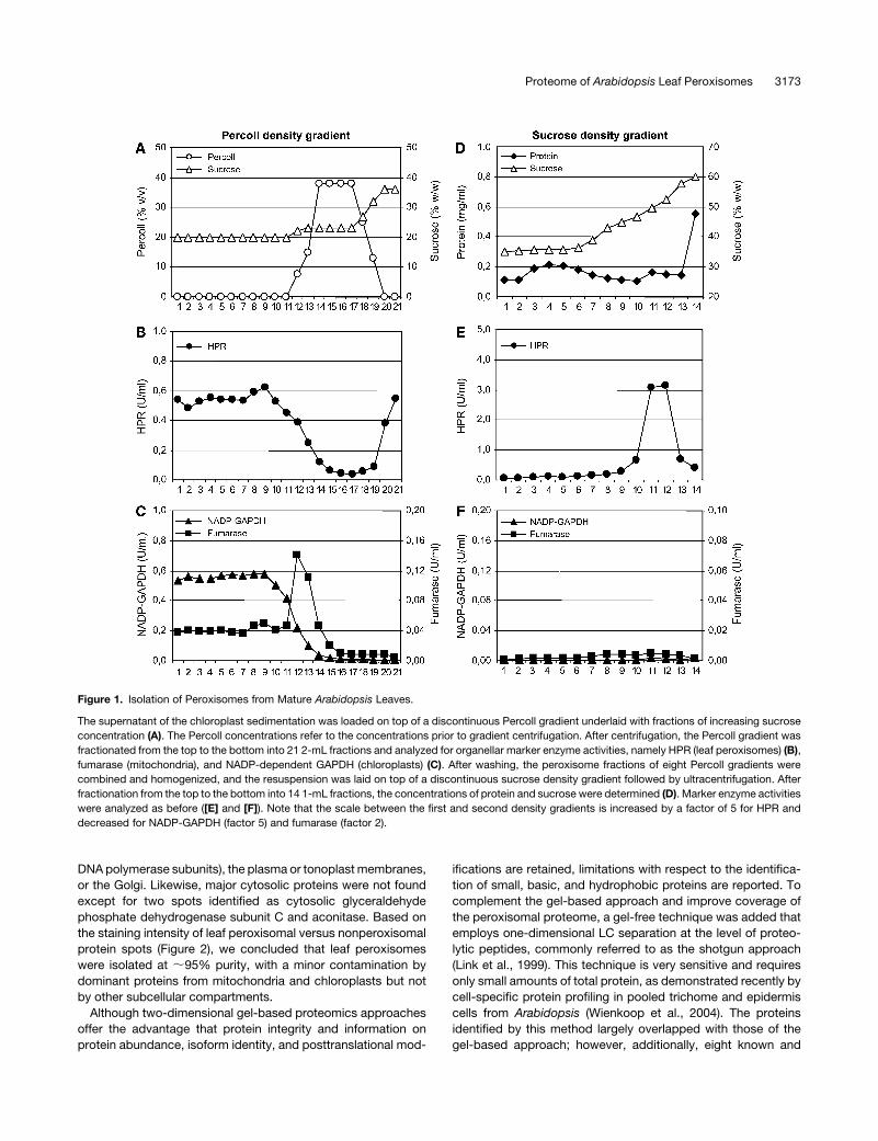
mately 7% of total activity of the leaf peroxisomal marker enzyme
hydroxypyruvate reductase (HPR) of the crude extract was
recovered in the first fraction of leaf peroxisomes (LP-P1) with
a yield of ;90 nkat HPR per 60 g fresh weight (Table 1). The
peroxisome fraction was carefully homogenized and laid on top
of a discontinuous sucrose density gradient (Figure 1D; see
Supplemental Figure 1 online). After ultracentrifugation, intact
leaf peroxisomes were enriched close to the bottom of the
gradient, visible as a sharp white band, at the typical density of
plant peroxisomes of ;50 to 53% (w/w) sucrose (Figures 1E and
1F; see Supplemental Figure 1 online). Even though only half of
the peroxisomes could be recovered, the contamination by
chloroplasts and mitochondria was further reduced by a factor
of three (Table 1). On average, 115 6 29 mg protein were ob-
tained with a specific HPR activity of 293 6 76 nkat/mg protein
(Table 1). The purity of leaf peroxisomes was high, as determined
by the activities of marker enzymes for leaf peroxisomes (HPR),
mitochondria (fumarase), and chloroplasts (NADP-dependent
glyceraldehyde-3-phosphate dehydrogenase [GAPDH]) and the
content of chlorophyll (thylakoids); contaminating chloroplasts
and mitochondria were estimated to comprise only 0.1 and 1.7%
of the total, respectively (Table 1).
Identification of Proteins from Mature Leaf Peroxisomes
Peroxisome fractions of highest purity (;400 nkat HPR/mg
protein) were analyzed by 2-DE to display the proteinaceous
constituents of Arabidopsis leaf peroxisomes as maps of intact
proteins (Figure 2). Upon gel staining with colloidal Coomassie
blue, ;180 separated protein spots were visualized, out of which
135 were identified with high confidence by peptide mass and
fragment fingerprinting, using an automated platform based on
matrix-assisted laser desorption/ionization time-of-flight mass
spectrometry (MALDI-TOF-MS) (Jahn et al., 2006). Summarized
over several two-dimensional gels, in addition to 28 known plant
peroxisomal proteins, 36 proteins were identified that had pre-
viously not been associated experimentally with plant peroxi-
somes and are referred to as novel proteins throughout this
study.
With respect to peroxisome purity, few minor spots derived
from dominant proteins of chloroplasts and mitochondria. For
instance, the presence of the large and small subunits of
ribulose-1,5-bisphosphate carboxylase/oxygenase, the most
abundant plant protein, and small subunits of the photosystems
(e.g., PSBP; Figure 2) indicated a low contamination with chlo-
roplasts. Similarly, a few tiny protein spots represented mito-
chondrial proteins, including the P and H subunits of Gly
decarboxylase (GDC-P and GDC-H), which is the most prom-
inent mitochondrial protein; subunit b of the ATP synthase com-
plex (ATPase b); and Ser-hydroxymethyltransferase (Figure 2).
By contrast, no dominant proteins were detected of the endo-
plasmic reticulum (ER) (e.g., BiP), the nucleus (e.g., histones and
3172 The Plant Cell
DNA polymerase subunits), the plasma or tonoplast membranes,
or the Golgi. Likewise, major cytosolic proteins were not found
except for two spots identified as cytosolic glyceraldehyde
phosphate dehydrogenase subunit C and aconitase. Based on
the staining intensity of leaf peroxisomal versus nonperoxisomal
protein spots (Figure 2), we concluded that leaf peroxisomes
were isolated at ;95% purity, with a minor contamination by
dominant proteins from mitochondria and chloroplasts but not
by other subcellular compartments.
Although two-dimensional gel-based proteomics approaches
offer the advantage that protein integrity and information on
protein abundance, isoform identity, and posttranslational mod-
ifications are retained, limitations with respect to the identifica-
tion of small, basic, and hydrophobic proteins are reported. To
complement the gel-based approach and improve coverage of
the peroxisomal proteome, a gel-free technique was added that
employs one-dimensional LC separation at the level of proteo-
lytic peptides, commonly referred to as the shotgun approach
(Link et al., 1999). This technique is very sensitive and requires
only small amounts of total protein, as demonstrated recently by
cell-specific protein profiling in pooled trichome and epidermis
cells from Arabidopsis (Wienkoop et al., 2004). The proteins
identified by this method largely overlapped with those of the
gel-based approach; however, additionally, eight known and
Figure 1. Isolation of Peroxisomes from Mature Arabidopsis Leaves.
The supernatant of the chloroplast sedimentation was loaded on top of a discontinuous Percoll gradient underlaid with fractions of increasing sucrose
concentration (A). The Percoll concentrations refer to the concentrations prior to gradient centrifugation. After centrifugation, the Percoll gradient was
fractionated from the top to the bottom into 21 2-mL fractions and analyzed for organellar marker enzyme activities, namely HPR (leaf peroxisomes) (B),
fumarase (mitochondria), and NADP-dependent GAPDH (chloroplasts) (C). After washing, the peroxisome fractions of eight Percoll gradients were
combined and homogenized, and the resuspension was laid on top of a discontinuous sucrose density gradient followed by ultracentrifugation. After
fractionation from the top to the bottom into 14 1-mL fractions, the concentrations of protein and sucrose were determined (D). Marker enzyme activities
were analyzed as before ([E] and [F]). Note that the scale between the first and second density gradients is increased by a factor of 5 for HPR and
decreased for NADP-GAPDH (factor 5) and fumarase (factor 2).
Proteome of Arabidopsis Leaf Peroxisomes 3173
six novel proteins were detected only by the shotgun strategy.
These proteins generally had either a basic isoelectric point (pI >
8.4) and/or were small proteins (<150 residues), underscoring the
complementary nature of the two approaches applied. In total,
we identified 78 nonredundant proteins, comprising 36 known
and 42 novel proteins (Table 2; see Supplemental Table 1 online).
Filtered mass spectra for protein identifications are stored in
ProMEX, a mass spectral reference library for plant proteomics,
that can be searched with peptide product ion spectra of
unknown samples (Hummel et al., 2007).
Known Plant Peroxisomal Proteins
Identified proteins already known as constituents of leaf perox-
isomes or glyoxysomes in any plant species are listed in Sup-
plemental Table 1 online. As valuable information on protein
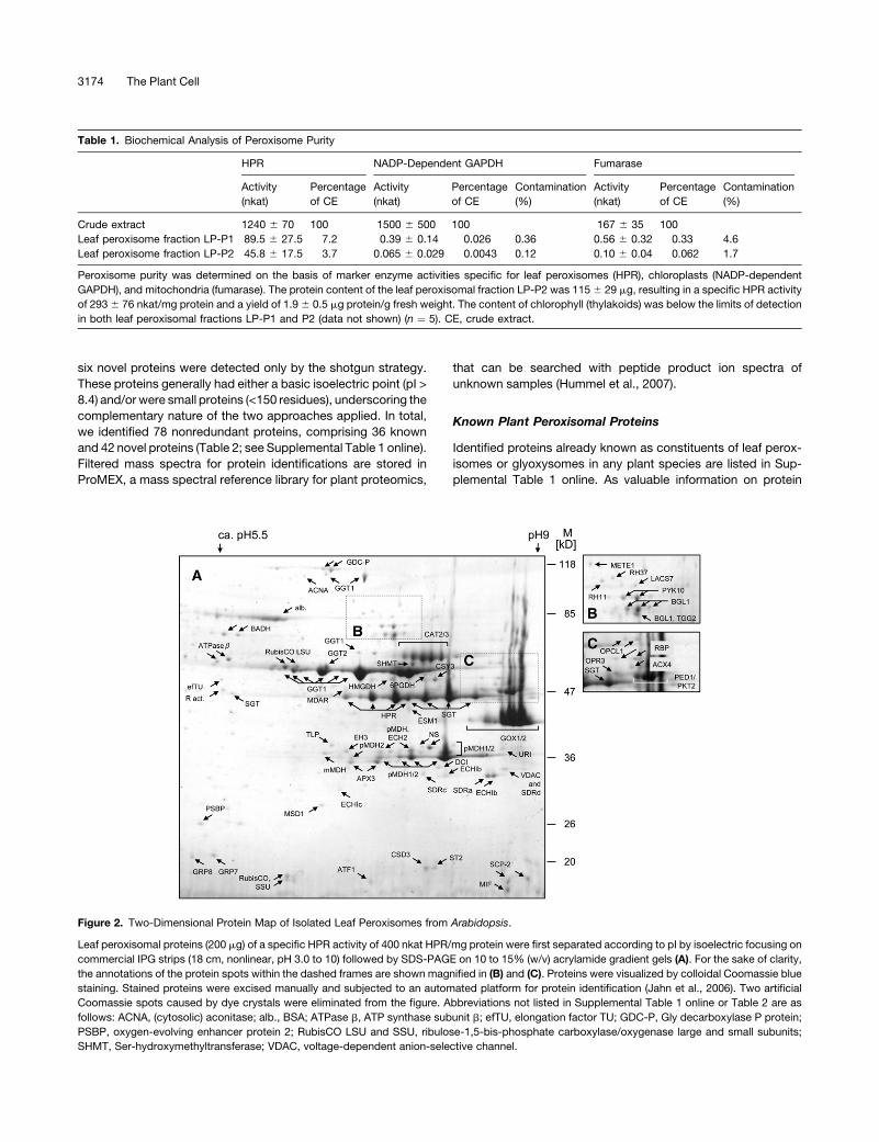
Table 1. Biochemical Analysis of Peroxisome Purity
HPR NADP-Dependent GAPDH Fumarase
Activity
(nkat)
Percentage
of CE
Activity
(nkat)
Percentage
of CE
Contamination
(%)
Activity
(nkat)
Percentage
of CE
Contamination
(%)
Crude extract 1240 6 70 100 1500 6 500 100 167 6 35 100
Leaf peroxisome fraction LP-P1 89.5 6 27.5 7.2 0.39 6 0.14 0.026 0.36 0.56 6 0.32 0.33 4.6
Leaf peroxisome fraction LP-P2 45.8 6 17.5 3.7 0.065 6 0.029 0.0043 0.12 0.10 6 0.04 0.062 1.7
Peroxisome purity was determined on the basis of marker enzyme activities specific for leaf peroxisomes (HPR), chloroplasts (NADP-dependent
GAPDH), and mitochondria (fumarase). The protein content of the leaf peroxisomal fraction LP-P2 was 115 6 29 mg, resulting in a specific HPR activity
of 293 6 76 nkat/mg protein and a yield of 1.9 6 0.5 mg protein/g fresh weight. The content of chlorophyll (thylakoids) was below the limits of detection
in both leaf peroxisomal fractions LP-P1 and P2 (data not shown) (n ¼ 5). CE, crude extract.
Figure 2. Two-Dimensional Protein Map of Isolated Leaf Peroxisomes from Arabidopsis.
Leaf peroxisomal proteins (200 mg) of a specific HPR activity of 400 nkat HPR/mg protein were first separated according to pI by isoelectric focusing on
commercial IPG strips (18 cm, nonlinear, pH 3.0 to 10) followed by SDS-PAGE on 10 to 15% (w/v) acrylamide gradient gels (A). For the sake of clarity,
the annotations of the protein spots within the dashed frames are shown magnified in (B) and (C). Proteins were visualized by colloidal Coomassie blue
staining. Stained proteins were excised manually and subjected to an automated platform for protein identification (Jahn et al., 2006). Two artificial
Coomassie spots caused by dye crystals were eliminated from the figure. Abbreviations not listed in Supplemental Table 1 online or Table 2 are as
follows: ACNA, (cytosolic) aconitase; alb., BSA; ATPase b, ATP synthase subunit b; efTU, elongation factor TU; GDC-P, Gly decarboxylase P protein;
PSBP, oxygen-evolving enhancer protein 2; RubisCO LSU and SSU, ribulose-1,5-bis-phosphate carboxylase/oxygenase large and small subunits;
SHMT, Ser-hydroxymethyltransferase; VDAC, voltage-dependent anion-selective channel.
3174 The Plant Cell
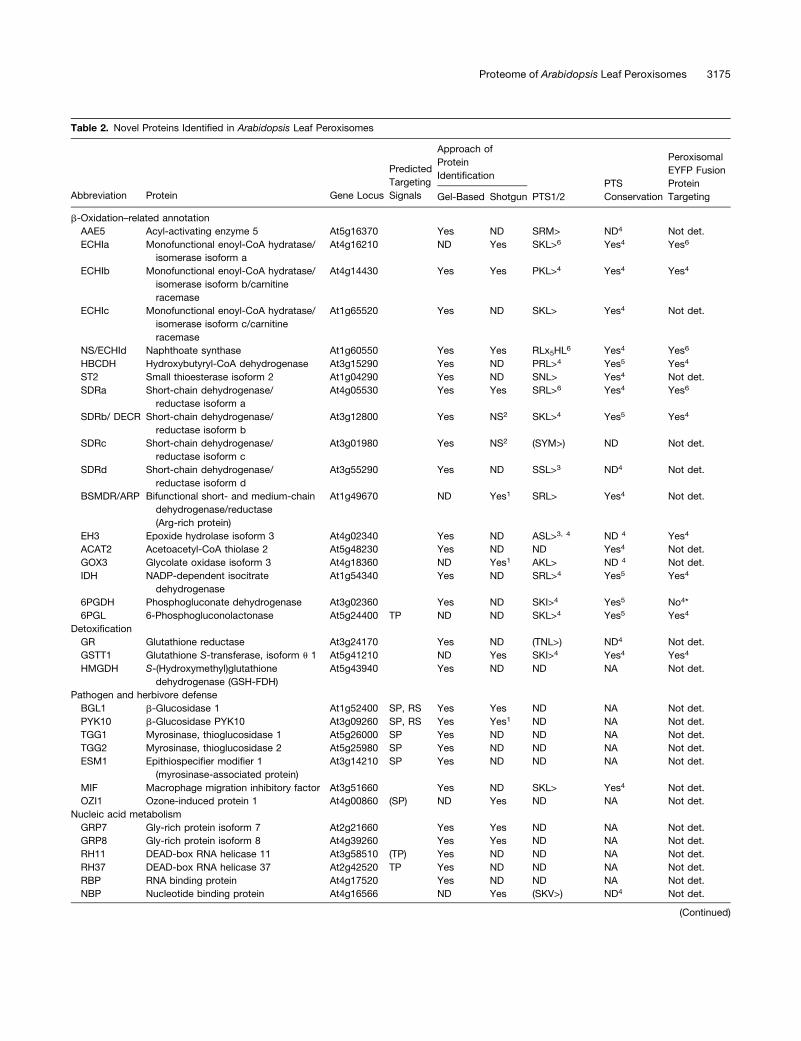
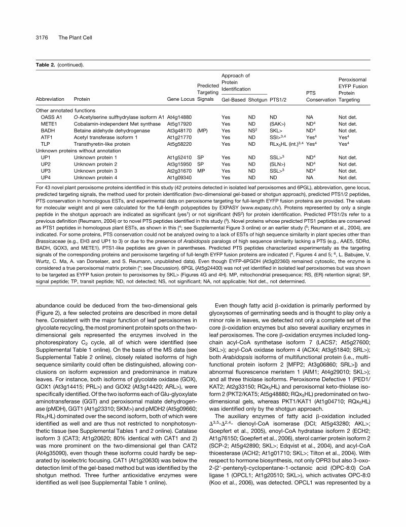
Table 2. Novel Proteins Identified in Arabidopsis Leaf Peroxisomes
Predicted
Targeting
Signals
Approach of
Protein
IdentificationPTS
Conservation
Peroxisomal
EYFP Fusion
Protein
TargetingAbbreviation Protein Gene Locus Gel-Based Shotgun PTS1/2
b-Oxidation–related annotation
AAE5 Acyl-activating enzyme 5 At5g16370 Yes ND SRM> ND4 Not det.
ECHIa Monofunctional enoyl-CoA hydratase/
isomerase isoform a
At4g16210 ND Yes SKL>6 Yes4 Yes6
ECHIb Monofunctional enoyl-CoA hydratase/
isomerase isoform b/carnitine
racemase
At4g14430 Yes Yes PKL>4 Yes4 Yes4
ECHIc Monofunctional enoyl-CoA hydratase/
isomerase isoform c/carnitine
racemase
At1g65520 Yes ND SKL> Yes4 Not det.
NS/ECHId Naphthoate synthase At1g60550 Yes Yes RLx5HL6 Yes4 Yes6
HBCDH Hydroxybutyryl-CoA dehydrogenase At3g15290 Yes ND PRL>4 Yes5 Yes4
ST2 Small thioesterase isoform 2 At1g04290 Yes ND SNL> Yes4 Not det.
SDRa Short-chain dehydrogenase/
reductase isoform a
At4g05530 Yes Yes SRL>6 Yes4 Yes6
SDRb/ DECR Short-chain dehydrogenase/
reductase isoform b
At3g12800 Yes NS2 SKL>4 Yes5 Yes4
SDRc Short-chain dehydrogenase/
reductase isoform c
At3g01980 Yes NS2 (SYM>) ND Not det.
SDRd Short-chain dehydrogenase/
reductase isoform d
At3g55290 Yes ND SSL>3 ND4 Not det.
BSMDR/ARP Bifunctional short- and medium-chain
dehydrogenase/reductase
(Arg-rich protein)
At1g49670 ND Yes1 SRL> Yes4 Not det.
EH3 Epoxide hydrolase isoform 3 At4g02340 Yes ND ASL>3, 4 ND 4 Yes4
ACAT2 Acetoacetyl-CoA thiolase 2 At5g48230 Yes ND ND Yes4 Not det.
GOX3 Glycolate oxidase isoform 3 At4g18360 ND Yes1 AKL> ND 4 Not det.
IDH NADP-dependent isocitrate
dehydrogenase
At1g54340 Yes ND SRL>4 Yes5 Yes4
6PGDH Phosphogluconate dehydrogenase At3g02360 Yes ND SKI>4 Yes5 No4*
6PGL 6-Phosphogluconolactonase At5g24400 TP ND ND SKL>4 Yes5 Yes4
Detoxification
GR Glutathione reductase At3g24170 Yes ND (TNL>) ND4 Not det.
GSTT1 Glutathione S-transferase, isoform u 1 At5g41210 ND Yes SKI>4 Yes4 Yes4
HMGDH S-(Hydroxymethyl)glutathione
dehydrogenase (GSH-FDH)
At5g43940 Yes ND ND NA Not det.
Pathogen and herbivore defense
BGL1 b-Glucosidase 1 At1g52400 SP, RS Yes Yes ND NA Not det.
PYK10 b-Glucosidase PYK10 At3g09260 SP, RS Yes Yes1 ND NA Not det.
TGG1 Myrosinase, thioglucosidase 1 At5g26000 SP Yes ND ND NA Not det.
TGG2 Myrosinase, thioglucosidase 2 At5g25980 SP Yes ND ND NA Not det.
ESM1 Epithiospecifier modifier 1
(myrosinase-associated protein)
At3g14210 SP Yes ND ND NA Not det.
MIF Macrophage migration inhibitory factor At3g51660 Yes ND SKL> Yes4 Not det.
OZI1 Ozone-induced protein 1 At4g00860 (SP) ND Yes ND NA Not det.
Nucleic acid metabolism
GRP7 Gly-rich protein isoform 7 At2g21660 Yes Yes ND NA Not det.
GRP8 Gly-rich protein isoform 8 At4g39260 Yes Yes ND NA Not det.
RH11 DEAD-box RNA helicase 11 At3g58510 (TP) Yes ND ND NA Not det.
RH37 DEAD-box RNA helicase 37 At2g42520 TP Yes ND ND NA Not det.
RBP RNA binding protein At4g17520 Yes ND ND NA Not det.
NBP Nucleotide binding protein At4g16566 ND Yes (SKV>) ND4 Not det.
(Continued)
Proteome of Arabidopsis Leaf Peroxisomes 3175
abundance could be deduced from the two-dimensional gels
(Figure 2), a few selected proteins are described in more detail
here. Consistent with the major function of leaf peroxisomes in
glycolate recycling, the most prominent protein spots on the two-
dimensional gels represented the enzymes involved in the
photorespiratory C2 cycle, all of which were identified (see
Supplemental Table 1 online). On the basis of the MS data (see
Supplemental Table 2 online), closely related isoforms of high
sequence similarity could often be distinguished, allowing con-
clusions on isoform expression and predominance in mature
leaves. For instance, both isoforms of glycolate oxidase (GOX),
GOX1 (At3g14415; PRL>) and GOX2 (At3g14420; ARL>), were
specifically identified. Of the two isoforms each of Glu-glyoxylate
aminotransferase (GGT) and peroxisomal malate dehydrogen-
ase (pMDH), GGT1 (At1g23310; SKM>) and pMDH2 (At5g09660;
RIx5HL) dominated over the second isoform, both of which were
identified as well and are thus not restricted to nonphotosyn-
thetic tissue (see Supplemental Tables 1 and 2 online). Catalase
isoform 3 (CAT3; At1g20620; 80% identical with CAT1 and 2)
was more prominent on the two-dimensional gel than CAT2
(At4g35090), even though these isoforms could hardly be sep-
arated by isoelectric focusing. CAT1 (At1g20630) was below the
detection limit of the gel-based method but was identified by the
shotgun method. Three further antioxidative enzymes were
identified as well (see Supplemental Table 1 online).
Even though fatty acid b-oxidation is primarily performed by
glyoxysomes of germinating seeds and is thought to play only a
minor role in leaves, we detected not only a complete set of the
core b-oxidation enzymes but also several auxiliary enzymes in
leaf peroxisomes. The core b-oxidation enzymes included long-
chain acyl-CoA synthetase isoform 7 (LACS7; At5g27600;
SKL>); acyl-CoA oxidase isoform 4 (ACX4; At3g51840; SRL>);
both Arabidopsis isoforms of multifunctional protein (i.e., multi-
functional protein isoform 2 [MFP2; At3g06860; SRL>]) and
abnormal fluorescence meristem 1 (AIM1; At4g29010; SKL>);
and all three thiolase isoforms. Peroxisome Defective 1 (PED1/
KAT2; At2g33150; RQx5HL) and peroxisomal keto-thiolase iso-
form 2 (PKT2/KAT5; At5g48880; RQx5HL) predominated on two-
dimensional gels, whereas PKT1/KAT1 (At1g04710; RQx5HL)
was identified only by the shotgun approach.
The auxiliary enzymes of fatty acid b-oxidation included
D3,5-D2,4- dienoyl-CoA isomerase (DCI; At5g43280; AKL>;
Goepfert et al., 2005), enoyl-CoA hydratase isoform 2 (ECH2;
At1g76150; Goepfert et al., 2006), sterol carrier protein isoform 2
(SCP-2; At5g42890; SKL>; Edqvist et al., 2004), and acyl-CoA
thioesterase (ACH2; At1g01710; SKL>; Tilton et al., 2004). With
respect to hormone biosynthesis, not only OPR3 but also 3-oxo-
2-(29-pentenyl)-cyclopentane-1-octanoic acid (OPC-8:0) CoA
ligase 1 (OPCL1; At1g20510; SKL>), which activates OPC-8:0
(Koo et al., 2006), was detected. OPCL1 was represented by a
Table 2. (continued).
Predicted
Targeting
Signals
Approach of
Protein
IdentificationPTS
Conservation
Peroxisomal
EYFP Fusion
Protein
TargetingAbbreviation Protein Gene Locus Gel-Based Shotgun PTS1/2
Other annotated functions
OASS A1 O-Acetylserine sulfhydrylase isoform A1 At4g14880 Yes ND ND NA Not det.
METE1 Cobalamin-independent Met synthase At5g17920 Yes ND (SAK>) ND4 Not det.
BADH Betaine aldehyde dehydrogenase At3g48170 (MP) Yes NS2 SKL> ND4 Not det.
ATF1 Acetyl transferase isoform 1 At1g21770 Yes ND SSI>3,4 Yes4 Yes4
TLP Transthyretin-like protein At5g58220 Yes ND RLx5HL (int.)3,4 Yes4 Yes4
Unknown proteins without annotation
UP1 Unknown protein 1 At1g52410 SP Yes ND SSL>3 ND4 Not det.
UP2 Unknown protein 2 At3g15950 SP Yes ND (SLN>) ND4 Not det.
UP3 Unknown protein 3 At2g31670 MP Yes ND SSL>3 ND4 Not det.
UP4 Unknown protein 4 At1g09340 Yes ND ND NA Not det.
For 43 novel plant peroxisome proteins identified in this study (42 proteins detected in isolated leaf peroxisomes and 6PGL), abbreviation, gene locus,
predicted targeting signals, the method used for protein identification (two-dimensional gel-based or shotgun approach), predicted PTS1/2 peptides,
PTS conservation in homologous ESTs, and experimental data on peroxisome targeting for full-length EYFP fusion proteins are provided. The values
for molecular weight and pI were calculated for the full-length polypeptides by EXPASY (www.expasy.ch/). Proteins represented by only a single
peptide in the shotgun approach are indicated as significant (yes1) or not significant (NS2) for protein identification. Predicted PTS1/2s refer to a
previous definition (Reumann, 2004) or to novel PTS peptides identified in this study (3). Novel proteins whose predicted PTS1 peptides are conserved
as PTS1 peptides in homologous plant ESTs, as shown in this (4; see Supplemental Figure 3 online) or an earlier study (5; Reumann et al., 2004), are
indicated. For some proteins, PTS conservation could not be analyzed owing to a lack of ESTs of high sequence similarity in plant species other than
Brassicaceae (e.g., EH3 and UP1 to 3) or due to the presence of Arabidopsis paralogs of high sequence similarity lacking a PTS (e.g., AAE5, SDRd,
BADH, GOX3, and METE1). PTS1-like peptides are given in parentheses. Predicted PTS peptides characterized experimentally as the targeting
signals of the corresponding proteins and peroxisome targeting of full-length EYFP fusion proteins are indicated (4, Figures 4 and 5; 6, L. Babujee, V.
Wurtz, C. Ma, A. van Dorselaer, and S. Reumann, unpublished data). Even though EYFP-6PGDH (At3g02360) remained cytosolic, the enzyme is
considered a true peroxisomal matrix protein (*; see Discussion). 6PGL (At5g24400) was not yet identified in isolated leaf peroxisomes but was shown
to be targeted as EYFP fusion protein to peroxisomes by SKL> (Figures 4G and 4H). MP, mitochondrial presequence; RS, (ER) retention signal; SP,
signal peptide; TP, transit peptide; ND, not detected; NS, not significant; NA, not applicable; Not det., not determined.
3176 The Plant Cell
low-abundance protein spot restricted to peroxisomes of highest
purity, consistent with its low expression level under standard
conditions and its induction by wounding. Two further acyl-CoA
acitivating enzymes (AAEs) included AAE7/ACN1 (At3g16910;
SRL>; Turner et al., 2005) and 4-coumarate-CoA ligase-like
protein 1 (4CLP1; At4g05160; SKM>; Schneider et al., 2005). In
agreement with the heat inducibility of one of the two sHsps from
plant peroxisomes (HSP15.7-Px; Ma et al., 2006), only the con-
stitutively expressed isoform ACD31.2-Px (At1g06460; RLx5HF)
was identified. As peroxisomal membranes were not further en-
riched prior to proteome analysis, the number of integral mem-
brane proteins identified was low (APX3, PEX11d, and PEX14),
which is consistent with the low content of membrane proteins in
peroxisomes in general (Fujiki et al., 1982; S. Reumann, unpub-
lished data).
In summary, the 36 known plant peroxisomal proteins identi-
fied in isolated leaf peroxisomes included all soluble enzymes
involved in photorespiration, ROS metabolism, and JA biosyn-
thesis. The detection of many core and auxiliary enzymes involved
in fatty acid b-oxidation and sulfur and nitrogen metabolism (see
Supplemental Table 1 online) indicated that (1) the dynamic
range of protein identification was sufficiently high to cover
low-abundance enzymes that are predominantly expressed in
peroxisome variants other than leaf peroxisomes and that (2)
b-oxidation–related enzymes may play a yet underestimated role
in leaf peroxisomal metabolism.
Several matrix enzymes, although encoded by single Arabi-
dopsis genes, often appeared on the two-dimensional gels as
multiple protein spots arranged in a characteristic horizontal
pattern. This pattern was most obvious for highly abundant ma-
trix enzymes, namely GGT1, Ser-glyoxylate aminotransferase,
HPR, and pMDH2, but also observed for minor enzymes, such as
ascorbate peroxidase (APX3) and OPCL1 (Figure 2; see Supple-
mental Figures 2A to 2C online). The horizontal spot series of
CAT3 differed slightly in that the differences in pI between spots
were smaller (see Supplemental Figure 2D online). Although
horizontal spot trains are often seen as artifacts of 2-DE, the
uniform and regular patterns observed here might be indicative
of the presence of charge-modifying posttranslational modifica-
tions, such as phosphorylation and acetylation.
Novel Plant Peroxisomal Proteins
Among the 42 novel proteins, a surprisingly large number
had annotated functions related to fatty acid b-oxidation
(Table 2). One protein is a yet uncharacterized isoform of the
extended superfamily of acyl-activating enzymes, namely AAE5
(At5g16370; SRM>), belonging to clade VI of largely unknown
PTS1-carrying AAEs, as does AAE7/ACN1 (Shockey et al., 2003;
Reumann et al., 2004; Turner et al., 2005). Regarding the super-
family of enoyl-CoA hydratases/isomerases (ECHIs; Reumann
et al., 2004), with MFP and AIM1 being the most prominent
enzymes, we now identified monofunctional members of plant
peroxisomes. The first ECHI, referred to as isoform a (ECHIa;
At4g16210; SKL>), belongs to clade VI of putative plant enoyl-
CoA hydratases. Two further monofunctional ECHIs identified,
namely, ECHIb (At4g14430; PKL>) and ECHIc (At1g65520;
SKL>), have been grouped in clade III and are the closest
Arabidopsis homologs of peroxisomal monofunctional D3-D2-
enoyl-CoA isomerase (PECI) from fungi and mammals (Geisbrecht
et al., 1999; Reumann et al., 2004). Most intriguing of this enzyme
family is ECHId, annotated as putative naphthoate synthase (NS/
ECHId; At1g60550; RLx5HL) because the enzyme is plant spe-
cific among eukaryotes and represents the Arabidopsis homolog
of cyanobacterial MenB (clade V), an enoyl-CoA hydratase
involved in phylloquinone biosynthesis (Johnson et al., 2001;
Gross et al., 2006, see also references therein). This provides
experimental evidence for peroxisome targeting of the single
Arabidopsis homolog of MenB, further supported by conserva-
tion of the predicted PTS2 in homologous ESTs (see Supple-
mental Figure 3O online), and indicates that phylloquinone
biosynthesis is partly compartmentalized in plant peroxisomes.
One protein spot was identified as a monofunctional hydroxyacyl-
CoA dehydrogenase, annotated as hydroxybutyryl-CoA dehy-
drogenase (HBCDH; At3g15290) and carrying the predicted
major PTS tripeptide PRL> (Reumann et al., 2004). Of the ex-
perimentally uncharacterized Arabidopsis family of small thioes-
terases (STs) with six PTS1-possessing members (Reumann et al.,
2004), the second isoform of clade I (ST2; At1g04290; SNL>) is
now experimentally associated with plant peroxisomes. Five
novel proteins belong to the superfamily of short (polypeptide)-
chain dehydrogenases/reductases (SDRs) and include four mono-
functional SDRs (SDRa, At4g05530, SRL>; SDRb, At3g12800,
SKL>; SDRc, At3g01980, SYM>; and SDRd, At3g55290, SSL>)
and a bifunctional short- and medium-chain dehydrogenase/
reductase (BSMDR; At1g49670; SRL>) with an N-terminal SDR
and a C-terminal medium-chain dehydrogenase/reductase do-
main of the subgroup of NADPH:quinone reductases (COG0604,
Qor). The latter was identified specifically by the gel-free
approach.
Further novel enzymes with predicted catalytic activities re-
lated to fatty acid b-oxidation included epoxide hydrolase iso-
form 3 (EH3; At4g02340; ASL>) and one of two Arabidopsis
homologs of acetoacetyl-CoA thiolase (ACAT2, EC 2.3.1.9,
At5g48230, which is 80% identical with ACAT1, At5g47720;
Carrie et al., 2007). GOX3 (At4g18360; AKL>) is a yet unknown
isoform that shares 85% identity at the amino acid level with
GOX1/2 but is predominantly expressed in nonphotosynthetic
tissue and presumably plays a role outside of photorespiration
(Kamada et al., 2003; Reumann et al., 2004). Enzymes associ-
ated with peroxisomal NADP metabolism included isoforms of
NADP-dependent isocitrate dehydrogenase (IDH; At1g54340;
SRL>) and 6-phosphogluconate dehydrogenase (6PGDH;
At3g02360; SKI>), one of three enzymes of the oxidative pentose
phosphate pathway (OPPP). Several novel plant peroxisomal
enzymes are predicted to catalyze GSH-dependent reactions.
These identified proteins included an isoform of glutathione
reductase (GR; At3g24170; TNL>), isoform u 1 of the extended
Arabidopsis family of glutathione S-transferases (GSTT1, formerly
GST10, At5g41210, SKI>, EC 2.5.1.18; Dixon et al., 2002), and
the single Arabidopsis homolog of S-(hydroxymethyl)glutathione
dehydrogenase (HMGDH; At5g43940; EC 1.1.1.284; Dolferus
et al., 1997).
Seven identified proteins are functionally associated with plant
defense mechanisms, which was rather unexpected. Four en-
zymes are b-glucosidases of the superfamily of glycoside
Proteome of Arabidopsis Leaf Peroxisomes 3177
hydrolase family 1, many of which are reported to mediate plant
defense against herbivores (Xu et al., 2004; Table 2). The
b-glucosidases included b-glucosidase 1 (BGL1; At1g52400;
Stotz et al., 2000), PYK10 (At3g09260), and the myrosinase ho-
molog thioglucoside glucohydrolase 2 (TGG2; At5g25980; Barth
and Jander, 2006). All three defense-related enzymes were ap-
parently upregulated in leaf peroxisomes isolated from senes-
cent plants (see Supplemental Figures 2E and 2F online); TGG1
(At5g26000; Barth and Jander, 2006) was specifically identified
in the latter (data not shown). The myrosinase-associated protein
EPITHIOSPECIFIER MODIFIER1 (ESM1; At3g14210), which al-
ters glucosinolate hydrolysis and insect resistance (Zhang et al.,
2006), was identified in highly purified leaf peroxisomes as well.
Two other defense proteins identified comprised one of three
Arabidopsis homologs of vertebrate macrophage migration in-
hibitory factor (MIF; At3g51660; SKL>) and ozone-induced pro-
tein 1 (OZI1; At4g00860), a plant-specific small polypeptide of
only 80 residues that is induced by ozone and pathogen attack
(Sharma and Davis, 1995).
Likewise unexpected was the identification of proteins with
annotated functions related to nucleic acid metabolism. These
six proteins included two Gly-rich RNA binding proteins (GRP7
and 8; At2g21660 and At4g39260; Staiger et al., 2003), two
DEAD-box RNA helicases (RH11, At3g58510; RH37, At2g42520),
an unknown RNA binding protein (RBP; At4g17520), and a nucle-
otide binding protein (NBP; At4g16566). Except for NBP carrying
the PTS1-like C-terminal tripeptide SKV>, all proteins related to
nucleic acid metabolism lack predicted PTSs.
Proteins with other annotated functions included two enzymes
reported to be involved in Cys and Met biosynthesis. O-acetylserine
sulfhydrylase (OASS A1; At4g14880) is a presumably cytosolic
isoform lacking an obvious PTS. Isoform 1 of cobalamin (vitamin
B12)-independent Met synthase (METE1; At5g17920; EC 2.1.1.14;
Ravanel et al., 2004) carries the PTS1-related C-terminal tripep-
tide SAK>. One of two Arabidopsis homologs of betaine aldehyde
dehydrogenase (BADH; At3g48170; SKL>) was identified as well,
homologs of which had previously been localized to the perox-
isome matrix in monocotyledons (Nakamura et al., 1997). An-
other protein spot (ATF1; At1g21770; SSI>) was identified as a
closely related homolog of a recently crystallized minimal acetyl
transferase (ATF2; At1g77540; SSI>; Tyler et al., 2006). The
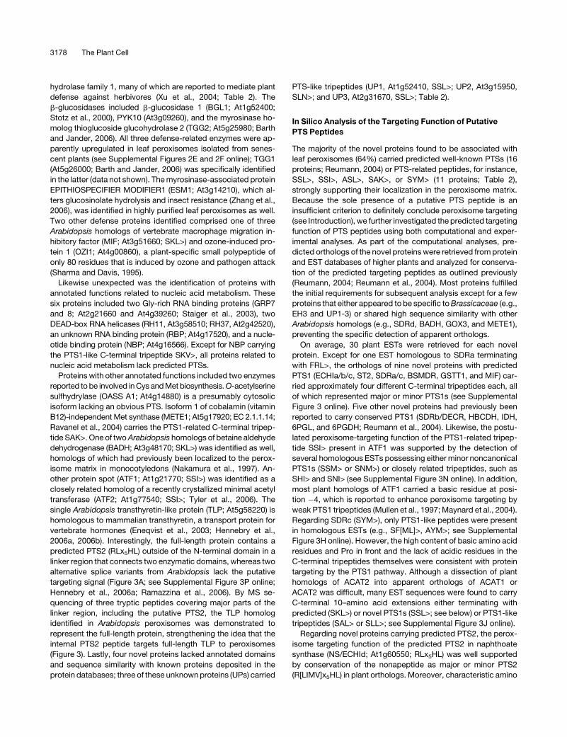
single Arabidopsis transthyretin-like protein (TLP; At5g58220) is
homologous to mammalian transthyretin, a transport protein for
vertebrate hormones (Eneqvist et al., 2003; Hennebry et al.,
2006a, 2006b). Interestingly, the full-length protein contains a
predicted PTS2 (RLx5HL) outside of the N-terminal domain in a
linker region that connects two enzymatic domains, whereas two
alternative splice variants from Arabidopsis lack the putative
targeting signal (Figure 3A; see Supplemental Figure 3P online;
Hennebry et al., 2006a; Ramazzina et al., 2006). By MS se-
quencing of three tryptic peptides covering major parts of the
linker region, including the putative PTS2, the TLP homolog
identified in Arabidopsis peroxisomes was demonstrated to
represent the full-length protein, strengthening the idea that the
internal PTS2 peptide targets full-length TLP to peroxisomes
(Figure 3). Lastly, four novel proteins lacked annotated domains
and sequence similarity with known proteins deposited in the
protein databases; three of these unknown proteins (UPs) carried
PTS-like tripeptides (UP1, At1g52410, SSL>; UP2, At3g15950,
SLN>; and UP3, At2g31670, SSL>; Table 2).
In Silico Analysis of the Targeting Function of Putative
PTS Peptides
The majority of the novel proteins found to be associated with
leaf peroxisomes (64%) carried predicted well-known PTSs (16
proteins; Reumann, 2004) or PTS-related peptides, for instance,
SSL>, SSI>, ASL>, SAK>, or SYM> (11 proteins; Table 2),
strongly supporting their localization in the peroxisome matrix.
Because the sole presence of a putative PTS peptide is an
insufficient criterion to definitely conclude peroxisome targeting
(see Introduction), we further investigated the predicted targeting
function of PTS peptides using both computational and exper-
imental analyses. As part of the computational analyses, pre-
dicted orthologs of the novel proteins were retrieved from protein
and EST databases of higher plants and analyzed for conserva-
tion of the predicted targeting peptides as outlined previously
(Reumann, 2004; Reumann et al., 2004). Most proteins fulfilled
the initial requirements for subsequent analysis except for a few
proteins that either appeared to be specific to Brassicaceae (e.g.,
EH3 and UP1-3) or shared high sequence similarity with other
Arabidopsis homologs (e.g., SDRd, BADH, GOX3, and METE1),
preventing the specific detection of apparent orthologs.
On average, 30 plant ESTs were retrieved for each novel
protein. Except for one EST homologous to SDRa terminating
with FRL>, the orthologs of nine novel proteins with predicted
PTS1 (ECHIa/b/c, ST2, SDRa/c, BSMDR, GSTT1, and MIF) car-
ried approximately four different C-terminal tripeptides each, all
of which represented major or minor PTS1s (see Supplemental
Figure 3 online). Five other novel proteins had previously been
reported to carry conserved PTS1 (SDRb/DECR, HBCDH, IDH,
6PGL, and 6PGDH; Reumann et al., 2004). Likewise, the postu-
lated peroxisome-targeting function of the PTS1-related tripep-
tide SSI> present in ATF1 was supported by the detection of
several homologous ESTs possessing either minor noncanonical
PTS1s (SSM> or SNM>) or closely related tripeptides, such as
SHI> and SNI> (see Supplemental Figure 3N online). In addition,
most plant homologs of ATF1 carried a basic residue at posi-
tion �4, which is reported to enhance peroxisome targeting by
weak PTS1 tripeptides (Mullen et al., 1997; Maynard et al., 2004).
Regarding SDRc (SYM>), only PTS1-like peptides were present
in homologous ESTs (e.g., SF[ML]>, AYM>; see Supplemental
Figure 3H online). However, the high content of basic amino acid
residues and Pro in front and the lack of acidic residues in the
C-terminal tripeptides themselves were consistent with protein
targeting by the PTS1 pathway. Although a dissection of plant
homologs of ACAT2 into apparent orthologs of ACAT1 or
ACAT2 was difficult, many EST sequences were found to carry
C-terminal 10–amino acid extensions either terminating with
predicted (SKL>) or novel PTS1s (SSL>; see below) or PTS1-like
tripeptides (SAL> or SLL>; see Supplemental Figure 3J online).
Regarding novel proteins carrying predicted PTS2, the perox-
isome targeting function of the predicted PTS2 in naphthoate
synthase (NS/ECHId; At1g60550; RLx5HL) was well supported
by conservation of the nonapeptide as major or minor PTS2
(R[LIMV]x5HL) in plant orthologs. Moreover, characteristic amino
3178 The Plant Cell
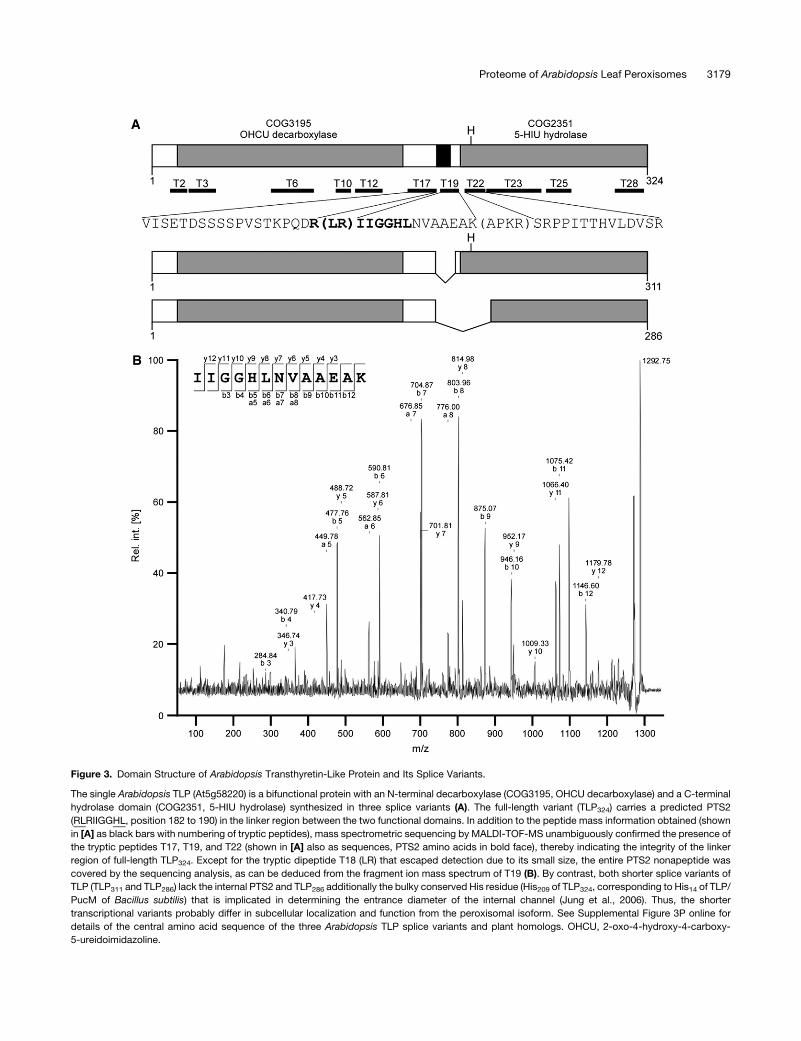
Figure 3. Domain Structure of Arabidopsis Transthyretin-Like Protein and Its Splice Variants.
The single Arabidopsis TLP (At5g58220) is a bifunctional protein with an N-terminal decarboxylase (COG3195, OHCU decarboxylase) and a C-terminal
hydrolase domain (COG2351, 5-HIU hydrolase) synthesized in three splice variants (A). The full-length variant (TLP324) carries a predicted PTS2
(RLRIIGGHL, position 182 to 190) in the linker region between the two functional domains. In addition to the peptide mass information obtained (shown
in [A] as black bars with numbering of tryptic peptides), mass spectrometric sequencing by MALDI-TOF-MS unambiguously confirmed the presence of
the tryptic peptides T17, T19, and T22 (shown in [A] also as sequences, PTS2 amino acids in bold face), thereby indicating the integrity of the linker
region of full-length TLP324. Except for the tryptic dipeptide T18 (LR) that escaped detection due to its small size, the entire PTS2 nonapeptide was
covered by the sequencing analysis, as can be deduced from the fragment ion mass spectrum of T19 (B). By contrast, both shorter splice variants of
TLP (TLP311 and TLP286) lack the internal PTS2 and TLP286 additionally the bulky conserved His residue (His209 of TLP324, corresponding to His14 of TLP/
PucM of Bacillus subtilis) that is implicated in determining the entrance diameter of the internal channel (Jung et al., 2006). Thus, the shorter
transcriptional variants probably differ in subcellular localization and function from the peroxisomal isoform. See Supplemental Figure 3P online for
details of the central amino acid sequence of the three Arabidopsis TLP splice variants and plant homologs. OHCU, 2-oxo-4-hydroxy-4-carboxy-
5-ureidoimidazoline.
Proteome of Arabidopsis Leaf Peroxisomes 3179
acid residues predicted to act as targeting-enhancing elements
(Arg and Pro) were present in close proximity to the PTS2
(Reumann, 2004; see Supplemental Figure 3O online). Likewise,
the predicted internal PTS2 (RLx5HL) of TLP was conserved as
putative major or minor PTS2s in all homologous plant ESTs
spanning this region (e.g., R[LIMV]x5HL and RIx5HM) except for
one slightly divergent nonapeptide (RVx5HV) present in Medicago
(see Supplemental Figure 3P online). Even though full-length TLP
variants appear to predominate among plant ESTs, some plant
species, such as Oryza and Triticum, were found to express
alternative splice variants similar to Arabidopsis that exactly lack
this nonapeptide (Figure 3B; see Supplemental Figure 3P online;
Hennebry et al., 2006a).
Experimental Verification of Predicted PTSs
To experimentally verify protein targeting to peroxisomes and the
peroxisome-targeting function of predicted PTSs, subcellular
targeting of seven representative fusion proteins with EYFP was
analyzed by fluorescence microscopy upon transient cDNA
expression in onion epidermal cells. Four proteins carried pre-
dicted major PTS1 peptides (SRL>, SKL>, or PRL>), whereas
three proteins possessed predicted minor PTS1s (SKI> or PKL>)
of lower peroxisome-targeting probability (Reumann, 2004). For
all proteins, subcellular targeting of two constructs was analyzed
(i.e., EYFP fusions of the full-length proteins of interest and
fusions attaching only the predicted PTS1 targeting domain
[PTD], comprising the C-terminal 10–amino acid residues, in-
cluding the putative PTS1 tripeptide, to EYFP).
NADP-dependent IDH (At1g54340; SRL>) was chosen as one
of the proteins carrying predicted major PTS1 peptides. NADP-
dependent IDH, fused at its N terminus with EYFP (EYFP-IDH),
was indeed detected in punctate structures in onion epidermal
cells (Figure 4A1). In double transformants expressing simul-
taneously a peroxisome-targeted cyan fluorescent protein
(CspMDH-CFP; Fulda et al., 2002; Ma et al., 2006), the yellow
fluorescent subcellular structures were identified conclusively as
peroxisomes (Figures 4A1 and 4A2). EYFP extended by the
C-terminal 10–amino acid residues of IDH, including the pre-
dicted PTS1 SRL> (EYFP-PTDIDH), was directed from the cytosol
to punctate structures, thereby identifying SRL> as the PTS of
peroxisomal IDH (Figure 4B). Two further proteins, namely, the
full-length fusion proteins of the predicted DECR homolog SDRb
(At3g12800; SKL>) and the monofunctional hydroxyacyl-CoA
dehydrogenase HBCDH (At3g15290; PRL>), were targeted to
peroxisomes as well (Figures 4C1, 4C2, 4E1, and 4E2). EYFP
extended by the PTDs of these proteins showed a punctate
fluorescence pattern, characterizing the C-terminal tripeptides
SKL> and PRL> as the targeting signals of SDRb/DECR and
HBCDH, respectively (Figures 4D and 4F).
Regarding proteins carrying predicted minor PTS1 peptides,
the fusion protein between EYFP and ECHIb (At4g14430; PKL>),
one of two predicted homologs of mammalian PECI (Geisbrecht
et al., 1999), was targeted to punctate structures in onion
epidermal cells that coincided with CFP-labeled peroxisomes
(Figures 4I1 and 4I2). EYFP, extended by the C-terminal 10–
amino acid residues of ECHIb, including the predicted PTS1
PKL> (EYFP-PTDECHIb), was directed from the cytosol to punc-
tate structures, thereby identifying the minor PTS1 PKL> as the
PTS of peroxisomal ECHIb (Figure 4J). Likewise, the isoform
GSTT1 (At5g41210) with the putative PTS1 SKI> was shown to
be located in peroxisomes, as the yellow fluorescent spots
coincided with cyan fluorescent peroxisomes upon expression
of EYFP-GSTT1 (Figures 4K1 and 4K2). The transferase was
imported into peroxisomes via the PTS1 pathway because
attachment of the C-terminal 10–amino acid domain of GSTT1
to EYFP showed a punctate pattern of fluorescence (Figure 4L).
The full-length protein of the NADPH-producing dehydrogenase
6PGDH (At3g02360; SKI>) fused to the C-terminal end of EYFP
was the only full-length fusion protein that remained cytosolic, for
yet unknown reasons (Figure 4M). The C-terminal 10–amino acid
residues of 6PGDH, however, directed EYFP to punctate sub-
cellular structures that moved along cytoplasmic strands and
coincided with peroxisomes (Figures 4N1 and 4N2). Although not
identified within the proteome analysis, subcellular targeting of
6P-gluconolactonase (6PGL), the precedent enzyme of 6PGDH
in the peroxisomal OPPP (Corpas et al., 1998, 1999), was also
investigated using one specific isoform (At5g24400) out of five
Arabidopsis homologs. This enzyme carries both a predicted
N-terminal transit peptide and a conserved PTS1 (SKL>;
Reumann et al., 2004). Whereas 6PGL-EYFP was targeted to
plastid-like structures consistent with the presence of a func-
tional N-terminal transit peptide (see Supplemental Figure 4
online), the inversely arranged fusion protein, EYFP-6PGL, was
targeted to peroxisomes (Figures 4G1 and 4G2), as was EYFP-
PTD6PGL with the PTS1 SKL> (Figure 4H). These data further
strengthen the idea that this 6PGL isoform is targeted to both
plastids and peroxisomes in vivo, probably regulated by alter-
native translation (Reumann et al., 2004).
Taken together, predicted peroxisome targeting of novel pro-
teins identified in isolated leaf peroxisomes was experimentally
confirmed for five of the six full-length candidate proteins and
additionally for 6PGL as a metabolically related enzyme; all
predicted PTS1s were characterized as the functional targeting
signals of the respective proteins. Peroxisome targeting of full-
length 6PGDH may have been disturbed by the N-terminal EYFP
tag or depend on yet unknown auxiliary import factors missing in
the standard expression system of onion epidermal cells.
Characterization of Novel Targeting Peptides
We next investigated whether representative novel proteins
carrying PTS1-related peptides at the extreme C terminus or
PTS2 peptides outside of the defined N-terminal domain are
indeed imported into peroxisomes and whether these peptides
function as targeting signals. The three PTS1-related tripeptides
chosen for these experiments were SSL>, SSI>, and ASL>. The
full-length fusion protein composed of EYFP and Arabidopsis
enoyl-CoA hydratase ECH2 (At1g76150; SSL>) was targeted to
peroxisomes (Figures 5A1 and 5A2). Extension of EYFP by the
C-terminal 10–amino acid residues of ECH2 directed the reporter
protein to punctate structures that coincided with peroxisomes
(Figures 5B1 and 5B2); thus, the C-terminal PTS1-like tripeptide
SSL> is the PTS1 of ECH2 and a novel functional PTS1 tripeptide
in higher plants. Likewise, EYFP-ATF1 (At1g21770) terminating
with SSI> was detected in peroxisomes (Figures 5C1 and 5C2).
3180 The Plant Cell
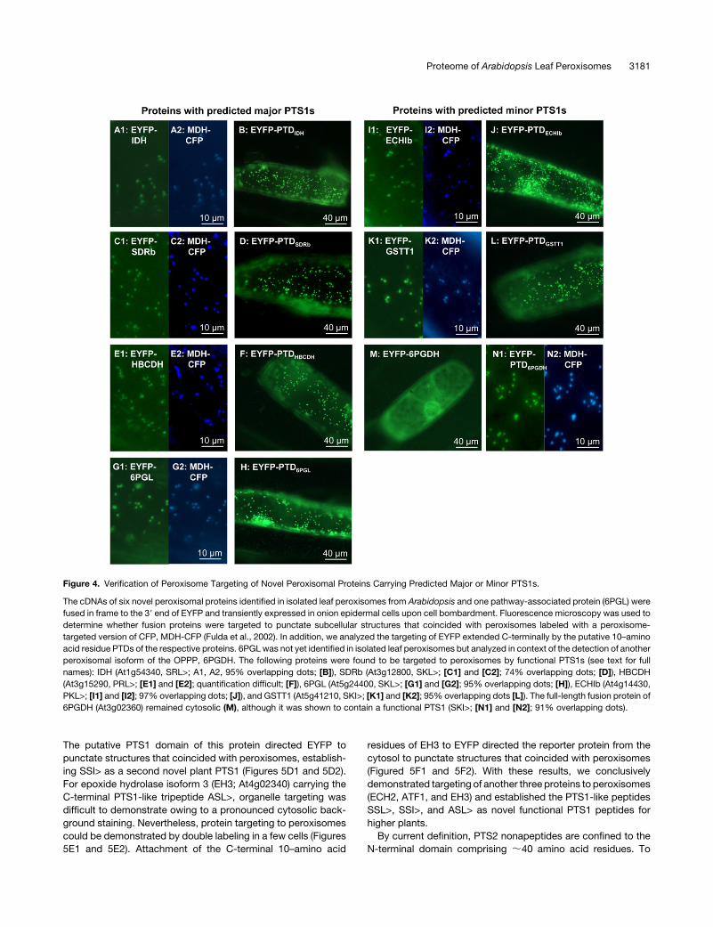
The putative PTS1 domain of this protein directed EYFP to
punctate structures that coincided with peroxisomes, establish-
ing SSI> as a second novel plant PTS1 (Figures 5D1 and 5D2).
For epoxide hydrolase isoform 3 (EH3; At4g02340) carrying the
C-terminal PTS1-like tripeptide ASL>, organelle targeting was
difficult to demonstrate owing to a pronounced cytosolic back-
ground staining. Nevertheless, protein targeting to peroxisomes
could be demonstrated by double labeling in a few cells (Figures
5E1 and 5E2). Attachment of the C-terminal 10–amino acid
residues of EH3 to EYFP directed the reporter protein from the
cytosol to punctate structures that coincided with peroxisomes
(Figured 5F1 and 5F2). With these results, we conclusively
demonstrated targeting of another three proteins to peroxisomes
(ECH2, ATF1, and EH3) and established the PTS1-like peptides
SSL>, SSI>, and ASL> as novel functional PTS1 peptides for
higher plants.
By current definition, PTS2 nonapeptides are confined to the
N-terminal domain comprising ;40 amino acid residues. To
Figure 4. Verification of Peroxisome Targeting of Novel Peroxisomal Proteins Carrying Predicted Major or Minor PTS1s.
The cDNAs of six novel peroxisomal proteins identified in isolated leaf peroxisomes from Arabidopsis and one pathway-associated protein (6PGL) were
fused in frame to the 39 end of EYFP and transiently expressed in onion epidermal cells upon cell bombardment. Fluorescence microscopy was used to
determine whether fusion proteins were targeted to punctate subcellular structures that coincided with peroxisomes labeled with a peroxisome-
targeted version of CFP, MDH-CFP (Fulda et al., 2002). In addition, we analyzed the targeting of EYFP extended C-terminally by the putative 10–amino
acid residue PTDs of the respective proteins. 6PGL was not yet identified in isolated leaf peroxisomes but analyzed in context of the detection of another
peroxisomal isoform of the OPPP, 6PGDH. The following proteins were found to be targeted to peroxisomes by functional PTS1s (see text for full
names): IDH (At1g54340, SRL>; A1, A2, 95% overlapping dots; [B]), SDRb (At3g12800, SKL>; [C1] and [C2]; 74% overlapping dots; [D]), HBCDH
(At3g15290, PRL>; [E1] and [E2]; quantification difficult; [F]), 6PGL (At5g24400, SKL>; [G1] and [G2]; 95% overlapping dots; [H]), ECHIb (At4g14430,
PKL>; [I1] and [I2]; 97% overlapping dots; [J]), and GSTT1 (At5g41210, SKI>; [K1] and [K2]; 95% overlapping dots [L]). The full-length fusion protein of
6PGDH (At3g02360) remained cytosolic (M), although it was shown to contain a functional PTS1 (SKI>; [N1] and [N2]; 91% overlapping dots).
Proteome of Arabidopsis Leaf Peroxisomes 3181
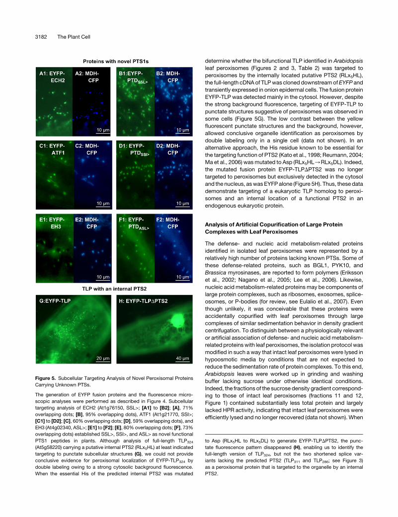
determine whether the bifunctional TLP identified in Arabidopsis
leaf peroxisomes (Figures 2 and 3, Table 2) was targeted to
peroxisomes by the internally located putative PTS2 (RLx5HL),
the full-length cDNA of TLP was cloned downstream of EYFP and
transiently expressed in onion epidermal cells. The fusion protein
EYFP-TLP was detected mainly in the cytosol. However, despite
the strong background fluorescence, targeting of EYFP-TLP to
punctate structures suggestive of peroxisomes was observed in
some cells (Figure 5G). The low contrast between the yellow
fluorescent punctate structures and the background, however,
allowed conclusive organelle identification as peroxisomes by
double labeling only in a single cell (data not shown). In an
alternative approach, the His residue known to be essential for
the targeting function of PTS2 (Kato et al., 1998; Reumann, 2004;
Ma et al., 2006) was mutated to Asp (RLx5HL/RLx5DL). Indeed,
the mutated fusion protein EYFP-TLPDPTS2 was no longer
targeted to peroxisomes but exclusively detected in the cytosol
and the nucleus, as was EYFP alone (Figure 5H). Thus, these data
demonstrate targeting of a eukaryotic TLP homolog to peroxi-
somes and an internal location of a functional PTS2 in an
endogenous eukaryotic protein.
Analysis of Artificial Copurification of Large Protein
Complexes with Leaf Peroxisomes
The defense- and nucleic acid metabolism-related proteins
identified in isolated leaf peroxisomes were represented by a
relatively high number of proteins lacking known PTSs. Some of
these defense-related proteins, such as BGL1, PYK10, and
Brassica myrosinases, are reported to form polymers (Eriksson
et al., 2002; Nagano et al., 2005; Lee et al., 2006). Likewise,
nucleic acid metabolism-related proteins may be components of
large protein complexes, such as ribosomes, exosomes, splice-
osomes, or P-bodies (for review, see Eulalio et al., 2007). Even
though unlikely, it was conceivable that these proteins were
accidentally copurified with leaf peroxisomes through large
complexes of similar sedimentation behavior in density gradient
centrifugation. To distinguish between a physiologically relevant
or artificial association of defense- and nucleic acid metabolism-
related proteins with leaf peroxisomes, the isolation protocol was
modified in such a way that intact leaf peroxisomes were lysed in
hypoosmotic media by conditions that are not expected to
reduce the sedimentation rate of protein complexes. To this end,
Arabidopsis leaves were worked up in grinding and washing
buffer lacking sucrose under otherwise identical conditions.
Indeed, the fractions of the sucrose density gradient correspond-
ing to those of intact leaf peroxisomes (fractions 11 and 12,
Figure 1) contained substantially less total protein and largely
lacked HPR activity, indicating that intact leaf peroxisomes were
efficiently lysed and no longer recovered (data not shown). When
Figure 5. Subcellular Targeting Analysis of Novel Peroxisomal Proteins
Carrying Unknown PTSs.
The generation of EYFP fusion proteins and the fluorescence micro-
scopic analyses were performed as described in Figure 4. Subcellular
targeting analysis of ECH2 (At1g76150, SSL>; [A1] to [B2]; [A], 71%
overlapping dots; [B], 95% overlapping dots), ATF1 (At1g21770, SSI>;
[C1] to [D2]; [C], 60% overlapping dots; [D], 59% overlapping dots), and
EH3 (At4g02340, ASL>; [E1] to [F2]; [E], 80% overlapping dots; [F], 73%
overlapping dots) established SSL>, SSI>, and ASL> as novel functional
PTS1 peptides in plants. Although analysis of full-length TLP324
(At5g58220) carrying a putative internal PTS2 (RLx5HL) at least indicated
targeting to punctate subcellular structures (G), we could not provide
conclusive evidence for peroxisomal localization of EYFP-TLP324 by
double labeling owing to a strong cytosolic background fluorescence.
When the essential His of the predicted internal PTS2 was mutated
to Asp (RLx5HL to RLx5DL) to generate EYFP-TLPDPTS2, the punc-
tate fluorescence pattern disappeared (H), enabling us to identify the
full-length version of TLP324, but not the two shortened splice var-
iants lacking the predicted PTS2 (TLP311 and TLP286; see Figure 3)
as a peroxisomal protein that is targeted to the organelle by an internal
PTS2.
3182 The Plant Cell
such a fraction volume corresponding to ;200 mg leaf perox-
isomal protein in the presence of osmoticum was subjected to
2-DE, none of the defense-related proteins (BGL1, TGG2, PYK10,
or ESM1) were detected by Coomassie staining (see Supple-
mental Figure 5 online). Similarly, the nucleic acid metabolism-
related proteins GRP7 and 8 and RBP were not visible. Although
two very faint spots may coincide with the RNA helicases RH11
and RH37 (see Supplemental Figure 5 online), these data as a
whole strongly argue against an artificial copurification of high
molecular weight complexes formed by the defense- or nucleic
acid metabolism-related proteins with leaf peroxisomes and
support a physiologically relevant association of most if not all of
these PTS-deficient proteins with leaf peroxisomes.
DISCUSSION
Only ;50 plant peroxisomal matrix proteins have been estab-
lished for Arabidopsis either experimentally or by homology to
peroxisomal proteins from other plant species (see summary at
www.araperox.uni-goettingen.de). The low number of known
peroxisomal enzymes in general and of plant-specific proteins in
particular severely restricts our current knowledge of the organ-
elle’s physiological functions and alternative protein targeting
mechanisms. The two most promising large-scale approaches
to ultimately characterize the proteome of plant peroxisomes in
its entity are (1) bioinformatics analyses predicting PTS1 and
PTS2 proteins from genome sequences in combination with
subcellular targeting studies of candidate fluorescent fusion
proteins, and (2) proteome analyses. As in silico predictions are
currently limited by several parameters (see Introduction) and
because highly pure peroxisomes can finally be isolated from
Arabidopsis, proteome analysis is the method of choice. This
study demonstrates that proteome data yield important infor-
mation not only directly on the identity of novel proteins and the
recognition of newfound metabolic pathways but also indirectly
on protein targeting. Leaf peroxisomal proteome analysis en-
ables us to (1) recognize novel targeting peptides, (2) extend the
location of PTS2 domains, and (3) posit the existence of novel
PTS1/2-independent targeting pathways.
Plant-specific peroxisome functions, such as photosynthesis-
related pathways, hormone biosynthesis, and pathogen defense
mechanisms, are expected to be most comprehensively de-
picted by proteome analyses of Arabidopsis peroxisomes from
mature rosette leaves rather than heterotrophic suspension-
cultured cells or germinating seeds, even though these tissues
offer significant technical advantages. The challenges in isolating
highly pure Arabidopsis leaf peroxisomes include not only obvi-
ous traits (e.g., low leaf fresh weight and predominance of
chloroplasts) but also unexpected parameters, such as the ex-
treme fragility of Arabidopsis leaf peroxisomes in aqueous ex-
tracts, probably owing to the high concentrations of secondary
metabolites, and their pronounced physical association with
chloroplasts and mitochondria in Brassicaceae. For in-depth
proteome analysis, we optimized our previous method (Ma et al.,
2006) in several aspects, such as plant growth and time of
harvest, differential centrifugation, and by the use of both Percoll
and sucrose density gradients in particular (see Results and
Methods). Contaminating proteins of chloroplasts and mitochon-
dria, the major contaminants of plant peroxisomes, were hardly
detectable in the final fraction by sensitive biochemical methods.
The higher apparent content of contaminants estimated from the
spot representation on two-dimensional gels (;5%) compared
with the activities of compartment-specific marker enzymes
(0.1% plastids and 1.7% mitochondria; Table 1) may reflect fac-
ilitated solubilization of the rather acidic nonperoxisomal pro-
teins compared with the neutral or basic peroxisomal proteins.
The Proteome Map of Arabidopsis Leaf Peroxisomes
The success of organellar proteome studies is determined by the
number of novel true positives that indeed reside in the com-
partment of interest. The gel-based and shotgun approaches
applied as complementary protein identification strategies max-
imized proteome coverage. The 78 proteins identified represent
approximately twice as many proteins as reported previously in
peroxisomes from rat liver (Kikuchi et al., 2004) or plants (Fukao
et al., 2002, 2003). The 33 known matrix proteins include 65%
of all currently known matrix proteins from plant peroxisomes
(51 Arabidopsis proteins; Table 3). As this ratio has been obtained
from only one of several plant peroxisomal variants and from
standard plants, the coverage is considered high. Comprehensive
proteome analyses of other tissue-specific and developmental
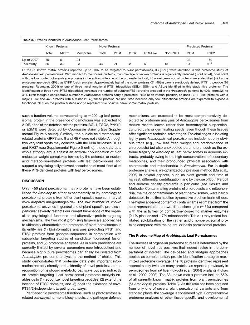
Table 3. Proteins Identified in Arabidopsis Leaf Peroxisomes
Known Proteins Novel Proteins Predicted Proteins
Total Matrix Membrane Total PTS1 PTS2 PTS-Like Non-PTS1 PTS1 PTS2
Up to 2007 75 51 24 – – – – 221 60
This study 36 33 3 43 21 2 5 15 311 611
Of the 51 known matrix proteins reported up to 2007 to be targeted to plant peroxisomes, 33 (65%) were identified in this proteome study of
Arabidopsis leaf peroxisomes. With respect to membrane proteins, the coverage of known proteins is significantly reduced (3 out of 24), consistent
with the low content of membrane proteins in the entire proteome of the organelle. In total, 43 novel peroxisomal proteins were identified (42 by the
proteome approach, 6PGL as EYFP fusion protein). Approximately half of the novel proteins (21; 49%) carry a previously defined PTS1 tripeptide (16
proteins; Reumann, 2004) or one of three novel functional PTS1 tripeptides (SSL>, SSI>, and ASL>) identified in this study (five proteins). The
identification of three novel PTS1 tripeptides increases the number of putative PTS1 proteins encoded in the Arabidopsis genome by 40%, from 221 to
311. Even though a considerable number of Arabidopsis proteins carry a predicted PTS2 at an internal position, as does TLP (1, 201 proteins with a
major PTS2 and 443 proteins with a minor PTS2), these proteins are not listed because only few bifunctional proteins are expected to expose a
functional PTS2 on the protein surface and to represent true positive peroxisomal matrix proteins.
Proteome of Arabidopsis Leaf Peroxisomes 3183
peroxisome variants (e.g., glyoxysomes and gerontosomes) and
from plants subjected to abiotic or biotic stresses are expected
to promote the detection of the remaining known and further
predicted proteins. A comprehensive membrane proteome anal-
ysis will be made possible by the availability of larger organelle
quantities and the selective enrichment of peroxisomal mem-
branes.
Forty-three novel proteins (42 proteins by the proteome ap-
proach and 6PGL as an EYFP fusion protein) had not previously
been localized to plant peroxisomes in any species and had not,
for the most part, been experimentally analyzed. Seventeen of
these novel proteins carry previously defined PTSs (16 PTS1 and
one PTS2) and had been predicted to be peroxisomal proteins
from the Arabidopsis genome (Reumann et al., 2004). Together
with six predicted PTS1 proteins localized experimentally to
peroxisomes in the meantime (4CLP1, AAE7/ACN1, ACH2, DCI,
OPCL1, and SCP-2), ;40% of the nonhypothetical Arabidopsis
proteins (23 out of 50) predicted to be peroxisome targeted with
high probability have been covered by our proteome study,
confirming reciprocally the in silico approach. The high number
of PTS-carrying proteins of this study (30 of 36 known and 22 of
42 novel proteins) contrasts with the low numbers of two previ-
ous two-dimensional gel-based peroxisomal proteome studies
from greening and etiolated cotyledons (one novel PTS protein
out of 20; Fukao et al., 2002; three out of 13; Fukao et al., 2003;
see Supplemental Figures 6A and 6B online). With respect to
novel protein identity, the overlap between the three proteome
studies is marginal (only one protein, i.e., NADP-dependent IDH,
At1g54340, SRL>) and difficult to rationalize in light of the large
number of b-oxidation–related enzymes reported here for leaf
peroxisomes but not previously for glyoxysomes, the peroxi-
some variant most specialized on fatty acid degradation (Fukao
et al., 2003; see Supplemental Figure 6A online). Compared with
the methods for peroxisome enrichment (single Percoll gradient
only) and protein identification (PMF alone) applied in the two
earlier studies, our sophisticated protocol for peroxisome puri-
fication and the highly confident protein identifications are
considered the major improvements toward reliable and com-
prehensive coverage of the plant peroxisomal proteome.
The horizontal spot pattern of several matrix enzymes is
suggestive of charge-modifying posttranslational modifications,
such as phosphorylation and acetylation, both of which are
currently being investigated in greater detail. Some peroxisomal
protein kinases are reported and several are predicted, along
with phosphatases, to be matrix targeted (Fukao et al., 2002,
2003; Dammann et al., 2003; Reumann et al., 2004). Acetylation
of internal Lys residues also decreases the pI of a polypeptide
and has recently been recognized as an important regulatory
signal in many cellular processes (for reviews, see Polevoda and
Sherman, 2002; Fuchs et al., 2006). ATF2 (At1g77540; SSI>),
whose x-ray structure has recently been resolved, is a minimal
acetyl transferase of unknown subcellular localization (Tyler
et al., 2006). We have now established its closely related homo-
log, ATF1 (At1g21770; SSI>; 80% identical with ATF1) as a novel
constituent of plant peroxisomes by multiple lines of evidence,
including in vivo EYFP targeting studies (Figures 5C and 5D).
Conservation of the novel PTS1 SSI> of ATF1 in ATF2 strongly
suggests that the entire two-member protein family is peroxi-
somal in Arabidopsis. Protein acetylation may thus be an as yet
unrecognized regulatory mechanism of plant peroxisomal en-
zymes. Proteins showing multiple horizontal spot patterns (see
Supplemental Figure 2 online) are reasonable candidate sub-
strates of ATF1.
Targeting Verification for Improving Targeting Prediction
By in silico analysis for conservation of the predicted PTS in
homologous plant ESTs (Reumann, 2004), the peroxisome tar-
geting function of the predicted PTS peptides was supported for
17 of 28 novel proteins (Table 2; see Supplemental Figure 3
online; Reumann et al., 2004). Exceptions included novel pro-
teins, whose PTS1-like peptides were unaltered in homologous
ESTs, and Brassicaceae-specific proteins. Notably, no evidence
against peroxisomal targeting, for instance, acidic amino acid
residues at any position of the tripeptide, was obtained for any of
the proteins. By subcellular analysis of reporter protein fusions,
peroxisome targeting of six out of seven representative novel
proteins with predicted PTS1s was confirmed. Artificial localiza-
tion data possibly caused by cDNA expression from the strong
35S cauliflower mosaic virus promoter were excluded by coloc-
alization of the proteins of interest with a cyan fluorescent
peroxisome marker, as applied in previous studies (Fulda et al.,
2002; Goepfert et al., 2006; Ma et al., 2006). Three enzymes with
b-oxidation–related annotations (SDRb, ECHIb, and HBCDH), a
glutathione S-transferase (GSTT1), and three enzymes involved
in NADP metabolism (IDH, 6PGL, and 6PGDH; see below) were
conclusively established for plant peroxisomes. Only full-length
EYFP-6PGDH remained cytosolic, possibly owing to interfer-
ence of the EYFP tag with PTS1 exposure or posttranslational
regulatory mechanisms. Nonetheless, this 6PGDH isoform is
considered peroxisomal owing to its conserved PTS1 and func-
tional PTS1 domain (SKI>; Figure 4N; Reumann et al., 2004), the
lack of predicted N-terminal targeting signals, and its apparent
absence in the proteomes of chloroplasts and mitochondria
(Kruft et al., 2001; Millar et al., 2001; Peltier et al., 2002).
The large number of 11 proteins with PTS1-related peptides
suggested their role as functional yet unknown PTS1s. The
PTS1-like peptides SSL>, SSI>, and ASL> were previously
absent from or below threshold in plant PTS1 sequences
(Reumann, 2004), and experimental data argued against a tar-
geting function of SSI> and SSL>. Conclusive experimental
support for an allowance of Ser at position �2 in general had
not been reported ([SACGT][RKHLN][LMIY]>, Mullen et al., 1997;
[SACP][RK][LMI]>, Hayashi et al., 1997; [SACGTP][RKHLN]
[LMIY]>, Kragler et al., 1998). Peroxisome targeting of the three
full-length fusion proteins and the corresponding PTS domain
constructs (Figure 5) identified ECH2, EH3, and ATF1 conclu-
sively as plant peroxisomal proteins and SSL>, SSI>, and ASL>
as novel noncanonical plant PTS1 tripeptides. Full-length ECH2
had previously been shown to be peroxisomal (Goepfert et al.,
2006). Our results on EH3 support earlier biochemical localiza-
tion data on Ricinus epoxide hydrolase in glyoxysomes (Stark
et al., 1995) and point to a physiological role of EH3 distinct from
lipid mobilization in germinating seeds.
The primary structure of the novel PTS1 peptides and their
remarkable frequency in the proteome of leaf peroxisomes (e.g.,
3184 The Plant Cell
four SSL> proteins), in contrast with their rare abundance in
previously known plant PTS1 proteins, raises further biological
questions. For instance, which structural feature specifically
allows Ser to replace the generally essential basic residue at
position �2 in plants? The emerging abundance of SSL>, SSI>,
or ASL> in leaf peroxisomal proteins may be partly attributed to
our ability to now detect weakly expressed peroxisomal matrix
proteins. We propose that the newfound noncanonical PTS1s
occur preferentially in low-abundance matrix proteins because
the tripeptides have a reduced affinity to the cytosolic PTS1
receptor PEX5 and lower the speed of cargo import into the
matrix. The previously known major plant peroxisomal matrix
proteins, by contrast, rely on highly efficient canonical PTS
peptides to adjust the rate of protein import to the intensity of
gene expression (G.E. Antonicelli and S. Reumann, unpublished
data). Thus, the identification of low-abundance proteins ap-
pears to be of major importance to fully cover the natural variety
of plant peroxisomal proteins and the range of functional PTS1/2
peptides in higher plants.
Even though our isolation method paves the way for analysis of
other abundant peroxisome variants from Arabidopsis, it remains
unrealistic to envision proteome studies of all plant microbody
forms in the near future (e.g., from flowers, trichomes, stems, and
roots). Prediction-based in silico studies will therefore remain the
second major large-scale approach to fully describe the plant
peroxisomal proteome. The prediction of plant peroxisomal pro-
teins benefits significantly from the proteome data generated in
the course of this study, in terms of both increased sensitivity and
specificity. First, the three novel PTS1 peptides allow extended ge-
nome screens for putative PTS1 proteins and a significant enlarge-
ment of the database AraPerox (www.araperox.uni-goettingen.de;
Table 3). The 100 additional new entries are predicted to include
numerous interesting true peroxisomal proteins (for example,
see Supplemental Figure 8 online) but may also comprise some
false positives, if peroxisome targeting of some proteins depends
on yet unknown enhancing elements. Experimental analysis of the
predicted peroxisome targeting function of further PTS1-related
peptides (e.g., GR, TNL>; METE1, SAK>; and NBP, SKV>) is
expected to reveal further functional PTS1s.
Second, upon experimental verification of the targeting func-
tion of predicted PTS peptides, novel peroxisomal matrix pro-
teins can be applied as input sequences to improve in silico
predictions of PTS1/2 proteins. EYFP extended by the C-terminal
domains of any of the 11 representative novel proteins showed a
punctate distribution, conclusively demonstrating that the pro-
teins carry functional PTS1s and use the PTS1 targeting pathway
(Figure 4). In addition, peroxisome targeting of three other pro-
teins identified in both Arabidopsis and spinach leaf peroxisomes
has been confirmed for the corresponding full-length Arabidop-
sis homologs as EYFP fusion proteins (ECHIa, ECHId/NS, and
SDRa) and their predicted targeting domains (SKL>, RLx5HL,
and SRL>, respectively; L. Babujee, V. Wurtz, C. Ma, F. Lueder,
A. van Dorselaer, and S. Reumann, unpublished data). In sum-
mary, the characterization of 26 novel PTS1/2 proteins at the
molecular level, combined with experimental definitions of their
targeting signals as reported in this and previous studies of the
past 3 years (12 additional proven or putative PTS1/2 proteins;
see Introduction), represents a significant extension of the pre-
vious number of known matrix proteins applicable as input
sequences for EST database searches and discriminative analy-
ses of PTS1 domains. The data will make possible a more
comprehensive definition of functional PTS peptides, the char-
acterization of auxiliary targeting elements, and an investigation
of any interdependence of these parameters in higher plants.
An Internal PTS2 in TLP
The characterization of a functional PTS2 in the Arabidopsis TLP
is biologically significant because (1) eukaryotic transthyretin
homologs previously have not been localized to peroxisomes
and (2) eukaryotic proteins have not been demonstrated to bear
functional PTS2s at an internal position. TLPs are homologous to
the mammalian thyroid-transport protein transthyretin, but they
lack hormone binding activity and are generally considered
cytosolic. The proteome data and the subcellular reporter protein
studies of native and modified EYFP constructs provide conclu-
sive evidence for peroxisome targeting of full-length Arabidopsis
TLP (Figures 2, 3, 5G, and 5H). The low import efficiency of EYFP-
TLP may reflect conformation-limited accessibility of the internal
PTS2 for PEX7 and/or its competition with another targeting
signal, for instance, the yet unknown peptide that directed the
inversely arranged full-length fusion protein, TLP-GFP, to the
plasma membrane (Nam and Li, 2004).
Genome screens for PTS2 proteins should thus be extended
beyond the N-terminal 40–amino acid domain (Table 3). Internal
PTS2s, however, are thought to remain rare in nature and to
occur preferentially in bifunctional proteins that evolved by gene
fusion, as deduced for plant TLP, a predicted bifunctional en-
zyme of purine metabolism. The two catalytic domains of plant
TLP were apparently fused shortly after the divergence of
Dictyostelium in the plant lineage, to facilitate stoichiometric
protein synthesis, peroxisome targeting, protein–protein inter-
action, and possibly metabolite channeling. Uricase, in fact,
produces 5-hydroxyisourate (5-HIU) rather than the presumed
pathway end product, allantoin. The enzyme that succeeds
uricase in purine catabolism and opens the ring (i.e., 5-HIU
hydrolase [COG2351]) turned out to be encoded by tlp (Figure
3A; Lee et al., 2005; Jung et al., 2006). The reaction product
is converted to (S)-allantoin by a decarboxylase (COG3195;
Ramazzina et al., 2006) that is homologous to the additional
N-terminal domain of bifunctional Arabidopsis TLP (Figure 3).
The likely role of Arabidopsis TLP in purine metabolism matches
peroxisome targeting of full-length TLP324 to the same compart-
ment as uricase. The shorter splice variants of plants lacking the
PTS2 are probably cytosolic (e.g., AtTLP311 and AtTLP286; Figure
3A; see Supplemental Figure 3P online) and are expected to
conduct distinct yet unknown physiological roles, possibly in-
cluding hormone transport-related functions (e.g., in brassino-
steroid signaling) (Nam and Li, 2004).
Peroxisomal Proteins Lacking Obvious PTSs Indicate
Alternative Targeting Pathways
A significant number of the novel leaf peroxisomal proteins were
expected to lack predicted PTS1/2 peptides (see Introduction).
Proteome of Arabidopsis Leaf Peroxisomes 3185
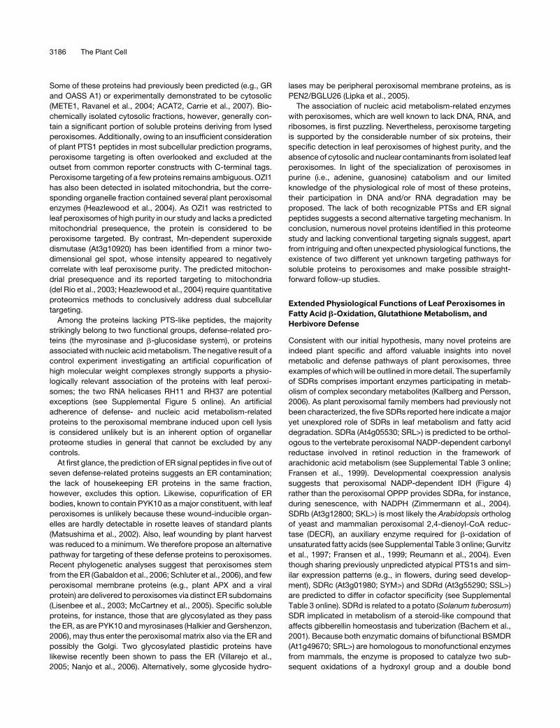
Some of these proteins had previously been predicted (e.g., GR
and OASS A1) or experimentally demonstrated to be cytosolic
(METE1, Ravanel et al., 2004; ACAT2, Carrie et al., 2007). Bio-
chemically isolated cytosolic fractions, however, generally con-
tain a significant portion of soluble proteins deriving from lysed
peroxisomes. Additionally, owing to an insufficient consideration
of plant PTS1 peptides in most subcellular prediction programs,
peroxisome targeting is often overlooked and excluded at the
outset from common reporter constructs with C-terminal tags.
Peroxisome targeting of a few proteins remains ambiguous. OZI1
has also been detected in isolated mitochondria, but the corre-
sponding organelle fraction contained several plant peroxisomal
enzymes (Heazlewood et al., 2004). As OZI1 was restricted to
leaf peroxisomes of high purity in our study and lacks a predicted
mitochondrial presequence, the protein is considered to be
peroxisome targeted. By contrast, Mn-dependent superoxide
dismutase (At3g10920) has been identified from a minor two-
dimensional gel spot, whose intensity appeared to negatively
correlate with leaf peroxisome purity. The predicted mitochon-
drial presequence and its reported targeting to mitochondria
(del Rio et al., 2003; Heazlewood et al., 2004) require quantitative
proteomics methods to conclusively address dual subcellular
targeting.
Among the proteins lacking PTS-like peptides, the majority
strikingly belong to two functional groups, defense-related pro-
teins (the myrosinase and b-glucosidase system), or proteins
associated with nucleic acid metabolism. The negative result of a
control experiment investigating an artificial copurification of
high molecular weight complexes strongly supports a physio-
logically relevant association of the proteins with leaf peroxi-
somes; the two RNA helicases RH11 and RH37 are potential
exceptions (see Supplemental Figure 5 online). An artificial
adherence of defense- and nucleic acid metabolism-related
proteins to the peroxisomal membrane induced upon cell lysis
is considered unlikely but is an inherent option of organellar
proteome studies in general that cannot be excluded by any
controls.
At first glance, the prediction of ER signal peptides in five out of
seven defense-related proteins suggests an ER contamination;
the lack of housekeeping ER proteins in the same fraction,
however, excludes this option. Likewise, copurification of ER
bodies, known to contain PYK10 as a major constituent, with leaf
peroxisomes is unlikely because these wound-inducible organ-
elles are hardly detectable in rosette leaves of standard plants
(Matsushima et al., 2002). Also, leaf wounding by plant harvest
was reduced to a minimum. We therefore propose an alternative
pathway for targeting of these defense proteins to peroxisomes.
Recent phylogenetic analyses suggest that peroxisomes stem
from the ER (Gabaldon et al., 2006; Schluter et al., 2006), and few
peroxisomal membrane proteins (e.g., plant APX and a viral
protein) are delivered to peroxisomes via distinct ER subdomains
(Lisenbee et al., 2003; McCartney et al., 2005). Specific soluble
proteins, for instance, those that are glycosylated as they pass
the ER, as are PYK10 and myrosinases (Halkier and Gershenzon,
2006), may thus enter the peroxisomal matrix also via the ER and
possibly the Golgi. Two glycosylated plastidic proteins have
likewise recently been shown to pass the ER (Villarejo et al.,
2005; Nanjo et al., 2006). Alternatively, some glycoside hydro-
lases may be peripheral peroxisomal membrane proteins, as is
PEN2/BGLU26 (Lipka et al., 2005).
The association of nucleic acid metabolism-related enzymes
with peroxisomes, which are well known to lack DNA, RNA, and
ribosomes, is first puzzling. Nevertheless, peroxisome targeting
is supported by the considerable number of six proteins, their
specific detection in leaf peroxisomes of highest purity, and the
absence of cytosolic and nuclear contaminants from isolated leaf
peroxisomes. In light of the specialization of peroxisomes in
purine (i.e., adenine, guanosine) catabolism and our limited
knowledge of the physiological role of most of these proteins,
their participation in DNA and/or RNA degradation may be
proposed. The lack of both recognizable PTSs and ER signal
peptides suggests a second alternative targeting mechanism. In
conclusion, numerous novel proteins identified in this proteome
study and lacking conventional targeting signals suggest, apart
from intriguing and often unexpected physiological functions, the
existence of two different yet unknown targeting pathways for
soluble proteins to peroxisomes and make possible straight-
forward follow-up studies.
Extended Physiological Functions of Leaf Peroxisomes in
Fatty Acid b-Oxidation, Glutathione Metabolism, and
Herbivore Defense
Consistent with our initial hypothesis, many novel proteins are
indeed plant specific and afford valuable insights into novel
metabolic and defense pathways of plant peroxisomes, three
examples of which will be outlined in more detail. The superfamily
of SDRs comprises important enzymes participating in metab-
olism of complex secondary metabolites (Kallberg and Persson,
2006). As plant peroxisomal family members had previously not
been characterized, the five SDRs reported here indicate a major
yet unexplored role of SDRs in leaf metabolism and fatty acid
degradation. SDRa (At4g05530; SRL>) is predicted to be orthol-
ogous to the vertebrate peroxisomal NADP-dependent carbonyl
reductase involved in retinol reduction in the framework of
arachidonic acid metabolism (see Supplemental Table 3 online;
Fransen et al., 1999). Developmental coexpression analysis
suggests that peroxisomal NADP-dependent IDH (Figure 4)
rather than the peroxisomal OPPP provides SDRa, for instance,
during senescence, with NADPH (Zimmermann et al., 2004).
SDRb (At3g12800; SKL>) is most likely the Arabidopsis ortholog
of yeast and mammalian peroxisomal 2,4-dienoyl-CoA reduc-
tase (DECR), an auxiliary enzyme required for b-oxidation of
unsaturated fatty acids (see Supplemental Table 3 online; Gurvitz
et al., 1997; Fransen et al., 1999; Reumann et al., 2004). Even
though sharing previously unpredicted atypical PTS1s and sim-
ilar expression patterns (e.g., in flowers, during seed develop-
ment), SDRc (At3g01980; SYM>) and SDRd (At3g55290; SSL>)
are predicted to differ in cofactor specificity (see Supplemental
Table 3 online). SDRd is related to a potato (Solanum tuberosum)
SDR implicated in metabolism of a steroid-like compound that
affects gibberellin homeostasis and tuberization (Bachem et al.,
2001). Because both enzymatic domains of bifunctional BSMDR
(At1g49670; SRL>) are homologous to monofunctional enzymes
from mammals, the enzyme is proposed to catalyze two sub-
sequent oxidations of a hydroxyl group and a double bond
3186 The Plant Cell
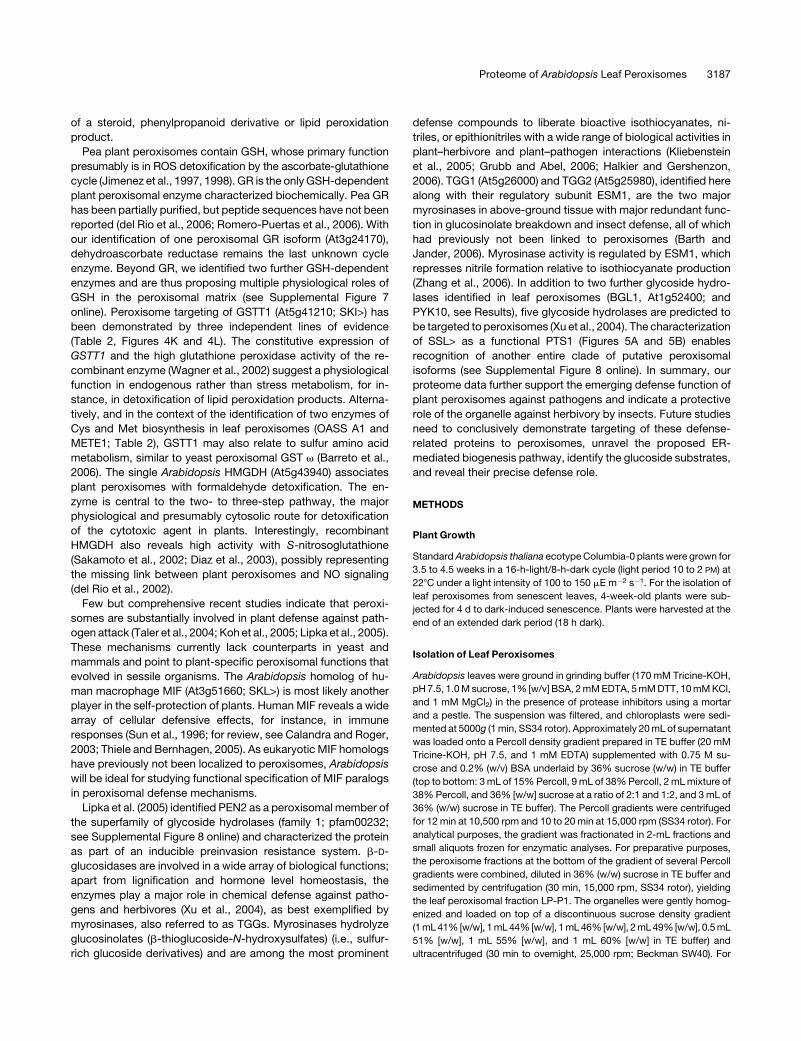
of a steroid, phenylpropanoid derivative or lipid peroxidation
product.
Pea plant peroxisomes contain GSH, whose primary function
presumably is in ROS detoxification by the ascorbate-glutathione
cycle (Jimenez et al., 1997, 1998). GR is the only GSH-dependent
plant peroxisomal enzyme characterized biochemically. Pea GR
has been partially purified, but peptide sequences have not been
reported (del Rio et al., 2006; Romero-Puertas et al., 2006). With
our identification of one peroxisomal GR isoform (At3g24170),
dehydroascorbate reductase remains the last unknown cycle
enzyme. Beyond GR, we identified two further GSH-dependent
enzymes and are thus proposing multiple physiological roles of
GSH in the peroxisomal matrix (see Supplemental Figure 7
online). Peroxisome targeting of GSTT1 (At5g41210; SKI>) has
been demonstrated by three independent lines of evidence
(Table 2, Figures 4K and 4L). The constitutive expression of
GSTT1 and the high glutathione peroxidase activity of the re-
combinant enzyme (Wagner et al., 2002) suggest a physiological
function in endogenous rather than stress metabolism, for in-
stance, in detoxification of lipid peroxidation products. Alterna-
tively, and in the context of the identification of two enzymes of
Cys and Met biosynthesis in leaf peroxisomes (OASS A1 and
METE1; Table 2), GSTT1 may also relate to sulfur amino acid
metabolism, similar to yeast peroxisomal GST v (Barreto et al.,
2006). The single Arabidopsis HMGDH (At5g43940) associates
plant peroxisomes with formaldehyde detoxification. The en-
zyme is central to the two- to three-step pathway, the major
physiological and presumably cytosolic route for detoxification
of the cytotoxic agent in plants. Interestingly, recombinant
HMGDH also reveals high activity with S-nitrosoglutathione
(Sakamoto et al., 2002; Diaz et al., 2003), possibly representing
the missing link between plant peroxisomes and NO signaling
(del Rio et al., 2002).
Few but comprehensive recent studies indicate that peroxi-
somes are substantially involved in plant defense against path-
ogen attack (Taler et al., 2004; Koh et al., 2005; Lipka et al., 2005).
These mechanisms currently lack counterparts in yeast and
mammals and point to plant-specific peroxisomal functions that
evolved in sessile organisms. The Arabidopsis homolog of hu-
man macrophage MIF (At3g51660; SKL>) is most likely another
player in the self-protection of plants. Human MIF reveals a wide
array of cellular defensive effects, for instance, in immune
responses (Sun et al., 1996; for review, see Calandra and Roger,
2003; Thiele and Bernhagen, 2005). As eukaryotic MIF homologs
have previously not been localized to peroxisomes, Arabidopsis
will be ideal for studying functional specification of MIF paralogs
in peroxisomal defense mechanisms.
Lipka et al. (2005) identified PEN2 as a peroxisomal member of
the superfamily of glycoside hydrolases (family 1; pfam00232;
see Supplemental Figure 8 online) and characterized the protein
as part of an inducible preinvasion resistance system. b-D-
glucosidases are involved in a wide array of biological functions;
apart from lignification and hormone level homeostasis, the
enzymes play a major role in chemical defense against patho-
gens and herbivores (Xu et al., 2004), as best exemplified by
myrosinases, also referred to as TGGs. Myrosinases hydrolyze
glucosinolates (b-thioglucoside-N-hydroxysulfates) (i.e., sulfur-
rich glucoside derivatives) and are among the most prominent
defense compounds to liberate bioactive isothiocyanates, ni-
triles, or epithionitriles with a wide range of biological activities in
plant–herbivore and plant–pathogen interactions (Kliebenstein
et al., 2005; Grubb and Abel, 2006; Halkier and Gershenzon,
2006). TGG1 (At5g26000) and TGG2 (At5g25980), identified here
along with their regulatory subunit ESM1, are the two major
myrosinases in above-ground tissue with major redundant func-
tion in glucosinolate breakdown and insect defense, all of which
had previously not been linked to peroxisomes (Barth and
Jander, 2006). Myrosinase activity is regulated by ESM1, which
represses nitrile formation relative to isothiocyanate production
(Zhang et al., 2006). In addition to two further glycoside hydro-
lases identified in leaf peroxisomes (BGL1, At1g52400; and
PYK10, see Results), five glycoside hydrolases are predicted to
be targeted to peroxisomes (Xu et al., 2004). The characterization
of SSL> as a functional PTS1 (Figures 5A and 5B) enables
recognition of another entire clade of putative peroxisomal
isoforms (see Supplemental Figure 8 online). In summary, our
proteome data further support the emerging defense function of
plant peroxisomes against pathogens and indicate a protective
role of the organelle against herbivory by insects. Future studies
need to conclusively demonstrate targeting of these defense-
related proteins to peroxisomes, unravel the proposed ER-
mediated biogenesis pathway, identify the glucoside substrates,
and reveal their precise defense role.
METHODS
Plant Growth
Standard Arabidopsis thaliana ecotype Columbia-0 plants were grown for
3.5 to 4.5 weeks in a 16-h-light/8-h-dark cycle (light period 10 to 2 PM) at
228C under a light intensity of 100 to 150 mE m�2 s�1. For the isolation of
leaf peroxisomes from senescent leaves, 4-week-old plants were sub-
jected for 4 d to dark-induced senescence. Plants were harvested at the
end of an extended dark period (18 h dark).
Isolation of Leaf Peroxisomes
Arabidopsis leaves were ground in grinding buffer (170 mM Tricine-KOH,
pH 7.5, 1.0 M sucrose, 1% [w/v] BSA, 2 mM EDTA, 5 mM DTT, 10 mM KCl,
and 1 mM MgCl2) in the presence of protease inhibitors using a mortar
and a pestle. The suspension was filtered, and chloroplasts were sedi-
mented at 5000g (1 min, SS34 rotor). Approximately 20 mL of supernatant
was loaded onto a Percoll density gradient prepared in TE buffer (20 mM
Tricine-KOH, pH 7.5, and 1 mM EDTA) supplemented with 0.75 M su-
crose and 0.2% (w/v) BSA underlaid by 36% sucrose (w/w) in TE buffer
(top to bottom: 3 mL of 15% Percoll, 9 mL of 38% Percoll, 2 mL mixture of
38% Percoll, and 36% [w/w] sucrose at a ratio of 2:1 and 1:2, and 3 mL of
36% (w/w) sucrose in TE buffer). The Percoll gradients were centrifuged
for 12 min at 10,500 rpm and 10 to 20 min at 15,000 rpm (SS34 rotor). For
analytical purposes, the gradient was fractionated in 2-mL fractions and
small aliquots frozen for enzymatic analyses. For preparative purposes,
the peroxisome fractions at the bottom of the gradient of several Percoll
gradients were combined, diluted in 36% (w/w) sucrose in TE buffer and
sedimented by centrifugation (30 min, 15,000 rpm, SS34 rotor), yielding
the leaf peroxisomal fraction LP-P1. The organelles were gently homog-
enized and loaded on top of a discontinuous sucrose density gradient
(1 mL 41% [w/w], 1 mL 44% [w/w], 1 mL 46% [w/w], 2 mL 49% [w/w], 0.5 mL
51% [w/w], 1 mL 55% [w/w], and 1 mL 60% [w/w] in TE buffer) and
ultracentrifuged (30 min to overnight, 25,000 rpm; Beckman SW40). For
Proteome of Arabidopsis Leaf Peroxisomes 3187
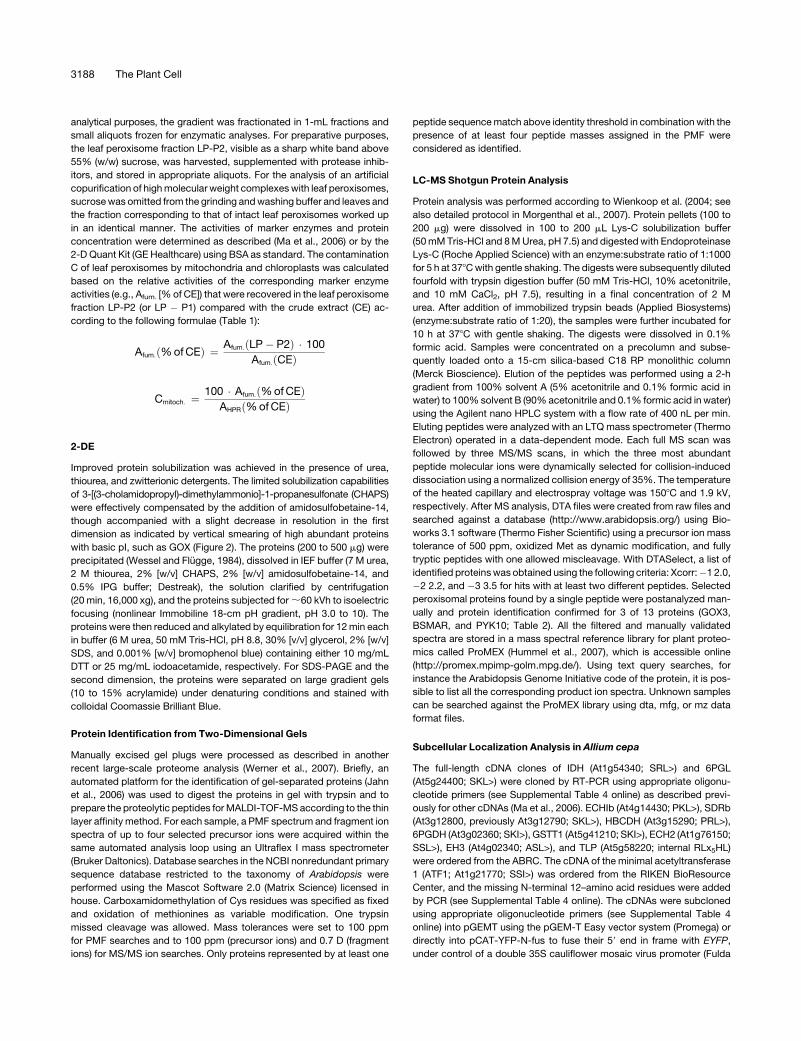
analytical purposes, the gradient was fractionated in 1-mL fractions and
small aliquots frozen for enzymatic analyses. For preparative purposes,
the leaf peroxisome fraction LP-P2, visible as a sharp white band above
55% (w/w) sucrose, was harvested, supplemented with protease inhib-
itors, and stored in appropriate aliquots. For the analysis of an artificial
copurification of high molecular weight complexes with leaf peroxisomes,
sucrose was omitted from the grinding and washing buffer and leaves and
the fraction corresponding to that of intact leaf peroxisomes worked up
in an identical manner. The activities of marker enzymes and protein
concentration were determined as described (Ma et al., 2006) or by the
2-D Quant Kit (GE Healthcare) using BSA as standard. The contamination
C of leaf peroxisomes by mitochondria and chloroplasts was calculated
based on the relative activities of the corresponding marker enzyme
activities (e.g., Afum. [% of CE]) that were recovered in the leaf peroxisome
fraction LP-P2 (or LP � P1) compared with the crude extract (CE) ac-
cording to the following formulae (Table 1):
Afum:ð% of CEÞ ¼ Afum:ðLP� P2Þ � 100
Afum:ðCEÞ
Cmitoch: ¼100 � Afum:ð% of CEÞ
AHPRð% of CEÞ
2-DE
Improved protein solubilization was achieved in the presence of urea,
thiourea, and zwitterionic detergents. The limited solubilization capabilities
of 3-[(3-cholamidopropyl)-dimethylammonio]-1-propanesulfonate (CHAPS)
were effectively compensated by the addition of amidosulfobetaine-14,
though accompanied with a slight decrease in resolution in the first
dimension as indicated by vertical smearing of high abundant proteins
with basic pI, such as GOX (Figure 2). The proteins (200 to 500 mg) were
precipitated (Wessel and Flugge, 1984), dissolved in IEF buffer (7 M urea,
2 M thiourea, 2% [w/v] CHAPS, 2% [w/v] amidosulfobetaine-14, and
0.5% IPG buffer; Destreak), the solution clarified by centrifugation
(20 min, 16,000 xg), and the proteins subjected for ;60 kVh to isoelectric
focusing (nonlinear Immobiline 18-cm pH gradient, pH 3.0 to 10). The
proteins were then reduced and alkylated by equilibration for 12 min each
in buffer (6 M urea, 50 mM Tris-HCl, pH 8.8, 30% [v/v] glycerol, 2% [w/v]
SDS, and 0.001% [w/v] bromophenol blue) containing either 10 mg/mL
DTT or 25 mg/mL iodoacetamide, respectively. For SDS-PAGE and the
second dimension, the proteins were separated on large gradient gels
(10 to 15% acrylamide) under denaturing conditions and stained with
colloidal Coomassie Brilliant Blue.
Protein Identification from Two-Dimensional Gels
Manually excised gel plugs were processed as described in another
recent large-scale proteome analysis (Werner et al., 2007). Briefly, an
automated platform for the identification of gel-separated proteins (Jahn
et al., 2006) was used to digest the proteins in gel with trypsin and to
prepare the proteolytic peptides for MALDI-TOF-MS according to the thin
layer affinity method. For each sample, a PMF spectrum and fragment ion
spectra of up to four selected precursor ions were acquired within the
same automated analysis loop using an Ultraflex I mass spectrometer
(Bruker Daltonics). Database searches in the NCBI nonredundant primary
sequence database restricted to the taxonomy of Arabidopsis were
performed using the Mascot Software 2.0 (Matrix Science) licensed in
house. Carboxamidomethylation of Cys residues was specified as fixed
and oxidation of methionines as variable modification. One trypsin
missed cleavage was allowed. Mass tolerances were set to 100 ppm
for PMF searches and to 100 ppm (precursor ions) and 0.7 D (fragment
ions) for MS/MS ion searches. Only proteins represented by at least one
peptide sequence match above identity threshold in combination with the
presence of at least four peptide masses assigned in the PMF were
considered as identified.
LC-MS Shotgun Protein Analysis
Protein analysis was performed according to Wienkoop et al. (2004; see
also detailed protocol in Morgenthal et al., 2007). Protein pellets (100 to
200 mg) were dissolved in 100 to 200 mL Lys-C solubilization buffer
(50 mM Tris-HCl and 8 M Urea, pH 7.5) and digested with Endoproteinase
Lys-C (Roche Applied Science) with an enzyme:substrate ratio of 1:1000
for 5 h at 378C with gentle shaking. The digests were subsequently diluted
fourfold with trypsin digestion buffer (50 mM Tris-HCl, 10% acetonitrile,
and 10 mM CaCl2, pH 7.5), resulting in a final concentration of 2 M
urea. After addition of immobilized trypsin beads (Applied Biosystems)
(enzyme:substrate ratio of 1:20), the samples were further incubated for
10 h at 378C with gentle shaking. The digests were dissolved in 0.1%
formic acid. Samples were concentrated on a precolumn and subse-
quently loaded onto a 15-cm silica-based C18 RP monolithic column
(Merck Bioscience). Elution of the peptides was performed using a 2-h
gradient from 100% solvent A (5% acetonitrile and 0.1% formic acid in
water) to 100% solvent B (90% acetonitrile and 0.1% formic acid in water)
using the Agilent nano HPLC system with a flow rate of 400 nL per min.
Eluting peptides were analyzed with an LTQ mass spectrometer (Thermo
Electron) operated in a data-dependent mode. Each full MS scan was
followed by three MS/MS scans, in which the three most abundant
peptide molecular ions were dynamically selected for collision-induced
dissociation using a normalized collision energy of 35%. The temperature
of the heated capillary and electrospray voltage was 1508C and 1.9 kV,
respectively. After MS analysis, DTA files were created from raw files and
searched against a database (http://www.arabidopsis.org/) using Bio-
works 3.1 software (Thermo Fisher Scientific) using a precursor ion mass
tolerance of 500 ppm, oxidized Met as dynamic modification, and fully
tryptic peptides with one allowed miscleavage. With DTASelect, a list of
identified proteins was obtained using the following criteria: Xcorr:�1 2.0,
�2 2.2, and �3 3.5 for hits with at least two different peptides. Selected
peroxisomal proteins found by a single peptide were postanalyzed man-
ually and protein identification confirmed for 3 of 13 proteins (GOX3,
BSMAR, and PYK10; Table 2). All the filtered and manually validated
spectra are stored in a mass spectral reference library for plant proteo-
mics called ProMEX (Hummel et al., 2007), which is accessible online
(http://promex.mpimp-golm.mpg.de/). Using text query searches, for
instance the Arabidopsis Genome Initiative code of the protein, it is pos-
sible to list all the corresponding product ion spectra. Unknown samples
can be searched against the ProMEX library using dta, mfg, or mz data
format files.
Subcellular Localization Analysis in Allium cepa
The full-length cDNA clones of IDH (At1g54340; SRL>) and 6PGL
(At5g24400; SKL>) were cloned by RT-PCR using appropriate oligonu-
cleotide primers (see Supplemental Table 4 online) as described previ-
ously for other cDNAs (Ma et al., 2006). ECHIb (At4g14430; PKL>), SDRb
(At3g12800, previously At3g12790; SKL>), HBCDH (At3g15290; PRL>),
6PGDH (At3g02360; SKI>), GSTT1 (At5g41210; SKI>), ECH2 (At1g76150;
SSL>), EH3 (At4g02340; ASL>), and TLP (At5g58220; internal RLx5HL)
were ordered from the ABRC. The cDNA of the minimal acetyltransferase
1 (ATF1; At1g21770; SSI>) was ordered from the RIKEN BioResource
Center, and the missing N-terminal 12–amino acid residues were added
by PCR (see Supplemental Table 4 online). The cDNAs were subcloned
using appropriate oligonucleotide primers (see Supplemental Table 4
online) into pGEMT using the pGEM-T Easy vector system (Promega) or
directly into pCAT-YFP-N-fus to fuse their 59 end in frame with EYFP,
under control of a double 35S cauliflower mosaic virus promoter (Fulda
3188 The Plant Cell
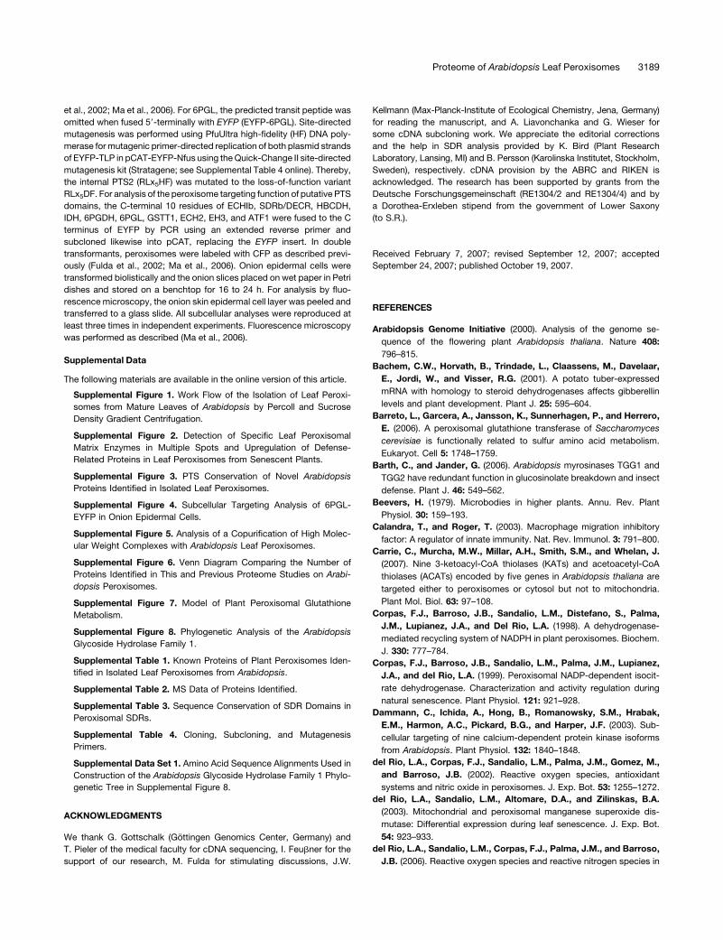
et al., 2002; Ma et al., 2006). For 6PGL, the predicted transit peptide was
omitted when fused 59-terminally with EYFP (EYFP-6PGL). Site-directed
mutagenesis was performed using PfuUltra high-fidelity (HF) DNA poly-
merase for mutagenic primer-directed replication of both plasmid strands
of EYFP-TLP in pCAT-EYFP-Nfus using the Quick-Change II site-directed
mutagenesis kit (Stratagene; see Supplemental Table 4 online). Thereby,
the internal PTS2 (RLx5HF) was mutated to the loss-of-function variant
RLx5DF. For analysis of the peroxisome targeting function of putative PTS
domains, the C-terminal 10 residues of ECHIb, SDRb/DECR, HBCDH,
IDH, 6PGDH, 6PGL, GSTT1, ECH2, EH3, and ATF1 were fused to the C
terminus of EYFP by PCR using an extended reverse primer and
subcloned likewise into pCAT, replacing the EYFP insert. In double
transformants, peroxisomes were labeled with CFP as described previ-
ously (Fulda et al., 2002; Ma et al., 2006). Onion epidermal cells were
transformed biolistically and the onion slices placed on wet paper in Petri
dishes and stored on a benchtop for 16 to 24 h. For analysis by fluo-
rescence microscopy, the onion skin epidermal cell layer was peeled and
transferred to a glass slide. All subcellular analyses were reproduced at
least three times in independent experiments. Fluorescence microscopy
was performed as described (Ma et al., 2006).
Supplemental Data
The following materials are available in the online version of this article.
Supplemental Figure 1. Work Flow of the Isolation of Leaf Peroxi-
somes from Mature Leaves of Arabidopsis by Percoll and Sucrose
Density Gradient Centrifugation.
Supplemental Figure 2. Detection of Specific Leaf Peroxisomal
Matrix Enzymes in Multiple Spots and Upregulation of Defense-
Related Proteins in Leaf Peroxisomes from Senescent Plants.
Supplemental Figure 3. PTS Conservation of Novel Arabidopsis
Proteins Identified in Isolated Leaf Peroxisomes.
Supplemental Figure 4. Subcellular Targeting Analysis of 6PGL-
EYFP in Onion Epidermal Cells.
Supplemental Figure 5. Analysis of a Copurification of High Molec-
ular Weight Complexes with Arabidopsis Leaf Peroxisomes.
Supplemental Figure 6. Venn Diagram Comparing the Number of
Proteins Identified in This and Previous Proteome Studies on Arabi-
dopsis Peroxisomes.
Supplemental Figure 7. Model of Plant Peroxisomal Glutathione
Metabolism.
Supplemental Figure 8. Phylogenetic Analysis of the Arabidopsis
Glycoside Hydrolase Family 1.
Supplemental Table 1. Known Proteins of Plant Peroxisomes Iden-
tified in Isolated Leaf Peroxisomes from Arabidopsis.
Supplemental Table 2. MS Data of Proteins Identified.
Supplemental Table 3. Sequence Conservation of SDR Domains in
Peroxisomal SDRs.
Supplemental Table 4. Cloning, Subcloning, and Mutagenesis
Primers.
Supplemental Data Set 1. Amino Acid Sequence Alignments Used in
Construction of the Arabidopsis Glycoside Hydrolase Family 1 Phylo-
genetic Tree in Supplemental Figure 8.
ACKNOWLEDGMENTS
We thank G. Gottschalk (Gottingen Genomics Center, Germany) and
T. Pieler of the medical faculty for cDNA sequencing, I. Feubner for the
support of our research, M. Fulda for stimulating discussions, J.W.
Kellmann (Max-Planck-Institute of Ecological Chemistry, Jena, Germany)
for reading the manuscript, and A. Liavonchanka and G. Wieser for
some cDNA subcloning work. We appreciate the editorial corrections
and the help in SDR analysis provided by K. Bird (Plant Research
Laboratory, Lansing, MI) and B. Persson (Karolinska Institutet, Stockholm,
Sweden), respectively. cDNA provision by the ABRC and RIKEN is
acknowledged. The research has been supported by grants from the
Deutsche Forschungsgemeinschaft (RE1304/2 and RE1304/4) and by
a Dorothea-Erxleben stipend from the government of Lower Saxony
(to S.R.).
Received February 7, 2007; revised September 12, 2007; accepted
September 24, 2007; published October 19, 2007.
REFERENCES
Arabidopsis Genome Initiative (2000). Analysis of the genome se-
quence of the flowering plant Arabidopsis thaliana. Nature 408:
796–815.
Bachem, C.W., Horvath, B., Trindade, L., Claassens, M., Davelaar,
E., Jordi, W., and Visser, R.G. (2001). A potato tuber-expressed
mRNA with homology to steroid dehydrogenases affects gibberellin
levels and plant development. Plant J. 25: 595–604.
Barreto, L., Garcera, A., Jansson, K., Sunnerhagen, P., and Herrero,
E. (2006). A peroxisomal glutathione transferase of Saccharomyces
cerevisiae is functionally related to sulfur amino acid metabolism.
Eukaryot. Cell 5: 1748–1759.
Barth, C., and Jander, G. (2006). Arabidopsis myrosinases TGG1 and
TGG2 have redundant function in glucosinolate breakdown and insect
defense. Plant J. 46: 549–562.
Beevers, H. (1979). Microbodies in higher plants. Annu. Rev. Plant
Physiol. 30: 159–193.
Calandra, T., and Roger, T. (2003). Macrophage migration inhibitory
factor: A regulator of innate immunity. Nat. Rev. Immunol. 3: 791–800.
Carrie, C., Murcha, M.W., Millar, A.H., Smith, S.M., and Whelan, J.
(2007). Nine 3-ketoacyl-CoA thiolases (KATs) and acetoacetyl-CoA
thiolases (ACATs) encoded by five genes in Arabidopsis thaliana are
targeted either to peroxisomes or cytosol but not to mitochondria.
Plant Mol. Biol. 63: 97–108.
Corpas, F.J., Barroso, J.B., Sandalio, L.M., Distefano, S., Palma,
J.M., Lupianez, J.A., and Del Rio, L.A. (1998). A dehydrogenase-
mediated recycling system of NADPH in plant peroxisomes. Biochem.
J. 330: 777–784.
Corpas, F.J., Barroso, J.B., Sandalio, L.M., Palma, J.M., Lupianez,
J.A., and del Rio, L.A. (1999). Peroxisomal NADP-dependent isocit-
rate dehydrogenase. Characterization and activity regulation during
natural senescence. Plant Physiol. 121: 921–928.
Dammann, C., Ichida, A., Hong, B., Romanowsky, S.M., Hrabak,
E.M., Harmon, A.C., Pickard, B.G., and Harper, J.F. (2003). Sub-
cellular targeting of nine calcium-dependent protein kinase isoforms
from Arabidopsis. Plant Physiol. 132: 1840–1848.
del Rio, L.A., Corpas, F.J., Sandalio, L.M., Palma, J.M., Gomez, M.,
and Barroso, J.B. (2002). Reactive oxygen species, antioxidant
systems and nitric oxide in peroxisomes. J. Exp. Bot. 53: 1255–1272.
del Rio, L.A., Sandalio, L.M., Altomare, D.A., and Zilinskas, B.A.
(2003). Mitochondrial and peroxisomal manganese superoxide dis-
mutase: Differential expression during leaf senescence. J. Exp. Bot.
54: 923–933.
del Rio, L.A., Sandalio, L.M., Corpas, F.J., Palma, J.M., and Barroso,
J.B. (2006). Reactive oxygen species and reactive nitrogen species in
Proteome of Arabidopsis Leaf Peroxisomes 3189
peroxisomes. Production, scavenging, and role in cell signaling. Plant
Physiol. 141: 330–335.
Diaz, M., Achkor, H., Titarenko, E., and Martinez, M.C. (2003). The
gene encoding glutathione-dependent formaldehyde dehydrogenase/
GSNO reductase is responsive to wounding, jasmonic acid and
salicylic acid. FEBS Lett. 543: 136–139.
Dixon, D.P., Lapthorn, A., and Edwards, R. (2002). Plant glutathione
transferases. Genome Biol. 3: REVIEWS3004.
Dolferus, R., Osterman, J.C., Peacock, W.J., and Dennis, E.S. (1997).
Cloning of the Arabidopsis and rice formaldehyde dehydrogenase
genes: Implications for the origin of plant ADH enzymes. Genetics
146: 1131–1141.
Edqvist, J., Ronnberg, E., Rosenquist, S., Blomqvist, K., Viitanen, L.,
Salminen, T.A., Nylund, M., Tuuf, J., and Mattjus, P. (2004). Plants
express a lipid transfer protein with high similarity to mammalian sterol
carrier protein-2. J. Biol. Chem. 279: 53544–53553.
Emanuelsson, O., Elofsson, A., von Heijne, G., and Cristobal, S.
(2003). In silico prediction of the peroxisomal proteome in fungi, plants
and animals. J. Mol. Biol. 330: 443–456.
Eneqvist, T., Lundberg, E., Nilsson, L., Abagyan, R., and Sauer-
Eriksson, A.E. (2003). The transthyretin-related protein family. Eur. J.
Biochem. 270: 518–532.
Eriksson, S., Andreasson, E., Ekbom, B., Graner, G., Pontoppidan,
B., Taipalensuu, J., Zhang, J., Rask, L., and Meijer, J. (2002).
Complex formation of myrosinase isoenzymes in oilseed rape seeds
are dependent on the presence of myrosinase-binding proteins. Plant
Physiol. 129: 1592–1599.
Eulalio, A., Behm-Ansmant, I., and Izaurralde, E. (2007). P bodies: At
the crossroads of post-transcriptional pathways. Nat. Rev. Mol. Cell
Biol. 8: 9–22.
Fransen, M., Van Veldhoven, P.P., and Subramani, S. (1999). Iden-
tification of peroxisomal proteins by using M13 phage protein VI
phage display: Molecular evidence that mammalian peroxisomes
contain a 2,4-dienoyl-CoA reductase. Biochem. J. 340: 561–568.
Fuchs, J., Demidov, D., Houben, A., and Schubert, I. (2006). Chro-
mosomal histone modification patterns – From conservation to diver-
sity. Trends Plant Sci. 11: 199–208.
Fujiki, Y., Fowler, S., Shio, H., Hubbard, A.L., and Lazarow, P.B.
(1982). Polypeptide and phospholipid composition of the membrane
of rat liver peroxisomes: Comparison with endoplasmic reticulum and
mitochondrial membranes. J. Cell Biol. 93: 103–110.
Fukao, Y., Hayashi, M., Hara-Nishimura, I., and Nishimura, M. (2003).
Novel glyoxysomal protein kinase, GPK1, identified by proteomic anal-
ysis of glyoxysomes in etiolated cotyledons of Arabidopsis thaliana.
Plant Cell Physiol. 44: 1002–1012.
Fukao, Y., Hayashi, M., and Nishimura, M. (2002). Proteomic analysis
of leaf peroxisomal proteins in greening cotyledons of Arabidopsis
thaliana. Plant Cell Physiol. 43: 689–696.
Fulda, M., Shockey, J., Werber, M., Wolter, F.P., and Heinz, E. (2002).
Two long-chain acyl-CoA synthetases from Arabidopsis thaliana in-
volved in peroxisomal fatty acid beta-oxidation. Plant J. 32: 93–103.
Gabaldon, T., Snel, B., van Zimmeren, F., Hemrika, W., Tabak, H.,
and Huynen, M.A. (2006). Origin and evolution of the peroxisomal
proteome. Biol. Direct 1: 8.
Geisbrecht, B.V., Zhang, D., Schulz, H., and Gould, S.J. (1999).
Characterization of PECI, a novel monofunctional Delta(3), Delta(2)-
enoyl-CoA isomerase of mammalian peroxisomes. J. Biol. Chem. 274:
21797–21803.
Glinski, M., and Weckwerth, W. (2006). The role of mass spectrometry
in plant systems biology. Mass Spectrom. Rev. 25: 173–214.
Goepfert, S., Hiltunen, J.K., and Poirier, Y. (2006). Identification and
functional characterization of a monofunctional peroxisomal enoyl-
CoA hydratase 2 that participates in the degradation of even cis-
unsaturated fatty acids in Arabidopsis thaliana. J. Biol. Chem. 281:
35894–35903.
Goepfert, S., Vidoudez, C., Rezzonico, E., Hiltunen, J.K., and Poirier,
Y. (2005). Molecular identification and characterization of the Arabi-
dopsis delta(3,5),delta(2,4)-dienoyl-coenzyme A isomerase, a perox-
isomal enzyme participating in the beta-oxidation cycle of unsaturated
fatty acids. Plant Physiol. 138: 1947–1956.
Goyer, A., Johnson, T.L., Olsen, L.J., Collakova, E., Shachar-Hill, Y.,
Rhodes, D., and Hanson, A.D. (2004). Characterization and meta-
bolic function of a peroxisomal sarcosine and pipecolate oxidase from
Arabidopsis. J. Biol. Chem. 279: 16947–16953.
Gross, J., Cho, W.K., Lezhneva, L., Falk, J., Krupinska, K.,
Shinozaki, K., Seki, M., Herrmann, R.G., and Meurer, J. (2006). A
plant locus essential for phylloquinone (vitamin K1) biosynthesis
originated from a fusion of four eubacterial genes. J. Biol. Chem.
281: 17189–17196.
Grubb, C.D., and Abel, S. (2006). Glucosinolate metabolism and its
control. Trends Plant Sci. 11: 89–100.
Gurvitz, A., Rottensteiner, H., Kilpelainen, S.H., Hartig, A., Hiltunen,
J.K., Binder, M., Dawes, I.W., and Hamilton, B. (1997). The Sac-
charomyces cerevisiae peroxisomal 2,4-dienoyl-CoA reductase is
encoded by the oleate-inducible gene SPS19. J. Biol. Chem. 272:
22140–22147.
Halkier, B.A., and Gershenzon, J. (2006). Biology and biochemistry of
glucosinolates. Annu. Rev. Plant Biol. 57: 303–333.
Hawkins, J., and Boden, M. (2006). Detecting and sorting targeting
peptides with neural networks and support vector machines. J.
Bioinform. Comput. Biol. 4: 1–18.
Hayashi, M., Aoki, M., Kondo, M., and Nishimura, M. (1997). Changes
in targeting efficiencies of proteins to plant microbodies caused by
amino acid substitutions in the carboxy-terminal tripeptide. Plant Cell
Physiol. 38: 759–768.
Hayashi, M., and Nishimura, M. (2006). Arabidopsis thaliana – A model
organism to study plant peroxisomes. Biochim. Biophys. Acta 1763:
1382–1391.
Heazlewood, J.L., Tonti-Filippini, J.S., Gout, A.M., Day, D.A.,
Whelan, J., and Millar, A.H. (2004). Experimental analysis of the
Arabidopsis mitochondrial proteome highlights signaling and regula-
tory components, provides assessment of targeting prediction pro-
grams, and indicates plant-specific mitochondrial proteins. Plant Cell
16: 241–256.
Helm, M., Luck, C., Prestele, J., Hierl, G., Huesgen, P.F., Frohlich, T.,
Arnold, G.J., Adamska, I., Gorg, A., Lottspeich, F., and Gietl, C.
(2007). Dual specificities of the glyoxysomal/peroxisomal processing
protease Deg15 in higher plants. Proc. Natl. Acad. Sci. USA 104:
11501–11506.
Hennebry, S.C., Law, R.H., Richardson, S.J., Buckle, A.M., and
Whisstock, J.C. (2006b). The crystal structure of the transthyretin-like
protein from Salmonella dublin, a prokaryote 5-hydroxyisourate hy-
drolase. J. Mol. Biol. 359: 1389–1399.
Hennebry, S.C., Wright, H.M., Likic, V.A., and Richardson, S.J. (2006a).
Structural and functional evolution of transthyretin and transthyretin-
like proteins. Proteins 64: 1024–1045.
Hummel, J., Niemann, M., Wienkoop, S., Schulze, W., Steinhauser,
D., Selbig, J., Walther, D., and Weckwerth, W. (2007). ProMEX: A
mass spectral reference database for proteins and protein phospho-
rylation sites. BMC Bioinformatics 8: 216.
Jahn, O., Hesse, D., Reinelt, M., and Kratzin, H.D. (2006). Technical
innovations for the automated identification of gel-separated pro-
teins by MALDI-TOF mass spectrometry. Anal. Bioanal. Chem. 386:
92–103.
Jimenez, A., Hernandez, J.A., Del Rio, L.A., and Sevilla, F. (1997).
Evidence for the presence of the ascorbate-glutathione cycle in
3190 The Plant Cell
mitochondria and peroxisomes of pea leaves. Plant Physiol. 114:
275–284.
Jimenez, A., Hernandez, J.A., Pastori, G., del Rio, L.A., and Sevilla,
F. (1998). Role of the ascorbate-glutathione cycle of mitochondria and
peroxisomes in the senescence of pea leaves. Plant Physiol. 118:
1327–1335.
Johnson, T.W., Zybailov, B., Jones, A.D., Bittl, R., Zech, S., Stehlik,
D., Golbeck, J.H., and Chitnis, P.R. (2001). Recruitment of a foreign
quinone into the A1 site of photosystem I. In vivo replacement of
plastoquinone-9 by media-supplemented naphthoquinones in phyllo-
quinone biosynthetic pathway mutants of Synechocystis sp. PCC
6803. J. Biol. Chem. 276: 39512–39521.
Jung, D.K., Lee, Y., Park, S.G., Park, B.C., Kim, G.H., and Rhee, S.
(2006). Structural and functional analysis of PucM, a hydrolase in the
ureide pathway and a member of the transthyretin-related protein
family. Proc. Natl. Acad. Sci. USA 103: 9790–9795.
Kallberg, Y., and Persson, B. (2006). Prediction of coenzyme speci-
ficity in dehydrogenases/reductases. A hidden Markov model-based
method and its application on complete genomes. FEBS J. 273:
1177–1184.
Kamada, T., Nito, K., Hayashi, H., Mano, S., Hayashi, M., and
Nishimura, M. (2003). Functional differentiation of peroxisomes
revealed by expression profiles of peroxisomal genes in Arabidopsis
thaliana. Plant Cell Physiol. 44: 1275–1289.
Kamigaki, A., Mano, S., Terauchi, K., Nishi, Y., Tachibe-Kinoshita,
Y., Nito, K., Kondo, M., Hayashi, M., Nishimura, M., and Esaka, M.
(2003). Identification of peroxisomal targeting signal of pumpkin
catalase and the binding analysis with PTS1 receptor. Plant J. 33:
161–175.
Kato, A., Takeda-Yoshikawa, Y., Hayashi, M., Kondo, M., Hara-
Nishimura, I., and Nishimura, M. (1998). Glyoxysomal malate dehy-
drogenase in pumpkin: Cloning of a cDNA and functional analysis of
its presequence. Plant Cell Physiol. 39: 186–195.
Kikuchi, M., Hatano, N., Yokota, S., Shimozawa, N., Imanaka, T.,
and Taniguchi, H. (2004). Proteomic analysis of rat liver peroxisome:
Presence of peroxisome-specific isozyme of Lon protease. J. Biol.
Chem. 279: 421–428.
Klein, A.T., van den Berg, M., Bottger, G., Tabak, H.F., and Distel, B.
(2002). Saccharomyces cerevisiae acyl-CoA oxidase follows a novel,
non-PTS1, import pathway into peroxisomes that is dependent on
Pex5p. J. Biol. Chem. 277: 25011–25019.
Kliebenstein, D.J., Kroymann, J., and Mitchell-Olds, T. (2005). The
glucosinolate-myrosinase system in an ecological and evolutionary
context. Curr. Opin. Plant Biol. 8: 264–271.
Koh, S., Andre, A., Edwards, H., Ehrhardt, D., and Somerville, S.
(2005). Arabidopsis thaliana subcellular responses to compatible
Erysiphe cichoracearum infections. Plant J. 44: 516–529.
Koo, A.J., Chung, H.S., Kobayashi, Y., and Howe, G.A. (2006).
Identification of a peroxisomal acyl-activating enzyme involved in
the biosynthesis of jasmonic acid in Arabidopsis. J. Biol. Chem. 281:
33511–33520.
Kragler, F., Lametschwandtner, G., Christmann, J., Hartig, A., and
Harada, J.J. (1998). Identification and analysis of the plant peroxi-
somal targeting signal 1 receptor NtPEX5. Proc. Natl. Acad. Sci. USA
95: 13336–13341.
Kruft, V., Eubel, H., Jansch, L., Werhahn, W., and Braun, H.P. (2001).
Proteomic approach to identify novel mitochondrial proteins in Arabi-
dopsis. Plant Physiol. 127: 1694–1710.
Lee, K.H., Piao, H.L., Kim, H.Y., Choi, S.M., Jiang, F., Hartung, W.,
Hwang, I., Kwak, J.M., Lee, I.J., and Hwang, I. (2006). Activation of
glucosidase via stress-induced polymerization rapidly increases ac-
tive pools of abscisic acid. Cell 126: 1109–1120.
Lee, Y., Lee, D.H., Kho, C.W., Lee, A.Y., Jang, M., Cho, S., Lee, C.H.,
Lee, J.S., Myung, P.K., Park, B.C., and Park, S.G. (2005). Trans-
thyretin-related proteins function to facilitate the hydrolysis of
5-hydroxyisourate, the end product of the uricase reaction. FEBS
Lett. 579: 4769–4774.
Leterrier, M., Corpas, F.J., Barroso, J.B., Sandalio, L.M., and del
Rio, L.A. (2005). Peroxisomal monodehydroascorbate reductase.
Genomic clone characterization and functional analysis under envi-
ronmental stress conditions. Plant Physiol. 138: 2111–2123.
Link, A.J., Eng, J., Schieltz, D.M., Carmack, E., Mize, G.J., Morris,
D.R., Garvik, B.M., and Yates III, J.R. (1999). Direct analysis of
protein complexes using mass spectrometry. Nat. Biotechnol. 17:
676–682.
Lipka, V., et al. (2005). Pre- and postinvasion defenses both contribute
to nonhost resistance in Arabidopsis. Science 310: 1180–1183.
Lisenbee, C.S., Heinze, M., and Trelease, R.N. (2003). Peroxisomal
ascorbate peroxidase resides within a subdomain of rough endo-
plasmic reticulum in wild-type Arabidopsis cells. Plant Physiol. 132:
870–882.
Lisenbee, C.S., Lingard, M.J., and Trelease, R.N. (2005). Arabi-
dopsis peroxisomes possess functionally redundant membrane and
matrix isoforms of monodehydroascorbate reductase. Plant J. 43:
900–914.
Ma, C., Haslbeck, M., Babujee, L., Jahn, O., and Reumann, S. (2006).
Identification and characterization of a stress-inducible and a consti-
tutive small heat-shock protein targeted to the matrix of plant perox-
isomes. Plant Physiol. 141: 47–60.
Matsushima, R., Hayashi, Y., Kondo, M., Shimada, T., Nishimura, M.,
and Hara-Nishimura, I. (2002). An endoplasmic reticulum-derived
structure that is induced under stress conditions in Arabidopsis. Plant
Physiol. 130: 1807–1814.
Maynard, E.L., Gatto, G.J., Jr., and Berg, J.M. (2004). Pex5p binding
affinities for canonical and noncanonical PTS1 peptides. Proteins 55:
856–861.
McCartney, A.W., Greenwood, J.S., Fabian, M.R., White, K.A., and
Mullen, R.T. (2005). Localization of the tomato bushy stunt virus
replication protein p33 reveals a peroxisome-to-endoplasmic reticu-
lum sorting pathway. Plant Cell 17: 3513–3531.
Millar, A.H., Sweetlove, L.J., Giege, P., and Leaver, C.J. (2001).
Analysis of the Arabidopsis mitochondrial proteome. Plant Physiol.
127: 1711–1727.
Morgenthal, K., Wienkoop, S., Wolschin, F., and Weckwerth, W.
(2007). Integrative profiling of metabolites and proteins: Improving
pattern recognition and biomarker selection for systems level ap-
proaches. Methods Mol. Biol. 358: 57–75.
Mullen, R.T., Lee, M.S., Flynn, C.R., and Trelease, R.N. (1997).
Diverse amino acid residues function within the type 1 peroxisomal
targeting signal. Implications for the role of accessory residues
upstream of the type 1 peroxisomal targeting signal. Plant Physiol.
115: 881–889.
Nagano, A.J., Matsushima, R., and Hara-Nishimura, I. (2005). Acti-
vation of an ER-body-localized beta-glucosidase via a cytosolic
binding partner in damaged tissues of Arabidopsis thaliana. Plant
Cell Physiol. 46: 1140–1148.
Nakamura, T., Yokota, S., Muramoto, Y., Tsutsui, K., Oguri, Y.,
Fukui, K., and Takabe, T. (1997). Expression of a betaine aldehyde
dehydrogenase gene in rice, a glycinebetaine nonaccumulator, and
possible localization of its protein in peroxisomes. Plant J. 11: 1115–
1120.
Nam, K.H., and Li, J. (2004). The Arabidopsis transthyretin-like protein
is a potential substrate of BRASSINOSTEROID-INSENSITIVE 1. Plant
Cell 16: 2406–2417.
Nanjo, Y., Oka, H., Ikarashi, N., Kaneko, K., Kitajima, A., Mitsui, T.,
Munoz, F.J., Rodriguez-Lopez, M., Baroja-Fernandez, E., and
Proteome of Arabidopsis Leaf Peroxisomes 3191
Pozueta-Romero, J. (2006). Rice plastidial N-glycosylated nucleotide
pyrophosphatase/phosphodiesterase is transported from the ER-
Golgi to the chloroplast through the secretory pathway. Plant Cell
18: 2582–2592.
Neuberger, G., Maurer-Stroh, S., Eisenhaber, B., Hartig, A., and
Eisenhaber, F. (2003a). Prediction of peroxisomal targeting signal
1 containing proteins from amino acid sequence. J. Mol. Biol. 328:
581–592.
Neuberger, G., Maurer-Stroh, S., Eisenhaber, B., Hartig, A., and
Eisenhaber, F. (2003b). Motif refinement of the peroxisomal targeting
signal 1 and evaluation of taxon-specific differences. J. Mol. Biol. 328:
567–579.
Nowak, K., Luniak, N., Witt, C., Wustefeld, Y., Wachter, A., Mendel,
R.R., and Hansch, R. (2004). Peroxisomal localization of sulfite
oxidase separates it from chloroplast-based sulfur assimilation. Plant
Cell Physiol. 45: 1889–1894.
Peck, S.C. (2005). Update on proteomics in Arabidopsis. Where do we
go from here? Plant Physiol. 138: 591–599.
Peltier, J.B., Emanuelsson, O., Kalume, D.E., Ytterberg, J., Friso, G.,
Rudella, A., Liberles, D.A., Soderberg, L., Roepstorff, P., von
Heijne, G., and van Wijk, K.J. (2002). Central functions of the lu-
menal and peripheral thylakoid proteome of Arabidopsis determined
by experimentation and genome-wide prediction. Plant Cell 14:
211–236.
Polevoda, B., and Sherman, F. (2002). The diversity of acetylated
proteins. Genome Biol 3: reviews0006.
Ramazzina, I., Folli, C., Secchi, A., Berni, R., and Percudani, R.
(2006). Completing the uric acid degradation pathway through
phylogenetic comparison of whole genomes. Nat. Chem. Biol. 2:
144–148.
Ravanel, S., Block, M.A., Rippert, P., Jabrin, S., Curien, G., Rebeille,
F., and Douce, R. (2004). Methionine metabolism in plants: Chloro-
plasts are autonomous for de novo methionine synthesis and can
import S-adenosylmethionine from the cytosol. J. Biol. Chem. 279:
22548–22557.
Reumann, S. (2002). The photorespiratory pathway of leaf peroxi-
somes. In Plant Peroxisomes: Biochemistry, Cell Biology and Bio-
technological Applications, A. Baker and I.A. Graham, eds (Dordrecht,
The Netherlands: Kluwer Academic Publishers), pp. 141–189.
Reumann, S. (2004). Specification of the peroxisome targeting signals
type 1 and type 2 of plant peroxisomes by bioinformatics analyses.
Plant Physiol. 135: 783–800.
Reumann, S., Ma, C., Lemke, S., and Babujee, L. (2004). AraPerox. A
database of putative Arabidopsis proteins from plant peroxisomes.
Plant Physiol. 136: 2587–2608.
Reumann, S., and Weber, A.P. (2006). Plant peroxisomes respire in the
light: Some gaps of the photorespiratory C2 cycle have become filled–
others remain. Biochim. Biophys. Acta 1763: 1496–1510.
Romero-Puertas, M.C., Corpas, F.J., Sandalio, L.M., Leterrier, M.,
Rodriguez-Serrano, M., Del Rio, L.A., and Palma, J.M. (2006).
Glutathione reductase from pea leaves: Response to abiotic stress
and characterization of the peroxisomal isozyme. New Phytol. 170:
43–52.
Sakamoto, A., Ueda, M., and Morikawa, H. (2002). Arabidopsis
glutathione-dependent formaldehyde dehydrogenase is an
S-nitrosoglutathione reductase. FEBS Lett. 515: 20–24.
Saleem, R.A., Smith, J.J., and Aitchison, J.D. (2006). Proteomics of
the peroxisome. Biochim. Biophys. Acta 1763: 1541–1551.
Schluter, A., Fourcade, S., Ripp, R., Mandel, J.L., Poch, O., and
Pujol, A. (2006). The evolutionary origin of peroxisomes: An ER-
peroxisome connection. Mol. Biol. Evol. 23: 838–845.
Schneider, K., Kienow, L., Schmelzer, E., Colby, T., Bartsch, M.,
Miersch, O., Wasternack, C., Kombrink, E., and Stuible, H.P.
(2005). A new type of peroxisomal acyl-coenzyme A synthetase from
Arabidopsis thaliana has the catalytic capacity to activate biosynthetic
precursors of jasmonic acid. J. Biol. Chem. 280: 13962–13972.
Sharma, Y.K., and Davis, K.R. (1995). Isolation of a novel Arabidopsis
ozone-induced cDNA by differential display. Plant Mol. Biol. 29: 91–98.
Shockey, J.M., Fulda, M.S., and Browse, J. (2003). Arabidopsis
contains a large superfamily of acyl-activating enzymes. Phylogenetic
and biochemical analysis reveals a new class of acyl-coenzyme A
synthetases. Plant Physiol. 132: 1065–1076.
Staiger, D., Zecca, L., Wieczorek Kirk, D.A., Apel, K., and Eckstein,
L. (2003). The circadian clock regulated RNA-binding protein AtGRP7
autoregulates its expression by influencing alternative splicing of its
own pre-mRNA. Plant J. 33: 361–371.
Stark, A., Houshmand, H., Sandberg, M., and Meijer, J. (1995).
Characterization of the activity of fatty-acid epoxide hydrolase in
seeds of castor bean (Ricinus communis L.). Planta 197: 84–88.
Stotz, H.U., Pittendrigh, B.R., Kroymann, J., Weniger, K., Fritsche,
J., Bauke, A., and Mitchell-Olds, T. (2000). Induced plant defense
responses against chewing insects. Ethylene signaling reduces resis-
tance of Arabidopsis against Egyptian cotton worm but not diamond-
back moth. Plant Physiol. 124: 1007–1018.
Strassner, J., Schaller, F., Frick, U.B., Howe, G.A., Weiler, E.W.,
Amrhein, N., Macheroux, P., and Schaller, A. (2002). Characteriza-
tion and cDNA-microarray expression analysis of 12-oxophytodienoate
reductases reveals differential roles for octadecanoid biosynthesis in
the local versus the systemic wound response. Plant J. 32: 585–601.
Sun, H.W., Bernhagen, J., Bucala, R., and Lolis, E. (1996). Crystal
structure at 2.6-A resolution of human macrophage migration inhib-
itory factor. Proc. Natl. Acad. Sci. USA 93: 5191–5196.
Taler, D., Galperin, M., Benjamin, I., Cohen, Y., and Kenigsbuch, D.
(2004). Plant eR genes that encode photorespiratory enzymes confer
resistance against disease. Plant Cell 16: 172–184.
Taylor, S.W., Fahy, E., and Ghosh, S.S. (2003). Global organellar
proteomics. Trends Biotechnol. 21: 82–88.
Thiele, M., and Bernhagen, J. (2005). Link between macrophage
migration inhibitory factor and cellular redox regulation. Antioxid.
Redox Signal. 7: 1234–1248.
Tilton, G.B., Shockey, J.M., and Browse, J. (2004). Biochemical and
molecular characterization of ACH2, an acyl-CoA thioesterase from
Arabidopsis thaliana. J. Biol. Chem. 279: 7487–7494.
Turner, J.E., Greville, K., Murphy, E.C., and Hooks, M.A. (2005).
Characterization of Arabidopsis fluoroacetate-resistant mutants re-
veals the principal mechanism of acetate activation for entry into the
glyoxylate cycle. J. Biol. Chem. 280: 2780–2787.
Tyler, R.C., Bitto, E., Berndsen, C.E., Bingman, C.A., Singh, S., Lee,
M.S., Wesenberg, G.E., Denu, J.M., Phillips, G.N., Jr., and Markley,
J.L. (2006). Structure of Arabidopsis thaliana At1g77540 protein, a
minimal acetyltransferase from the COG2388 family. Biochemistry 45:
14325–14336.
Villarejo, A., et al. (2005). Evidence for a protein transported through
the secretory pathway en route to the higher plant chloroplast. Nat.
Cell Biol. 7: 1224–1231.
Wagner, U., Edwards, R., Dixon, D.P., and Mauch, F. (2002). Probing
the diversity of the Arabidopsis glutathione S-transferase gene family.
Plant Mol. Biol. 49: 515–532.
Werner, H.B., et al. (2007). Proteolipid protein is required for transport
of sirtuin 2 into CNS myelin. J. Neurosci. 27: 7717–7730.
Wessel, D., and Flugge, U.I. (1984). A method for the quantitative
recovery of protein in dilute solution in the presence of detergents and
lipids. Anal. Biochem. 138: 141–143.
Wienkoop, S., Zoeller, D., Ebert, B., Simon-Rosin, U., Fisahn, J.,
Glinski, M., and Weckwerth, W. (2004). Cell-specific protein profiling
in Arabidopsis thaliana trichomes: Identification of trichome-located
3192 The Plant Cell
proteins involved in sulfur metabolism and detoxification. Phytochem-
istry 65: 1641–1649.
Xu, Z., Escamilla-Trevino, L., Zeng, L., Lalgondar, M., Bevan, D.,
Winkel, B., Mohamed, A., Cheng, C.L., Shih, M.C., Poulton, J., and
Esen, A. (2004). Functional genomic analysis of Arabidopsis thaliana
glycoside hydrolase family 1. Plant Mol. Biol. 55: 343–367.
Zhang, Z., Ober, J.A., and Kliebenstein, D.J. (2006). The gene con-
trolling the quantitative trait locus EPITHIOSPECIFIER MODIFIER1
alters glucosinolate hydrolysis and insect resistance in Arabidopsis.
Plant Cell 18: 1524–1536.
Zimmermann, P., Hirsch-Hoffmann, M., Hennig, L., and Gruissem,
W. (2004). GENEVESTIGATOR. Arabidopsis microarray database and
analysis toolbox. Plant Physiol. 136: 2621–2632.
Zolman, B.K., Monroe-Augustus, M., Thompson, B., Hawes, J.W.,
Krukenberg, K.A., Matsuda, S.P., and Bartel, B. (2001). chy1, an Arabi-
dopsis mutant with impaired beta-oxidation, is defective in a peroxisomal
beta-hydroxyisobutyryl-CoA hydrolase. J. Biol. Chem. 276: 31037–31046.
Zolman, B.K., Nyberg, M., and Bartel, B. (2007). IBR3, a novel perox-
isomal acyl-CoA dehydrogenase-like protein required for indole-3-
butyric acid response. Plant Mol. Biol. 64: 59–72.
Proteome of Arabidopsis Leaf Peroxisomes 3193
DOI 10.1105/tpc.107.050989; originally published online October 19, 2007; 2007;19;3170-3193Plant Cell
Antonicelli, Nicolas Rasche, Franziska Lüder, Wolfram Weckwerth and Olaf JahnSigrun Reumann, Lavanya Babujee, Changle Ma, Stephanie Wienkoop, Tanja Siemsen, Gerardo E.
Pathways, and Defense Mechanisms Leaf Peroxisomes Reveals Novel Targeting Peptides, MetabolicArabidopsisProteome Analysis of
This information is current as of January 2, 2020
Supplemental Data /content/suppl/2007/09/25/tpc.107.050989.DC1.html
References /content/19/10/3170.full.html#ref-list-1
This article cites 118 articles, 53 of which can be accessed free at:
Permissions https://www.copyright.com/ccc/openurl.do?sid=pd_hw1532298X&issn=1532298X&WT.mc_id=pd_hw1532298X
eTOCs http://www.plantcell.org/cgi/alerts/ctmain
Sign up for eTOCs at:
CiteTrack Alerts http://www.plantcell.org/cgi/alerts/ctmain
Sign up for CiteTrack Alerts at:
Subscription Information http://www.aspb.org/publications/subscriptions.cfm
is available at:Plant Physiology and The Plant CellSubscription Information for
ADVANCING THE SCIENCE OF PLANT BIOLOGY © American Society of Plant Biologists