Embed Size (px)
Citation preview

2215RESEARCH ARTICLE
INTRODUCTIONAfter axons reach their targets, secreted and membrane proteinsactivate signalling cascades to initiate the assembly of synapticsites. At central synapses, secreted signals such as Wnts, FGFs andneurotrophins activate specific receptors on target cells, resultingin structural and functional changes in the presynaptic terminal andon the postsynaptic receptive dendrite (Ciani and Salinas, 2005;Fox and Umemori, 2006; Hardin and King, 2008). Little is known,however, about the localization and identity of the receptorsinvolved.
Wnt proteins function as synaptic organizers in both vertebratesand invertebrates (Salinas and Zou, 2008; Speese and Budnik,2007). In a retrograde or anterograde manner, Wnts regulate axonremodelling, required for the conversion of growth cones intosynaptic boutons (Hall et al., 2000; Packard et al., 2002), andinduce the recruitment of pre- and postsynaptic components tofuture synaptic sites (Hall et al., 2000; Packard et al., 2002). Wntsinduce presynaptic differentiation through the activation of thecanonical or -catenin pathway (Davis et al., 2008; Hall et al.,2000). Studies in Drosophila and C. elegans have shown that Wntsactivate the seven transmembrane Frizzled (Fz) receptors toregulate synapse formation (Ataman et al., 2008; Klassen andShen, 2007). At vertebrate synapses, by contrast, the receptorsactivated by Wnts during synaptic assembly remain elusive.
Three distinct receptor families mediate Wnt signalling: Fzs, thesingle-pass LRP5/6 co-receptors and the atypical tyrosine kinasereceptors Ror2 and Ryk (Angers and Moon, 2009; van Amerongenet al., 2008). In the nervous system, Fzs mediate a range offunctions from neuronal differentiation (Van Raay et al., 2005) tocell polarity (Prasad and Clark, 2006), cell migration (Pan et al.,2006; Vivancos et al., 2009), axon guidance (Lyuksyutova et al.,2003; Wang et al., 2002) and cell survival (Wang et al., 2001). InDrosophila, the Wnt protein Wingless (Wg) activates the Fz2receptor, present at both sides of the neuromuscular synapse, toregulate pre- and postsynaptic signalling (Ataman et al., 2008;Packard et al., 2002). In C. elegans, by contrast, activation of theextrasynaptic Fz receptor Lin-17 inhibits synapse formation(Klassen and Shen, 2007). At vertebrate central synapses, Wntssuch as Wnt7a, Wnt3a and Wnt5a stimulate synapse formation inthe cerebellum, spinal cord and hippocampal neurons (Ahmad-Annuar et al., 2006; Davis et al., 2008; Hall et al., 2000; Krylovaet al., 2002). In cultured hippocampal neurons, Fz1 is present atpresynaptic sites and mediates Wnt3a-induced synapse formation(Varela-Nallar et al., 2009). However, the receptors for keysynaptogenic factors such as Wnt7a remain poorly characterized.
Neuronal activity plays a crucial role in synapse formation andfunction. Activity regulates synapse formation through changes inaxonal and dendritic filopodia dynamics (Hua and Smith, 2004)and by recruiting pre- and postsynaptic proteins to synaptic sites(Craig et al., 2006; McAllister, 2007). In addition, activity regulatesthe localization of membrane proteins that control the formationand function of neuronal circuits. Depolarization or induction oflong-term potentiation (LTP) recruits N-cadherin (Bozdagi et al.,2000; Tanaka et al., 2000), as well as AMPA and NMDA receptors(Friedman et al., 2000; Hayashi et al., 2000; Heynen et al., 2000)to synaptic sites, whereas field stimulation or depolarization
Development 137, 2215-2125 (2010) doi:10.1242/dev.046722© 2010. Published by The Company of Biologists Ltd
1Department of Cell and Developmental Biology and 2Department of Neuroscience,Physiology and Pharmacology, University College London, London WC1E 6BT, UK.
*Author for correspondence ([email protected])
Accepted 30 April 2010
SUMMARYWnt proteins play a crucial role in several aspects of neuronal circuit formation. Wnts can signal through different receptorsincluding Frizzled, Ryk and Ror2. In the hippocampus, Wnt7a stimulates the formation of synapses; however, its receptor remainspoorly characterized. Here, we demonstrate that Frizzled-5 (Fz5) is expressed during the peak of synaptogenesis in the mousehippocampus. Fz5 is present in synaptosomes and colocalizes with the pre- and postsynaptic markers vGlut1 and PSD-95.Expression of Fz5 during early stages of synaptogenesis increases the number of presynaptic sites in hippocampal neurons.Conversely, Fz5 knockdown or the soluble Fz5-CRD domain (Fz5CRD), which binds to Wnt7a, block the ability of Wnt7a tostimulate synaptogenesis. Increased neuronal activity induced by K+ depolarization or by high-frequency stimulation (HFS), knownto induce synapse formation, raises the levels of Fz5 at the cell surface. Importantly, both stimuli increase the localization of Fz5at synapses, an effect that is blocked by Wnt antagonists or Fz5CRD. Conversely, low-frequency stimulation, which reduces thenumber of synapses, decreases the levels of surface Fz5 and the percentage of synapses containing the receptor. Interestingly,Fz5CRD abolishes HFS-induced synapse formation. Our results indicate that Fz5 mediates the synaptogenic effect of Wnt7a andthat its localization to synapses is regulated by neuronal activity, a process that depends on endogenous Wnts. These findingssupport a model where neuronal activity and Wnts increase the responsiveness of neurons to Wnt signalling by recruiting Fz5receptor at synaptic sites.
KEY WORDS: Wnt signalling, Synaptic assembly, Hippocampus, Dendrite, Neuronal activity, Mouse
Frizzled-5, a receptor for the synaptic organizer Wnt7a,regulates activity-mediated synaptogenesisMacarena Sahores1, Alasdair Gibb2 and Patricia C. Salinas1,*
DEVELO
PMENT
2216
increases TrkB surface levels (Du et al., 2000; Meyer-Franke et al.,1998). By contrast, low-frequency stimulation (LFS)-evoked long-term depression (LTD) is associated with internalization of synapticAMPA and NMDA receptors (Carroll et al., 2001; Shi et al., 1999)as well as TrkB receptors (Du et al., 2003). However, little isknown about the role of neuronal activity in the localization of Wntreceptors.
Here, we report that the Wnt receptor Fz5 is expressed in themouse hippocampus and in cultured hippocampal neurons duringthe period of synaptogenesis, where it localizes to both pre- andpostsynaptic sites. Gain-of-function studies demonstrate that Fz5increases the number of synapsin I and bassoon puncta, a hallmarkof presynaptic assembly. In addition, Wnt7a binds to theextracellular cysteine-rich domain (CRD) of Fz5, which is crucialfor binding to Wnts and for signalling (Povelones and Nusse, 2005;Rulifson et al., 2000). Importantly, Fz5 knockdown or the solubleFz5-CRD domain (Fz5CRD) block the synaptogenic activity ofWnt7a. In addition, neuronal activity induced by depolarization orhigh-frequency stimulation (HFS) increases the mobilization of Fz5to the cell surface and its localization to synapses. By contrast, LFSdecreases the trafficking of Fz5 to the surface and its insertion tosynaptic sites. Fz5CRD or the Wnt antagonists Sfrp1 and Sfrp3,block the activity-dependent localization of Fz5 at synapses.Moreover, HFS-induced synapse formation is completely blockedby Fz5CRD, suggesting a role for Wnt-Fz5 signalling in thisprocess. We therefore propose that activation of Fz5 by Wnt7a,together with neuronal activity, regulate synaptic assembly inhippocampal neurons and that both neuronal activity and Wntsregulate Fz5 localization to synapses.
MATERIALS AND METHODSNeuronal cultures and cell transfectionPrimary hippocampal cultures were prepared from embryonic day 18 (E18)Sprague-Dawley rat embryos and cultured as described previously (Rossoet al., 2005). Cells were cultured at a density of 30-50 cells/mm2 forimmunostaining or at 250 cells/mm2 for biotinylation assays. Hippocampalneurons were transfected at 3-5 days in vitro (DIV) with EGFP or hFz5-HA constructs using Lipofectamine 2000 (Invitrogen), treated as indicatedand fixed at 9-11 DIV. Transfected neurons were exposed to recombinantWnt7a (50 ng/ml, R&D Systems) for 16 hours. Untransfected neurons (9-12 DIV) were exposed for 16 hours to control or Fz5CRD-containingconditioned media obtained from transfected QT6 cells.
Surface receptor staining, immunofluorescence andimmunohistochemistryCultured neurons were fixed with 4% paraformaldehyde (PFA) in PBSwith 4% sucrose, and stained with primary antibodies against -III tubulin(Tuj-1; Chemicon), HA (Roche), GFP (Molecular Probes and Upstate),Myc (Sigma), bassoon (Bioquote Limited), synapsin I (Transduction Labs),vGlut-1 (Chemicon), NR1 (Synaptic Systems), MAP-2 (Sigma) or frizzled5 (Abcam). Secondary antibodies were from Molecular Probes. For surfaceexpression, anti-Fz5 antibody was added to live cells and incubated for 30minutes on ice or 15 minutes at 37°C. Mouse brains were fixed with 4%PFA in PBS at 4°C and then incubated in 30% sucrose in PBS at 4°C.Cryostat sections (16 mm) were incubated in blocking solution (0.2%gelatine, 0.25% Triton X-100 in PBS) followed by primary antibodiesagainst vGlut1, PSD-95 (Affinity Bioreagents) and Fz5 and finally byappropriate secondary antibodies. Fluorescence images were captured withan Olympus BX60 microscope or with a Leica TCS SP1 confocalmicroscope. Images were acquired with either Metamorph or Leicasoftware and analyzed using Metamorph or Volocity software. Single-planeconfocal images or maximal projections obtained with the 3D feature onVolocity were used to determine colocalization.
RESEARCH ARTICLE Development 137 (13)
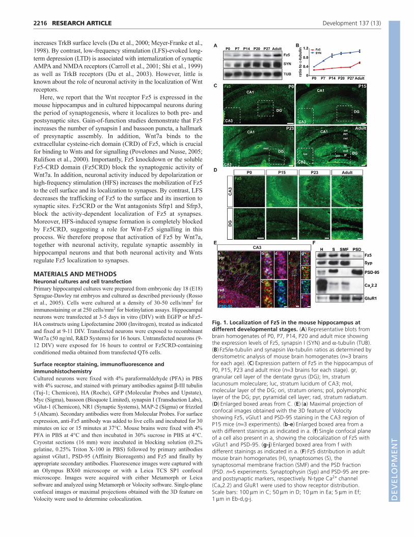
Fig. 1. Localization of Fz5 in the mouse hippocampus atdifferent developmental stages. (A)Representative blots frombrain homogenates of P0, P7, P14, P20 and adult mice showingthe expression levels of Fz5, synapsin I (SYN) and -tubulin (TUB).(B)Fz5/-tubulin and synapsin I/-tubulin ratios as determined bydensitometric analysis of mouse brain homogenates (n3 brainsfor each age). (C)Expression pattern of Fz5 in the hippocampus ofP0, P15, P23 and adult mice (n3 brains for each stage). gr,granular cell layer of the dentate gyrus (DG); lm, stratumlacunosum moleculare; luc, stratum lucidum of CA3; mol,molecular layer of the DG; ori, stratum oriens; pol, polymorphiclayer of the DG; pyr, pyramidal cell layer; rad, stratum radiatum.(D)Enlarged boxed areas from C. (E)(a) Maximal projection ofconfocal images obtained with the 3D feature of Volocityshowing Fz5, vGlut1 and PSD-95 staining in the CA3 region ofP15 mice (n3 experiments). (b-e)Enlarged boxed area from awith different stainings as indicated in a. (f)Single confocal planeof a cell also present in a, showing the colocalization of Fz5 withvGlut1 and PSD-95. (g-j)Enlarged boxed area from f withdifferent stainings as indicated in a. (F)Fz5 distribution in adultmouse brain homogenates (H), synaptosomes (S), thesynaptosomal membrane fraction (SMF) and the PSD fraction(PSD. n5 experiments. Synaptophysin (Syp) and PSD-95 are pre-and postsynaptic markers, respectively. N-type Ca2+ channel(Cav2.2) and GluR1 were used to show receptor distribution.Scale bars: 100m in C; 50m in D; 10m in Ea; 5m in Ef;1m in Eb-d,g-j. D
EVELO
PMENT

Western blot analysisBrain homogenates were prepared with RIPA buffer using a tissuehomogenizer at 0°C. Equal amounts of protein (Lowry assay) wereanalyzed by SDS-PAGE and western blot using antibodies for synapsin I(Chemicon), Fz5 and -tubulin (Sigma). Membranes were probed withHRP-coupled secondary antibodies and developed with ECL reagent(Amersham). Quantification of band intensity was performed using ImageJsoftware.
Synaptosomal preparationSynaptosomes were prepared as previously described (Cohen et al.,1977), with minor modifications (Ahmad-Annuar et al., 2006). In brief,synaptosomes were treated with TX-100, followed by centrifugation at82,500 g for 45 minutes. Then the supernatant, corresponding to thesynaptosomal membrane fraction (SMF), was removed and the pellet,corresponding to the postsynaptic density (PSD) fraction, wasresuspended in buffer B. Equal amounts of proteins were loaded onto anSDS-PAGE. Primary antibodies against Fz5, synaptophysin (Chemicon),PSD-95, N-type Ca2+ channel (1B subunit, Sigma) and GluR1 (Upstate)were used.
Binding assayQT6 cells were used to prepare Wnt7a-HA-containing conditionedmedia. Cos-7 cells were transfected with the CRD domain of human Fz5or Drosophila Dfz2, both containing a glycosyl-phosphatidylinositol(GPI) sequence (Fz5CRD-myc-GPI and Dfz2CRD-myc-GPI). Livetransfected Cos-7 cells were incubated for 1 hour at RT with control orWnt7a-HA-containing conditioned media. Cells were fixed with 4%PFA and 4% sucrose in PBS, and incubated with primary antibodies toHA and Myc followed by incubation with biotinylated secondaryantibodies (Amersham), Alexa 488 conjugated secondary antibodies andHoechst (Molecular Probes). HRP staining was developed according tothe manufacturer’s protocol (Vectastain Elite ABC Kit, VectorLaboratories).
RNA interference to Fz5Fz5 knockdown was achieved by using SureSilencing shRNA plasmids torat Fz5 (SABiosciences). The kit contains 4 shRNA vectors specificallydirected to the rat Fz5 and an shRNA vector with a scrambled artificialsequence as a negative control (nc-shRNA). These plasmids also containthe GFP gene to identify transfected cells. NRK cells were transfected withthe shRNAs using Lipofectamine 2000. NRK cells were fixed andprocessed for immunostaining or lysed and prepared for western blotanalysis 48 hours after transfection. Primary hippocampal neurons weretransfected at 4 DIV with either the mixture of Fz5 shRNAs or nc-shRNAusing Lipofectamine 2000. At 9 DIV, these neurons were incubated withrecombinant Wnt7a (50 ng/ml) for 16 hours, fixed and processed forimmunocytochemistry.
Cell surface biotinylationSurface Fz5 was detected by cell surface biotinylation using Sulfo-NHS-LC-LC-biotin (Pierce) and Streptavidin Sepharose High Performance (GEHealthcare) (Du et al., 2000). Cell lysates were prepared from 14 DIVhippocampal neurons after K+ depolarization, HFS or LFS.
Stimulation of cultured hippocampal neuronsFor K+ depolarization studies, 14 DIV neurons were treated with 50 mMNaCl or KCl. For HFS and LFS studies, neurons were electricallystimulated using a Grass48 stimulator (Grass Instruments). Trains of HFSwere used, with train duration of 400 milliseconds containing 20consecutive pulses at 50 Hz. The interval between trains lasted 5 seconds.LTD-like LFS used a constant stimulation frequency of 4 Hz. Electricalstimulation was delivered for 1 hour at 37°C in a 6 cm-diameter dish viatwo Ag/AgCl electrodes. Neurons were exposed to control or Fz5CRD-containing conditioned media obtained from transfected QT6 cells or torecombinant Sfrp1 and Sfrp3 (2.5 mg/ml and 250 ng/ml respectively,R&D).
To assess depolarization and HFS, the levels of CaMKIIphosphorylation, which increase under these experimental conditions, wereanalyzed by western blot (Blitzer et al., 1998; Pettit et al., 1994). The levelof CaMKII activation was determined by its phosphorylation at threonine286 and normalized to total CaMKII levels (Cell Signaling).
To assess the effect of LFS, the levels of NR1 were analyzed. FollowingLFS, cells displayed a 54% reduction in the number NR1 puncta (data notshown) and a 49% reduction in the number of synapses (Fig. 7J), indicativethat LTD-like induction was achieved (Heynen et al., 2000).
Statistical analysesValues given are mean ± s.e.m. Statistical significance was determined usingtwo-tailed Student’s t-test and ANOVA. Levels of significance were labelledas follows: ****, P<0.0001; ***, P<0.001; **, P<0.01 and *, P<0.05.
2217RESEARCH ARTICLEFz5 stimulates synapse formation
Fig. 2. Fz5 distribution in cultured hippocampal neurons.(A)Expression of Fz5 in 7, 14 and 21 DIV cultured hippocampalneurons (n3 experiments). Middle panels: Fz5 colocalization withMAP-2 and tubulin (Tuj1). Lower panels: enlarged boxed areas.(B)Representative images showing the colocalization of sFz5 withvGlut1 and NR1. Lower panels: enlarged boxed areas. (C)Percentage ofsFz5 that colocalizes with both vGlut1 and NR1 in 14 and 21 DIVcultured neurons (n3 experiments). (D)Percentage of sFz5 thatcolocalizes with both synaptic and extrasynaptic vGlut1 (grey) and NR1(white) in 14 and 21 DIV neurons (n3 experiments). Scale bars: 10m(top and middle panels) and 2m (lower panels) in A; 5m (upperpanels) and 1m (lower panels) in B. *, P<0.05. D
EVELO
PMENT
2218
RESULTSFz5 is expressed during synapse formation in thehippocampusTo determine whether Fz receptors play a role in central synapseformation, we first examined the protein levels of Fz5 in brainhomogenates from different stages. Fz5 is expressed at low levelsin newborn mice but it increases during the postnatal period,reaching a plateau at postnatal day 27 (P27), with a further increasein the adult brain (Fig. 1A). When compared with synapsin I, apresynaptic protein and an indicator of synaptogenesis (Chin et al.,1995; Rosahl et al., 1995), Fz5 expression increases with a slightdelay (Fig. 1B). These results indicate that Fz5 expression followssynaptic development and maturation.
We next examined the distribution of Fz5 in the mousehippocampus by immunofluorescence microscopy. Fz5 isparticularly enriched in neurons and neuropil (Fig. 1C,D). At P0, Fz5is present in the cell body and processes of pyramidal neurons (CA1to CA3 regions) and granule cells of the dentate gyrus (DG; Fig.1C,D). From P15, a progressive increase in Fz5 levels is observed.In the CA3 region, there is a clear elevation in Fz5 expression at P15that is maintained throughout development. In the DG, however, theincrease in Fz5 expression is more gradual and reaches a maximumin the adult. Interestingly, Fz5 exhibits a laminar-selectivedistribution, which becomes more evident from P23. Fz5 is presentat higher levels in the molecular than in the granular cell layer of theDG (Fig. 1C,D). In CA3 and CA1 regions, Fz5 is highly localized tothe stratum lacunosum moleculare (Fig. 1C). At P23, this pattern ismaintained. In the adult hippocampus, Fz5 is highly expressed in thepolymorphic layer of the DG and it is more evident in the stratumoriens of the CA1 region (Fig. 1C,D). Together, these resultsdemonstrate that Fz5 expression increases during postnatalhippocampal development.
A pool of Fz5 localizes to synapsesGiven that Fz5 is expressed by neurons, we investigated whether itlocalizes to synapses. In P15 mice, Fz5 presents a punctate patternand colocalizes with the pre- and postsynaptic markers vGlut1 andPSD-95, respectively, in CA3 pyramidal neurons (Fig. 1E). Fz5 alsocolocalizes with vGlut1 and PSD-95 in the polymorphic layer of theDG, presumably along the axons of the granule cells (see Fig. S1 inthe supplementary material). Consistent with its synaptic localization,Fz5 is present in synaptosomes isolated from adult mouse brain.Synaptophysin, N-type Ca2+ channel, PSD-95 and GluR1 were usedas markers for different synaptic fractions. Fz5 is present insynaptosomes (S), and a pool of Fz5 is present in the synaptosomalmembrane fraction (SMF) and also in the postsynaptic density (PSD)fraction (Fig. 1F). These results indicate that Fz5 is present at bothpre- and postsynaptic sites.
We next examined the distribution of Fz5 in culturedhippocampal neurons. At 7 DIV, Fz5 is present in the soma ofpyramidal neurons (Fig. 2A) and exhibits a punctate appearancealong dendrites (MAP-2 positive) and axons (MAP-2 negative),which becomes more evident as neurons mature. At 14 and 21 DIV,Fz5 is clearly present along neurites and particularly at high levelsin areas where axons wrap around dendrites (Fig. 2A). Thus, Fz5localization is developmentally regulated in cultured hippocampalneurons, as occurs in vivo.
We next examined the distribution of surface Fz5 (sFz5) atdifferent stages of synaptogenesis by following its colocalizationwith the pre- and postsynaptic markers vGlut1 and NR1. Synapticlocalization was defined by the receptor colocalization with bothvGlut1 and NR1. At 14 DIV, at the peak of synaptogenesis, 23%of sFz5 colocalizes with both synaptic markers but increases to33% by 21 DIV (Fig. 2B,C). The percentage of the receptorassociated with NR1 puncta, which is not in contact with vGlut1
RESEARCH ARTICLE Development 137 (13)
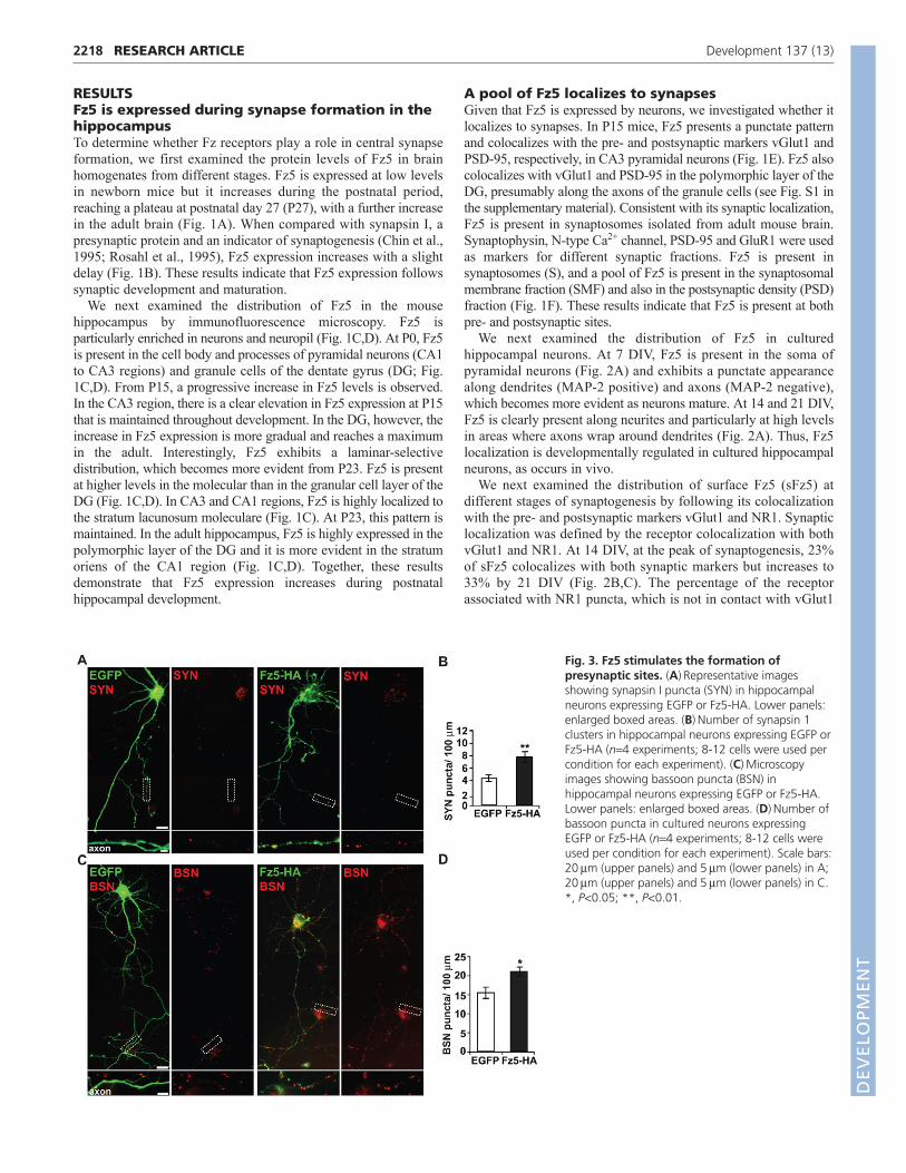
Fig. 3. Fz5 stimulates the formation ofpresynaptic sites. (A)Representative imagesshowing synapsin I puncta (SYN) in hippocampalneurons expressing EGFP or Fz5-HA. Lower panels:enlarged boxed areas. (B)Number of synapsin 1clusters in hippocampal neurons expressing EGFP orFz5-HA (n4 experiments; 8-12 cells were used percondition for each experiment). (C)Microscopyimages showing bassoon puncta (BSN) inhippocampal neurons expressing EGFP or Fz5-HA.Lower panels: enlarged boxed areas. (D)Number ofbassoon puncta in cultured neurons expressingEGFP or Fz5-HA (n4 experiments; 8-12 cells wereused per condition for each experiment). Scale bars:20m (upper panels) and 5m (lower panels) in A;20m (upper panels) and 5m (lower panels) in C.*, P<0.05; **, P<0.01.
DEVELO
PMENT

puncta, also increases during neuronal maturation (Fig. 2D).Although the number of sFz5 and extrasynaptic vGlut1 punctaincreases between 14 and 21 DIV, the percentage of sFz5 thatcolocalizes with extrasynaptic vGlut1 puncta does not significantlychange during this period (Fig. 2D). Together, our data suggest thatFz5 is present at synaptic sites and that its synaptic localizationincreases during synapse formation.
Fz5 stimulates the formation of synaptic sitesThe synaptic localization of sFz5 led us to test its possible rolein synaptogenesis by performing gain-of-function studies.Hippocampal neurons (9-11 DIV) expressing control EGFP or Fz5(Fz5-HA) were examined for the formation of presynaptic puncta(labelled with the synaptic vesicle protein synapsin I and the activezone protein bassoon) along axons (Fig. 3A,B). Axonal Fz5expression induces a 78% and a 36% increase in the number ofsynapsin I and bassoon puncta, respectively (Fig. 3C,D). Thus,expression of Fz5 in axons induces presynaptic differentiation in asimilar manner to the synaptogenic factor Wnt7a.
Fz5 functions as a receptor for Wnt7a duringsynapse formationWnt7a induces the formation of synapses in hippocampal neurons(Cerpa et al., 2008) but the receptor mediating this effect has notbeen identified. Therefore, we tested whether Fz5 functions as a
2219RESEARCH ARTICLEFz5 stimulates synapse formation
Fig. 4. Fz5 promotes synapse formation by binding to Wnt7a.(A)Wnt7a-HA binding (dark precipitate, black arrows) to Fz5CRD-myc-GPI (white arrows) but not to Dfz2CRD-myc-GPI-expressing Cos-7 cells(n3 experiments). Blue: Hoechst nuclei staining. (B)Synapsin I (SYN)and bassoon (BSN) clustering in cultured hippocampal neurons treatedwith Fz5CRD and/or Wnt7a. Tubulin: Tuj1. (C)Number of synapsin 1clusters in hippocampal neurons treated with Fz5CRD and/or Wnt7a(n3 experiments; 8-12 cells were used per condition for eachexperiment). EGFP-CM: conditioned medium from EFGP-transfectedQT6 cells. (D)Bassoon cluster number in cultured neurons treated withWnt7a and/or Fz5CRD (n3 experiments; 8-12 cells were used percondition for each experiment). Scale bars: 20m in A,B. *, P<0.05;**, P<0.01; ***, P<0.001; ****, P<0.0001.
Fig. 5. Fz5 knockdown abolishes Wnt7a-induced synapseformation. (A)Representative blots showing Fz5 and -tubulin (TUB)protein levels in NRK cells transfected with shRNAs to Fz5 (n3experiments). nc-shRNA, negative control shRNA; shRNA 1 to 4,Fz5shRNAs; untransf, untransfected cells; Fz5-shRNAs, mixture ofFz5shRNAs. (B)Confocal images show Fz5 expression in NRK cellstransfected with Fz5-shRNAs or nc-shRNAs (n3 experiments). (C)Fz5expression in cultured hippocampal neurons transfected with Fz5-shRNAs or nc-shRNA (n3 experiments). Insets show an enlarged viewof boxed areas. (D)Bassoon (BSN) puncta in cultured hippocampalneurons transfected with Fz5-shRNAs or nc-shRNAs and treated withWnt7a. (E)Number of bassoon clusters in hippocampal neurons treatedwith Wnt7a and transfected with Fz5-shRNAs or nc-shRNAs (n3experiments; 8-12 cells were used per condition for each experiment).Scale bars: 10m; 5 m in inset in C. *, P<0.05; ***, P<0.001;****, P<0.0001.
DEVELO
PMENT
2220
receptor for Wnt7a. Previous studies have shown that Wnt ligandsbind to the cysteine-rich domain (CRD) of Fz and that this domainis required for signalling (Liu et al., 2008; Povelones and Nusse,2005). Importantly, the CRD domain of Fz5 can block Wntsignalling during early embryonic patterning (Kemp et al., 2007;Liu et al., 2008). We expressed the CRD domains of human Fz5(Fz5CRD-GPI) and the Drosophila Dfz2 (Dfz2CRD-GPI) in Cos-7 cells. Transfected cells were then incubated with control orWnt7a-HA-containing conditioned medium. Immunocytochemicalassays reveal that Wnt7a binds to cells expressing Fz5CRD-GPIbut not Dfz2CRD-GPI or EGFP (Fig. 4A; data not shown). Theseresults demonstrate that Wnt7a specifically binds to the CRDdomain of Fz5.
We then investigated whether Fz5 functions as a receptor forWnt7a in hippocampal neurons. To test this, we examined whethera soluble version of the CRD domain of Fz5 (Fz5CRD) has aneffect on synaptogenesis. Hippocampal neurons were exposed tocontrol or Fz5CRD-containing conditional media either with orwithout recombinant Wnt7a for 16 hours. Fz5CRD alone decreasedthe number of synapsin I and bassoon puncta by 45% and 34%,respectively, when compared with controls (Fig. 4B-D). Thesefindings suggest that Fz5CRD blocks endogenous synaptogenicWnts. We next tested if Fz5CRD could block the ability ofexogenous Wnt7a to regulate synaptogenesis. Wnt7a alone induceda 52% and a 79% increase in the number of synapsin I and bassoonpuncta, respectively (Fig. 4B-D). By contrast, neurons exposed toboth Wnt7a and Fz5CRD exhibited a decrease of 82% in synapsinI puncta and 70% in bassoon puncta, when compared with Wnt7a-treated cells (Fig. 4B-D). Therefore, Fz5CRD completely blocksthe synaptogenic activity of Wnt7a, suggesting that Fz5 mediatesWnt7a function at synapses.
To confirm that Fz5 is a receptor for Wnt7a in hippocampalneurons, we knocked down its expression using shRNAs. The levelof knockdown was assessed by transfecting each of the fourdifferent shRNAs (Fz5-shRNA 1 to 4) or a mixture of the four intoNRK cells. A scrambled artificial sequence shRNA (nc-shRNA)was used as control. Expression of the EGFP gene present in theshRNA plasmids allowed identification of transfected cells.
Western blot analyses and immunocytochemistry indicated thatexpression of each single shRNA does not affect the levels ofendogenous Fz5 (Fig. 5A). By contrast, combined expression of thefour Fz5-shRNAs decreased the expression of endogenous Fz5when compared with untransfected cells or cells expressing nc-shRNA (Fig. 5A,B). In hippocampal neurons, the Fz5 shRNAmixture significantly decreased the level of endogenous Fz5,whereas nc-shRNA had no effect (Fig. 5C).
To test the effect of Fz5 knockdown in synapse formation,hippocampal neurons were transfected with Fz5-shRNAs or nc-shRNA. We found that Fz5-shRNAs decreased the number ofbassoon puncta by 23% when compared with nc-shRNA-expressing cells (Fig. 5D,E). These findings demonstrate thatendogenous Fz5 regulates synaptogenesis. A significant increase inthe number of Bassoon clusters (59%) was observed in thepresence of Wnt7a in nc-shRNA-transfected neurons (Fig. 5D,E).However, Fz5 knockdown abolished the ability of exogenousWnt7a to induce the bassoon clustering (Fig. 5D,E). These resultsdemonstrate that Fz5 functions as a receptor for Wnt7a duringsynapse formation in hippocampal neurons.
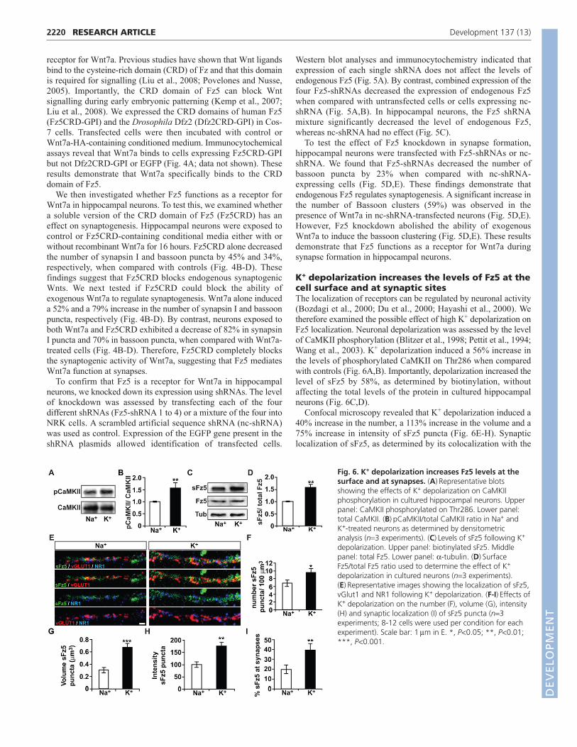
K+ depolarization increases the levels of Fz5 at thecell surface and at synaptic sitesThe localization of receptors can be regulated by neuronal activity(Bozdagi et al., 2000; Du et al., 2000; Hayashi et al., 2000). Wetherefore examined the possible effect of high K+ depolarization onFz5 localization. Neuronal depolarization was assessed by the levelof CaMKII phosphorylation (Blitzer et al., 1998; Pettit et al., 1994;Wang et al., 2003). K+ depolarization induced a 56% increase inthe levels of phosphorylated CaMKII on Thr286 when comparedwith controls (Fig. 6A,B). Importantly, depolarization increased thelevel of sFz5 by 58%, as determined by biotinylation, withoutaffecting the total levels of the protein in cultured hippocampalneurons (Fig. 6C,D).
Confocal microscopy revealed that K+ depolarization induced a40% increase in the number, a 113% increase in the volume and a75% increase in intensity of sFz5 puncta (Fig. 6E-H). Synapticlocalization of sFz5, as determined by its colocalization with the
RESEARCH ARTICLE Development 137 (13)
Fig. 6. K+ depolarization increases Fz5 levels at thesurface and at synapses. (A)Representative blotsshowing the effects of K+ depolarization on CaMKIIphosphorylation in cultured hippocampal neurons. Upperpanel: CaMKII phosphorylated on Thr286. Lower panel:total CaMKII. (B)pCaMKII/total CaMKII ratio in Na+ andK+-treated neurons as determined by densitometricanalysis (n3 experiments). (C)Levels of sFz5 following K+
depolarization. Upper panel: biotinylated sFz5. Middlepanel: total Fz5. Lower panel: -tubulin. (D)SurfaceFz5/total Fz5 ratio used to determine the effect of K+
depolarization in cultured neurons (n3 experiments).(E)Representative images showing the localization of sFz5,vGlut1 and NR1 following K+ depolarization. (F-I)Effects ofK+ depolarization on the number (F), volume (G), intensity(H) and synaptic localization (I) of sFz5 puncta (n3experiments; 8-12 cells were used per condition for eachexperiment). Scale bar: 1m in E. *, P<0.05; **, P<0.01;***, P<0.001.
DEVELO
PMENT

synaptic markers vGlut1 and NR1, demonstrated thatdepolarization increases the level of sFz5 at synapses from 20% to39% (Fig. 6E,I). Together, these results demonstrate thatdepolarization not only increases the trafficking of Fz5 to the cellsurface but also stimulates its mobilization to synapses.
Different patterns of electrical stimulationdifferentially modulate Fz5 levels at the cellsurface and synaptic sitesTo further investigate the role of neuronal activity on Fz5localization, we examined the effect of patterned electricalstimulation, shown to regulate the distribution of a number ofreceptors (Du et al., 2000; Hayashi et al., 2000; Rodriguez et al.,2008; Shi et al., 1999). We used two different electrical stimuli toinduce neuronal activity. Intermittent high-frequency stimulation(HFS), where a regular stimulation pattern comprising trains of 20stimuli at 50 Hz (10 millisecond interval) delivered every 5 secondswas used to mimic average neuronal activity in vivo (an average of4 action potentials per second) (Schoenbaum et al., 1999; Attwelland Laughlin, 2001). We also used low-frequency (LFS)stimulation at 4 Hz (250 millisecond interval), known to induceLTD (Zamani et al., 2000; Dudek and Bear, 1992). Neuronalactivation induced by HFS was confirmed by a 74% increase in thelevels of CaMKII Thr286 phosphorylation when compared withunstimulated cells (Fig. 7A,B). Then, sFz5 levels were measuredby biotinylation after HFS or LFS. Surface Fz5 increased by 52%in HFS cells, whereas LFS induced a 35% decrease in sFz5,without changing the total level of Fz5 (Fig. 7C,D). These resultsshow that patterned electrical stimulation can differentially regulatethe amount of Fz5 present at the cell surface.
We next examined the effect of HFS and LFS on sFz5 byconfocal microscopy. These two stimuli exert opposing effects onsFz5 localization. HFS increased the number of sFz5 puncta by47%, the volume by 90% and their intensity by 57% (Fig. 7E-H),whereas LFS decreased the number of sFz5 puncta by 61% withoutchanging their volume or intensity (Fig. 7E-H). In addition, HFSincreased the amount of sFz5 at synapses from 21% to 43%,whereas LFS decreased synaptic sFz5 to 15% (Fig. 7E,I). When thenumber of synapses was analyzed (as determined by thecolocalization of vGlut1 and NR1), we found that HFS increasedthe number of synapses by 152%, whereas LFS decreased thesynapse number by 49% (Fig. 7E,J), consistent with previousreports (Bastrikova et al., 2008; Antonova et al., 2001; Bozdagi etal., 2000). Under basal conditions, 45% of synapses containedsFz5, whereas after HFS, 65% of synapses contained sFz5 (Fig.7E,K). Following LFS, however, only 20% of synapses containedsFz5 (Fig. 7E,K). These results demonstrate that neuronal activitymodulates the localization of sFz5 to synapses and that the outcomedepends on the stimulation frequency used. Neuronal activitytriggered by HFS enhances the trafficking of Fz5 to the cell surfaceand its localization to synaptic sites. By contrast, LFS significantlyreduces the amount of sFz5 and impairs its localization to synapses.
Wnts contribute to HFS-induced synapseformation and synaptic localization of Fz5Neuronal activity regulates the expression of Wnts in hippocampalneurons (Wayman et al., 2006; Yu and Malenka, 2003; Gogolla etal., 2009). Therefore, HFS could modulate the mobilization of Fz5through changes in endogenous Wnt proteins. To test thishypothesis, we blocked endogenous Wnts that bind to Fz5 by usingFz5CRD. Cultured neurons were exposed to control or Fz5CRD-containing conditional media during HFS. Surface biotinylation
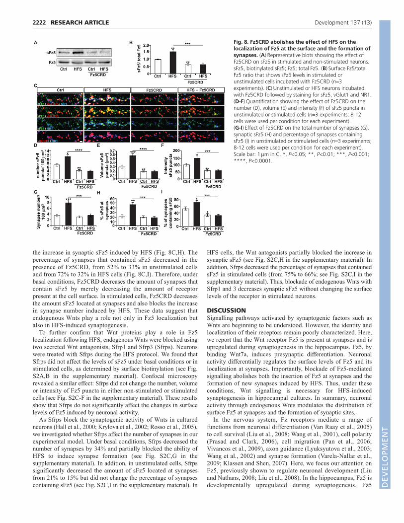
indicated that, in unstimulated cells, Fz5CRD decreased theamount of receptor present at the surface by 40%, whereas itcompletely abolished the increase in sFz5 induced by HFS withoutaffecting the total level of the protein (Fig. 8A,B). Using confocalmicroscopy, we found that, under basal conditions, Fz5CRDdecreases the number, volume and intensity of sFz5 puncta (by41%, 47% and 41%, respectively), while it completely blocks theeffect of HFS on sFz5 levels (Fig. 8C-F).
As Fz5CRD blocked Wnt-mediated synaptogenesis after 16hours of exposure (Fig. 4), we tested whether it affects theincrease in synapse number induced after 1 hour of HFS. Inunstimulated cells, Fz5CRD did not significantly change thenumber of synapses (Fig. 8C,G). By contrast, Fz5CRD preventedthe increase in synapse number that follows HFS (Fig. 8C,G),suggesting that Wnts participate in the formation of synapsesinduced by activity. In addition, Fz5CRD completely abolished
2221RESEARCH ARTICLEFz5 stimulates synapse formation
Fig. 7. High-frequency stimulation and low-frequencystimulation have opposite effects on Fz5 levels at the surfaceand at synapses. (A)Effect of high-frequency stimulation (HFS) onCaMKII phosphorylation in cultured hippocampal neurons. Upperpanel: CaMKII phosphorylated on Thr286. Lower panel: total CaMKII.(B)Quantification showing the level of phosphorylation ofThr286CaMKII after normalization to total CaMKII. (C)Surface Fz5levels following HFS and low-frequency stimulation (LFS). sFz5,biotinylated sFz5; Fz5, total Fz5; Tub, -tubulin. (D)Surface Fz5/totalFz5 ratio calculated to show the effects of HFS and LFS on Fz5 levels.(E)Representative images of sFz5, vGlut1 and NR1 staining followingHFS and LFS. (F-I)Effects of HFS and LFS on the number (F), volume (G),intensity (H) and synaptic localization (I) of sFz5 puncta. (n3experiments; 8-12 cells were used per condition for each experiment).(J)Quantification of synapse number following HFS and LFS (n3experiments; 8-12 cells were used per condition for each experiment).(K)Percentage of synapses containing sFz5 after HFS and LFS (n3experiments; 8-12 cells were used per condition for each experiment).Scale bar: 2m in E. *, P<0.05; **, P<0.01; ***, P<0.001. D
EVELO
PMENT
2222
the increase in synaptic sFz5 induced by HFS (Fig. 8C,H). Thepercentage of synapses that contained sFz5 decreased in thepresence of Fz5CRD, from 52% to 33% in unstimulated cellsand from 72% to 32% in HFS cells (Fig. 8C,I). Therefore, underbasal conditions, Fz5CRD decreases the amount of synapses thatcontain sFz5 by merely decreasing the amount of receptorpresent at the cell surface. In stimulated cells, Fz5CRD decreasesthe amount sFz5 located at synapses and also blocks the increasein synapse number induced by HFS. These data suggest thatendogenous Wnts play a role not only in Fz5 localization butalso in HFS-induced synaptogenesis.
To further confirm that Wnt proteins play a role in Fz5localization following HFS, endogenous Wnts were blocked usingtwo secreted Wnt antagonists, Sfrp1 and Sfrp3 (Sfrps). Neuronswere treated with Sfrps during the HFS protocol. We found thatSfrps did not affect the levels of sFz5 under basal conditions or instimulated cells, as determined by surface biotinylation (see Fig.S2A,B in the supplementary material). Confocal microscopyrevealed a similar effect: Sfrps did not change the number, volumeor intensity of Fz5 puncta in either non-stimulated or stimulatedcells (see Fig. S2C-F in the supplementary material). These resultsshow that Sfrps do not significantly affect the changes in surfacelevels of Fz5 induced by neuronal activity.
As Sfrps block the synaptogenic activity of Wnts in culturedneurons (Hall et al., 2000; Krylova et al., 2002; Rosso et al., 2005),we investigated whether Sfrps affect the number of synapses in ourexperimental model. Under basal conditions, Sfrps decreased thenumber of synapses by 34% and partially blocked the ability ofHFS to induce synapse formation (see Fig. S2C,G in thesupplementary material). In addition, in unstimulated cells, Sfrpssignificantly decreased the amount of sFz5 located at synapsesfrom 21% to 15% but did not change the percentage of synapsescontaining sFz5 (see Fig. S2C,I in the supplementary material). In
HFS cells, the Wnt antagonists partially blocked the increase insynaptic sFz5 (see Fig. S2C,H in the supplementary material). Inaddition, Sfrps decreased the percentage of synapses that containedsFz5 in stimulated cells (from 75% to 66%; see Fig. S2C,I in thesupplementary material). Thus, blockade of endogenous Wnts withSfrp1 and 3 decreases synaptic sFz5 without changing the surfacelevels of the receptor in stimulated neurons.
DISCUSSIONSignalling pathways activated by synaptogenic factors such asWnts are beginning to be understood. However, the identity andlocalization of their receptors remain poorly characterized. Here,we report that the Wnt receptor Fz5 is present at synapses and isupregulated during synaptogenesis in the hippocampus. Fz5, bybinding Wnt7a, induces presynaptic differentiation. Neuronalactivity differentially regulates the surface levels of Fz5 and itslocalization at synapses. Importantly, blockade of Fz5-mediatedsignalling abolishes both the insertion of Fz5 at synapses and theformation of new synapses induced by HFS. Thus, under theseconditions, Wnt signalling is necessary for HFS-inducedsynaptogenesis in hippocampal cultures. In summary, neuronalactivity through endogenous Wnts modulates the distribution ofsurface Fz5 at synapses and the formation of synaptic sites.
In the nervous system, Fz receptors mediate a range offunctions from neuronal differentiation (Van Raay et al., 2005)to cell survival (Liu et al., 2008; Wang et al., 2001), cell polarity(Prasad and Clark, 2006), cell migration (Pan et al., 2006;Vivancos et al., 2009), axon guidance (Lyuksyutova et al., 2003;Wang et al., 2002) and synapse formation (Varela-Nallar et al.,2009; Klassen and Shen, 2007). Here, we focus our attention onFz5, previously shown to regulate neuronal development (Liuand Nathans, 2008; Liu et al., 2008). In the hippocampus, Fz5 isdevelopmentally upregulated during synaptogenesis. Fz5
RESEARCH ARTICLE Development 137 (13)
Fig. 8. Fz5CRD abolishes the effect of HFS on thelocalization of Fz5 at the surface and the formation ofsynapses. (A)Representative blots showing the effect ofFz5CRD on sFz5 in stimulated and non-stimulated neurons.sFz5, biotinylated sFz5; Fz5; total Fz5. (B)Surface Fz5/totalFz5 ratio that shows sFz5 levels in stimulated orunstimulated cells incubated with Fz5CRD (n3experiments). (C)Unstimulated or HFS neurons incubatedwith Fz5CRD followed by staining for sFz5, vGlut1 and NR1.(D-F)Quantification showing the effect of Fz5CRD on thenumber (D), volume (E) and intensity (F) of sFz5 puncta inunstimulated or stimulated cells (n3 experiments; 8-12cells were used per condition for each experiment).(G-I)Effect of Fz5CRD on the total number of synapses (G),synaptic sFz5 (H) and percentage of synapses containingsFz5 (I) in unstimulated or stimulated cells (n3 experiments;8-12 cells were used per condition for each experiment).Scale bar: 1m in C. *, P<0.05; **, P<0.01; ***, P<0.001;****, P<0.0001.
DEVELO
PMENT

exhibits a punctate distribution in axons, where it colocalizeswith vGlut1, and in dendrites, where it colocalizes with PSD-95and NMDA receptors. Fz5 is also present at sites where both pre-and postsynaptic markers colocalize. Consistently, Fz5 is foundin synaptosomes, the SMF and the PSD. Therefore, Fz5 ispresent at both pre- and postsynaptic sites at the peak ofsynaptogenesis, where it could mediate bidirectional Wntsignalling to regulate synaptic assembly.
Fz5 is required for Wnt7a-mediated synapse formation inhippocampal neurons. Several pieces of evidence suggest thatFz5 functions as a receptor for Wnt7a. First, Wnt7a (Davis et al.,2008) and Fz5 are expressed in the hippocampus during the peakof synaptogenesis. Second, Wnt7a binds to the CRD domain ofFz5, crucial for signalling (Povelones and Nusse, 2005). Third,axonal Fz5 expression induces the clustering of presynapticmarkers as observed with Wnt7a (Ahmad-Annuar et al., 2006;Cerpa et al., 2008). Fourth, Fz5 knockdown or Fz5CRD blocksthe ability of Wnt7a to induce presynaptic differentiation. Fz5might not be the only receptor that mediates the synaptogeniceffect of Wnt7a, as Fz1 also stimulates synapse formation inhippocampal neurons (Varela-Nallar et al., 2009). However, ourfindings strongly support the idea that endogenous Fz5 functionsas a receptor for Wnt7a to regulate synapse formation inhippocampal neurons.
Neuronal activity regulates the localization of transmembraneproteins to sub-cellular compartments. For example, activityincreases the level of surface TrkB receptors (Du et al., 2000;
Meyer-Franke et al., 1998) and the mobilization of N-cadherinto synapses (Bozdagi et al., 2000; Tanaka et al., 2000). Neuronalactivity also drives the synaptic incorporation of AMPAR andNMDAR and their lateral diffusion between synaptic andextrasynaptic sites (Lau and Zukin, 2007; Newpher and Ehlers,2008; Heynen et al., 2000). Here, we show that neuronal activityhas a profound effect on the distribution and synapticlocalization of Fz5 (Fig. 9). Depolarization and HFS increase thelevels of Fz5 at the cell surface and the percentage of synapsesthat contain Fz5. By contrast, LFS decreases the amount of Fz5at the surface and the percentage of synaptic Fz5. The totallevels of Fz5 remain unaffected, suggesting that neuronal activityregulates the trafficking of the receptor from intracellular storesto the plasma membrane and to synaptic sites. Importantly, inthese experiments, the patterns of HFS and LFS were chosensuch that they both deliver, on average, 4 stimulation pulses persecond. As the cultures received the same number of stimuli, itis clear that distinct temporal patterns of synaptic activation,such as HFS and LFS, modulate the insertion of Fz5 receptors inopposing ways.
How does neuronal activity induce Fz5 recruitment tosynapses? Activity has previously been shown to regulate theexpression and/or secretion of Wnt proteins. Depolarizationstimulates Wnt2 transcription in cultured hippocampal neurons,resulting in increased dendritic arborization (Wayman et al.,2006). The Wnt inhibitor Dkk1 blocks depolarization-induceddendrite growth in cultured hippocampal neurons (Yu andMalenka, 2003). Moreover, experience-induced plasticityenhances Wnt7a/b expression, which regulates hippocampalaxon remodelling (Gogolla et al., 2009). At the Drosophilaneuromuscular junction, neuronal activity increases the secretionof Wg to stimulate pre- and postsynaptic assembly as well asdendritic refinement (Singh et al., 2010; Ataman et al., 2008).Thus, neuronal activity modulates the levels of Wnts, which inturn could regulate Fz receptor localization. Indeed, Wnt proteinscontrol the localization of Fz receptors in neurons (Ataman et al.,2008; Klassen and Shen, 2007). However, a role for neuronalactivity in Fz localization to synapses has not been documented.By blocking Wnts using two different approaches, wedemonstrate that secreted Wnts contribute to the regulation ofsurface Fz5 localization to synapses elicited by neuronal activity.
Neuronal activity regulates the formation and maintenance ofsynapses (Craig et al., 2006; Blankenship and Feller, 2010). Here,we demonstrate that Wnts and Fz5 are necessary for stimulation-evoked synaptogenesis in 14 DIV hippocampal cultures, asblockade of Fz5 signalling with Fz5CRD completely abolishes thesynaptogenic effect of HFS. We propose a model where neuronalactivity modulates the secretion of Wnts, which then stimulate theformation of synapses through binding to Fz5. Our results providea link between neuronal activity, Wnt-Fz5 signalling and synapseformation.
AcknowledgementsWe thank Drs Jeremy Nathans and Xi He for constructs. We also thankmembers of our laboratory for useful discussion and comments on themanuscript. The Wellcome Trust and MRC supported this work. Deposited inPMC for release after 6 months.
Competing interests statementThe authors declare no competing financial interests.
Supplementary materialSupplementary material for this article is available athttp://dev.biologists.org/lookup/suppl/doi:10.1242/dev.046722/-/DC1
2223RESEARCH ARTICLEFz5 stimulates synapse formation
Fig. 9. Regulation of sFz5 localization and synapse formation byneuronal activity and secreted Wnts. The insertion of Fz5 to synapsesis differentially regulated by distinct temporal patterns of synapticactivation. LFS drives Fz5 out of synapses, whereas HFS, through amechanism that involves secreted Wnts, increases the levels of Fz5 at thesurface and at synaptic sites. In addition, Wnt-Fz5 signalling participatesin the formation of new synapses induced by neuronal activity.
DEVELO
PMENT
2224
ReferencesAhmad-Annuar, A., Ciani, L., Simeonidis, I., Herreros, J., Fredj, N. B.,
Rosso, S. B., Hall, A., Brickley, S. and Salinas, P. C. (2006). Signaling acrossthe synapse: a role for Wnt and Dishevelled in presynaptic assembly andneurotransmitter release. J. Cell Biol. 174, 127-139.
Angers, S. and Moon, R. T. (2009). Proximal events in Wnt signal transduction.Nat. Rev. Mol. Cell Biol. 10, 468-477.
Antonova, I., Arancio, O., Trillat, A. C., Wang, H. G., Zablow, L., Udo, H.,Kandel, E. R. and Hawkins, R. D. (2001). Rapid increase in clusters ofpresynaptic proteins at onset of long-lasting potentiation. Science 294, 1547-1550.
Ataman, B., Ashley, J., Gorczyca, M., Ramachandran, P., Fouquet, W.,Sigrist, S. J. and Budnik, V. (2008). Rapid activity-dependent modifications insynaptic structure and function require bidirectional Wnt signaling. Neuron 57,705-718.
Attwell, D. and Laughlin, S. B. (2001). An energy budget for signaling in thegrey matter of the brain. J. Cereb. Blood Flow Metab. 21, 1133-1145.
Bastrikova, N., Gardner, G. A., Reece, J. M., Jeromin, A. and Dudek, S. M.(2008). Synapse elimination accompanies functional plasticity in hippocampalneurons. Proc. Natl. Acad. Sci. USA 105, 3123-3127.
Blankenship, A. G. and Feller, M. B. (2010). Mechanisms underlyingspontaneous patterned activity in developing neural circuits. Nat. Rev.Neurosci. 11, 18-29.
Blitzer, R. D., Connor, J. H., Brown, G. P., Wong, T., Shenolikar, S., Iyengar,R. and Landau, E. M. (1998). Gating of CaMKII by cAMP-regulated proteinphosphatase activity during LTP. Science 280, 1940-1942.
Bozdagi, O., Shan, W., Tanaka, H., Benson, D. L. and Huntley, G. W. (2000).Increasing numbers of synaptic puncta during late-phase LTP: N-cadherin issynthesized, recruited to synaptic sites, and required for potentiation. Neuron28, 245-259.
Carroll, R. C., Beattie, E. C., von Zastrow, M. and Malenka, R. C. (2001).Role of AMPA receptor endocytosis in synaptic plasticity. Nat. Rev. Neurosci. 2,315-324.
Cerpa, W., Godoy, J. A., Alfaro, I., Farias, G. G., Metcalfe, M. J.,Fuentealba, R., Bonansco, C. and Inestrosa, N. C. (2008). Wnt-7amodulates the synaptic vesicle cycle and synaptic transmission in hippocampalneurons. J. Biol. Chem. 283, 5918-5927.
Chin, L. S., Li, L., Ferreira, A., Kosik, K. S. and Greengard, P. (1995).Impairment of axonal development and of synaptogenesis in hippocampalneurons of synapsin I-deficient mice. Proc. Natl. Acad. Sci. USA 92, 9230-9234.
Ciani, L. and Salinas, P. C. (2005). WNTs in the vertebrate nervous system: frompatterning to neuronal connectivity. Nat. Rev. Neurosci. 6, 351-362.
Cohen, R. S., Blomberg, F., Berzins, K. and Siekevitz, P. (1977). The structureof postsynaptic densities isolated from dog cerebral cortex. I. Overallmorphology and protein composition. J. Cell Biol. 74, 181-203.
Craig, A. M., Graf, E. R. and Linhoff, M. W. (2006). How to build a centralsynapse: clues from cell culture. Trends Neurosci. 29, 8-20.
Davis, E. K., Zou, Y. and Ghosh, A. (2008). Wnts acting through canonical andnoncanonical signaling pathways exert opposite effects on hippocampalsynapse formation. Neural Dev. 3, 32.
Du, J., Feng, L., Yang, F. and Lu, B. (2000). Activity- and Ca(2+)-dependentmodulation of surface expression of brain-derived neurotrophic factorreceptors in hippocampal neurons. J. Cell Biol. 150, 1423-1434.
Du, J., Feng, L., Zaitsev, E., Je, H. S., Liu, X. W. and Lu, B. (2003). Regulationof TrkB receptor tyrosine kinase and its internalization by neuronal activity andCa2+ influx. J. Cell Biol. 163, 385-395.
Dudek, S. M. and Bear, M. F. (1992). Homosynaptic long-term depression inarea CA1 of hippocampus and effects of N-methyl-D-aspartate receptorblockade. Proc. Natl. Acad. Sci. USA 89, 4363-4367.
Fox, M. A. and Umemori, H. (2006). Seeking long-term relationship: axon andtarget communicate to organize synaptic differentiation. J. Neurochem. 97,1215-1231.
Friedman, H. V., Bresler, T., Garner, C. C. and Ziv, N. E. (2000). Assembly ofnew individual excitatory synapses: time course and temporal order of synapticmolecule recruitment. Neuron 27, 57-69.
Gogolla, N., Galimberti, I., Deguchi, Y. and Caroni, P. (2009). Wnt signalingmediates experience-related regulation of synapse numbers and mossy fiberconnectivities in the adult hippocampus. Neuron 62, 510-525.
Hall, A. C., Lucas, F. R. and Salinas, P. C. (2000). Axonal remodeling andsynaptic differentiation in the cerebellum is regulated by WNT-7a signaling.Cell 100, 525-535.
Hardin, J. and King, R. S. (2008). The long and the short of Wnt signaling in C.elegans. Curr. Opin. Genet. Dev. 18, 362-367.
Hayashi, Y., Shi, S. H., Esteban, J. A., Piccini, A., Poncer, J. C. and Malinow,R. (2000). Driving AMPA receptors into synapses by LTP and CaMKII:requirement for GluR1 and PDZ domain interaction. Science 287, 2262-2267.
Heynen, A. J., Quinlan, E. M., Bae, D. C. and Bear, M. F. (2000). Bidirectional,activity-dependent regulation of glutamate receptors in the adulthippocampus in vivo. Neuron 28, 527-536.
Hua, J. Y. and Smith, S. J. (2004). Neural activity and the dynamics of centralnervous system development. Nat. Neurosci. 7, 327-332.
Kemp, C. R., Willems, E., Wawrzak, D., Hendrickx, M., Agbor Agbor, T. andLeyns, L. (2007). Expression of Frizzled5, Frizzled7, and Frizzled10 duringearly mouse development and interactions with canonical Wnt signaling. Dev.Dyn. 236, 2011-2019.
Klassen, M. P. and Shen, K. (2007). Wnt signaling positions neuromuscularconnectivity by inhibiting synapse formation in C. elegans. Cell 130, 704-716.
Krylova, O., Herreros, J., Cleverley, K. E., Ehler, E., Henriquez, J. P.,Hughes, S. M. and Salinas, P. C. (2002). WNT-3, expressed by motoneurons,regulates terminal arborization of neurotrophin-3-responsive spinal sensoryneurons. Neuron 35, 1043-1056.
Lau, C. G. and Zukin, R. S. (2007). NMDA receptor trafficking in synapticplasticity and neuropsychiatric disorders. Nat. Rev. Neurosci. 8, 413-426.
Liu, C. and Nathans, J. (2008). An essential role for frizzled 5 in mammalianocular development. Development 135, 3567-3576.
Liu, C., Wang, Y., Smallwood, P. M. and Nathans, J. (2008). An essential rolefor Frizzled5 in neuronal survival in the parafascicular nucleus of the thalamus.J. Neurosci. 28, 5641-5653.
Lyuksyutova, A. I., Lu, C. C., Milanesio, N., King, L. A., Guo, N., Wang, Y.,Nathans, J., Tessier-Lavigne, M. and Zou, Y. (2003). Anterior-posteriorguidance of commissural axons by Wnt-frizzled signaling. Science 302, 1984-1988.
McAllister, A. K. (2007). Dynamic aspects of CNS synapse formation. Annu.Rev. Neurosci. 30, 425-450.
Meyer-Franke, A., Wilkinson, G. A., Kruttgen, A., Hu, M., Munro, E.,Hanson, M. G., Jr, Reichardt, L. F. and Barres, B. A. (1998). Depolarizationand cAMP elevation rapidly recruit TrkB to the plasma membrane of CNSneurons. Neuron 21, 681-693.
Newpher, T. M. and Ehlers, M. D. (2008). Glutamate receptor dynamics indendritic microdomains. Neuron 58, 472-497.
Packard, M., Koo, E. S., Gorczyca, M., Sharpe, J., Cumberledge, S. andBudnik, V. (2002). The Drosophila Wnt, wingless, provides an essential signalfor pre- and postsynaptic differentiation. Cell 111, 319-330.
Pan, C. L., Howell, J. E., Clark, S. G., Hilliard, M., Cordes, S., Bargmann, C.I. and Garriga, G. (2006). Multiple Wnts and frizzled receptors regulateanteriorly directed cell and growth cone migrations in Caenorhabditis elegans.Dev. Cell 10, 367-377.
Pettit, D. L., Perlman, S. and Malinow, R. (1994). Potentiated transmissionand prevention of further LTP by increased CaMKII activity in postsynaptichippocampal slice neurons. Science 266, 1881-1885.
Povelones, M. and Nusse, R. (2005). The role of the cysteine-rich domain ofFrizzled in Wingless-Armadillo signaling. EMBO J. 24, 3493-3503.
Prasad, B. C. and Clark, S. G. (2006). Wnt signaling establishes anteroposteriorneuronal polarity and requires retromer in C. elegans. Development 133,1757-1766.
Rodriguez, J. J., Dallerac, G. M., Tabuchi, M., Davies, H. A., Colyer, F. M.,Stewart, M. G. and Doyere, V. (2008). N-methyl-D-aspartate receptorindependent changes in expression of polysialic acid-neural cell adhesionmolecule despite blockade of homosynaptic long-term potentiation andheterosynaptic long-term depression in the awake freely behaving rat dentategyrus. Neuron Glia Biol. 4, 169-178.
Rosahl, T. W., Spillane, D., Missler, M., Herz, J., Selig, D. K., Wolff, J. R.,Hammer, R. E., Malenka, R. C. and Sudhof, T. C. (1995). Essential functionsof synapsins I and II in synaptic vesicle regulation. Nature 375, 488-493.
Rosso, S. B., Sussman, D., Wynshaw-Boris, A. and Salinas, P. C. (2005). Wntsignaling through Dishevelled, Rac and JNK regulates dendritic development.Nat. Neurosci. 8, 34-42.
Rulifson, E. J., Wu, C. H. and Nusse, R. (2000). Pathway specificity by thebifunctional receptor frizzled is determined by affinity for wingless. Mol. Cell6, 117-126.
Salinas, P. C. and Zou, Y. (2008). Wnt signaling in neural circuit assembly.Annu. Rev. Neurosci. 31, 339-358.
Schoenbaum, G., Chiba, A. A. and Gallagher, M. (1999). Neural encoding inorbitofrontal cortex and basolateral amygdala during olfactory discriminationlearning. J. Neurosci. 19, 1876-1884.
Shi, S. H., Hayashi, Y., Petralia, R. S., Zaman, S. H., Wenthold, R. J.,Svoboda, K. and Malinow, R. (1999). Rapid spine delivery and redistributionof AMPA receptors after synaptic NMDA receptor activation. Science 284,1811-1816.
Singh, A. P., Vijayraghavan, K. and Rodrigues, V. (2010). Dendriticrefinement of an identified neuron in the Drosophila CNS is regulated byneuronal activity and Wnt signaling. Development 137, 1351-1360.
Speese, S. D. and Budnik, V. (2007). Wnts: up-and-coming at the synapse.Trends Neurosci. 30, 268-275.
Tanaka, H., Shan, W., Phillips, G. R., Arndt, K., Bozdagi, O., Shapiro, L.,Huntley, G. W., Benson, D. L. and Colman, D. R. (2000). Molecularmodification of N-cadherin in response to synaptic activity. Neuron 25, 93-107.
RESEARCH ARTICLE Development 137 (13)
DEVELO
PMENT
van Amerongen, R., Mikels, A. and Nusse, R. (2008). Alternative wntsignaling is initiated by distinct receptors. Sci. Signal. 1, re9.
Van Raay, T. J., Moore, K. B., Iordanova, I., Steele, M., Jamrich, M., Harris,W. A. and Vetter, M. L. (2005). Frizzled 5 signaling governs the neuralpotential of progenitors in the developing Xenopus retina. Neuron 46, 23-36.
Varela-Nallar, L., Grabowski, C. P., Alfaro, I. E., Alvarez, A. R. andInestrosa, N. C. (2009). Role of the Wnt receptor Frizzled-1 in presynapticdifferentiation and function. Neural Dev. 4, 41.
Vivancos, V., Chen, P., Spassky, N., Qian, D., Dabdoub, A., Kelley, M.,Studer, M. and Guthrie, S. (2009). Wnt activity guides facial branchiomotorneuron migration, and involves the PCP pathway and JNK and ROCK kinases.Neural Dev. 4, 7.
Wang, H., Shimizu, E., Tang, Y. P., Cho, M., Kyin, M., Zuo, W., Robinson, D.A., Alaimo, P. J., Zhang, C., Morimoto, H. et al. (2003). Inducible proteinknockout reveals temporal requirement of CaMKII reactivation for memoryconsolidation in the brain. Proc. Natl. Acad. Sci. USA 100, 4287-4292.
Wang, Y., Huso, D., Cahill, H., Ryugo, D. and Nathans, J. (2001). Progressivecerebellar, auditory, and esophageal dysfunction caused by targeted disruptionof the frizzled-4 gene. J. Neurosci. 21, 4761-4771.
Wang, Y., Thekdi, N., Smallwood, P. M., Macke, J. P. and Nathans, J. (2002).Frizzled-3 is required for the development of major fiber tracts in the rostralCNS. J. Neurosci. 22, 8563-8573.
Wayman, G. A., Impey, S., Marks, D., Saneyoshi, T., Grant, W. F., Derkach,V. and Soderling, T. R. (2006). Activity-dependent dendritic arborizationmediated by CaM-kinase I activation and enhanced CREB-dependenttranscription of Wnt-2. Neuron 50, 897-909.
Yu, X. and Malenka, R. C. (2003). Beta-catenin is critical for dendriticmorphogenesis. Nat. Neurosci. 6, 1169-1177.
Zamani, M. R., Desmond, N. L. and Levy, W. B. (2000). Estradiol modulateslong-term synaptic depression in female rat hippocampus. J. Neurophysiol. 84,1800-1808.
2225RESEARCH ARTICLEFz5 stimulates synapse formation
DEVELO
PMENT