Embed Size (px)
Citation preview
Foster, A., Mitchell, S., & Wall, R. (2015). Cattle ectoparasites in GreatBritain. Cattle Practice, 23(2), 280-287.
Publisher's PDF, also known as Version of record
License (if available):Unspecified
Link to publication record in Explore Bristol ResearchPDF-document
This is the final published version of the article (version of record). It first appeared via BAVC. Please refer toany applicable terms of use of the publisher.
University of Bristol - Explore Bristol ResearchGeneral rights
This document is made available in accordance with publisher policies. Please cite only the publishedversion using the reference above. Full terms of use are available:http://www.bristol.ac.uk/pure/about/ebr-terms
CATTLE PRACTICE VOLUME 23 PART 2
2015 280
Cattle ectoparasites in Great BritainFoster, A.1, Mitchell, S.2, Wall, R.3,1School of Veterinary Sciences, University of Bristol, Langford House, Langford, BS40 5DU 2Carmarthen Veterinary Investigation Centre, Animal and Plant Health Agency, Job’s Well Rd, Johnstown, Carmarthen, SA31 3EZ3Veterinary Parasitology and Ecology Group, University of Bristol, Bristol Life Sciences Building, Bristol, BS8 1TQ
ABSTRACTEctoparasites are almost ubiquitous on British cattle, reflecting the success of these parasites at retaining a residual population in the national herd.
Lice infestation is common and may be associated with significant disease especially in young moribund calves. The chewing louse Bovicola bovis is a particular challenge to eradicate given its limited response to various therapies and emerging evidence of reduced susceptibility to pyrethroids.
Chorioptes is the most common cause of mange in cattle and given its surface feeding habits can be difficult to eradicate with current treatments.
Psoroptic mange has re-emerged in British cattle in recent years and while the prevalence of infestation is low this parasite poses a significant challenge for treatment especially in dairy cattle.
Scabies is rare in British cattle but, like psoroptic mange, can cause significant pruritus and skin disease. Furthermore it is a potential zoonosis.
Diagnosis of such ectoparasites is usually made by interpretation of signs of skin disease; definitive diagnosis requires microscopic examination of the ectoparasite which can more accurately inform the implementation of control measures.
In the future, control measures for such ectoparasites may need to move away from the reliance on synthetic pyrethroids and macrocyclic lactones, to consider alternative topical agents.
skin, rather than only at the margins. Scabies mites can be very difficult to find on
an infested animal and repeated superficial skin scrapes from the margins of affected areas (away from crusts and erosions) may enable detection.
Severe mange due to Chorioptes, Psoroptes or Sarcoptes sp mites can only be definitively diagnosed through microscopic examination of skin scrape material; should be examined under low power with liquid paraffin as the mounting agent and with a cover slip. When submitting samples to a diagnostic laboratory scrape material should be placed in a clean bijou or universal container without any liquid paraffin.
In animals with mange the skin lesions can be extensive and severe; consequently, mites are more likely to be detected at the margins of affected area; to facilitate detection through skin scraping it may be helpful to clip away the hair coat at the margin of affected areas.
A note on the management of cattle ectoparasites The selection of appropriate measures for the control of ectoparasites requires:
• careful assessment of the nature of the clinical problems they cause.
INTRODUCTIONThe aim of this article is to briefly review the lice and mite species that may be found on British cattle. This article reflects the information published on the COWS website (http://www.cattleparasites.org.uk/) about the control of ectoparasites on cattle (written by Richard Wall).
A note on diagnostic samplingLiceWhen collecting samples for microscopic examination it can be helpful to clip a small window in the hair coat to reach the skin and use a blunt scalpel blade to scrape the lice, eggs attached to hairs and skin scales. These can be examined at the practice laboratory or on farm, if facilities permit, using liquid paraffin to mount the material and then examine under low power with a cover slip. Skin scrape material sent to a diagnostic laboratory should be placed in a clean container (bijou or universal container) with no liquid paraffin.
MitesPsoroptes mites may be visible to the naked eye or with a hand lens. They are usually numerous and may be present throughout an area of affected
CATTLE PRACTICE VOLUME 23 PART 2
2015 281
• correct identification of the parasite an understanding of the epidemiology, phenology and life cycle of the parasites.
• an assessment of the cost/benefit of the intended outcome.
• a careful assessment of the current resistance status of the parasite in question to the insecticides and acaricides available.
When considering the particular parasites it may be construed that the ubiquitous lice and Chorioptes mites are difficult to eradicate and in many cases do not cause substantial disease that can have an impact on production (milk or meat) or hide quality.
Psoroptic and sarcoptic mange are currently uncommon; even so, their introduction to a herd can have a significant impact on cattle welfare because these mites usually lead to severe pruritus and secondary skin changes; furthermore, scabies is a potential zoonosis. The risk of introduction of such mites through new stock coming onto a farm can be ameliorated somewhat by isolation and treatment before introduction to the herd.
Furthermore, ectoparasite control measures used on a farm need to fit alongside the concurrent control strategy for endoparasites and form part of any herd health programme.
LICE Lice are obligate parasites that spend their entire life-cycle on a host. They are very common parasites of cattle. All life cycle stages are found simultaneously on the host. A nymph, which closely resembles the adult, hatches from an egg and its size increases through a succession of nymphal moults until the adult stage is reached.
Lice are conveniently divided into two functional groups: chewing lice and sucking lice. Chewing lice feed on skin and hair, while sucking lice have piercing mouthparts and feed on blood. These two groups of lice are easily distinguished based on the shape of the head; correct differentiation between them is important when selecting the product and form of application that is likely to be most effective in achieving control. Low burdens of lice are very common and should not necessarily be considered to be of any immediate pathogenic importance, lice being almost normal inhabitants of the coat of cattle, especially in winter. However, louse populations can increase very rapidly. Moderate infestations are associated only with a mild chronic dermatitis and are usually well tolerated. In heavier infestations there is intense itching, with rubbing and licking; if sucking lice are present in large
numbers there will be a degree of anaemia. A heavy louse infestation may be a sign of
another underlying condition, such as malnutrition or chronic disease such as pneumonia or enteritis, because debilitated animals may not groom themselves effectively. Lice are often considered primarily as indicators of ill thrift rather than being of pathogenic significance themselves.
Transfer of lice between animals or herds is usually by direct physical contact. Because lice do not survive for long off their host, usually about three to five days depending on the weather, the potential for animals to pick up infestations from dirty housing is limited, although it cannot be ignored.
Lice and eggs are easily found by parting the hair, especially along the midline. The lice are present next to the skin and the eggs are scattered like coarse powder throughout the hair. Resting a warm hand on an animal’s coat for a minute or so can encourage chewing lice, in particular, to move to the surface where they can be easily observed, particularly on light-coloured cattle.
In the UK, the heaviest infestations are seen in late winter and early spring, when the coat is at its thickest, giving a sheltered, bulky and humid habitat for optimal multiplication. The most rapid annual increase in louse populations is seen when cattle are winter-housed and lice numbers can build up quickly. In late spring, there is usually an abrupt fall in the numbers of lice as most of the parasites and eggs are shed with the winter coat. Numbers generally remain low throughout the summer, partly because the thinness of the coat provides a restricted habitat, but partly because high skin surface temperatures and direct sunlight limit multiplication and may even be lethal.
Four species of lice have been recorded in British cattle, one species of chewing louse and three species of sucking lice.
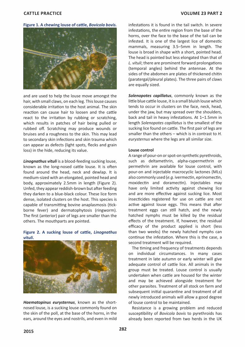
Bovicola bovis is a chewing louse formerly called Damalinia bovis. It is one of the commonest cattle parasites and is usually found on the head, especially the curly hair of the poll and forehead, the neck, shoulders, back and rump, and occasionally the tail switch. If infestations reach high levels the lice may spread down the sides and may cover the rest of the body. This louse is a reddish-brown in colour with dark transverse bands on the abdomen (Figure 1). Adults measure up to 2mm in length and 0.35-0.55mm in width. The head is relatively large, as wide as the body and is rounded anteriorly, with the mouthparts adapted for chewing (Figure 1). The legs are slender
CATTLE PRACTICE VOLUME 23 PART 2
2015 282
and are used to help the louse move amongst the hair, with small claws, on each leg. This louse causes considerable irritation to the host animal. The skin reaction can cause hair to loosen and the cattle react to the irritation by rubbing or scratching, which results in patches of hair being pulled or rubbed off. Scratching may produce wounds or bruises and a roughness to the skin. This may lead to secondary skin infections and skin trauma which can appear as defects (light spots, flecks and grain loss) in the hide, reducing its value.
Linognathus vituli is a blood-feeding sucking louse, known as the long-nosed cattle louse. It is often found around the head, neck and dewlap. It is medium-sized with an elongated, pointed head and body, approximately 2.5mm in length (Figure 2). Unfed, they appear reddish-brown but after feeding they darken to a blue-black colour. These lice form dense, isolated clusters on the host. This species is capable of transmitting bovine anaplasmosis (tick-borne fever) and dermatophytosis (ringworm). The first (anterior) pair of legs are smaller than the others. The mouthparts are pointed.
infestations it is found in the tail switch. In severe infestations, the entire region from the base of the horns, over the face to the base of the tail can be infested. It is one of the largest lice of domestic mammals, measuring 3.5–5mm in length. The louse is broad in shape with a short, pointed head. The head is pointed but less elongated than that of L. vituli; there are prominent forward prolongations (temporal angles) behind the antennae. At the sides of the abdomen are plates of thickened chitin (paratergal/pleural plates). The three pairs of claws are equally sized.
Solenopotes capillatus, commonly known as the little blue cattle louse, it is a small bluish louse which tends to occur in clusters on the face, neck, head, under the jaw, but may spread over the shoulders, back and tail in heavy infestations. At 1–1.5mm in length Solenopotes capillatus is the smallest of the sucking lice found on cattle. The first pair of legs are smaller than the others – which is in contrast to H. eurysternus where the legs are all similar size.
Louse control A range of pour-on or spot-on synthetic pyrethroids, such as deltamethrin, alpha-cypermethrin or permethrin are available for louse control, with pour-on and injectable macrocyclic lactones (MLs) also commonly used (e.g. ivermectin, eprinomectin, moxidectin and doramectin). Injectables may have only limited activity against chewing lice and are more effective against sucking lice. Most insecticides registered for use on cattle are not active against louse eggs. This means that after treatment eggs can still hatch, and the newly hatched nymphs must be killed by the residual effects of the treatment. If, however, the residual efficacy of the product applied is short (less than two weeks) the newly hatched nymphs can continue the infestation. Where this is the case, a second treatment will be required.
The timing and frequency of treatments depends on individual circumstances. In many cases treatment in late autumn or early winter will give adequate control of cattle lice. All animals in the group must be treated. Louse control is usually undertaken when cattle are housed for the winter and may be achieved alongside treatment for other parasites. Treatment of all stock on farm and subsequent initial quarantine and treatment of all newly introduced animals will allow a good degree of louse control to be maintained.
Resistance is a growing problem and reduced susceptibility of Bovicola bovis to pyrethroids has already been reported from two herds in the UK
Figure 1. A chewing louse of cattle, Bovicola bovis.
Figure 2. A sucking louse of cattle, Linognathus vituli.
Haematopinus eurysternus, known as the short-nosed louse, is a sucking louse commonly found on the skin of the poll, at the base of the horns, in the ears, around the eyes and nostrils, and even in mild
CATTLE PRACTICE VOLUME 23 PART 2
2015 283
(Sands and others 2015). Two treatments of an aqueous (5% v/v) suspension of tea tree oil applied topically to the skin, two-weeks apart, has also been demonstrated to be effective in the management of equine lice and may be a useful alternative in organic cattle husbandry or where resistance is suspected (Ellse and others 2015).
MANGE MITES • Chorioptic – commonest in UK• Psoroptic mange – rare in UK, found in
Europe• Sarcoptic mange – uncommon in the UK
Infestation by mites (acariasis) can result in severe skin disease, often called mange. The ectoparasitic mites of cattle feed on lymph, blood and or sebaceous secretions, which they scavenge from the skin surface or obtain from epidermal lesions. Eggs hatch into a six legged larva, which then moult through eight-legged protonymph, tritonymph and adult stages. This may be completed in only 14 days. All life cycle stages are found simultaneously on the host and spend their entire lives in intimate contact with their host. Transmission from host to host is primarily by physical contact but may also occur through contact with a contaminated environment (bedding, housing, trailers, etc.).
Chorioptic mangeThe commonest mange affecting UK cattle is caused by the mite Chorioptes bovis. Chorioptes texanus is also present in the UK, although the difference between C. bovis and C. texanus is of no clinical consequence. The names Chorioptes ovis, Chorioptes equi, Chorioptes caprae and Chorioptes cuniculi have been used to describe the chorioptic mites found on sheep, horses, goats and rabbits respectively, but are now all thought to be synonyms of C. bovis/C. texanus.
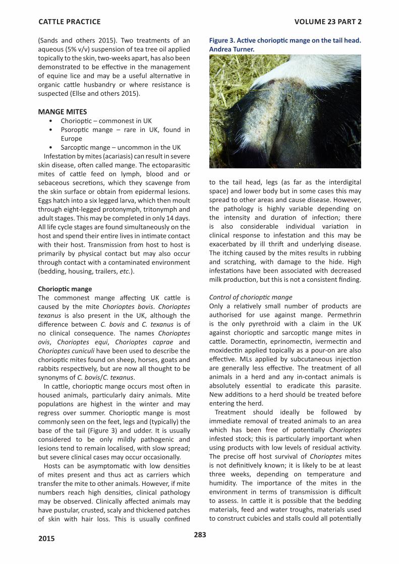
In cattle, chorioptic mange occurs most often in housed animals, particularly dairy animals. Mite populations are highest in the winter and may regress over summer. Chorioptic mange is most commonly seen on the feet, legs and (typically) the base of the tail (Figure 3) and udder. It is usually considered to be only mildly pathogenic and lesions tend to remain localised, with slow spread; but severe clinical cases may occur occasionally.
Hosts can be asymptomatic with low densities of mites present and thus act as carriers which transfer the mite to other animals. However, if mite numbers reach high densities, clinical pathology may be observed. Clinically affected animals may have pustular, crusted, scaly and thickened patches of skin with hair loss. This is usually confined
to the tail head, legs (as far as the interdigital space) and lower body but in some cases this may spread to other areas and cause disease. However, the pathology is highly variable depending on the intensity and duration of infection; there is also considerable individual variation in clinical response to infestation and this may be exacerbated by ill thrift and underlying disease. The itching caused by the mites results in rubbing and scratching, with damage to the hide. High infestations have been associated with decreased milk production, but this is not a consistent finding.
Control of chorioptic mange Only a relatively small number of products are authorised for use against mange. Permethrin is the only pyrethroid with a claim in the UK against chorioptic and sarcoptic mange mites in cattle. Doramectin, eprinomectin, ivermectin and moxidectin applied topically as a pour-on are also effective. MLs applied by subcutaneous injection are generally less effective. The treatment of all animals in a herd and any in-contact animals is absolutely essential to eradicate this parasite. New additions to a herd should be treated before entering the herd.
Treatment should ideally be followed by immediate removal of treated animals to an area which has been free of potentially Chorioptes infested stock; this is particularly important when using products with low levels of residual activity. The precise off host survival of Chorioptes mites is not definitively known; it is likely to be at least three weeks, depending on temperature and humidity. The importance of the mites in the environment in terms of transmission is difficult to assess. In cattle it is possible that the bedding materials, feed and water troughs, materials used to construct cubicles and stalls could all potentially
Figure 3. Active chorioptic mange on the tail head. Andrea Turner.
CATTLE PRACTICE VOLUME 23 PART 2
2015 284
be contaminated. One item that might require consideration is the use of grooming brushes – if a pruritic cow was to use such a brush to rub she could transmit mites to the next cow that uses the brush system.
As yet, no acaricidal resistance has been recorded in Chorioptes mites in Europe.
It can prove difficult to eradicate this parasite from a herd and the factors of environmental contamination and residual infestation on some individuals in a herd may contribute to persistent infestation (Villarroel and Halliburton 2013). It is likely that in dairy herds, at least, that only severely affected animals will be treated and that eradication will not be pursued given the limited evidence that a systematic programme of treatment will pay dividends in terms of improved milk production.
Psoroptic mange Psoroptic mange has only rarely been reported in cattle in the UK, although it is common in parts of mainland Europe, particularly in breeds such as the Belgian Blue. However, the disease was diagnosed in South West Wales in 2006 and has since been diagnosed on more than 20 premises, the majority in Wales, with one farm in England and one in Scotland (Jones and others 2008, Millar and others 2011, Jones and others 2014). Psoroptic mange has also been reported in Ireland. There have also been anecdotal reports of disease diagnosed on other holdings in GB. Most animals infested were beef cattle but there was evidence of recurrent disease in some dairy herds. It appears probable that this initial outbreak has now been controlled, but there is a continuing threat of importing the disease from abroad.
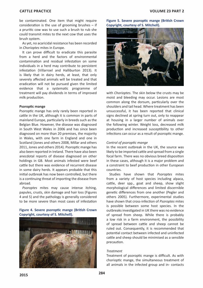
Psoroptes mites may cause intense itching, papules, crusts, skin damage and hair loss (Figures 4 and 5) and the pathology is generally considered to be more severe than most cases of infestation
with Chorioptes. The skin below the crusts may be moist and bleeding may occur. Lesions are most common along the dorsum, particularly over the shoulders and tail head. Where treatment has been unsuccessful, it has been reported that clinical signs declined at spring turn out, only to reappear at housing in a larger number of animals over the following winter. Weight loss, decreased milk production and increased susceptibility to other infections can occur as a result of psoroptic mange.
Control of psoroptic mange In the recent outbreak in the UK, the source was likely to be imported cattle and spread from a single focal farm. There was no obvious breed disposition in these cases, although it is a major problem and a constraint to beef production in other European countries.
Studies have shown that Psoroptes mites from a variety of host species including alpaca, cattle, deer spp., goat and sheep, show slight morphological differences and limited discernible genetic differences from one another (Pegler and others 2005). Furthermore, experimental studies have shown that cross-infection of Psoroptes mites is possible between some host species. In the outbreaks investigated in UK there was no evidence of spread from sheep. While there is probably a low risk in a farm environment, the possibility of spread between cattle and sheep cannot be ruled out. Consequently, it is recommended that potential contact between infected and uninfected cattle and sheep should be minimised as a sensible precaution.
TreatmentTreatment of psoroptic mange is difficult. As with chorioptic mange, the simultaneous treatment of all animals in the infected group and in- contacts
Figure 4. Severe psoroptic mange (British Crown Copyright, courtesy of S. Mitchell).
Figure 5. Severe psoroptic mange (British Crown Copyright, courtesy of S. Mitchell).
CATTLE PRACTICE VOLUME 23 PART 2
2015 285
is essential where this mite is diagnosed regardless of clinical signs. Removal of the crusts by clipping and, if necessary, washing prior to treatment is essential. The crusts harbour large numbers of mites and eggs and should be destroyed.
Treatment should ideally be followed by immediate removal to an area which has been free of potentially infested animals, particularly for products with low levels of residual activity. The off host survival of Psoroptes mites is about 18 days, depending on prevailing weather conditions.
The only products licensed for treatment of psoroptic mange in the UK are the MLs given by injection, or moxidectin or doramectin as pour-ons. However, differences in efficacies between ivermectin formulations have been reported (Genchi and others 2008) and often repeated treatments are needed to kill all mites. Treatment should always be monitored for success using skin scrapes to detect live mites.
The cases seen in south Wales were not cured by licensed macrocyclic lactones, despite careful veterinary administered treatment in some cases. Large numbers of live mites were detected after treatment and clinical signs reoccurred. Success was achieved using a 4 per cent permethrin pour-on given at an increased frequency of treatment (three treatments at two-weekly intervals) to all at-risk animals. Clinical signs resolved quickly with this treatment schedule, but three treatments were necessary to ensure that all of the mites were killed. This product was used under the cascade system by the farmers’ veterinary surgeons following failure of the licensed treatment (see VMD Guidance Note 13 https://www.vmd.defra.gov.uk/pdf/vmgn/VMGNote13.pdf)
In dairy cattle, treatment is even more difficult as the licensed products are not to be used in lactating animals and the use of the permethrin pour-on at an increased frequency of treatment incurs a seven-day milk withdrawal after the second and third treatments. Amitraz, a drug available in Europe, is used as a spray for treatment of psoroptic mange in dairy cattle in some countries. If used in the UK, the farmer’s veterinary surgeon has to apply for a special import certificate, available via the VMD website (https://www.vmd.defra.gov.uk/sis/default.aspx). Its use would be under the rules of the cascade (see VMD Guidance note 13).
The control of psoroptic mange in cattle is challenging and there appears to be considerable variation between populations in their response to different acaricides; elements of tolerance, resistance and host-adaptation may all be involved in creating this variable response to treatment in
different mite populations. However, it is also often difficult to disentangle poor treatment efficacy from poor administration practice, particularly where only clinically affected animals are treated.
Risk to the UKPsoroptic mange in cattle is present in mainland Europe and Ireland, as well as other areas of the world. It is more common in beef cattle, but dairy herds have also been infected. In Belgium, it is considered the most economically important ectoparasitic disease of cattle. There is a high risk of importing disease from these countries, particularly when the animals are carrying small numbers of mites and the skin lesions are small or absent. As yet, there is no test that is able to identify these animals as infected when they are not showing clinical signs.
Psoroptic mange is a severe skin disease in cattle, with serious welfare implications if not identified quickly and treated correctly. It has the potential to become established in Great Britain because of the movement of animals and the difficulties of treatment.
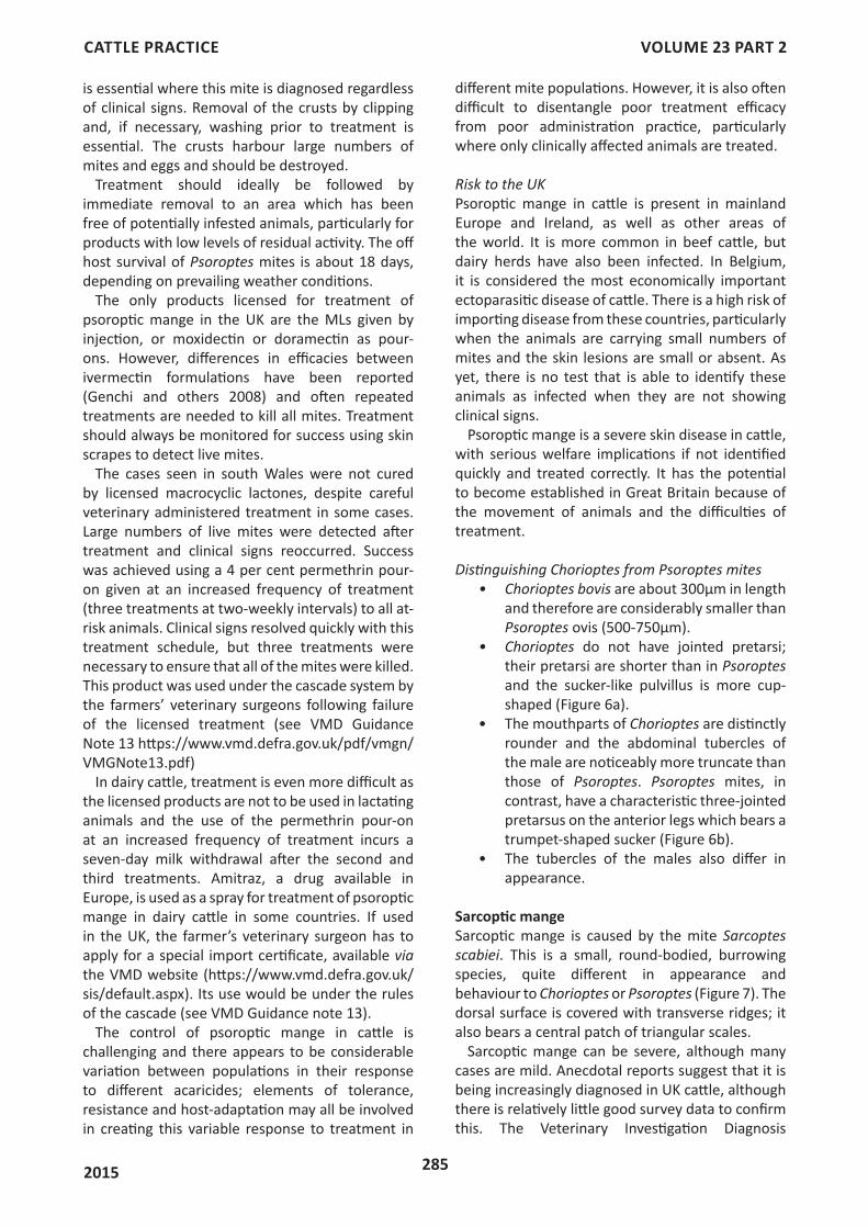
Distinguishing Chorioptes from Psoroptes mites• Chorioptes bovis are about 300μm in length
and therefore are considerably smaller than Psoroptes ovis (500-750μm).
• Chorioptes do not have jointed pretarsi; their pretarsi are shorter than in Psoroptes and the sucker-like pulvillus is more cup-shaped (Figure 6a).
• The mouthparts of Chorioptes are distinctly rounder and the abdominal tubercles of the male are noticeably more truncate than those of Psoroptes. Psoroptes mites, in contrast, have a characteristic three-jointed pretarsus on the anterior legs which bears a trumpet-shaped sucker (Figure 6b).
• The tubercles of the males also differ in appearance.
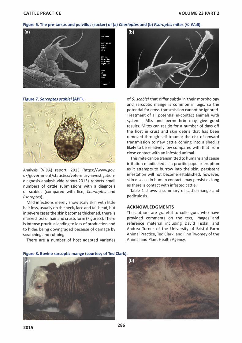
Sarcoptic mangeSarcoptic mange is caused by the mite Sarcoptes scabiei. This is a small, round-bodied, burrowing species, quite different in appearance and behaviour to Chorioptes or Psoroptes (Figure 7). The dorsal surface is covered with transverse ridges; it also bears a central patch of triangular scales.
Sarcoptic mange can be severe, although many cases are mild. Anecdotal reports suggest that it is being increasingly diagnosed in UK cattle, although there is relatively little good survey data to confirm this. The Veterinary Investigation Diagnosis
CATTLE PRACTICE VOLUME 23 PART 2
2015 286
Analysis (VIDA) report, 2013 (https://www.gov.uk/government/statistics/veterinary-investigation-diagnosis-analysis-vida-report-2013) reports small numbers of cattle submissions with a diagnosis of scabies (compared with lice, Chorioptes and Psoroptes).
Mild infections merely show scaly skin with little hair loss, usually on the neck, face and tail head, but in severe cases the skin becomes thickened, there is marked loss of hair and crusts form (Figure 8). There is intense pruritus leading to loss of production and to hides being downgraded because of damage by scratching and rubbing.
There are a number of host adapted varieties
Figure 6. The pre-tarsus and pulvillus (sucker) of (a) Chorioptes and (b) Psoroptes mites (© Wall).
(a) (b)
Figure 7. Sarcoptes scabiei (APF).
Figure 8. Bovine sarcoptic mange (courtesy of Ted Clark).(a) (b)
of S. scabiei that differ subtly in their morphology and sarcoptic mange is common in pigs, so the potential for cross-transmission cannot be ignored. Treatment of all potential in-contact animals with systemic MLs and permethrin may give good results. Mites can reside for a number of days off the host in crust and skin debris that has been removed through self trauma; the risk of onward transmission to new cattle coming into a shed is likely to be relatively low compared with that from close contact with an infested animal.
This mite can be transmitted to humans and cause irritation manifested as a pruritic papular eruption as it attempts to burrow into the skin; persistent infestation will not become established, however, skin disease in human contacts may persist as long as there is contact with infested cattle.
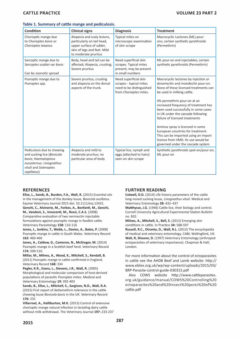
Table 1 shows a summary of cattle mange and pediculosis.
ACKNOWLEDGMENTSThe authors are grateful to colleagues who have provided comments on the text, images and reference material including David Tisdall and Andrea Turner of the University of Bristol Farm Animal Practice, Ted Clark, and Finn Twomey of the Animal and Plant Health Agency.
CATTLE PRACTICE VOLUME 23 PART 2
2015 287
REFERENCESEllse, L., Sands, B., Burden, F.A., Wall, R. (2015) Essential oils in the management of the donkey louse, Bovicola ocellatus. Equine Veterinary Journal 2015 doi: 10.1111/evj.12431.Genchi, C., Alvinerie, M., Forbes, A., Bonfanti, M., Genchi, M., Vandoni, S., Innocenti, M., Rossi, C.A.S. (2008) Comparative evaluation of two ivermectin injectable formulations against psoroptic mange in feedlot cattle. Veterinary Parasitology 158: 110-116 Jones, J., Jenkins, T., Webb, L., Davies, A., Bates, P. (2008) Psoroptic mange in cattle in South Wales. Veterinary Record 162: 460-460 Jones, A., Caldow, G., Cameron, N., McGregor, M. (2014) Psoroptic mange in a Scottish beef herd. Veterinary Record 174: 509-510 Millar, M., Milnes, A., Wood, K., Mitchell, S., Kendall, B. (2011) Psoroptic mange in cattle confirmed in England. Veterinary Record 168: 334Pegler, K.R., Evans, L., Stevens, J.R., Wall, R. (2005) Morphological and molecular comparison of host-derived populations of parasitic Psoroptes mites. Medical and Veterinary Entomology 19: 392-403Sands, B., Ellse, L., Mitchell, S., Sargison, N.D., Wall, R.A. (2015) First report of deltamethrin tolerance in the cattle chewing louse Bovicola bovis in the UK. Veterinary Record 176: 231 Villarroel, A., Halliburton, M.K. (2013) Control of extensive chorioptic mange natural infection in lactating dairy cattle without milk withdrawal. The Veterinary Journal 197: 233-237
Table 1. Summary of cattle mange and pediculosis. Condition Clinical signs Diagnosis TreatmentChorioptic mange due to Chorioptes bovis or Chorioptes texanus
Alopecia and scaly lesions, particularly on tail head, upper surface of udder, skin of legs and feet. Mild to moderate pruritus
Typical mites on microscopic examination of skin scrape
Macrocyclic Lactones (ML) pour-ons; certain synthetic pyrethroids (Permethrin)
Sarcoptic mange due to Sarcoptes scabiei var bovis
Can be zoonotic spread
Body, head and tail can be affected; Alopecia, crusting; Severe pruritus
Need superficial skin scrapes. Typical mites present; may be present in small numbers
ML pour-on and injectables; certain synthetic pyrethroids (Permethrin)
Psoroptic mange due to Psoroptes spp.
Severe pruritus, crusting and alopecia on the dorsal aspects of the trunk.
Need superficial skin scrapes - typical mites need to be distinguished from Chorioptes mites.
Macrocyclic lactones by injection or doramectin and moxidectin pour-on. None of these licensed treatments can be used in milking cattle.
4% permethrin pour-on at an increased frequency of treatment has been used successfully in some cases in UK under the cascade following failure of licensed treatments
Amitraz spray is licensed in some European countries for treatment. This can be imported using an import licence from VMD. Its use would be governed under the cascade system
Pediculosis due to chewing and sucking lice (Bovicola bovis; Haematopinus eurysternus. Linognathus vituli and Solenoptes capillatus)
Alopecia and mild to moderate pruritus; no particular area of body
Typical lice, nymph and eggs (attached to hairs) seen on skin scrape
Synthetic pyrethroids spot-on/pour-on; ML pour-on
FURTHER READINGColwell, D.D. (2014) Life history parameters of the cattle long-nosed sucking louse, Linognathus vituli. Medical and Veterinary Entomology 28: 432–437 Matthysse, J.G. (1946) Cattle lice, their biology and control. Cornell University Agricultural Experimental Station Bulletin no. 832.Milnes, A., Mitchell, S., Bell, S. (2012) Emerging skin conditions in cattle. In Practice 34: 588-597 Russell, R.C., Otranto, D., Wall, R.L. (2013) The encyclopedia of medical and veterinary entomology. CABI; Wallingford, UK. Wall, R, Shearer, D. (1997) Veterinary Entomology (arthropod ectoparasites of veterinary importance). Chapman & Hall; London.
For more information about the control of ectoparasites in cattle see the AHDB Beef and Lamb website: http://www.eblex.org.uk/wp/wp-content/uploads/2015/03/BRP-Parasite-control-guide-030315.pdf
Also COWS website http://www.cattleparasites.org.uk/guidance/manual/COWS%20Controlling%20ectoparasites%20and%20insect%20pests%20of%20cattle.pdf


![Characterization of mitochondrial COX-1 gene of Sarcoptes ...bdvets.binbd.com/JAVAR/V6I4/f366_pp445-450.pdfscabies infected rabbits. Research by Lastuti et al. [21] demonstrated that](https://img.pdfslide.us/doc/110x75/5e7acf2400f88a75f80c9dcf/characterization-of-mitochondrial-cox-1-gene-of-sarcoptes-scabies-infected-rabbits.jpg)