Embed Size (px)
Citation preview
Submitted 28 February 2018Accepted 2 July 2018Published 27 July 2018
Corresponding authorMartina Jelocnik,[email protected]
Academic editorCarlos Gutiérrez
Additional Information andDeclarations can be found onpage 11
DOI 10.7717/peerj.5291
Copyright2018 Fraser et al.
Distributed underCreative Commons CC-BY 4.0
OPEN ACCESS
A Sarcoptes scabiei specific isothermalamplification assay for detection of thisimportant ectoparasite of wombats andother animalsTamieka A. Fraser1,2, Scott Carver2, Alynn M. Martin2, Kate Mounsey1,3,Adam Polkinghorne1 and Martina Jelocnik1
1USC Animal Research Centre, Faculty of Science, Health, Education and Engineering, University of theSunshine Coast, Sippy Downs, Australia
2Department of Biological Sciences, University of Tasmania, Sandy Bay, Australia3 School of Health and Sport Sciences, University of the Sunshine Coast, Sippy Downs, Australia
ABSTRACTBackground. The globally distributed epidermal ectoparasite, Sarcoptes scabiei, is aserious health and welfare burden to at-risk human and animal populations. Rapidand sensitive detection of S. scabiei infestation is critical for intervention strategies.While direct microscopy of skin scrapings is a widely utilised diagnostic method, it haslow sensitivity. PCR, alternatively, has been shown to readily detect mite DNA even inmicroscopy-negative skin scrapings. However, a limitation to the latter method is therequirements for specialised equipment and reagents. Such resourcesmay not be readilyavailable in regional or remote clinical settings and are an important consideration indiagnosis of this parasitic disease.Methodology. A Loop Mediated Isothermal Amplification (LAMP) assay targeting theITS-2 gene for S. scabiei was developed and evaluated on clinical samples from varioushosts, previously screened with conventional S. scabies-specific PCR. Species specificityof the newly developed LAMP assay was tested against a range of DNA samples fromother arthropods. The LAMP assays were performed on a real-time fluorometer aswell as thermal cycler to evaluate an end-point of detection. Using skin scrapings, arapid sample processing method was assessed to eliminate extensive processing timesinvolved with DNA extractions prior to diagnostic assays, including LAMP.Results. The S. scabiei LAMP assay was demonstrated to be species-specific and able todetect DNA extracted from a single mite within a skin scraping in under 30 minutes.Application of this assay to DNA extracts from skin scrapings taken from a rangeof hosts revealed 92.3% congruence (with 92.50% specificity and 100% sensitivity)to the conventional PCR detection of S. scabiei. Preliminary results have indicatedthat diagnostic outcome from rapidly processed dry skin scrapings using our newlydeveloped LAMP is possible in approximately 40 minutes.Discussion. We have developed a novel, rapid and robust molecular assay for detectingS. scabiei infesting humans and animals. Based on these findings, we anticipate that thisassay will serve an important role as an ancillary diagnostic tool at the point-of-care,complementing existing diagnostic protocols for S. scabiei.
How to cite this article Fraser et al. (2018), A Sarcoptes scabiei specific isothermal amplification assay for detection of this important ec-toparasite of wombats and other animals. PeerJ 6:e5291; DOI 10.7717/peerj.5291
Subjects Parasitology, Veterinary Medicine, Public HealthKeywords LAMP, Diagnostics, Sarcoptic mange, Skin scraping, PCR, One health, Australianwildlife, Sarcoptes scabiei, Wombats
INTRODUCTIONSarcoptes scabiei is an ectoparasite that resides in the epidermal layer of its hosts causing arange of clinical signs of disease including pruritis, dermal inflammation, hyperkeratosisand alopecia, which may lead to bacterial sepsis (McCarthy et al., 2004). S. scabiei is listedamong the top 50 most prevalent diseases in humans with over 100 million people globallypredicted to be infested (Hay et al., 2014; Romani et al., 2015). Beyond its role in humandisease, a wide range of domestic animals, wild canids, and other wildlife suffer extensivelyfrom sarcoptic mange, and transmission to at-risk animal populations can result inpopulation declines and localised extinctions (Forchhammer & Asferg, 2000; Gakuya etal., 2011; Graczyk et al., 2001; Martin et al., 2018; Perrucci et al., 2016). With the endemicinfestation of humans in tropical and subtropical areas, the large variety of animal speciesinfested and the knowledge that S. scabiei is the same mite infesting all, pathogen dispersaland spill-over has been suggested to be the causative consequence of global infestations(Fraser et al., 2016;Walton & Currie, 2007).
As with many infectious diseases, the successful treatment of affected individuals andthe application of appropriate disease management strategies relies on rapid and accuratedetection of the infectious agent. Diagnosis of scabies (also classified as mange in animals)is typically made by assessment of clinical features alone (Hardy, Engelman & Steer, 2017;Walton & Currie, 2007). When atypical appearances are presented, however, the diagnosiscan be challenging as other skin conditions can mimic clinical signs of scabies (Hardy,Engelman & Steer, 2017). A skin scraping of the affected area provides a more definitivediagnosis as mites, mite eggs and faecal pellets can be identified via microscopy due totheir distinct morphology (Leung & Miller, 2011). Although the diagnosis is more specificusing microscopy, detection of early mange has been shown to have limited sensitivity,primarily due to the difficulties in sampling and visualising mites when the mite burden islow (Fraser et al., 2018; Skerratt, 2005; Walton & Currie, 2007). Recent studies have shownthat diagnosis of S. scabiei by clinical features and microscopy are unreliable methods forearly stage infestations (Fraser et al., 2018;Wong et al., 2015).
Besides microscopy, alternative diagnostic methods for S. scabiei have been evaluatedwith varying sensitivity and specificity. Several studies have attempted to use serologicaltechniques (i.e., ELISAs) as a more targeted diagnostic method (Arlian, Feldmeier &Morgan, 2015; Löwenstein, Kahlbacher & Peschke, 2004; Rambozzi et al., 2004; Rodríguez-Cadenas et al., 2010; Zhao et al., 2014). However, as reviewed by Arlian & Morgan (2017),significant limitations for this method exist including the time taken for the host to developS. scabiei-specific antibodies and cross-reactivity between S. scabiei antigens and thosefrom other mites. Molecular techniques using nucleic acid amplification tests (NAATs)as a diagnostic tool for S. scabiei are relatively new but show promising results (Fraser etal., 2018;Wong et al., 2015). Two studies, analysing samples collected from humans (Wong
Fraser et al. (2018), PeerJ, DOI 10.7717/peerj.5291 2/15
et al., 2015) and animals (Fraser et al., 2018), have demonstrated that PCRs have a highersensitivity and specificity than microscopy, revealing high rates of false negative samplespreviously screened by microscopy. However, NAATs are not well adapted for clinicalsettings, particularly for diseases like scabies which are common in remote or resource-limited communities and/or in field settings with limited access to diagnostic laboratoriesand necessary equipment (Walton & Currie, 2007). Recent advances in this field haveutilised hand held devices resulting in promising outcomes for detecting infectious diseasesquickly. This includes the Biomeme Inc. portable PCR machine with thermocycler andfluorometer which can dock into an iPhone resulting in rapid results and the AmplifyRP R©
portable florescence reader (Marx, 2015; Zhang et al., 2014).Loopmediated isothermal amplification (LAMP) is one of the expanding range of NAAT
techniques that is showing capacity at the Point of Care (POC). LAMP assays are low cost,rapid and can be used with simple ‘bench-top’ equipment. Visual result interpretation withthe use of different DNA binding dyes in these assays further support LAMP use at the POC.There have beenmultiple successful LAMPassays developed for other human and veterinaryparasites including Plasmodium spp. (Lucchi et al., 2016), Toxoplasma gondii (Kong etal., 2012) and Leishmania spp. (Adams et al., 2010), and bacteria including Chlamydiaspp. (Jelocnik et al., 2017), Mycoplasma pneumoniae (Saito et al., 2005) and Streptococcusagalactiae (McKenna et al., 2017). To overcome on some limitations associated with LAMPassays, including misleading of results using turbidity techniques and cross contaminationas a result of opening tubes, the use of a fluorometer and a signature melt for ampliconcharacteristics can account for these limitations.
This study aimed to develop a LAMP assay for the detection of S. scabiei in animalsand assess its reliability against PCR and demonstrate its potential as a POC test. Wehave utilised a unique sample set of skin scrapings taken from a range of hosts and testedextracted DNA from those with the newly developed S. scabiei specific LAMP assay. TheLAMP assay was evaluated against microscopy and a recently described conventionalS. scabiei-specific PCR assay. In an attempt to reduce sample processing time, we alsooptimised a rapid DNA extraction method on a small subset of skin scrapings, furtherhighlighting the potential for this assay to be deployed at the POC.
METHODS AND MATERIALSLAMP assay designThe S. scabiei internal transcribed spacer 2 (ITS-2) gene is a highly conserved gene andwas used as the LAMP target in this study. A ClustalW alignment of 87 ITS-2 sequences(represented as haplotypes) from S. scabiei mites from humans and a variety of animalsacross Australia, Europe, North America and Asia available in GenBank was obtained toidentify polymorphisms in this gene (Fig. S1, Table S1). In addition, we have includedITS-2 sequences from other mite species, including the house dust mite (Dermatophagoidesfarinae), the chorioptic mange mite (Chorioptes sp), the notoedric mange mite (Notoedrescati), the psoroptic mange mite (Psoroptes sp) and ticks (Ixodes sp). This 450 bp fragmentwas also subjected to a discontiguous megablast search in Basic Local Alignment Search
Fraser et al. (2018), PeerJ, DOI 10.7717/peerj.5291 3/15
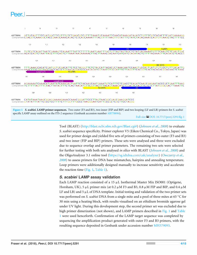
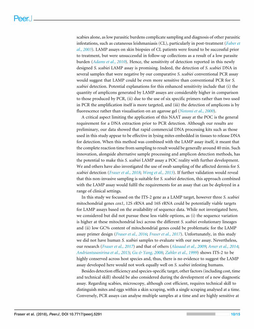
Figure 1 S. scabiei. LAMP primer sequences. Two outer (F3 and B3), two inner (FIP and BIP) and two looping (LF and LB) primers for S. scabieispecific LAMP assay outlined on the ITS-2 sequence (Genbank accession number AB778896).
Full-size DOI: 10.7717/peerj.5291/fig-1
Tool (BLAST) (http://blast.ncbi.nlm.nih.gov/Blast.cgi#) (Johnson et al., 2008) to evaluateS. scabiei sequence specificity. Primer explorer V5 (Eiken Chemical Co., Tokyo, Japan) wasused for primer design and yielded five sets of primers consisting of two outer (F3 and B3)and two inner (FIP and BIP) primers. These sets were analysed and three were excludeddue to sequence overlap and primer parameters. The remaining two sets were selectedfor further testing with both sets analysed in silico with BLAST (Johnson et al., 2008) andthe OligoAnalyzer 3.1 online tool (https://sg.idtdna.com/calc/analyzer) (Owczarzy et al.,2008) to assess primers for DNA base mismatches, hairpins and annealing temperature.Loop primers were additionally designed manually to increase sensitivity and acceleratethe reaction time (Fig. 1, Table 1).
S. scabiei LAMP assay validationEach LAMP reaction consisted of a 15 µL Isothermal Master Mix ISO001 (Optigene,Horsham, UK), 5 µL primer mix (at 0.2 µM F3 and B3, 0.8 µM FIP and BIP, and 0.4 µMLF and LB) and 5 µL of DNA template. Initial testing and validation of the two primer setswas performed on S. scabiei DNA from a single mite and a pool of three mites at 65 ◦C for30 min using a heating block, with results visualised on an ethidium bromide agarose gelunder UV light. During this development step, the second primer set was excluded due tohigh primer dimerization (not shown), and LAMP primers described in Fig. 1 and Table1 were used henceforth. Confirmation of the LAMP target sequence was completed bysequencing the amplification product generated with outer F3 and B3 primers, with theresulting sequence deposited in Genbank under accession number MH379093.
Fraser et al. (2018), PeerJ, DOI 10.7717/peerj.5291 4/15
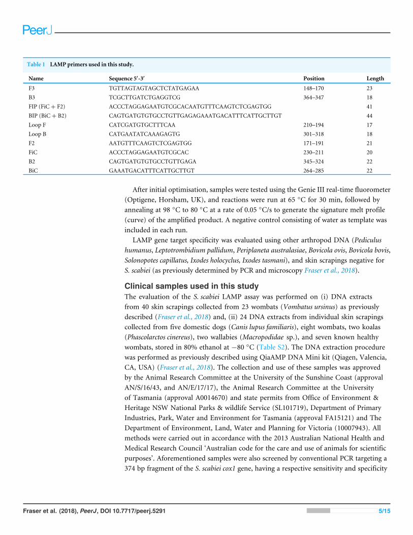
Table 1 LAMP primers used in this study.
Name Sequence 5′-3′ Position Length
F3 TGTTAGTAGTAGCTCTATGAGAA 148–170 23B3 TCGCTTGATCTGAGGTCG 364–347 18FIP (FiC+ F2) ACCCTAGGAGAATGTCGCACAATGTTTCAAGTCTCGAGTGG 41BIP (BiC+ B2) CAGTGATGTGTGCCTGTTGAGAGAAATGACATTTCATTGCTTGT 44Loop F CATCGATGTGCTTTCAA 210–194 17Loop B CATGAATATCAAAGAGTG 301–318 18F2 AATGTTTCAAGTCTCGAGTGG 171–191 21FiC ACCCTAGGAGAATGTCGCAC 230–211 20B2 CAGTGATGTGTGCCTGTTGAGA 345–324 22BiC GAAATGACATTTCATTGCTTGT 264–285 22
After initial optimisation, samples were tested using the Genie III real-time fluorometer(Optigene, Horsham, UK), and reactions were run at 65 ◦C for 30 min, followed byannealing at 98 ◦C to 80 ◦C at a rate of 0.05 ◦C/s to generate the signature melt profile(curve) of the amplified product. A negative control consisting of water as template wasincluded in each run.
LAMP gene target specificity was evaluated using other arthropod DNA (Pediculushumanus, Leptotrombidium pallidum, Periplaneta australasiae, Bovicola ovis, Bovicola bovis,Solonopotes capillatus, Ixodes holocyclus, Ixodes tasmani), and skin scrapings negative forS. scabiei (as previously determined by PCR and microscopy Fraser et al., 2018).
Clinical samples used in this studyThe evaluation of the S. scabiei LAMP assay was performed on (i) DNA extractsfrom 40 skin scrapings collected from 23 wombats (Vombatus ursinus) as previouslydescribed (Fraser et al., 2018) and, (ii) 24 DNA extracts from individual skin scrapingscollected from five domestic dogs (Canis lupus familiaris), eight wombats, two koalas(Phascolarctos cinereus), two wallabies (Macropodidae sp.), and seven known healthywombats, stored in 80% ethanol at −80 ◦C (Table S2). The DNA extraction procedurewas performed as previously described using QiaAMP DNA Mini kit (Qiagen, Valencia,CA, USA) (Fraser et al., 2018). The collection and use of these samples was approvedby the Animal Research Committee at the University of the Sunshine Coast (approvalAN/S/16/43, and AN/E/17/17), the Animal Research Committee at the Universityof Tasmania (approval A0014670) and state permits from Office of Environment &Heritage NSW National Parks & wildlife Service (SL101719), Department of PrimaryIndustries, Park, Water and Environment for Tasmania (approval FA15121) and TheDepartment of Environment, Land, Water and Planning for Victoria (10007943). Allmethods were carried out in accordance with the 2013 Australian National Health andMedical Research Council ‘Australian code for the care and use of animals for scientificpurposes’. Aforementioned samples were also screened by conventional PCR targeting a374 bp fragment of the S. scabiei cox1 gene, having a respective sensitivity and specificity
Fraser et al. (2018), PeerJ, DOI 10.7717/peerj.5291 5/15
of 100% and 84.62% in concordance to microscopy, as previously described (Fraser etal., 2018). PCR positivity for cox1 was determined by visualisation of the 374bp fragmentfollowing agarose gel electrophoresis under UV light.
In order to confirm negative samples and to test for isothermal amplification inhibition,a subset of six negative samples were spiked with 10 µL mite only DNA and tested againby LAMP.
Evaluation of rapid skin scraping DNA extractionIn order to assess the use of LAMP at the POC, eleven wombat skin scrapings, withmite counts previously assessed by microscopy, were submerged with 0.3M PotassiumHydroxide (KOH), pH 13, and heated at 95 ◦C for 10 min in order to lyse the tissue andrelease DNA from the cells. After vortexing, 5 µL of the tissue suspension was used as atemplate in each reaction, also consisting of 15 µL of Lyse’n’Lamp master mix (Optigene,Horsham,UK) and 5µL primermix as described above. The LAMP reaction was performedin the Genie III fluorometer using the same cycling conditions as described above. Negativecontrols of water only and an aliquot of 0.3M KOH only were included in the assays. Thesame samples were also tested with the cox 1 PCR after performing DNA extractions, asdescribed above, on the KOH skin suspensions.
Statistical analysisThe performance of the LAMP assay compared to the reference PCR assay conductedon the same samples was estimated by calculating Kappa values, overall agreement,sensitivity and specificity. Direct comparisons were conducted using EpiTools online(http://epitools.ausvet.com.au) (Sergeant, 2017). Kappa values are interpreted as follows:values ≤0 as indicating no agreement, 0.01–0.20 as none to slight, 0.21–0.40 as fair, 0.41–0.60 as moderate, 0.61–0.80 as substantial, and 0.81–1.00 as almost perfect agreement.
RESULTSS. scabiei LAMP assay development and validationThe LAMP primers were predicted to amplify a 217 bp fragment of the ITS-2 gene.The alignment of the available S. scabiei ITS-2 gene sequences (n= 87) revealed 96.5%–100% sequence identity (Fig. S1). ’’-In silico analysis of ITS-2 sequences obtained fromDermatophagoides farinae (KT724354), Chorioptes sp. (AF123084), Ixodes pavlovskyi(KP242014), Ixodes persulcatus (KR136379), Notoedres cati (AF251801), Psoroptesnatalensis (AB968091), Psoroptes cuniculi (KP676689) and Psoroptes ovis (EF429259)indicated that the S. scabiei LAMP primers are likely to be specific, as we identified 101to 239 nucleotide polymorphisms between our primers and other arthropod sequences(Fig. S1).
The S. scabiei LAMP assay was initially assessed in house on a thermal block, with theassay run for 30 min at 65 ◦C. A single mite as well as pooled mite DNA extracts weredetectable by LAMP, as visualised by the amplicons on the gel. We also tested 10-folddilutions of a mite and mite-positive skin scraping DNA samples by LAMP on the thermalcycler in two independent runs using the same run conditions (Fig. S2). We successfully
Fraser et al. (2018), PeerJ, DOI 10.7717/peerj.5291 6/15
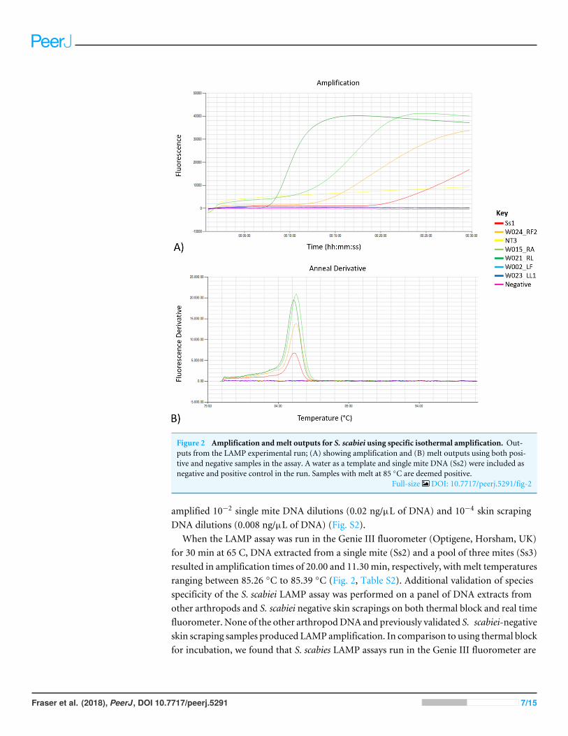
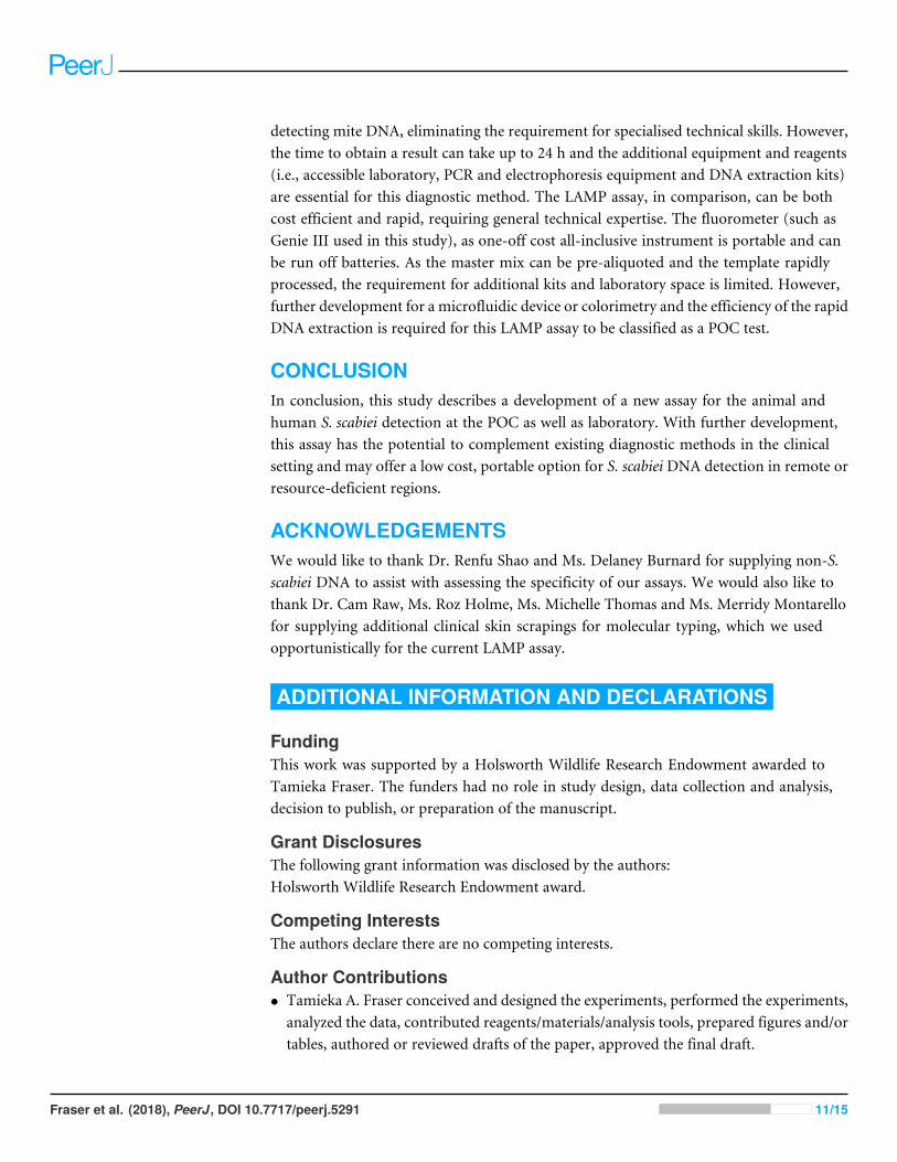
Figure 2 Amplification andmelt outputs for S. scabiei using specific isothermal amplification. Out-puts from the LAMP experimental run; (A) showing amplification and (B) melt outputs using both posi-tive and negative samples in the assay. A water as a template and single mite DNA (Ss2) were included asnegative and positive control in the run. Samples with melt at 85 ◦C are deemed positive.
Full-size DOI: 10.7717/peerj.5291/fig-2
amplified 10−2 single mite DNA dilutions (0.02 ng/µL of DNA) and 10−4 skin scrapingDNA dilutions (0.008 ng/µL of DNA) (Fig. S2).
When the LAMP assay was run in the Genie III fluorometer (Optigene, Horsham, UK)for 30 min at 65 C, DNA extracted from a single mite (Ss2) and a pool of three mites (Ss3)resulted in amplification times of 20.00 and 11.30min, respectively, withmelt temperaturesranging between 85.26 ◦C to 85.39 ◦C (Fig. 2, Table S2). Additional validation of speciesspecificity of the S. scabiei LAMP assay was performed on a panel of DNA extracts fromother arthropods and S. scabiei negative skin scrapings on both thermal block and real timefluorometer. None of the other arthropodDNA and previously validated S. scabiei-negativeskin scraping samples produced LAMP amplification. In comparison to using thermal blockfor incubation, we found that S. scabies LAMP assays run in the Genie III fluorometer are
Fraser et al. (2018), PeerJ, DOI 10.7717/peerj.5291 7/15
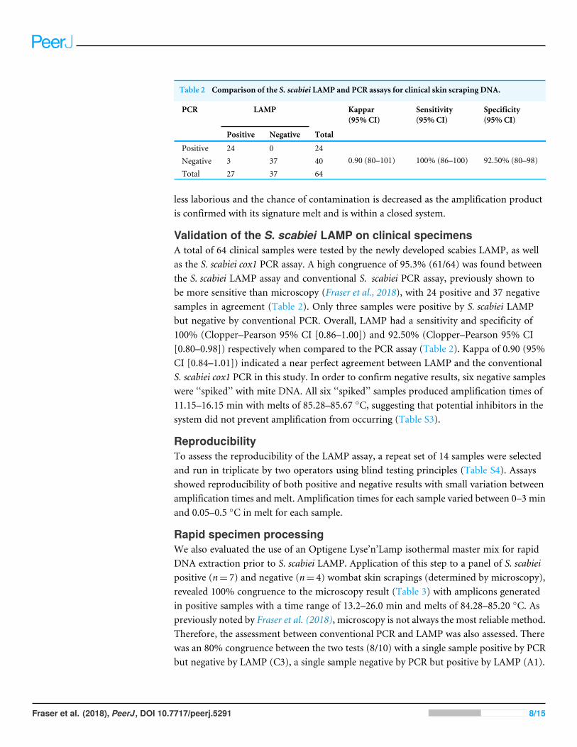
Table 2 Comparison of the S. scabiei LAMP and PCR assays for clinical skin scraping DNA.
PCR LAMP Kappar(95% CI)
Sensitivity(95% CI)
Specificity(95% CI)
Positive Negative Total
Positive 24 0 24Negative 3 37 40Total 27 37 64
0.90 (80–101) 100% (86–100) 92.50% (80–98)
less laborious and the chance of contamination is decreased as the amplification productis confirmed with its signature melt and is within a closed system.
Validation of the S. scabiei LAMP on clinical specimensA total of 64 clinical samples were tested by the newly developed scabies LAMP, as wellas the S. scabiei cox1 PCR assay. A high congruence of 95.3% (61/64) was found betweenthe S. scabiei LAMP assay and conventional S. scabiei PCR assay, previously shown tobe more sensitive than microscopy (Fraser et al., 2018), with 24 positive and 37 negativesamples in agreement (Table 2). Only three samples were positive by S. scabiei LAMPbut negative by conventional PCR. Overall, LAMP had a sensitivity and specificity of100% (Clopper–Pearson 95% CI [0.86–1.00]) and 92.50% (Clopper–Pearson 95% CI[0.80–0.98]) respectively when compared to the PCR assay (Table 2). Kappa of 0.90 (95%CI [0.84–1.01]) indicated a near perfect agreement between LAMP and the conventionalS. scabiei cox1 PCR in this study. In order to confirm negative results, six negative sampleswere ‘‘spiked’’ with mite DNA. All six ‘‘spiked’’ samples produced amplification times of11.15–16.15 min with melts of 85.28–85.67 ◦C, suggesting that potential inhibitors in thesystem did not prevent amplification from occurring (Table S3).
ReproducibilityTo assess the reproducibility of the LAMP assay, a repeat set of 14 samples were selectedand run in triplicate by two operators using blind testing principles (Table S4). Assaysshowed reproducibility of both positive and negative results with small variation betweenamplification times and melt. Amplification times for each sample varied between 0–3 minand 0.05–0.5 ◦C in melt for each sample.
Rapid specimen processingWe also evaluated the use of an Optigene Lyse’n’Lamp isothermal master mix for rapidDNA extraction prior to S. scabiei LAMP. Application of this step to a panel of S. scabieipositive (n= 7) and negative (n= 4) wombat skin scrapings (determined by microscopy),revealed 100% congruence to the microscopy result (Table 3) with amplicons generatedin positive samples with a time range of 13.2–26.0 min and melts of 84.28–85.20 ◦C. Aspreviously noted by Fraser et al. (2018), microscopy is not always the most reliable method.Therefore, the assessment between conventional PCR and LAMP was also assessed. Therewas an 80% congruence between the two tests (8/10) with a single sample positive by PCRbut negative by LAMP (C3), a single sample negative by PCR but positive by LAMP (A1).
Fraser et al. (2018), PeerJ, DOI 10.7717/peerj.5291 8/15
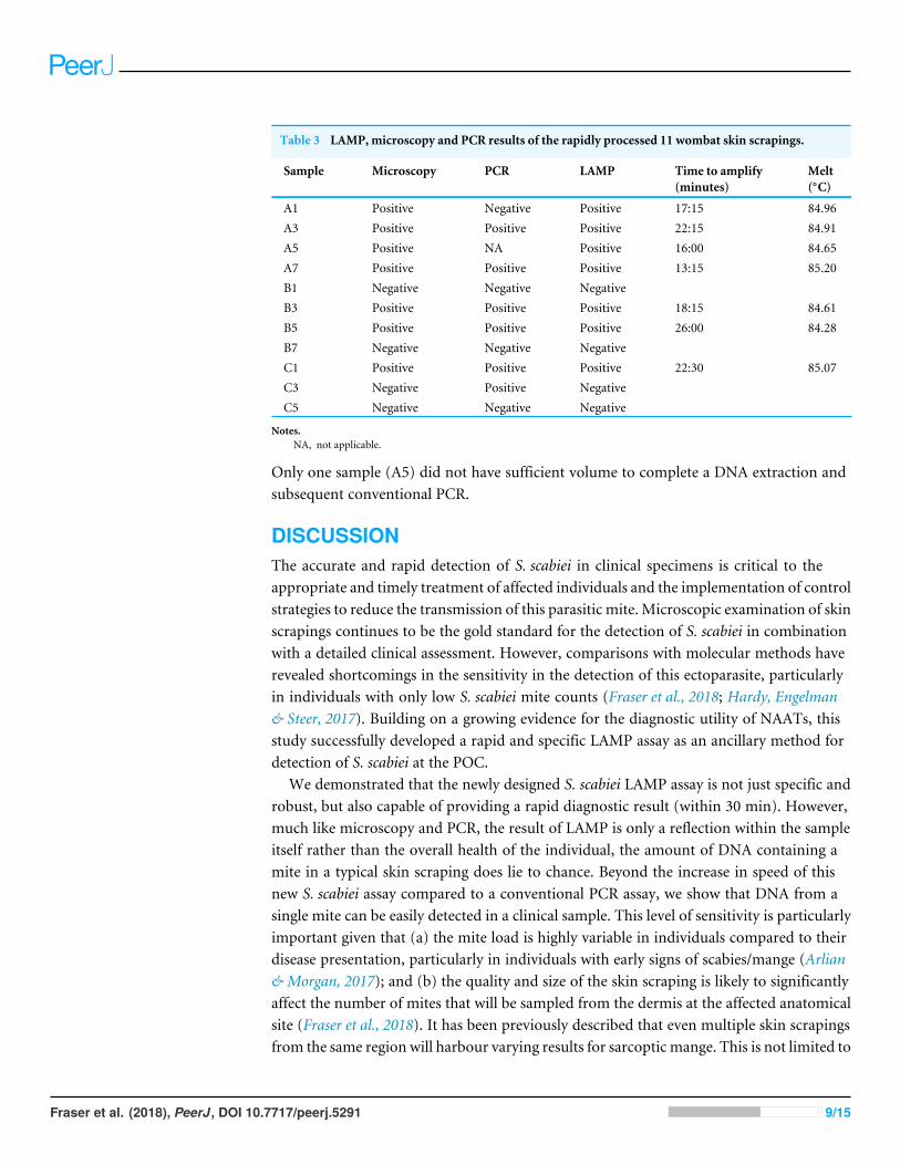
Table 3 LAMP, microscopy and PCR results of the rapidly processed 11 wombat skin scrapings.
Sample Microscopy PCR LAMP Time to amplify(minutes)
Melt(◦C)
A1 Positive Negative Positive 17:15 84.96A3 Positive Positive Positive 22:15 84.91A5 Positive NA Positive 16:00 84.65A7 Positive Positive Positive 13:15 85.20B1 Negative Negative NegativeB3 Positive Positive Positive 18:15 84.61B5 Positive Positive Positive 26:00 84.28B7 Negative Negative NegativeC1 Positive Positive Positive 22:30 85.07C3 Negative Positive NegativeC5 Negative Negative Negative
Notes.NA, not applicable.
Only one sample (A5) did not have sufficient volume to complete a DNA extraction andsubsequent conventional PCR.
DISCUSSIONThe accurate and rapid detection of S. scabiei in clinical specimens is critical to theappropriate and timely treatment of affected individuals and the implementation of controlstrategies to reduce the transmission of this parasitic mite. Microscopic examination of skinscrapings continues to be the gold standard for the detection of S. scabiei in combinationwith a detailed clinical assessment. However, comparisons with molecular methods haverevealed shortcomings in the sensitivity in the detection of this ectoparasite, particularlyin individuals with only low S. scabiei mite counts (Fraser et al., 2018; Hardy, Engelman& Steer, 2017). Building on a growing evidence for the diagnostic utility of NAATs, thisstudy successfully developed a rapid and specific LAMP assay as an ancillary method fordetection of S. scabiei at the POC.
We demonstrated that the newly designed S. scabiei LAMP assay is not just specific androbust, but also capable of providing a rapid diagnostic result (within 30 min). However,much like microscopy and PCR, the result of LAMP is only a reflection within the sampleitself rather than the overall health of the individual, the amount of DNA containing amite in a typical skin scraping does lie to chance. Beyond the increase in speed of thisnew S. scabiei assay compared to a conventional PCR assay, we show that DNA from asingle mite can be easily detected in a clinical sample. This level of sensitivity is particularlyimportant given that (a) the mite load is highly variable in individuals compared to theirdisease presentation, particularly in individuals with early signs of scabies/mange (Arlian& Morgan, 2017); and (b) the quality and size of the skin scraping is likely to significantlyaffect the number of mites that will be sampled from the dermis at the affected anatomicalsite (Fraser et al., 2018). It has been previously described that even multiple skin scrapingsfrom the same region will harbour varying results for sarcoptic mange. This is not limited to
Fraser et al. (2018), PeerJ, DOI 10.7717/peerj.5291 9/15
scabies alone, as low parasitic burdens complicate sampling and diagnosis of other parasiticinfestations, such as cutaneous leishmaniasis (CL), particularly in post-treatment (Faber etal., 2003). LAMP assays on skin biopsies of CL patients were found to be successful priorto treatment, but were unsuccessful in follow-up collections as a result of a low parasiteburden (Adams et al., 2010). Hence, the sensitivity of detection reported in this newlydesigned S. scabiei LAMP assay is promising. Indeed, the detection of S. scabiei DNA inseveral samples that were negative by our comparative S. scabiei conventional PCR assaywould suggest that LAMP could be even more sensitive than conventional PCR for S.scabiei detection. Potential explanations for this enhanced sensitivity include that (i) thequantity of amplicons generated by LAMP assays are considerably higher in comparisonto those produced by PCR, (ii) due to the use of six specific primers rather than two usedin PCR the amplification itself is more targeted, and (iii) the detection of amplicons is byfluorescence rather than visualisation on an agarose gel (Notomi et al., 2000).
A critical aspect limiting the application of this NAAT assay at the POC is the generalrequirement for a DNA extraction prior to PCR detection. Although our results arepreliminary, our data showed that rapid commercial DNA processing kits such as thoseused in this study appear to be effective in lysing mites embedded in tissues to release DNAfor detection. When this method was combined with the LAMP assay itself, it meant thatthe complete reaction time from sampling to result would be generally around 40min. Suchinnovation, alongside alternative sample processing and amplicon detection methods, hasthe potential to make this S. scabiei LAMP assay a POC reality with further development.We and others have also investigated the use of swab sampling of the affected dermis for S.scabiei detection (Fraser et al., 2018; Wong et al., 2015). If further validation would revealthat this non-invasive sampling is suitable for S. scabiei detection, this approach combinedwith the LAMP assay would fulfil the requirements for an assay that can be deployed in arange of clinical settings.
In this study we focussed on the ITS-2 gene as a LAMP target, however three S. scabieimitochondrial genes cox1, 12S rRNA and 16S rRNA could be potentially viable targetsfor LAMP assays based on the availability of sequence data. While not investigated here,we considered but did not pursue these less viable options, as (i) the sequence variationis higher at these mitochondrial loci across the different S. scabiei evolutionary lineagesand (ii) low GC% content of mitochondrial genes could be problematic for the LAMPassay primer design (Fraser et al., 2016; Fraser et al., 2017). Unfortunately, in this studywe did not have human S. scabiei samples to evaluate with our new assay. Nevertheless,our research (Fraser et al., 2017) and that of others (Alasaad et al., 2009; Amer et al., 2014;Andriantsoanirina et al., 2015; Gu & Yang, 2008; Zahler et al., 1999) shows ITS-2 to behighly conserved across host species and, thus, there is no evidence to suggest the LAMPassay developed here would not work equally well on S. scabiei infesting humans.
Besides detection efficiency and species-specific target, other factors (including cost, timeand technical skill) should be also considered during the development of a new diagnosticassay. Regarding scabies, microscopy, although cost efficient, requires technical skill todistinguish mites and eggs within a skin scraping, with a single scraping analysed at a time.Conversely, PCR assays can analyse multiple samples at a time and are highly sensitive at
Fraser et al. (2018), PeerJ, DOI 10.7717/peerj.5291 10/15
detecting mite DNA, eliminating the requirement for specialised technical skills. However,the time to obtain a result can take up to 24 h and the additional equipment and reagents(i.e., accessible laboratory, PCR and electrophoresis equipment and DNA extraction kits)are essential for this diagnostic method. The LAMP assay, in comparison, can be bothcost efficient and rapid, requiring general technical expertise. The fluorometer (such asGenie III used in this study), as one-off cost all-inclusive instrument is portable and canbe run off batteries. As the master mix can be pre-aliquoted and the template rapidlyprocessed, the requirement for additional kits and laboratory space is limited. However,further development for a microfluidic device or colorimetry and the efficiency of the rapidDNA extraction is required for this LAMP assay to be classified as a POC test.
CONCLUSIONIn conclusion, this study describes a development of a new assay for the animal andhuman S. scabiei detection at the POC as well as laboratory. With further development,this assay has the potential to complement existing diagnostic methods in the clinicalsetting and may offer a low cost, portable option for S. scabiei DNA detection in remote orresource-deficient regions.
ACKNOWLEDGEMENTSWe would like to thank Dr. Renfu Shao and Ms. Delaney Burnard for supplying non-S.scabiei DNA to assist with assessing the specificity of our assays. We would also like tothank Dr. Cam Raw, Ms. Roz Holme, Ms. Michelle Thomas and Ms. Merridy Montarellofor supplying additional clinical skin scrapings for molecular typing, which we usedopportunistically for the current LAMP assay.
ADDITIONAL INFORMATION AND DECLARATIONS
FundingThis work was supported by a Holsworth Wildlife Research Endowment awarded toTamieka Fraser. The funders had no role in study design, data collection and analysis,decision to publish, or preparation of the manuscript.
Grant DisclosuresThe following grant information was disclosed by the authors:Holsworth Wildlife Research Endowment award.
Competing InterestsThe authors declare there are no competing interests.
Author Contributions• Tamieka A. Fraser conceived and designed the experiments, performed the experiments,analyzed the data, contributed reagents/materials/analysis tools, prepared figures and/ortables, authored or reviewed drafts of the paper, approved the final draft.
Fraser et al. (2018), PeerJ, DOI 10.7717/peerj.5291 11/15
• Scott Carver analyzed the data, contributed reagents/materials/analysis tools, authoredor reviewed drafts of the paper, approved the final draft.• Alynn M. Martin and Kate Mounsey performed the experiments, contributedreagents/materials/analysis tools, approved the final draft.• Adam Polkinghorne analyzed the data, contributed reagents/materials/analysis tools,authored or reviewed drafts of the paper, approved the final draft.• Martina Jelocnik conceived and designed the experiments, analyzed the data, contributedreagents/materials/analysis tools, prepared figures and/or tables, authored or revieweddrafts of the paper, approved the final draft.
Animal EthicsThe following information was supplied relating to ethical approvals (i.e., approving bodyand any reference numbers):
The collection and use of these samples was approved by the Animal ResearchCommitteeat the University of the Sunshine Coast (approval AN/S/16/43, and AN/E/17/17), theAnimal Research Committee at the University of Tasmania (approval A0014670) andstate permits from Office of Environment & Heritage NSW National Parks & WildlifeService (SL101719), Department of Primary Industries, Park, Water and Environmentfor Tasmania (approval FA15121) and the Department of Environment, Land, Water andPlanning for Victoria (10007943). All methods were carried out in accordance with the2013 Australian National Health and Medical Research Council ‘Australian code for thecare and use of animals for scientific purposes’.
DNA DepositionThe following information was supplied regarding the deposition of DNA sequences:
The 217 bp ITS gene fragment, described in this study is deposited in Genbank underaccession number MH379093.
Data AvailabilityThe following information was supplied regarding data availability:
The raw data are provided in Table S2.
Supplemental InformationSupplemental information for this article can be found online at http://dx.doi.org/10.7717/peerj.5291#supplemental-information.
REFERENCESAdams ER, Schoone GJ, El Safi S, Schallig HD. 2010. Development of a reverse tran-
scriptase loop-mediated isothermal amplification (LAMP) assay for the sensitivedetection of Leishmania parasites in clinical samples. The American Journal ofTropical Medicine and Hygiene 82:591–596 DOI 10.4269/ajtmh.2010.09-0369.
Alasaad S, Soglia D, Spalenza V, Maione S, Soriguer RC, Perez JM, Rasero R,Degiorgis MP, Nimmervoll H, Zhu XQ, Rossi L. 2009. Is ITS-2 rDNA suit-able marker for genetic characterization of Sarcoptesmites from different wild
Fraser et al. (2018), PeerJ, DOI 10.7717/peerj.5291 12/15
animals in different geographic areas? Veterinary Parasitology 159:181–185DOI 10.1016/j.vetpar.2008.10.001.
Amer S, El Wahab TA, Metwaly AEN, Ye J, Roellig D, Feng Y, Xiao L. 2014. Preliminarymolecular characterizations of Sarcoptes scaibiei (Acari: Sarcoptidae) from farmanimals in Egypt. PLOS ONE 9:e94705 DOI 10.1371/journal.pone.0094705.
Andriantsoanirina V, Ariey F, Izri A, Bernigaud C, Fang F, Guillot J, ChosidowO,Durand R. 2015.Wombats acquired scabies from humans and/or dogs from outsideAustralia. Parasitology Research 114:2079–2083 DOI 10.1007/s00436-015-4422-2.
Arlian LG, Feldmeier H, MorganMS. 2015. The potential for a blood test for scabies.PLOS Neglected Tropical Diseases 9:e0004188 DOI 10.1371/journal.pntd.0004188.
Arlian LG, MorganMS. 2017. A review of Sarcoptes scabiei: past, present and future.Parasites & Vectors 10:297 DOI 10.1186/s13071-017-2234-1.
FaberWR, Oskam L, Van Gool T, Kroong NC, Knegt-Junk KJ, Hofwegenf H,Van derWal AC, Kager PA. 2003. Value of diagnostic techniques for cuta-neous leishmaniasis. Journal of the American Academy of Dermatology 49:70–74DOI 10.1067/mjd.2003.492.
ForchhammerMC, Asferg T. 2000. Invading parasites cause a structural shift in redfox dynamics. Proceedings of the Royal Society of London B: Biological Sciences267:779–786 DOI 10.1098/rspb.2000.1071.
Fraser TA, CharlestonM,Martin A, Polkinghorne A, Carver S. 2016. The emergenceof sarcoptic mange in Australian wildlife: an unresolved debate. Parasites & Vectors9:1–11 DOI 10.1186/s13071-016-1578-2.
Fraser TA, Martin A, Polkinghorne A, Carver S. 2018. Comparative diagnostics revealsPCR assays on skin scrapings is the most reliable method to detect Sarcoptes scabieiinfestations. Veterinary Parasitology 251:119–124 DOI 10.1016/j.vetpar.2018.01.007.
Fraser TA, Shao R, Fountain-Jones NM, CharlestonM,Martin A,Whiteley P, HolmeR, Carver S, Polkinghorne A. 2017.Mitochondrial genome sequencing revealspotential origins of the scabies mite Sarcoptes scabiei infesting two iconic Australianmarsupials. BMC Evolutionary Biology 17:233 DOI 10.1186/s12862-017-1086-9.
Gakuya F, Rossi L, Ombui J, Maingi N, Muchemi G, OgaraW, Soriguer RC, Alasaad S.2011. The curse of the prey: Sarcoptesmite molecular analysis reveals potential prey-to-predator parasitic infestation in wild animals from Masai Mara, Kenya. Parasites& Vectors 4:193 DOI 10.1186/1756-3305-4-193.
Graczyk TK, Mudakikwa AB, Cranfield MR, Eilenberger U. 2001.Hyperkeratoticmange caused by Sarcoptes scabiei (Acariformes: Sarcoptidae) in juvenile human-habituated mountain gorillas (Gorilla gorilla beringei). Parasitology Research87:1024–1028.
Gu X-B, Yang G-Y. 2008. A study on the genetic relationship of mites in the genus Sar-coptes (Acari: Sarcoptidae) in China. International Journal of Acarology 34:183–190DOI 10.1080/01647950808683722.
HardyM, Engelman D, Steer A. 2017. Scabies: a clinical update. Australian FamilyPhysician 46:264–268.
Fraser et al. (2018), PeerJ, DOI 10.7717/peerj.5291 13/15
Hay RJ, Johns NE,Williams HC, Bolliger IW, Dellavalle RP, Margolis DJ, Marks R,Naldi L, WeinstockMA,Wulf SK. 2014. The global burden of skin disease in 2010:an analysis of the prevalence and impact of skin conditions. Journal of InvestigativeDermatology 134:1527–1534 DOI 10.1038/jid.2013.446.
Jelocnik M, IslamMM,Madden D, Jenkins C, Branley J, Carver S, Polkinghorne A.2017. Development and evaluation of rapid novel isothermal amplification assays forimportant veterinary pathogens: Chlamydia psittaci and Chlamydia pecorum. PeerJ5:e3799 DOI 10.7717/peerj.3799.
JohnsonM, Zaretskaya I, Raytselis Y, Merezhuk Y, McGinnis S, Madden TL.2008. NCBI BLAST: a better web interface. Nucleic Acids Research 36:W5–W9DOI 10.1093/nar/gkn201.
Kong Q-M, Lu S-H, Tong Q-B, Lou D, Chen R, Zheng B, Kumagai T, Wen L-Y,Ohta N, Zhou X-N. 2012. Loop-mediated isothermal amplification (LAMP):early detection of Toxoplasma gondii infection in mice. Parasites & Vectors 5:2DOI 10.1186/1756-3305-5-2.
Leung V, Miller M. 2011. Detection of scabies: a systematic review of diagnostic methods.Canadian Journal of Infectious Diseases and Medical Microbiology 22:143–146DOI 10.1155/2011/698494.
LöwensteinM, Kahlbacher H, Peschke R. 2004. On the substantial variation in sero-logical responses in pigs to Sarcoptes scabiei var. suis using different commerciallyavailable indirect enzyme-linked immunosorbent assays. Parasitology Research94:24–30.
Lucchi NW, GayeM, Goldman IF, Ljolje D, Deme AB, Badiane A, Ndiaye YD, BarnwellJW, Udhayakumar V, Ndiaye D. 2016. Evaluation of the illumigene malaria LAMP:A robust molecular diagnostic tool for malaria parasites. Scientific Reports 6:36808DOI 10.1038/srep36808.
Martin AM, Burridge CP, Ingram J, Fraser TA, Carver S. 2018. Invasive pathogen driveshost population collapse: effects of a travelling wave of sarcoptic mange on bare-nosed wombats. Journal of Applied Ecology 55:331–341DOI 10.1111/1365-2664.12968.
Marx V. 2015. PCR heads into the field. Nature Methods 12:393–397DOI 10.1038/nmeth.3369.
McCarthy J, KempDJ,Walton SF, Currie BJ. 2004. Scabies: more than just an irritation.Postgraduate Medical Journal 80:382–387 DOI 10.1136/pgmj.2003.014563.
McKenna JP, Cox C, Fairley DJ, Burke R, Shields MD,Watt A, Coyle PV. 2017.Loop-mediated isothermal amplification assay for rapid detection of Streptococcusagalactiae (group B Streptococcus) in vaginal swabs—a proof of concept study.Journal of Medical Microbiology 66:294–300 DOI 10.1099/jmm.0.000437.
Notomi T, Okayama H, Masubuchi H, Yonekawa T,Watanabe K, Amino N, HaseT. 2000. Loop-mediated isothermal amplification of DNA. Nucleic Acids Research28:e63–e63.
Fraser et al. (2018), PeerJ, DOI 10.7717/peerj.5291 14/15
Owczarzy R, Tataurov AV,Wu Y, Manthey JA, McQuisten KA, Almabrazi HG, Peder-sen KF, Lin Y, Garretson J, McEntaggart NO. 2008. IDT SciTools: a suite for anal-ysis and design of nucleic acid oligomers. Nucleic Acids Research 36:W163–W169DOI 10.1093/nar/gkn198.
Perrucci S, Verin R, Mancianti F, Poli A. 2016. Sarcoptic mange and other ectoparasiticinfections in a red fox (Vulpes vulpes) population from central Italy. ParasiteEpidemiology and Control 1:66–71 DOI 10.1016/j.parepi.2016.03.007.
Rambozzi L, Menzano A, Lavin S, Rossi L. 2004. Biotin-avidin amplified ELISA fordetection of antibodies to Sarcoptes scabiei in chamois (Rupicapra spp.). VeterinaryResearch 35:701–708 DOI 10.1051/vetres:2004039.
Rodríguez-Cadenas F, Carbajal-González M, Fregeneda-Grandes J, Aller-GancedoJ, Huntley J, Rojo-Vázquez F. 2010. Development and evaluation of an antibodyELISA for sarcoptic mange in sheep and a comparison with the skin-scrapingmethod. Preventive Veterinary Medicine 96:82–92DOI 10.1016/j.prevetmed.2010.05.009.
Romani L, Steer AC,Whitfeld MJ, Kaldor JM. 2015. Prevalence of scabies and im-petigo worldwide: a systematic review. The Lancet Infectious Diseases 15:960–967DOI 10.1016/S1473-3099(15)00132-2.
Saito R, Misawa Y, Moriya K, Koike K, Ubukata K, Okamura N. 2005. Developmentand evaluation of a loop-mediated isothermal amplification assay for rapid detec-tion ofMycoplasma pneumoniae. Journal of Medical Microbiology 54:1037–1041DOI 10.1099/jmm.0.46071-0.
Sergeant. 2017. Epitools epidemiological calculators. Available at http:// epitools.ausvet.com.au.
Skerratt LF. 2005. Sarcoptes scabiei: an important exotic pathogen of wombats.Microbiol-ogy Australia 26:79–81.
Walton SF, Currie BJ. 2007. Problems in diagnosing scabies, a global disease inhuman and animal populations. Clinical Microbiology Reviews 20:268–279DOI 10.1128/CMR.00042-06.
Wong SS, Poon RW, Chau S,Wong SC, To KK, Cheng VC, Fung KS, Yuen K. 2015.Development of conventional and realtime quantitative polymerase chain reactionassay in the diagnosis and monitoring of scabies. Journal of Clinical Microbiology53:2095–2102 DOI 10.1128/JCM.00073-15.
Zahler M, Essig A, Gothe R, Rinder H. 1999.Molecular analyses suggest monospecificityof the genus Sarcoptes (Acari: Sarcoptidae). International Journal for Parasitology29:759–766 DOI 10.1016/S0020-7519(99)00034-X.
Zhang S, RavelonandroM, Russell P, McOwen N, Briard P, Bohannon S, Vrient A.2014. Rapid diagnostic detection of plum pox virus in Prunus plants by isothermalAmplifyRP R© using reverse transcription-recombinase polymerase amplification.Journal of Virological Methods 207:114–120 DOI 10.1016/j.jviromet.2014.06.026.
Zhao Y, Cao Z, Cheng J, Hu L, Ma J, Yang Y,Wang X, Zeng J, Wang T. 2014. Popu-lation identification of Sarcoptes hominis and Sarcoptes canis in China using DNAsequences. Parasitology Research 10:1001–1010.
Fraser et al. (2018), PeerJ, DOI 10.7717/peerj.5291 15/15