Embed Size (px)
Citation preview
Visitor address: ulls väg 2 B. Postal address. 751 89 Uppsala Telephone. +46 18 67 40 00 Fax. +46 18 30 91 62 E-mail. [email protected] Web. www.sva.se
Editor: Erik Ågren Authors: Caroline Bröjer, Gete Hestvik, Aleksija Neimanis, Jonas Malmsten, Torsten Mörner, Henrik Uhlhorn, Erik Ågren Photo, cover: Fredrik Olsson. Photo credits: See each photo. Layout: Gun-Britt Rydén, Erik Ågren Suggestion citation: Wildlife disease surveillance in Sweden 2015. National Veterinary Institute, SVA, Uppsala SVA’s report series 33 ISSN 1654-7098

Table of contents
Table of contents 1 Introduction 1 Wildlife disease surveillance in Sweden 2 Staff working with wildlife diseases 3
Wildlife section 2015 3 The four large carnivores 3 Other staff working with wildlife 3 Wildlife diseases of interest 4
Skin ulcers in moose 4 Myxomatosis 5 Tularemia outbreak, hares 5 Lead poisoning in mute swans 5 Avian flu in mute swans 6 Trichomonas in greenfinches 6 Pigeon paramyxovirus 6
Wildlife disease surveillance 2015 7 Statistics wildlife cases 7
Cases submitted to SVA 7 Cases of wildlife per species 2015 8 Common Diagnoses 9
Game species 10 Moose 10 Roe deer 10 Wild boar 11 Hare 11 Red fox 11 Birds 11
Targeted wildlife disease surveillance 12 Echinococcus 12 Serologic screening of wild boar 12 Avian flu 13 Trichinella 13
OIE report 14 Acute projects 2015 15
Moose calf mortality on Öland 15 Rabbit hemorrhagic disease type 2 15 PCR analysis for myxomatosis 16
The four large carnivores 17
Brown bear 17 Wolverine 17 Lynx 17 Wolf 17
Museum of Natural History 19 Marine mammals 19 Eagles 20 Otters 20
Wildlife research projects at SVA 21 Hepatitis E virus 21 Tularemia 21 Nodular onchocercosis in red deer 22
Miscellaneous 23 Forensic investigations 23 Biobank 23 International disease surveillance 23 Disseminating knowledge 23
Visits and lectures 23 Contacting the wildlife section 24
Telephone and email 24 Web-based reporting 24 Transport of dead wildlife 24
Publications 2015 25

1
Introduction The health status of wildlife in Sweden is monitored through SVA's wildlife disease surveillance program VSÖP. This annual report summarizes the work and results from the VSÖP, highlighting wildlife disease events of significance from 2015. Uppsala, August 2016 Erik Ågren, head of the Wildlife Section Dolores Gavier-Widén, Head of Department, Department of Pathology and Wildlife Diseases Torsten Mörner, State veterinarian, Department of Epidemiology and Disease Control
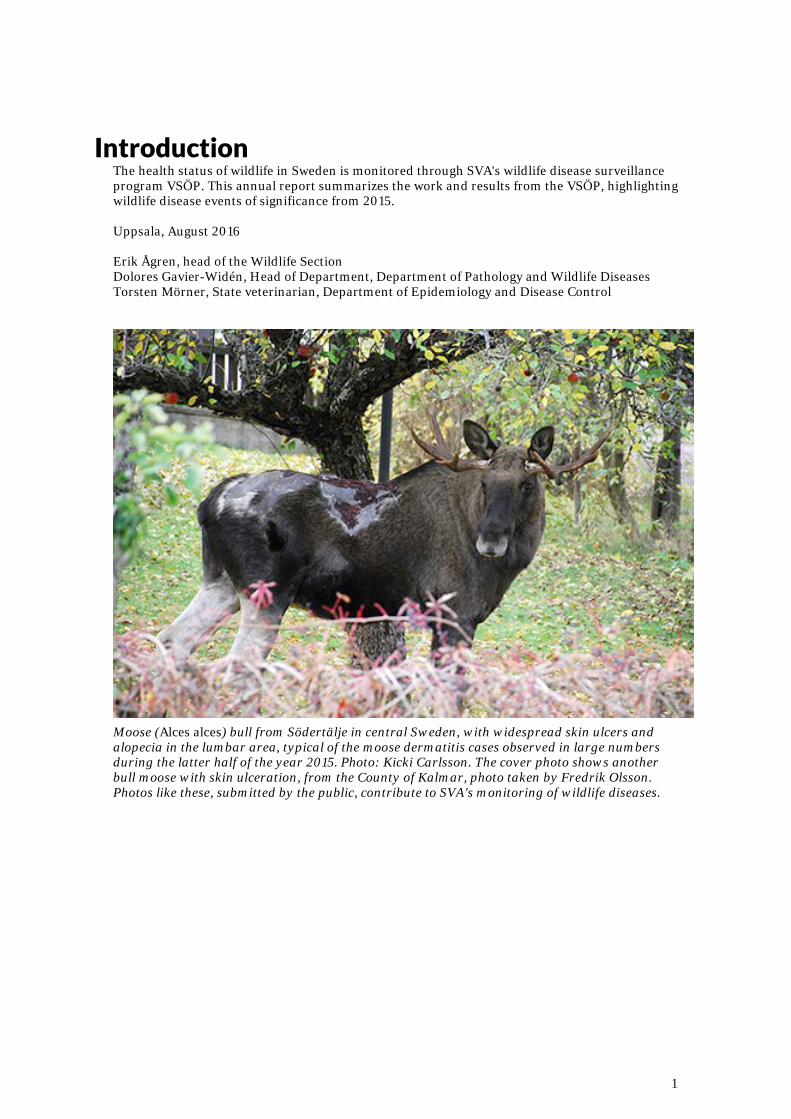
Moose (Alces alces) bull from Södertälje in central Sweden, with widespread skin ulcers and alopecia in the lumbar area, typical of the moose dermatitis cases observed in large numbers during the latter half of the year 2015. Photo: Kicki Carlsson. The cover photo shows another bull moose with skin ulceration, from the County of Kalmar, photo taken by Fredrik Olsson. Photos like these, submitted by the public, contribute to SVA's monitoring of wildlife diseases.
2
Wildlife disease surveillance in Sweden The Government's directive specifies that the veterinary expert authority SVA shall do a comprehensive assessment and analysis of the status of infectious diseases as well as the state of health in general of domestic and wild animals in Sweden. SVA is the only Swedish veterinary laboratory systematically working on disease surveillance of wild animals. The work is mainly based on the pathological examination of wildlife found dead, or samples from sick and euthanized wildlife, with the addition of samples collected from hunted game, for the monitoring of certain infectious agents. SVA also collaborates with other research groups and projects involved in wildlife studies, to obtain a more complete picture of disease issues in wildlife. Here we report the main activities and results of interest concerning wildlife disease monitoring during 2015. Systematic Wildlife Disease Surveillance has been performed since the 1940’s at SVA. The main component consists of general disease surveillance (fallen wildlife monitoring), supplemented with targeted monitoring and investigative efforts. The present Wildlife Disease Surveillance Programme (Viltsjukdomsövervakningsprogrammet) is possible through funding from the State Wildlife Fund (generated from Swedish hunting license fees) as well as funding from the government and the Environmental Protection Agency. The Wildlife Disease Council (Viltsjukdomsrådet) is a group of experts and officials from the Environmental Protection Agency and SVA responsible for exchanging information on wildlife surveillance, wildlife management and wildlife disease surveillance and to jointly discuss appropriate Active disease surveillance activities on wildlife in Sweden. In 2015, the Council consisted of Klas Allander, and Ola Inghe from the EPA and Dolores Gavier-Widén, Torsten Mörner, and Erik Ågren, with Henrik Uhlhorn as a secretary, from SVA. VSR held two meetings in 2015. The Hunters Association's Game sampler’s organization (Viltprovtagarna) is a voluntary network of hunters within the Swedish Association for Hunting and Wildlife Management (Svenska Jägareförbundet, SJF). The network is active in all 21 counties of Sweden and assists in reporting disease and mortality events in wildlife, assists with the collection of wildlife tissue samples from hunted game species and aids the public through submission of fallen wildlife for general disease monitoring. DEFINITIONS
General disease surveillance involves diagnosing diseases by necropsies, histopathology and ancillary testing of found dead wildlife or euthanized sick wildlife. Targeted disease surveillance involves targeted sampling and examination of sick or healthy wildlife to investigate specific diseases or disease agents. Most often, these investigations are initiated by results found through general disease surveillance, or when information about emerging diseases or ongoing outbreaks are reported within Sweden or in neighboring countries.
3
Staff at SVA working with wildlife diseases The wildlife section is part of the Department of Pathology and Wildlife Diseases. The wildlife section work is focused on pathology of wildlife and the majority of employees are veterinary pathologists. We collaborate with the other specialized laboratories and veterinary experts throughout SVA regarding analyses of infectious agents (e.g. bacteria, viruses, parasites) and chemical substances, and with specialists in epidemiology, to diagnose, study and report on the status of wildlife diseases. Wildlife section 2015
Erik Ågren, head of section, Veterinary officer, Dipl. ECVP, DipECZM (Wildlife population health) Caroline Bröjer, Veterinary officer, MSc, PhD, DipECZM (Wildlife population health) Gete Hestvik, Veterinary officer Jonas Malmsten, Veterinary officer, PhD, DipECZM (Wildlife population health) Aleksija Neimanis, Veterinary officer, BSc, MSc, Dipl ACVP, MVetSci. Henry Uhlhorn, Veterinary officer, PhD Tomas Meijer, researcher, PhD. Jessica Åsbrink; research engineer, MSc. Ewa Backman and Carina Bohlin, Secretaries at the wildlife section. The four large carnivores
Tomas Meijer, Jessica Åsbrink, Erik Ågren, Jonas Malmsten. Other staff at SVA working with wildlife
Necropsy assistants Hans Kanbjer, Johan Karevik, Lars Hammarsten. Necropsy technicians Marit Liljefors, Sandra Karevik. Dolores Gavier-Widén, PhD, head of Department. Histological laboratory technicians Torsten Mörner, State veterinarian of wildlife diseases, PhD, Associate Professor, Department of Epidemiology and Disease Control. OIE National Focal point for wildlife diseases.
Wildlife section staff 2015. From left: Gete Hestvik, Carina Bohlin, Henrik Uhlhorn, Tomas Meijer, Ewa Backman, Aleksija Neimanis, Jonas Malmsten, Caroline Bröjer, Erik Ågren. Photo: SVA
4
Wildlife diseases of particular interest 2015
SKIN ULCERS IN MOOSE
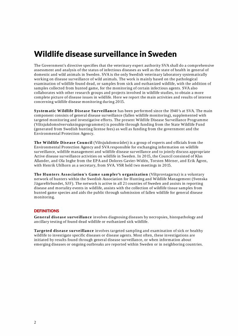
During the late summer of 2015, SVA received a large number of reports on moose (Alces alces) with widespread and severe skin lesions on the back. Similar changes have been reported in the past, but only as occasional, single cases. The Swedish Environmental Protection Agency funded an investigation into the increased number of cases in moose. A questionnaire was sent out to all Swedish hunting units, and further reporting and submission of cases was encouraged. This resulted in 149 cases reported from October 2015 until March 2016, found scattered across the southern half of Sweden. No cases were reported from northern Sweden. All cases but one were seen in adult males. Carcasses or skin samples from 58 cases were submitted for gross and microscopic examination, as well as parasitology and bacteriologic cultures. In almost all cases, the lesions were observed in the skin of the dorsal lumbar area, and in some cases with spread along the spine or over the rump or sides of the body. The largest wounds were over one meter in length. Deep, suppurative ulcerations covered by thick dried scabs were surrounded by areas of alopecic skin. The ulcerated skin was greatly thickened by fibrosis, which indicated a prolonged disease process. The pathologic changes were similar to pyotraumatic dermatitis, suggesting a background of chronic pruritis with secondary bacterial infection. Most investigated moose skins had high loads of deer ked (Lipoptena cervi). In some cases, moose ear mites (Chorioptes sp.), were observed in the skin. In 40 of 45 skin samples, the bacterium Staphylococcus aureus was isolated through culture. This bacterium can cause skin infections under some conditions. The pathogenesis of these skin ulcers is not fully understood, but it is possible that ectoparasites initiate a severe itch, leading to chronic scratching. Male moose with large antlers can use the antlers to scratch on the lumbar area of the back. Chronic scratching would lead to minor skin lesions, that then could be infected. In combination with warm, moist weather conditions, it is
conceivable that these infections then spread over larger areas of the skin. These suppurative ulcerations differ from the numerous cases of extensive alopecia in moose previously reported, especially in 2007 and 2011. These alopecic moose had abundant deer ked infestations and were noted in both Sweden and Norway, but only very few animals were seen to also have severe skin ulcers. The study of moose skin ulcerations continues in 2016.
Bull moose with widespread dorsal skin ulcers. Picture from the moose hunt 2015. Photo: E. Jenhall
5
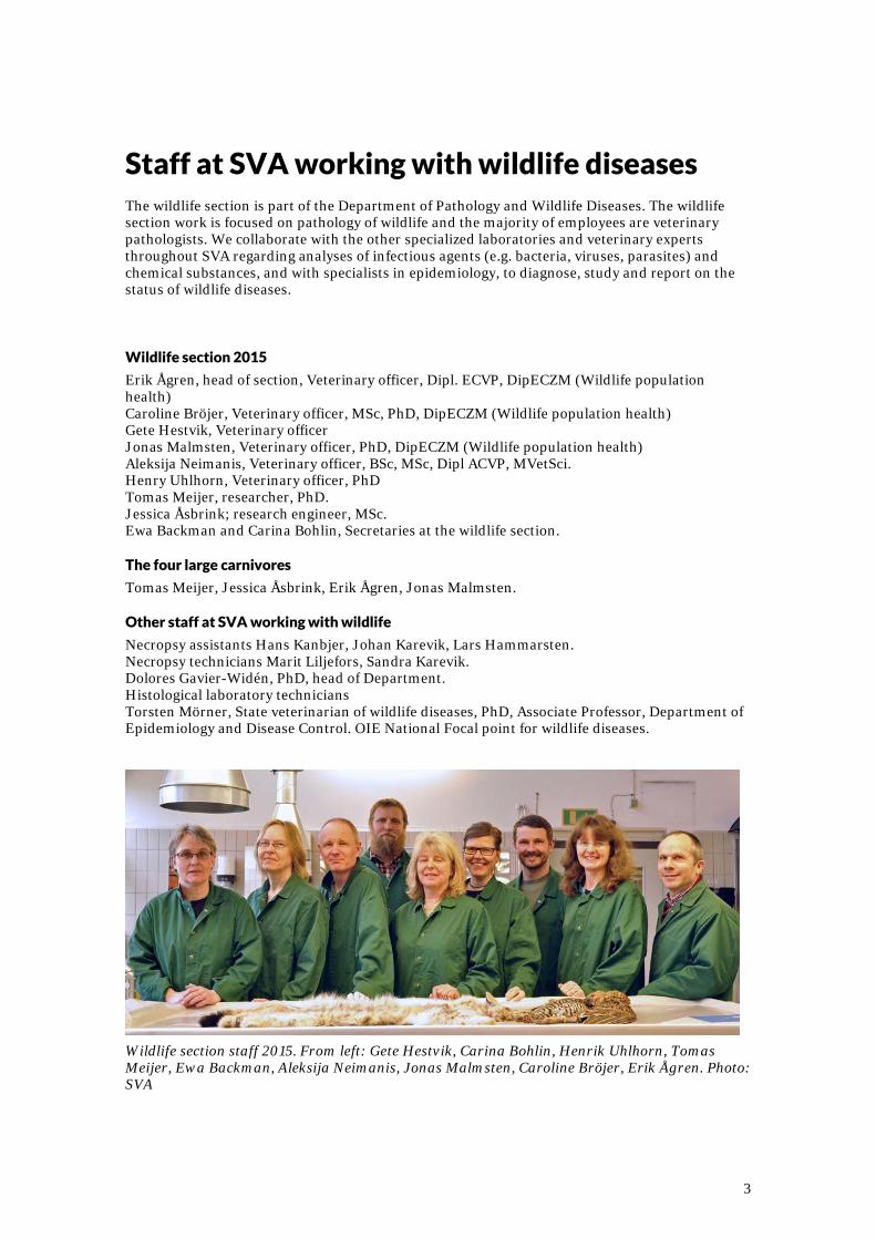
MYXOMATOSIS A widespread outbreak of myxomatosis in wild rabbits continued in 2015 in the southern counties of Sweden, where wild rabbits are more common. Reports and photos of sick and dead rabbits with severely swollen eyelids were submitted to SVA from many cities and towns in the county of Skåne, with fewer reports from the counties of Halland, Blekinge and Kronoberg. The disease is notifiable when diagnosed by a laboratory, so all verified cases are reported to the Board of Agriculture, and every six months to the OIE (the World Health Organization for animals).
Wild rabbit (Oryctolagus cuniculus) with swollen eyelids, typical myxomatosis lesions, in Skåne County in southern Sweden, 2015. Photo: Helene Okhagen TULAREMIA OUTBREAK, HARES
From late July to early September 2015 SVA received a large number of reports of mountain hares (Lepus timidus) found dead in the northern counties of Västerbotten and Norrbotten, especially from the coastal areas. With a lot of help from staff at the local Swedish Association for Hunting and Game Management office, and from the interested public, carcasses of 31 mountain hares were sent to SVA for examination. There are no European brown hares (Lepus europaeus) in these counties. After necropsy and bacteriologic investigation, the disease tularemia, caused by the bacterium Francisella tularensis, was confirmed in 24 of the submitted hares. At necropsy, there were severe inflammatory changes typical of tularemia in several organs, usually in the spleen, liver, and bone marrow. Hares die after a very short period of illness from septicaemia. Major outbreaks of tularemia occur every few years. During this year's
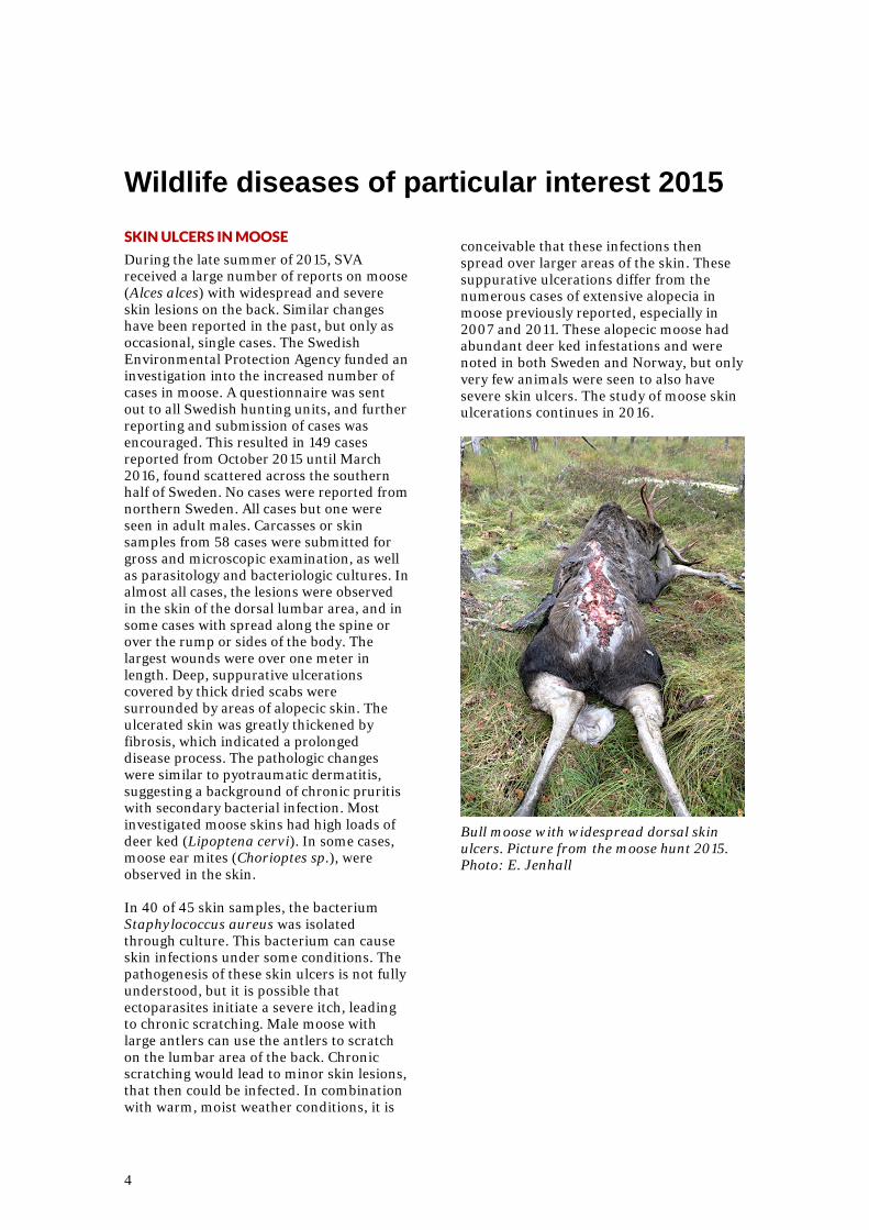
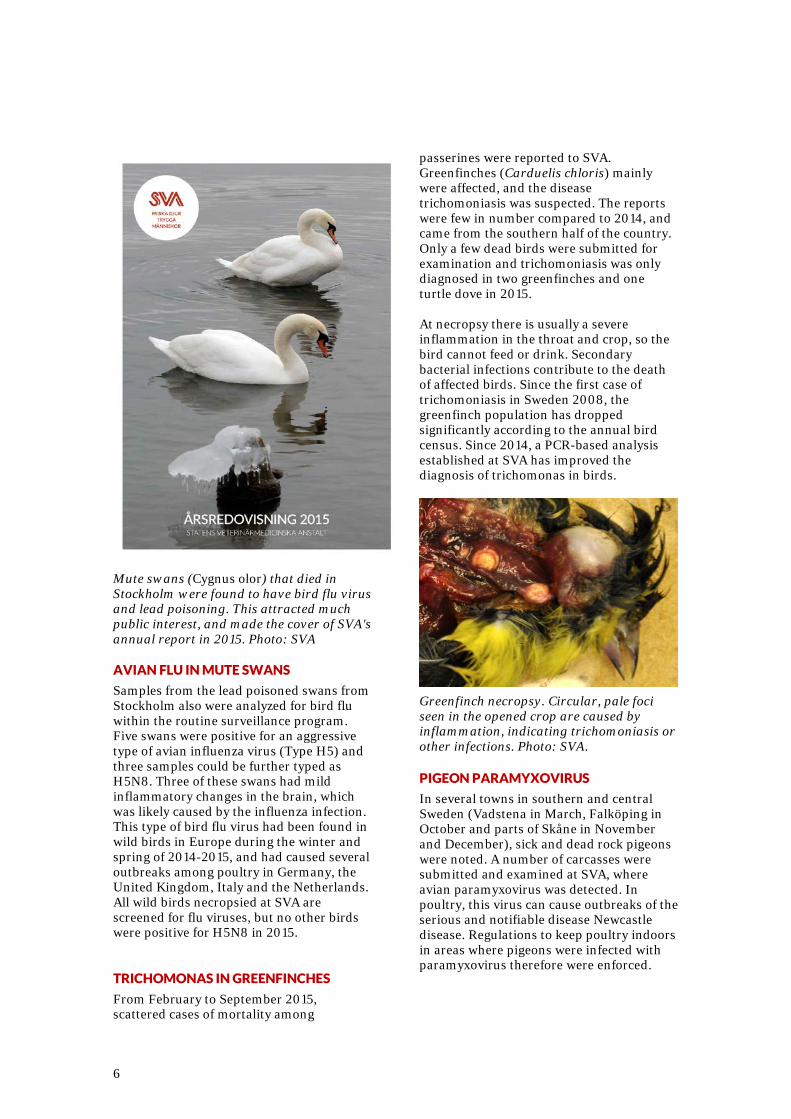
outbreak, the public health authority (Folkhälsomyndigheten) reported a large number of tularemia cases in people, which coincided with the hare mortalities. Concurrently, there were reports of tularemia cases in hares and people in neighboring Finland. People can contract tularemia when handling a sick hare, but infection by mosquitoes that act as carriers of the bacterium is thought to be more common. Although most species can become infected with tularemia, most reported cases come from hares and humans. It is likely that mice, voles and other small rodents also are affected during outbreaks, but the small carcasses are more difficult to note or find in the field. AVIAN INFLUENZA AND LEAD POISONING IN MUTE SWANS During February and March 2015, a number of dead or dying mute swans (Cygnus olor) were found in central Stockholm, generating media attention as these large birds are rather obvious in the city. Twelve of these swans were submitted to SVA for necropsy. Emaciation was noted in four swans, six were in poor body condition and two were in normal body condition. Most cases had a distended and impacted esophagus and intestinal tract, which can be an indication of lead poisoning. Chemical analysis of liver and kidney tissue showed that all examined birds had elevated lead values. The highest lead level in kidney tissue was 178 µg/g wet weight, which is extremely high considering that the lower range of lead that causes fatal lead poisoning is around 5 µg/g wet weight. Lead poisoning only occurs if swans ingest lead shot or lead fragments. At necropsy, there were no apparent lead particles found in the stomachs of the swans. The origin of the high lead levels causing lead poisoning in these swans could not be determined. Avian influenza virus also was detected in several of these swans (see below).
6
Mute swans (Cygnus olor) that died in Stockholm were found to have bird flu virus and lead poisoning. This attracted much public interest, and made the cover of SVA's annual report in 2015. Photo: SVA AVIAN FLU IN MUTE SWANS
Samples from the lead poisoned swans from Stockholm also were analyzed for bird flu within the routine surveillance program. Five swans were positive for an aggressive type of avian influenza virus (Type H5) and three samples could be further typed as H5N8. Three of these swans had mild inflammatory changes in the brain, which was likely caused by the influenza infection. This type of bird flu virus had been found in wild birds in Europe during the winter and spring of 2014-2015, and had caused several outbreaks among poultry in Germany, the United Kingdom, Italy and the Netherlands. All wild birds necropsied at SVA are screened for flu viruses, but no other birds were positive for H5N8 in 2015. TRICHOMONAS IN GREENFINCHES
From February to September 2015, scattered cases of mortality among
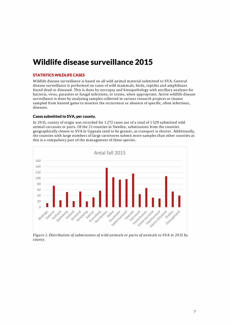
passerines were reported to SVA. Greenfinches (Carduelis chloris) mainly were affected, and the disease trichomoniasis was suspected. The reports were few in number compared to 2014, and came from the southern half of the country. Only a few dead birds were submitted for examination and trichomoniasis was only diagnosed in two greenfinches and one turtle dove in 2015. At necropsy there is usually a severe inflammation in the throat and crop, so the bird cannot feed or drink. Secondary bacterial infections contribute to the death of affected birds. Since the first case of trichomoniasis in Sweden 2008, the greenfinch population has dropped significantly according to the annual bird census. Since 2014, a PCR-based analysis established at SVA has improved the diagnosis of trichomonas in birds.
Greenfinch necropsy. Circular, pale foci seen in the opened crop are caused by inflammation, indicating trichomoniasis or other infections. Photo: SVA. PIGEON PARAMYXOVIRUS
In several towns in southern and central Sweden (Vadstena in March, Falköping in October and parts of Skåne in November and December), sick and dead rock pigeons were noted. A number of carcasses were submitted and examined at SVA, where avian paramyxovirus was detected. In poultry, this virus can cause outbreaks of the serious and notifiable disease Newcastle disease. Regulations to keep poultry indoors in areas where pigeons were infected with paramyxovirus therefore were enforced.
7
Wildlife disease surveillance 2015
STATISTICS WILDLIFE CASES
Wildlife disease surveillance is based on all wild animal material submitted to SVA. General disease surveillance is performed on cases of wild mammals, birds, reptiles and amphibians found dead or diseased. This is done by necropsy and histopathology with ancillary analyses for bacteria, virus, parasites or fungal infections, or toxins, when appropriate. Active wildlife disease surveillance is done by analyzing samples collected in various research projects or tissues sampled from hunted game to monitor the occurrence or absence of specific, often infectious, diseases. Cases submitted to SVA, per county.
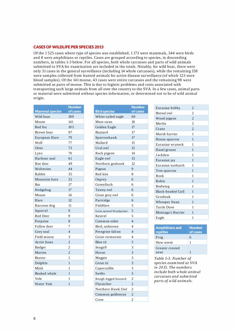
In 2015, county of origin was recorded for 1 272 cases out of a total of 1 529 submitted wild animal carcasses or parts. Of the 21 counties in Sweden, submissions from the counties geographically closest to SVA in Uppsala tend to be greater, as transport is shorter. Additionally, the counties with large numbers of large carnivores submit more samples than other counties as this is a compulsory part of the management of these species.
Figure 1. Distribution of submissions of wild animals or parts of animals to SVA in 2015 by county.
0
20
40
60
80
100
120
140
160
Antal fall 2015
8
CASES OF WILDLIFE PER SPECIES 2015
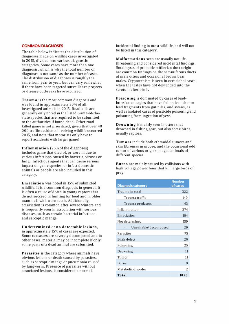
Of the 1 525 cases where type of species was established, 1 173 were mammals, 344 were birds and 8 were amphibians or reptiles. Cases are grouped according to species, in descending numbers, in tables 1-3 below. For all species, both whole carcasses and parts of wild animals submitted to SVA for examination are included in the totals. Notably, for wild boar, there were only 31 cases in the general surveillance (including 14 whole carcasses), while the remaining 158 were samples collected from hunted animals for active disease surveillance (of which 123 were blood samples). Of the 141 moose, 43 cases were entire carcasses and the remaining 98 were submitted as parts of moose. This is due to logistic problems and costs associated with transporting such large animals from all over the country to the SVA. In a few cases, animal parts or material were submitted without species information, or determined not to be of wild animal origin.
Mammal species Number of cases
Wild boar 189 Moose 141 Red fox 103 Brown bear 97 European Hare 79 Wolf 77 Otter 73 Lynx 70 Harbour seal 61 Roe deer 49 Wolverine 44 Rabbit 35 Mountain hare 32 Bat 17 Hedgehog 17 Mouse 14 Hare 12 Raccoon dog 11 Squirrel 9 Red Deer 9 Porpoise 8 Fallow deer 7 Grey seal 4 Field mouse 3 Arctic foxes 2 Badger 2 Marten 2 Beaver 1 Dolphin 1 Mink 1 Beaked whale 1 Vole 1 Water Vole 1
Bird species Number of cases
White-tailed eagle 69 Mute swan 18 Golden Eagle 17 Buzzard 17 Sparrowhawk 17 Mallard 15 Ural owl 15 Rock pigeon 14 Eagle owl 13 Northern goshawk 12 Pigeon 9 Red kite 8 Osprey 6 Greenfinch 6 Tawny owl 6 Great grey owl 6 Partridge 6 Fieldfare 5 Great spotted Woodpecker 5 Kestrel 5 Common eider 4 Bird, unknown 4 Peregrine falcon 4 Great cormorant 4 Blue tit 3 Seagull 3 Heron 3 Magpie 3 Great tit 3 Capercaillie 3 Swifts 3 Rough-legged buzzard 2 Flycatcher 2 Northern Hawk Owl 2 Common goldeneye 2 Crow 2
Eurasian hobby 2 Boreal owl 2 Wood pigeon 2 Merlin 2 Crane 2 Marsh harrier 1 House sparrow 1 Eurasian wryneck 1 Hazel grouse 1 Jackdaw 1 Eurasian jay 1 Eurasian nuthatch 1 Tree sparrow 1 Rook 1 Robin 1 Redwing 1 Black-headed Gull 1 Grosbeak 1 Whooper Swan 1 Turtle Dove 1 Montagu's Harrier 1 Eagle 1
Amphibians and reptiles
Number of cases
Frog 6 Slow worm 1
Greater crested newt 1
Table 1-3. Number of species examined at SVA in 2015. The numbers include both whole animal carcasses and submitted parts of wild animals.
9
COMMON DIAGNOSES
The table below indicates the distribution of diagnoses made on wildlife cases investigated in 2015, divided into various diagnostic categories. Some cases have more than one diagnosis, which is why the total number of diagnoses is not same as the number of cases. The distribution of diagnoses is roughly the same from year to year, but can vary somewhat if there have been targeted surveillance projects or disease outbreaks have occurred. Trauma is the most common diagnosis and was found in approximately 30% of all investigated animals in 2015. Road kills are generally only noted in the listed Game-of-the-state species that are required to be submitted to the authorities if found dead. Other road killed game is not prioritized, given that over 48 000 traffic accidents involving wildlife occurred 2015, and note that motorists only have to report accidents with larger game! Inflammation (25% of the diagnoses) includes game that died of, or were ill due to various infections caused by bacteria, viruses or fungi. Infectious agents that can cause serious impact on game species, or infect domestic animals or people are also included in this category. Emaciation was noted in 15% of submitted wildlife. It is a common diagnosis in general. It is often a cause of death in young raptors that do not succeed in hunting for food and in older mammals with worn teeth. Additionally, emaciation is common after severe winters and is frequently seen in association with serious diseases, such as certain bacterial infections and sarcoptic mange. Undetermined or no detectable lesions, in approximately 15% of cases are expected. Some carcasses are severely decomposed and in other cases, material may be incomplete if only some parts of a dead animal are submitted. Parasites is the category where animals have obvious lesions or death caused by parasites, such as sarcoptic mange or pneumonia caused by lungworm. Presence of parasites without associated lesions, is considered a normal,
incidental finding in most wildlife, and will not be listed in this category. Malformations seen are usually not life-threatening and considered incidental findings. Small cysts of probable müllerian duct origin are common findings on the seminiferous ducts of male otters and occasional brown bear males. Cryptorchism is seen in occasional cases when the testes have not descended into the scrotum after birth. Poisoning is dominated by cases of lead-intoxicated eagles that have fed on lead shot or lead fragments from gut piles, and swans, as well as isolated cases of pesticide poisoning and poisoning from ingestion of yew. Drowning is mainly seen in otters that drowned in fishing gear, but also some birds, usually raptors. Tumors include both ethmoidal tumors and skin fibromas in moose, and the occasional odd tumor of various origins in aged animals of different species. Burns are mainly caused by collisions with high voltage power lines that kill large birds of prey.
Diagnosis category Number of cases
Trauma in total 322
Trauma traffic 140
Trauma predators 43
Inflammation 274
Emaciation 164
Not determined 159
- Unsuitable/decomposed 29
Parasites 75
Birth defect 26
Poisoning 25
Drowning 11
Tumor 11
Burns 9
Metabolic disorder 2
Total 1078
10
Diseases and causes of mortality in game species Half of the funding for the general disease surveillance of wildlife at SVA comes from the Wildlife Fund (Viltvårdsfonden), which is financed by part of the compulsory annual state hunting fee for all hunters. Here we report diagnoses and findings in some common game species in 2015. MOOSE
In all, 148 moose or parts of moose were submitted in 2015. For 129 moose, a total of 134 diagnoses were made (see table below), in addition to a number of minor findings. The most common diagnosis was inflammation/ infection (54%) where dermatitis cases dominated (see page 4). Parasite findings of note include nasal bots, meningeal worm and Cysticercus tapeworm larvae. Emaciation as well as trauma from traffic or predators were common diagnoses. Tumors were predominately ethmoidal tumors and skin fibromas.
Diagnosis category Number of cases
Inflammation/infection 70 Parasites 13 Emaciation 11 Trauma 10 Tumor 9 No pathological changes 7 Poisoning 4 Not determined 4 Birth defect 3 Shock 2 Abortion 1
Table 5. Distribution of diagnosis categories for 129 moose cases examined at SVA in 2015. ROE DEER
SVA received 49 roe deer cases in 2015. Inflammation and trauma were the most common diagnoses. Diarrhea in combination with emaciation was also common. Roe deer diarrhea was reported from various areas, but has not caused widespread mortality. Eye infections caused by Listeria bacteria have been noted in some roe deer, similar to cases in sheep, that were associated with feeding with silage. The trauma category was dominated by predation. Domestic dogs occasionally kill roe deer in suburban areas, which can be a legal offence.
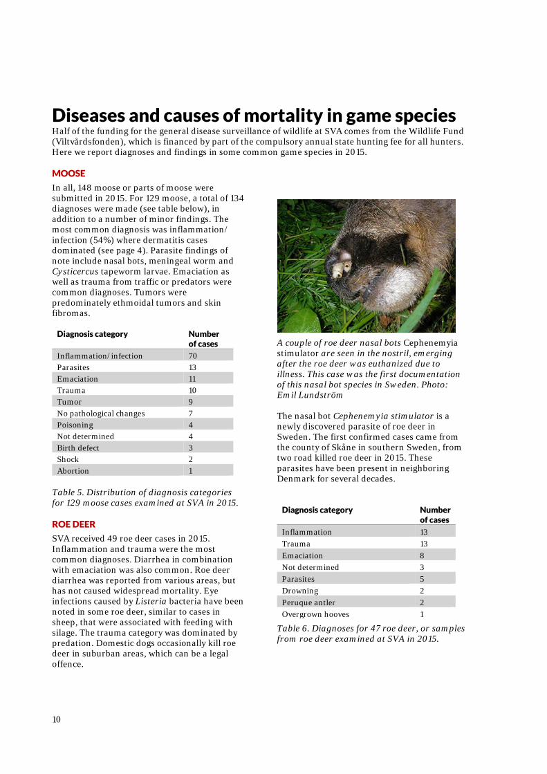
A couple of roe deer nasal bots Cephenemyia stimulator are seen in the nostril, emerging after the roe deer was euthanized due to illness. This case was the first documentation of this nasal bot species in Sweden. Photo: Emil Lundström The nasal bot Cephenemyia stimulator is a newly discovered parasite of roe deer in Sweden. The first confirmed cases came from the county of Skåne in southern Sweden, from two road killed roe deer in 2015. These parasites have been present in neighboring Denmark for several decades.
Diagnosis category Number of cases
Inflammation 13 Trauma 13 Emaciation 8 Not determined 3 Parasites 5 Drowning 2 Peruque antler 2 Overgrown hooves 1
Table 6. Diagnoses for 47 roe deer, or samples from roe deer examined at SVA in 2015.
11
WILD BOAR
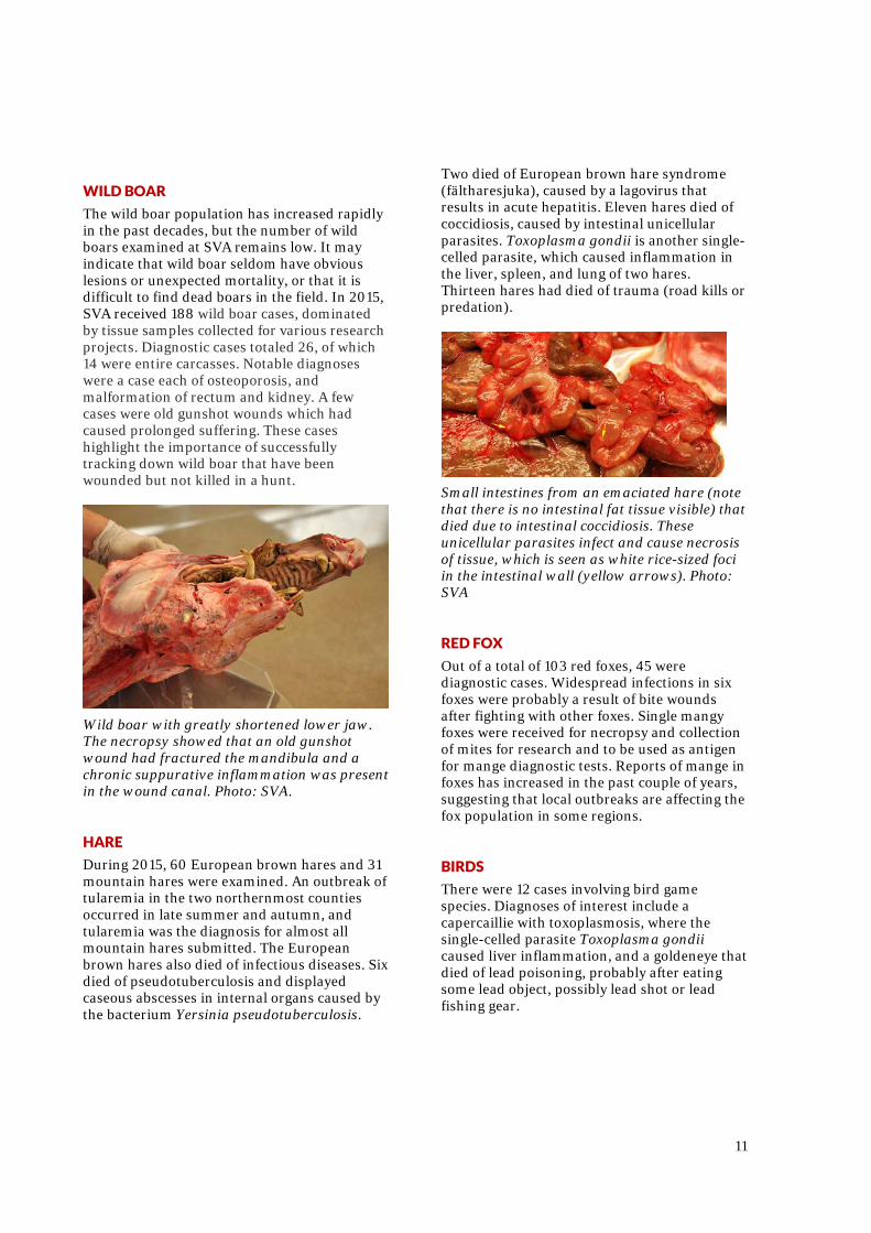
The wild boar population has increased rapidly in the past decades, but the number of wild boars examined at SVA remains low. It may indicate that wild boar seldom have obvious lesions or unexpected mortality, or that it is difficult to find dead boars in the field. In 2015, SVA received 188 wild boar cases, dominated by tissue samples collected for various research projects. Diagnostic cases totaled 26, of which 14 were entire carcasses. Notable diagnoses were a case each of osteoporosis, and malformation of rectum and kidney. A few cases were old gunshot wounds which had caused prolonged suffering. These cases highlight the importance of successfully tracking down wild boar that have been wounded but not killed in a hunt.
Wild boar with greatly shortened lower jaw. The necropsy showed that an old gunshot wound had fractured the mandibula and a chronic suppurative inflammation was present in the wound canal. Photo: SVA. HARE
During 2015, 60 European brown hares and 31 mountain hares were examined. An outbreak of tularemia in the two northernmost counties occurred in late summer and autumn, and tularemia was the diagnosis for almost all mountain hares submitted. The European brown hares also died of infectious diseases. Six died of pseudotuberculosis and displayed caseous abscesses in internal organs caused by the bacterium Yersinia pseudotuberculosis.
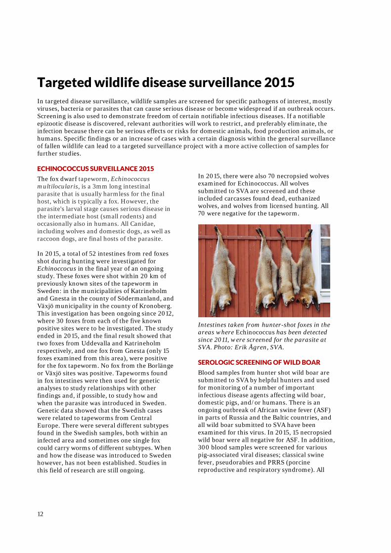
Two died of European brown hare syndrome (fältharesjuka), caused by a lagovirus that results in acute hepatitis. Eleven hares died of coccidiosis, caused by intestinal unicellular parasites. Toxoplasma gondii is another single-celled parasite, which caused inflammation in the liver, spleen, and lung of two hares. Thirteen hares had died of trauma (road kills or predation).
Small intestines from an emaciated hare (note that there is no intestinal fat tissue visible) that died due to intestinal coccidiosis. These unicellular parasites infect and cause necrosis of tissue, which is seen as white rice-sized foci in the intestinal wall (yellow arrows). Photo: SVA RED FOX
Out of a total of 103 red foxes, 45 were diagnostic cases. Widespread infections in six foxes were probably a result of bite wounds after fighting with other foxes. Single mangy foxes were received for necropsy and collection of mites for research and to be used as antigen for mange diagnostic tests. Reports of mange in foxes has increased in the past couple of years, suggesting that local outbreaks are affecting the fox population in some regions. BIRDS
There were 12 cases involving bird game species. Diagnoses of interest include a capercaillie with toxoplasmosis, where the single-celled parasite Toxoplasma gondii caused liver inflammation, and a goldeneye that died of lead poisoning, probably after eating some lead object, possibly lead shot or lead fishing gear.
12
Targeted wildlife disease surveillance 2015 In targeted disease surveillance, wildlife samples are screened for specific pathogens of interest, mostly viruses, bacteria or parasites that can cause serious disease or become widespread if an outbreak occurs. Screening is also used to demonstrate freedom of certain notifiable infectious diseases. If a notifiable epizootic disease is discovered, relevant authorities will work to restrict, and preferably eliminate, the infection because there can be serious effects or risks for domestic animals, food production animals, or humans. Specific findings or an increase of cases with a certain diagnosis within the general surveillance of fallen wildlife can lead to a targeted surveillance project with a more active collection of samples for further studies. ECHINOCOCCUS SURVEILLANCE 2015
The fox dwarf tapeworm, Echinococcus multilocularis, is a 3mm long intestinal parasite that is usually harmless for the final host, which is typically a fox. However, the parasite's larval stage causes serious disease in the intermediate host (small rodents) and occasionally also in humans. All Canidae, including wolves and domestic dogs, as well as raccoon dogs, are final hosts of the parasite. In 2015, a total of 52 intestines from red foxes shot during hunting were investigated for Echinoccocus in the final year of an ongoing study. These foxes were shot within 20 km of previously known sites of the tapeworm in Sweden: in the municipalities of Katrineholm and Gnesta in the county of Södermanland, and Växjö municipality in the county of Kronoberg. This investigation has been ongoing since 2012, where 30 foxes from each of the five known positive sites were to be investigated. The study ended in 2015, and the final result showed that two foxes from Uddevalla and Katrineholm respectively, and one fox from Gnesta (only 15 foxes examined from this area), were positive for the fox tapeworm. No fox from the Borlänge or Växjö sites was positive. Tapeworms found in fox intestines were then used for genetic analyses to study relationships with other findings and, if possible, to study how and when the parasite was introduced in Sweden. Genetic data showed that the Swedish cases were related to tapeworms from Central Europe. There were several different subtypes found in the Swedish samples, both within an infected area and sometimes one single fox could carry worms of different subtypes. When and how the disease was introduced to Sweden however, has not been established. Studies in this field of research are still ongoing.
In 2015, there were also 70 necropsied wolves examined for Echinococcus. All wolves submitted to SVA are screened and these included carcasses found dead, euthanized wolves, and wolves from licensed hunting. All 70 were negative for the tapeworm.
Intestines taken from hunter-shot foxes in the areas where Echinococcus has been detected since 2011, were screened for the parasite at SVA. Photo: Erik Ågren, SVA. SEROLOGIC SCREENING OF WILD BOAR
Blood samples from hunter shot wild boar are submitted to SVA by helpful hunters and used for monitoring of a number of important infectious disease agents affecting wild boar, domestic pigs, and/or humans. There is an ongoing outbreak of African swine fever (ASF) in parts of Russia and the Baltic countries, and all wild boar submitted to SVA have been examined for this virus. In 2015, 15 necropsied wild boar were all negative for ASF. In addition, 300 blood samples were screened for various pig-associated viral diseases; classical swine fever, pseudorabies and PRRS (porcine reproductive and respiratory syndrome). All
13
samples were negative, except a single sample that indicated a positive case of PRRS. A follow-up with targeted sampling of several wild boar and domestic swine in the immediate area was done, and all tests were negative for PRRS. The positive test was considered to be a test reaction that was not caused by an infection with PRRS virus, which is known to happen in some cases. AVIAN FLU SURVEILLANCE
All wild birds necropsied at SVA are routinely screened for avian flu virus, when possible. The screening results are used by the Board of Agriculture for reporting to the EU. In 2015, 221 wild birds of almost 50 different species (see table page 8, in which most, but not all, birds were sampled). For the first time since the major outbreak of avian flu in 2006, the aggressive type of virus (highly pathogenic AIV) was found in Swedish wild birds. Avian influenza virus was found in in five of 12 mute swans found ill or dead in central Stockholm during February and March. A highly pathogenic virus type was identified in two swans, but further typing was not possible in the other three swan samples. The swans also had high levels of lead in internal organs and may have died of lead poisoning, bird flu, or in a combination. The table below shows the 10 most common species analyzed in the avian flu surveillance program.
Species of bird Number of cases
White-tailed eagle 47
Mute swan 17
Mallard 14
Buzzard 13
Sparrowhawk 12
Rock pigeon 11
Ural owl 9
Golden Eagle 7
Eagle owl 6
Kestrel 5
The ten most common bird species screened for avian influenza at SVA in 2015.
TRICHINELLA SCREENING
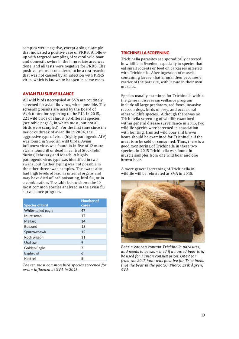
Trichinella parasites are sporadically detected in wildlife in Sweden, especially in species that eat small rodents or feed on carcasses infested with Trichinella. After ingestion of muscle containing larvae, that animal then becomes a carrier of the parasite, with larvae in their own muscles. Species usually examined for Trichinella within the general disease surveillance program include all large predators, red foxes, invasive raccoon dogs, birds of prey, and occasional other wildlife species. Although there was no Trichinella screening of wildlife examined within general disease surveillance in 2015, two wildlife species were screened in association with hunting. Hunted wild boar and brown bears should be examined for Trichinella if the meat is to be sold or consumed. Thus, there is a good monitoring of Trichinella in these two species. In 2015 Trichinella was found in muscle samples from one wild boar and one brown bear. A more general screening of Trichinella in wildlife will be reinstated at SVA in 2016.
Bear meat can contain Trichinella parasites, and needs to be examined if a hunted bear is to be used for human consumption. One bear from the 2015 hunt was positive for Trichinella (not the bear in the photo). Photo: Erik Ågren, SVA.
14
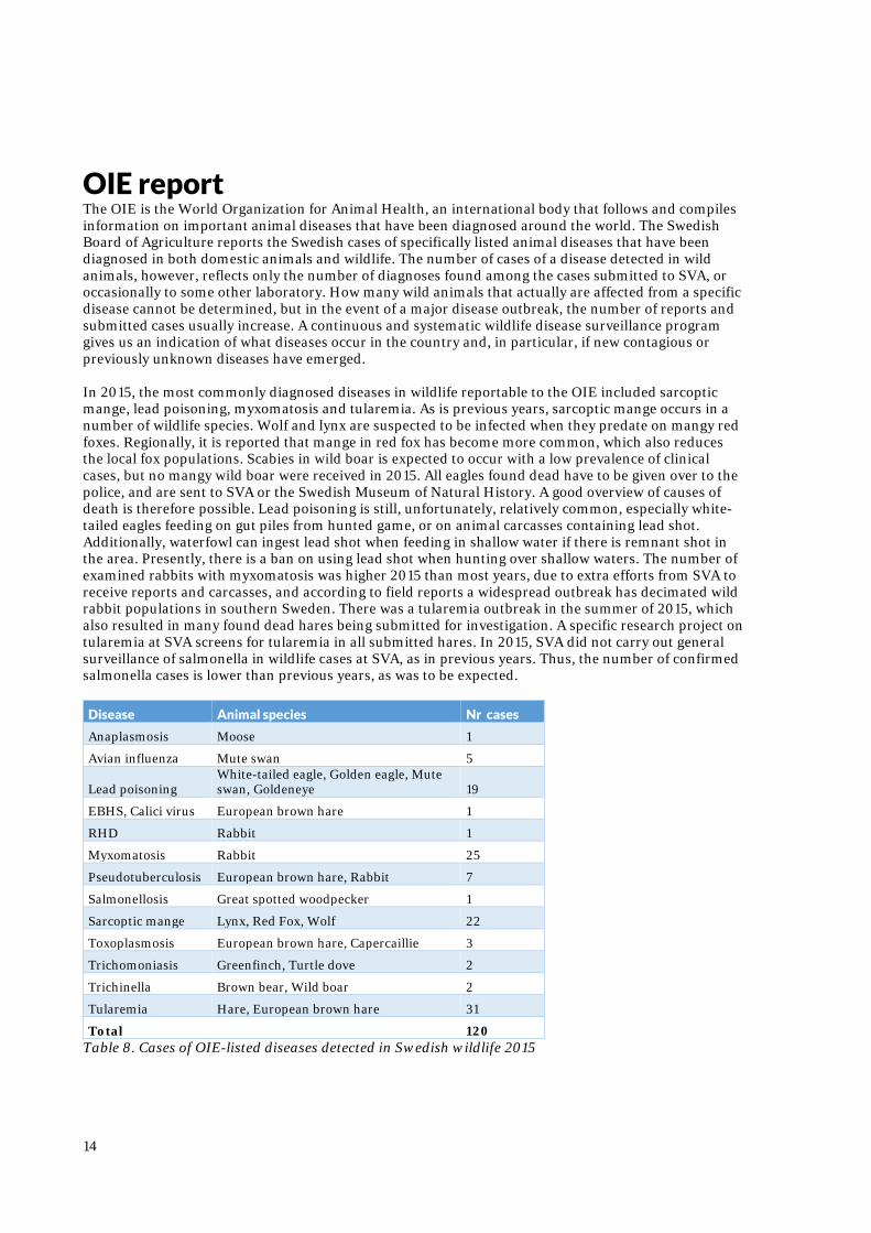
OIE report The OIE is the World Organization for Animal Health, an international body that follows and compiles information on important animal diseases that have been diagnosed around the world. The Swedish Board of Agriculture reports the Swedish cases of specifically listed animal diseases that have been diagnosed in both domestic animals and wildlife. The number of cases of a disease detected in wild animals, however, reflects only the number of diagnoses found among the cases submitted to SVA, or occasionally to some other laboratory. How many wild animals that actually are affected from a specific disease cannot be determined, but in the event of a major disease outbreak, the number of reports and submitted cases usually increase. A continuous and systematic wildlife disease surveillance program gives us an indication of what diseases occur in the country and, in particular, if new contagious or previously unknown diseases have emerged. In 2015, the most commonly diagnosed diseases in wildlife reportable to the OIE included sarcoptic mange, lead poisoning, myxomatosis and tularemia. As is previous years, sarcoptic mange occurs in a number of wildlife species. Wolf and lynx are suspected to be infected when they predate on mangy red foxes. Regionally, it is reported that mange in red fox has become more common, which also reduces the local fox populations. Scabies in wild boar is expected to occur with a low prevalence of clinical cases, but no mangy wild boar were received in 2015. All eagles found dead have to be given over to the police, and are sent to SVA or the Swedish Museum of Natural History. A good overview of causes of death is therefore possible. Lead poisoning is still, unfortunately, relatively common, especially white-tailed eagles feeding on gut piles from hunted game, or on animal carcasses containing lead shot. Additionally, waterfowl can ingest lead shot when feeding in shallow water if there is remnant shot in the area. Presently, there is a ban on using lead shot when hunting over shallow waters. The number of examined rabbits with myxomatosis was higher 2015 than most years, due to extra efforts from SVA to receive reports and carcasses, and according to field reports a widespread outbreak has decimated wild rabbit populations in southern Sweden. There was a tularemia outbreak in the summer of 2015, which also resulted in many found dead hares being submitted for investigation. A specific research project on tularemia at SVA screens for tularemia in all submitted hares. In 2015, SVA did not carry out general surveillance of salmonella in wildlife cases at SVA, as in previous years. Thus, the number of confirmed salmonella cases is lower than previous years, as was to be expected.
Disease Animal species Nr cases
Anaplasmosis Moose 1
Avian influenza Mute swan 5
Lead poisoning White-tailed eagle, Golden eagle, Mute swan, Goldeneye 19
EBHS, Calici virus European brown hare 1
RHD Rabbit 1
Myxomatosis Rabbit 25
Pseudotuberculosis European brown hare, Rabbit 7
Salmonellosis Great spotted woodpecker 1
Sarcoptic mange Lynx, Red Fox, Wolf 22
Toxoplasmosis European brown hare, Capercaillie 3
Trichomoniasis Greenfinch, Turtle dove 2
Trichinella Brown bear, Wild boar 2
Tularemia Hare, European brown hare 31
Total 120 Table 8. Cases of OIE-listed diseases detected in Swedish wildlife 2015
15
Acute projects 2015 – targeted surveillance
The Swedish Environmental Protection Agency (EPA) has a fund for targeted surveillance projects of more acute character. SVA plans the projects and makes an application for funding of pilot studies that need to be launched at short notice when increased morbidity or mortality in wildlife occurs. Time is usually of the essence to be able to collect suitable samples during a disease outbreak in wildlife. The acute projects that have been running in 2015 are described below. Some of the projects are a direct result of disease outbreaks that happened in 2015 and are described in more detail above, under the heading of Wildlife diseases of particular interest 2015, page 4-6.
MOOSE CALF MORTALITY ON ÖLAND
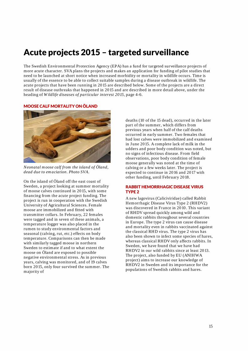
Neonatal moose calf from the island of Öland, dead due to emaciation. Photo SVA. On the island of Öland off the east coast of Sweden, a project looking at summer mortality of moose calves continued in 2015, with some financing from the acute project funding. The project is run in cooperation with the Swedish University of Agricultural Sciences. Female moose are immobilized and fitted with transmitter collars. In February, 22 females were tagged and in seven of these animals, a temperature logger was also placed in the rumen to study environmental factors and seasonal (calving, rut, etc.) effects on body temperature. Comparisons can then be made with similarly tagged moose in northern Sweden to estimate if and to what extent the moose on Öland are exposed to possible negative environmental stress. As in previous years, calving was monitored, and of 19 calves born 2015, only four survived the summer. The majority of
deaths (10 of the 15 dead), occurred in the later part of the summer, which differs from previous years when half of the calf deaths occurred in early summer. Two females that had lost calves were immobilized and examined in June 2015. A complete lack of milk in the udders and poor body condition was noted, but no signs of infectious disease. From field observations, poor body condition of female moose generally was noted at the time of calving or a few weeks later. The project is expected to continue in 2016 and 2017 with other funding, until February 2018. RABBIT HEMORRHAGIC DISEASE VIRUS TYPE 2
A new lagovirus (Caliciviridae) called Rabbit Hemorrhagic Disease Virus Type 2 (RHDV2) was discovered in France in 2010. This variant of RHDV spread quickly among wild and domestic rabbits throughout several countries in Europe. The type 2 virus can cause disease and mortality even in rabbits vaccinated against the classical RHD virus. The type 2 virus has also been shown to infect some species of hares, whereas classical RHDV only affects rabbits. In Sweden, we have found that we have had RHDV2 in our wild rabbits since at least 2013. The project, also funded by EU (ANIHWA project) aims to increase our knowledge of RHDV2 in Sweden and its importance for the populations of Swedish rabbits and hares.
16
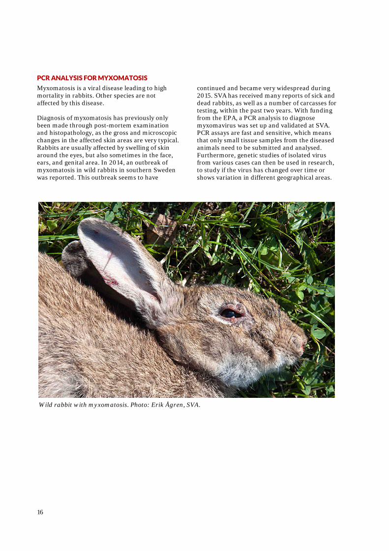
PCR ANALYSIS FOR MYXOMATOSIS
Myxomatosis is a viral disease leading to high mortality in rabbits. Other species are not affected by this disease. Diagnosis of myxomatosis has previously only been made through post-mortem examination and histopathology, as the gross and microscopic changes in the affected skin areas are very typical. Rabbits are usually affected by swelling of skin around the eyes, but also sometimes in the face, ears, and genital area. In 2014, an outbreak of myxomatosis in wild rabbits in southern Sweden was reported. This outbreak seems to have
continued and became very widespread during 2015. SVA has received many reports of sick and dead rabbits, as well as a number of carcasses for testing, within the past two years. With funding from the EPA, a PCR analysis to diagnose myxomavirus was set up and validated at SVA. PCR assays are fast and sensitive, which means that only small tissue samples from the diseased animals need to be submitted and analysed. Furthermore, genetic studies of isolated virus from various cases can then be used in research, to study if the virus has changed over time or shows variation in different geographical areas.
Wild rabbit with myxomatosis. Photo: Erik Ågren, SVA.
17
The four large carnivores, 2015 A substantial proportion of wild animals or animal parts coming to SVA are from the four large predators; brown bear, wolf, lynx and wolverine. These species belong to the State's wildlife, and according to the regulations of the Swedish Environmental Protection Agency, all animals of these species found dead have to be submitted at SVA. Additionally, hunted wolves, lynx and wolverines (typically the carcass without the pelt) must be sent to SVA for sampling and disease surveillance. Hunted bears are sampled in the field by inspectors, so only certain tissue samples and a tooth from shot animals are submitted to SVA. BROWN BEAR
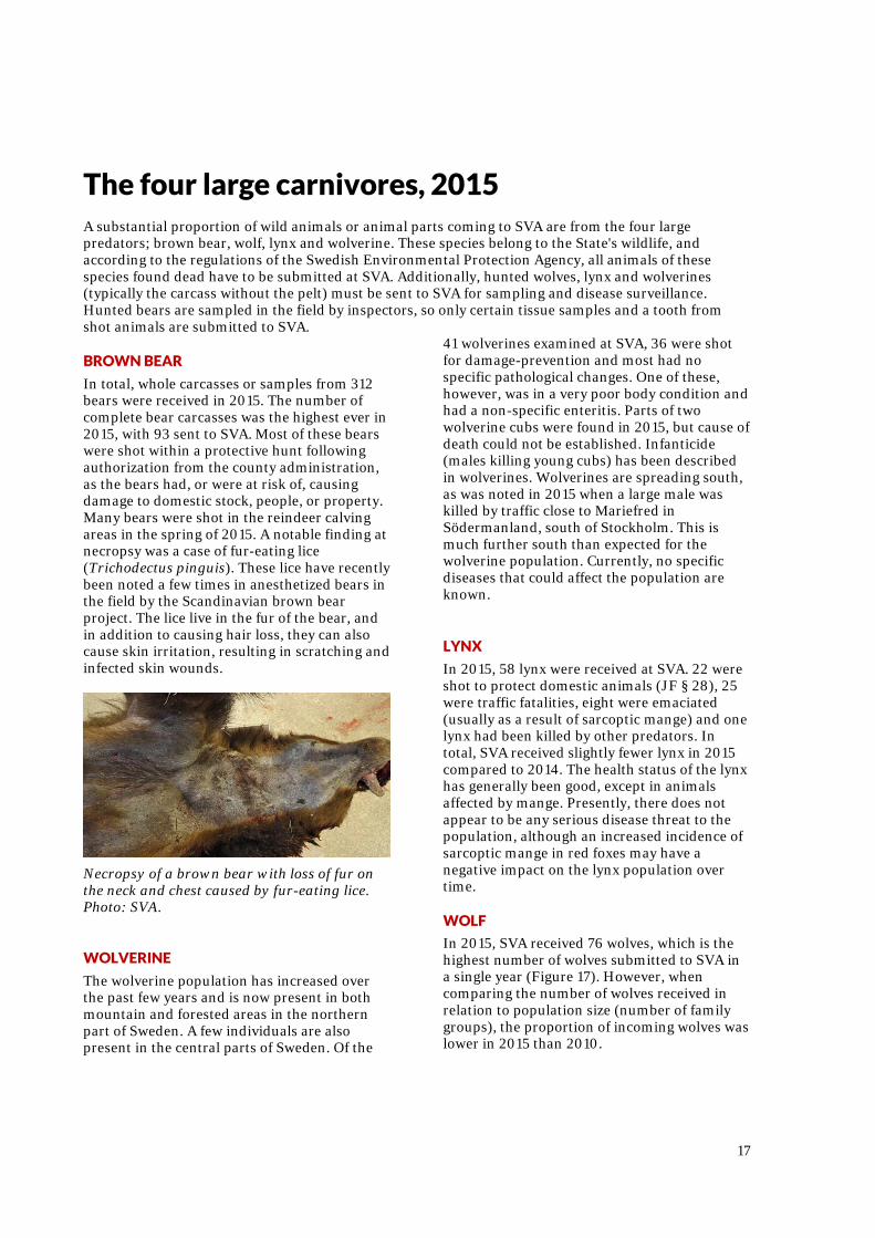
In total, whole carcasses or samples from 312 bears were received in 2015. The number of complete bear carcasses was the highest ever in 2015, with 93 sent to SVA. Most of these bears were shot within a protective hunt following authorization from the county administration, as the bears had, or were at risk of, causing damage to domestic stock, people, or property. Many bears were shot in the reindeer calving areas in the spring of 2015. A notable finding at necropsy was a case of fur-eating lice (Trichodectus pinguis). These lice have recently been noted a few times in anesthetized bears in the field by the Scandinavian brown bear project. The lice live in the fur of the bear, and in addition to causing hair loss, they can also cause skin irritation, resulting in scratching and infected skin wounds.
Necropsy of a brown bear with loss of fur on the neck and chest caused by fur-eating lice. Photo: SVA. WOLVERINE
The wolverine population has increased over the past few years and is now present in both mountain and forested areas in the northern part of Sweden. A few individuals are also present in the central parts of Sweden. Of the
41 wolverines examined at SVA, 36 were shot for damage-prevention and most had no specific pathological changes. One of these, however, was in a very poor body condition and had a non-specific enteritis. Parts of two wolverine cubs were found in 2015, but cause of death could not be established. Infanticide (males killing young cubs) has been described in wolverines. Wolverines are spreading south, as was noted in 2015 when a large male was killed by traffic close to Mariefred in Södermanland, south of Stockholm. This is much further south than expected for the wolverine population. Currently, no specific diseases that could affect the population are known. LYNX
In 2015, 58 lynx were received at SVA. 22 were shot to protect domestic animals (JF § 28), 25 were traffic fatalities, eight were emaciated (usually as a result of sarcoptic mange) and one lynx had been killed by other predators. In total, SVA received slightly fewer lynx in 2015 compared to 2014. The health status of the lynx has generally been good, except in animals affected by mange. Presently, there does not appear to be any serious disease threat to the population, although an increased incidence of sarcoptic mange in red foxes may have a negative impact on the lynx population over time. WOLF
In 2015, SVA received 76 wolves, which is the highest number of wolves submitted to SVA in a single year (Figure 17). However, when comparing the number of wolves received in relation to population size (number of family groups), the proportion of incoming wolves was lower in 2015 than 2010.
18
In 2015, the causes of death of wolves was distributed as follows: -44 license hunting (JF § 23 c) -6 protective hunting (JF § 23a) -7 protective hunting on an individual´s initiative (JF § 28) -12 traffic fatalities -2 euthanised due to animal welfare concerns (JF section 40b) -5 other causes The health status of necropsied wolves has generally been good, except for those with injuries or sarcoptic mange leading to emaciation. Three of the 35 males were cryptorchid, which means that one or both testicles had not migrated down into the scrotum properly. One of the three wolves was bilateral cryptorchid, which results in sterility. That 9% of the males had defects in the testicles is a slightly higher than the average result in wolves received the last 10 years (6%), but this is within the margin of error and is most probably due to the small number of cases. Defects of this type can likely be related to inbreeding, similar to the finding of defects in certain dog breeds with a limited number of breeding individuals.
Figure 2. The number of wolves that were submitted to SVA 2005-2015
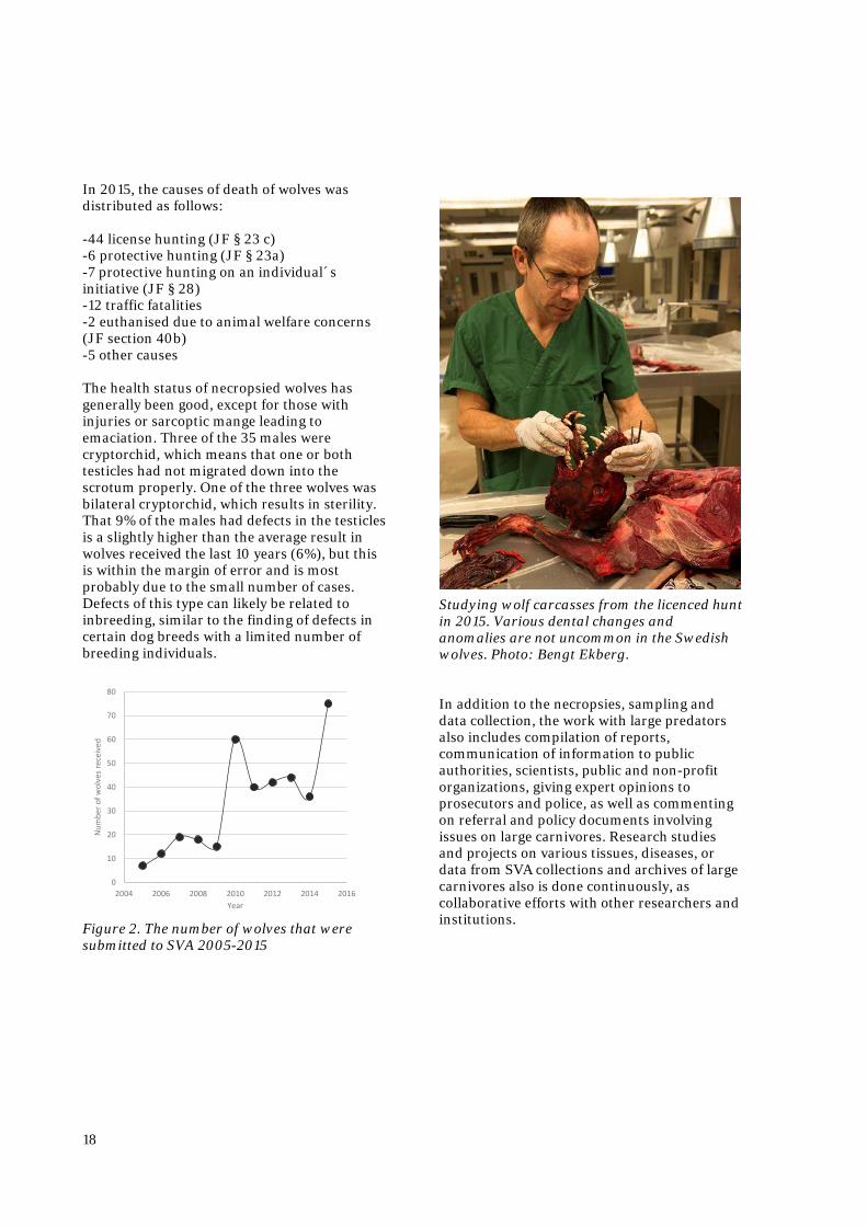
Studying wolf carcasses from the licenced hunt in 2015. Various dental changes and anomalies are not uncommon in the Swedish wolves. Photo: Bengt Ekberg. In addition to the necropsies, sampling and data collection, the work with large predators also includes compilation of reports, communication of information to public authorities, scientists, public and non-profit organizations, giving expert opinions to prosecutors and police, as well as commenting on referral and policy documents involving issues on large carnivores. Research studies and projects on various tissues, diseases, or data from SVA collections and archives of large carnivores also is done continuously, as collaborative efforts with other researchers and institutions.
0
10
20
30
40
50
60
70
80
2004 2006 2008 2010 2012 2014 2016
Num
ber o
f wol
ves r
ecei
ved
Year
19
Museum of Natural History SVA collaborates with the Swedish Museum of Natural History (NRM) in Stockholm regarding large carnivores, investigation of the health status of marine mammals, as well as necropsies and pathological studies of other species of Wildlife of the State, in particular otters and eagles. SVA performs necropsies on all species belonging to the State's Wildlife and the carcasses are then sent on to NRM for further biological and environmental studies and become part of the museum's collection. The cooperation between the SVA and NRM is an excellent example of interdisciplinary work involving biology, ecology, and veterinary medicine. MARINE MAMMALS
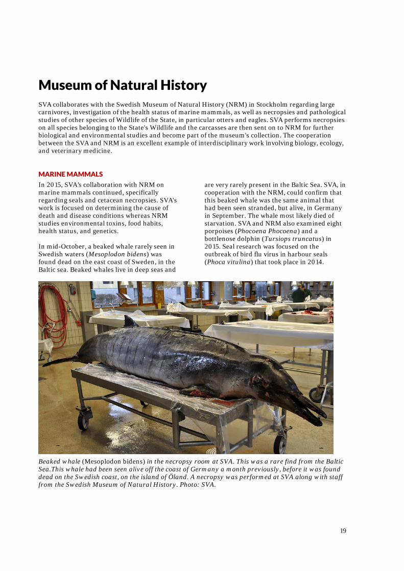
In 2015, SVA’s collaboration with NRM on marine mammals continued, specifically regarding seals and cetacean necropsies. SVA's work is focused on determining the cause of death and disease conditions whereas NRM studies environmental toxins, food habits, health status, and genetics. In mid-October, a beaked whale rarely seen in Swedish waters (Mesoplodon bidens) was found dead on the east coast of Sweden, in the Baltic sea. Beaked whales live in deep seas and
are very rarely present in the Baltic Sea. SVA, in cooperation with the NRM, could confirm that this beaked whale was the same animal that had been seen stranded, but alive, in Germany in September. The whale most likely died of starvation. SVA and NRM also examined eight porpoises (Phocoena Phocoena) and a bottlenose dolphin (Tursiops truncatus) in 2015. Seal research was focused on the outbreak of bird flu virus in harbour seals (Phoca vitulina) that took place in 2014.
Beaked whale (Mesoplodon bidens) in the necropsy room at SVA. This was a rare find from the Baltic Sea.This whale had been seen alive off the coast of Germany a month previously, before it was found dead on the Swedish coast, on the island of Öland. A necropsy was performed at SVA along with staff from the Swedish Museum of Natural History. Photo: SVA.
20
EAGLES
All eagles found dead have to be sent to SVA or the Swedish Museum of Natural History. In 2015, SVA received 87 eagles (69 white-tailed eagles, 17 golden eagles and one unidentified eagle carcass). The table below shows the different categories of causes of death. Some eagles may have more than one diagnosis. Road and railroad traffic is the most important cause of death among white-tailed eagles. Power lines and wind turbines also causes several deaths. Eagles are found shot each year, even though it is illegal to hunt eagles. Lead shot or lead bullet fragments in gut piles from hunting are probable origins of lead that is ingested by eagles, causing lead poisoning. Eagles fighting with each other can occasionally cause fatal injuries. Infections are relatively rare among the necropsied eagles.
Diagnoses
White-tailed eagle
Golden Eagle Total
Lead poisoning 7 2 9 Bleeding 1 1 Shock 1 1 Undetermined 10 3 13 Electric shock 1 3 4 Joint inflammation 1 1 Lung inflammation 1 1 Gunshot wound 5 1 6 Traffic 20 1 21 Train 10 10 Trauma, other cause 9 1 10 Trauma, another eagle 1 1 Emaciation 2 4 6 Wind turbines 5 5
Table 6. Overview of causes of death of white-tailed eagles and golden eagles necropsied at SVA in 2015.
OTTERS
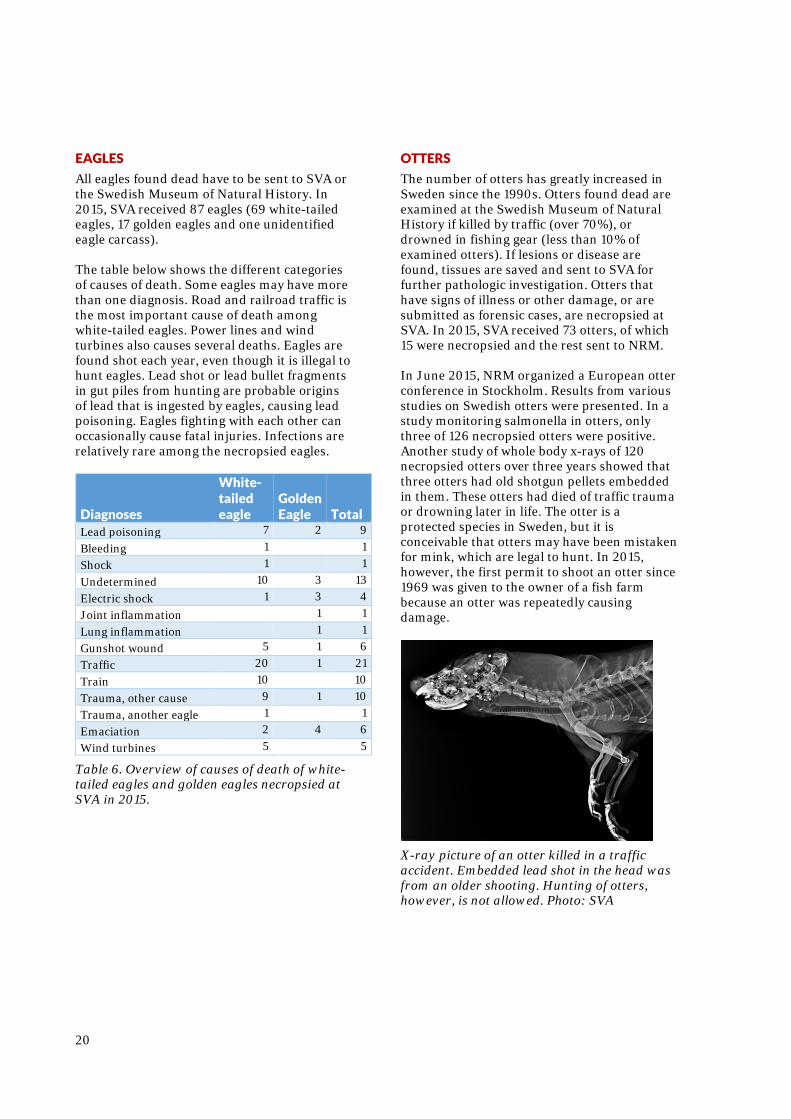
The number of otters has greatly increased in Sweden since the 1990s. Otters found dead are examined at the Swedish Museum of Natural History if killed by traffic (over 70%), or drowned in fishing gear (less than 10% of examined otters). If lesions or disease are found, tissues are saved and sent to SVA for further pathologic investigation. Otters that have signs of illness or other damage, or are submitted as forensic cases, are necropsied at SVA. In 2015, SVA received 73 otters, of which 15 were necropsied and the rest sent to NRM. In June 2015, NRM organized a European otter conference in Stockholm. Results from various studies on Swedish otters were presented. In a study monitoring salmonella in otters, only three of 126 necropsied otters were positive. Another study of whole body x-rays of 120 necropsied otters over three years showed that three otters had old shotgun pellets embedded in them. These otters had died of traffic trauma or drowning later in life. The otter is a protected species in Sweden, but it is conceivable that otters may have been mistaken for mink, which are legal to hunt. In 2015, however, the first permit to shoot an otter since 1969 was given to the owner of a fish farm because an otter was repeatedly causing damage.
X-ray picture of an otter killed in a traffic accident. Embedded lead shot in the head was from an older shooting. Hunting of otters, however, is not allowed. Photo: SVA
21
Wildlife research projects at SVA
Here we present the targeted wildlife disease surveillance carried out as specific projects and other larger, ongoing projects involving wildlife in 2015. HEPATITIS E VIRUS
A major research work on hepatitis E virus was finalized in 2015. The study was done in cooperation with, among others, SLU and the University of Gothenburg. Tissues from wild boar, roe deer, red deer, fallow deer, and moose collected by Swedish hunters were examined. Hepatitis E virus is a zoonotic disease, capable of transmission between animals and humans. Studies have shown that humans can become infected with hepatitis E virus originating from domestic swine, wild boar, and deer. One part of the study showed that 22% of 245 examined wild ungulates carried or had carried this virus. The highest incidence of hepatitis E infection was in moose: it was detected in 29% of the 231 surveyed moose. The hepatitis E virus in moose however, differed genetically from the hepatitis E virus in other ungulate species. This strain is believed to pose a low risk of infection to humans, as there has not been any cases of human infection with the moose hepatitis E virus to date. TULAREMIA
Tularemia is a disease caused by the bacterium Francisella tularensis. A wide variety of animal species, including humans, may be susceptible to the disease. In 2015, two projects on tularemia were completed. In one project, 646 blood samples from red fox, wild boar, lynx, wolf, wolverine, brown bear, and raccoon dog were examined for antibodies against F. tularensis. These species were chosen because they hunt or feed on hares and small rodents, and may serve as indicators of the spread of tularemia into new geographic areas. Antibodies were detected in the blood of seven wild boar, two red foxes, a wolverine, and a bear. In some of these cases, examination of lymph nodes was done to look for presence of
the actual bacterium, but in no case was bacteria found. In the second project, muscle tissue from hares positive for tularemia was examined to study if the bacterium also was present in the meat. As there sometimes are no visible changes in the internal organs of infected animals, there would be a risk of infection for people handling or eating meat from these animals. In 39 of the 43 hares, the bacterium could be detected in the muscle by PCR technology. On microscopic examination, the bacterium could be visualized in tissues and usually was localized in connective tissue surrounding the muscle fibers. The conclusion is that hare meat could be a source of infection for humans. However, if this actually happens and is another cause of human cases has not yet been studied in detail. At the end of 2015, a new project was initiated to study tissues collected from mountain hares necropsied during the summer outbreak in the North. This study will include investigations of various internal organs and further typing of the bacterium strain found during the outbreak.
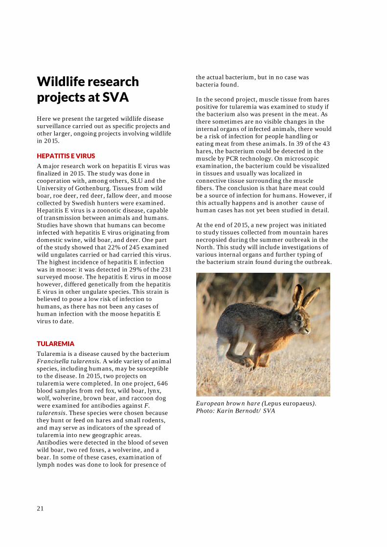
European brown hare (Lepus europaeus). Photo: Karin Bernodt/ SVA
22
NODULAR ONCHOCERCOSIS IN RED DEER
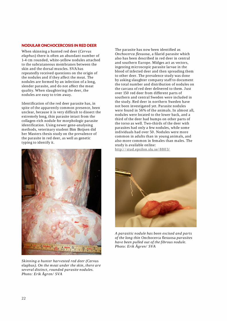
When skinning a hunted red deer (Cervus elaphus) there is often an abundant number of 1-4 cm rounded, white-yellow nodules attached to the subcutaneous membranes between the skin and the dorsal muscles. SVA has repeatedly received questions on the origin of the nodules and if they affect the meat. The nodules are formed by an infection of a long, slender parasite, and do not affect the meat quality. When slaughtering the deer, the nodules are easy to trim away. Identification of the red deer parasite has, in spite of the apparently common presence, been unclear, because it is very difficult to dissect the extremely long, thin parasite intact from the collagen-rich nodule for morphologic parasite identification. Using newer gene-analysing methods, veterinary student Bim Boijsen did her Masters thesis study on the prevalence of the parasite in red deer, as well as genetic typing to identify it.
Skinning a hunter harvested red deer (Cervus elaphus). On the meat under the skin, there are several distinct, rounded parasite nodules. Photo: Erik Ågren/ SVA
The parasite has now been identified as Onchocerca flexuosa, a filarid parasite which also has been described in red deer in central and southern Europe. Midges act as vectors, ingesting microscopic parasite larvae in the blood of infected deer and then spreading them to other deer. The prevalence study was done by asking slaughter company staff to document the total number and distribution of nodules on the carcass of red deer delivered to them. Just over 150 red deer from different parts of southern and central Sweden were included in the study. Red deer in northern Sweden have not been investigated yet. Parasite nodules were found in 56% of the animals. In almost all, nodules were located to the lower back, and a third of the deer had bumps on other parts of the torso as well. Two-thirds of the deer with parasites had only a few nodules, while some individuals had over 50. Nodules were more common in adults than in young animals, and also more common in females than males. The study is available online: http://stud.epsilon.slu.se/8803/
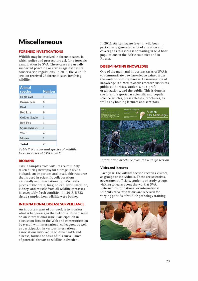
A parasitic nodule has been excised and parts of the long thin Onchocerca flexuosa parasites have been pulled out of the fibrous nodule. Photo: Erik Ågren/ SVA
23
Miscellaneous FORENSIC INVESTIGATIONS
Wildlife may be involved in forensic cases, in which police and prosecutors ask for a forensic examination by SVA. These cases are usually suspected poaching or crimes against nature conservation regulations. In 2015, the Wildlife section received 25 forensic cases involving wildlife. Animal species Number
Eagle owl 1
Brown bear 8
Bird 1
Red kite 6
Golden Eagle 1
Red Fox 1
Sparrowhawk 1
Wolf 4
Moose 1
Total 25
Table 7. Number and species of wildlife forensic cases at SVA in 2015. BIOBANK
Tissue samples from wildlife are routinely taken during necropsy for storage in SVA's biobank, an important and invaluable resource that is used in scientific collaborations nationally and internationally. SVA banks pieces of the brain, lung, spleen, liver, intestine, kidney, and muscle from all wildlife carcasses in acceptably fresh condition. In 2015, 5 533 tissue samples from wildlife were banked. INTERNATIONAL DISEASE SURVEILLANCE
An important part of our work is to monitor what is happening in the field of wildlife disease on an international scale. Participation in discussion lists on the Web and communication by e-mail with international colleagues, as well as participation in various international associations involved in wildlife health and disease, forms the basis of this surveillance of potential threats to wildlife in Sweden.
In 2015, African swine fever in wild boar particularly generated a lot of attention and coverage as this virus is spreading in wild boar populations in the Baltic countries and in Russia. DISSEMINATING KNOWLEDGE
One of the main and important tasks of SVA is to communicate new knowledge gained from the work on wildlife disease. Dissemination of knowledge is aimed towards research institutes, public authorities, students, non-profit organizations, and the public. This is done in the form of reports, as scientific and popular science articles, press releases, brochures, as well as by holding lectures and seminars.
Information brochure from the wildlife section Visits and lectures
Each year, the wildlife section receives visitors, as groups or individuals. These are scientists, government officials, students or study groups, visiting to learn about the work at SVA. Externships for national or international students or veterinarians are received for varying periods of wildlife pathology training.
24
CONTACTING THE WILDLIFE SECTION
Telephone and email
Questions on wildlife, and reports of observations or findings regarding wildlife from the public is a large part of the daily work of the wildlife section. During working hours, a wildlife pathologist is always available to answer questions on the phone or e-mail ([email protected]). During 2015, questions and answers regarding wildlife resulted in an archive of slightly over 1600 e-mails. Most emails concerned moose (over 300 mails), in which skin ulceration reports dominated. Many questions, as is typical, concerned large carnivores; with about 250 e-mails in 2015. A total of 712 incoming phone calls were registered in 2015 regarding questions or reports about wildlife. This is an increase of 50% from the previous year. Wildlife species affected by major disease outbreaks or have high public interest, are the most common subject of telephone calls: 158 calls about moose, 178 about hare, 57 about rabbit, 44 about roe deer and 19 calls about swans. Web-based reporting of dead wildlife
When sick or dead wildlife is found, the public can also report to SVA online (www.sva.se). The collective incoming information gives SVA an indication of when and where unusual mortality events or disease outbreaks occur in wildlife. In 2015, 65 reports were submitted by the online form.
To further facilitate reporting to SVA, an App for smartphones is scheduled to be developed in 2016. Transport of dead wildlife
To transport dead wildlife to SVA, special cardboard boxes approved by the postal service are shipped to those who contact the wildlife section and have a wildlife case suitable for examination. In 2015, over 400 shipment boxes for wildlife weighing up to 20 kg, were distributed. Wildlife carcasses weighing over 20 kg must be transported by trailer or truck.
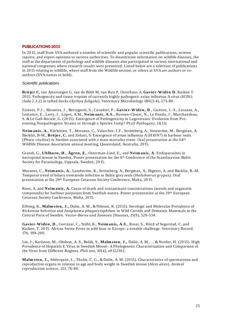
All wildlife material that is sent to SVA must be correctly packaged, to avoid leakage of fluids. A form with relevant information must accompany the carcass. Instructions, forms, and heavy duty plastic bags are sent out with the cardboard box to the submitter of wildlife material. Photo: Rickard Wolrath/SVA
25
PUBLICATIONS 2015
In 2015, staff from SVA authored a number of scientific and popular scientific publications, written reports, and expert opinions to various authorities. To disseminate information on wildlife diseases, the staff at the department of pathology and wildlife diseases also participated in various international and national congresses where research results were presented. Listed below are a selection of publications in 2015 relating to wildlife, where staff from the Wildlife section, or others at SVA are authors or co-authors (SVA names in bold).
Scientific publications Bröjer C, van Amerongen G, van de Bildt M, van Run P, Osterhaus A, Gavier-Widén D, Kuiken T. 2015. Pathogencity and tissue tropism of currently highly pathogenic avian influenza A virus (H5N1; clade 2.3.2) in tufted ducks (Aythya fuligula). Veterinary Microbiology 180(3-4), 273-80. Esteves, P.J., Abrantes, J., Bertagnoli, S., Cavadini, P., Gavier-Widen, D., Guitton, J.-S., Lavazza, A., Lemaitre, E., Letty, J., Lopes, A.M., Neimanis, A.S., Ruvoen-Clouet, N., Le Pendu, J., Marchandeau, S. & Le Gall-Recule, G. (2015). Emergence of Pathogenicity in Lagoviruses: Evolution from Pre-existing Nonpathogenic Strains or through a Species Jump? PLoS Pathogens, 11(11).
Neimanis, A., Härkönen, T., Moraeus, C., Valarcher, J.F., Strömberg, A., Stenström, M., Bergman, A. Bäcklin, B-M., Bröjer, C. and Zohari, S. Emergence of avian influenza A (H10N7) in harbour seals (Phoca vitulina) in Sweden associated with a mass mortality event. Oral presentation at the 64th Wildlife Disease Association annual meeting, Queensland, Australia, 2015. Grandi, G., Uhlhorn, H., Ågren, E., Osterman-Lind, E., and Neimanis, A. Endoparasites in necropsied moose in Sweden. Poster presentation for the 6th Conference of the Scandinavian-Baltic Society for Parasitology, Uppsala, Sweden, 2015. Moraeus, C., Neimanis, A., Lundström, K., Strömberg, A., Bergman, A., Bignert, A. and Bäcklin, B.-M. Temporal trend of biliary trematode infection in Baltic grey seals (Halichoerus grypus). Oral presentation at the 29th European Cetacean Society Conference, Malta, 2015. Roos, A. and Neimanis, A. Cause of death and contaminant concentrations (metals and organotin compounds) for harbour porpoises from Swedish waters. Poster presentation at the 29th European Cetacean Society Conference, Malta, 2015. Elfving, K., Malmsten, J., Dalin, A. M., & Nilsson, K. (2015). Serologic and Molecular Prevalence of Rickettsia helvetica and Anaplasma phagocytophilum in Wild Cervids and Domestic Mammals in the Central Parts of Sweden. Vector-Borne and Zoonotic Diseases, 15(9), 529-534. Gavier-Widén, D., Gortázar, C., Ståhl, K., Neimanis, A.S., Rossi, S., Hård af Segerstad, C. and Kuiken, T. 2015. African Swine Fever in wild boar in Europe: a notable challenge. Veterinary Record. 176: 199-200. Lin, J., Karlsson, M., Olofson, A. S., Belák, S., Malmsten, J., Dalin, A. M., ... & Norder, H. (2015). High Prevalence of Hepatitis E Virus in Swedish Moose–A Phylogenetic Characterization and Comparison of the Virus from Different Regions. PloS one, 10(4), e0122102. Malmsten, J., Söderquist, L., Thulin, C. G., & Dalin, A. M. (2015). Characteristics of spermatozoa and reproductive organs in relation to age and body weight in Swedish moose (Alces alces). Animal reproduction science, 153, 76-86.
26
Norén K, Statham MJ, Ågren EO, Isomursu M, Flagstad Ø, Eide NE, Berg TB, Bech-Sanderhoff L, Sacks BN. 2015. Genetic footprints reveal geographic patterns of expansion in Fennoscandian red foxes. Glob Chang Biol. 21(9):3299-312. Thulin, C. G., Malmsten, J., & Ericsson, G. (2015). Opportunities and challenges with growing wildlife populations and zoonotic diseases in Sweden. European Journal of Wildlife Research, 61(5), 649-656. Wensman J, C G das Neves, A H Kautto, U Rockström, E Ågren, B Åhman, S Alenius. Temporal and geographical variation of pestivirus and alphaherpes virus infection in Swedish semi-domesticated reindeer (Rangifer t. tarandus). Scientific poster presented at X th International Congress of Veterinary Virology ESVV2015 and 9th Annual Meeting of EPIZONE.
Scientific presentations Ågren E Roos A, Bröjer C, Hestvik G. Screening of Salmonella in Swedish Otters. Scientific poster presented at European Otter workshop, Stockholm, 8-10 June 2015. Ågren E, Roos A, Bröjer C. Evidence of shotgun wounded otters (Lutra lutra) in Sweden. Scientific poster presented at European Otter workshop, Stockholm, 8-10 June 2015. Ågren E, Paul E, Åhman B, Sirkkola H, Skarin, A. Comparison of two pregnancy test methods in semi-domesticated reindeer (Rangifer t. tarandus). Scientific poster presented at the 14th International Arctic Ungulate Conference, 16-21 August 2015, Røros, Norway. Ågren E, Söderberg A. Malformations in brown bear (Ursus arctos). Scientific poster presented at Nordic section of the Wildlife Disease Association NWDA, Hjerkinn, Norway, 10-12 June 2015, and Veterinary Congress, Uppsala, Sweden 5-6 nov 2015. Ågren E, Uhlhorn H, Bröjer C, Gavier-Widén D. Studies on peruke antlers in roe deer. Scientific poster presented at ESVP-ECVP, Helsinki 2-5 sept 2015. Abstract in: J Comp Path 2016, 154, 98.
Popular scientific publications and reports Roos A, Loso K, Ågren E. 2015. Uttrar i samhällets tjänst [Otters in the service of society]. Fauna & Flora 110:3, 2-6. Ågren E, Sylvan L. 2015. Perukhorn hos råget [Peruke antlers in a roe deer female]. Fauna & Flora 110:3, 30-33.

Visitor address: ulls väg 2 B. Postal address. 751 89 Uppsala Telephone. +46 18 67 40 00 Fax. +46 18 30 91 62 E-mail. [email protected] Web. www.sva.se