Embed Size (px)
Citation preview

Flat Adenoma-Adenocarcinoma Sequence in the Colon of Rats Carlos Rubio, M.D., Ph.D., Jayant Shetye, M.D. (Path.) From the Departments of Pathology, Karolinska Institute and Hospital, Stockholm, Sweden
PURPOSE: As there is an increased awareness of the exis- tence of a "flat adenoma-adenocarcinoma sequence" in the colonic mucosa of human subjects, the aims of the study were to assess whether flat colonic adenocarcinomas in rats are also preceded by flat adenomas, as is reported in hu- mans, and to determine the frequency of flat lesions com- pared with exophytic lesions in the colon of rats. METHOD: The colonotropic carcinogen 1,2-dimethylhydrazine was in- jected subcutaneously in 300 Sprague-Dawley rats for 27 weeks. RESULTS: A total of 358 tumors developed in 278 of the 300 rats. Of the 60 adenomas found at histology, 25 percent were flat adenomas. Of the 298 adenocarcinomas, 12.7 percent had originated in a flat adenoma. Of the 180 colonic neoplasias (adenomas or adenocarcinomas), 29.4 percent were flat neoplasias (flat adenomas or adenocarci- nomas arising in a flat adenoma), and the remaining 70.6 percent were exophytic neoplasias (tubulo or villous ade- nomas or adenocarcinomas arising in exophytic adenomas). From the 298 colonic adenocarcinomas, 1 was a intramu- cosal adenocarcinoma, 87 were overt adenocarcinomas, and 90 were lymphoid-associated carcinomas; in those 298 adenocarcinomas, no preneoplastic lesion could be re- corded. In 208 animals, biopsies were taken from macro- scopically visible colonic lesions, and, in the remaining 70 animals, the entire colon was processed for histologic ex- amination. Flat adenomas were found in 3.8 percent of the 208 biopsy specimens and in 10 percent of the 70 colec- tomy specimens. Further, of the 40 adenomas found in biopsy specimens, 20 percent were flat adenomas, and, of the 20 adenomas found in colectomy specimens, 35 per- cent were flat adenomas. CONCLUSIONS: The study re- ported herein indicates the existence of a "flat adenoma- adenocarcinoma sequence" in the colonic mucosa of Sprague-Dawley rats. The flat lesions of the colon consti- tuted approximately one-third of the total neoplastic lesions seen in the rat following injections of 1,2-dimethylhy- drazine. More flat adenomas were detected at histologic examination of the entire colon than in biopsies obtained from the macroscopically visible colonic lesions. Conse- quently, flat adenomas may be overlooked by naked-eye examination. [Key words: Flat adenomas; Flat adenocarci- nomas; Colonic mucosa; Rats]
Rubio C, Shetye J. Flat adenoma-adenocarcinoma sequence in the colon of rats. Dis Colon Rectum 1994;37:1300-1306.
F o l lowing the d i scovery that cycasin was ab le to
induce a d e n o c a r c i n o m a s in the co lon of rats, ~
m a n y invest igators s tud ied the characteris t ics of those
colonic tumors, inc luding his togenesis , 2 ultrastruc-
Supported by the Cancer Society, the Cancer Fund, and the Karo- linska Institute, Stockholm, Sweden. Address reprint requests to Dr. Rubio: Department of Pathology, Karolinska Hospital, S-171 76 Stockholm, Sweden.
ture, 3 b iochemica l proper t ies , 4 t ransplantabi l i ty , 5 as
wel l as t rea tment wi th c h e m o t h e r a p y r or i m m u n o -
t h e r a p y ]
At p resen t there is much d iscuss ion in the l i terature
regard ing the type of mucosa l les ion(s) that p r e c e d e s
invasive a d e n o c a r c i n o m a in the co lon of rats. Al-
t hough some have r e p o r t e d that all co lon ic tumors
arise "de novo" (i.e., f rom the no rma l co lon ic mu-
cosaS), o thers have f o u n d that the tumors or iginate in
the mucosa cover ing discrete l y m p h o i d aggrega tes 9
or in the focal areas of exophy t i c prol i fera t ion (i.e., exophy t i c adenoma l~ The ongo ing d iscuss ion is of
p a r a m o u n t impor t ance b e c a u s e the rat has b e e n con-
s ide red useful as a m o d e l to invest igate not on ly the
h is togenes is bu t also the b io log ic at t r ibutes that pos -
s ibly mimic the cascade of events occurr ing dur ing
co lonic ca rc inogenes i s in h u m a n subjects.
The type of les ion(s) that p r e c e d e s colonic adeno -
ca rc inomas in h u m a n s has also b e e n a mat ter of much
concern . Before 1934, it was b e l i e v e d that all a d e n o -
ca rc inomas in the rectal m u c o s a in h u m a n s arose de novo. 11 The w o r k of Swinton and Wal ren , 11 p r e s e n t e d
in 1939 demons t ra t ed , however , that rectal adenoca r -
c inomas of ten arose in exophyt ic , p re-ex is t ing adeno -
ma tous po lyps . Those f indings we re conf i rmed b y
m a n y workers , but, dur ing the ear ly fifties, Cas t leman
and Krickstein, ~2 and Spratt and Ackerman , ~3 recon-
s ide red the poss ib i l i ty that rectal a d e n o c a r c i n o m a s
or ig ina ted in n o n e x o p h y t i c lesions.
The i m p r o v e m e n t o f the d o u b l e x- ray t echn ique
and e n d o s c o p i c examina t ion has pe rmi t t ed b i o p s y of
exophy t i c les ions of the colonic m u c o s a as well . Re-
suits of that w o r k ind ica ted that co lon ic adenoca r -
c inomas of ten or ig ina ted in exophy t i c adenomas .
These f indings we re a c c e p t e d b y bo th Wes te rn and
Asian s tudents , and the interest for the " a d e n o m a
(exophy t i c ) - ca rc inoma sequence" as a p a t h w a y of
colorecta l ca rc inogenes i s in h u m a n s was r e o p e n e d .
Fur ther i m p r o v e m e n t in f iberopt ics in later years
has pe rmi t t ed bo th the de tec t ion and remova l no t
on ly of exophy t i c les ions but also of n o n e l e v a t e d
les ions (i.e., fiat l e s i o n s in the co lonic mucosa14).
1300

Vol. 37, No. 12
Some of the latter lesions were found, at histology, to be fiat adenomas or fiat adenocarcinomas.
In a previous work, 15 we found an occasional fiat neoplastic lesion in the colorectal mucosa of 1,2- dimethylhydrazine (DMH)-treated Sprague-Dawley rats. In that work, the percentage of fiat to exophytic neoplasms was, however, not highlighted. The pres- ent investigation was carried out to assess the frequency of fiat and exophytic neoplastic lesions occurring in the colon of Sprague-Dawley rats treated with DMH.
FLAT ADENOMA-ADENOCARCINOMA IN RATS 1301
ently normal colonic mucosa. 16 Mucosal lesions that were at the same level or slightly more elevated than the surrounding normal colonic mucosa were consid- ered as fiat.
Exophytic and fiat mucosal lesions were found at histology to be either adenomas, adenocarcinomas, or lymphoid-associated adenocarcinomas (LAC).
Normal lymphoid plaques covered by histologically normal colonic mucosa could also protrude into the lumen of the organ. These lymphoid aggregates could occasionally be confused at gross examination for an exophytic tumor.
MATERIALS A N D METHODS
Three-hundred male Sprague-Dawley rats weigh- ing 200 grams received weekly subcutaneous injec- tions of 21 mg of DMH/kg of body weight for 27 weeks. At the end of the scheduled time, animals were sacrified in a concentrated CO2 atmosphere. The entire colon was removed in 75 of 300 rats, washed in physiologic saline, and prepared as a Swiss-roll be- fore fixation (in methanol-acetic acid or neutral for- malin) as reported elsewhere. 15 In the remaining 225 rats, the colon was washed in physiologic saline and carefully inspected. All abnormal changes in the mu- cosa were cut and fixed in methanol-acetic acid or neutral formalin. Paraffin sections were cut at 4 /xm and stained with hematoxylin and eosin.
Definitions
Exophytic lesions were considered those elevated polypoid protrusions that were surrounded by appar-
A d e n o m a s
Adenomas were histologically divided into 1) exo- phytic adenomas (tubular and tubulo-villous) (Fig. 1) and 2) fiat adenomas (tubular) (Fig. 2). Pure villous adenomas were not recorded.
Adenocarcinomas
Adenocarcinomas were classified into those arising in 1) exophytic adenomas (Fig. 3), 2) flat adenomas (Fig. 4), 3) the colonic mucosa covering lymphoid aggregates (i.e., LAC; Fig. 5), 4) intramucosal adeno- carcinomas (i.e., signet-ring cell adenocarcinomas in- vading the lamina propria mucosa exclusively; Fig. 6), and finally 5) overt invasive adenocarcinomas (either signet-ring cell adenocarcinomas or mixed tubulo- signet-ring cells adenocarcinomas). The histogenesis (i.e., the structure from where the adenocarcinoma
Figure 1. Low-power view of exophytic tubular adenoma (hematoxylin and eosin; •
1302 RUBIO AND SHETYE Dis Colon Rectum, December 1994
Figure 2. Low-power view of a flat adenoma in a colectomy specimen (hematoxylin and eosin; •
originated) in intramucosal and overt adenocarcino-
mas was impossible to ascertain. With regard to LAC,
some of them were found to have nonprotruding
dysplastic glands at the luminal aspect of the lym- phoid plaque. 15 However, the lymphoid aggregate
was, by itself, thicker than the surrounding mucosa and, therefore, protruded into the lumen of the organ.
Thus, the protrusion was many times not caused by the tumoral part of the LAC. Because of this snare and
because some of the early LAC were not accompanied
by dysplastic glands at the lumen of the organ, they could not be classified as flat or as protruding into the
lumen (i.e., exophytic). In addition, the histogenesis in those tumors was difficult to assess. Consequently,
LAC tumors were herein also regarded as being of uncertain origin.
RESULTS
Of 300 DMH-treated rats, 278 developed colonic
tumors. The results in Table 1 show that a total of 358 tumors were found in the colon of 278 tumor-bearing rats.
Results of histologic examination shown in Table 1 demonstrate that 60 were adenomast, and 298 were adenocarcinomas. Of the 60 adenon~as, 15 (25 per- cent) were fiat adenomas. Of 298 adenocarcinomas, 38 (12.7 percent) were found to have originated in a fiat adenoma.
Of 180 colonic fiat and exophytic adenomas and adenocarcinomas, 53 (29.4 percent) were fiat neopla-
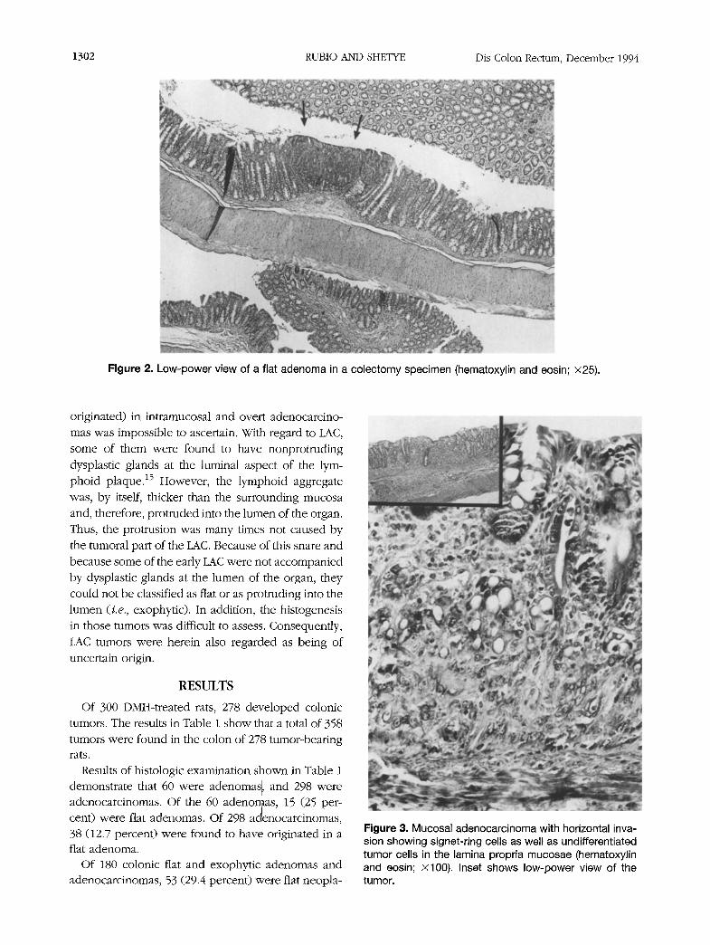
Figure 3. Mucosal adenocarcinoma with horizontal inva- sion showing signet-ring cells as well as undifferentiated tumor cells in the lamina propria mucosae (hematoxylin and eosin; • Inset shows low-power view of the tumor.

Vol. 37, No. 12 FLAT ADENOMA-ADENOCARCINOMA IN RATS 1303
Figure 4. LAC. Note remnants of lymphoid tissue and mucous secretion in invading glands (hematoxylin and eosin; • 100). Inset shows low-power view of the same tumor.
Figure 5, Flat adenocarcinoma originating in a flat tubular adenoma (hematoxylin and eosin; xlO0). Inset shows low-power view of the tumor.
sias (fiat adenomas or adenocarcinomas arising in a
flat adenoma), and the remaining 127 (70.6 percent) were exophytic neoplasias (tubulo or villous adeno-
mas or adenocarcinomas arising in exophytic adeno-
mas). Of 358 tumors recorded in all animals, 178 (49.7
percent) were tumors in which the origin could not be ascertained (87 overt adenocarcinomas, 1 intramuco- sal adenocarcinoma, and 90 LAC).
The number of tumors per rat was similar in 70
animals in which the entire colon had been removed for histologic examination (Swiss-rolled) and in 208
animals in which only macroscopically visible tumors had been biopsied (1.25 tumors/rat and 1.37 tumors/
rat, respectively). However, when the type of lesion was investigated, it was found that of 40 adenomas
found at biopsy, 8 (20 percent) were fiat adenomas.

1304 RUBIO AND SHETYE Dis Colon Rectum, December 1994
Figure 6. Exophytic adenocarcinoma originating in a tubulo-villous adenoma (hematoxylin and eosin; x l00). Inset shows low-power view of the tumor.
Table 1. Histologic Type of 358 Colonic Tumors Recorded in 278 Sprague-Dawley Rats Subjected to Weekly Subcutaneous
Injections (n = 27 wk) of 1,2-Dimethylhydrazine
Histology
Adenocarcinomas Adenomas
No. of All Overt All Adeno- Rats Exophytic Flat Adenomas Exophytic Flat LAC Intramucosal carcinoma carcinomas
At biopsy 208 32 8 40 70 27 69 1 55 222 At colectomy 70 13 7 20 12 11 21 0 32 76 All 278 45 15 60 82 38 90 1 87 298
On the other hand, of 20 adenomas found in colec-
tomy specimens, 7 (35 percent) were flat adenomas.
Furthermore, 8 (3.8 percent) fiat adenomas were found at biopsy in 208 animals. On the other hand, 7 (10 percent) flat adenomas were found in 70 animals in which the entire colon was available for histologic
examination. Of 206 adenomas or adenocarcinomas of known
origin found at biopsy, 35 (16.9 percent) were fiat adenomas or fiat adenocarcinomas. The rate for fiat lesions found in colectomy specimens was 28.1 per- cent (i.e., 18 fiat lesions in 64 specimens).
DISCUSSION
Results of the present work demonstrated that un- der conditions of husbandry at this laboratory, fiat adenomas as well as exophytic adenomas developed
in the colonic mucosa of Sprague-Dawley rats treated
with DMH for 27 weeks. Results also demonstrated that adenocarcinomas could originate both in fiat adenomas and exophytic adenomas. Thus, both a "fiat adenoma-carcinoma sequence" and an "exophy- tic adenoma-carcinoma sequence" could be demon-
strated in our rats. Those two pathways of colon carcinogenesis are claimed by some authors to occur in human subjects. 14' 17 Kino,18 however, believes that
epithelial cells of atypical glands change to cancer rapidly without first forming a polyp. This theory is called "carcinogenesis by progression." In accor- dance with Inamori et al., 19 we consider, however,
that fiat colonic adenocarcinomas in experimental an- imals are antedated by minute adenomas (i.e., mea- suring -< 1 cm in diameter).
One case of intramucosal adenocarcinoma with sig- net-ring cells was found during our study. Although

Vol. 37, No. 12
the origin of this tumor could not be traced to a
particular pre-existing lesion, it is conceivable that it
may have originated in the mucous-producing cells of
crypts of the colonic mucosa.
The experience accumulated in later years indicates
that colonic adenocarcinomas often originate in large
(->2 cm in diameter, 2~ exophytic adenomas. Appar-
ently, the exophytic growth must attain a large size
before invasion ensues. In the present work with
rats, a similar observation was done, only large exo-
phytic adenomas demonstrated invasive growth. On
the other hand, tiny fiat adenomas could demon-
strate invasive growth. This suggests that exophytic
and fiat adenomas may be two biologically different
lesions. Interestingly, it has been recently found that
the expression of proliferative cell nuclear antigen is
quantitatively higher in flat adenomas than in exo-
phytic adenomas (Shetye J, Rubio C, unpublished
data).
As reported above, the examination of colectomy
specimens revealed that 35 percent of the adenomas
were fiat. When macroscopically detected lesions
were obtained at biopsy only, 20 percent of the ade-
nomas were fiat adenomas. These findings suggest
that many more fiat adenomas were detected at the
histologic examination of colectomy specimens and
that the naked-eye examination failed to detect some
of those lesions (because they had not been sampled
at biopsy for histologic examination). Similarly, fiat
mucosal lesions in the human colon seem to have
been overlooked in the past. A possible explanation
may be that colonic endoscopists harvested mainly
exophytic lesions (i.e., lesions protruding into the
lumen of the organ).
Further improvement of fiberoptics in recent
years has permitted detection of nonexophytic (i.e.,
fiat) mucosal lesions in the hunlan colon. 14 Some
of those fiat lesions have been demonstrated re-
cently21, 22 to be fiat adenomas or adenocarcinomas
arising thereof.
C O N C L U S I O N
From the results presented above, it appears that
Sprague-Dawley rats treated with DMH may be a
suitable model for investigating development of not
only exophytic but also fiat adenomas as well as
exophytic and fiat adenocarcinomas arising in the
above-ment ioned pre-existing mucosal lesions.
FLAT ADENOMA-ADENOCARCINOMA IN RATS 1305
A C K N O W L E D G M E N T
The authors thank Ms. Ingeborg May for preparing
the photomicrographs.
REFERENCES
1. Laqueur GL, Mickelson O, Whiting MG, Kurland L. Carcinogenic properties of nuts from cycas circinalis
L. indigenous to Guam. J Natl Cancer Inst 1963;31: 919-51.
2. Pozharisski KM. Morphology and morphogenesis of experimental epithelial tumors of the intestine. J Natl Cancer Inst 1975;54:1115-35.
3. Jacobs LR. Mterations in surface ultrastructure and anionic sites of rat dimethylhydrazine-induced intest- inal tumors. Virchows Arch B Cell Pathol 1981;37: 207-16.
4. Tamaru T, Kobayashi H, Kishimoto S, Kajiyama G, Shimamoto F, Brown WR. Histochemical study of co- lonic cancer in experimental colitis of rats. Dig Dis Sci 1993;38:529-37.
5. Rubio CA, Wallin B, Sveander M, Duvander A. Research model for the study of metastases from colonic tumors by autotransplantation. Dis Colon Rectum 1987;30: 884-7.
6. Rubio CA, Wallin B, Ware J, Sveander M, Duvander A. Effect of indomethacin in autotransplanted colonic tu- mors. Dis Colon Rectum 1989;32:488-91.
7. Bansal BR, Mobini J, Bansal SC. Multimodel immuno- therapy of primary gastrointestinal tumors in rats: I. Histologic correlation. Cancer 1978;42:2070-96.
8. Maskens AP, Dujardin-Loits R-M. Experimental adeno- mas and carcinomas of the large intestine behave as distinct entities: most carcinomas arise de novo in fiat mucosa. Cancer 1981;47:81-9.
9. Ward JM. Morphogenesis of chemically induced neo- plasms of the colon and small intestine in rats. Lab Invest 1974;30:505-16.
10. Madara JL, Harte P, DeasyJ, Ross D, Lahey S, Steele G. Evidence for an adenoma-carcinoma sequence in di- methylhydrazine-induced neoplasms of rat intestinal epithelium. Am J Pathol 1983;110:230-5.
11. Swinton NW, Warren S. Polyps of the colon and rectum and their relation to malignancy. JAMA 1939;113: 1927-36.
12. Castleman B, Krickstein HI. Do adenomatous polyps of the colon become malignant? N Engl J Med 1962;267: 469-75.
13. Spratt JS, Ackerman LV. Small primary adenocarcino- mas of the colon and rectum. JAMA 1962;179:125-34.
14. Muto T, KamiyaJ, Sawada T, et al. Small "fiat adenoma" of the large bowel with special reference to its clin- icopathologic features. Dis Colon Rectum 1985;28: 847-51.
15. Rubio CA, Nylander G, Wahlin B, Sveander M, et aL

1306
Monitoring the histogenesis of colonic tumors in the Sprague-Dawley rats. J Surg Oncol 1986;31:225-8.
16. Rubio CA, Nylancter G, Sveander M, Duvander A, Alun M-L. Minimal invasive carcinoma of the colon in rats. Am J Pathol 1986;123:161-5.
17. Morson BC. Precancerous lesions of the colon and rec- tum. JAMA 1962;179:316-21.
18. Kino I. Pathological characteristics of colonic early can- cers--from the viewpoint of histogenesis. Stomach and Intestine 1980;15:357-62.
RUBIO AND SHETYE Dis Colon Rectum, December 1994
19. Inamori I, Misumi A, Murakami A, Akagi M. The histo- genesis of DMH-induced colonic carcinoma in rats. Gastroenterol Jpn 1987;22:7-17.
20. Fenoglio CM. Polyp-cancer controversy updated. N Y State J Med 1978;78:1889-91.
21. Karita M, Tada M, Okita K, Kodama T. Endoscopic therapy for early colon cancer: the strip biopsy resec- tion technique. Gastrointest Endosc 1991;37:128-32.
22. Kuramoto S, Oohara T. Minute cancers arising de n o v o
in the human large intestine. Cancer 1988;61:829-34.


![Ductal Adenocarcinoma Ex Pleomorphic Adenoma of the ... · lesions [2, 5]. Carcinoma ex pleomorphic adenoma (Ca ex PA) is a rare transformation of a benign primary PA to a malignant](https://img.pdfslide.us/doc/110x75/60bd399bb7acaf776f026cd1/ductal-adenocarcinoma-ex-pleomorphic-adenoma-of-the-lesions-2-5-carcinoma.jpg)