Embed Size (px)
Citation preview
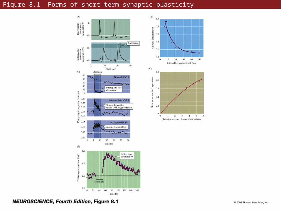
Figure 8.1 Forms of short-term synaptic plasticity
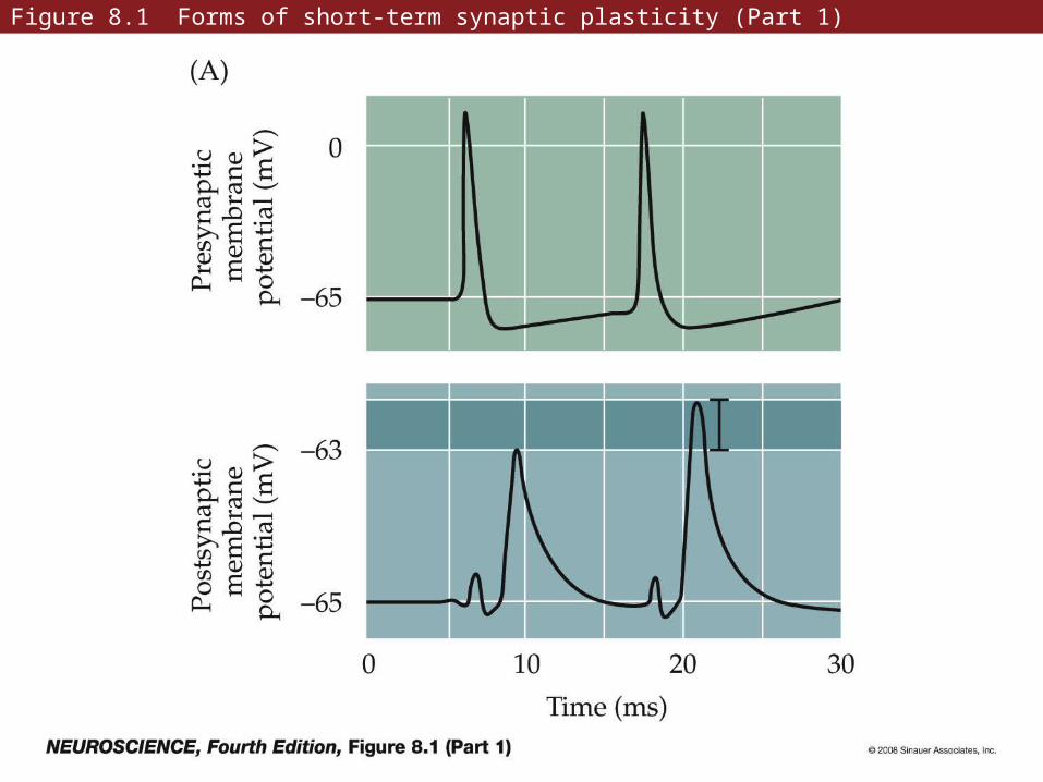
Figure 8.1 Forms of short-term synaptic plasticity (Part 1)
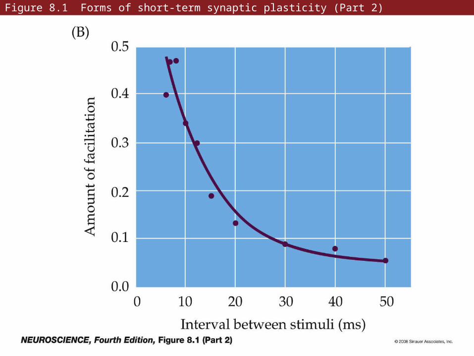
Figure 8.1 Forms of short-term synaptic plasticity (Part 2)
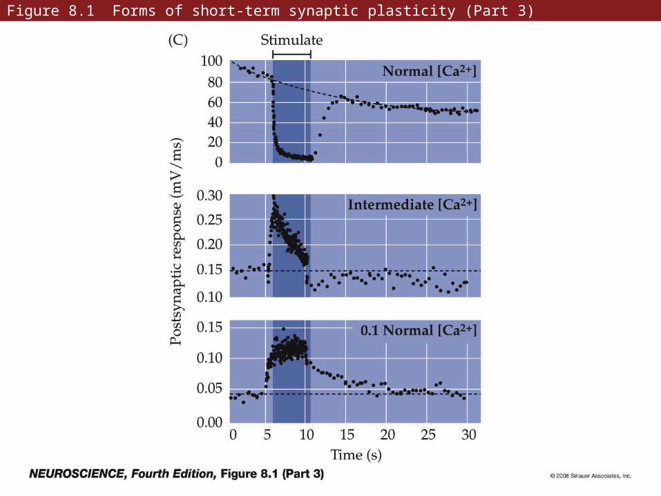
Figure 8.1 Forms of short-term synaptic plasticity (Part 3)
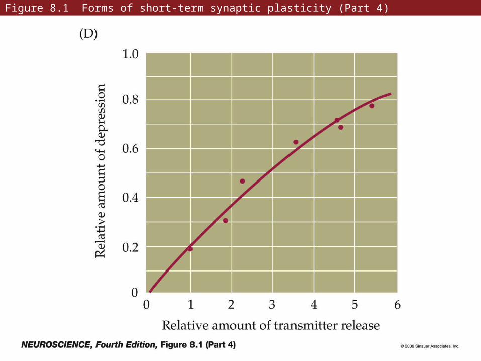
Figure 8.1 Forms of short-term synaptic plasticity (Part 4)
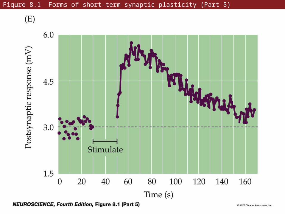
Figure 8.1 Forms of short-term synaptic plasticity (Part 5)
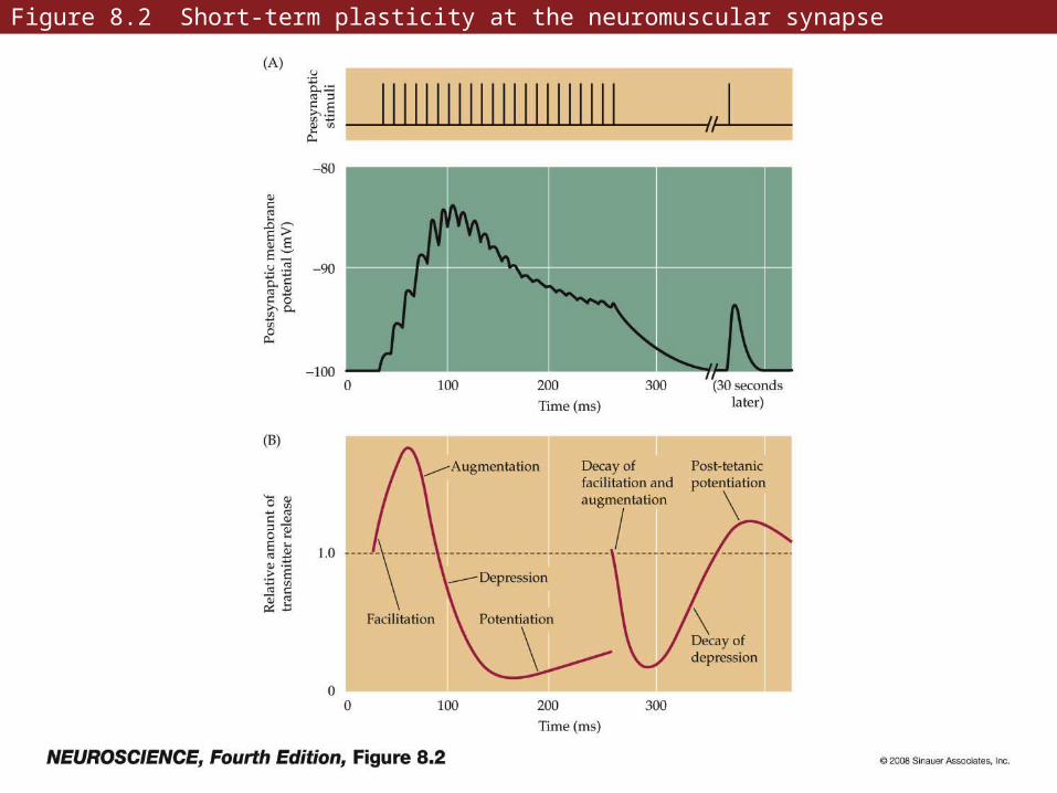
Figure 8.2 Short-term plasticity at the neuromuscular synapse
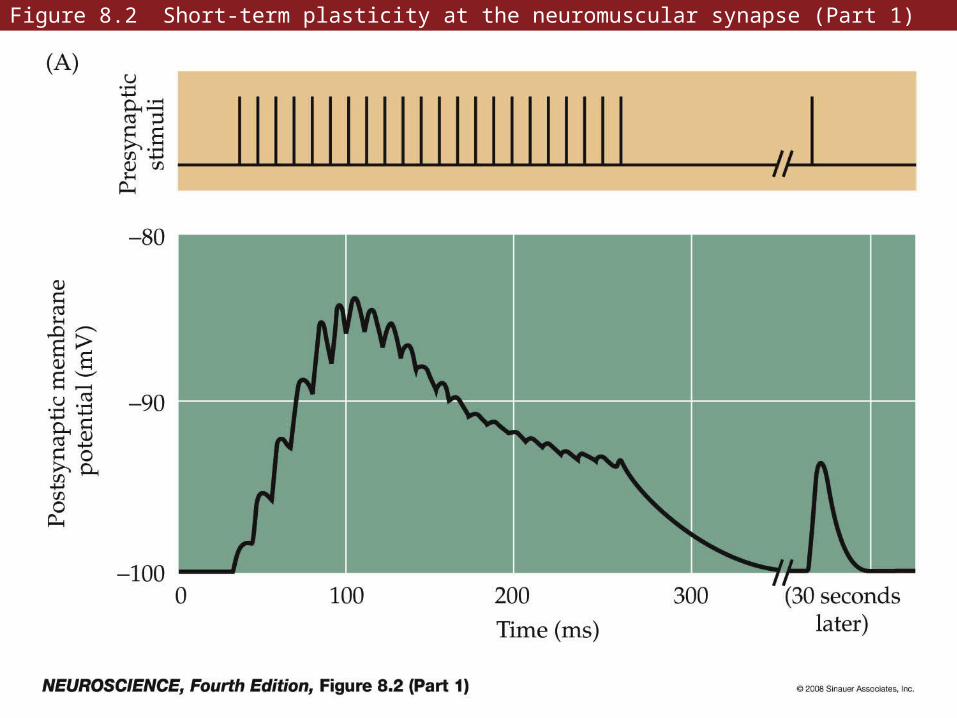
Figure 8.2 Short-term plasticity at the neuromuscular synapse (Part 1)
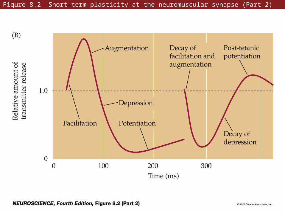
Figure 8.2 Short-term plasticity at the neuromuscular synapse (Part 2)
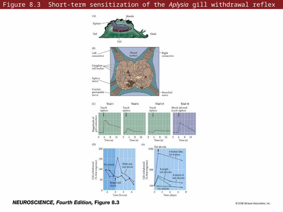
Figure 8.3 Short-term sensitization of the Aplysia gill withdrawal reflex
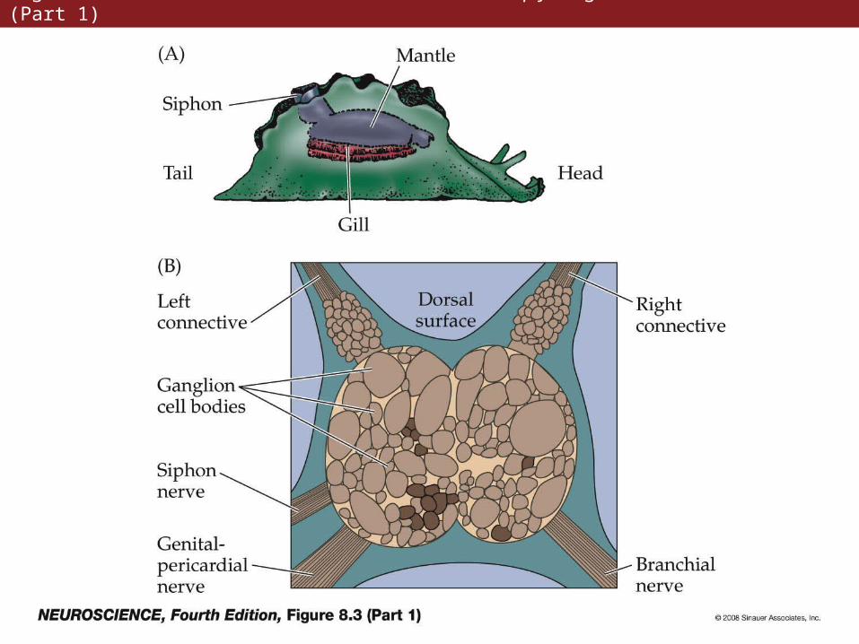
Figure 8.3 Short-term sensitization of the Aplysia gill withdrawal reflex (Part 1)
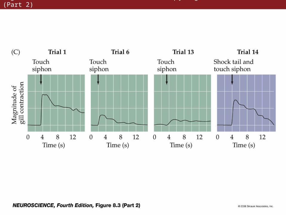
Figure 8.3 Short-term sensitization of the Aplysia gill withdrawal reflex (Part 2)
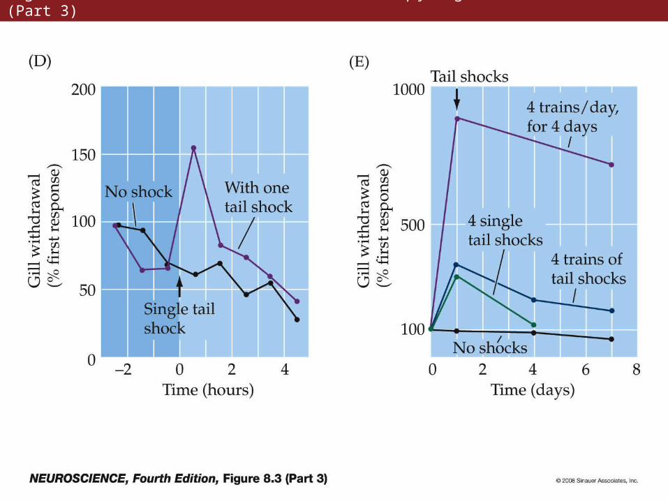
Figure 8.3 Short-term sensitization of the Aplysia gill withdrawal reflex (Part 3)
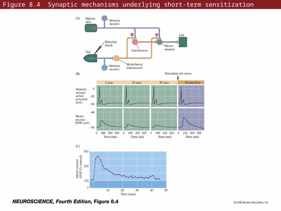
Figure 8.4 Synaptic mechanisms underlying short-term sensitization
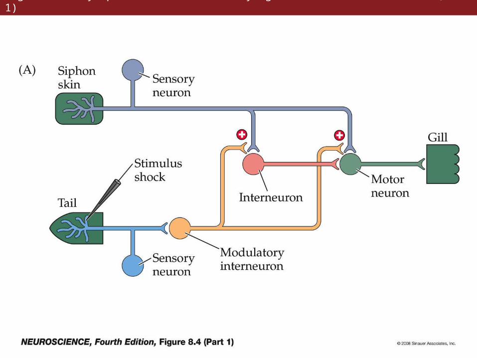
Figure 8.4 Synaptic mechanisms underlying short-term sensitization (Part 1)
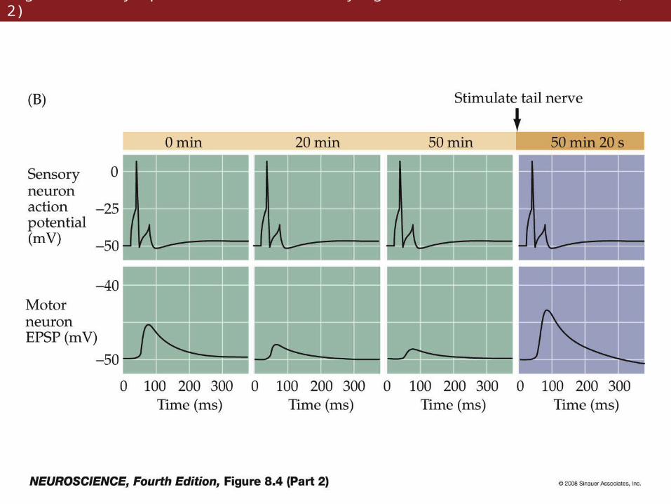
Figure 8.4 Synaptic mechanisms underlying short-term sensitization (Part 2)
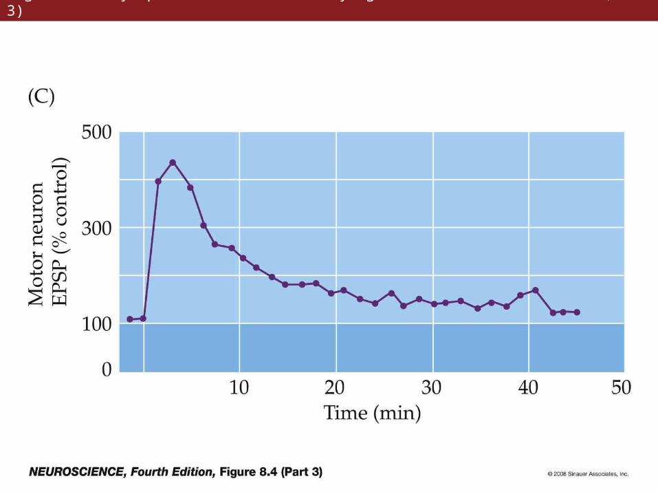
Figure 8.4 Synaptic mechanisms underlying short-term sensitization (Part 3)
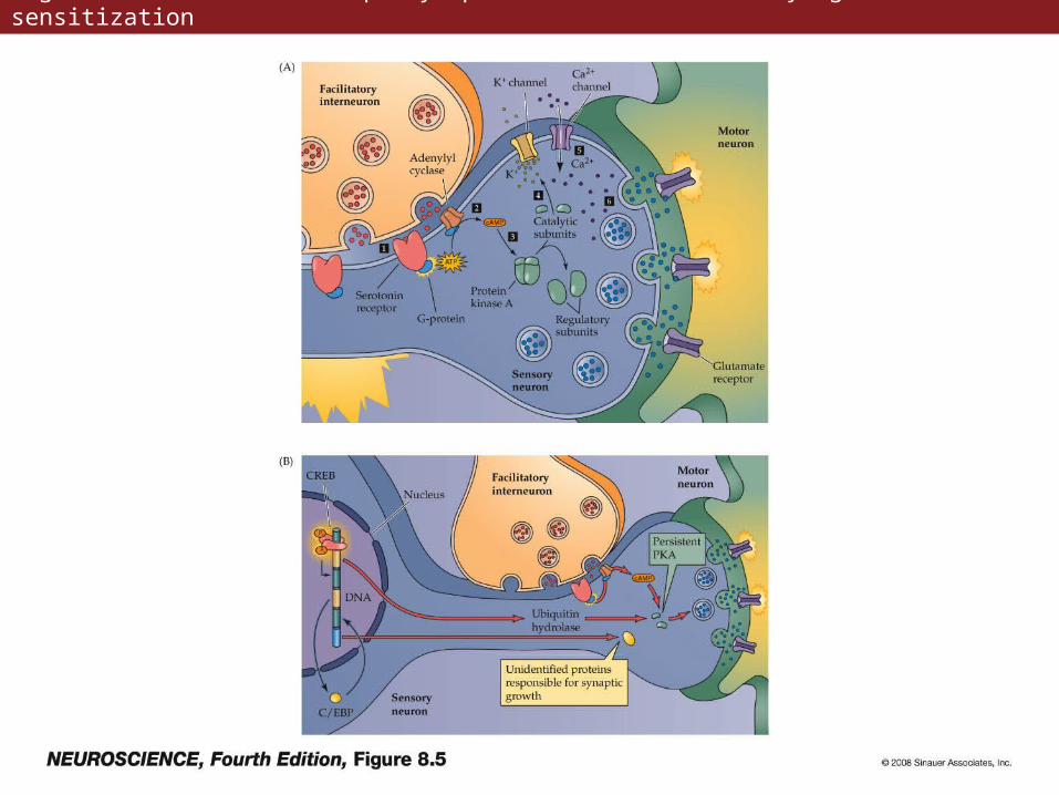
Figure 8.5 Mechanism of presynaptic enhancement underlying behavioral sensitization
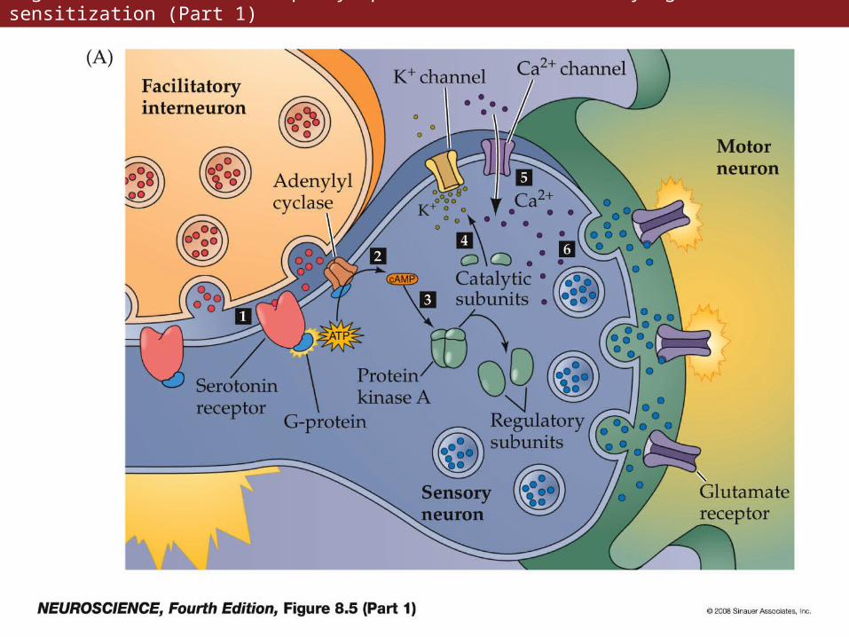
Figure 8.5 Mechanism of presynaptic enhancement underlying behavioral sensitization (Part 1)
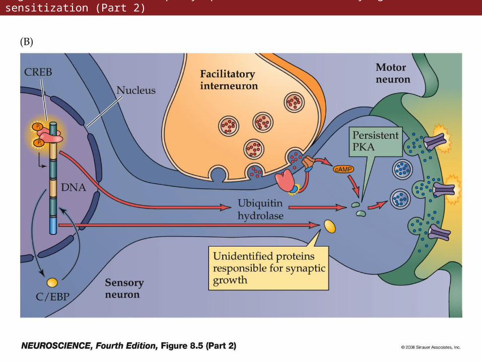
Figure 8.5 Mechanism of presynaptic enhancement underlying behavioral sensitization (Part 2)
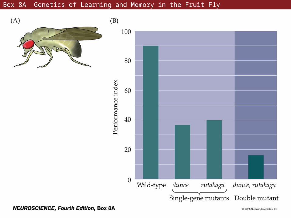
Box 8A Genetics of Learning and Memory in the Fruit Fly
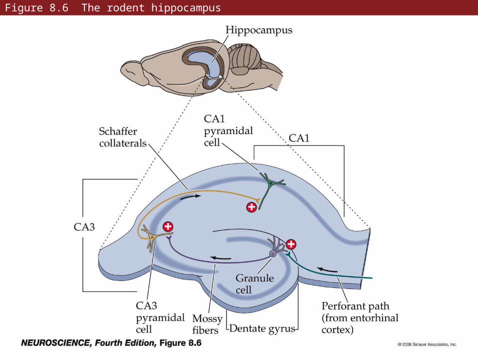
Figure 8.6 The rodent hippocampus
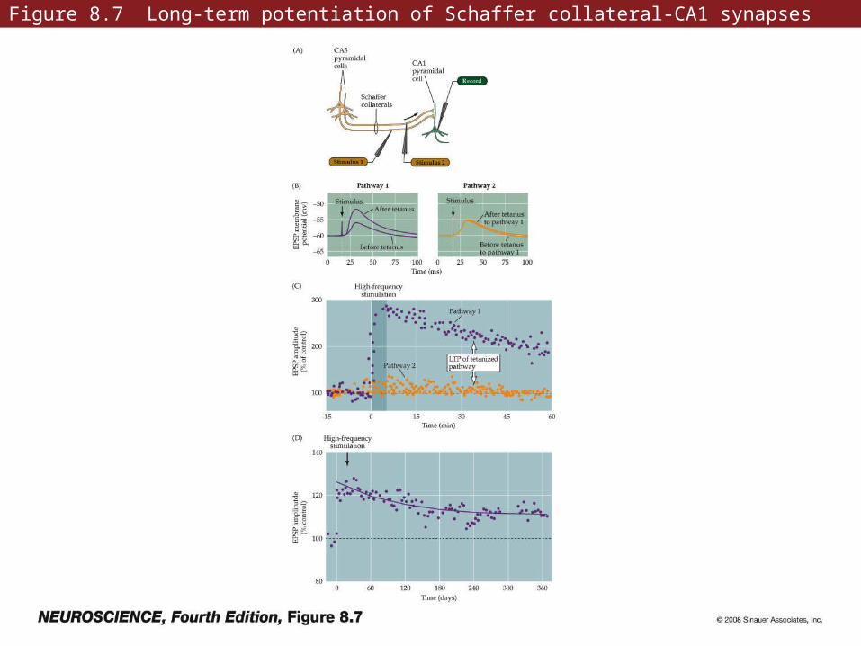
Figure 8.7 Long-term potentiation of Schaffer collateral-CA1 synapses
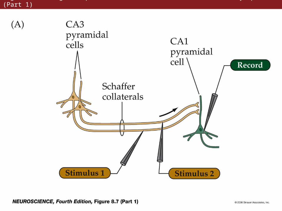
Figure 8.7 Long-term potentiation of Schaffer collateral-CA1 synapses (Part 1)
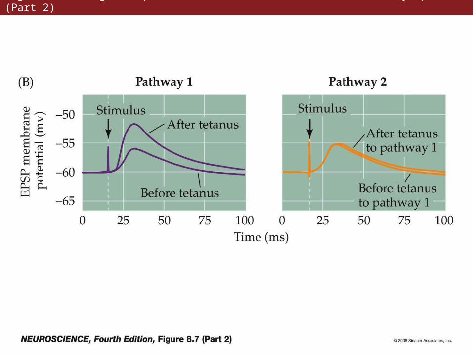
Figure 8.7 Long-term potentiation of Schaffer collateral-CA1 synapses (Part 2)
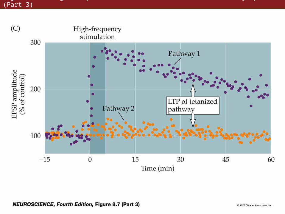
Figure 8.7 Long-term potentiation of Schaffer collateral-CA1 synapses (Part 3)
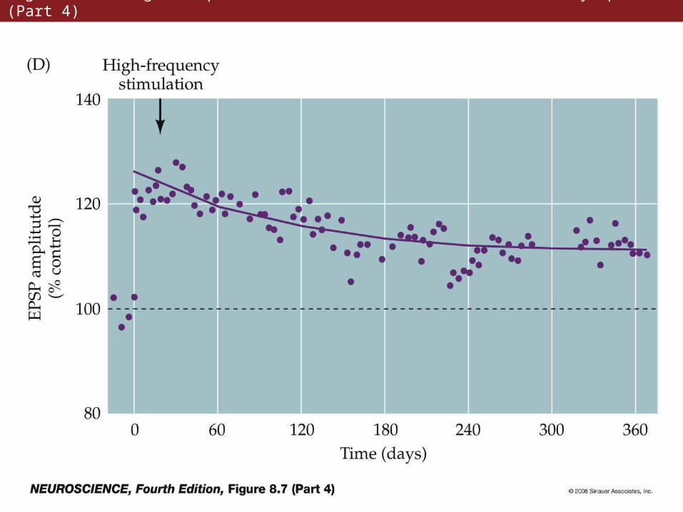
Figure 8.7 Long-term potentiation of Schaffer collateral-CA1 synapses (Part 4)
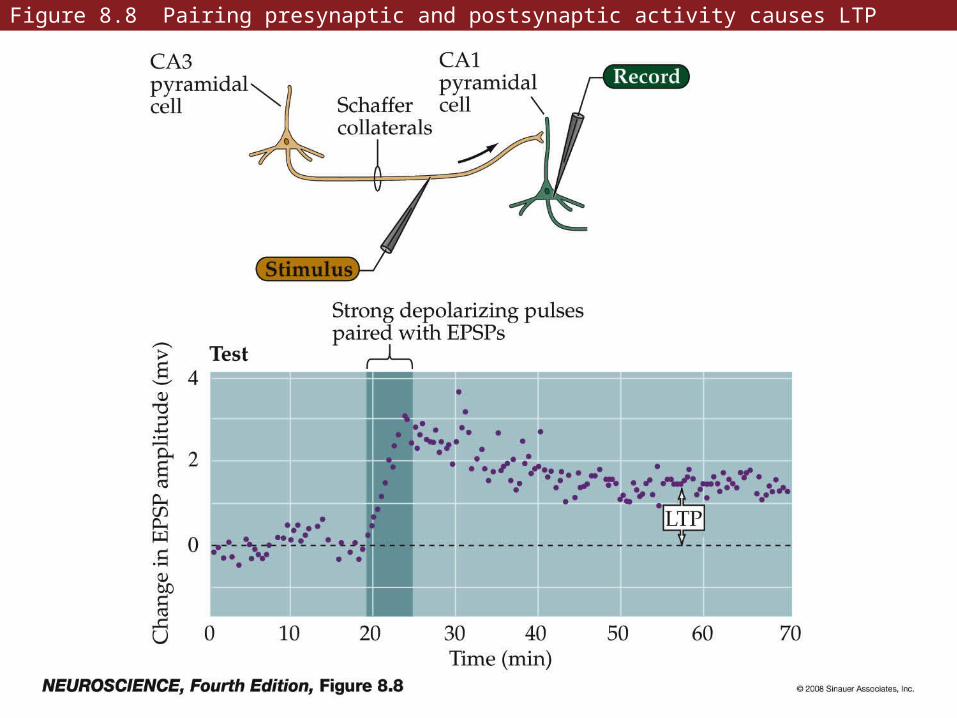
Figure 8.8 Pairing presynaptic and postsynaptic activity causes LTP
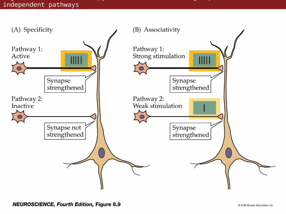
Figure 8.9 LTP at a CA1 pyramidal neuron receiving inputs from two independent pathways
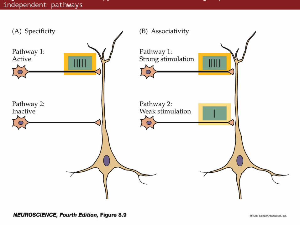
Figure 8.9 LTP at a CA1 pyramidal neuron receiving inputs from two independent pathways
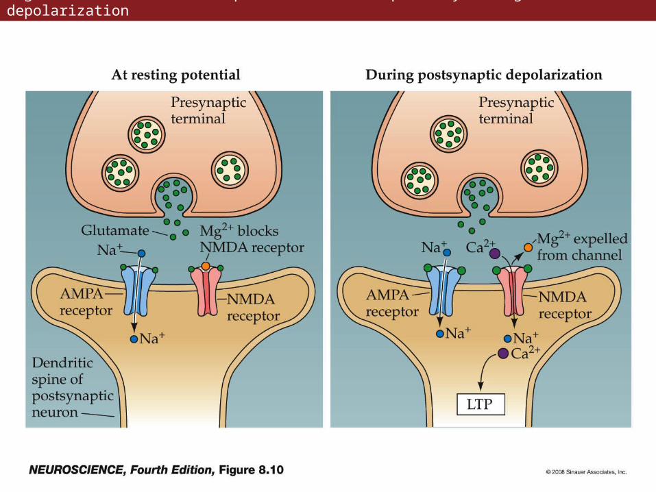
Figure 8.10 The NMDA receptor channel can open only during depolarization
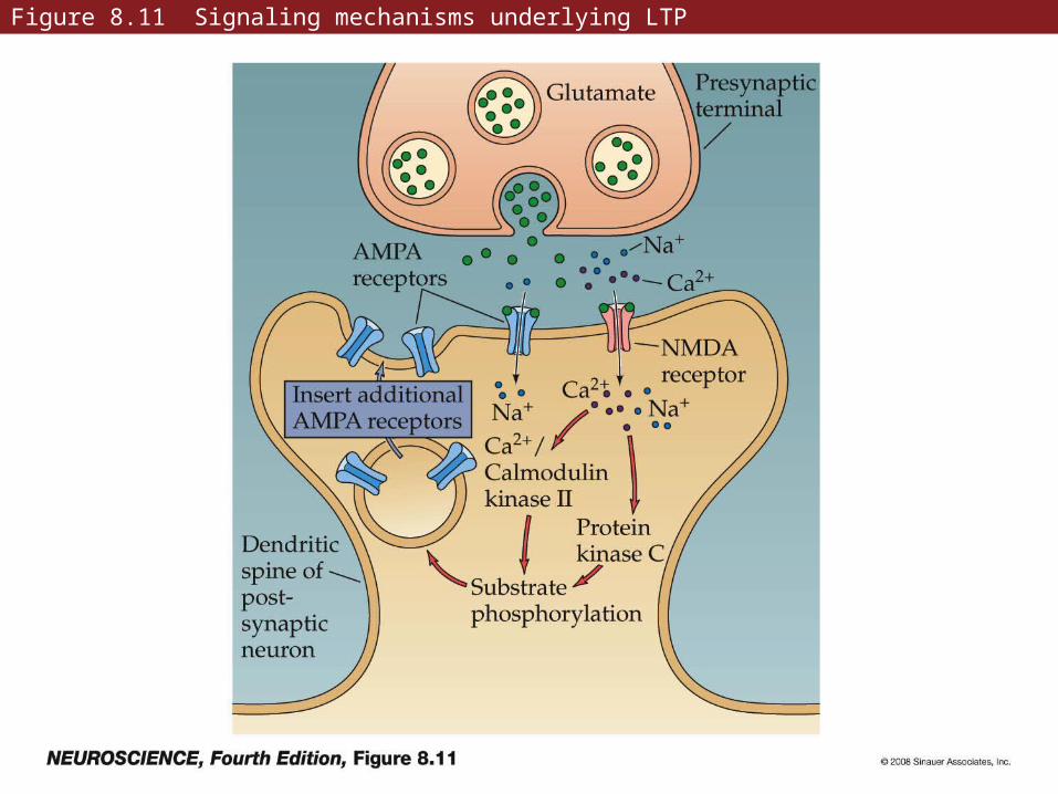
Figure 8.11 Signaling mechanisms underlying LTP
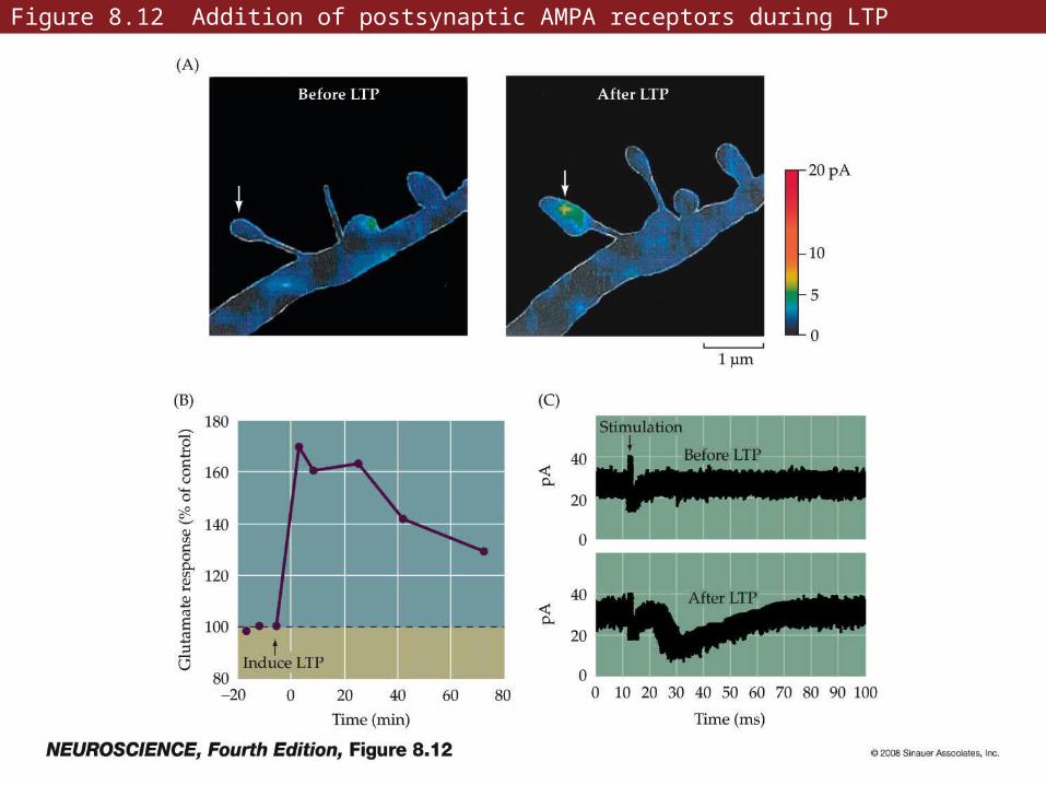
Figure 8.12 Addition of postsynaptic AMPA receptors during LTP
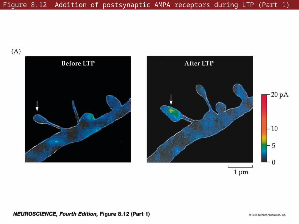
Figure 8.12 Addition of postsynaptic AMPA receptors during LTP (Part 1)

Figure 8.12 Addition of postsynaptic AMPA receptors during LTP (Part 2)
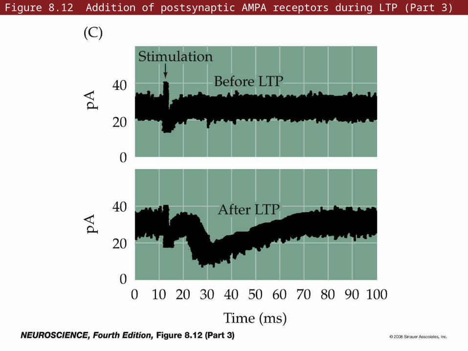
Figure 8.12 Addition of postsynaptic AMPA receptors during LTP (Part 3)
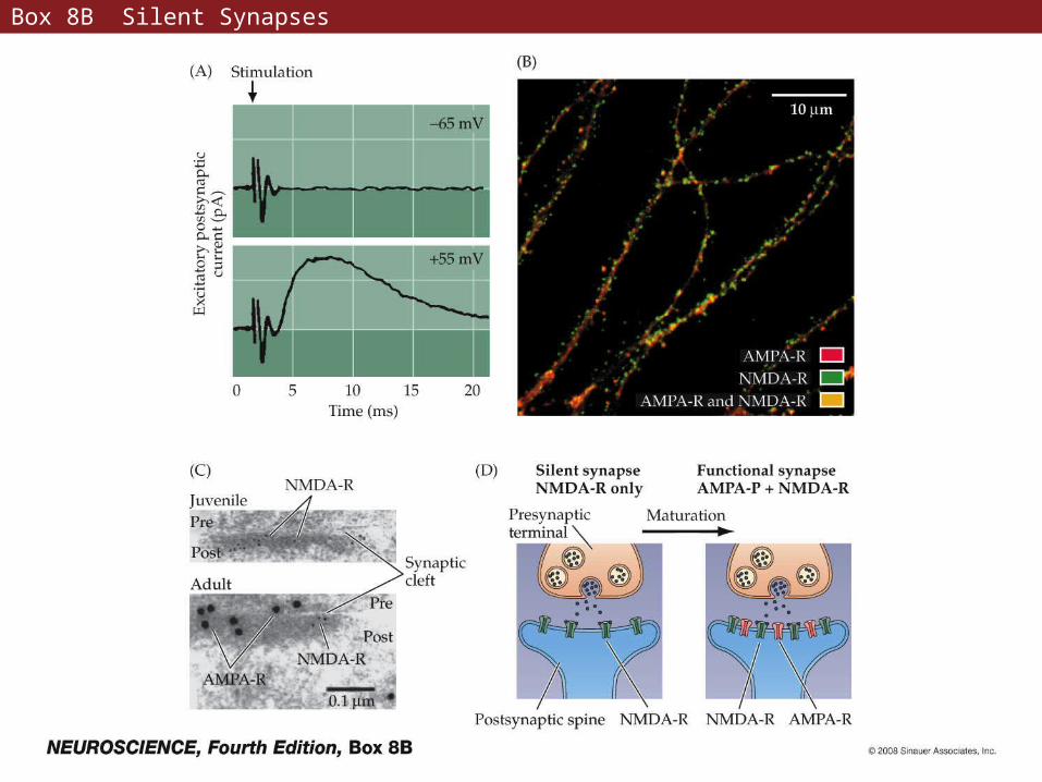
Box 8B Silent Synapses
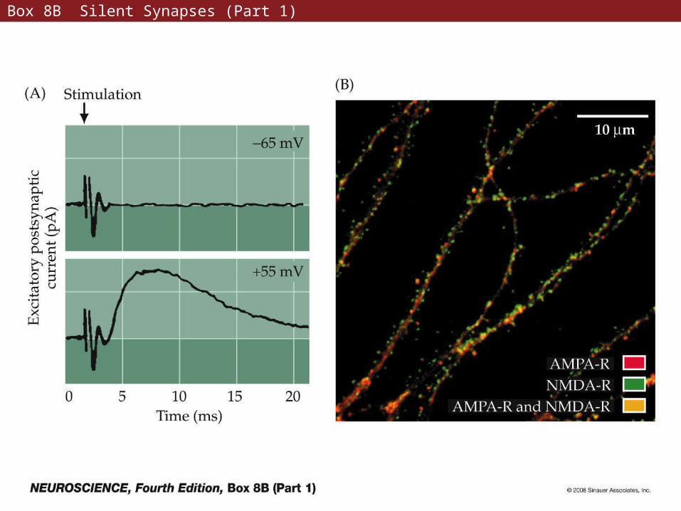
Box 8B Silent Synapses (Part 1)
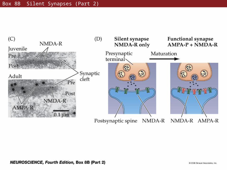
Box 8B Silent Synapses (Part 2)
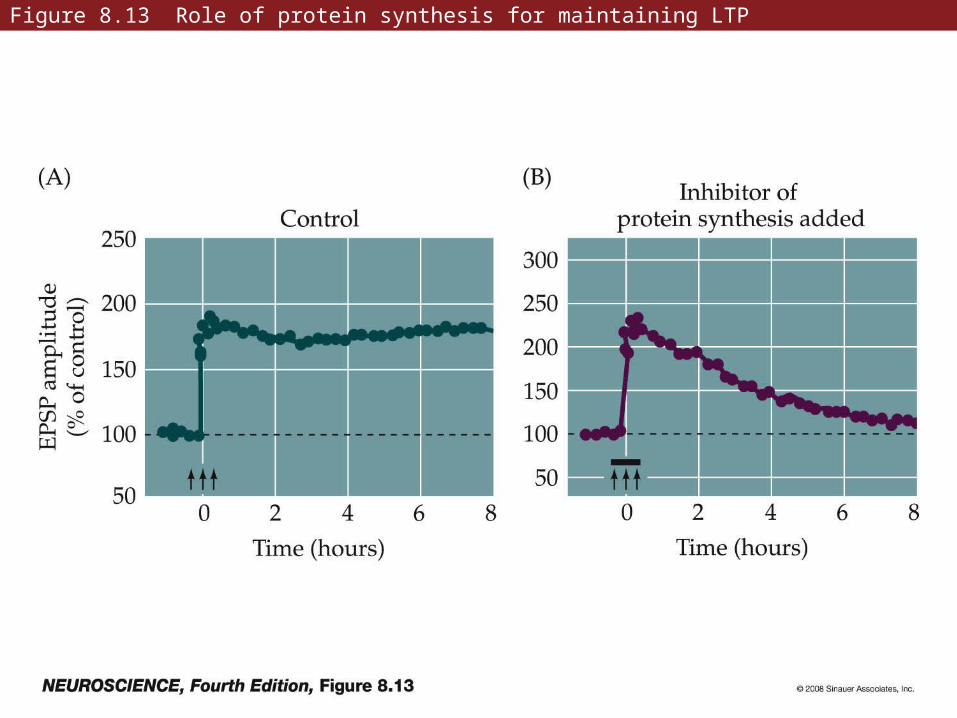
Figure 8.13 Role of protein synthesis for maintaining LTP
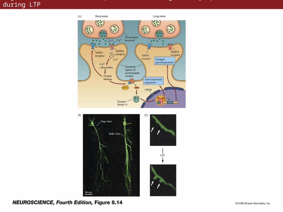
Figure 8.14 Mechanisms responsible for changes in synaptic transmission during LTP
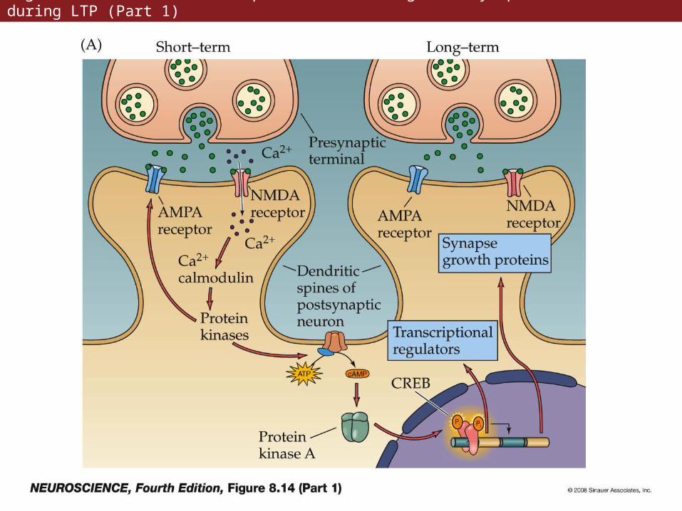
Figure 8.14 Mechanisms responsible for changes in synaptic transmission during LTP (Part 1)
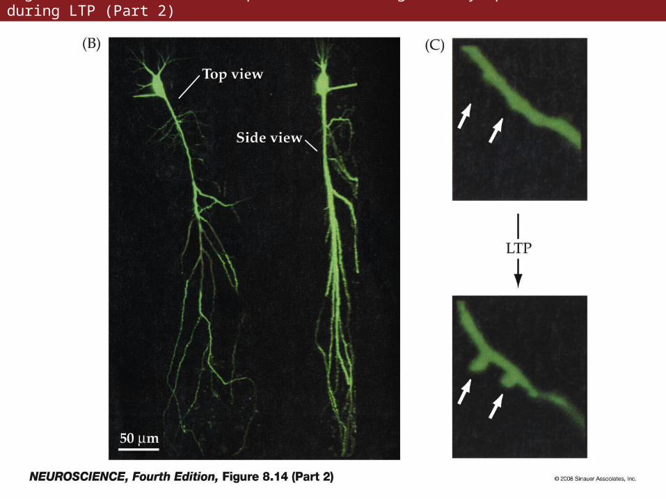
Figure 8.14 Mechanisms responsible for changes in synaptic transmission during LTP (Part 2)
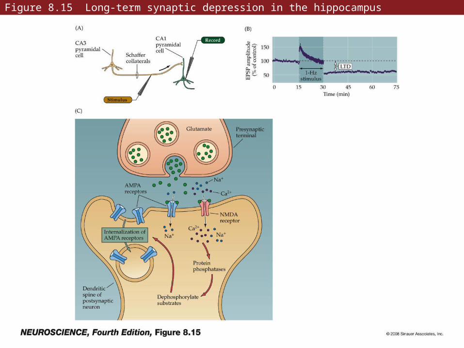
Figure 8.15 Long-term synaptic depression in the hippocampus
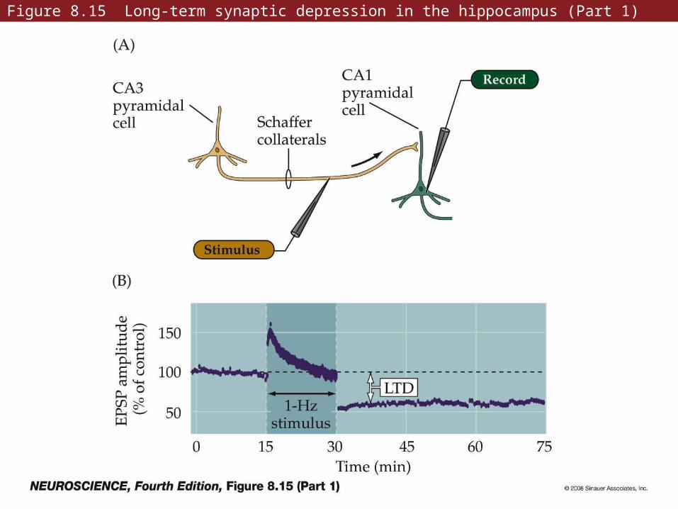
Figure 8.15 Long-term synaptic depression in the hippocampus (Part 1)
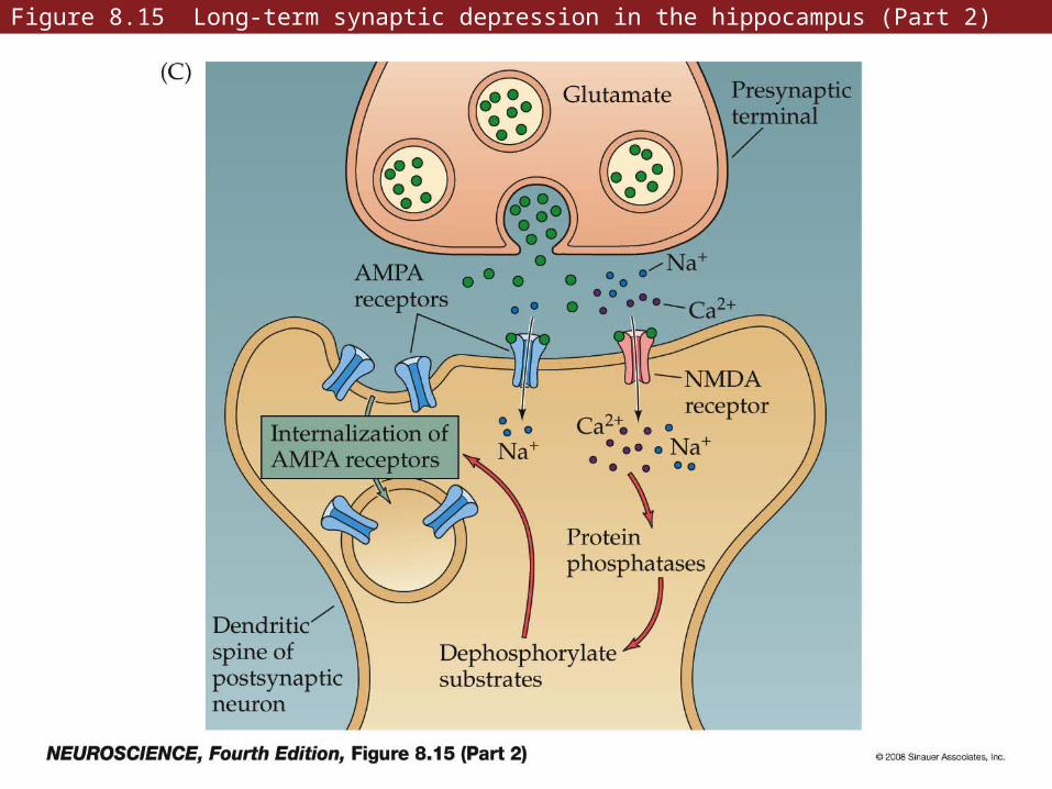
Figure 8.15 Long-term synaptic depression in the hippocampus (Part 2)
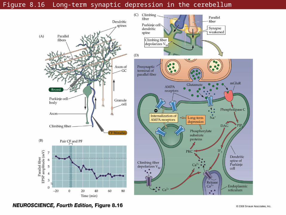
Figure 8.16 Long-term synaptic depression in the cerebellum
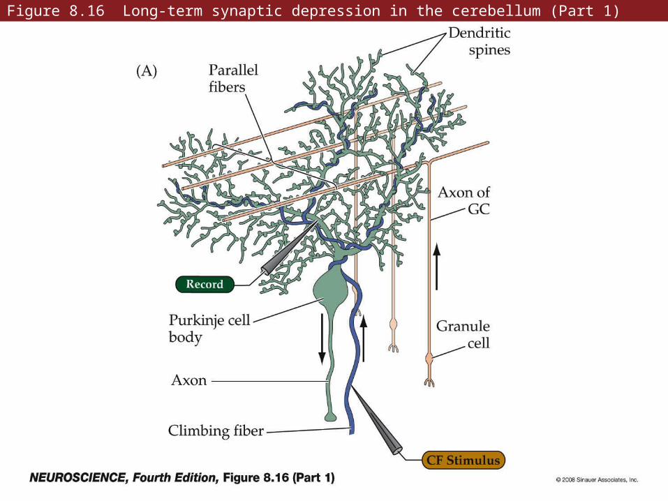
Figure 8.16 Long-term synaptic depression in the cerebellum (Part 1)
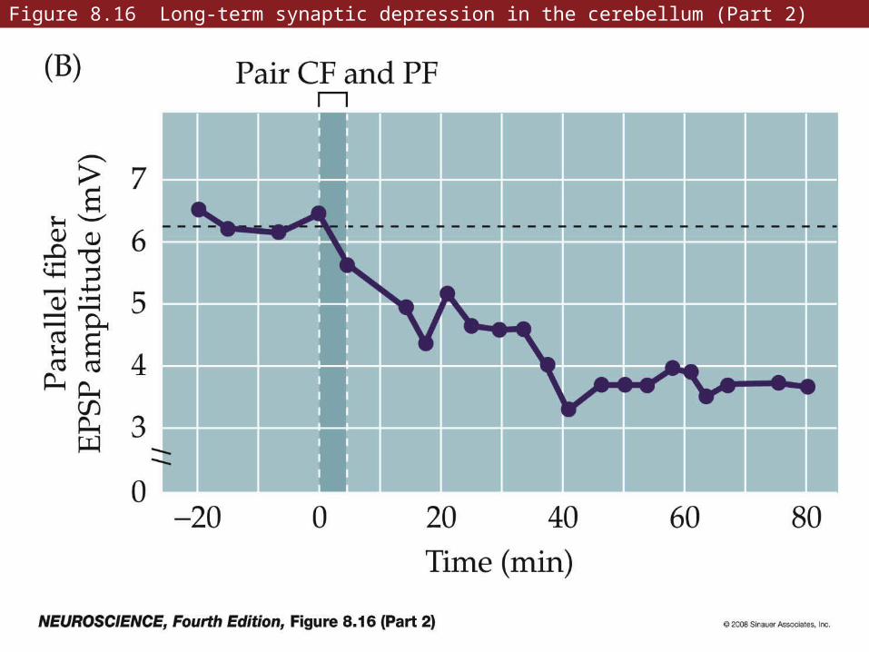
Figure 8.16 Long-term synaptic depression in the cerebellum (Part 2)
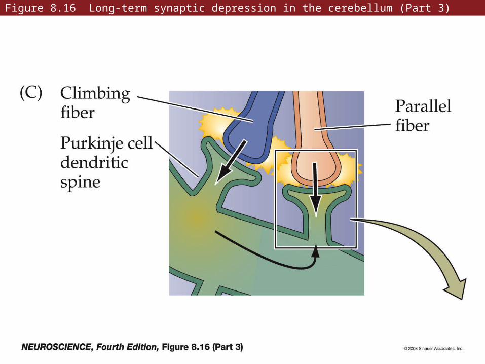
Figure 8.16 Long-term synaptic depression in the cerebellum (Part 3)
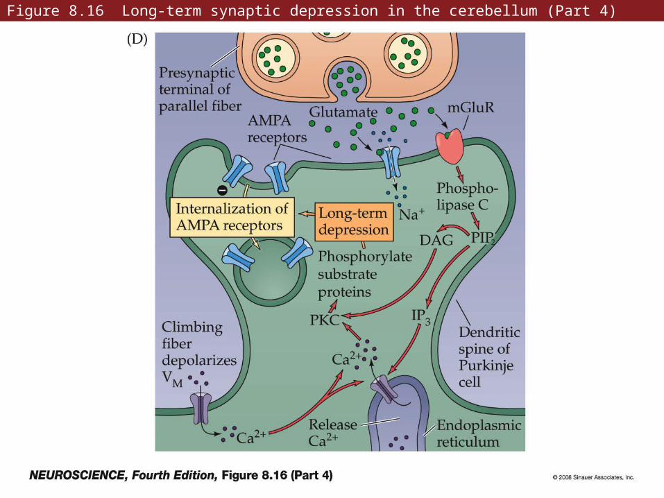
Figure 8.16 Long-term synaptic depression in the cerebellum (Part 4)
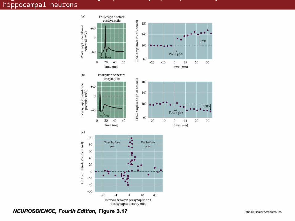
Figure 8.17 Spike timing-dependent synaptic plasticity in cultured hippocampal neurons
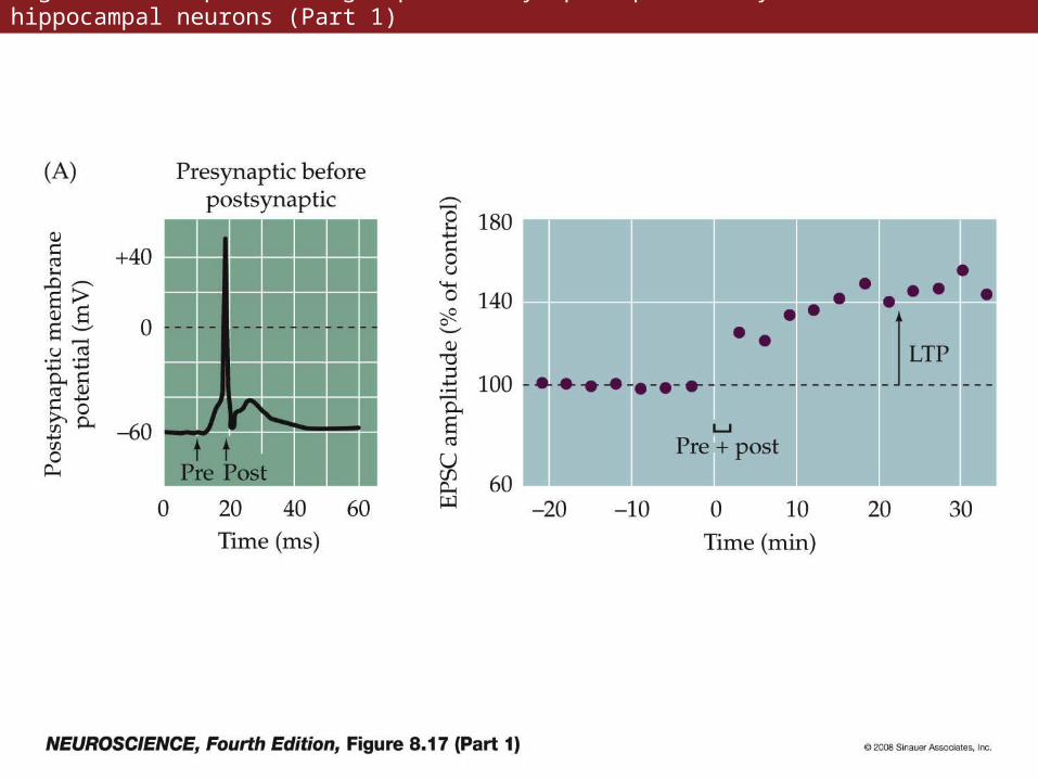
Figure 8.17 Spike timing-dependent synaptic plasticity in cultured hippocampal neurons (Part 1)
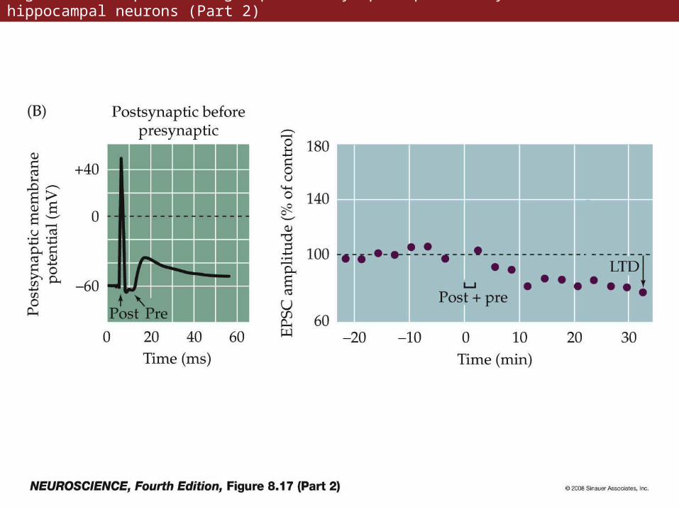
Figure 8.17 Spike timing-dependent synaptic plasticity in cultured hippocampal neurons (Part 2)
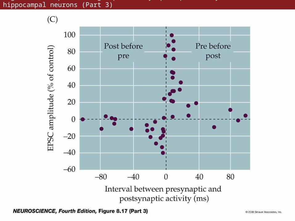
Figure 8.17 Spike timing-dependent synaptic plasticity in cultured hippocampal neurons (Part 3)
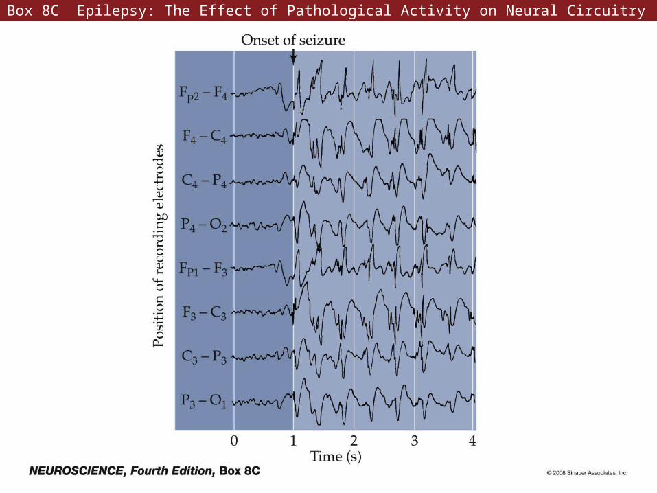
Box 8C Epilepsy: The Effect of Pathological Activity on Neural Circuitry