Embed Size (px)
Citation preview
The Corticohippocampal Circuit, SynapticPlasticity, and Memory
Jayeeta Basu1 and Steven A. Siegelbaum2,3,4
1Department of Neuroscience and Physiology, NYU Neuroscience Institute, New York University Schoolof Medicine, New York, New York 10016
2Kavli Institute for Brain Science, Columbia University, New York, New York 100323Department of Neuroscience, Columbia University, New York, New York 100324Department of Pharmacology, Columbia University, New York, New York 10032
Correspondence: [email protected]
Synaptic plasticity serves as a cellular substrate for information storage in the central nervoussystem. The entorhinal cortex (EC) and hippocampus are interconnected brain areas sup-porting basic cognitive functions important for the formation and retrieval of declarativememories. Here, we discuss how information flow in the EC–hippocampal loop is organizedthrough circuit design. We highlight recently identified corticohippocampal and intrahippo-campal connections and how these long-range and local microcircuits contribute to learn-ing. This review also describes various forms of activity-dependent mechanisms that changethe strength of corticohippocampal synaptic transmission. A key point to emerge from thesestudies is that patterned activityand interaction of coincident inputs gives rise to associationalplasticity and long-term regulation of information flow. Finally, we offer insights about howlearning-related synaptic plasticity within the corticohippocampal circuit during sensoryexperiences may enable adaptive behaviors for encoding spatial, episodic, social, and con-textual memories.
Ever since the description by Scoville andMilner (1957) of the profound anterograde
amnesia in patient H.M. following bilateraltemporal lobe resection, the hippocampus andsurrounding temporal lobe structures have beenextensively studied for their role in memorystorage (Squire and Wixted 2011). Fifteen yearsafter this initial finding, our understandingof the neurophysiological bases of hippocam-pal function were greatly enhanced by twobreakthroughs: Bliss and Lomo’s (1973) findingof activity-dependent long-term potentiation(LTP) of synaptic transmission in the hippo-
campus, and the discovery of hippocampalplace cells, neurons that encode the spatial po-sition of an animal, by O’Keefe and Dostrovsky(1971). These discoveries stimulated a numberof subsequent advances in our understandingof various forms of long-term synaptic plastic-ity at different stages of information processingin the corticohippocampal circuit, and howsuch plasticity contributes to memory storageand spatial representation.
Changes in synaptic efficacy ultimately actby altering the flow of information throughneural circuits. Thus, a deeper understanding
Editors: Eric R. Kandel, Yadin Dudai, and Mark R. Mayford
Additional Perspectives on Learning and Memory available at www.cshperspectives.org
Copyright # 2015 Cold Spring Harbor Laboratory Press; all rights reserved; doi: 10.1101/cshperspect.a021733
Cite this article as Cold Spring Harb Perspect Biol 2015;7:a021733
1
on January 4, 2020 - Published by Cold Spring Harbor Laboratory Press http://cshperspectives.cshlp.org/Downloaded from
of how synaptic plasticity may subserve the en-coding of memory requires a detailed knowl-edge of the paths of information flow throughthe corticohippocampal circuit, and how neuralactivity alters such information processing.Because there have been a number of recentexcellent reviews on the importance of variousforms of hippocampal synaptic plasticity inlearning and memory (Morris et al. 2013; Ban-nerman et al. 2014), we largely focus on recentstudies elucidating new features of the cor-ticohippocampal circuit and new forms of plas-ticity that are tuned to the dynamics of thiscircuit.
INFORMATION FLOW THROUGH THECORTICOHIPPOCAMPAL CIRCUIT
The hippocampus is important for both spatialand nonspatial forms of declarative or explic-it memory (Squire et al. 2004), our knowledgeof people, places, things, and events. In additionto encoding spatial information (Burgess andO’Keefe 1996), hippocampal neurons may alsoencode time during episodic events (Pastalkovaet al. 2008; Kraus et al. 2013; Macdonald et al.2013). How does the hippocampus encode andstore these diverse memories? Although a defin-itive answer is lacking, our knowledge of thecorticohippocampal circuit has greatly expand-ed in recent years, giving us a new appreciationfor the multiple pathways by which informationis processed in the hippocampus, an importantstep to achieving a circuit-level understandingof how this brain region stores memories.
Neurons wire up during development toform circuits that provide the architecturalframework for information flow in the brain,enabling one brain area to influence another.Learning requires the association of informa-tion from different coactive brain areas duringsensory experiences or the reprocessing of inter-nal representations. Such associations result inplastic changes in synaptic and cell-wide func-tions that enable the formation of preferentiallyconnected cell assemblies. At the level of a singleneuron, this could occur through integrationand association of inputs that coincide in timeor space to influence the neuron’s output.
As initially defined by Lorente de No (1934),the hippocampal region is composed of severalsubregions, including dentate gyrus (DG), andthe CA3, CA2, and CA1 regions of the hippo-campus proper. In the rat brain, there are esti-mated to be �1,000,000 DG granule neurons,300,000 CA3 pyramidal neurons, �30,000 CA2pyramidal neurons, and 300,000 CA1 pyra-midal neurons (Amaral and Witter 1989). Inaddition to these excitatory, principal neurons,there are many classes of inhibitory neurons inthe hippocampus, although the total numberof these inhibitory interneurons is only about10% to 20% that of the principal cells. In CA1alone, more than 20 types of GABAergic inter-neurons, have been classified according to theirmorphology, location, molecular and electro-physiological properties, and synaptic targets(Klausberger and Somogyi 2008). A key goalin hippocampal research is to gain an under-standing as to how these different subregionsprocess their inputs during learning to generatean output contributing to distinct aspects ofmemory encoding.
The Corticohippocampal Circuit.I: Classical Pathways
A typical CA1 pyramidal neuron in the rat re-ceives a total of �30,000 glutamatergic synapticinputs distributed throughout its dendritic tree.In addition, it receives �1700 GABAergic in-puts (Megias et al. 2001). The major source ofglutamatergic input to hippocampus comesfrom the entorhinal cortex (EC), a polymodalsensory association area that conveys both non-spatial sensory information (from the lateralentorhinal cortex [LEC]) and spatial infor-mation (from the medial entorhinal cortex[MEC]). This sensory information is then pro-cessed within the hippocampus by severalparallel circuits, ultimately leaving the hippo-campus through CA1, the major output path-way (van Strien et al. 2009).
One striking feature of certain neurons inlayers II and III of EC (EC LII and LIII), termedgrid cells, is that they show spatially tuned firingpatterns consisting of a hexagonally spaced ar-ray of grid-like firing fields that cover the extent
J. Basu and S.A. Siegelbaum
2 Cite this article as Cold Spring Harb Perspect Biol 2015;7:a021733
on January 4, 2020 - Published by Cold Spring Harbor Laboratory Press http://cshperspectives.cshlp.org/Downloaded from
of a two-dimensional environment (Fyhn et al.2004; Hafting et al. 2005; Buzsaki and Moser2013). Convergent input from a number of ECgrid cells with slightly shifted firing fields hasbeen proposed to produce an interference-likepattern that gives rise to the single well-definedplace field of a hippocampal place cell (O’Keefeand Burgess 2005; Solstad et al. 2006; Bush et al.2014).
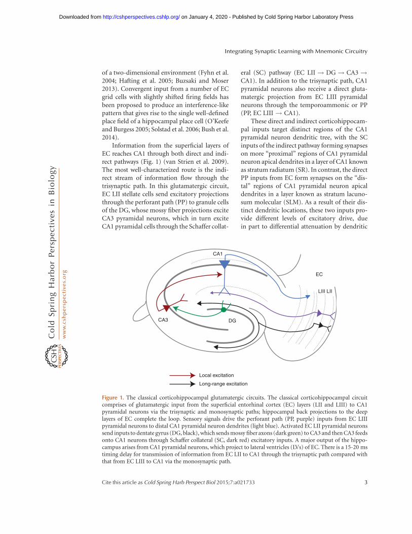
Information from the superficial layers ofEC reaches CA1 through both direct and indi-rect pathways (Fig. 1) (van Strien et al. 2009).The most well-characterized route is the indi-rect stream of information flow through thetrisynaptic path. In this glutamatergic circuit,EC LII stellate cells send excitatory projectionsthrough the perforant path (PP) to granule cellsof the DG, whose mossy fiber projections exciteCA3 pyramidal neurons, which in turn exciteCA1 pyramidal cells through the Schaffer collat-
eral (SC) pathway (EC LII ! DG ! CA3!CA1). In addition to the trisynaptic path, CA1pyramidal neurons also receive a direct gluta-matergic projection from EC LIII pyramidalneurons through the temporoammonic or PP(PP, EC LIII ! CA1).
These direct and indirect corticohippocam-pal inputs target distinct regions of the CA1pyramidal neuron dendritic tree, with the SCinputs of the indirect pathway forming synapseson more “proximal” regions of CA1 pyramidalneuron apical dendrites in a layer of CA1 knownas stratum radiatum (SR). In contrast, the directPP inputs from EC form synapses on the “dis-tal” regions of CA1 pyramidal neuron apicaldendrites in a layer known as stratum lacuno-sum molecular (SLM). As a result of their dis-tinct dendritic locations, these two inputs pro-vide different levels of excitatory drive, duein part to differential attenuation by dendritic
CA3
Local excitation
Long-range excitation
DG
CA1
EC
LIII LII
Figure 1. The classical corticohippocampal glutamatergic circuits. The classical corticohippocampal circuitcomprises of glutamatergic input from the superficial entorhinal cortex (EC) layers (LII and LIII) to CA1pyramidal neurons via the trisynaptic and monosynaptic paths; hippocampal back projections to the deeplayers of EC complete the loop. Sensory signals drive the perforant path (PP, purple) inputs from EC LIIIpyramidal neurons to distal CA1 pyramidal neuron dendrites (light blue). Activated EC LII pyramidal neuronssend inputs to dentate gyrus (DG, black), which sends mossy fiber axons (dark green) to CA3 and then CA3 feedsonto CA1 neurons through Schaffer collateral (SC, dark red) excitatory inputs. A major output of the hippo-campus arises from CA1 pyramidal neurons, which project to lateral ventricles (LVs) of EC. There is a 15-20 mstiming delay for transmission of information from EC LII to CA1 through the trisynaptic path compared withthat from EC LIII to CA1 via the monosynaptic path.
Integrating Synaptic Learning with Mnemonic Circuitry
Cite this article as Cold Spring Harb Perspect Biol 2015;7:a021733 3
on January 4, 2020 - Published by Cold Spring Harbor Laboratory Press http://cshperspectives.cshlp.org/Downloaded from
cable properties, with the PP synapses of thedirect pathway providing weak excitation atthe soma compared with the strong excitationprovided by the SC inputs of the indirect path-way. These two classes of glutamatergic inputsalso activate a number of different classes ofGABAergic interneurons that inhibit CA1 out-put in a feedforward manner (Fig. 2).
The Corticohippocampal Circuit.II: Neoclassical Pathways
More recent results show that, in addition totheir classical inputs from CA3 and EC, CA1neurons also receive strong excitatory inputfrom the CA2 region of the hippocampus(CA2 ! CA1) (Chevaleyre and Siegelbaum2010), a relatively small area nestled betweenCA3 and CA1 (Fig. 3). Although first identifiedby Lorente de No in 1934, the CA2 region hasremained relatively unexplored, due in part toits small size and transitional location between
CA3 and CA1. However, recent studies usinggenetic approaches clearly show the unique mo-lecular identity of CA2 pyramidal neurons (Leinet al. 2005; Hitti and Siegelbaum 2014; Koharaet al. 2014) and suggest the importance of theCA2 region in certain hippocampal functions(Piskorowski and Chevaleyre 2012).
Similar to CA1 neurons, CA2 pyramidalneurons receive both direct input from LII ECneurons (EC LII ! CA2) onto their distal den-drites (Cui et al. 2013; Hitti and Siegelbaum2014) and indirect input from the CA3 SC path-way (EC LII! DG ! CA3 ! CA2) ontotheir proximal dendrites (Chevaleyre and Sie-gelbaum 2010). In addition, CA2 also receivesweaker mossy fiber excitatory input directlyfrom DG granule cells (EC LII ! DG ! CA2)(Kohara et al. 2014).
In striking contrast to CA1, CA2 pyramidalneurons are excited much more strongly bytheir direct EC input compared with their SCinput (Chevaleyre and Siegelbaum 2010). Fur-
CA3
Local excitation
Long-range excitation
Local inhibition
Long-range inhibition
DG
CA1
EC
LIII LII
Figure 2. Local and long-range GABAergic connections. CA1 has several local GABAergic interneurons (red).These target the CA1 pyramidal neuron soma, axon, and dendrite to modulate pyramidal neuron activity in adomain-specific manner. Schaffer collateral (SC)- and entorhinal cortex (EC)-associated inhibitory microcir-cuits provide feedforward inhibition, whereas feedback inhibition is recruited recurrently when the CA1 pyra-midal neuron fires an action potential. Long-range inhibitory projections (green) from the EC provide directinhibition preferentially to local interneurons (INs) in CA1. Long-range projections from GABAergic neuronsin stratum oriens (SO) of hippocampus to layer II/III (LII/LIII) of the EC have also been described.
J. Basu and S.A. Siegelbaum
4 Cite this article as Cold Spring Harb Perspect Biol 2015;7:a021733
on January 4, 2020 - Published by Cold Spring Harbor Laboratory Press http://cshperspectives.cshlp.org/Downloaded from
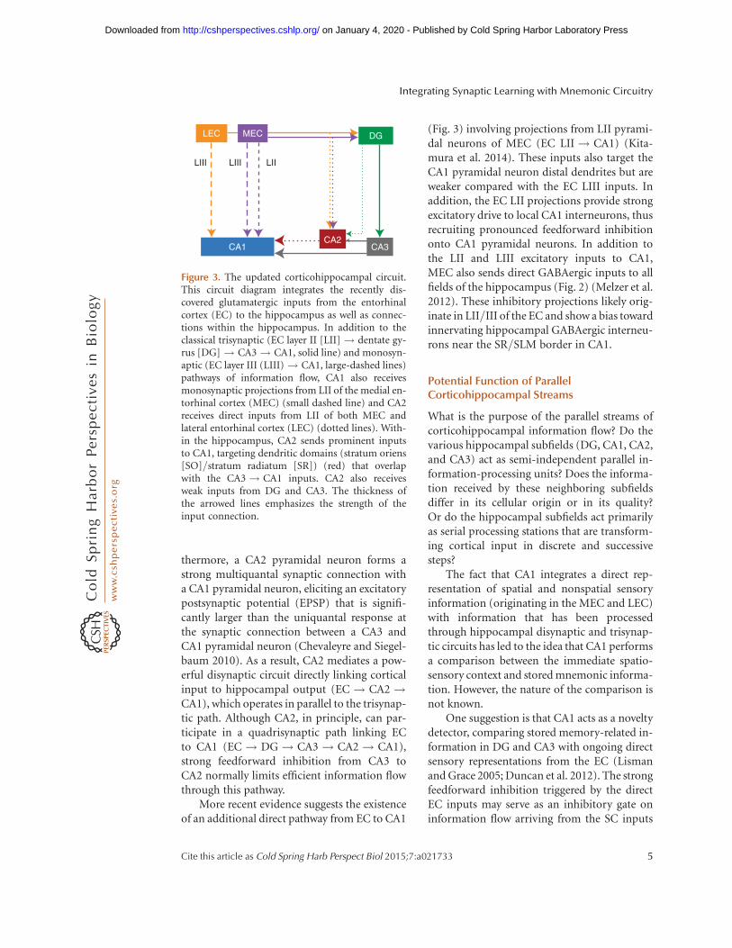
thermore, a CA2 pyramidal neuron forms astrong multiquantal synaptic connection witha CA1 pyramidal neuron, eliciting an excitatorypostsynaptic potential (EPSP) that is signifi-cantly larger than the uniquantal response atthe synaptic connection between a CA3 andCA1 pyramidal neuron (Chevaleyre and Siegel-baum 2010). As a result, CA2 mediates a pow-erful disynaptic circuit directly linking corticalinput to hippocampal output (EC! CA2 !CA1), which operates in parallel to the trisynap-tic path. Although CA2, in principle, can par-ticipate in a quadrisynaptic path linking ECto CA1 (EC! DG ! CA3 ! CA2! CA1),strong feedforward inhibition from CA3 toCA2 normally limits efficient information flowthrough this pathway.
More recent evidence suggests the existenceof an additional direct pathway from EC to CA1
(Fig. 3) involving projections from LII pyrami-dal neurons of MEC (EC LII! CA1) (Kita-mura et al. 2014). These inputs also target theCA1 pyramidal neuron distal dendrites but areweaker compared with the EC LIII inputs. Inaddition, the EC LII projections provide strongexcitatory drive to local CA1 interneurons, thusrecruiting pronounced feedforward inhibitiononto CA1 pyramidal neurons. In addition tothe LII and LIII excitatory inputs to CA1,MEC also sends direct GABAergic inputs to allfields of the hippocampus (Fig. 2) (Melzer et al.2012). These inhibitory projections likely orig-inate in LII/III of the EC and show a bias towardinnervating hippocampal GABAergic interneu-rons near the SR/SLM border in CA1.
Potential Function of ParallelCorticohippocampal Streams
What is the purpose of the parallel streams ofcorticohippocampal information flow? Do thevarious hippocampal subfields (DG, CA1, CA2,and CA3) act as semi-independent parallel in-formation-processing units? Does the informa-tion received by these neighboring subfieldsdiffer in its cellular origin or in its quality?Or do the hippocampal subfields act primarilyas serial processing stations that are transform-ing cortical input in discrete and successivesteps?
The fact that CA1 integrates a direct rep-resentation of spatial and nonspatial sensoryinformation (originating in the MEC and LEC)with information that has been processedthrough hippocampal disynaptic and trisynap-tic circuits has led to the idea that CA1 performsa comparison between the immediate spatio-sensory context and stored mnemonic informa-tion. However, the nature of the comparison isnot known.
One suggestion is that CA1 acts as a noveltydetector, comparing stored memory-related in-formation in DG and CA3 with ongoing directsensory representations from the EC (Lismanand Grace 2005; Duncan et al. 2012). The strongfeedforward inhibition triggered by the directEC inputs may serve as an inhibitory gate oninformation flow arriving from the SC inputs
LEC MEC DG
LIILIII
CA1CA2
CA3
LIII
Figure 3. The updated corticohippocampal circuit.This circuit diagram integrates the recently dis-covered glutamatergic inputs from the entorhinalcortex (EC) to the hippocampus as well as connec-tions within the hippocampus. In addition to theclassical trisynaptic (EC layer II [LII] ! dentate gy-rus [DG]! CA3! CA1, solid line) and monosyn-aptic (EC layer III (LIII) ! CA1, large-dashed lines)pathways of information flow, CA1 also receivesmonosynaptic projections from LII of the medial en-torhinal cortex (MEC) (small dashed line) and CA2receives direct inputs from LII of both MEC andlateral entorhinal cortex (LEC) (dotted lines). With-in the hippocampus, CA2 sends prominent inputsto CA1, targeting dendritic domains (stratum oriens[SO]/stratum radiatum [SR]) (red) that overlapwith the CA3! CA1 inputs. CA2 also receivesweak inputs from DG and CA3. The thickness ofthe arrowed lines emphasizes the strength of theinput connection.
Integrating Synaptic Learning with Mnemonic Circuitry
Cite this article as Cold Spring Harb Perspect Biol 2015;7:a021733 5
on January 4, 2020 - Published by Cold Spring Harbor Laboratory Press http://cshperspectives.cshlp.org/Downloaded from
by means of the action of dopaminergic noveltysignals (Lisman and Grace 2005). Another sug-gestion is that the direct inputs may provideinstructive signals for assessing the salience ofinformation flow through the trisynaptic pathusing a timing-dependent plasticity rule, whichis tuned to the delay-line architecture of the hip-pocampal circuit (Dudman et al. 2007). Becauseof the two extra synapses and conduction andintegration delays, information flowing throughthe trisynaptic path arrives at CA1 neuronssome 15 to 20 msec after the arrival of informa-tion through the direct EC inputs (Yeckel andBerger 1990). Such a circuit design allows forintegration and comparison of temporally co-ordinated inputs that may contribute to the en-coding of sequential episodic events (Dudmanet al. 2007; Ahmed and Mehta 2009; Mizusekiet al. 2009; Basu et al. 2013).
The Corticohippocampal Circuit.III: Outputs of the Hippocampus
CA1 pyramidal neurons provide the major out-put from the hippocampus, sending projec-tions to a number of brain regions, includingthe neighboring subiculum, perirhinal cortex,prefrontal cortex, and amygdala (van Groenand Wyss 1990). A small fraction of pyramidalneurons from dorsomedial CA1 also project tothe restrosplenial cortex (Wyss and Van Groen1992). One particularly strong output goes backto the EC, in which CA1 axons excite layer Vpyramidal cells, which in turn send excitatoryfeedback input to the EC LII/III, there-by completing an EC ! hippocampus! ECloop (Figs. 1 and 3) (Naber et al. 2001). In ad-dition to excitatory pyramidal neuronal out-puts, certain GABAergic neurons in specificlayers of CA1 project directly to retrohippocam-pal and cortical areas. These include long-range GABAergic projections from somatosta-tin (SOM) and mGluR1a expressing inhibitoryneurons in stratum oriens (SO) to the subicu-lum and medial septum (MS) (Jinno et al. 2007;Fuentealba et al. 2008). GABAergic cells locatedin the border of SR and SLM (often express-ing muscarinic AChRs or mGluR1as) projectto retrosplenial cortex and indusium gresium
(Jinno et al. 2007). There are also SOM-express-ing GABAergic neurons in SO of CA1 and hilusof DG that send direct projections to the super-ficial layers of MEC (Fig. 2) and striatum(Melzer et al. 2012). The long-range inhibitoryprojections are often highly myelinated (Jinnoet al. 2007) and predominantly target localGABAergic interneurons at the projection sites(Melzer et al. 2012). These properties have led tothe suggestion that long-range inhibitory pro-jections may be important for coordinating thetiming between the hippocampus and its corti-cal targets (Buzsaki and Chrobak 1995) andcould serve a disinhibitory role (Caputi et al.2013).
At present, there are conflicting data as towhether CA2 neurons project outside of thehippocampus. One study using a cell-type-spe-cific rabies virus retrograde tracing strategyreported that CA2 pyramidal neurons send pro-jections to EC LII neurons (Rowland et al.2013), the source of the direct cortical inputto CA2 (Hitti and Siegelbaum 2014; Koharaet al. 2014). CA2 has also been reported to pro-ject to the supramammillary nucleus (Cui et al.2013), a hypothalamic region long known toprovide strong input to CA2 (Haglund et al.1984; Vertes 1992; Magloczky et al. 1994;Ochiishi et al. 1999; Kiss et al. 2000).
The Corticohippocampal Circuit.IV: Heterogeneity within the CA1Pyramidal Neuron Population
Although many studies treat CA1 pyramidalneurons as a uniform population, there is in-creasing evidence for heterogeneity along eachof the three spatial axes of the hippocampus: theseptotemporal (or dorsoventral) longitudinalaxis, the proximodistal transverse axis (CA2 tosubiculum), and the deep superficial radial axisin the stratum pyramidale (SP) cell body layer(with deep referring to pyramidal neurons clos-er to SO and superficial referring to pyramidalneurons closer to SR).
Along the transverse axis, proximal CA1neurons (closer to the CA2 border) receivedirect input primarily from the MEC, whereasdistal CA1 neurons (closer to the border with
J. Basu and S.A. Siegelbaum
6 Cite this article as Cold Spring Harb Perspect Biol 2015;7:a021733
on January 4, 2020 - Published by Cold Spring Harbor Laboratory Press http://cshperspectives.cshlp.org/Downloaded from
subiculum) receive direct input primarily fromthe LEC (Fig. 3) (Ishizuka et al. 1990; Witter andAmaral 1991). This topographical arrangementis reversed in subiculum. Pyramidal neurons inCA1 and subiculum also show a strong proxi-modistal gradient from regular firing in CA1 toprominent burst firing in subiculum (and CA3)(Jarsky et al. 2008; Kim and Spruston 2012).
Marked differences in pyramidal neuron-firing properties along the transverse axis havealso been recorded in vivo. Thus, distal CA1pyramidal neurons, which receive largely non-spatial input from the LEC, show dispersed fir-ing during spatial navigation and tend to havemultiple place fields (Henriksen et al. 2010) anddisplay increased 20–40 Hz coupling with theLEC during spatial associational learning be-havior (Igarashi et al. 2014). As expected, thefiring of proximal CA1 pyramidal neurons aremore strongly coupled to MEC neuron firing attheta frequencies and show greater spatial mod-ulation and more compact place fields com-pared with their distal counterparts (Henriksenet al. 2010).
Since the initial studies of Lorente de No(1934), it has been suggested that there may betwo separate sublayers of CA1 pyramidal neu-rons along the radial axis: a relatively tight super-ficial layer (closer to SR) and a broader moredispersed deep layer (closer to SO) (Slomiankaet al. 2011). Molecular evidence supportingthis view comes from the finding that the super-ficial neurons, but not deep pyramidal neurons,are enriched in calbindin and zinc (Baimbridgeand Miller 1982; Baimbridge et al. 1982; Donget al. 2008; Slomianka et al. 2011). During de-velopment, expression of Sox5 specifies pyra-midal neurons in the deep layers, whereas thecoexpression of Satb2 and Zbtb20 may deter-mine the fate of superficial neurons (Nielsenet al. 2010; Xie et al. 2010). Morphologically,the deep neurons have fewer oblique dendritesin SR and more extensive branching in SO thando superficial neurons (Bannister and Larkman1995b). The two sublayers also have distinctinput–output connectivity, with deep neuronsreceiving preferential input from CA2 pyrami-dal neurons (Kohara et al. 2014) and local in-hibitory parvalbumin-positive basket cells (Lee
et al. 2014), the latter distinction first reportedby Lorente de No (1934).
Importantly, the two layers show differentfunctional properties in vitro and during be-havior. The presence in superficial neuronsof calbindin, which buffers calcium and in-terferes with N-methyl-D-aspartate receptor(NMDAR)-dependent LTP, may lead to plastic-ity differences in the two populations (Araiet al. 1994; Bannister and Larkman 1995a,b).The superficial cells show a larger influence ofthe hyperpolarization-activated cation current,Ih, which contributes to the resting integrativeproperties of the neurons (Jarsky et al. 2008). Invivo, the deep neurons show a greater tendencyfor spatial tuning during navigation as well asstronger modulation during slow wave sleep(Mizuseki et al. 2011).
Finally, there are functional and structuraldifferences along the longitudinal axis of thehippocampus, including differences in proteinexpression along the dorsoventral axis thatactually define three distinct regions: dorsal,intermediate, and ventral hippocampus (Donget al. 2010). In addition, ventral hippocampalCA1 pyramidal neurons are more excitable thandorsal neurons, perhaps resulting from a strong-er expression of the HCN1 cation channel in theventral hippocampus (Dougherty et al. 2013).
Dorsal hippocampal place cells show moreprecise place fields compared with ventral hip-pocampal place cells, whose place fields are morediffuse (Jung et al. 1994), although this maynot degrade the ability of the ventral hippocam-pus to encode spatial position because of thehigher ventral cell-firing rates (Keinath et al.2014). This difference in place-field size corre-sponds to a dorsoventral gradient of increasinggrid field size and spacing in MEC LII grid cells(Brun et al. 2008), which show a topographicdorsoventral projection to the hippocampus(van Strien et al. 2009). Dorsal and ventralhippocampi also differ in their outputs. Ven-tral hippocampus projects more strongly toprefrontal cortex and amygdala compared withdorsal hippocampus (Ishikawa and Nakamura2006). Moreover, these differences reflect thedistinct behavioral roles of these two regions,with dorsal hippocampus more important for
Integrating Synaptic Learning with Mnemonic Circuitry
Cite this article as Cold Spring Harb Perspect Biol 2015;7:a021733 7
on January 4, 2020 - Published by Cold Spring Harbor Laboratory Press http://cshperspectives.cshlp.org/Downloaded from
spatial and contextual memory and ventral hip-pocampus more important for emotional- andanxiety-related behaviors (Fanselow and Dong2010).
ROLE OF DISTINCT REGIONS OF THECORTICOHIPPOCAMPAL CIRCUIT INLEARNING AND MEMORY
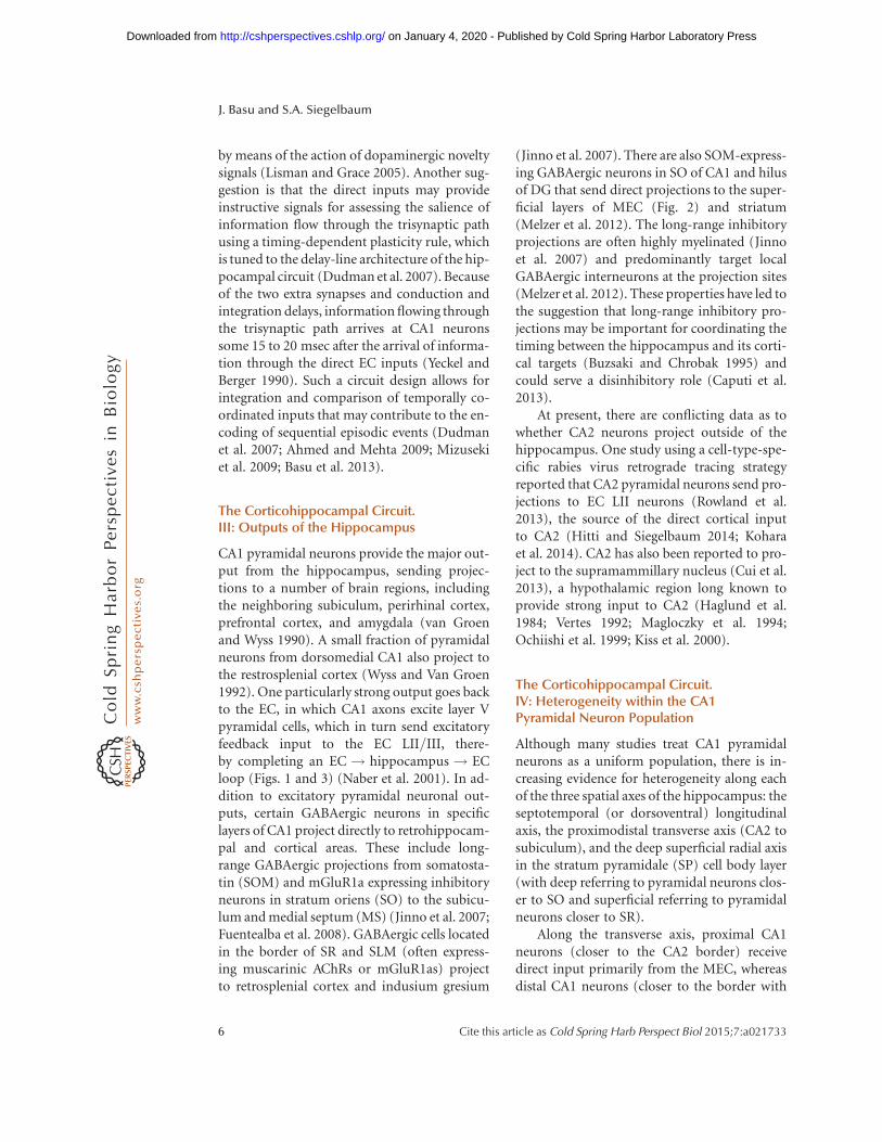
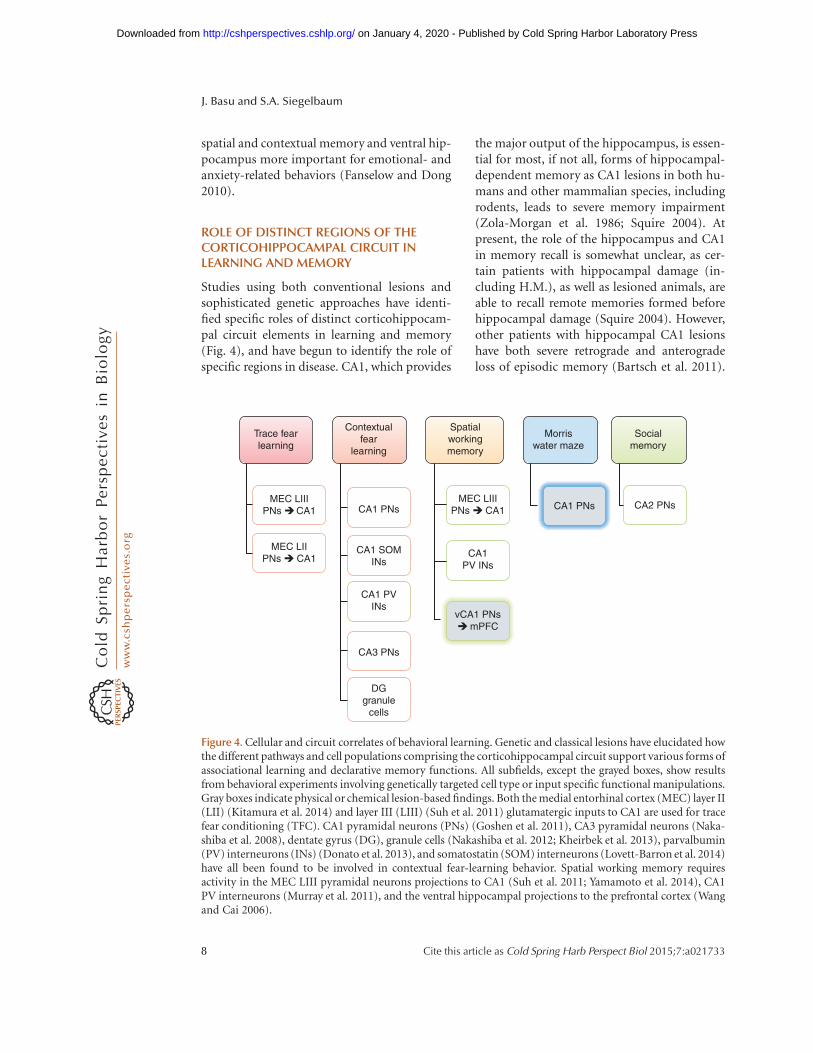
Studies using both conventional lesions andsophisticated genetic approaches have identi-fied specific roles of distinct corticohippocam-pal circuit elements in learning and memory(Fig. 4), and have begun to identify the role ofspecific regions in disease. CA1, which provides
the major output of the hippocampus, is essen-tial for most, if not all, forms of hippocampal-dependent memory as CA1 lesions in both hu-mans and other mammalian species, includingrodents, leads to severe memory impairment(Zola-Morgan et al. 1986; Squire 2004). Atpresent, the role of the hippocampus and CA1in memory recall is somewhat unclear, as cer-tain patients with hippocampal damage (in-cluding H.M.), as well as lesioned animals, areable to recall remote memories formed beforehippocampal damage (Squire 2004). However,other patients with hippocampal CA1 lesionshave both severe retrograde and anterogradeloss of episodic memory (Bartsch et al. 2011).
Trace fearlearning
Contextualfear
learning
Spatialworkingmemory
Morriswater maze
Socialmemory
CA2 PNsCA1 PNsMEC LIII
PNs � CA1
vCA1 PNs� mPFC
CA3 PNs
DGgranule
cells
CA1 PVINs
CA1 SOMINs
CA1 PNsMEC LIII
PNs �CA1
MEC LIIPNs � CA1 CA1
PV INs
Figure 4. Cellular and circuit correlates of behavioral learning. Genetic and classical lesions have elucidated howthe different pathways and cell populations comprising the corticohippocampal circuit support various forms ofassociational learning and declarative memory functions. All subfields, except the grayed boxes, show resultsfrom behavioral experiments involving genetically targeted cell type or input specific functional manipulations.Gray boxes indicate physical or chemical lesion-based findings. Both the medial entorhinal cortex (MEC) layer II(LII) (Kitamura et al. 2014) and layer III (LIII) (Suh et al. 2011) glutamatergic inputs to CA1 are used for tracefear conditioning (TFC). CA1 pyramidal neurons (PNs) (Goshen et al. 2011), CA3 pyramidal neurons (Naka-shiba et al. 2008), dentate gyrus (DG), granule cells (Nakashiba et al. 2012; Kheirbek et al. 2013), parvalbumin(PV) interneurons (INs) (Donato et al. 2013), and somatostatin (SOM) interneurons (Lovett-Barron et al. 2014)have all been found to be involved in contextual fear-learning behavior. Spatial working memory requiresactivity in the MEC LIII pyramidal neurons projections to CA1 (Suh et al. 2011; Yamamoto et al. 2014), CA1PV interneurons (Murray et al. 2011), and the ventral hippocampal projections to the prefrontal cortex (Wangand Cai 2006).
J. Basu and S.A. Siegelbaum
8 Cite this article as Cold Spring Harb Perspect Biol 2015;7:a021733
on January 4, 2020 - Published by Cold Spring Harbor Laboratory Press http://cshperspectives.cshlp.org/Downloaded from
Moreover, a number of studies have shown theactivation of the hippocampus, including CA1,during memory recall (Rugg and Vilberg 2013).Furthermore, optogenetic experiments haveshown that temporally precise and transient in-activation of dorsal CA1 pyramidal neurons inrodents markedly impairs recall (Goshen et al.2011). This suggests that long-term inactivationof the hippocampus through genetic or physicallesions may result in a variable degree of com-pensatory changes in other brain regions thatenable recall in some individuals but not inothers.
One surprising finding is that lesions of dor-sal CA1 lead to a disruption of grid cell spatialtuning patterns within the EC, which providesthe major input to the hippocampus (Bonnevieet al. 2013). In addition, hippocampal place-cellactivity can precede the appearance of well-de-fined EC grid-cell firing during early postnataldevelopment (Langston et al. 2010; Wills et al.2010). These results suggest that feedback fromthe hippocampus may be necessary for optimalgrid-field formation. As “border cells” that firewhen an animal reaches the edges of an environ-ment develop in the EC before grid cells, it hasbeen proposed that these border cells may pro-vide the input that gives rise to hippocampalplace cells (Bjerknes et al. 2014). Furthermore,removal of CA1 inputs to the EC converts gridcells into head direction cells (Bonnevie et al.2013).
In contrast to the profound changes pro-duced by lesions in CA1, lesions of DG appearto cause more subtle changes in memory per-formance. Although such lesions do not disrupta basic form of contextual fear conditioning,they do result in an impaired ability to distin-guish between closely related environments, aprocess termed pattern separation (Leutgebet al. 2007; Sahay et al. 2011; Nakashiba et al.2012), the ability to distinguish between closelyrelated environments. Interestingly, DG is aprominent site of adult neurogenesis (Drewet al. 2013) and the newborn neurons appearpreferentially involved in pattern separation(Nakashiba et al. 2012). DG is also affectedpreferentially during age-related memory loss(Small et al. 2011; Pavlopoulos et al. 2013),
whereas Alzheimer’s disease initially targetsthe EC (Braak and Braak 1985). Interestingly,a recent study (Kheirbek et al. 2013) using op-togenetic activation and silencing of granulecells shows that dorsal DG is important forencoding but not retrieval of contextual fearmemories. Genetic knockout studies suggestthat CA3 is important for pattern completion(Nakazawa et al. 2002), the ability to recall amemory from partial cues, and one-trial formsof contextual learning (Fig. 4), in which a strongaversive stimulus results in rapid memory for-mation without the usual need for repeated tri-als and spatial reference memory.
The primary role of the trisynaptic path inmemory formation and spatial encoding wascalled into question by physical (Brun et al.2002) and genetic (Nakashiba et al. 2008) le-sion studies, which showed that removal ofCA3 input to CA1 had surprisingly little effecton spatial reference memory performance inthe Morris water maze (MWM), or on therate of CA1 neuron place-cell firing in vivo.To explain these findings, it was postulatedthat the direct EC LIII (and possibly EC LII)inputs to CA1 might be sufficient to drive nor-mal CA1 firing rates to support hippocampus-dependent memory.
In support of the importance of the directEC LIII inputs in spatial memory, chemicaland electrolytic lesions of the MEC inputs toCA1 were found to lead to a degradation ofplace-field precision (Brun et al. 2008) and def-icits in consolidation of spatial long-term mem-ory (Remondes and Schuman 2004). However,highly selective genetic lesions or optogeneticsilencing of glutamatergic MEC LIII inputs toCA1 impaired trace fear conditioning (TFC), aform of temporal association memory, and spa-tial working memory, but did not perturb spa-tial reference memory, contextual fear condi-tioning, or place-cell firing (Fig. 4) (Suh et al.2011; Kitamura et al. 2014; Yamamoto et al.2014). The recently identified EC LII pyramidalneuron projections to CA1 are also involved intrace fear learning but have an opposite role toEC LIII inputs, acting to inhibit fear memory byrecruiting a population of dendritic targetingCA1 inhibitory neurons (Kitamura et al. 2014).
Integrating Synaptic Learning with Mnemonic Circuitry
Cite this article as Cold Spring Harb Perspect Biol 2015;7:a021733 9
on January 4, 2020 - Published by Cold Spring Harbor Laboratory Press http://cshperspectives.cshlp.org/Downloaded from
The suggestion that the direct EC inputs toCA1 can compensate for loss of CA3 inputs isdifficult to reconcile with the finding that thedirect EC inputs to CA1 provide only a weakexcitatory drive and so cannot elicit CA1 out-put, except when stimulated in high-frequencybursts (Jarsky et al. 2005). One alternative expla-nation is that the residual function of CA1 pyra-midal neurons and memory task performancefollowing CA3 lesions is maintained by activitythrough the disynaptic pathway mediated byCA2. However, when output from CA2 pyra-midal neurons was silenced using a Cre-depen-dent viral vector to express tetanus toxin in theAmigo2-Cre mouse line, there was little changein spatial reference memory (MWM), objector odor recognition (including social odors),or context memory (contextual fear condition-ing). Surprisingly, inactivation of CA2 did causea profound deficit in social memory, the abilityof an animal to recognize and remember indi-vidual members of its species (Fig. 4) (Hitti andSiegelbaum 2014). The role of the hippocampusin social memory in humans is well illustratedby the case of H.M., who could no longer formmemories of new individuals following tempo-ral lobe resection (Corkin 2002).
The finding that spatial and contextual fearconditioning are intact following lesioning of allthree major classes of inputs to CA1 is some-what surprising, and could indicate that one ortwo input pathways may be able to maintainmemory storage capacity in the absence of thethird pathway. Perhaps this explains why thereare the parallel processing routes for informa-tion flow from the EC to hippocampus thatare sufficient but not necessary to sustain thecritical functions of the hippocampus, namely,spatial and contextual encoding. Another pos-sibility is that long-term lesions and cell-type-specific genetic ablations may lead to compen-satory mechanisms that result in strengtheningor redistribution of functional gain of otherwiseweaker pathways, thus enabling them to sustainthe function of the lesioned circuit. Studiesusing temporally precise inactivation of pairsof hippocampal regions and inputs may helpdetermine whether different regions do indeedprovide such compensation.
EXTRAHIPPOCAMPAL ANDNEUROMODULATORY TUNING OFCORTICOHIPPOCAMPAL CIRCUITSDURING LEARNING AND MEMORY
There are several neuromodulatory inputs tothe various subfields of the hippocampus thatstrongly influence synaptic transmission andactivity in the corticohippocampal circuit andcontribute significantly to learning behaviors.These projections often target specific subpop-ulations of GABAergic interneurons in addi-tion to their glutamatergic counterparts. Fur-thermore, there are substantial differences inthe innervation patterns and behaviorally trig-gered activity of such inputs.
Aminergic modulatory inputs from mid-brain and brain stem can markedly alter thefunction of various circuit elements and mod-ulate memory formation. The importance ofdopamine (DA) in regulating short-term syn-aptic transmission, long-term plasticity, andlearning and memory has received a great dealof attention (Jay 2003; Lisman and Grace 2005).Studies using bath application of dopaminergicagonists generally report a strong suppression ofthe direct EC input to CA1, with little effect onthe SC input (Lisman and Grace 2005; Ito andSchuman 2007).
Other studies show that DA facilitates theinduction of a late phase of LTP at the SC!CA1 pyramidal neuron synapses by activationof D1/D5 receptors and an increase in cAMPlevels both in vitro (Huang and Kandel 1995)and in vivo (Lemon and Manahan-Vaughan2006). Moreover, DA has been found to beimportant for both hippocampal memory for-mation (Wilkerson and Levin 1999; Rossatoet al. 2009; Bethus et al. 2010) and the stabilityof place-field representations (Kentros et al.1998). Such results have led to the suggestionthat DA release in response to novelty acts as areward signal to enhance memory storage byincreasing the relative influence of the hippo-campal SC inputs to CA1 versus the EC inputs(Lisman and Grace 2005).
Cholinergic inputs also play a key role inregulating hippocampal function and memory.A recent study (Lovett-Barron et al. 2014) found
J. Basu and S.A. Siegelbaum
10 Cite this article as Cold Spring Harb Perspect Biol 2015;7:a021733
on January 4, 2020 - Published by Cold Spring Harbor Laboratory Press http://cshperspectives.cshlp.org/Downloaded from
that cholinergic input from the MS activatesa class of SOM-positive dendritic targeting in-terneurons during the unconditioned aversivestimulus in a contextual fear-conditioning task.Interfering with this circuit-based mechanismby silencing dendrite-targeting inhibition inCA1 during presentation of the unconditionedstimulus impaired fear learning (Fig. 4). Theincreased inhibition at CA1 pyramidal neurondistal dendrites acts to suppress any coincidentexcitatory inputs conveyed by the EC directpathway. The investigators postulate that sucha mechanism may be useful for preventing cor-tical sensory information representing the aver-sive stimuli from becoming confounded withthe representation of the context, enabling recallof the context alone to trigger a fear response.
Another extrahippocampal modulatory in-put to CA1 comes from the thalamic nucleusreuniens (NuRe), which receives input from theprefrontal cortex. Anatomical and electrophys-iological studies show that the NuRe sends ex-citatory inputs to the SLM region of CA1, whereit innervates both local interneurons and thedistal dendrites of pyramidal neurons (Wouter-lood et al. 1990; Hirayasu and Wada 1992; Dol-leman-Van der Weel et al. 1997, 2009). Xu andSudhof (2013) recently found that this pathwayis important for specificity of contextual repre-sentations by preventing the generalization offear memory, based on experiments in whichthe NuRe projections were selectively silenced.
The hippocampal CA2 region also receivesneuromodulatory inputs from various mid-brain nuclei in addition to its EC LII and in-trahippocampal inputs. These include the re-ciprocal projections between CA2 and thesupramammillary nucleus (SUM), MS, diago-nal band of Broca (DBB) as well as afferent vaso-pressinergic projections from paraventricularnuclei of the hypothalamus to CA2 (Cui et al.2013; Hitti and Siegelbaum 2014). In fact, thestrong input from the hypothalamic nuclei aswell as a high level of expression of the argininevasopressin receptor AVPR1b likely contributesto the ability of the CA2 region to participatein a specific circuit for the storage and recallof social memory (Young et al. 2006; Hittiand Siegelbaum 2014). A global knockout of
AVPR1b (Wersinger et al. 2002) shows decreasedsocial memory, the ability to remember interac-tions with conspecifics, and decreased temporalmemory for event order (DeVito et al. 2009).Whereas AVPR1b is widely expressed (albeit atlower levels) throughout the brain, including inhypothalamus, the deficit in socially motivatedaggressive behavior in the knockout mousewas partially rescued by selective expression ofAVPR1b in dorsal CA2 using spatially, but notgenetically, targeted viral injections (Paganiet al. 2014). It will be of interest to determinewhether AVPR1b expression in CA2 is also re-quired for formation of social memory (Hittiand Siegelbaum 2014).
LONG-TERM SYNAPTIC PLASTICITYIN THE CORTICOHIPPOCAMPAL CIRCUITIN LEARNING AND MEMORY
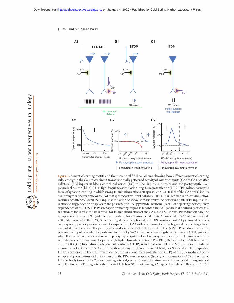
One of the striking features of synapses in thecentral nervous system, especially those in thecorticohippocampal circuit, is the extent towhich their strength can be regulated for pro-longed periods of time by different patterns ofsynaptic activity, a process termed activity-de-pendent synaptic plasticity. In some instances,the plasticity is homosynaptic in that activitywithin a given synaptic pathway leads to alteredstrength of synaptic communication at the samesynapses that were activated (Fig. 5A,B). Otherexamples of synaptic plasticity are heterosynap-tic in that activity in one synaptic pathway in-fluences the function of another pathway (Fig.5C). Although there is a great deal of correlativeevidence linking forms of plasticity to hippo-campal-dependent memory formation, the pre-cise role of plasticity mechanisms in learningand memory formation remains controversial(see reviews Morris 2013; Bannerman et al.2014). This complexity likely reflects the factthat there are multiple forms of plasticity thatdiffer in the pattern of activity required for theirinduction, the molecular mechanisms for boththeir induction and expression, and the dura-tion of the plastic changes (Fig. 5).
Homosynaptic Plasticity
The importance of homosynaptic forms of ac-tivity-dependent plasticity was first postulated
Integrating Synaptic Learning with Mnemonic Circuitry
Cite this article as Cold Spring Harb Perspect Biol 2015;7:a021733 11
on January 4, 2020 - Published by Cold Spring Harbor Laboratory Press http://cshperspectives.cshlp.org/Downloaded from
CA3
LTP
CA3
LTP
CA3
LTP
HFS LTP ITDPSTDP
A1 B1 C1
A2 B2 C2
LTP
EC EC
10 msec
300
200
100
Syn
aptic
res
pons
e (%
)
Syn
aptic
res
pons
e (%
)
Syn
aptic
res
pons
e (%
)
–40
–200
–150
–100 –5
010
100
200
100
200
20 100 200 0 50 –30
–20
–10 0 10
EC–SC pairing interval (msec)
20
Prepost pairing interval (msec)Interstimulus interval (msec)
Presynaptic SC input activation
Presynaptic EC input activation
Presynaptic input activation
Postsynaptic action potential
20 msec
HomosynapticHebbian
Heterosynapticnon-Hebbian
Figure 5. Synaptic learning motifs and their temporal fidelity. Scheme showing how different synaptic learningrules emerge in the CA1 microcircuit from temporally patterned activity of synaptic inputs (CA3 to CA1 Schaffercollateral [SC] inputs in black; entorhinal cortex [EC] to CA1 inputs in purple) and the postsynaptic CA1pyramidal neuron (blue). (A1) High-frequencystimulation long-term potentiation (HFS LTP) is a homosynapticform of synaptic learning in which strong tetanic stimulation (200 pulses at 20–100 Hz) of the CA3 or EC inputscan strengthen the synaptic output of that specific active input pathway. HFS LTP is Hebbian in that its inductionrequires Schaffer collateral (SC) input stimulation to evoke somatic spikes, or perforant path (PP) input stim-ulation to trigger dendritic spikes in the postsynaptic CA1 pyramidal neurons. (A2) Plot depicting the frequencydependence of SC HFS LTP. Postsynaptic excitatory response recorded in CA1 pyramidal neurons plotted as afunction of the interstimulus interval for tetanic stimulation of the CA3–CA1 SC inputs. Preinduction baselinesynaptic response is 100%. (Adapted, with values, from Thomas et al. 1996; Aihara et al. 1997; Zakharenko et al.2003; Alarcon et al. 2004.) (B1) Spike-timing-dependent plasticity (STDP) is induced in CA1 pyramidal neuronsby temporally precise pairing of synaptic inputs from CA3 with a postsynaptic spike triggered by injecting a briefcurrent step in the soma. The pairing is typically repeated 50–100 times at 10 Hz. (B2) LTP is induced when thepresynaptic input precedes the postsynaptic spike by 5–20 msec, whereas long-term depression (LTD) prevailswhen the pairing sequence is reversed (postsynaptic spike before the presynaptic input). (2) Timing intervalsindicate pre- before postsynaptic pairing. (Adapted from data in Bi and Poo 1998; Debanne et al. 1998; Nishiyamaet al. 2000.) (C1) Input-timing-dependent plasticity (ITDP) is induced when EC and SC inputs are stimulated20 msec apart (EC before SC) at subthreshold strengths (hence, non-Hebbian) for 90 sec at a 1 Hz frequency.ITDP is expressed in the CA1 pyramidal neuron as a long-term potentiation (LTP) of the SC- mediated post-synaptic depolarization without a change in the PP-evoked response (hence, heterosynaptic). (C2) Induction ofITDP is finely tuned to the 20 msec pairing interval, even a 10 msec deviation from this preferred timing intervalis ineffective. (2) Timing intervals indicate EC before SC input pairing. (Adapted from data in Basu et al. 2013.)
12 Cite this article as Cold Spring Harb Perspect Biol 2015;7:a021733
J. Basu and S.A. Siegelbaum
on January 4, 2020 - Published by Cold Spring Harbor Laboratory Press http://cshperspectives.cshlp.org/Downloaded from
by Donald Hebb (1949) on theoretical groundsas a mechanism for forming neural assemblies.According to Hebb:
When an axon of cell A is near enough to excitea cell B and repeatedly or persistently takes partin firing it, some growth process or metabolicchange takes place in one or both cells suchthat A’s efficiency, as one of the cells firing B, isincreased.
Hebb’s idea was that this type of synapticlearning rule would provide for the wiring ofneuronal assemblies with common responseproperties. For example, it could explain howa linear array of neighboring retinal ganglioncells and their lateral geniculate targets, whichboth have circular receptive fields, are able toconnect to a common cortical neuron in pri-mary visual cortex to generate typical responseselectivity to oriented bars of light. However,Hebbian plasticity is also ideally suited for theformation of neural ensembles that encode agiven memory.
Hebbian LTP
LTPof synaptic transmission represents the clas-sic example of a Hebbian synaptic learning rule.Bliss and Lomo induced LTP by a brief, strongtetanic stimulation of the PP inputs to DG inanesthetized rabbits, which produced a long-lasting enhancement in the strength of excita-tory synaptic transmission from PP to DG thatlasted for hours to days (Bliss and Lomo 1973).Following its initial discovery, LTP was subse-quently found to be inducible at nearly all stagesof hippocampal synaptic transmission.
At most synapses, LTP follows Hebb’s syn-aptic learning rules in that it requires strongsynaptic activity that is able to drive spike firingin the postsynaptic cells (Bliss and Collingridge1993; Bliss et al. 2014). The one exception ismossy fiber LTP from DG granules cells toCA3 pyramidal neurons, which is found inmost studies to require only presynaptic activitywith no requirement for action potential firingin the postsynaptic neuron (Nicoll and Malenka1995). Tetanic stimulation of the SCs fromCA3 pyramidal neurons also induces HebbianLTP at the synapses these axons make onto other
CA3 pyramidal neurons (recurrent collaterals)as well as at their synapses with CA1 pyramidalneurons (Fig. 5A). In contrast, tetanic high-fre-quency stimulation (HFS) does not normallyinduce LTP in the classical Hebbian sense atthe SC synapses onto CA2 pyramidal neurons(Zhao et al. 2007) because of their strong expres-sion of the G-protein regulatory protein RGS14(Lee et al. 2010) and enhanced Ca2þ pump ac-tivity (Simons et al. 2009). However, a form ofLTP was recently described at SC! CA2 syn-apses that relies on suppression of feedforwardinhibition (see below) (Piskorowski and Che-valeyre 2013). Finally, LTP can also be inducedby tetanic stimulation of the direct EC inputsto CA1 and CA2, although the extent of LTPonto CA1 is typically quite small. Thus, HFSLTP is a widespread form of homosynapticactivity-dependent plasticity found at all excit-atory synapses throughout the hippocampalcircuit.
Our understanding of the synaptic andmolecular mechanisms underlying LTP hasprogressed greatly since its initial discovery.However, a number of fundamental questionsconcerning the basic properties of LTP, as well asthe precise role that different forms of LTP playin learning and memory, remain unanswered.
The key characteristics of activity-depen-dent Hebbian LTP are established by the prop-erties of the NMDARs, whose activation is crit-ical for the induction of LTP at many synapses,including the SC! CA1 synapses (Colling-ridge et al. 1983). Fast excitatory synapses relyon two classes of ionotropic glutamate re-ceptors, the NMDARs and a-amino-3-hydroxy-5-methyl-4-isoxazolepropionic acid receptors(AMPARs). NMDARs differ from most io-notropic ligand-gated channels (including theAMPARs), in that they require membranedepolarization in addition to glutamate neuro-transmitter to function. At typical negative rest-ing potentials, the pore of the NMDAR isblocked by an Mg2þ ion. As a result, basalsynaptic transmission normally relies on ac-tivation of the AMPARs. However, when pre-synaptic activity is coupled with strong post-synaptic depolarization, such as during tetanicstimulation, the NMDARs are relieved from
Integrating Synaptic Learning with Mnemonic Circuitry
Cite this article as Cold Spring Harb Perspect Biol 2015;7:a021733 13
on January 4, 2020 - Published by Cold Spring Harbor Laboratory Press http://cshperspectives.cshlp.org/Downloaded from
their Mg2þ blockade through electrostatic re-pulsion. NMDARs are also distinguished frommost AMPARs by their high permeability toCa2þ, which acts as a second messenger insidecells to activate a number of downstream sig-naling cascades. As a result, NMDAR activa-tion during strong synaptic stimulation causesa rise in intracellular Ca2þ levels, which leads toactivation of the Ca2þ-calmodulin-dependentprotein kinase II (CaMKII), a step critical forthe induction of LTP (Malenka et al. 1989;Malinow et al. 1989).
A major question that has dominated thefield is whether LTP results from a presynapticchange, involving an increase in glutamate re-lease, or a postsynaptic change, resulting froman increase in the postsynaptic response to glu-tamate, or a coordinated change in presynapticand postsynaptic properties. This has led to alively controversy in the field that remains todate. A number of investigators state the casethat LTP is largely postsynaptic, resulting fromthe insertion of AMPARs into the postsynapticmembrane (Huganir and Nicoll 2013; Nicolland Roche 2013). Others argue that LTP is large-ly presynaptic (Enoki et al. 2009) or is a mixtureof distinct pre and postsynaptic processes (Blisset al. 2014).
Much of the controversy likely results fromthe fact that LTP is not a unitary phenomenon(Mayford et al. 2012). Rather a neuron can ex-press multiple forms of long-lasting synapticplasticity that differ in the pattern of activityneeded for their induction, underlying molec-ular mechanism, time scale of onset and dura-tion, and role in learning and memory (Fig. 5).The behaviorally linked activity state and thecoupled molecular tuning of upstream inputsand downstream targets will also determinethe ability of a particular circuit to serve as asubstrate for plasticity, a process termed meta-plasticity (Abraham and Tate 1997), and therebyallow the modulation of information flow. Oneclear indication of this diversity is provided bythe observation that certain forms of LTP thatappear to have a presynaptic locus of expres-sion do not require activation of NMDARs butare primarily mediated by Ca2þ influx throughvoltage-gated calcium channels (Grover and
Teyler 1990). Another important finding isthat deletion of the GluR1 AMPA receptor sub-unit blocks LTP induced by tetanic stimulationbut only partially inhibits LTP induced by thetaburst stimulation (Hoffman et al. 2002) andhas little effect on synaptic potentiation duringspike-timing-dependentplasticity (STDP)(Freyet al. 2009), a form of plasticity induced by thepairing of an EPSP with a postsynaptic actionpotential (Markram et al. 1997).
Another important distinction among dif-ferent forms of synaptic plasticity lies in thetime course and duration of the change in syn-aptic efficacy. LTP in the hippocampus has bothearly and late phases. For example, stimulationof SC inputs using one train of tetanic stimula-tion (at 100 Hz for 1 sec) produces an earlyphase of potentiation (E-LTP) that lasts for 1to 3 h and does not require protein synthesis.Stimulation with four or more identical trainsspaced several minutes apart recruits a latephase of potentiation (L-LTP), which can lastfor 24 h or more (Huang and Kandel 1994).Unlike E-LTP, L-LTP requires new protein syn-thesis and also depends on the activation ofprotein kinase A (PKA) (Frey et al. 1993; Abelet al. 1997) through the action of modulatorytransmitters, such as DA (Huang and Kandel1995). The role of L-LTP has been investigatedgenetically using mice that express a mutantgene that blocks the catalytic subunit of PKA,or carry an inhibitory mutation in the CREB-1gene. Both lines of mice have a serious defect inlong-term spatial memory and similar defectsin LTP. The early phase is normal but the latephase is blocked, providing evidence linking thephases of LTP to the phases of memory storage(Silva et al. 1992; Abel et al. 1997; Bourtchou-ladze et al. 1998). Theoretical studies suggestthat the expression of different temporal phasesof LTP enables the stability of long-term mem-ory traces as new memories are encodedthrough ongoing plastic changes in synapticfunction (Fusi et al. 2005).
Long-Term Depression
The presence of LTP raises the prospect thatsynapses may become saturated during the life-
J. Basu and S.A. Siegelbaum
14 Cite this article as Cold Spring Harb Perspect Biol 2015;7:a021733
on January 4, 2020 - Published by Cold Spring Harbor Laboratory Press http://cshperspectives.cshlp.org/Downloaded from
time of an animal, and thus no longer able toencode new memories. This potential problemis averted by the antagonistic processes of de-potentiation and long-term depression (LTD)(Bear and Abraham 1996), which can be in-duced by prolonged periods (5–15 min) oflow frequency stimulation (1–5 Hz). Depoten-tiation refers to the reversal of LTP seen whenlow frequency stimulation is delivered shortlyafter induction of LTP. LTD is induced by lowfrequency stimulation in the absence of priorinduction of LTP. Similar to LTP, there are alsodifferent forms of LTD. One prominent formrequires activation of NMDARs and an influxof postsynaptic Ca2þ, similar to NMDAR-de-pendent LTP (Bear and Abraham 1996). Anoth-er form of LTD requires activation of metabo-tropic glutamate receptors (Luscher and Huber2010).
How can Ca2þ influx through NMDARsdifferentially trigger LTP versus LTD? The an-swer seems to depend on the magnitude andduration of the postsynaptic Ca2þ signal, witha large elevation in Ca2þ triggering LTP and amore long-lasting but low Ca2þ signal resultingin LTD (Neveu and Zucker 1996). These differ-ences may reflect the distinct molecular mech-anisms of LTP versus LTD, with LTP requiringactivation of CaMKII, whereas LTD requires ac-tivation of the calcium-calmodulin-dependentphosphatase calcineurin (Mulkey et al. 1994),which is activated by lower levels of Ca2þ.
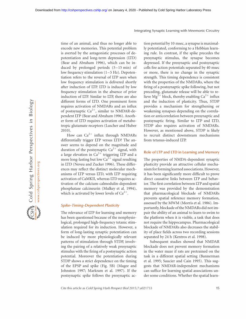
Spike-Timing-Dependent Plasticity
The relevance of LTP for learning and memoryhas been questioned because of the nonphysio-logical, prolonged high-frequency tetanic stim-ulation required for its induction. However, aform of long-lasting synaptic potentiation canbe induced by more physiologically relevantpatterns of stimulation through STDP, involv-ing the pairing of a relatively weak presynapticstimulus with the firing of a postsynaptic actionpotential. Moreover the potentiation duringSTDP shows a strict dependence on the timingof the EPSP and spike (Fig. 5B) (Magee andJohnston 1997; Markram et al. 1997). If thepostsynaptic spike follows the presynaptic ac-
tion potential by 10 msec, a synapse is maximal-ly potentiated, conforming to a Hebbian learn-ing rule. In contrast, if the spike precedes thepresynaptic stimulus, the synapse becomesdepressed. If the presynaptic and postsynapticcells fire action potentials separated by 40 msecor more, there is no change in the synapticstrength. This timing dependence is consistentwith the properties of the NMDARs, where thefiring of a postsynaptic spike following, but notpreceding, glutamate release will be able to re-lieve Mg2þ block, thereby enabling Ca2þ influxand the induction of plasticity. Thus, STDPprovides a mechanism for strengthening orweakening synapses depending on the correla-tion or anticorrelation between presynaptic andpostsynaptic firing. Similar to LTP and LTD,STDP also requires activation of NMDARs.However, as mentioned above, STDP is likelyto recruit distinct downstream mechanismsfrom tetanus-induced LTP.
Role of LTP and LTD in Learning and Memory
The properties of NMDA-dependent synapticplasticity provide an attractive cellular mecha-nism for forming learned associations. However,it has been significantly more difficult to provedirect causative links between LTP and behav-ior. The first correlation between LTPand spatialmemory was provided by the demonstrationthat pharmacological blockade of NMDARsprevents spatial reference memory formation,assessed by the MWM (Morris et al. 1986). Im-portantly, blockade of the NMDARs did not im-pair the ability of an animal to learn to swim tothe platform when it is visible, a task that doesnot require the hippocampus. Pharmacologicalblockade of NMDARs also decreases the stabil-ity of place fields across two recording sessionsseparated by 24 h (Kentros et al. 1998).
Subsequent studies showed that NMDARblockade does not prevent memory formationin the water maze if rats are pretrained on thetask in a different spatial setting (Bannermanet al. 1995; Saucier and Cain 1995). This sug-gests that NMDAR-independent mechanismscan suffice for learning spatial associations un-der some conditions. Whether the spatial learn-
Integrating Synaptic Learning with Mnemonic Circuitry
Cite this article as Cold Spring Harb Perspect Biol 2015;7:a021733 15
on January 4, 2020 - Published by Cold Spring Harbor Laboratory Press http://cshperspectives.cshlp.org/Downloaded from
ing occurs through non-NMDAR-dependentforms of LTP or through another mechanismis not known.
Genetic evidence linking LTP in the CA1region of the hippocampus with learning wasagain provided by experiments that rely on ma-nipulation of NMDAR function. Mice lackingan essential NMDAR subunit (NR1) in the CA1region (Tsien et al. 1996) show a loss of tetanus-induced LTP at the SC ! CA1 pathway andhave a profound deficit in the MWM. However,subsequent NR1 knockout-based studies haveraised questions about these initial conclusions(Bannerman et al. 2014). First, the initial NR1deletion mouse line was found to have a loss ofNR1 outside the hippocampus. Furthermore, asecond transgenic mouse line in which deletionof NR1 was indeed restricted to DG and CA1showed no deficit in spatial reference memory,whereas spatial working memory performancewas impaired (Bannerman et al. 2012). Interest-ingly, these mice did show difficulties in relearn-ing a new location of the MWM platform afterinitial training. This result has led to the sug-gestion that hippocampal NMDAR-dependentLTP is necessary for resolving conflicts betweenstored information and the current sensorycontext rather than for encoding paired associa-tive memories (Bannerman et al. 2014).
It is also important to realize that the estab-lishment of a link between NMDARs and certainhippocampal-dependent memory tasks doesnot necessarily imply that the memory forma-tion results from NMDAR-dependent LTP. Thisis because NMDARs subserve a number of func-tions apart from the induction of LTP, includinggeneration of a late phase of the glutamatergicEPSP and regenerative dendritic voltage signalstermed NMDAR spikes (Golding et al. 1999,2002; Schiller et al. 2000). Conversely, the find-ing that pharmacological blockade or geneticdeletion of the NMDAR does not alter learningand memory in certain tasks suggests a potentialrole of non-NMDAR-dependent forms of LTP.
An important line of evidence linking LTPto hippocampal learning and memory comesfrom in vivo extracellular recording experi-ments during learning behaviors. For example,one study (Whitlock et al. 2006) found that
one-trial inhibitory avoidance learning pro-duces an enhancement in synaptic responsesevoked by electrical stimulation of SC! CA1pyramidal neuron inputs. Moreover, learningbehaviors brought about the same changes inAMPAR phosphorylation and membrane traf-ficking as seen during induction of LTP by te-tanic stimulation.
Although less thoroughly examined thanLTP, several lines of evidence suggest the impor-tance of LTD in both learning and its extinction.Pharmacological blockade of NMDAR-depen-dent hippocampal LTD was found to impairconsolidation of long-term spatial memory inthe MWM (Ge et al. 2010). Furthermore, Kempand Manahan-Vaughan (2004) showed an en-hanced induction of CA1 LTD by low-frequencystimulation when rats explored novel objects ina new environment. Links between mGluR-dependent hippocampal LTD and object-placelearning was provided by a recent study (DiPrisco et al. 2014) in mice carrying a phosphor-ylation-deficient mutation of the translationalfactor EF2a.
Some of the best evidence linking LTP andLTD to learning and memory comes from stud-ies in amygdala, in which the relatively simplecircuitry and its role in well-established behav-ioral paradigms provide an important experi-mental advantage (McNally et al. 2011). Earlystudies showed that cued fear conditioning, inwhich animals learn to associate a tone with ashock, led to an LTP-like enhancement in thesynaptic response both to the auditory condi-tioned stimulus recorded from the amygdala invivo (Rogan et al. 1997) and to electrical stim-ulation of thalamic input to the amygdala inan in vitro slice preparation (McKernan andShinnick-Gallagher 1997). Cued fear condi-tioning also occluded subsequent induction ofLTP at corticoamygdala synapses in acute amyg-dala slices (Tsvetkov et al. 2002). Interestingly, alearned safety response in which an auditorystimulus signals the absence of aversive stimuliled to an LTD-like decrease in the synaptic re-sponse of lateral amygdala to the auditory cue(Rogan et al. 2005). Recent in vivo experimentsusing optogenetics to directly stimulate the cor-ticothalamic auditory inputs to amygdala show
J. Basu and S.A. Siegelbaum
16 Cite this article as Cold Spring Harb Perspect Biol 2015;7:a021733
on January 4, 2020 - Published by Cold Spring Harbor Laboratory Press http://cshperspectives.cshlp.org/Downloaded from
that induction of LTD can suppress fear condi-tioning memory and that the subsequent in-duction of LTP can restore the fearful memory(Nabavi et al. 2014), providing some of thestrongest evidence linking memory to plasticchanges that enhance or diminish synaptic re-sponses.
Inhibitory Circuits in Plasticity, Learning, andMemory
In addition to plastic changes at the excitatorysynapses onto CA1 pyramidal neurons, a num-ber of studies have now described activity-dependent plastic changes in the inhibitorysynapses these neurons receive. Several recentstudies have begun to address the role of inhi-bition and inhibitory plasticity in learning andmemory, as discussed next (see also the recentreviews by Kullmann et al. 2012; Wester andMcBain 2014).
One prominent form of activity-dependentplasticity of inhibitory synaptic transmissionis mediated by the endocannabinoid-signalingpathway (Castillo et al. 2012; Younts and Cas-tillo 2014). Although endocannabinoids havebeen reported to contribute to LTD and LTP atexcitatory SC synapses in some experimentalparadigms (Ohno-Shosaku et al. 2002; Peterfiet al. 2012), the most prominent action of thesesignaling molecules involves the suppressionof inhibition. This results from the presynapticinhibition of GABA release caused by the bind-ing of endocannabinoids to G-protein-coupledCB1 receptors present on presynaptic terminalsof cholecystokinin-positive (CCKþ) inhibitoryneurons. Endocannabinoids were first foundto mediate a short-lasting suppression of inhi-bition observed following strong postsynapticdepolarization of the CA1 pyramidal neuron(Wilson and Nicoll 2001). Later studies foundthat tetanic or theta burst stimulation of the SCpathway could induce an endocannabinoid-dependent LTD of GABA release (Katona et al.1999; Chevaleyre and Castillo 2003; Freundand Katona 2007). Interestingly, in addition tothese stronger stimulation paradigms (Cheva-leyre and Castillo 2003, 2004), endocannabi-noid CB1R-mediated synaptic modulation can
be induced by coincident recruitment of pre-synaptic inputs with subthreshold depolariza-tions and activation of mGluRs (Hashimoto-dani et al. 2007; Basu et al. 2013). Suchendocannabinoid-mediated plasticity of inhibi-tion can have a long-lasting effect to enhancethe output of the excitatory CA1 circuit (seebelow) (Chevaleyre and Castillo 2004; Zhuand Lovinger 2007; Basu et al. 2013; Yountset al. 2013).
Several studies have used pharmacologicaland genetic approaches to address the role ofCB1Rs in hippocampal-dependent memory be-haviors. Unconditional deletion of the CB1R inmice results in heightened freezing responses tohippocampal-dependent CFC and an increasedovergeneralization of fear memories to a strongunconditioned aversive stimulus. This behav-ioral effect was accompanied by an increase ofLTP at the PP inputs to DG (Jacob et al. 2012).Evidence suggesting the importance of hip-pocampal CB1Rs comes from the finding thatspecific knockdown of these receptors in pyra-midal cells and interneurons of mouse dorsalhippocampus both impairs associative learningduring hippocampal-dependent trace eyeblinkconditioning and reduces HFS-induced SC LTP(Madronal et al. 2012).
One surprising finding comes from a studyshowing that selective deletion of the CB1Rfrom astrocytes impairs both in vivo LTD in-duced by the cannabinoid agonist THC as wellas performance in a spatial working memoryversion of a MWM task (Han et al. 2012). Thisform of LTD also requires activation of NR2Bsubunit containing NMDARs and down-regu-lation of surface AMPARs. In contrast, selectivedeletion of the CB1R from cortical and hip-pocampal glutamatergic or GABAergic neuronsproduced little change in working memory or invivo endocannabinoid-dependent LTD.
As described above, tetanic stimulation nor-mally fails to elicit classical homosynaptic LTPat glutamatergic SC–CA2 pyramidal neuronsynapses (Caruana et al. 2012). However, wheninhibition is intact, high-frequency 10 Hz ortheta-burst stimulation of CA3 inputs to CA2can induce potentiation of information flow inthis pathway. Such activity leads to a d-opioid-
Integrating Synaptic Learning with Mnemonic Circuitry
Cite this article as Cold Spring Harb Perspect Biol 2015;7:a021733 17
on January 4, 2020 - Published by Cold Spring Harbor Laboratory Press http://cshperspectives.cshlp.org/Downloaded from
dependent LTD of GABAergic feedforward in-hibition mediated by parvalbumin (PV) inter-neurons. The decreased inhibition results ina long-term enhancement of the ability of SCstimulation to excite the CA2 pyramidal neu-rons (Piskorowski and Chevaleyre 2013).
Neuromodulatory tuning of PV interneu-rons in the hippocampal CA1 region also occursthrough the actions of oxytocin (Owen et al.2013). This hormone enhances the spontaneousfiring of bistratified and basket PV interneu-rons, leading to the suppression of spontaneousspiking activity in CA1 pyramidal neurons. Atthe same time, oxytocin decreases feedforwardinhibition onto CA1 pyramidal neurons inresponse to activation of the SC inputs, likelya result of short-term synaptic depression ofGABA release caused by the elevated spontane-ous firing rate. The reduction in feedforwardinhibition enhances the firing of synapticallyevoked action potentials in CA1 pyramidal neu-rons. These dual actions of oxytocin to inhibitspontaneous pyramidal neuron firing, while en-hancing net evoked synaptic excitation, greatlyincrease the signal to noise ratio in informationtransfer through the trisynaptic circuit.
Genetic silencing of PV interneurons indorsal hippocampus results in a deficit in spatialworking memory but does not impair long-term spatial memory (Fig. 4) (Murray et al.2011). Although PV interneurons were silencedby targeted injections into the CA1 region of aviral vector expressing tetanus toxin, some ofthe behavioral changes may have resulted fromexpression of the toxin in the neighboring CA2region, which has an unusually dense popula-tion of PV interneurons. Silencing PV interneu-rons also increases the firing rates of place cellswithin the place field and shifted spike timing inrelation to spatially modulated theta phase;however, it does not affect their place-field size(Royer et al. 2012).
The oriens-lacunosum moleculare (OLM)subpopulation of SOM-expressing interneu-rons has also been shown to powerfully regulatememory encoding. These neurons are located inSO where they are recruited in a recurrent fash-ion by CA1 pyramidal neuron output and bysubcortical cholinergic inputs. The OLM neu-
rons send their axons to SLM where theypowerfully inhibit the distal dendrites of CA1pyramidal neurons, thereby suppressing the ex-citatory effects of the direct EC inputs to CA1.Activation of the OLM neurons by cholinergicinputs in response to a fearful stimulus (footshock) has been found to be important for en-coding of contextual fear conditioning (Lovett-Barron et al. 2014).
Corticohippocampal HeterosynapticPlasticity
LTP and STDP are nonsupervised, homosynap-tic Hebbian learning rules in which activity in asingle class of excitatory synaptic inputs altersthe efficacy of those same inputs. In contrast,cerebellar LTD (Ito 2001) represents a hetero-synaptic supervised learning rule, in which ac-tivity in one set of excitatory synapses (climbingfibers), is thought to provide an error signal thatinduces plasticity (LTD) at another set of excit-atory synapses (parallel fibers), which plays animportant role in certain forms of motor learn-ing. Might the complex and convergent set ofcortical and hippocampal inputs to a CA1 pyr-amidal neuron also support activity-dependentheterosynaptic learning rules?
The fact that CA1 pyramidal neurons re-ceive both weak direct sensory input from theEC and strong processed or mnemonic inputfrom CA3 has led a number of groups to inves-tigate the possible function of this dual input.Strong paired activation of the EC and SC in-puts antagonizes the induction of HFS LTP atthe SC inputs because the EC inputs produce astrong inhibitory response in the CA1 pyrami-dal neurons (Levy et al. 1998; Remondes andSchuman 2004). Other groups have found thatstimulation of the SC pathway before PP activa-tion can potentiate the propagation of the ECEPSP to the soma (Jarsky et al. 2005; Ang et al.2006).
In contrast to the suppressive effect of ECinput stimulation on SC LTP noted above, thepairing of a brief 100 Hz burst of four EC stim-uli with a single SC stimulus at theta frequency(5 Hz) causes a small long-lasting potentiationin the SC pathway (25% increase) when the SC
J. Basu and S.A. Siegelbaum
18 Cite this article as Cold Spring Harb Perspect Biol 2015;7:a021733
on January 4, 2020 - Published by Cold Spring Harbor Laboratory Press http://cshperspectives.cshlp.org/Downloaded from
stimulus occurs within 70 msec of the end of theburst (Judge and Hasselmo 2004). Theta burststimulation of the EC pathway alone can pro-duce a slow, small (20%–40%) potentiation ofthe SC pathway, with no change in the EC syn-aptic response (Han and Heinemann 2013).Simultaneous theta burst stimulation of the en-torhinal cortex and SC inputs results in dendrit-ic plateau potentials and a 30% to 60% LTP ofthe PP EPSP (Takahashi and Magee 2009).
One particularly interesting feature of thedirect and trisynaptic corticohippocampal in-puts to CA1 pyramidal neurons is that they areorganized in a delay line architecture, in whichinformation carried by the direct entorhinalcortex inputs arrive at CA1 pyramidal neuronssome 15–20 msec before the arrival of informa-tion propagated through the trisynaptic path(Yeckel and Berger 1990). Our laboratory (Dud-man et al. 2007) examined whether such a de-lay-line architecture might be used to imple-ment a timing-dependent synaptic learningrule. The paired activation of the entorhinalcortex and SC inputs at a 20 msec delay interval(PP before SC) in hippocampal slices induces asurprisingly large (100%–300%) potentiationof the SC synaptic response, without alteringthe entorhinal cortex-evoked response. In con-trast, pairings at slightly different intervals (10or 30 msec) or reversed timing (SC before en-torhinal cortex) produced little or no long-last-ing potentiation (Fig. 5C) (Dudman et al. 2007;Basu et al. 2013). This phenomenon was termedinput timing-dependent plasticity (ITDP),(Dudman et al. 2007), by analogy to STDP.
ITDP shares key features with SC HFS LTP inthat it requires Ca2þ influx through NMDARsand some postsynaptic depolarization (Dud-man et al. 2007; Basu et al. 2013). It differsfrom LTP in that it does not require a large post-synaptic depolarization or CA1 action potentialoutput. ITDP also requires activation of themGluR1a metabotropic glutamate receptorand IP3 receptor-dependent Ca2þ release frominternal stores (Dudman et al. 2007; Basu et al.2013).
What is responsible for the large enhance-ment in synaptic transmission observed duringITDP? One of the unusual properties of ITDP is
that it can be robustly induced when inhibitionis intact. Indeed blockade of GABA receptorsgreatly reduces the magnitude of ITDP, indi-cating the importance of inhibitory synaptictransmission (Xu et al. 2012; Basu et al. 2013).Because the PSP generated in a CA1 pyramidalneuron in response to SC stimulation is deter-mined by the overlapping SC EPSP and thefeedforward IPSP, the enhanced depolarizingresponse during ITDP could result, in principle,from either an increase in the EPSP or a decreasein the feedforward IPSP. Our laboratory (Basuet al. 2013) approached this question by exam-ining the EPSP and IPSP separately and foundthat ITDP results from both a long-lasting po-tentiation of the EPSP (E-LTP) and a long-last-ing depression of the IPSP (I-LTD). Moreover,I-LTD was found to result from a selective de-crease in perisomatic inhibition from CCK-ex-pressing interneurons mediated by the releaseof endocannabinoids and the activation ofCB1 G-protein-coupled receptors.
The fact that ITDP is highly tuned to thetiming delay for propagation of informationthrough the trisynaptic versus direct paths toCA1 pyramidal neurons suggests that it mightbe useful for assessing the salience of mnemonicinformation processed through DG and CA3 tothe immediate sensory context encoded by ECinputs. A role for ITDP in learning and memoryis consistent with the deficit in learned temporalassociations seen on inactivation of LIII neu-rons in the MEC (Suh et al. 2011; Kitamuraet al. 2014) and with the learning defects seenon deletion of the CB1 receptors describedabove. A possible role for ITDP in spatial codingis also suggested by its dependence on CCKinhibitory basket cells, which fire synchronouslyduring spatially tuned theta activity at a phasethat just precedes place-cell firing as the animalenters the corresponding place field (Klaus-berger et al. 2005). Endocannabinoid-depen-dent modulation of feedforward inhibition me-diated by CCK interneurons may be a plausibleway for the CA1 microcircuit to generate highercontrast for functionally linked pyramidal neu-ron ensembles in a use-dependent manner, suchas during contextual or spatial coding. Duringsensory experience-driven associative learning,
Integrating Synaptic Learning with Mnemonic Circuitry
Cite this article as Cold Spring Harb Perspect Biol 2015;7:a021733 19
on January 4, 2020 - Published by Cold Spring Harbor Laboratory Press http://cshperspectives.cshlp.org/Downloaded from

ITDP could be useful for the assignment ofweights to previously stored hippocampal rep-resentations based on the online cortical sen-sory information stream.
SUMMARY
One of the most difficult problems in linkingsynaptic plasticity mechanisms to declarativememory is the sparse and distributed natureof the functionally linked circuits. How thevarious neural representations of the environ-ment are modified with learning, the locationof the critical sites of plasticity, and how thesemodified circuits are recruited to alter motoroutput during a behavioral memory task arestill largely unclear. This makes interpretingthe effects of a single type of pharmacologicalmanipulation quite difficult. However, recentgenetic-based approaches allowing the mark-ing of neural assemblies activated by learningparadigms and reactivated during memory re-call (Garner et al. 2012; Liu et al. 2012; Ramirezet al. 2013; Denny et al. 2014) offer an excitingapproach for the identification and spatiotem-poral dissection of the specific circuit elementsthat are involved in formation of specific mem-ories.
Given that different forms of synaptic plas-ticity are tightly tuned to the temporal patternsof activation, defined circuit elements thatparticipate in the induction or expression ofspecific forms of synaptic plasticity could beoptogenetically manipulated during behaviorto simulate how such temporal codes contrib-ute to learning (Nabavi et al. 2014). Cell-type aswell as compartment-specific in vivo functionalimaging during learning behaviors will alsoprovide glimpses of cellular dynamics, synapsestrengthening or weakening as memories areformed in the live animal (Holtmaat et al.2006; Lai et al. 2012; Donato et al. 2013; Zivet al. 2013; Grienberger et al. 2014; Lovett-Bar-ron et al. 2014). We expect that such advanceswill greatly enhance our understanding of howchanges in information flow through the corti-cohippocampal circuit through both homosyn-aptic and heterosynaptic plasticity mechanisms
contribute to hippocampal-dependent learningand memory.
REFERENCES
Abel T, Nguyen PV, Barad M, Deuel TA, Kandel ER,Bourtchouladze R. 1997. Genetic demonstration of arole for PKA in the late phase of LTP and in hippocam-pus-based long-term memory. Cell 88: 615–626.
Abraham WC, Tate WP. 1997. Metaplasticity: A new vistaacross the field of synaptic plasticity. Prog Neurobiol 52:303–323.
Ahmed OJ, Mehta MR. 2009. The hippocampal rate code:Anatomy, physiology and theory. Trends Neurosci 32:329–338.
Aihara T, Tsukada M, Crair MC, Shinomoto S. 1997. Stim-ulus-dependent induction of long-term potentiation inCA1 area of the hippocampus: Experiment and model.Hippocampus 7: 416–426.
Alarcon JM, Hodgman R, Theis M, Huang YS, Kandel ER,Richter JD. 2004. Selective modulation of some formsof Schaffer collateral-CA1 synaptic plasticity in micewith a disruption of the CPEB-1 gene. Learn Mem11: 318–327.
Amaral DG, Witter MP. 1989. The three-dimensional orga-nization of the hippocampal formation: A review of an-atomical data. Neuroscience 31: 571–591.
Ang CW, Carlson GC, Coulter DA. 2006. Massive and spe-cific dysregulation of direct cortical input to the hippo-campus in temporal lobe epilepsy. J Neurosci 26: 11850–11856.
Arai A, Black J, Lynch G. 1994. Origins of the variations inlong-term potentiation between synapses in the basalversus apical dendrites of hippocampal neurons. Hippo-campus 4: 1–9.
Baimbridge KG, Miller JJ. 1982. Immunohistochemical lo-calization of calcium-binding protein in the cerebellum,hippocampal formation and olfactory bulb of the rat.Brain Res 245: 223–229.
Baimbridge KG, Miller JJ, Parkes CO. 1982. Calcium-bind-ing protein distribution in the rat brain. Brain Res 239:519–525.
Bannerman DM, Good MA, Butcher SP, Ramsay M, MorrisRG. 1995. Distinct components of spatial learning re-vealed by prior training and NMDA receptor blockade.Nature 378: 182–186.
Bannerman DM, Bus T, Taylor A, Sanderson DJ, Schwarz I,Jensen V, Hvalby Ø, Rawlins JN, Seeburg PH, Sprengel R.2012. Dissecting spatial knowledge from spatial choice byhippocampal NMDA receptor deletion. Nat Neurosci 15:1153–1159.
Bannerman DM, Sprengel R, Sanderson DJ, McHugh SB,Rawlins JN, Monyer H, Seeburg PH. 2014. Hippocampalsynaptic plasticity, spatial memory and anxiety. Nat RevNeurosci 15: 181–192.
Bannister NJ, Larkman AU. 1995a. Dendritic morphology ofCA1 pyramidal neurones from the rat hippocampus. I:Branching patterns. J Comp Neurol 360: 150–160.
Bannister NJ, Larkman AU. 1995b. Dendritic morphologyof CA1 pyramidal neurones from the rat hippocam-
J. Basu and S.A. Siegelbaum
20 Cite this article as Cold Spring Harb Perspect Biol 2015;7:a021733
on January 4, 2020 - Published by Cold Spring Harbor Laboratory Press http://cshperspectives.cshlp.org/Downloaded from
pus. II: Spine distributions. J Comp Neurol 360: 161–171.
Bartsch T, Dohring J, Rohr A, Jansen O, Deuschl G. 2011.CA1 neurons in the human hippocampus are criti-cal for autobiographical memory, mental time travel,and autonoetic consciousness. Proc Natl Acad Sci 108:17562–17567.
Basu J, Srinivas KV, Cheung SK, Taniguchi H, Huang ZJ,Siegelbaum SA. 2013. A cortico-hippocampal learningrule shapes inhibitory microcircuit activity to enhancehippocampal information flow. Neuron 79: 1208–1221.
Bear MF, Abraham WC. 1996. Long-term depression in hip-pocampus. Annu Rev Neurosci 19: 437–462.
Bethus I, Tse D, Morris RG. 2010. Dopamine and memory:Modulation of the persistence of memory for novel hip-pocampal NMDA receptor-dependent paired associates.J Neurosci 30: 1610–1618.
Bi GQ, Poo MM. 1998. Synaptic modifications in culturedhippocampal neurons: Dependence on spike timing, syn-aptic strength, and postsynaptic cell type. J Neurosci 18:10464–10472.
Bjerknes TL, Moser EI, Moser MB. 2014. Representationof geometric borders in the developing rat. Neuron 82:71–78.
Bliss TV, Collingridge GL. 1993. A synaptic model of mem-ory: Long-term potentiation in the hippocampus. Nature361: 31–39.
Bliss TV, Lomo T. 1973. Long-lasting potentiation of synap-tic transmission in the dentate area of the anaesthetizedrabbit following stimulation of the perforant path. J Phys-iol 232: 331–356.
Bliss TV, Collingridge GL, Morris RG. 2014. Synaptic plas-ticity in health and disease: Introduction and overview.Philos Trans R Soc Lond B Biol Sci 369: 20130129.
Bonnevie T, Dunn B, Fyhn M, Hafting T, Derdikman D,Kubie JL, Roudi Y, Moser EI, Moser MB. 2013. Grid cellsrequire excitatory drive from the hippocampus. Nat Neu-rosci 16: 309–317.
Bourtchouladze R, Abel T, Berman N, Gordon R, Lapidus K,Kandel ER. 1998. Different training procedures recruiteither one or two critical periods for contextual memoryconsolidation, each of which requires protein synthesisand PKA. Learn Mem 5: 365–374.
Braak H, Braak E. 1985. On areas of transition betweenentorhinal allocortex and temporal isocortex in the hu-man brain. Normal morphology and lamina-specific pa-thology in Alzheimer’s disease. Acta Neuropathol 68:325–332.
Brun VH, Otnass MK, Molden S, Steffenach HA, Witter MP,Moser MB, Moser EI. 2002. Place cells and place recog-nition maintained by direct entorhinal-hippocampal cir-cuitry. Science 296: 2243–2246.
Brun VH, Leutgeb S, Wu HQ, Schwarcz R, Witter MP, MoserEI, Moser MB. 2008. Impaired spatial representation inCA1 after lesion of direct input from entorhinal cortex.Neuron 57: 290–302.
Burgess N, O’Keefe J. 1996. Neuronal computations under-lying the firing of place cells and their role in navigation.Hippocampus 6: 749–762.
Bush D, Barry C, Burgess N. 2014. What do grid cells con-tribute to place cell firing? Trends Neurosci 37: 136–145.
Buzsaki G, Chrobak JJ. 1995. Temporal structure in spatiallyorganized neuronal ensembles: A role for interneuronalnetworks. Curr Opin Neurobiol 5: 504–510.
Buzsaki G, Moser EI. 2013. Memory, navigation and thetarhythm in the hippocampal-entorhinal system. Nat Neu-rosci 16: 130–138.
Caputi A, Melzer S, Michael M, Monyer H. 2013. The longand short of GABAergic neurons. Curr Opin Neurobiol23: 179–186.
Caruana DA, Alexander GM, Dudek SM. 2012. New insightsinto the regulation of synaptic plasticity from an unex-pected place: Hippocampal area CA2. Learn Mem 19:391–400.
Castillo PE, Younts TJ, Chavez AE, Hashimotodani Y. 2012.Endocannabinoid signaling and synaptic function. Neu-ron 76: 70–81.
Chevaleyre V, Castillo PE. 2003. Heterosynaptic LTD of hip-pocampal GABAergic synapses: A novel role of endocan-nabinoids in regulating excitability. Neuron 38: 461–472.
Chevaleyre V, Castillo PE. 2004. Endocannabinoid-mediat-ed metaplasticity in the hippocampus. Neuron 43: 871–881.
Chevaleyre V, Siegelbaum SA. 2010. Strong CA2 pyramidalneuron synapses define a powerful disynaptic cortico-hippocampal loop. Neuron 66: 560–572.
Collingridge GL, Kehl SJ, McLennan H. 1983. Excitatoryamino acids in synaptic transmission in the Schaffer col-lateral-commissural pathway of the rat hippocampus. JPhysiol 334: 33–46.
Corkin S. 2002. What’s new with the amnesic patient H.M.?Nat Rev Neurosci 3: 153–160.
Cui Z, Gerfen CR, Young WS III. 2013. Hypothalamic andother connections with dorsal CA2 area of the mousehippocampus. J Comp Neurol 521: 1844–1866.
Debanne D, Gahwiler BH, Thompson SM. 1998. Long-termsynaptic plasticity between pairs of individual CA3 pyra-midal cells in rat hippocampal slice cultures. J Physiol507: 237–247.
Denny CA, Kheirbek MA, Alba EL, Tanaka KF, BrachmanRA, Laughman KB, Tomm NK, Turi GF, Losonczy A, HenR. 2014. Hippocampal memory traces are differentiallymodulated by experience, time, and adult neurogenesis.Neuron 83: 189–201.
DeVito LM, Konigsberg R, Lykken C, Sauvage M, Young WSIII, Eichenbaum H. 2009. Vasopressin 1b receptor knock-out impairs memory for temporal order. J Neurosci 29:2676–2683.
Di Prisco GV, Huang W, Buffington SA, Hsu CC, BonnenPE, Placzek AN, Sidrauski C, Krnjevic K, Kaufman RJ,Walter P, et al. 2014. Translational control of mGluR-dependent long-term depression and object-place learn-ing by eIF2a. Nat Neurosci 17: 1073–1082.
Dolleman-Van der Weel MJ, Lopes da Silva FH, Witter MP.1997. Nucleus reuniens thalami modulates activity inhippocampal field CA1 through excitatory and inhibito-ry mechanisms. J Neurosci 17: 5640–5650.
Dolleman-van der Weel MJ, Morris RG, Witter MP. 2009.Neurotoxic lesions of the thalamic reuniens or medio-dorsal nucleus in rats affect non-mnemonic aspects ofwatermaze learning. Brain Struct Funct 213: 329–342.
Integrating Synaptic Learning with Mnemonic Circuitry
Cite this article as Cold Spring Harb Perspect Biol 2015;7:a021733 21
on January 4, 2020 - Published by Cold Spring Harbor Laboratory Press http://cshperspectives.cshlp.org/Downloaded from
Donato F, Rompani SB, Caroni P. 2013. Parvalbumin-ex-pressing basket-cell network plasticity induced by expe-rience regulates adult learning. Nature 504: 272–276.
Dong Z, Han H, Cao J, Zhang X, Xu L. 2008. Coincidentactivity of converging pathways enables simultaneouslong-term potentiation and long-term depression in hip-pocampal CA1 network in vivo. PLoS ONE 3: e2848.
Dong S, Rogan SC, Roth BL. 2010. Directed molecular evo-lution of DREADDs: A generic approach to creatingnext-generation RASSLs. Nat Protoc 5: 561–573.
Dougherty KA, Nicholson DA, Diaz L, Buss EW, NeumanKM, Chetkovich DM, Johnston D. 2013. Differentialexpression of HCN subunits alters voltage-dependentgating of h-channels in CA1 pyramidal neurons fromdorsal and ventral hippocampus. J Neurophysiol 109:1940–1953.
Drew LJ, Fusi S, Hen R. 2013. Adult neurogenesis in themammalian hippocampus: Why the dentate gyrus? LearnMem 20: 710–729.
Dudman JT, Tsay D, Siegelbaum SA. 2007. A role for syn-aptic inputs at distal dendrites: Instructive signals forhippocampal long-term plasticity. Neuron 56: 866–879.
Duncan K, Ketz N, Inati SJ, Davachi L. 2012. Evidence forarea CA1 as a match/mismatch detector: A high-resolu-tion fMRI study of the human hippocampus. Hippocam-pus 22: 389–398.
Enoki R, Hu YL, Hamilton D, Fine A. 2009. Expression oflong-term plasticity at individual synapses in hippocam-pus is graded, bidirectional, and mainly presynaptic:Optical quantal analysis. Neuron 62: 242–253.
Fanselow MS, Dong HW. 2010. Are the dorsal and ventralhippocampus functionally distinct structures? Neuron65: 7–19.
Freund TF, Katona I. 2007. Perisomatic inhibition. Neuron56: 33–42.
Frey U, Huang YY, Kandel ER. 1993. Effects of cAMP sim-ulate a late stage of LTP in hippocampal CA1 neurons.Science 260: 1661–1664.
Frey MC, Sprengel R, Nevian T. 2009. Activity pattern-de-pendent long-term potentiation in neocortex and hip-pocampus of GluA1 (GluR-A) subunit-deficient mice. JNeurosci 29: 5587–5596.
Fuentealba P, Begum R, Capogna M, Jinno S, Marton LF,Csicsvari J, Thomson A, Somogyi P, Klausberger T. 2008.Ivy cells: A population of nitric-oxide-producing, slow-spiking GABAergic neurons and their involvement inhippocampal network activity. Neuron 57: 917–929.
Fusi S, Drew PJ, Abbott LF. 2005. Cascade models of synap-tically stored memories. Neuron 45: 599–611.
Fyhn M, Molden S, Witter MP, Moser EI, Moser MB. 2004.Spatial representation in the entorhinal cortex. Science305: 1258–1264.
Garner AR, Rowland DC, Hwang SY, Baumgaertel K, RothBL, Kentros C, Mayford M. 2012. Generation of a syn-thetic memory trace. Science 335: 1513–1516.
Ge Y, Dong Z, Bagot RC, Howland JG, Phillips AG, Wong TP,Wang YT. 2010. Hippocampal long-term depression isrequired for the consolidation of spatial memory. ProcNatl Acad Sci 107: 16697–16702.
Golding NL, Jung HY, Mickus T, Spruston N. 1999. Den-dritic calcium spike initiation and repolarization are con-
trolled by distinct potassium channel subtypes in CA1pyramidal neurons. J Neurosci 19: 8789–8798.
Golding NL, Staff NP, Spruston N. 2002. Dendritic spikes asa mechanism for cooperative long-term potentiation.Nature 418: 326–331.
Goshen I, Brodsky M, Prakash R, Wallace J, Gradinaru V,Ramakrishnan C, Deisseroth K. 2011. Dynamics of re-trieval strategies for remote memories. Cell 147: 678–689.
Grienberger C, Chen X, Konnerth A. 2014. NMDA receptor-dependent multidendrite Ca2þ spikes required for hip-pocampal burst firing in vivo. Neuron 81: 1274–1281.
Grover LM, Teyler TJ. 1990. Two components of long-termpotentiation induced by different patterns of afferent ac-tivation. Nature 347: 477–479.
Hafting T, Fyhn M, Molden S, Moser MB, Moser EI. 2005.Microstructure of a spatial map in the entorhinal cortex.Nature 436: 801–806.
Haglund L, Swanson LW, Kohler C. 1984. The projection ofthe supramammillary nucleus to the hippocampal forma-tion: An immunohistochemical and anterograde trans-port study with the lectin PHA-L in the rat. J Comp Neurol229: 171–185.
Han EB, Heinemann SF. 2013. Distal dendritic inputs con-trol neuronal activity by heterosynaptic potentiation ofproximal inputs. J Neurosci 33: 1314–1325.
Han J, Kesner P, Metna-Laurent M, Duan T, Xu L, Georges F,Koehl M, Abrous DN, Mendizabal-Zubiaga J, Grandes P,et al. 2012. Acute cannabinoids impair working memorythrough astroglial CB1 receptor modulation of hippo-campal LTD. Cell 148: 1039–1050.
Hashimotodani Y, Ohno-Shosaku T, Kano M. 2007. Ca2þ-assisted receptor-driven endocannabinoid release: Mech-anisms that associate presynaptic and postsynaptic activ-ities. Curr Opin Neurobiol 17: 360–365.
Hebb DO. 1949. The organization of behavior. Wiley, NewYork.
Henriksen EJ, Colgin LL, Barnes CA, Witter MP, Moser MB,Moser EI. 2010. Spatial representation along the proxi-modistal axis of CA1. Neuron 68: 127–137.
Hirayasu Y, Wada JA. 1992. N-methyl-D-aspartate injectioninto the massa intermedia facilitates development of lim-bic kindling in rats. Epilepsia 33: 965–970.
Hitti FL, Siegelbaum SA. 2014. The hippocampal CA2 re-gion is essential for social memory. Nature 508: 88–92.
Hoffman DA, Sprengel R, Sakmann B. 2002. Molecular dis-section of hippocampal theta-burst pairing potentiation.Proc Natl Acad Sci 99: 7740–7745.
Holtmaat A, Wilbrecht L, Knott GW, Welker E, Svoboda K.2006. Experience-dependent and cell-type-specific spinegrowth in the neocortex. Nature 441: 979–983.
Huang YY, Kandel ER. 1994. Recruitment of long-lastingand protein kinase A-dependent long-term potentiationin the CA1 region of hippocampus requires repeated tet-anization. Learn Mem 1: 74–82.
Huang YY, Kandel ER. 1995. D1/D5 receptor agonists in-duce a protein synthesis-dependent late potentiation inthe CA1 region of the hippocampus. Proc Natl Acad Sci92: 2446–2450.
Huganir RL, Nicoll RA. 2013. AMPARs and synaptic plas-ticity: The last 25 years. Neuron 80: 704–717.
J. Basu and S.A. Siegelbaum
22 Cite this article as Cold Spring Harb Perspect Biol 2015;7:a021733
on January 4, 2020 - Published by Cold Spring Harbor Laboratory Press http://cshperspectives.cshlp.org/Downloaded from
Igarashi KM, Lu L, Colgin LL, Moser MB, Moser EI. 2014.Coordination of entorhinal-hippocampal ensemble ac-tivity during associative learning. Nature 510: 143–147.
Ishikawa A, Nakamura S. 2006. Ventral hippocampal neu-rons project axons simultaneously to the medial prefron-tal cortex and amygdala in the rat. J Neurophysiol 96:2134–2138.
Ishizuka N, Weber J, Amaral DG. 1990. Organization ofintrahippocampal projections originating from CA3 py-ramidal cells in the rat. J Comp Neurol 295: 580–623.
Ito M. 2001. Cerebellar long-term depression: Characteriza-tion, signal transduction, and functional roles. PhysiolRev 81: 1143–1195.
Ito HT, Schuman EM. 2007. Frequency-dependent gating ofsynaptic transmission and plasticity by dopamine. FrontNeural Circuits 1: 1.
Jacob W, Marsch R, Marsicano G, Lutz B, Wotjak CT. 2012.Cannabinoid CB1 receptor deficiency increases contex-tual fear memory under highly aversive conditions andlong-term potentiation in vivo. Neurobiol Learn Mem 98:47–55.
Jarsky T, Roxin A, Kath WL, Spruston N. 2005. Conditionaldendritic spike propagation following distal synapticactivation of hippocampal CA1 pyramidal neurons. NatNeurosci 8: 1667–1676.
Jarsky T, Mady R, Kennedy B, Spruston N. 2008. Distribu-tion of bursting neurons in the CA1 region and the sub-iculum of the rat hippocampus. J Comp Neurol 506: 535–547.
Jay TM. 2003. Dopamine: A potential substrate for synapticplasticity and memory mechanisms. Prog Neurobiol 69:375–390.
Jinno S, Klausberger T, Marton LF, Dalezios Y, Roberts JD,Fuentealba P, Bushong EA, Henze D, Buzsaki G, SomogyiP. 2007. Neuronal diversity in GABAergic long-rangeprojections from the hippocampus. J Neurosci 27: 8790–8804.
Judge SJ, Hasselmo ME. 2004. Theta rhythmic stimulationof stratum lacunosum-moleculare in rat hippocampuscontributes to associative LTP at a phase offset in stratumradiatum. J Neurophysiol 92: 1615–1624.
Jung MW, Wiener SI, McNaughton BL. 1994. Comparisonof spatial firing characteristics of units in dorsal and ven-tral hippocampus of the rat. J Neurosci 14: 7347–7356.
Katona I, Sperlagh B, Sik A, Kafalvi A, Vizi ES, Mackie K,Freund TF. 1999. Presynaptically located CB1 cannabi-noid receptors regulate GABA release from axon termi-nals of specific hippocampal interneurons. J Neurosci 19:4544–4558.
Keinath AT, Wang ME, Wann EG, Yuan RK, Dudman JT,Muzzio IA. 2014. Precise spatial coding is preserved alongthe longitudinal hippocampal axis. Hippocampus 24:1533–1543.
Kemp A, Manahan-Vaughan D. 2004. Hippocampal long-term depression and long-term potentiation encode dif-ferent aspects of novelty acquisition. Proc Natl Acad Sci101: 8192–8197.
Kentros C, Hargreaves E, Hawkins RD, Kandel ER, ShapiroM, Muller RV. 1998. Abolition of long-term stability ofnew hippocampal place cell maps by NMDA receptorblockade. Science 280: 2121–2126.
Kheirbek MA, Drew LJ, Burghardt NS, Costantini DO, Tan-nenholz L, Ahmari SE, Zeng H, Fenton AA, Hen R. 2013.Differential control of learning and anxiety along thedorsoventral axis of the dentate gyrus. Neuron 77: 955–968.
Kim Y, Spruston N. 2012. Target-specific output patternsare predicted by the distribution of regular-spiking andbursting pyramidal neurons in the subiculum. Hippo-campus 22: 693–706.
Kiss J, Csaki A, Bokor H, Shanabrough M, Leranth C. 2000.The supramammillo-hippocampal and supramammillo-septal glutamatergic/aspartatergic projections in the rat:A combined [3H] D-aspartate autoradiographic and im-munohistochemical study. Neuroscience 97: 657–669.
Kitamura T, Pignatelli M, Suh J, Kohara K, Yoshiki A, Abe K,Tonegawa S. 2014. Island cells control temporal associa-tion memory. Science 343: 896–901.
Klausberger T, Somogyi P. 2008. Neuronal diversity andtemporal dynamics: The unity of hippocampal circuitoperations. Science 321: 53–57.
Klausberger T, Marton LF, O’Neill J, Huck JH, Dalezios Y,Fuentealba P, Suen WY, Papp E, Kaneko T, Watanabe M,et al. 2005. Complementary roles of cholecystokinin- andparvalbumin-expressing GABAergic neurons in hippo-campal network oscillations. J Neurosci 25: 9782–9793.
Kohara K, Pignatelli M, Rivest AJ, Jung HY, Kitamura T, SuhJ, Frank D, Kajikawa K, Mise N, Obata Y, et al. 2014. Celltype-specific genetic and optogenetic tools reveal hippo-campal CA2 circuits. Nat Neurosci 17: 269–279.
Kraus BJ, Robinson RJ II, White JA, Eichenbaum H, Has-selmo ME. 2013. Hippocampal “time cells”: Time versuspath integration. Neuron 78: 1090–1101.
Kullmann DM, Moreau AW, Bakiri Y, Nicholson E. 2012.Plasticity of inhibition. Neuron 75: 951–962.
Lai CS, Franke TF, Gan WB. 2012. Opposite effects of fearconditioning and extinction on dendritic spine remod-elling. Nature 483: 87–91.
Langston RF, Ainge JA, Couey JJ, Canto CB, Bjerknes TL,Witter MP, Moser EI, Moser MB. 2010. Development ofthe spatial representation system in the rat. Science 328:1576–1580.
Lee SE, Simons SB, Heldt SA, Zhao M, Schroeder JP, VellanoCP, Cowan DP, Ramineni S, Yates CK, Feng Y, et al. 2010.RGS14 is a natural suppressor of both synaptic plasticityin CA2 neurons and hippocampal-based learning andmemory. Proc Natl Acad Sci 107: 16994–16998.
Lee SH, Marchionni I, Bezaire M, Varga C, Danielson N,Lovett-Barron M, Losonczy A, Soltesz I. 2014. Parvalbu-min-positive basket cells differentiate among hippocam-pal pyramidal cells. Neuron 82: 1129–1144.
Lein ES, Callaway EM, Albright TD, Gage FH. 2005. Rede-fining the boundaries of the hippocampal CA2 subfieldin the mouse using gene expression and 3-dimensionalreconstruction. J Comp Neurol 485: 1–10.
Lemon N, Manahan-Vaughan D. 2006. Dopamine D1/D5
receptors gate the acquisition of novel informationthrough hippocampal long-term potentiation andlong-term depression. J Neurosci 26: 7723–7729.
Leutgeb JK, Leutgeb S, Moser MB, Moser EI. 2007. Patternseparation in the dentate gyrus and CA3 of the hippo-campus. Science 315: 961–966.
Integrating Synaptic Learning with Mnemonic Circuitry
Cite this article as Cold Spring Harb Perspect Biol 2015;7:a021733 23
on January 4, 2020 - Published by Cold Spring Harbor Laboratory Press http://cshperspectives.cshlp.org/Downloaded from
Levy WB, Desmond NL, Zhang DX. 1998. Perforant pathactivation modulates the induction of long-term poten-tiation of the Schaffer collateral–hippocampal CA1 re-sponse: Theoretical and experimental analyses. LearnMem 4: 510–518.
Lisman JE, Grace AA. 2005. The hippocampal-VTA loop:Controlling the entry of information into long-termmemory. Neuron 46: 703–713.
Liu X, Ramirez S, Pang PT, Puryear CB, Govindarajan A,Deisseroth K, Tonegawa S. 2012. Optogenetic stimulationof a hippocampal engram activates fear memory recall.Nature 484: 381–385.
Lorente de No R. 1934. Studies on the structure of the ce-rebral cortex. II: Continuation of the study of the am-monic system. J Psychol Neurol 46: 113–177.
Lovett-Barron M, Kaifosh P, Kheirbek MA, Danielson N,Zaremba JD, Reardon TR, Turi GF, Hen R, ZemelmanBV, Losonczy A. 2014. Dendritic inhibition in the hippo-campus supports fear learning. Science 343: 857–863.
Luscher C, Huber KM. 2010. Group 1 mGluR-dependentsynaptic long-term depression: Mechanisms and impli-cations for circuitry and disease. Neuron 65: 445–459.
Macdonald CJ, Carrow S, Place R, Eichenbaum H. 2013.Distinct hippocampal time cell sequences representodor memories in immobilized rats. J Neurosci 33:14607–14616.
Madronal N, Gruart A, Valverde O, Espadas I, Moratalla R,Delgado-Garcia JM. 2012. Involvement of cannabinoidCB1 receptor in associative learning and in hippocampalCA3-CA1 synaptic plasticity. Cereb Cortex 22: 550–566.
Magee JC, Johnston D. 1997. A synaptically controlled, as-sociative signal for Hebbian plasticity in hippocampalneurons. Science 275: 209–213.
Magloczky Z, Acsady L, Freund TF. 1994. Principal cells arethe postsynaptic targets of supramammillary afferents inthe hippocampus of the rat. Hippocampus 4: 322–334.
Malenka RC, Kauer JA, Perkel DJ, Mauk MD, Kelly PT,Nicoll RA, Waxham MN. 1989. An essential role for post-synaptic calmodulin and protein kinase activity in long-term potentiation. Nature 340: 554–557.
Malinow R, Schulman H, Tsien RW. 1989. Inhibition ofpostsynaptic PKC or CaMKII blocks induction but notexpression of LTP. Science 245: 862–866.
Markram H, Lubke J, Frotscher M, Sakmann B. 1997. Reg-ulation of synaptic efficacy by coincidence of postsynap-tic APs and EPSPs. Science 275: 213–215.
Mayford M, Siegelbaum SA, Kandel ER. 2012. Synapsesand memory storage. Cold Spring Harb Perspect Biol 4:a005751.
McKernan MG, Shinnick-Gallagher P. 1997. Fear condition-ing induces a lasting potentiation of synaptic currents invitro. Nature 390: 607–611.
McNally GP, Johansen JP, Blair HT. 2011. Placing predictioninto the fear circuit. Trends Neurosci 34: 283–292.
Megias A, Martinez-Senac MM, Delgado J, Saborido A.2001. Regulation of transverse tubule ecto-ATPase activ-ity in chicken skeletal muscle. Biochem J 353: 521–529.
Melzer S, Michael M, Caputi A, Eliava M, Fuchs EC, Whit-tington MA, Monyer H. 2012. Long-range-projectingGABAergic neurons modulate inhibition in hippocam-pus and entorhinal cortex. Science 335: 1506–1510.
Mizuseki K, Sirota A, Pastalkova E, Buzsaki G. 2009. Thetaoscillations provide temporal windows for local circuitcomputation in the entorhinal-hippocampal loop. Neu-ron 64: 267–280.
Mizuseki K, Diba K, Pastalkova E, Buzsaki G. 2011. Hippo-campal CA1 pyramidal cells form functionally distinctsublayers. Nat Neurosci 14: 1174–1181.
Morris RG. 2013. NMDA receptors and memory encoding.Neuropharmacology 74: 32–40.
Morris RG, Hagan JJ, Rawlins JN. 1986. Allocentric spatiallearning by hippocampectomised rats: A further test ofthe “spatial mapping” and “working memory” theoriesof hippocampal function. Q J Exp Psychol B 38: 365–395.
Morris RG, Steele RJ, Bell JE, Martin SJ. 2013. N-methyl-D-aspartate receptors, learning and memory: Chronic in-traventricular infusion of the NMDA receptor antagonistD-AP5 interacts directly with the neural mechanisms ofspatial learning. Eur J Neurosci 37: 700–717.
Mulkey RM, Endo S, Shenolikar S, Malenka RC. 1994. In-volvement of a calcineurin/inhibitor-1 phosphatase cas-cade in hippocampal long-term depression. Nature 369:486–488.
Murray AJ, Sauer JF, Riedel G, McClure C, Ansel L, CheyneL, Bartos M, Wisden W, Wulff P. 2011. Parvalbumin-pos-itive CA1 interneurons are required for spatial workingbut not for reference memory. Nat Neurosci 14: 297–299.
Nabavi S, Fox R, Proulx CD, Lin JY, Tsien RY, Malinow R.2014. Engineering a memory with LTD and LTP. Nature511: 348–352.
Naber PA, Lopes da Silva FH, Witter MP. 2001. Reciprocalconnections between the entorhinal cortex and hippo-campal fields CA1 and the subiculum are in register withthe projections from CA1 to the subiculum. Hippocam-pus 11: 99–104.
Nakashiba T, Young JZ, McHugh TJ, Buhl DL, Tonegawa S.2008. Transgenic inhibition of synaptic transmission re-veals role of CA3 output in hippocampal learning. Science319: 1260–1264.
Nakashiba T, Cushman JD, Pelkey KA, Renaudineau S, BuhlDL, McHugh TJ, Rodriguez Barrera V, Chittajallu R, Iwa-moto KS, McBain CJ, et al. 2012. Young dentate granulecells mediate pattern separation, whereas old granule cellsfacilitate pattern completion. Cell 149: 188–201.
Nakazawa K, Quirk MC, Chitwood RA, Watanabe M, YeckelMF, Sun LD, Kato A, Carr CA, Johnston D, Wilson MA, etal. 2002. Requirement for hippocampal CA3 NMDA re-ceptors in associative memory recall. Science 297: 211–218.
Neveu D, Zucker RS. 1996. Postsynaptic levels of [Ca2þ]i
needed to trigger LTD and LTP. Neuron 16: 619–629.
Nicoll RA, Malenka RC. 1995. Contrasting properties of twoforms of long-term potentiation in the hippocampus.Nature 377: 115–118.
Nicoll RA, Roche KW. 2013. Long-term potentiation: Peel-ing the onion. Neuropharmacology 74: 18–22.
Nielsen JV, Blom JB, Noraberg J, Jensen NA. 2010. Zbtb20-induced CA1 pyramidal neuron development and areaenlargement in the cerebral midline cortex of mice. CerebCortex 20: 1904–1914.
J. Basu and S.A. Siegelbaum
24 Cite this article as Cold Spring Harb Perspect Biol 2015;7:a021733
on January 4, 2020 - Published by Cold Spring Harbor Laboratory Press http://cshperspectives.cshlp.org/Downloaded from
Nishiyama M, Hong K, Mikoshiba K, Poo MM, Kato K.2000. Calcium stores regulate the polarity and input spe-cificity of synaptic modification. Nature 408: 584–588.
Ochiishi T, Saitoh Y, Yukawa A, Saji M, Ren Y, Shirao T,Miyamoto H, Nakata H, Sekino Y. 1999. High level ofadenosine A1 receptor-like immunoreactivity in theCA2/CA3a region of the adult rat hippocampus. Neuro-science 93: 955–967.
Ohno-Shosaku T, Tsubokawa H, Mizushima I, Yoneda N,Zimmer A, Kano M. 2002. Presynaptic cannabinoid sen-sitivity is a major determinant of depolarization-inducedretrograde suppression at hippocampal synapses. J Neu-rosci 22: 3864–3872.
O’Keefe J, Burgess N. 2005. Dual phase and rate coding inhippocampal place cells: Theoretical significance and re-lationship to entorhinal grid cells. Hippocampus 15: 853–866.
O’Keefe J, Dostrovsky J. 1971. The hippocampus as a spatialmap. Preliminary evidence from unit activity in the freelymoving rat. Brain Res 34: 171–175.
Owen SF, Tuncdemir SN, Bader PL, Tirko NN, Fishell G,Tsien RW. 2013. Oxytocin enhances hippocampal spiketransmission by modulating fast-spiking interneurons.Nature 500: 458–462.
Pagani JH, Zhao M, Cui Z, Williams Avram SK, CaruanaDA, Dudek SM, Young WS. 2014. Role of the vasopressin1b receptor in rodent aggressive behavior and synapticplasticity in hippocampal area CA2. Mol Psychiatry 20:490–499.
Pastalkova E, Itskov V, Amarasingham A, Buzsaki G. 2008.Internally generated cell assembly sequences in the rathippocampus. Science 321: 1322–1327.
Pavlopoulos E, Jones S, Kosmidis S, Close M, Kim C, Kova-lerchik O, Small SA, Kandel ER. 2013. Molecular mech-anism for age-related memory loss: The histone-bindingprotein RbAp48. Sci Transl Med 5: 200ra115.
Peterfi Z, Urban GM, Papp OI, Nemeth B, Monyer H, SzaboG, Erdelyi F, Mackie K, Freund TF, Hajos N, et al. 2012.Endocannabinoid-mediated long-term depression ofafferent excitatory synapses in hippocampal pyrami-dal cells and GABAergic interneurons. J Neurosci 32:14448–14463.
Piskorowski RA, Chevaleyre V. 2012. Synaptic integration bydifferent dendritic compartments of hippocampal CA1and CA2 pyramidal neurons. Cell Mol Life Sci 69: 75–88.
Piskorowski RA, Chevaleyre V. 2013. d-Opioid receptorsmediate unique plasticity onto parvalbumin-expressinginterneurons in area CA2 of the hippocampus. J Neurosci33: 14567–14578.
Ramirez S, Liu X, Lin PA, Suh J, Pignatelli M, Redondo RL,Ryan TJ, Tonegawa S. 2013. Creating a false memory inthe hippocampus. Science 341: 387–391.
Remondes M, Schuman EM. 2004. Role for a cortical inputto hippocampal area CA1 in the consolidation of a long-term memory. Nature 431: 699–703.
Rogan MT, Staubli UV, LeDoux JE. 1997. Fear conditioninginduces associative long-term potentiation in the amyg-dala. Nature 390: 604–607.
Rogan MT, Leon KS, Perez DL, Kandel ER. 2005. Distinctneural signatures for safety and danger in the amygdalaand striatum of the mouse. Neuron 46: 309–320.
Rossato JI, Bevilaqua LR, Izquierdo I, Medina JH, Camma-rota M. 2009. Dopamine controls persistence of long-term memory storage. Science 325: 1017–1020.
Rowland DC, Weible AP, Wickersham IR, Wu H, MayfordM, Witter MP, Kentros CG. 2013. Transgenically targetedrabies virus demonstrates a major monosynaptic projec-tion from hippocampal area CA2 to medial entorhinallayer II neurons. J Neurosci 33: 14889–14898.
Royer S, Zemelman BV, Losonczy A, Kim J, Chance F, MageeJC, Buzsaki G. 2012. Control of timing, rate and bursts ofhippocampal place cells by dendritic and somatic inhibi-tion. Nat Neurosci 15: 769–775.
Rugg MD, Vilberg KL. 2013. Brain networks underlying epi-sodic memory retrieval. Curr Opin Neurobiol 23: 255–260.
Sahay A, Scobie KN, Hill AS, O’Carroll CM, Kheirbek MA,Burghardt NS, Fenton AA, Dranovsky A, Hen R. 2011.Increasing adult hippocampal neurogenesis is sufficientto improve pattern separation. Nature 472: 466–470.
Saucier D, Cain DP. 1995. Spatial learning without NMDAreceptor-dependent long-term potentiation. Nature 378:186–189.
Schiller J, Major G, Koester HJ, Schiller Y. 2000. NMDAspikes in basal dendrites of cortical pyramidal neurons.Nature 404: 285–289.
Scoville WB, Milner B. 1957. Loss of recent memory afterbilateral hippocampal lesions. J Neurol Neurosurg Psychi-atry 20: 11–21.
Silva AJ, Paylor R, Wehner JM, Tonegawa S. 1992. Impairedspatial learning in a-calcium-calmodulin kinase II mu-tant mice. Science 257: 206–211.
Simons SB, Escobedo Y, Yasuda R, Dudek SM. 2009. Region-al differences in hippocampal calcium handling provide acellular mechanism for limiting plasticity. Proc Natl AcadSci 106: 14080–14084.
Slomianka L, Amrein I, Knuesel I, Sorensen JC, Wolfer DP.2011. Hippocampal pyramidal cells: The reemergence ofcortical lamination. Brain Struct Funct 216: 301–317.
Small SA, Schobel SA, Buxton RB, Witter MP, Barnes CA.2011. A pathophysiological framework of hippocampaldysfunction in ageing and disease. Nat Rev Neurosci 12:585–601.
Solstad T, Moser EI, Einevoll GT. 2006. From grid cells toplace cells: A mathematical model. Hippocampus 16:1026–1031.
Squire LR. 2004. Memory systems of the brain: A brief his-tory and current perspective. Neurobiol Learn Mem 82:171–177.
Squire LR, Wixted JT. 2011. The cognitive neuroscience ofhuman memory since H.M. Annu Rev Neurosci 34: 259–288.
Squire LR, Stark CE, Clark RE. 2004. The medial temporallobe. Annu Rev Neurosci 27: 279–306.
Suh J, Rivest AJ, Nakashiba T, Tominaga T, Tonegawa S.2011. Entorhinal cortex layer III input to the hippocam-pus is crucial for temporal association memory. Science334: 1415–1420.
Takahashi H, Magee JC. 2009. Pathway interactions andsynaptic plasticity in the dendritic tuft regions of CA1pyramidal neurons. Neuron 62: 102–111.
Thomas MJ, Moody TD, Makhinson M, O’Dell TJ. 1996.Activity-dependent b-adrenergic modulation of low fre-
Integrating Synaptic Learning with Mnemonic Circuitry
Cite this article as Cold Spring Harb Perspect Biol 2015;7:a021733 25
on January 4, 2020 - Published by Cold Spring Harbor Laboratory Press http://cshperspectives.cshlp.org/Downloaded from
quency stimulation induced LTP in the hippocampalCA1 region. Neuron 17: 475–482.
Tsien JZ, Huerta PT, Tonegawa S. 1996. The essential role ofhippocampal CA1 NMDA receptor-dependent synapticplasticity in spatial memory. Cell 87: 1327–1338.
Tsvetkov E, Carlezon WA, Benes FM, Kandel ER, BolshakovVY. 2002. Fear conditioning occludes LTP-inducedpresynaptic enhancement of synaptic transmission inthe cortical pathway to the lateral amygdala. Neuron 34:289–300.
Van Groen T, Wyss JM. 1990. Extrinsic projections from areaCA1 of the rat hippocampus: Olfactory, cortical, subcor-tical, and bilateral hippocampal formation projections.J Comp Neurol 302: 515–528.
Van Strien NM, Cappaert NL, Witter MP. 2009. The anato-my of memory: An interactive overview of the parahip-pocampal-hippocampal network. Nat Rev Neurosci 10:272–282.
Vertes RP. 1992. PHA-L analysis of projections from thesupramammillary nucleus in the rat. J Comp Neurol326: 595–622.
Wang GW, Cai JX. 2006. Disconnection of the hippocam-pal-prefrontal cortical circuits impairs spatial workingmemory performance in rats. Behav Brain Res 175: 329–336.
Wersinger SR, Ginns EI, O’Carroll AM, Lolait SJ, Young WSIII. 2002. Vasopressin V1b receptor knockout reducesaggressive behavior in male mice. Mol Psychiatry 7: 975–984.
Wester JC, McBain CJ. 2014. Behavioral state-dependentmodulation of distinct interneuron subtypes and conse-quences for circuit function. Curr Opin Neurobiol 29C:118–125.
Whitlock JR, Heynen AJ, Shuler MG, Bear MF. 2006. Learn-ing induces long-term potentiation in the hippocampus.Science 313: 1093–1097.
Wilkerson A, Levin ED. 1999. Ventral hippocampal dopa-mine D1 and D2 systems and spatial working memory inrats. Neuroscience 89: 743–749.
Wills TJ, Cacucci F, Burgess N, O’Keefe J. 2010. Develop-ment of the hippocampal cognitive map in preweanlingrats. Science 328: 1573–1576.
Wilson RI, Nicoll RA. 2001. Endogenous cannabinoids me-diate retrograde signalling at hippocampal synapses.Nature 410: 588–592.
Witter MP, Amaral DG. 1991. Entorhinal cortex of the mon-key: V. Projections to the dentate gyrus, hippocampus,and subicular complex. J Comp Neurol 307: 437–459.
Wouterlood FG, Saldana E, Witter MP. 1990. Projectionfrom the nucleus reuniens thalami to the hippocampalregion: Light and electron microscopic tracing study inthe rat with the anterograde tracer Phaseolus vulgaris-leucoagglutinin. J Comp Neurol 296: 179–203.
Wyss JM, Van Groen T. 1992. Connections between the ret-rosplenial cortex and the hippocampal formation in therat: A review. Hippocampus 2: 1–11.
Xie Z, Ma X, Ji W, Zhou G, Lu Y, Xiang Z, Wang YX, ZhangL, Hu Y, Ding YQ, et al. 2010. Zbtb20 is essential for thespecification of CA1 field identity in the developing hip-pocampus. Proc Natl Acad Sci 107: 6510–6515.
Xu W, Sudhof TC. 2013. A neural circuit for memory spe-cificity and generalization. Science 339: 1290–1295.
Xu JY, Zhang J, Chen C. 2012. Long-lasting potentiationof hippocampal synaptic transmission by direct corticalinput is mediated via endocannabinoids. J Physiol 590:2305–2315.
Yamamoto J, Suh J, Takeuchi D, Tonegawa S. 2014. Success-ful execution of working memory linked to synchronizedhigh-frequency g oscillations. Cell 157: 845–857.
Yeckel MF, Berger TW. 1990. Feedforward excitation of thehippocampus by afferents from the entorhinal cortex:Redefinition of the role of the trisynaptic pathway. ProcNatl Acad Sci 87: 5832–5836.
Young WS, Li J, Wersinger SR, Palkovits M. 2006. The va-sopressin 1b receptor is prominent in the hippocampalarea CA2 where it is unaffected by restraint stress or ad-renalectomy. Neuroscience 143: 1031–1039.
Younts TJ, Castillo PE. 2014. Endogenous cannabinoid sig-naling at inhibitory interneurons. Curr Opin Neurobiol26: 42–50.
Younts TJ, Chevaleyre V, Castillo PE. 2013. CA1 pyramidalcell theta-burst firing triggers endocannabinoid-mediat-ed long-term depression at both somatic and dendriticinhibitory synapses. J Neurosci 33: 13743–13757.
Zakharenko SS, Patterson SL, Dragatsis I, Zeitlin SO, Siegel-baum SA, Kandel ER, Morozov A. 2003. PresynapticBDNF required for a presynaptic but not postsynapticcomponent of LTP at hippocampal CA1-CA3 synapses.Neuron 39: 975–990.
Zhao M, Choi YS, Obrietan K, Dudek SM. 2007. Synapticplasticity (and the lack thereof ) in hippocampal CA2neurons. J Neurosci 27: 12025–12032.
Zhu PJ, Lovinger DM. 2007. Persistent synaptic activityproduces long-lasting enhancement of endocannabinoidmodulation and alters long-term synaptic plasticity. JNeurophysiol 97: 4386–4389.
Ziv Y, Burns LD, Cocker ED, Hamel EO, Ghosh KK, KitchLJ, El Gamal A, Schnitzer MJ. 2013. Long-term dynamicsof CA1 hippocampal place codes. Nat Neurosci 16: 264–266.
Zola-Morgan S, Squire LR, Amaral DG. 1986. Human am-nesia and the medial temporal region: Enduring memoryimpairment following a bilateral lesion limited to fieldCA1 of the hippocampus. J Neurosci 6: 2950–2967.
J. Basu and S.A. Siegelbaum
26 Cite this article as Cold Spring Harb Perspect Biol 2015;7:a021733
on January 4, 2020 - Published by Cold Spring Harbor Laboratory Press http://cshperspectives.cshlp.org/Downloaded from
2015; doi: 10.1101/cshperspect.a021733Cold Spring Harb Perspect Biol Jayeeta Basu and Steven A. Siegelbaum The Corticohippocampal Circuit, Synaptic Plasticity, and Memory
Subject Collection Learning and Memory
Behaving MammalsMethods for Studies of Long-Term Memory in Large-Scale Fluorescence Calcium-Imaging
SchnitzerPablo Jercog, Thomas Rogerson and Mark J.
Persistence of MemoryThe Role of Functional Prion-Like Proteins in the
Kausik Si and Eric R. Kandel
Activity-Based GeneticsExploring Memory Representations with
Mark Mayford and Leon ReijmersRealization of IntentionsWorking Memory: Maintenance, Updating, and the
Lars Nyberg and Johan Eriksson
Pavlovian ConditioningThe Origins and Organization of Vertebrate
Michael S. Fanselow and Kate M. Wassum
Memory Retrieval in Mice and MenAya Ben-Yakov, Yadin Dudai and Mark R. Mayford
Plasticity, and MemoryThe Corticohippocampal Circuit, Synaptic
Jayeeta Basu and Steven A. SiegelbaumMemoryReconsolidation and the Dynamic Nature of
Karim NaderMotor Learning and the Cerebellum
Chris I. De Zeeuw and Michiel M. Ten BrinkeMemory Consolidation
Larry R. Squire, Lisa Genzel, John T. Wixted, et al.The Striatum: Where Skills and Habits Meet
Ann M. Graybiel and Scott T. Grafton Memory ConsolidationStructural Components of Synaptic Plasticity and
HarrisCraig H. Bailey, Eric R. Kandel and Kristen M.
Corticohippocampal CircuitsMolecular Genetic Strategies in the Study of
Christopher C. Angelakos and Ted Abel
Associative Learning in InvertebratesRobert D. Hawkins and John H. Byrne
Nonassociative Learning in InvertebratesJohn H. Byrne and Robert D. Hawkins Consolidation
The Regulation of Transcription in Memory
Cristina M. Alberini and Eric R. Kandel
http://cshperspectives.cshlp.org/cgi/collection/ For additional articles in this collection, see
Copyright © 2015 Cold Spring Harbor Laboratory Press; all rights reserved
on January 4, 2020 - Published by Cold Spring Harbor Laboratory Press http://cshperspectives.cshlp.org/Downloaded from