Embed Size (px)
Citation preview
1
Nitric oxide signalling in hippocampal synaptic
plasticity
Beatrice Marina Pigott
Thesis submitted in fulfilment of the degree of Doctor of Philosophy, University
College London (UCL).
2
I, Beatrice Marina Pigott, confirm that the work presented in this thesis is my own.
Where information has been derived from other sources, I confirm that this has been
indicated in the thesis.
Some of the data presented in Chapter 3 has been published in abstract form (Pigott
and Garthwaite, 2009).
October, 2011
3
Abstract
Nitric oxide (NO) is a freely diffusible transmitter acting throughout the mammalian
nervous system via guanylyl cyclase activation and cGMP production. Since
neuronal NO synthesis is linked to NMDA receptor activation, much research has
focused on the role of NO in NMDA receptor-dependent long-term potentiation
(LTP). The proposed role predicts that exogenous NO, paired with a standard LTP
induction protocol, should restore the NO-dependent component of LTP when
NMDA receptors are blocked. Surprisingly, however, tests of this prediction have
not been reported. Here, it was found that exogenous NO, paired with a 1-s, 100-Hz
tetanus during NMDA receptor blockade yielded a slowly-rising, long-lasting
potentiation of CA1 field EPSPs in hippocampal slices. Like NO-dependent LTP,
this potentiation required the tetanus and was guanylyl cyclase-dependent. Contrary
to predictions, however, the NO-induced potentiation was additive with subsequent
LTP. At CA1 and other synapses, NO is viewed as a putative retrograde transmitter,
generated postsynaptically and acting presynaptically. Discordant with this role, the
NO-induced potentiation was not associated with a persistent change in paired-pulse
facilitation, an index of presynaptic function. However, endogenous NO did appear
to facilitate neurotransmitter release under conditions of basal stimulation. In this
case, NO generated by endothelial cells was responsible, perhaps explaining the
requirement for endothelium-derived NO in LTP. An NMDA receptor-independent
form of LTP involving L-type voltage-gated Ca2+ channels has previously been
described at CA1 synapses. Unexpectedly, we found that this type of LTP also
required NO, apparently derived solely from neurons. Unfortunately, supposed
inhibitors of neuronal NO synthesis, though widely used, were found to be
inadequately selective to be of use diagnostically. Finally, presynaptic effects of NO,
such as those described above, have been reported to require the guanylyl cyclase α1
subunit. Accordingly, immunohistochemistry was used to investigate the location of
this subunit in the hippocampus.
4
Acknowledgements
I would like to express my sincere gratitude to my supervisor, Professor John
Garthwaite, for his help, guidance and advice throughout this project and for sharing
his knowledge, time and enthusiasm with me. I am especially grateful to Doctor
Andrew Batchelor for so generously giving me his time, for sharing his expertise and
for all of his input, and Doctor Giti Garthwaite for lending me her invaluable help,
and for her constant encouragement and support throughout my studies. I would also
like to thank Kathryn Harris for her kind assistance with a number of aspects of this
project, and Doctor Jeff Vernon, for sharing all his useful tips and hints. Lastly I
would like to acknowledge Doctor Frances Edwards for her input and to thank
everyone in the Garthwaite group for making the laboratory such a friendly and
enjoyable place to learn.
I cannot over express my gratitude to my parents, Marina and Charles, my sister,
Florence, and the rest of my family, especially my grandmothers, Despina and
Hannah, for their love, support and encouragement, without which I would not have
reached this stage in my education. For their unrelenting friendship and faith, I would
like to express my heartfelt appreciation to Bill, Holly and too many other people to
list here. Lastly, I am indebted to John Hanks for introducing me to neuroscience.
This work was supported by the Wellcome Trust (London, UK) and the
Biotechnology and Biological Sciences Research Council (Wiltshire, UK). Funding
for conferences was generously provided by Guarantors of Brain (London, UK) and
the UCL graduate school (London, UK).

5
Table of Contents
Chapter 1: General introduction
1.1 Discovery of endogenous NO…………………………………………......................... 15
1.2 Synthesis of endogenous NO………………………………………….......................... 19
1.3 NO signal transduction…………………………………………………........................ 29
1.4 Characteristics of NO/cGMP signals…………………………...................................... 37
1.5 Major cGMP targets…………………………………………………............................ 41
1.6 Pharmacology of NOS and NO-targeted guanylyl cyclase……………......................... 53
1.7 Endogenous activators of NO-targeted guanylyl cyclase other than NO........................ 54
1.8 NO-targeted guanylyl cyclase-independent NO signal transduction….......................... 55
1.9 NO signalling in brain…………………………………………………......................... 56
1.10 NO and synaptic plasticity in adults……………………………………........................ 58
1.11 LTP, NO and learning and memory……………………………………........................ 65
1.12 The hippocampus……………………………………………………............................ 72
1.13 General aim……………………………………………………………......................... 80
Chapter 2: General materials and methods
2.1 Materials…...................................................................................................................... 83
2.2 General Methods………………………………………………………......................... 93
Chapter 3: NO-induced long-lasting potentiation at hippocampal CA1
synapses
3.1 Introduction..................................................................................................................... 103
3.2 Aim.................................................................................................................................. 108
3.3 Methods........................................................................................................................... 109
3.4 Results………………………………………………………………............................. 112
3.5 Discussion……………………………………………………………........................... 144
3.6 Conclusion………………………………………………………………....................... 154
Chapter 4: Modulation of basal synaptic efficacy by NO in area CA1 of the
hippocampus
4.1 Introduction..................................................................................................................... 157
4.2 Aim.................................................................................................................................. 159
4.3 Methods........................................................................................................................... 160
4.4 Results………………………………………………………………............................. 163
4.5 Discussion……………………………………………………………........................... 172
4.6 Conclusion………………………………………………………………....................... 175
6
Chapter 5: NO and NMDA receptor-independent, L-type voltage-gated Ca2+
channel-dependent LTP
5.1 Introduction..................................................................................................................... 179
5.2 Aim.................................................................................................................................. 186
5.3 Methods........................................................................................................................... 186
5.4 Results………………………………………………………………............................. 190
5.5 Discussion……………………………………………………………........................... 204
5.6 Conclusion………………………………………………………………....................... 210
Chapter 6: Evaluation of nNOS inhibitors using intact tissues
6.1 Introduction..................................................................................................................... 213
6.2 Aim.................................................................................................................................. 215
6.3 Methods........................................................................................................................... 218
6.4 Results………………………………………………………………............................. 220
6.5 Discussion……………………………………………………………........................... 229
Chapter 7: The location of NO-targeted guanylyl cyclase in adult mouse
hippocampus
7.1 Introduction..................................................................................................................... 234
7.2 Aim.................................................................................................................................. 236
7.3 Methods........................................................................................................................... 237
7.4 Results………………………………………………………………............................. 244
7.5 Discussion……………………………………………………………........................... 261
Chapter 8: Summary
8.1 NO and NMDA receptor-dependent LTP ...................................................................... 268
8.2 NO and NMDA receptor-independent LTP ................................................................... 271
8.3 Some general outstanding issues..................................................................................... 273
Appendix 1: Intracellular recording of synaptic activity in area CA1
using sharp electrodes…………………………………………………………
276
Appendix 2: Mechanism of K+-induced, NOS-dependent cGMP
accumulation in hippocampus………………………………………………...
282
References……………………….……………………………………………..
288
7
List of Figures
1.1 Domain structures of the mammalian NOS isozymes…… ............................................ 21
1.2 Suggested pathway of electron flow through NOS……………………......................... 22
1.3 Reaction for NO synthesis by NOS……………………………………......................... 22
1.4 Scheme for regulation of eNOS by caveolin 1…………………………........................ 23
1.5 The domain structure of NO-targeted guanylyl cyclase……………….......................... 32
1.6 Proposed means of cGMP synthesis from GTP by NO-targeted guanylyl cyclase…… 33
1.7 Two-step model for NO-targeted guanylyl cyclase activation by NO…........................ 34
1.8 Predicted structure of PKGI……………………………………………........................ 42
1.9 Predicted structure of PDE 5……………………………………………....................... 45
1.10 Predicted topology of CNG and HCN channel subunits………………......................... 49
1.11 Role of HCN channel activity in oscillatory activities in excitable cells........................ 51
1.12 NMDA receptor-dependent LTP induction and possible expression through NO……. 64
1.13 Current taxonomy of memory…………………………………………......................... 66
1.14 Location of the hippocampus in human and rodent brain………………....................... 73
1.15 Strata of the hippocampus………………………………………………....................... 75
1.16 Major connections of the hippocampal formation………………………...................... 78
2.1 General NONOate structure............................................................................................ 90
2.2 Predicted profiles of NO release from two NONOates………....................................... 91
2.3 The location of the hippocampus within the rodent brain and the orientation of
transverse slices...............................................................................................................
94
2.4 Stimulating and recording from Shaffer collateral-CA1 synapses in transverse
hippocampal slices..........................................................................................................
96
2.5 An example of absorbance at 562 nm by a series of BSA standards following the
BCA protein assay...........................................................................................................
97
2.6 An example standard curve obtained by radioimmunoassay.......................................... 99
3.1 Genotyping wild-type and αCaMKIIT286A mice.............................................................. 111
3.2 Characterisation of HFS-induced LTP............................................................................ 112
3.3 Requirement of LTP for NMDA receptor channel opening............................................ 114
3.4 Involvement of NOS in LTP........................................................................................... 116
3.5 Dependency of LTP on extracellular L-arginine............................................................. 117
3.6 Effect of PAPA/NONOate application during HFS and NMDA receptor blockade on
synaptic efficacy..............................................................................................................
120
3.7 Impact of the NO-induced potentiation on subsequent HFS-induced LTP………….. 122
3.8 Correlation between the magnitude of the NO-induced potentiation and subsequent
HFS-induced LTP...........................................................................................................
124
3.9 LTP saturation subsequent to NO-induced potentiation................................................. 126
3.10 The number of HFS required to saturate LTP and the effect of exogenous NO on
8
saturated LTP.................................................................................................................. 128
3.11 Effect of exogenous NO on control LTP................……………………......................... 129
3.12 Activity-dependence of NO-induced potentiation.......................................................... 130
3.13 Impact of L-VGCC inhibition on NO-induced potentiation........................................... 132
3.14 Requirement of NO-induced potentiation for NOS........................................................ 133
3.15 Dependence of NO-induced potentiation on NO-targeted guanylyl cyclase.........……. 134
3.16 Involvement of αCaMKII in NO-induced potentiation……........................................... 136
3.17 PPF of CA1 fEPSPs........................................................................................................ 138
3.18 Effect of 2-Cl-adenosine and forskolin on PPF............................................................... 139
3.19 Changes in PPF subsequent to NO-induced potentiation...................................………. 141
3.20 Control HFS-induced LTP and PPF................................................................................ 143
3.21 Working scheme for the involvement of NO in NMDA receptor-dependent LTP…… 150
4.1 A typical example of PPF................................................................................................ 161
4.2 Genotyping wild-type and eNOS-/- mice......................................................................... 162
4.3 Involvement of endogenous NO in basal PPF................................................................. 165
4.4 Contribution of NO-targeted guanylyl cyclase to basal PPF.......................................... 167
4.5 Effect of NOS inhibition on the magnitude of PPF in slices from eNOS-/- mice…........ 169
4.6 NMDA receptor-dependency of basal PPF in wild-type mice........................................ 171
5.1 Predicted subunit structure and composition of L-VGCCs based on channels purified
from skeletal muscle........................................................................................................
179
5.2 Example extracellular recordings of synaptic activity made in one area of the stratum
pyramidale.......................................................................................................................
187
5.3 LTP induced by high frequency (200 Hz) burst stimulation........................................... 191
5.4 Effect of NMDA receptor and L-VGCC inhibition on LTP induced by 200 Hz burst
stimulation.......................................................................................................................
192
5.5 Involvement of NOS in NMDA receptor-independent, L-VGCC-dependent LTP........ 195
5.6 Requirement of L-VGCC-dependent, NMDA receptor-independent LTP for NO-
targeted guanylyl cyclase................................................................................................
196
5.7 NMDA receptor-independent, L-VGCC-dependent LTP in eNOS-/- mice..................... 198
5.8 K+-induced cGMP accumulation in hippocampal slices................................................. 201
5.9 Effect of L-VGCC modulators on K+-induced cGMP accumulation.............................. 202
5.10 Estimating Vm and Ek as a function of [K+]o................................................................... 204
6.1 Effect of L-VNIO on NMDA-induced cGMP accumulation and nNOS-dependent
LTP in adult mouse hippocampal slices..........................................................................
221
6.2 Chemical analysis of L-VNIO......................................................................................... 223
6.3 Effect of 1400-W on NMDA-induced cGMP accumulation in adult mouse
hippocampal slices..........................................................................................................
224
6.4 Chemical analysis of 1400-W......................................................................................... 225
6.5 Characterisation of the putative nNOS inhibitors, FX-5043 and JK-5, using immature

9
rat cerebellar slices.......................................................................................................... 227
6.6 Effect of 100 µM FX-5043 and JK-5, pre-incubated for 90 min, on NMDA-induced
cGMP accumulation in rat cerebellar slices....................................................................
228
6.7 Effect of 100 µM FX-5043 and JK-5, pre-incubated for 90 min, on ACh-induced
cGMP accumulation in rat aortic rings............................................................................
229
7.1 Immunoperoxidase staining for the NO-targeted guanylyl cyclase α1 subunit in
transverse hippocampal sections fixed with 4 % PFA....................................................
245
7.2 Immunoperoxidase staining for the NO-targeted guanylyl cyclase α1 subunit in
transverse hippocampal sections fixed with 1 % PFA....................................................
247
7.3 Toluidine blue staining of a section of the same tissue as Figure 7.2............................. 248
7.4 Immunoperoxidase staining for β1 in transverse hippocampal sections fixed with 4 %
PFA using an antibody provided by Prof. S. Behrends...................................................
250
7.5 Immunoperoxidase staining for β1 in transverse hippocampal sections fixed with 4 %
PFA using a primary antibody obtained from Cayman...................................................
251
7.6 Immunoperoxidase staining for nNOS............................................................................ 253
7.7 Western blots of rat and mouse cerebellum and forebrain lysates for α1....................... 254
7.8 Western blot analysis of NOGCα1-/- cerebellum lysate................................................... 255
7.9 Immunoperoxidase staining of NOGCα1-/- and wild-type tissue for the NO-targeted
guanylyl cyclase α1 subunit............................................................................................
257
7.10 Immunoperoxidase staining of NOGCα1-/- and wild-type tissue for the NO-targeted
guanylyl cyclase α1 subunit............................................................................................
259
7.11 Immunoperoxidase staining for α1 in NOGCα1-/- (KO) and wild-type (WT) cortex
(transverse sections) fixed with 4 % PFA.......................................................................
260
9.1 Intracellular recording of pyramidal cell activity in hippocampal slices using sharp
electrodes.........................................................................................................................
277
9.2 PPF of pyramidal cell EPSPs and fEPSPs in hippocampal slices................................... 278
9.3 LTP of adjacent fEPSPs and pyramidal neuron EPSPs.................................................. 279
10.1 Pharmacological profile of high [K+]o-evoked cGMP accumulation in hippocampal
slices……………………………………………………................................................
286
List of Tables
1.1 Key findings relating to the discovery that NO is an effector of activated macrophage
cytotoxicity……………………………………..............................................................
18
1.2 Potential PKG substrates………………………………………………......................... 44
1.3 cGMP-hydrolysing PDEs………………………………………………........................ 47
1.4 Major pharmacological tools used for the manipulation of NO/cGMP signalling……. 53
2.1 Pharmacological compounds used.................................................................................. 83
10
2.2 Antibodies used............................................................................................................... 86
2.3 Primers used for PCR...................................................................................................... 87
2.4 Special chemicals, reagents and enzymes used............................................................... 87
2.5 Suppliers of materials...................................................................................................... 89
2.6 Structure and properties of PAPA/NONOate.................................................................. 90
3.1 Evidence consistent with retrograde NO transmission during NMDA-receptor
dependent LTP in CA1....................................................................................................
104
3.2 LoxP primers used for PCR of αCaMKIIT286A mouse DNA........................................... 110
4.1 PCR primers used for genotyping eNOS-/- mice............................................................. 161
5.1 Summary of the pharmacological properties, tissue distribution and function of L-
VGCC subtypes...............................................................................................................
181
6.1 Summary of NOS inhibitors discussed........................................................................... 217
7.1 Antibodies used for immunoperoxidase staining, their concentration and supplier....... 239
7.2 Summary of antibodies used for Western blotting, their concentration and supplier..... 243
7.3 Summary of results.......................................................................................................... 261
10.1 Inhibitors used to identify the molecular mechanism of K+-induced, nNOS-dependent
cGMP accumulation in hippocampal slices....................................................................
283
Abbreviations
1400-W [N-(3-aminoethylyl)benzyl]-acetamide
200 Hz HFS 10 200 ms, 200 Hz trains delivered every 5 s at a stimulus intensity that evokes
a 0.5-1 mV population spike in the stratum pyramidale.
8-Br-cGMP 8- Bromoguanosine- 3', 5'- cyclic monophosphate
ACh Acetylcholine
aCSF Artificial cerebrospinal fluid
AMPA α-amino-3-hydroxyl-5-methyl-4-isoxazole-propionate
ANOVA Analysis of variance
ATP Adenosine-5'-triphosphate
BCA Bicinchoninic acid
BDNF Brain derived neurotrophic factor
CA Cornu Ammonis
CaM Calmodulin
CAMK Calcium/calmodulin-activated kinase
cAMP Cyclic adenosine monophosphate
cGMP Cyclic guanosine monophosphate
CNBD Cyclic nucleotide binding domain
CNG Cyclic nucleotide-gated
CNQX 6-cyano-7-nitroquinoxaline-2,3-dione

11
CNS Central nervous system
CO Carbon monoxide
CREB cAMP response element binding
D-AP5 D-(-)-2-Amino-5-phosphonopentanoic acid
DEA/NONOate 2-(N,N-Diethylamino)-diazenolate-2-oxide diethylammonium salt
DMSO Dimethyl sulfoxide
DNA Deoxyribonucleic acid
DPX Di-N-Butyle Phthalate in Xylene
early-LTP Early phase (within 1 hr of induction) LTP
EC50 Value of half-maximal activation
EDRF Endothelium-derived relaxing factor (NO)
EDTA N,N'-1,2-Ethanediylbis[N-(carboxymethyl)]glycine
EGTA Ethylene glycol-bis(2-aminoethylether)-N,N,N',N'-tetraacetic acid
Ek Potassium equilibrium potential
eNOS Endothelial nitric oxide synthase
eNOS-/-
Endothelial nitric oxide-deficient
EPSC/P Excitatory postsynaptic current/potential
fEPSP Field excitatory postsynaptic potential
FX-5043 6-[(3R,4R)-4-(2-[2,2-difluoro-2-(3-
fluorophenyl)ethyl]aminoethoxy)pyrrolidin-3-yl]methyl-4-methylpyridin-2-
amine
GABA γ-Aminobutyric acid
GMP Guanosine monophosphate
GTP Guanosine-5'-triphosphate
HCN Hyperpolarisation-activated, cyclic nucleotide-gated
HEK Human embryonic kidney
HFS High frequency stimulation (1-s, 100-Hz tetanus)
Hsp Heat shock protein
IC50 Value of half-maximal inhibition
Ih HCN channel current
iNOS Inducible nitric oxide synthase
IPSC/P Inhibitory postsynaptic current/potential
ISI Inter-stimulus interval
JK-5 6-[(3R,4R)-4-(2-[2-(3-chloro-5-fluorophenyl)-2,2-
difluoroethyl]aminoethoxy)pyrrolidin-3-yl]methyl-4-methylpyridin-2-amine
trihydrochloride
Ki Dissociation constant for inhibitor binding
[K+]o Concentration of extracellular K+
late-LTP Late phase (> 3 hr post induction) LTP
12
L-NAME NG-nitro-L-arginine-methyl ester
L-NMMA L-NG-monomethyl arginine
L-NNA NG-Nitro-L-arginine
LTD Long-term depression
LTP Long-term potentiation
L-VGCC L-type voltage gated calcium channel
L-VNIO N5-(1-Iminio-3-butenyl)-L-orthinine
MK-801 5-methyl-10,11- dihydro-5H-dibenzo[a,d]cyclohepten-5,10-imine
mRNA Messenger RNA
NADPH Nicotinamide adenine dinucleotide phosphate
NANC Non-adrenergic, non-cholinergic
NEO Neomycin
NMDA N-methyl-D-aspartic acid
nNOS Neuronal nitric oxide synthase
NO Nitric oxide
NOGCα1-/-
NO targeted guanylyl cyclase α1 subunit deficient
NOGCα2-/-
NO targeted guanylyl cyclase α2 subunit deficient
NONOate 1-substituted diazen-1-ium-1,2-diolate
NOS Nitric oxide synthase
NOSIP NOS interacting protein
OCT Optimal cutting temperature
ODQ 1H-[1,2,4]Oxadiazolo[4,3-a]quinoxalin-1-one
P Probability
PAPA/NONOate (Z)-1-[N-(3-Ammoniopropyl)-N-(n-propyl)amino]diazen-1-ium-1,2-diolate
PB Phosphate buffer
PCR Polymerase chain reaction
PDE Phosphodiesterase
PFA Paraformaldehyde
pKa Acid dissociation constant
PKA cAMP-dependent protein kinase
PKG cGMP-dependent protein kinase
PNS Peripheral nervous system
PPF Paired-pulse facilitation
PPR Paired-pulse ratio
PSD Postsynaptic density
PTP Post-tetanic potentiation
SDS Sodium dodecyl sulphate
SEM Standard error of the mean
SERCA Sarco/endoplasmic reticulum Ca2+ ATPase
13
SPM Synaptic plasticity and memory
TAE Tris base, acetic acid and EDTA
TBS Tris-buffered saline
TBS-T Tris-buffered saline containing triton
TBS-tween Tris-buffered saline containing tween 20
TTX Tetrodotoxin
UV Ultra-violet
V0.5 Voltage of half-maximal activation
VASP Vasodilator-stimulated phosphoprotein
VGCC Voltage-gated calcium channel
Vm Membrane potential

Chapter 1:
General introduction

Chapter 1: General introduction
15
Nitric oxide (NO) is a free radical gas. It is an air pollutant found in cigarette smoke
and car exhaust fumes, and yet is also an endogenously produced, freely-diffusible
transmitter active throughout the body. The effects of endogenous NO signalling
typically fall into three categories: vasodilation, neurotransmission and immune
defence. By extension, NO is involved in huge number of physiological processes
including, amongst others, neurodevelopment, platelet aggregation,
phototransduction, digestion, respiration, cardiovascular function and reproduction.
Accordingly, disordered NO signalling has been implicated in myriad pathologies,
such as arthritis, asthma, hypertension, diabetes, stroke and Alzheimer’s disease.
In the nervous system, a major role of NO that appears to have been evolutionarily
conserved is in the regulation of synaptic plasticity, which is thought to underlie
various aspects of neurodevelopment, as well as learning and memory in the adult.
Since neuronal NO synthesis is linked to NMDA receptor channel opening, the
involvement of NO in NMDA-receptor dependent long-term potentiation (LTP) has
received much attention. LTP is a form of synaptic plasticity that can be induced in
the laboratory and is a putative correlate of learning. In the mammalian brain,
NMDA receptor-dependent LTP is archetypal at hippocampal Schaffer collateral-
CA1 synapses. Under various conditions, this LTP is NO-dependent. However, the
precise role of NO remains ill-defined, and some long-standing hypotheses, most
notably that NO is a retrograde messenger, are poorly evidenced. In this project, NO-
dependent plasticity at CA1 synapses in the hippocampus has been investigated,
paying particular attention to the role of NO in LTP.
1.1 Discovery of endogenous NO
NO was first described by Joseph Priestly, who also discovered oxygen, as a
colourless, toxic gas with a short half-life (Priestley, 1775). Indeed, toxic effects of
inhaled NO were reported early on in the study of the molecule: first in 1800 by the
anaesthetist, Sir Humphrey Davy (Davy, 1800), whose research interests lay in
nitrous oxide (N2O); then in 1967 after N2O contaminated with NO killed patients in
the Bristol Royal Infirmary, UK (Clutton-Brock, 1967). The first indication that NO
is a by-product of normal metabolism was the observation made by Mitchell et al.

Chapter 1: General introduction
16
(1916) that healthy male volunteers were capable of excreting more nitrate (NO3-), a
metabolite of NO, than they consume. However, it was not until the 1980s that the
roles of endogenous NO as a vasodilator, neurotransmitter and cytotoxin were
discovered.
Beginning in 1977, Ferid Murad published a series of papers revealing that various
nitrate-based vasodilators, such as nitroglycerine, caused an increase in guanylyl
cyclase activity in tissues including brain, kidney and liver. Nitroglycerine had been
manufactured by Alfred Nobel (the founder of the Nobel prize) as an explosive and
used since the mid-19th century to relieve angina; although, the mechanism
underlying its action was unknown (reviewed by Marsh and Marsh, 2000). Murad
found that in pre-contracted smooth muscle preparations, such as guinea pig trachea,
cGMP increase was associated with relaxation. Both the increase in cGMP and the
relaxation could be mimicked by nitrate-based vasodilators, NO donors, such as
sodium nitroprusside, and exogenous NO (Arnold et al., 1977; Katsuki et al., 1977a;
Katsuki et al., 1977b; Ignarro et al., 1981). The vasodilatory properties of exogenous
NO and concomitant increase in cGMP were then confirmed by a group led by Louis
Ignarro using pre-contracted strips of bovine coronary artery (Gruetter et al., 1980a;
Gruetter et al., 1980b).
At roughly the same time as this work, Robert Furchgott discovered an apparently
freely-diffusible, endothelium-derived relaxing factor (EDRF) responsible for
acetylcholine (ACh)-mediated smooth muscle relaxation in aorta (Furchgott and
Zawadzki, 1980). Subsequently, numerous similarities between exogenous NO and
EDRF were reported. For example, EDRF signalling was cGMP-dependent
(Rapoport et al., 1983). Then, in 1987, definitive evidence that endogenously-
produced NO was EDRF was reported by 2 groups. One group, led by Ignarro,
showed that EDRF derived from bovine pulmonary artery and vein, and exogenous
NO applied to endothelium-denuded tissues, elicited identical cGMP production and
vasorelaxation. Using a colorimetric assay, they showed that NO is produced and
released from artery and vein upon stimulation with a Ca2+
ionophore. Moreover,
using spectrophotometry, NO and EDRF were demonstrated to react with a complex
molecule (reduced haemoglobin) to form an identical product (nitrosylhaemoglobin),
Chapter 1: General introduction
17
thus providing chemical evidence consistent with NO being EDRF (Ignarro et al.,
1987). At the same time, a group led by Salvador Moncada showed that NO, detected
using a chemiluminescent assay, was produced by cultured porcine endothelial cells
upon stimulation with the hormone, bradykinin, and was sufficient to account for the
vasodilatory effects of EDRF produced by the porcine endothelial cells on rabbit
aorta (Palmer et al., 1987). Thus, for the first time, a free radical, freely-diffusible
transmitter was found to be active in the mammalian body, and the active component
of nitrate-based vasodilators was elucidated.
The identification of NO as an intercellular transmitter in brain occurred in 1988.
Years prior to the identification of EDRF as NO, it had been found that various
agents known to depolarise excitable cells, including K+, the Na
+/K
+-ATPase
inhibitor, ouabain, the Na+ channel enhancer, veratridine, and glutamate (Ferrendelli
et al., 1973; Ferrendelli et al., 1974; Ferrendelli et al., 1976) elicited Ca2+
-dependent
cGMP accumulation in cerebellar and cortical brain slices. In 1977, 2 groups had
shown that exogenous NO activated guanylyl cyclase in cerebellar and cortical
homogenates, leading to cGMP accumulation (Arnold et al., 1977; Miki et al., 1977).
Later, L-arginine was identified as an endogenous activator of a guanylyl cyclase that
had been partially purified from the soluble fraction of neuroblastoma cells. NO was
also found to activate the cyclase in a manner that was non-additive with the effect of
L-arginine (Deguchi and Yoshioka, 1982). In 1985, John Garthwaite found that
glutamate-induced cGMP accumulation in dissociated cerebellar cells was NMDA
receptor-dependent (Garthwaite, 1985). By selectively ablating different cell types in
cerebellar slices, it was discovered that the NMDA-induced cGMP accumulation
required an intercellular transmitter, because, although granule cells were necessary
for ~ 90% of depolarisation-induced cGMP accumulation in whole slices, the
neurons were not required for cGMP accumulation in response to exogenous NO
(Garthwaite and Garthwaite, 1987). A year later, Garthwaite characterised the
missing transmitter as NO/EDRF and found it to be released from brain slices in a
Ca2+
-dependent manner following NMDA receptor activation (Garthwaite et al.,
1988).
Chapter 1: General introduction
18
At the same time that endogenous NO was identified as a neurotransmitter, research
by several groups combined to assert a role for (higher concentrations of) NO as a
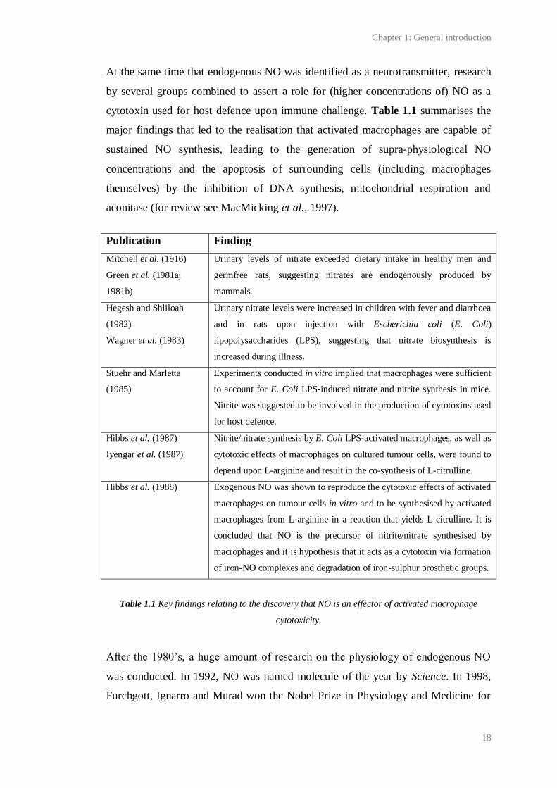
cytotoxin used for host defence upon immune challenge. Table 1.1 summarises the
major findings that led to the realisation that activated macrophages are capable of
sustained NO synthesis, leading to the generation of supra-physiological NO
concentrations and the apoptosis of surrounding cells (including macrophages
themselves) by the inhibition of DNA synthesis, mitochondrial respiration and
aconitase (for review see MacMicking et al., 1997).
Publication Finding
Mitchell et al. (1916)
Green et al. (1981a;
1981b)
Urinary levels of nitrate exceeded dietary intake in healthy men and
germfree rats, suggesting nitrates are endogenously produced by
mammals.
Hegesh and Shliloah
(1982)
Wagner et al. (1983)
Urinary nitrate levels were increased in children with fever and diarrhoea
and in rats upon injection with Escherichia coli (E. Coli)
lipopolysaccharides (LPS), suggesting that nitrate biosynthesis is
increased during illness.
Stuehr and Marletta
(1985)
Experiments conducted in vitro implied that macrophages were sufficient
to account for E. Coli LPS-induced nitrate and nitrite synthesis in mice.
Nitrite was suggested to be involved in the production of cytotoxins used
for host defence.
Hibbs et al. (1987)
Iyengar et al. (1987)
Nitrite/nitrate synthesis by E. Coli LPS-activated macrophages, as well as
cytotoxic effects of macrophages on cultured tumour cells, were found to
depend upon L-arginine and result in the co-synthesis of L-citrulline.
Hibbs et al. (1988) Exogenous NO was shown to reproduce the cytotoxic effects of activated
macrophages on tumour cells in vitro and to be synthesised by activated
macrophages from L-arginine in a reaction that yields L-citrulline. It is
concluded that NO is the precursor of nitrite/nitrate synthesised by
macrophages and it is hypothesis that it acts as a cytotoxin via formation
of iron-NO complexes and degradation of iron-sulphur prosthetic groups.
Table 1.1 Key findings relating to the discovery that NO is an effector of activated macrophage
cytotoxicity.
After the 1980’s, a huge amount of research on the physiology of endogenous NO
was conducted. In 1992, NO was named molecule of the year by Science. In 1998,
Furchgott, Ignarro and Murad won the Nobel Prize in Physiology and Medicine for
Chapter 1: General introduction
19
their discoveries relating to the vasodilatory effects of NO, a fitting award since
Alfred Nobel (the founder of the prize) was among the first people to recognise
nitroglycerine as a vasodilator (Marsh and Marsh, 2000). Now, NO is one of the
most researched signalling molecules active in the mammalian body. Research on the
physiology of organisms such as slime moulds (Golderer et al., 2001), jellyfish
(Moroz et al., 2004), molluscs (Park et al., 1998), fireflies (Dudzinski et al., 2006)
and even plants (reviewed by Wojtaszek, 2000), has combined to show that NO
signalling has been highly evolutionary conserved. In accordance with histological
data showing a wide distribution of the enzymes responsible for NO synthesis and
signal transduction throughout the mammalian body (see 1.2.2 and 1.3.2), it is
accepted that NO has a huge number of consequences for mammalian health and
disease. Furthermore, the NO signalling pathway is highly researched as a putative
target of therapeutic strategies. Some successful outcomes of this research include
anti-anginals, sildenafil (Viagra) and the use of inhaled NO to treat neonates with
respiratory failure.
1.2 Synthesis of endogenous NO
Soon after NO was identified as EDRF, an assay based on the conversion of L-
arginine to L-citrulline and NO was used to isolate the enzyme responsible for NO
synthesis, NO synthase (NOS), from rat cerebellum and identify it as nicotinamide
adenine dinucleotide phosphate (NADPH)- and calmodulin (CaM)-dependent (Bredt
and Snyder, 1990). This led to the cloning of brain-derived NOS (Bredt et al., 1991b)
and its localisation to vascular endothelial cells, nerves of the peripheral nervous
system (PNS) and discrete populations of neurons throughout the brain (Bredt et al.,
1990; Bredt et al., 1991a).
There are now three identified mammalian NOS isozymes, each coded for by a
distinct gene. Two, the neuronal NOS (nNOS) and endothelial NOS (eNOS), are
constitutively expressed throughout the nervous system and relate NO production to
intracellular changes in Ca2+
by their dependence on Ca2+
/CaM binding for catalytic
activity. The third, inducible NOS (iNOS), is the isoform expressed in immune cells
such as macrophages and microglia in response to products of infection (such as
Chapter 1: General introduction
20
endotoxins) and inflammatory mediators (such as cytokines). Since iNOS expression
is only prevalent under pathological states, it will not be considered in detail here
(reviewed by Stuehr, 1999; Alderton et al., 2001; Daff, 2010). The existence of a
distinct, constitutively expressed mitochondrial NOS is under debate (see Lacza et
al., 2006 for a review).
All three well-known NOS isozymes synthesise NO from L-arginine by two steps of
monooxygenation and share a common general structure with 50-60 % homology
(Figure 1.1). Each is conferred with distinct functionality, not only by differences in
tissue distribution, but also by multiple differences in the regulation of their activity.
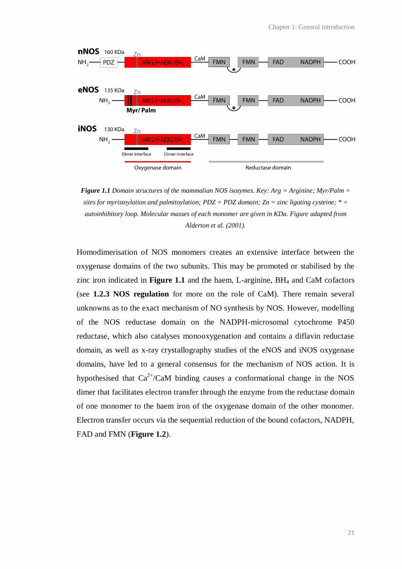
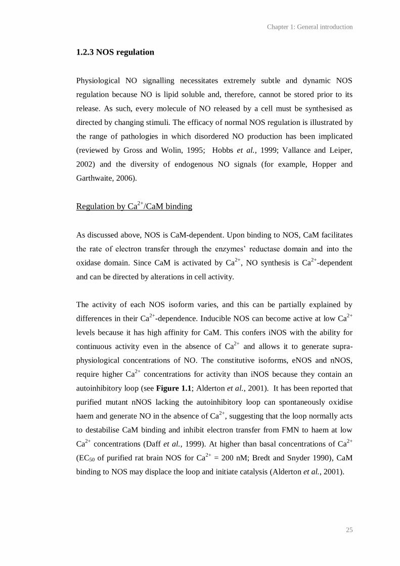
1.2.1 NOS structure and reaction mechanism
Functional NOS exists as a homodimer, each monomer consisting of an N-terminal
oxygenase domain, comprising binding sites for the cofactors haem and
tetrahydrobiopterin (BH4), and the substrate L-arginine, and a C-terminal reductase
domain, containing sites for flavin adenine dinucleotide (FAD), flavin
mononucleotide (FMN) and NADPH binding. The oxygenase and reductase domains
are linked by a series of amino acids constituting a Ca2+
/CaM-binding domain
(Figure 1.1).
The N-terminal of the most abundantly expressed (> 90 % of total) nNOS splice
variant in the brain (Huang et al., 1993), nNOSα, contains a PDZ domain which
allows its physical association with various proteins, most notably the NR2B NMDA
receptor subunit, via the adaptor protein, post-synaptic density 95 (PSD-95; Brenman
et al., 1996; Christopherson et al., 1999). The N-terminal of eNOS contains
consensus sequences for myristoylation and cysteine palmitoylation that allow its
association with the membrane of endothelial cells, specifically at their caveolae,
which are protein-rich invaginations of the membrane. Inducible NOS lacks the
ability to associate with membranes and is cytosolic (see Alderton et al., 2001; Daff,
2010 for reviews).
Chapter 1: General introduction
21
Figure 1.1 Domain structures of the mammalian NOS isozymes. Key: Arg = Arginine; Myr/Palm =
sites for myristoylation and palmitoylation; PDZ = PDZ domain; Zn = zinc ligating cysteine; * =
autoinhibitory loop. Molecular masses of each monomer are given in KDa. Figure adapted from
Alderton et al. (2001).
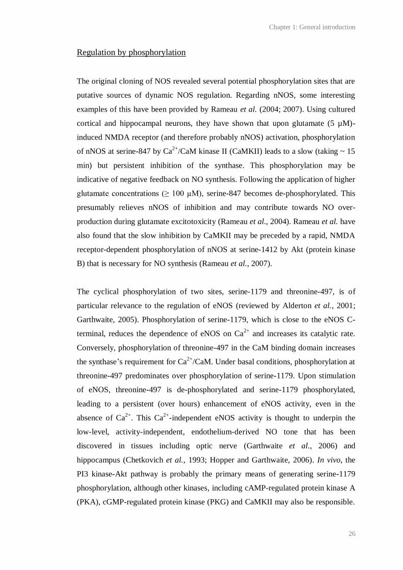
Homodimerisation of NOS monomers creates an extensive interface between the
oxygenase domains of the two subunits. This may be promoted or stabilised by the
zinc iron indicated in Figure 1.1 and the haem, L-arginine, BH4 and CaM cofactors
(see 1.2.3 NOS regulation for more on the role of CaM). There remain several
unknowns as to the exact mechanism of NO synthesis by NOS. However, modelling
of the NOS reductase domain on the NADPH-microsomal cytochrome P450
reductase, which also catalyses monooxygenation and contains a diflavin reductase
domain, as well as x-ray crystallography studies of the eNOS and iNOS oxygenase
domains, have led to a general consensus for the mechanism of NOS action. It is
hypothesised that Ca2+
/CaM binding causes a conformational change in the NOS
dimer that facilitates electron transfer through the enzyme from the reductase domain
of one monomer to the haem iron of the oxygenase domain of the other monomer.
Electron transfer occurs via the sequential reduction of the bound cofactors, NADPH,
FAD and FMN (Figure 1.2).
Chapter 1: General introduction
22
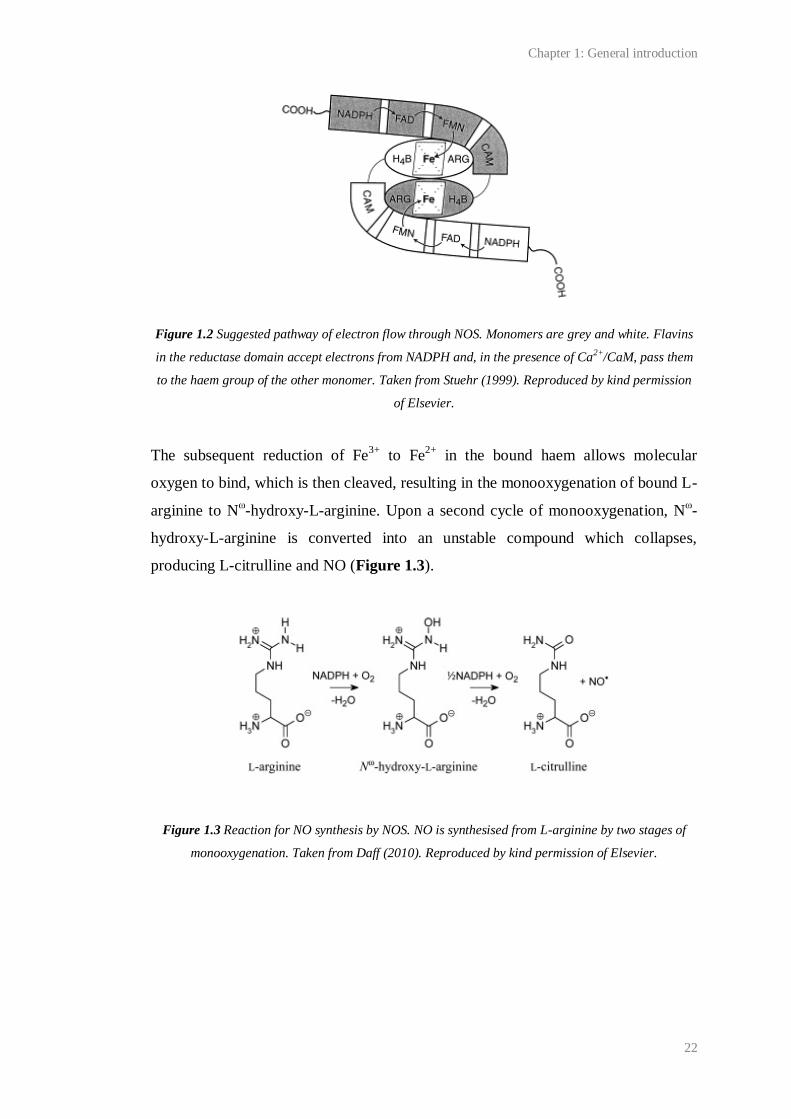
Figure 1.2 Suggested pathway of electron flow through NOS. Monomers are grey and white. Flavins
in the reductase domain accept electrons from NADPH and, in the presence of Ca2+/CaM, pass them
to the haem group of the other monomer. Taken from Stuehr (1999). Reproduced by kind permission
of Elsevier.
The subsequent reduction of Fe3+
to Fe2+
in the bound haem allows molecular
oxygen to bind, which is then cleaved, resulting in the monooxygenation of bound L-
arginine to Nω-hydroxy-L-arginine. Upon a second cycle of monooxygenation, N
ω-
hydroxy-L-arginine is converted into an unstable compound which collapses,
producing L-citrulline and NO (Figure 1.3).
Figure 1.3 Reaction for NO synthesis by NOS. NO is synthesised from L-arginine by two stages of
monooxygenation. Taken from Daff (2010). Reproduced by kind permission of Elsevier.
Chapter 1: General introduction
23
1.2.2 Location of NOS in brain and its intracellular distribution
nNOS
The first studies of the location of NOS in brain used NADPH-diaphorase staining
(Vincent and Kimura, 1992; Southam and Garthwaite, 1993), which relies upon the
reduction of tetrazolium salts to visible formazans in a NADPH- and NOS-dependent
manner. Since then, immunohistochemistry (Bredt et al., 1991a; Rodrigo et al., 1994;
de Vente et al., 1998; Burette et al., 2002) and in situ hybridisation (Keilhoff et al.,
1996) have also been used to locate nNOS protein and mRNA, and this has led to the
consensus that nNOS is expressed throughout the entire rodent and primate brain,
albeit at varying levels in different areas. In the cerebellum, most neurons are
immunopositive for nNOS (Bredt et al., 1990; Southam et al., 1992). Areas such as
the hippocampus and olfactory bulb also appear rich in the enzyme (Southam and
Garthwaite, 1993). In other brain regions, nNOS appears to be restricted to
populations of interneurons, as in the cerebral cortex. However, even in these areas, a
dense network of nNOS positive fibres has been discovered, suggesting that the
majority of brain cells could be contacted by NO (Vincent and Kimura, 1992;
Rodrigo et al., 1994).
The intracellular distribution (and physiology) of nNOS is largely dictated by its
interaction with PDZ-containing proteins, such as PSD-95. PDZ domains are motifs
for protein-protein interaction. By binding with a PDZ domain in the N-terminal of
nNOSα, and another in the C-terminal domain of the NMDA receptor NR2B subunit,
PSD-95 physically links the synthase to the NMDA receptor (Christopherson et al.,
1999). In this way, nNOS is anchored to a major site of activity-dependent Ca2+
influx to cells and is therefore thought to be preferentially activated by NMDA
receptor opening. Consistent with this, NMDA causes NO synthesis in vitro
(Garthwaite et al., 1988; Garthwaite et al., 1989) and in vivo (Wood et al., 1990). In
tissue supernatants prepared from various brain regions, Ca2+
-dependent NO
synthesis has been shown to be nNOS-dependent (Huang et al., 1993), and
suppression of PSD-95 in cultured cortical neurons by an anti-sense oligonucleotide
has been found to inhibit NMDA-induced cGMP production by > 60% (Sattler et al.,
Chapter 1: General introduction
24
1999). Neuronal NOS and PDS-95 co-localise throughout the brain (Brenman et al.,
1996), and in the hippocampus, immunofluorescent staining has shown that nNOS,
NR2 and PSD-95 co-localise in PSDs (Burette et al., 2002).
In addition to nNOSα, there are two other splice variants of nNOS: β and γ. These
lack a PDZ domain and are cytosolic. The γ variant appears to be inactive, though the
β may be functional in several brain areas, including cortex, hippocampus, olfactory
bulb and cerebellum (Brenman et al. 1996; Eliasson et al., 1997; Huang et al., 1993).
eNOS
An initial immunohistochemical study of the location of eNOS in the brain found it
to be expressed in hippocampal pyramidal neurons (Dinerman et al., 1994) but this
result has not been replicated. Rather, data collected using in situ hybridisation
(Seidel et al., 1997; Demas et al., 1999; Blackshaw et al., 2003),
immunohistochemistry (Stanarius et al., 1997; Topel et al., 1998) and polymerase
chain reaction (PCR) of DNA from dissociated hippocampal neurons (Chiang et al.,
1994) has combined to assert the consensus that eNOS is exclusively expressed in
the endothelium of blood vessels.
As discussed above, eNOS has been found to associate with the membrane of
endothelial cells, specifically in the cells’ caveolae (Garcia-Cardena et al., 1996).
Caveolae are enriched in cholesterol and lipids. It is thought that their limited fluidity
draws proteins together, thereby promoting protein-protein interactions (Razani et
al., 2002). Binding of eNOS to caveolae membranes is thought to occur via the
enzyme’s N-terminal, which contains consensus sequences for myristoylation, which
is irreversible, and palmitoylation, which is reversible (Garcia-Cardena et al., 1996;
Alderton et al., 2001). Palmitoylation of eNOS may be subject to dynamic
regulation, since prolonged stimulation of eNOS has been reported to cause the
enzymes de-palmitoylation and translocation into the cytosol, this presumably
limiting the opportunity for eNOS activation (see 1.2.3).
Chapter 1: General introduction
25
1.2.3 NOS regulation
Physiological NO signalling necessitates extremely subtle and dynamic NOS
regulation because NO is lipid soluble and, therefore, cannot be stored prior to its
release. As such, every molecule of NO released by a cell must be synthesised as
directed by changing stimuli. The efficacy of normal NOS regulation is illustrated by
the range of pathologies in which disordered NO production has been implicated
(reviewed by Gross and Wolin, 1995; Hobbs et al., 1999; Vallance and Leiper,
2002) and the diversity of endogenous NO signals (for example, Hopper and
Garthwaite, 2006).
Regulation by Ca2+
/CaM binding
As discussed above, NOS is CaM-dependent. Upon binding to NOS, CaM facilitates
the rate of electron transfer through the enzymes’ reductase domain and into the
oxidase domain. Since CaM is activated by Ca2+
, NO synthesis is Ca2+
-dependent
and can be directed by alterations in cell activity.
The activity of each NOS isoform varies, and this can be partially explained by
differences in their Ca2+
-dependence. Inducible NOS can become active at low Ca2+
levels because it has high affinity for CaM. This confers iNOS with the ability for
continuous activity even in the absence of Ca2+
and allows it to generate supra-
physiological concentrations of NO. The constitutive isoforms, eNOS and nNOS,
require higher Ca2+
concentrations for activity than iNOS because they contain an
autoinhibitory loop (see Figure 1.1; Alderton et al., 2001). It has been reported that
purified mutant nNOS lacking the autoinhibitory loop can spontaneously oxidise
haem and generate NO in the absence of Ca2+
, suggesting that the loop normally acts
to destabilise CaM binding and inhibit electron transfer from FMN to haem at low
Ca2+
concentrations (Daff et al., 1999). At higher than basal concentrations of Ca2+
(EC50 of purified rat brain NOS for Ca2+
= 200 nM; Bredt and Snyder 1990), CaM
binding to NOS may displace the loop and initiate catalysis (Alderton et al., 2001).
* *
Chapter 1: General introduction
26
Regulation by phosphorylation
The original cloning of NOS revealed several potential phosphorylation sites that are
putative sources of dynamic NOS regulation. Regarding nNOS, some interesting
examples of this have been provided by Rameau et al. (2004; 2007). Using cultured
cortical and hippocampal neurons, they have shown that upon glutamate (5 µM)-
induced NMDA receptor (and therefore probably nNOS) activation, phosphorylation
of nNOS at serine-847 by Ca2+
/CaM kinase II (CaMKII) leads to a slow (taking ~ 15
min) but persistent inhibition of the synthase. This phosphorylation may be
indicative of negative feedback on NO synthesis. Following the application of higher
glutamate concentrations (≥ 100 µM), serine-847 becomes de-phosphorylated. This
presumably relieves nNOS of inhibition and may contribute towards NO over-
production during glutamate excitotoxicity (Rameau et al., 2004). Rameau et al. have
also found that the slow inhibition by CaMKII may be preceded by a rapid, NMDA
receptor-dependent phosphorylation of nNOS at serine-1412 by Akt (protein kinase
B) that is necessary for NO synthesis (Rameau et al., 2007).
The cyclical phosphorylation of two sites, serine-1179 and threonine-497, is of
particular relevance to the regulation of eNOS (reviewed by Alderton et al., 2001;
Garthwaite, 2005). Phosphorylation of serine-1179, which is close to the eNOS C-
terminal, reduces the dependence of eNOS on Ca2+
and increases its catalytic rate.
Conversely, phosphorylation of threonine-497 in the CaM binding domain increases
the synthase’s requirement for Ca2+
/CaM. Under basal conditions, phosphorylation at
threonine-497 predominates over phosphorylation of serine-1179. Upon stimulation
of eNOS, threonine-497 is de-phosphorylated and serine-1179 phosphorylated,
leading to a persistent (over hours) enhancement of eNOS activity, even in the
absence of Ca2+
. This Ca2+
-independent eNOS activity is thought to underpin the
low-level, activity-independent, endothelium-derived NO tone that has been
discovered in tissues including optic nerve (Garthwaite et al., 2006) and
hippocampus (Chetkovich et al., 1993; Hopper and Garthwaite, 2006). In vivo, the
PI3 kinase-Akt pathway is probably the primary means of generating serine-1179
phosphorylation, although other kinases, including cAMP-regulated protein kinase A
(PKA), cGMP-regulated protein kinase (PKG) and CaMKII may also be responsible.
Chapter 1: General introduction
27
These kinases, as well as Akt, may be activated in response to stimuli including shear
stress, oestrogens, insulin and vascular endothelial growth factor (Garthwaite, 2008).
Regulation by protein-protein interaction
As discussed above, protein-protein interactions, for example, between nNOS and
PSD-95, serve to anchor the constitutive NOS isoforms to cell membranes where
they may be switched on by a rise in intracellular Ca2+
. Importantly, the binding of e-
or nNOS to cell membranes is reversible. Indeed, the intracellular distributions of e-
and nNOS, and thus the capacity for their activation, are subject to dynamic
regulation by various other binding proteins.
The C-terminal PDZ ligand of NOS (CAPON), is an adaptor protein that was
identified by a yeast two-hybrid screen with nNOS. Immunohistochemistry for
CAPON shows that it is expressed throughout the brain in a distribution overlapping
that of nNOS. It contains a C-terminal domain which competes with PSD-95 for
binding to the PDZ domain of the synthase. This causes the translocation of nNOS
away from the PSD and therefore, may limit neuronal NO synthesis (Jaffrey et al.,
1998). In presynaptic terminals, interaction between a phosphotyrosine binding
domain in the N-terminal of CAPON and synapsin 1 may direct nNOS to the
membrane (Jaffrey et al., 2002) where nNOS may be activated by voltage-gated Ca2+
channels (VGCCs), as in the PNS (reviewed by Vincent, 2010).
NOS interacting protein (NOSIP) was also discovered by yeast two-hybrid screening
with nNOS and may also modulate nNOS by altering its intracellular distribution.
Dreyer et al. (2004) have found that NOSIP and nNOS can be co-
immunoprecipitated from rat brain lysates, and co-occur in multiple brain areas
including the hippocampus, cortex and cerebellum. They also report that expression
of NOSIP leads to a reduction in Ca2+
-induced NO synthesis in an immortalised cell
line containing nNOS, and a (moderate) shift in the location of nNOS from the
dendrites to the soma of dissociated hippocampal neurons.
Chapter 1: General introduction
28
Other protein regulators of nNOS include ‘protein-inhibitor of nNOS’, a dynein light
chain that may bind to and regulate the axonal transport of nNOS (Rodriguez-Crespo
et al., 1998) and heat shock protein 90 (Hsp90), which has been shown to facilitate
nNOS activity in vitro, likely by increasing its affinity for CaM (Song et al., 2001).
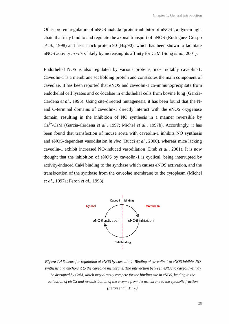
Endothelial NOS is also regulated by various proteins, most notably caveolin-1.
Caveolin-1 is a membrane scaffolding protein and constitutes the main component of
caveolae. It has been reported that eNOS and caveolin-1 co-immunoprecipitate from
endothelial cell lysates and co-localise in endothelial cells from bovine lung (Garcia-
Cardena et al., 1996). Using site-directed mutagenesis, it has been found that the N-
and C-terminal domains of caveolin-1 directly interact with the eNOS oxygenase
domain, resulting in the inhibition of NO synthesis in a manner reversible by
Ca2+
/CaM (Garcia-Cardena et al., 1997; Michel et al., 1997b). Accordingly, it has
been found that transfection of mouse aorta with caveolin-1 inhibits NO synthesis
and eNOS-dependent vasodilation in vivo (Bucci et al., 2000), whereas mice lacking
caveolin-1 exhibit increased NO-induced vasodilation (Drab et al., 2001). It is now
thought that the inhibition of eNOS by caveolin-1 is cyclical, being interrupted by
activity-induced CaM binding to the synthase which causes eNOS activation, and the
translocation of the synthase from the caveolae membrane to the cytoplasm (Michel
et al., 1997a; Feron et al., 1998).
Figure 1.4 Scheme for regulation of eNOS by caveolin-1. Binding of caveolin-1 to eNOS inhibits NO
synthesis and anchors it to the caveolae membrane. The interaction between eNOS to caveolin-1 may
be disrupted by CaM, which may directly compete for the binding site in eNOS, leading to the
activation of eNOS and re-distribution of the enzyme from the membrane to the cytosolic fraction
(Feron et al., 1998).

Chapter 1: General introduction
29
Hsp90 may also bind directly to eNOS in a Ca2+
-dependent manner. It has been
found to co-immunoprecipitate from endothelial cells with eNOS and caveolin-1
(Gratton et al., 2000), and has been hypothesised to facilitate the displacement of
caveolin-1 from eNOS by CaM. Indeed, the EC50 of Ca2+
and CaM for eNOS is
reduced in the presence of Hsp90 (Takahashi and Mendelsohn, 2003a). Additionally,
physiological stimuli for eNOS, such as vascular endothelial growth factor or shear
stress, have been reported to increase the interaction of Hsp90 and eNOS in isolated
cells, whereas an antibiotic-based Hsp90 inhibitor has been found to inhibit eNOS-
dependent ACh-induced vasodilation of rat aortic rings (Garcia-Cardena et al.,
1998). Co-immunoprecipitation studies also suggest that Hsp90 may facilitate a
physical interaction between eNOS and Akt (Garcia-Cardena et al., 1998), consistent
with findings that the effects of Hsp90 and Akt on eNOS activity are synergistic at
low Ca2+
concentrations (Takahashi and Mendelsohn, 2003b).
In caveolae, eNOS may also directly interact with bradykinin B2 receptors, which,
are upstream of the phospholipase C-phosphatidylinositol 4,5-bisphosphate (PIP2)
pathway, and the arginine transporter, cationic amino acid transporter, which may
facilitate eNOS activity (reviewed by Nedvetsky et al., 2002). NOSIP may also
regulate eNOS in the same way that it does nNOS: Dedio et al. (2001) have found
that the co-expression of eNOS and NOSIP in Chinese hamster ovary cells causes a
reduction in NO synthesis and the redistribution of eNOS from the caveolae
membrane to the cytoplasm.
1.3 NO signal transduction
The identification of NO as EDRF was preceded by the discovery that NO elicits
cGMP accumulation in tissues such as aorta, lung and brain, and that a rise in cGMP
accompanies the relaxation of smooth muscle (see 1.1 Discovery of endogenous
NO). About 20 years prior to this, cGMP had been detected in mammalian urine and
various tissues. At around the same time, cAMP, produced by adenylyl cyclases, was
recognised as a biological second messenger. This spurred research which led to the
discovery of two major variants of guanylyl cyclase that synthesise cGMP from
Chapter 1: General introduction
30
GTP: one that is membrane-bound and consists of seven isoforms, each of which
contain an extracellular binding domain for ligands such as natriuretic peptides and
are unresponsive to NO; and another that does not span the membrane and contains a
prosthetic haem group able to bind NO (reviewed by Potter, 2011; Schulz et al.,
1989). The latter cyclase was initially termed ‘soluble’, but it is now known to
associate with membranes under some conditions (see 1.3.2) and therefore has been
renamed ‘NO-targeted’ or ‘NO-activated’.
To date, NO is the only known physiological activator of NO-targeted guanylyl
cyclase. Cyclic GMP accumulation via the activation of this enzyme is the only
accepted means of physiological NO signal transduction (see 1.8 and Garthwaite,
2008). Amongst the research that has led to this consensus are findings that mice
lacking eNOS or the NO-targeted guanylyl cyclase are incapable of NO-induced
vasodilation (Huang et al., 1995; Friebe et al., 2007) and that NADPH diaphorase
histochemistry for NOS in rodent brain is remarkably coincident with
immunohistochemistry for exogenous NO-induced cGMP accumulation (Southam
and Garthwaite, 1993).
1.3.1 NO-targeted guanylyl cyclase structure and reaction mechanism
Isoforms of NO-targeted guanylyl cyclase
NO-targeted guanylyl cyclase is an obligate heterodimer comprising one β and one α
subunit (Nakane et al., 1990; Buechler et al., 1991; Harteneck et al., 1991). To date,
two endogenous, functional isoforms of NO-targeted guanylyl cyclase have been
discovered: the α1β1- and α2β1-containing enzymes. The isoforms appear to have a
similar sensitivity to exogenous NO, capacity for cGMP production and
pharmacology (Russwurm et al., 1998; Gibb et al., 2003), but different intracellular
distributions (see 1.3.2). The α1β1 isoform was first purified from rat and bovine
lung and subsequently both participating subunits were cloned and sequenced
(Koesling et al., 1988; Nakane et al., 1988; Koesling et al., 1990; Nakane et al.,
1990; Russwurm et al., 1998). The α2 subunit was identified by homology screening
with the α1 subunit. Subsequently, functional α2β1 dimers were reported to form in
Chapter 1: General introduction
31
cells transfected with both subunits (Harteneck et al., 1991) and, in 1998, were
discovered in human placenta (Russwurm et al., 1998). Message for the α1, α2 and
β1 subunits has now been found throughout the mammalian body and brain (Gibb
and Garthwaite, 2001; Mergia et al., 2003).
Two other NO-targeted guanylyl cyclase subunits, namely α3 and β3, have been
cloned but identified as human variants of the α1 and β1 subunits (Zabel et al.,
1998). Messenger RNA for a β2 NO-targeted guanylyl cyclase subunit has also been
detected in rodents, and in various organs (Mergia et al., 2003). The expression of
this subunit with α1 in COS-7 cells has been reported to result in a functional
cyclase, although with reduced sensitivity to NO compared to the α1β1 and α2β1
isoforms (Gupta et al., 1997; Gibb et al., 2003). However, the transfection of other
types of cells with α1 and β2 NO-targeted guanylyl cyclase subunits has failed to
yield a functional enzyme (Gibb et al., 2003). Furthermore, message for the β2
subunit in brain and other organs is negligible, and there have been no reports of an
endogenous functional β2-containing NO-targeted guanylyl cyclase (Gibb and
Garthwaite, 2001; Mergia et al., 2003).
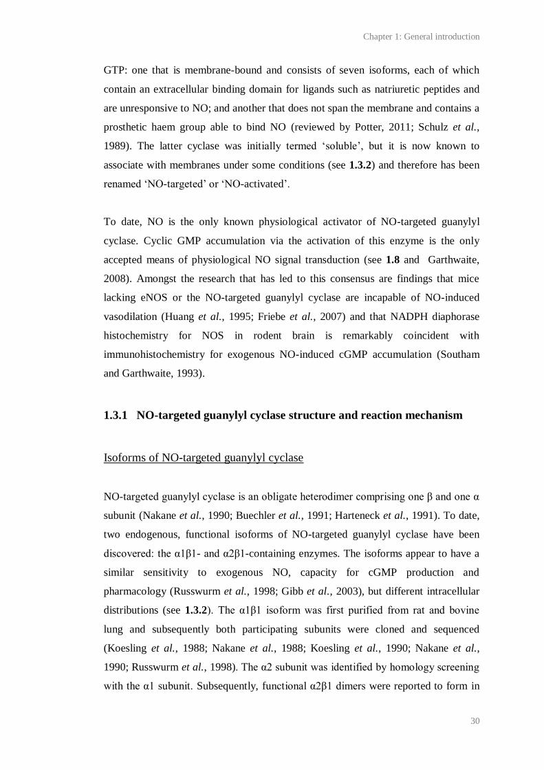
General structure of heterodimers
Each functionally relevant NO-targeted guanylyl cyclase subunit contains a C-
terminal, catalytic domain, a dimerisation domain and an N-terminal, regulatory
domain (see Figure 1.5). The catalytic domain appears to have been highly
conserved across each NO-targeted guanylyl cyclase subunit, and, within the
functional enzyme, is so homologous to that of adenylyl cyclase that substitution of
three amino acids produces a NO-targeted, cAMP-synthesising enzyme (Sunahara et
al., 1998). It is highly likely that the catalytic domain contains the site for GTP
binding, and consistent with this, studies using site-directed mutagenesis have found
the catalytic domain to be sufficient for un-stimulated cGMP production (Wedel et
al., 1995).
The N-terminal regulatory domain of each heterodimer binds one haem prosthetic
group, primarily through an interaction with the haem Fe2+
and His-105 of the β1
Chapter 1: General introduction
32
subunit (Wedel et al., 1994). The α subunits, which differ significantly from each
other within the N-terminal region, may also be necessary for haem binding (Wedel
et al., 1995; Foerster et al., 1996; although see Koglin and Behrends, 2003), thus
partly explaining why NO-targeted guanylyl cyclase is an obligate heterodimer. The
haem is the NO-binding site within the cyclase. It has long been known that NO
binds to haem; indeed its interaction with reduced haemoglobin was critical to the
identification of NO as EDRF (Ignarro et al., 1987). As such, the haem component of
NO-targeted guanylyl cyclase was discovered relatively soon after initial attempts to
purify the enzyme (Craven and DeRubertis, 1978; Gerzer et al., 1981) and was
immediately identified as a putative NO binding site. Now the evidence in favour of
this is convincing. Studies have shown, for example, that haem loss (Foerster et al.,
1996), truncation of the N-terminal domain (Wedel et al., 1995; Foerster et al., 1996)
or substitution of His-105 with phenylalanine (Wedel et al., 1994) renders the
guanylyl cyclase NO-insensitive.
Figure 1.5 The domain structure of NO-targeted guanylyl cyclase. The haem is shown in grey.
Adapted from Bartus (2009).
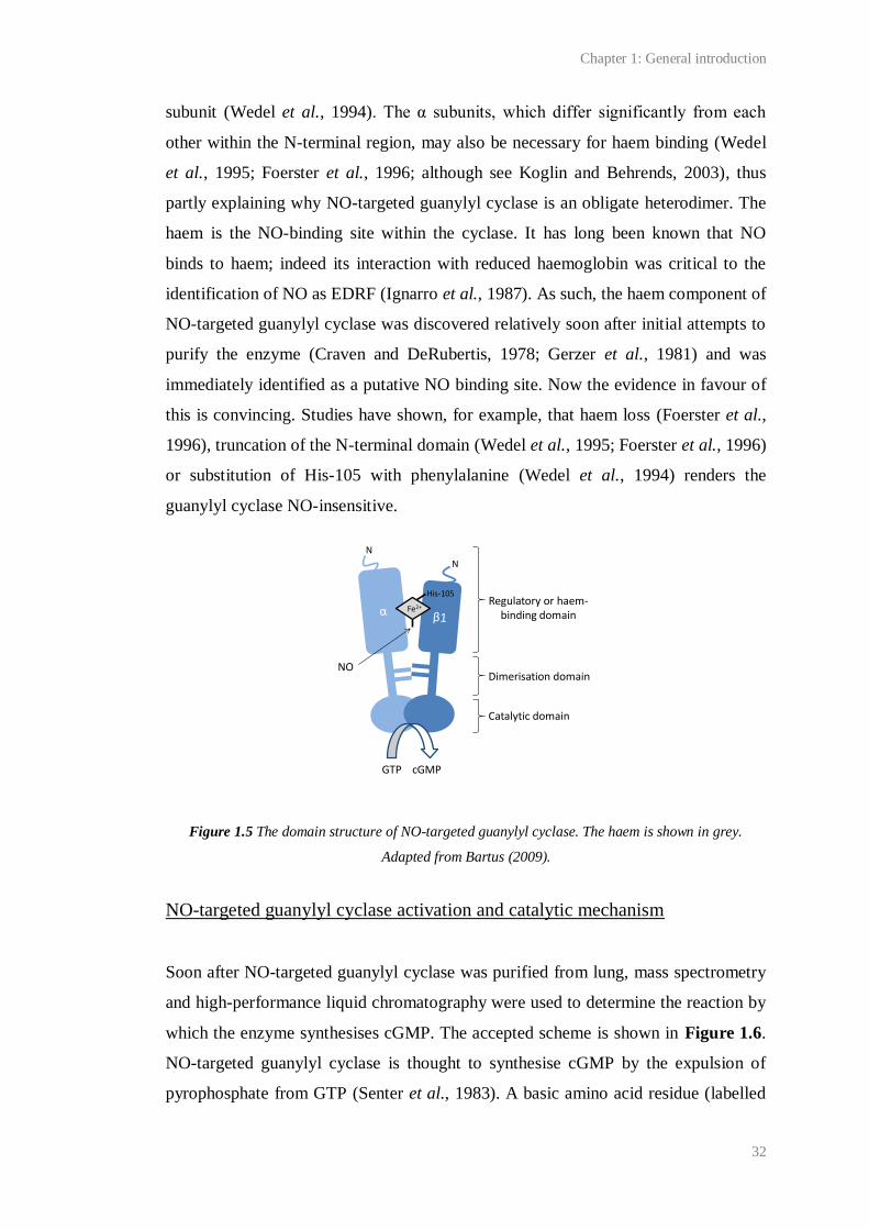
NO-targeted guanylyl cyclase activation and catalytic mechanism
Soon after NO-targeted guanylyl cyclase was purified from lung, mass spectrometry
and high-performance liquid chromatography were used to determine the reaction by
which the enzyme synthesises cGMP. The accepted scheme is shown in Figure 1.6.
NO-targeted guanylyl cyclase is thought to synthesise cGMP by the expulsion of
pyrophosphate from GTP (Senter et al., 1983). A basic amino acid residue (labelled
GTP cGMP
N
N
Fe2+
His-105
NO
Regulatory or haem-binding domain
Dimerisation domain
Catalytic domain

Chapter 1: General introduction
33
X in the figure), the identity of which is currently unknown, is required to accept a
proton from GTP during the reaction.
Figure 1.6 Proposed means of cGMP synthesis from GTP by NO-targeted guanylyl cyclase. A basic
residue in the NO-targeted guanylyl cyclase (shown as X) accepts a proton from the hydroxyl group at
position five of the ribose moiety of GTP. This leads to the displacement of pyrophosphate (PPiO)
from the molecule and the formation of cGMP.
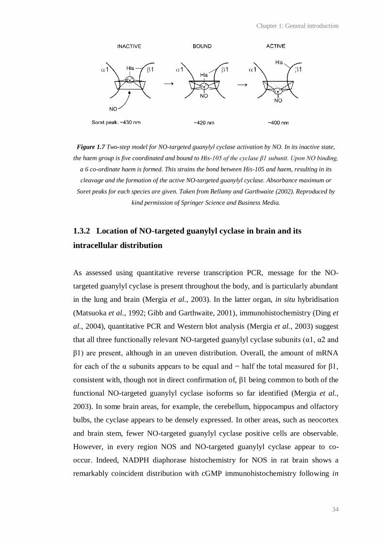
Detailed structure-function studies are required to elucidate how NO binding to the
NO-targeted guanylyl cyclase haem catalyses this reaction. However, studies on a
homologous cyanobacterial NO detector, in conjunction with the analysis of UV-
visible absorbance spectra for different haem species formed during NO binding,
have led to a general scheme in which the cyclase passes through inactive, NO-
bound and active states (Figure 1.7; Bellamy and Garthwaite, 2002). When the
enzyme is inactive, the haem appears to be five-coordinated, its Fe2+
centre
covalently bound to the cyclase via His-105 in the N-terminal of the β1 subunit. NO
binding to the haem, which is thought to be so rapid that it is almost diffusion-
limited, forms a six-coordinated haem and is thought to cause the haem to pivot,
leading to the rapid (within 1 ms) translocation and subsequent rupture of the bond
between it and His-105. Rupture of the bond is assumed to cause a conformational
change in the cyclase that propagates to the catalytic domain by some unknown
mechanism and causes up to a 1000-fold increase in the rate of cGMP synthesis. The
propagation of this conformational change is thought to be the rate-limiting step in
activation of the enzyme and is hypothesised to facilitate access of GTP to the
catalytic site.
X XH
PPiO
Guanosine
PPiO
Chapter 1: General introduction
34
Figure 1.7 Two-step model for NO-targeted guanylyl cyclase activation by NO. In its inactive state,
the haem group is five coordinated and bound to His-105 of the cyclase β1 subunit. Upon NO binding,
a 6 co-ordinate haem is formed. This strains the bond between His-105 and haem, resulting in its
cleavage and the formation of the active NO-targeted guanylyl cyclase. Absorbance maximum or
Soret peaks for each species are given. Taken from Bellamy and Garthwaite (2002). Reproduced by
kind permission of Springer Science and Business Media.
1.3.2 Location of NO-targeted guanylyl cyclase in brain and its
intracellular distribution
As assessed using quantitative reverse transcription PCR, message for the NO-
targeted guanylyl cyclase is present throughout the body, and is particularly abundant
in the lung and brain (Mergia et al., 2003). In the latter organ, in situ hybridisation
(Matsuoka et al., 1992; Gibb and Garthwaite, 2001), immunohistochemistry (Ding et
al., 2004), quantitative PCR and Western blot analysis (Mergia et al., 2003) suggest
that all three functionally relevant NO-targeted guanylyl cyclase subunits (α1, α2 and
β1) are present, although in an uneven distribution. Overall, the amount of mRNA
for each of the α subunits appears to be equal and ~ half the total measured for β1,
consistent with, though not in direct confirmation of, β1 being common to both of the
functional NO-targeted guanylyl cyclase isoforms so far identified (Mergia et al.,
2003). In some brain areas, for example, the cerebellum, hippocampus and olfactory
bulbs, the cyclase appears to be densely expressed. In other areas, such as neocortex
and brain stem, fewer NO-targeted guanylyl cyclase positive cells are observable.
However, in every region NOS and NO-targeted guanylyl cyclase appear to co-
occur. Indeed, NADPH diaphorase histochemistry for NOS in rat brain shows a
remarkably coincident distribution with cGMP immunohistochemistry following in
Chapter 1: General introduction
35
vivo perfusion of the NO donor, sodium nitroprusside (Southam and Garthwaite,
1993).
Message for the β subunit is almost always accompanied by mRNA for one or, more
typically, both of the α subunits. Some areas appear to contain more RNA for one α
subunit than the other. For example, the hippocampus and cerebellum appear richer
in α2, whereas the caudate putamen and nucleus accumbens appear richer in α1
(Gibb and Garthwaite, 2001). These trends have been confirmed by quantitative real-
time PCR (Mergia et al., 2003).
Within cells, the α1β1 and α2β1 appear to differ in their location. This arises due to
the ability of the α2 subunit to interact with PDZ-containing synaptic proteins,
including PSD-95 and synapse associated protein-97, through its C-terminal
(Russwurm et al., 2001). In this way, the α2β1 isoform may be anchored to the
membrane and in remarkable proximity to sites of NO synthesis. In contrast, the
α1β1 isoform appears to be mainly cytosolic, although, in platelets and lung
endothelial cells, it has been found to translocate to the membrane upon raised
concentrations of intracellular Ca2+
. Translocation to the membrane has been found
to increase the sensitivity of the cyclase to NO (Zabel et al., 2002), perhaps by
placing the cyclase closer to sites of NO synthesis.
1.3.3 Regulation of NO-targeted guanylyl cyclase
Compared to NOS, relatively little is known about how NO-targeted guanylyl
cyclase activity is regulated. Some putative examples of regulation are given below,
although more work is needed to clarify whether these are physiologically relevant
and what effect they have on NO-induced cGMP accumulation.
Regulation by co-factors
Several co-factors are required for the conversion of GTP to cGMP. Two Mg2+
per
cyclase are required for catalytic activity and may facilitate the binding of GTP to the
cyclase. Additionally, ATP inhibits NO-targeted guanylyl cyclase, perhaps by
Chapter 1: General introduction
36
binding to a regulatory site in competition with GTP (Ruiz-Stewart et al., 2004; Roy
and Garthwaite, 2006). Apart from the regulation of the α1β1 intracellular
distribution by Ca2+
(see 1.3.2), this cation also inhibits cGMP synthesis under
physiological conditions (Kazerounian et al., 2002).
Regulation by phosphorylation
Both the α and β subunits contain several putative phosphorylation sites that might
confer the cyclase with dynamic regulation (reviewed by Pyriochou and
Papapetropoulos, 2005). The effect of kinases including PKA and protein kinase C
on NO-targeted guanylyl cyclases have been researched, although studies have
yielded contradictory results. Two studies have shown that PKG may inhibit NO-
targeted guanylyl cyclase, thereby providing cGMP production with negative
feedback (Ferrero et al., 2000; Murthy, 2001). Murthy (2001) found that the NO
donor, sodium nitroprusside, caused an increase in PKG-dependent 32
P incorporation
into NO-targeted guanylyl cyclase in gastric smooth muscle that was accompanied
by a reduction in cGMP synthesis. Ferrero et al. (2000) have reported that NO-
targeted guanylyl cyclase is phosphorylated under basal conditions by PKG in
chromaffin cells (neuroendocrine cells of the sympathetic nervous system) and that a
cGMP analogue or PKG activation leads to the activation of a phosphatase,
dephosphorylation of NO-targeted guanylyl cyclase and a subsequent decrease in
sodium nitroprusside-induced cGMP synthesis.
Regulation by protein-protein interactions
Several proteins may regulate the intracellular distribution of the NO-targeted
guanylyl cyclases. For example, in endothelial cells, Hsp90 may physically link the
NO-targeted guanylyl cyclase β1 subunit to eNOS (Venema et al., 2003).
Chapter 1: General introduction
37
1.4 Characteristics of NO/cGMP signals
In the brain, NO may act as a neurotransmitter (discussed 1.9.1). Research suggests
that bursts of NO are synthesised by nNOS in response to synaptic stimuli that cause
a rise in intracellular Ca2+
(Park et al., 1998; Batchelor et al., 2010). However, unlike
classical neurotransmitters, immunohistochemistry for NOS and NO-targeted
guanylyl cyclase, in accordance with functional studies of NO transmission, suggest
that NO may act as anterograde (for example, Park et al., 1998), retrograde (for
example, Arancio et al., 1995; Arancio et al., 1996; Arancio et al., 2001), and/or
intracellular transmitter (for example, Burette et al., 2002). These effects may be
synapse specific (see below). Tonic NO signals synthesised by continuous eNOS
activity have also been found to effect paracrine transmission between blood vessels
and groups of neurons (for example, Garthwaite et al., 2006; Hopper and Garthwaite,
2006). To describe the ability of a ‘cloud’ of NO to diffuse freely from a source and
potentially affect all receptive structures contacted by a physiologically relevant
concentration, the term ‘volume signalling’ has been used. Despite intense research
on the dynamics of NO signalling, details that are vital to our understanding of how
NO is capable of such diverse signalling, such as what constitutes a physiological or
pathological concentration of NO, or how far a physiological concentration of NO
can spread through brain tissue from a site of synthesis, remain unclear.
1.4.1 Concentration of physiological NO signals
Initial attempts to measure endogenous NO employed NO/cGMP assays and NO
electrodes, which translate a chemical reaction between NO and the electrode tip into
an electric potential. Unfortunately, these approaches have many drawbacks, and
have led to a wide spread of measurements (from femtomolar to low micromolar
values). More recently, NO biosensors have been used to measure the concentration
of endogenous NO signals elicited by physiological stimuli. These biosensors are
composed of cGMP binding sites connected to fluorescent proteins. Cyclic GMP
binding causes a conformational change in the protein and concomitant change in its
fluorescence. This type of biosensor takes advantage of the amplification of NO
signals by cGMP production and the high selectivity of cGMP-binding proteins over
Chapter 1: General introduction
38
cAMP and GTP (reviewed by Hall and Garthwaite, 2009). Using a fluorescence
resonance energy transfer-based biosensor incorporating the cGMP-binding domain
of NO-targeted guanylyl cyclase, Sato et al. have detected 1 nM endogenous NO
inside un-stimulated endothelial cells. Approximately 100 pM NO was detected
inside a cell placed next to hippocampal neurons under the influence of spontaneous,
oscillatory network activity or endothelial cells stimulated with ATP (Sato et al.,
2005; Sato et al., 2006). In support of such low concentrations of NO representing
physiological signals, studies using human embryonic kidney (HEK) cells
transfected with a biosensor composed of the cGMP-binding domain of PKG fused
to a circularly permutated enhanced green fluorescent protein have shown that even
smaller concentrations of exogenous NO (1-3 pM) can be detected by NO-targeted
guanylyl cyclase, even in the presence of a phosphodiesterase (PDE; a
phosphohydrolase that degrades endogenous cGMP (see 1.4.4); Batchelor et al.,
2010). Very recently, HEK cells containing this biosensor have been used to detect ~
100-200 pM endogenous NO from overlying cerebellar and hippocampal slices upon
stimulation with NMDA (Wood et al., 2011). Consistent with such low amplitude
physiological NO signals, only low nanomolar concentrations of NO have been
recorded in cerebellar slices and hippocampal slice cultures following extreme
stimuli, such as ischemia, maximal NMDA receptor activation and iNOS activation
(reviewed by Hall and Garthwaite, 2009).
1.4.2 Spread of NO through tissues
NO is predicted to diffuse rapidly through tissues (tissue diffusion constant of 8.48
µm2/s). At low nanomolar concentrations, the NO free radical is predicted to be
relatively stable. For example, autoxidation of NO will be minimal. However, NO
will react with lipid peroxyl radicals and haemoglobin in blood vessels, and it has
been suggested that this will limit the half-life of NO in tissue to ~ 1 s (reviewed by
Garthwaite, 2008). Further to this, the spread of NO in brain tissue appears to be
hugely limited by an unknown means of NO inactivation. In cerebellar slices, this is
predicted to limit the half-life of < 10 nM NO to ~ 10 ms. NO inactivation by brain
tissue has been illustrated by a marked gradient of immunostaining for NO-induced
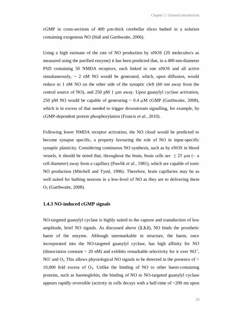
Chapter 1: General introduction
39
cGMP in cross-sections of 400 µm-thick cerebellar slices bathed in a solution
containing exogenous NO (Hall and Garthwaite, 2006).
Using a high estimate of the rate of NO production by nNOS (20 molecules/s as
measured using the purified enzyme) it has been predicted that, in a 400-nm-diameter
PSD containing 50 NMDA receptors, each linked to one nNOS and all active
simultaneously, ~ 2 nM NO would be generated, which, upon diffusion, would
reduce to 1 nM NO on the other side of the synaptic cleft (60 nm away from the
central source of NO), and 250 pM 1 µm away. Upon guanylyl cyclase activation,
250 pM NO would be capable of generating ~ 0.4 M cGMP (Garthwaite, 2008),
which is in excess of that needed to trigger downstream signalling, for example, by
cGMP-dependent protein phosphorylation (Francis et al., 2010).
Following lower NMDA receptor activation, the NO cloud would be predicted to
become synapse specific, a property favouring the role of NO in input-specific
synaptic plasticity. Considering continuous NO synthesis, such as by eNOS in blood
vessels, it should be noted that, throughout the brain, brain cells are ≤ 25 µm (~ a
cell diameter) away from a capillary (Pawlik et al., 1981), which are capable of tonic
NO production (Mitchell and Tyml, 1996). Therefore, brain capillaries may be as
well suited for bathing neurons in a low-level of NO as they are to delivering them
O2 (Garthwaite, 2008).
1.4.3 NO-induced cGMP signals
NO-targeted guanylyl cyclase is highly suited to the capture and transduction of low
amplitude, brief NO signals. As discussed above (1.3.1), NO binds the prosthetic
haem of the enzyme. Although unremarkable in structure, the haem, once
incorporated into the NO-targeted guanylyl cyclase, has high affinity for NO
(dissociation constant ~ 20 nM) and exhibits remarkable selectivity for it over NO+,
NO- and O2. This allows physiological NO signals to be detected in the presence of >
10,000 fold excess of O2. Unlike the binding of NO to other haem-containing
proteins, such as haemoglobin, the binding of NO to NO-targeted guanylyl cyclase
appears rapidly reversible (activity in cells decays with a half-time of ~200 ms upon
Chapter 1: General introduction
40
removal of NO). This endows the activation of the purified cyclase with a half-life of
~ 2-5 s following the addition of a NO scavenger. This half-life, coupled with almost
instantaneous (within 20 ms) guanylyl cyclase activity upon NO binding, allows the
faithful transduction of transient NO signals. It is interesting to note that NMDA
receptor activation, which is thought to be the preferential means of stimulating
nNOS in vivo, follows similar kinetics (reviewed by Bellamy and Garthwaite, 2002;
Koesling et al., 2004; Garthwaite, 2005; Garthwaite, 2008).
Surprisingly, physiological signals are predicted to be several 1000-fold lower than
the EC50 of NO-targeted guanylyl cyclase predicted using platelets and cerebellar cell
suspensions (10 nM). Nevertheless, the remarkable sensitivity of NO-targeted
guanylyl cyclase for picomolar concentrations of NO may be explained by a large
receptor excess. This in turn explains why smooth muscles are capable of NO-
induced relaxation despite the deletion of > 90 % of NO-targeted guanylyl cyclase
(Mergia et al., 2006). An excess of NO-targeted guanylyl cyclase is predicted to act
as a sink for NO, which will create a gradient for the diffusion of NO into receptor
pools, thereby promoting the diffusion of NO towards its targets (Batchelor et al.,
2010).
1.4.4 Termination of cGMP signals
Independent of the removal of NO and rapid deactivation of NO-targeted guanylyl
cyclase, the declining phase of a cGMP signal is shaped by desensitisation of the
cyclase and rapid (typically within 1 s) degradation of cGMP by PDEs. These
mechanisms further enable the transduction of NO signals with high fidelity and may
also prevent the generation of saturating concentrations of cGMP. Furthermore,
diversity in the kinetics, sub-cellular location, tissue distribution and regulation of
PDEs may allow NO signals to generate hugely diverse cGMP signals in different
cells and within different intracellular compartments (see Table 1.3 and Fischmeister
et al., 2005).
PDE’s catalyse the hydrolysis of cyclic nucleotides to non-cyclised monophosphates
(i.e. 3’, 5’-cGMP to 5’-GMP) by degrading the 3’ cyclic phosphate bond in the cyclic
Chapter 1: General introduction
41
nucleotide molecule (see Figure 1.9B). PDEs have been found to exist in all cell
types tested. They are a superfamily comprising 11 distinct enzymes. The existence
of multiple active subtypes of each enzyme and of splice variants has led to estimates
that over 50 different PDEs exist in mammals. PDEs appear to exist as homodimers
with a C-terminal catalytic domain and an N-terminal regulatory domain containing
sites for interaction with proteins capable of influencing the enzyme’s catalytic
activity and subcellular location. Overall, PDEs appear to share less than 30 %
homology in structure, and this is consistent with diversity in their sub-cellular
location, tissue distribution and activity (see Table 1.3; reviewed by Bender and
Beavo, 2006; Francis et al., 2010).
Desensitisation of NO-targeted guanylyl cyclase has been demonstrated in cerebellar
astrocytes, platelets and striatal neurons. In intact cerebellar cells, it occurs within
seconds (or less), increases with NO concentration (EC50 10-20 nM NO), and is slow
to reverse (half-time of 16 min). Its mechanism is unclear, although desensitisation
does not occur in cell lysates or to the purified enzyme, suggesting that some cellular
factor(s) is required (Bellamy and Garthwaite, 2002; Garthwaite, 2008).
1.5 Major cGMP targets
1.5.1 cGMP-dependent protein kinase (PKG)
PKG is a serine/threonine kinase dependent upon cGMP binding for catalytic
activity. Two isoforms of PKG, encoded by two genes, have been discovered: PKGI,
of which there are two functional splice variants named α and β, and PKGII. All
PKGs exist as homodimers, each subunit containing: a N-terminal domain
responsible for dimerisation, interaction with regulatory proteins,
autophosphorylation and autoinhibition; a regulatory domain with two homologous
allosteric binding sites for cGMP; and a C-terminal catalytic domain with substrate-
and ATP-binding sites (reviewed by Feil et al., 2005a; see Figure 1.8). The N-
terminal of PKGII, but not PKGIα or β, contains consensus sequences for
myristoylation which allows the association of this isoform with plasma membranes
(Vaandrager et al., 1996). Additionally, PKGIα and β vary in the N-terminal region,
Chapter 1: General introduction
42
and this may confer each variant with differences in sensitivity, substrate specificity
and localisation (reviewed by Francis et al., 2010).
It is unclear exactly how cGMP binding to PKG causes an increase in
phosphorylation. Currently, it is thought that, in the absence of cGMP, the catalytic
domain of the kinase is covalently bound to and inhibited by the N-terminal. Binding
of cGMP to the catalytic domain is hypothesised to cause a conformational change or
elongation of PKG that distances the N-terminal from the catalytic domain, leading
to the relief of autoinhibition and a three- to ten-fold increase in phosphotransferase
activity. The binding of four cGMPs to PKG (two per monomer) is required for full
activity (Feil et al., 2005a). PKGI contains one high affinity and one low affinity
cGMP site, whereas PKGII contains two low affinity sites. This may partly explain
why the PKG isoforms are differentially sensitive to cGMP. Sensitivity to cGMP
follows the order: PKGIα > PKGIβ > PKGII (Gamm et al., 1995). Gamm et al.
(1995) report that the Ka values of recombinant PKGIα and PKGII purified from
mouse brain and expressed in HEK 293 and Sf9 cells are 0.092 µM and 0.80 µM
cGMP, respectively. The sensitivities of all the PKGs to cGMP may be increased by
autophosphorylation at a site overlapping the autoinhibition domain in the N-
terminal, leading to an increase in the enzymes activity at basal cGMP concentrations
and prolonged activation (Francis et al., 2010).
Figure 1.8 Predicted structure of PKGI. Monomers are labelled. The leucine zipper is the proposed
site of homodimerisation. In the absence of cGMP, the catalytic site is inhibited by binding to the
autoinhibitory subdomain. Binding of cGMP causes a conformational change that is proposed to
Monomer 1
Monomer 2

Chapter 1: General introduction
43
remove the catalytic site from autoinhibitory contacts (see red arrow). Adapted from Francis et al.
(2010). Reproduced by kind permission of ASPET Journals.
Like NOS and NO-targeted guanylyl cyclase, PKG appears to be widely expressed in
the mammalian body, and is predominant in in brain, platelets and the cardiovascular
system. Messenger RNA and protein for both PKG isoforms has been detected
throughout the rodent brain, including in the cerebellum, olfactory bulbs, cortex and
hippocampus. The distributions of each isoform appear to overlap (el-Husseini et al.,
1995; de Vente et al., 2001; Feil et al., 2005b). Using immunoblotting, it has also
been shown that both the α and β PKG I splice variants are present, although to
varying ratios in different brain regions (Feil et al., 2005b).
Through the use of techniques such as 32
P-labelling, several potential PKG targets
have been identified (Table 1.2), and PKG-mediated phosphorylation has been
linked to multifarious processes, including synaptic plasticity, cytoskeletal dynamics
and smooth muscle relaxation (Feil et al., 2005a; Francis et al., 2010). Given
problems such as the lack of a known PKG phosphorylation consensus sequence, it is
thought that many PKG substrates/effects of PKG-dependent phosphorylation are
unknown. Nevertheless, the effects of PKG phosphorylation so far identified
correlate well with the phenotype of PKG knock-out mice (Schlossmann et al.,
2005).
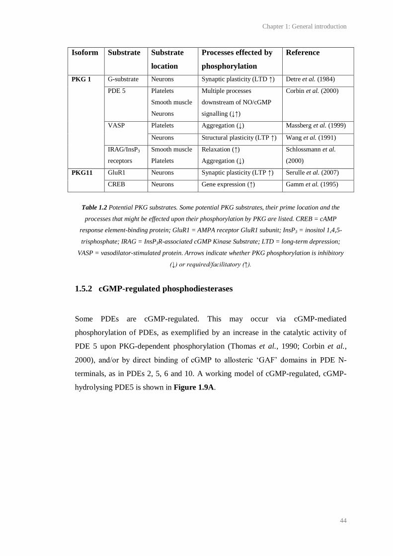
Chapter 1: General introduction
44
Table 1.2 Potential PKG substrates. Some potential PKG substrates, their prime location and the
processes that might be effected upon their phosphorylation by PKG are listed. CREB = cAMP
response element-binding protein; GluR1 = AMPA receptor GluR1 subunit; InsP3 = inositol 1,4,5-
trisphosphate; IRAG = InsP3R-associated cGMP Kinase Substrate; LTD = long-term depression;
VASP = vasodilator-stimulated protein. Arrows indicate whether PKG phosphorylation is inhibitory
(↓) or required/facilitatory (↑).
1.5.2 cGMP-regulated phosphodiesterases
Some PDEs are cGMP-regulated. This may occur via cGMP-mediated
phosphorylation of PDEs, as exemplified by an increase in the catalytic activity of
PDE 5 upon PKG-dependent phosphorylation (Thomas et al., 1990; Corbin et al.,
2000), and/or by direct binding of cGMP to allosteric ‘GAF’ domains in PDE N-
terminals, as in PDEs 2, 5, 6 and 10. A working model of cGMP-regulated, cGMP-
hydrolysing PDE5 is shown in Figure 1.9A.
Isoform Substrate Substrate
location
Processes effected by
phosphorylation
Reference
PKG 1 G-substrate Neurons Synaptic plasticity (LTD ↑) Detre et al. (1984)
PDE 5 Platelets
Smooth muscle
Neurons
Multiple processes
downstream of NO/cGMP
signalling (↓↑)
Corbin et al. (2000)
VASP Platelets Aggregation (↓) Massberg et al. (1999)
Neurons Structural plasticity (LTP ↑) Wang et al. (1991)
IRAG/InsP3
receptors
Smooth muscle
Platelets
Relaxation (↑)
Aggregation (↓)
Schlossmann et al.
(2000)
PKG11 GluR1 Neurons Synaptic plasticity (LTP ↑) Serulle et al. (2007)
CREB Neurons Gene expression (↑) Gamm et al. (1995)
Chapter 1: General introduction
45
Figure 1.9 Predicted structure of PDE 5. A) PDE 5 is predicted to be a homodimer, each monomer
comprising catalytic and regulatory domains. The catalytic site is found within the catalytic domain,
close to a Zn2+ and another divalent metal ion (perhaps Mg2+ or Mn2+) which facilitate the
polarisation of a hydroxyl ion from water for breaking the cyclic phosphate ring. Catalysis can be
enhanced by PKGI phosphorylation at serine-102 near the amino terminus, and/or by cGMP binding
to GAF-A. B) PDEs degrade the 3’ cyclic phosphate bond in cyclic nucleotides. Picture shows cAMP.
Adapted from Francis et al. (2010) and Bender and Beavo (2006). Reproduced by kind permission of
ASPET Journals.
‘GAF’ domains, named after an acronym of the first three classes of protein in which
they were discovered (cGMP-regulated PDEs, cyanobacterial adenylyl cyclase and
Escherichia coli transcription factor Fh1a), are common to proteins involved in
cyclic nucleotide signalling. Two GAF domains (A and B) are found in the N-
terminal of cGMP-regulated PDEs 2, 5, 6, and 10. In different PDEs, cGMP may
selectively bind one or the other GAF domain, causing either an increase or decrease
in cyclic nucleotide hydrolysis (see Table 1.3). It is unclear why cGMP
preferentially binds one GAF domain over the other, and what the function(s) are of
GAF domains that do not bind cGMP. PDE 11, which is found in skeletal muscle,
prostate, kidney, liver, testes and pituitary glands, also contains GAF domains,
although one is truncated and the PDE’s activity is insensitive to cGMP (reviewed by
Bender and Beavo, 2006).
PDE activity not only regulates the NO-cGMP pathway, but may also facilitate
cross-talk between cAMP and cGMP-dependent signalling cascades. For example, it
has been advanced that a rise in intracellular cGMP may lead to the competitive
Chapter 1: General introduction
46
inhibition of cAMP hydrolysis by PDEs that are not substrate-selective, for example
PDE 1C. It has also been discovered that the hydrolysis of cAMP by PDE 3 is
inhibited by a rise in cGMP, presumably because the enzyme has high affinity for
cGMP but hydrolyses it slowly (Degerman et al., 1997). In human cardiac myocytes,
cGMP-mediated inhibition of PDE 3 is thought to occur downstream of NO, leading
to cAMP accumulation, PKA activation and a phosphorylation-dependent increase in
the activity of L-type VGCCs, which are critical to cardiac function (Kirstein et al.,
1995). The NO-cGMP-PDE 3 pathway has also been implicated in the regulation of
other channels required for cardiac function, such as hyperpolarisation-activated,
cyclic nucleotide-gated (HCN) channels, and is thought to act in concert with other
cGMP-regulated PDEs, including cGMP-activated PDE 2 (Fischmeister et al., 2005).
Outside cardiac myocytes, a cGMP-induced increase in cAMP hydrolysis by PDE 2
has been implicated in platelet aggregation and hormone secretion from the adrenal
gland (reviewed by Bender and Beavo, 2006). Other processes, including
neurodevelopment and synaptic plasticity, might also be affected by PDE-mediated
cAMP and cGMP cross-talk, since these nucleotides often act in parallel signalling
pathways during these phenomena.
Given the above, the diversity in PDE tissue distribution, substrate selectivity and
kinetics (summarised in Table 1.3), as well as the intracellular compartmentalisation
of PDEs by their association with kinases and scaffolding proteins, PDE activity is
likely to have huge consequences for the diversity of cGMP signals that could be
elicited by one NO signal in one cell (reviewed by Fischmeister et al., 2005; Bender
and Beavo, 2006; Francis et al., 2010).
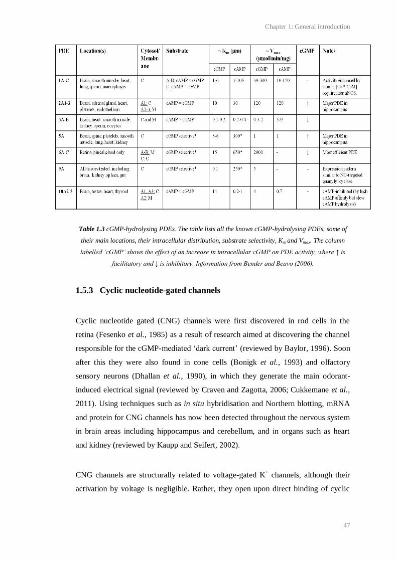
Chapter 1: General introduction
47
Table 1.3 cGMP-hydrolysing PDEs. The table lists all the known cGMP-hydrolysing PDEs, some of
their main locations, their intracellular distribution, substrate selectivity, Km and Vmax. The column
labelled ‘cGMP’ shows the effect of an increase in intracellular cGMP on PDE activity, where ↑ is
facilitatory and ↓ is inhibitory. Information from Bender and Beavo (2006).
1.5.3 Cyclic nucleotide-gated channels
Cyclic nucleotide gated (CNG) channels were first discovered in rod cells in the
retina (Fesenko et al., 1985) as a result of research aimed at discovering the channel
responsible for the cGMP-mediated ‘dark current’ (reviewed by Baylor, 1996). Soon
after this they were also found in cone cells (Bonigk et al., 1993) and olfactory
sensory neurons (Dhallan et al., 1990), in which they generate the main odorant-
induced electrical signal (reviewed by Craven and Zagotta, 2006; Cukkemane et al.,
2011). Using techniques such as in situ hybridisation and Northern blotting, mRNA
and protein for CNG channels has now been detected throughout the nervous system
in brain areas including hippocampus and cerebellum, and in organs such as heart
and kidney (reviewed by Kaupp and Seifert, 2002).
CNG channels are structurally related to voltage-gated K+ channels, although their
activation by voltage is negligible. Rather, they open upon direct binding of cyclic
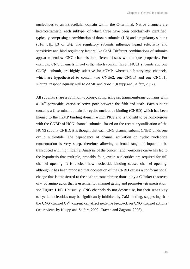
Chapter 1: General introduction
48
nucleotides to an intracellular domain within the C-terminal. Native channels are
heterotetrameric, each subtype, of which three have been conclusively identified,
typically comprising a combination of three α subunits (1-3) and a regulatory subunit
(β1α, β1β, β3 or α4). The regulatory subunits influence ligand selectivity and
sensitivity and bind regulatory factors like CaM. Different combinations of subunits
appear to endow CNG channels in different tissues with unique properties. For
example, CNG channels in rod cells, which contain three CNGα1 subunits and one
CNGβ1 subunit, are highly selective for cGMP, whereas olfactory-type channels,
which are hypothesised to contain two CNGα2, one CNGα4 and one CNGβ1β
subunit, respond equally well to cAMP and cGMP (Kaupp and Seifert, 2002).
All subunits share a common topology, comprising six transmembrane domains with
a Ca2+
-permeable, cation selective pore between the fifth and sixth. Each subunit
contains a C-terminal domain for cyclic nucleotide binding (CNBD) which has been
likened to the cGMP binding domain within PKG and is thought to be homologous
with the CNBD of HCN channel subunits. Based on the recent crystallisation of the
HCN2 subunit CNBD, it is thought that each CNG channel subunit CNBD binds one
cyclic nucleotide. The dependence of channel activation on cyclic nucleotide
concentration is very steep, therefore allowing a broad range of inputs to be
transduced with high fidelity. Analysis of the concentration-response curve has led to
the hypothesis that multiple, probably four, cyclic nucleotides are required for full
channel opening. It is unclear how nucleotide binding causes channel opening,
although it has been proposed that occupation of the CNBD causes a conformational
change that is transferred to the sixth transmembrane domain by a C-linker (a stretch
of ~ 80 amino acids that is essential for channel gating and promotes tetramerisation;
see Figure 1.10). Unusually, CNG channels do not desensitise, but their sensitivity
to cyclic nucleotides may be significantly inhibited by CaM binding, suggesting that
the CNG channel Ca2+
current can affect negative feedback on CNG channel activity
(see reviews by Kaupp and Seifert, 2002; Craven and Zagotta, 2006).
Chapter 1: General introduction
49
Figure 1.10 Predicted topology of CNG and HCN channel subunits. Each subunit constitutes six
transmembrane domains (S1–6), a pore loop between S5-6, and a cyclic nucleotide‐binding domain
(CNBD) in the C-terminal connected to S6 by a C-linker (*). CNG channels conduct Ca2+ and Na+.
HCN channels conduct Na+ and K+. It is unclear whether they conduct Ca2+. CNG channels are
activated upon binding of either cAMP or cGMP to the CNBD, depending on the channel type. HCN
channels are activated upon membrane hyperpolarisation which causes the movement of positively
charged residues (+) in S4 and the opening of the pore. Binding of cAMP or cGMP to the CNBD
causes a depolarising shift in voltage dependence.
The ability of CNG channels to directly translate NO/cGMP signals into rises in
intracellular Ca2+
has generated intense interest in their physiology, although many
aspects of CNG function remain unclear. Among the processes in which the
NO/cGMP/CNG pathway has been implicated are axonal guidance in chick retina
during development (Wu et al., 1994), regulation of membrane potential (Vm) and
activity-dependent conductance in frog and rat olfactory neurons (Schmachtenberg et
al., 2003), neurotransmitter release from lizard cones (Savchenko et al., 1997) and
synaptic plasticity (LTP) at CA1 synapses in mouse hippocampal slices (Parent et
al., 1998).
1.5.4 Hyperpolarisation-activated, cyclic nucleotide-regulated channels
HCN channels were first discovered in sinoatrial node cells in the heart and then in
neurons, including hippocampal neurons, in the late 1970s and early 1980s. They
have now been detected throughout the rodent central nervous system, including in
brainstem, retina, olfactory bulbs, cerebellum and spinal cord (Notomi and
1 2 3
++4++
5 6
N
C
1 2 3 4 5 6
N
C
Ca2+ Na+ Na+ K+
CNBD CNBD
Extracellular
Intracellular
CNG HCN
cAMP cAMPcGMP cGMP?
* *
Chapter 1: General introduction
50
Shigemoto, 2004; Milligan et al., 2006). They are closely phylogenetically related to
CNG channels and are similar in structure, being tetramers comprised of varying
(unknown) combinations of four different subunits (1-4), each including six
transmembrane domains and an intracellular CNBD (Figure 1.10). Unlike CNG
channels, they are primarily voltage-gated. As in voltage-gated K+ channels,
positively charged residues in the fourth transmembrane domain of each subunit
serve as a voltage sensor which move inwards during hyperpolarisation. Unusually,
this sensor, by some unknown downstream mechanism, elicits channel opening in
response to hyperpolarisation (at potentials negative to -50 to -60 mV (resting Vm)).
Channel opening leads to an inward Na+
current known as Ih (hyperpolarisation), Iq
(queer), or If (funny) and depolarisation of the membrane towards the action potential
threshold. Consequently, HCN channels have been linked to the generation of
oscillatory activity in excitable cells, most notably in sinoatrial node cells, which are
pacemakers for heart rate, and thalamocortical neurons, which generate rhythmic
‘burst’ activity during non-rapid eye movement sleep (see Figure 1.11; reviewed by
Craven and Zagotta, 2006).
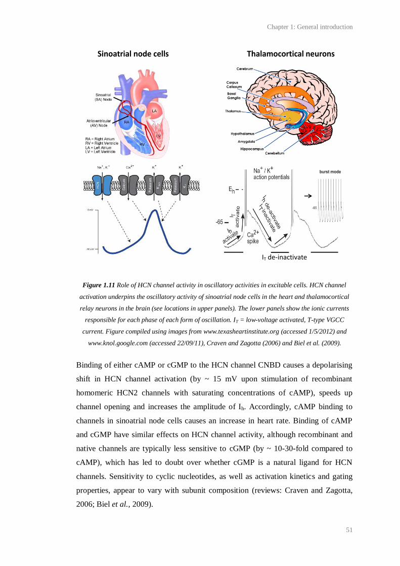
Chapter 1: General introduction
51
Figure 1.11 Role of HCN channel activity in oscillatory activities in excitable cells. HCN channel
activation underpins the oscillatory activity of sinoatrial node cells in the heart and thalamocortical
relay neurons in the brain (see locations in upper panels). The lower panels show the ionic currents
responsible for each phase of each form of oscillation. IT = low-voltage activated, T-type VGCC
current. Figure compiled using images from www.texasheartinstitute.org (accessed 1/5/2012) and
www.knol.google.com (accessed 22/09/11), Craven and Zagotta (2006) and Biel et al. (2009).
Binding of either cAMP or cGMP to the HCN channel CNBD causes a depolarising
shift in HCN channel activation (by ~ 15 mV upon stimulation of recombinant
homomeric HCN2 channels with saturating concentrations of cAMP), speeds up
channel opening and increases the amplitude of Ih. Accordingly, cAMP binding to
channels in sinoatrial node cells causes an increase in heart rate. Binding of cAMP
and cGMP have similar effects on HCN channel activity, although recombinant and
native channels are typically less sensitive to cGMP (by ~ 10-30-fold compared to
cAMP), which has led to doubt over whether cGMP is a natural ligand for HCN
channels. Sensitivity to cyclic nucleotides, as well as activation kinetics and gating
properties, appear to vary with subunit composition (reviews: Craven and Zagotta,
2006; Biel et al., 2009).
Sinoatrial node cells Thalamocortical neurons
IT de-inactivate

Chapter 1: General introduction
52
The unusual properties of Ih have implicated HCN channels in the setting of resting
Vm, which will be subject to a depolarising shift in the presence of HCN channels,
and the regulation of resting membrane resistance, which will be lowered in the
presence of Ih. Accordingly, Ohm’s law dictates that an input current (either hyper-
or depolarising) will evoke a smaller change in Vm when Ih is active. This will lead to
the stabilisation of resting Vm under basal conditions and an increase in the
amplitude attenuation of excitatory postsynaptic potentials (EPSPs) as they travel to
the soma. Therefore, Ih is thought to be involved in dendritic integration and EPSP
summation. Through the above processes, Ih has been found to regulate
neurotransmission and synaptic plasticity in multiple areas of the nervous system, in
particular the hippocampus (Biel et al., 2009).
HCN channels have only recently been identified as potential targets of NO/cGMP.
Some of the first research in support of this was performed in guinea pig and cat
thalamocortical neurons. Pape and Mager (1992) found that the depolarisation of
resting Vm via the NO/cGMP/HCN channel pathway causes a reduction in bursting
by limiting the de-inactivation of T-type VGCCs and consequent rebound
depolarisation. In this way, NO/cGMP has been hypothesised to regulate the switch
in thalamocortical neurons from bursting behaviour during non-rapid eye movement
sleep to ‘single spike’ mode which prevails during rapid eye movement sleep and
wakefulness.
More recently, the NO/cGMP/HCN channel has been found to regulate the
excitability of neurons in the spine (Kim et al., 2005), optic nerve (Garthwaite et al.,
2006) and hippocampus (Neitz et al., 2011). Depolarisation of spinal neurons by
HCN channels has been postulated to underlie central sensitisation (Kim et al.,
2005), a process likened to LTP and a correlate of chronic pain (reviewed by Ji et al.,
2003). In the hippocampus, the modulation of HCN channels by endothelium-
derived NO is thought to set a basal level of neurotransmitter release at CA1
synapses (Neitz et al., 2011).
Several aspects of NO/cGMP/HCN signalling remain ambiguous and more work will
be needed to elucidate its impact on physiology. Given the apparent insensitivity of
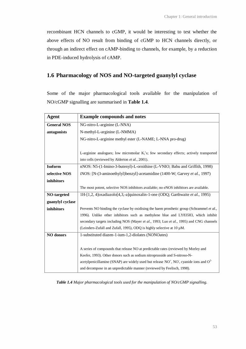
Chapter 1: General introduction
53
recombinant HCN channels to cGMP, it would be interesting to test whether the
above effects of NO result from binding of cGMP to HCN channels directly, or
through an indirect effect on cAMP-binding to channels, for example, by a reduction
in PDE-induced hydrolysis of cAMP.
1.6 Pharmacology of NOS and NO-targeted guanylyl cyclase
Some of the major pharmacological tools available for the manipulation of
NO/cGMP signalling are summarised in Table 1.4.
Agent Example compounds and notes
General NOS
antagonists
NG-nitro-L-arginine (L-NNA)
N-methyl-L-arginine (L-NMMA)
NG-nitro-L-arginine methyl ester (L-NAME; L-NNA pro-drug)
L-arginine analogues; low micromolar Ki’s; few secondary effects; actively transported
into cells (reviewed by Alderton et al., 2001).
Isoform
selective NOS
inhibitors
nNOS: N5-(1-Imino-3-butenyl)-L-ornithine (L-VNIO; Babu and Griffith, 1998)
iNOS: [N-(3-aminoethylyl)benzyl]-acetamidine (1400-W; Garvey et al., 1997)
The most potent, selective NOS inhibitors available; no eNOS inhibitors are available.
NO-targeted
guanylyl cyclase
inhibitors
1H-[1,2, 4]oxadiazolo[4,3,-a]quinoxalin-1-one (ODQ; Garthwaite et al., 1995)
Prevents NO binding the cyclase by oxidising the haem prosthetic group (Schrammel et al.,
1996). Unlike other inhibitors such as methylene blue and LY83583, which inhibit
secondary targets including NOS (Mayer et al., 1993; Luo et al., 1995) and CNG channels
(Leinders-Zufall and Zufall, 1995), ODQ is highly selective at 10 µM.
NO donors 1-substituted diazen-1-ium-1,2-diolates (NONOates)
A series of compounds that release NO at predictable rates (reviewed by Morley and
Keefer, 1993). Other donors such as sodium nitroprusside and S-nitroso-N-
acetylpenicillamine (SNAP) are widely used but release NO+, NO-, cyanide ions and O2-
and decompose in an unpredictable manner (reviewed by Feelisch, 1998).
Table 1.4 Major pharmacological tools used for the manipulation of NO/cGMP signalling.
Chapter 1: General introduction
54
1.7 Endogenous activators of NO-targeted guanylyl cyclase other
than NO
Carbon monoxide (CO) has been postulated by several researchers to activate NO-
targeted guanylyl cyclase. Like NO, CO is short-lived, freely-diffusible and
endogenously produced. The enzyme responsible for its synthesis, haem oxygenase,
is primarily responsible for degrading haem in old erythrocytes (Dawson and Snyder,
1994). In situ hybridisation has shown that mRNA for haem oxygenase is present
throughout the CNS in a distribution complementary to that of NO-targeted guanylyl
cyclase (Verma et al., 1993).
Endogenous CO signalling has been implicated in various processes ranging from
olfaction (Verma et al., 1993), to LTP at hippocampal synapses (Zhuo et al., 1993),
to NANC transmission and smooth muscle relaxation (Xue et al., 2000). Many
studies, mostly performed on the enteric nervous system, suggest that CO and NO
are co-transmitters.
However, unlike mice lacking NOS, mice deficient in haem oxygenase display no
gross behavioural abnormalities and have normal whole brain cGMP levels
(reviewed by Boehning and Snyder, 2003). Some of the specific effects of CO on
physiology are also controversial. For example, it has been reported that LTP is
normal in mice lacking haem oxygenase, and that supposed haem oxygenase
inhibitors, though widely used, inhibit LTP in wild-type and haem oxygenase-
deficient mice to a similar extent (Poss et al., 1995). Furthermore, serious doubts
over whether CO is a physiological activator of guanylyl cyclase have been raised by
the finding that millimolar concentrations of CO yield only a four-fold increase in the
activity of purified NO-targeted guanylyl cyclase (Brune and Ullrich, 1987). The
poor sensitivity of NO-targeted guanylyl cyclase to CO may be explained by the
finding that CO binding to the cyclase fails to break the bond between His-105 and
the haem prosthetic group (Stone and Marletta, 1994). Additionally, doubts over the
availability of the haem required for CO synthesis, and the apparent lack of
regulation of haem oxygenase (reviewed by Boehning and Snyder, 2003) have led to
Chapter 1: General introduction
55
a general consensus that CO is not a physiologically relevant activator of NO-
targeted guanylyl cyclase.
1.8 NO-targeted guanylyl cyclase-independent NO signal
transduction
Some examples of presumed physiological NO signal transduction independent of
NO-targeted guanylyl cyclase exist. For example, the heterosynaptic spread of LTP
at synapses between rat cerebellar parallel fibres and Purkinje cells reported by
Jacoby et al. (2001) was found to be prevented by NOS inhibition and NO
scavenging but not by a concentration of the NO-targeted guanylyl cyclase
antagonist, ODQ (5 µM), that was effective in blocking cerebellar LTD.
It has been hypothesised that effects of NO independent of NO-targeted guanylyl
cyclase might be transduced by the S-nitrosation of cysteine thiol (R-SH) groups. S-
nitrosation can be evoked using high concentrations of NO that can react with
oxygen to produce nitrosating species such as N2O3. Changes in the function of
proteins upon S-nitrosation are thought to result from changes in their tertiary
structure, which is in part determined by the location of cysteine thiols (Dudzinski et
al., 2006). S-nitrosation was exemplified by the negative feedback of NO on synaptic
NMDA receptors reported by Lipton et al. (2002), who proposed that it occurred via
S-nitrosation of NMDA receptor cysteine residues. However, this was later found to
be an artefact of un-caging supra-physiological concentrations of exogenous NO
using UV light, which may have led to the spurious generation of nitrosating species
(Hopper et al., 2004). Perhaps in part because of the difficulty of studying nitrosated
proteins, which are very unstable (Dudzinski et al., 2006), there appear to be no
unambiguous examples of physiological NO signalling via S-nitrosation, or by a
related process called S-nitrosylation. Moreover, the requirement for high NO
concentrations means that, in vivo, S-nitrosation and S-nitrosylation are likely to be
restricted to certain pathological conditions involving raised NO concentrations, for
example, following iNOS activation (Zhang and Hogg, 2005).
Chapter 1: General introduction
56
Mitochondrial cytochrome C oxidase, which is the last enzyme in the respiratory
electron transport chain, has also been suggested as an alternative target for NO,
leading to speculation that NO might, by competing with O2 for binding, inhibit
mitochondrial respiration (Erusalimsky and Moncada, 2007). An example of this in
normal physiology is found in the firefly, in which phasic bursts of NO generated in
the insect’s lantern may transiently inhibit mitochondrial respiration, therefore
allowing free oxygen to accumulate to a level necessary for the initiation of the
biochemical cascade underlying the characteristic firefly flash (Dudzinski et al.,
2006). However, in mammals at physiological O2 concentrations (20-30 µM), the
EC50 of cytochrome oxidase C for NO (~ 120 nm) is significantly greater than even
generous estimates of physiological NO concentrations, which are probably within
the low picomolar range (see 1.4.1). Furthermore, endogenous NO is unlikely to
exceed a few nanomolar following even intense stimulation of NOS (reviewed by
Hall and Garthwaite, 2009). Thus, mitochondrial inhibition by NO is also likely to be
restricted to conditions involving supra-physiological concentrations of NO, and/or
very low concentrations of O2. As such, NO-targeted guanylyl cyclase is the only
recognised physiological NO receptor.
1.9 NO signalling in brain
1.9.1 NO-mediated neurotransmission and modulation of cell excitability
NO acts as a neurotransmitter in the brain (Garthwaite, 2008). As in the PNS,
anterograde neurotransmission may occur upon postsynaptic nNOS activation in
response to action potential-dependent, intra-axonal Ca2+
influx via N-type VGCCs
and/or other Ca2+
channels (reviewed by Vincent, 2010). Anterograde
neurotransmission by NO has been exemplified at a synapse between two identifiable
neurons of the buccal ganglia of the pond snail, Lymnaea stagnalis. The reliance of
this synapse on NO as a neurotransmitter means that, when the participating neurons
are co-cultured, depolarisation of one can cause an EPSP in the other, regardless of a
physical connection (Park et al., 1998).
Chapter 1: General introduction
57
Retrograde NO transmission may occur following the opening of postsynaptic
NMDA receptors and has been exemplified by Arancio et al.. They have shown that,
during LTP induction at synapses between dissociated hippocampal neurons, NMDA
receptor activation leads to postsynaptic NO generation and a subsequent increase in
neurotransmitter release through presynaptic NO-targeted guanylyl cyclase (Arancio
et al., 1995; Arancio et al., 1996; Arancio et al., 2001). The relevance of this to intact
tissues remains unclear (see Chapter 3).
NO may also modulate neurotransmission by other molecules. Studies,
predominantly of neurotransmitter efflux from tissue preparations in vitro, have
implicated tonic and activity-dependent NO production in the regulation of the
release of neurotransmitters including ACh, noradrenaline, dopamine, glutamate and
GABA from brain areas as diverse as cortex, striatum, hypothalamus and
hippocampus (see Chapter 4 for examples). Interestingly, the release of one
neurotransmitter may be both up- and down-regulated by NO depending on the
concentration of NO involved and the tissue under study (reviewed by Prast and
Philippu, 2001).
Most of these effects appear to be underpinned by NO-mediated changes in
membrane excitability. Common targets for NO are K+ and Ca
2+ channels (reviewed
by Garthwaite, 2008). Other targets include, for example, GABAA receptors, as
illustrated in dissociated cerebellar granule cells by Robello et al. (1996), serotonin
receptors, as illustrated using invertebrate neurons (Straub et al., 2007), and the
serotonin uptake transporter (reviewed by Garthwaite, 2007). Several effects appear
to involve PKG-mediated phosphorylation. As well as an increase or decrease in cell
excitability caused by NO, studies, for example, using hippocampal slices (Makara et
al., 2007) and in the hippocampus in vivo (Hada et al., 2003), suggest that NO can
cause the disinhibition of synaptic activity via a reduction of GABAergic
transmission.
Chapter 1: General introduction
58
1.9.2 NO signalling between blood vessels and neurons
In the brain, NO may signal from central neurons to blood vessels, and this may link
local synaptic activity with vasodilation. For example, application of NMDA to
hippocampal slices has been reported to cause NO-dependent vasodilation of
microvessels (Lovick et al., 1999). Similar effects in other brain areas such as
cerebellum and cortex (reviewed by Garthwaite, 2008) have been found to be
sensitive to inhibition by the Na+
channel-inhibitor, tetrodotoxin (TTX), suggesting
that the underlying mechanism is action potential-dependent.
Interestingly, NO also signals from blood vessels to neurons. Convincing evidence
for vasculoneuronal NO transmission has been provided using optic nerves, which
contain only blood vessels and axons and no nNOS. It has been found that a low-
level NO tone produced by capillary eNOS causes tonic depolarisation of axons via a
pathway involving HCN channels (Garthwaite et al., 2006). A low-level (~ 0.1 nM),
endothelium-derived NO tone has also been discovered in the hippocampus (Hopper
and Garthwaite, 2006), and eNOS appears to be necessary for NO-dependent LTP at
CA1 (Kantor et al., 1996; Wilson et al., 1999; Bon and Garthwaite, 2003; Hopper
and Garthwaite, 2006) and mossy fibre (Doreulee et al., 2001) synapses. Mice
deficient in eNOS also exhibit impaired LTP in the neocortex (Haul et al., 1999) and
striatum (Doreulee et al., 2003), suggesting that vasculoneuronal NO signals are
active in multiple brain areas. In vivo, this form of NO signal may enable blood-
borne agents to influence neuronal activity.
1.10 NO and synaptic plasticity in adults
1.10.1 Synaptic plasticity
The term synaptic plasticity was coined by Jerzy Konorski in 1948 to describe the
ability of neurons to change the strength of their connections in response to activity.
He, along with many scientists, assumed synaptic plasticity to be critical for
information storage in the brain. This idea was first proposed by Santiago Ramon y
Chapter 1: General introduction
59
Cajal in 1894 but it was not until 1949, that this postulate was formalised into a
hypothesis (see Andersen et al., 2007 for a review). At this time, Donald Hebb
proposed that if two connected neurons are simultaneously and repeatedly active,
then the efficacy of the synapse involved will increase (Hebb, 1949).
1.10.2 Long-term potentiation
Hebb’s hypothesis that ‘neurons that fire together, wire together’ attracted much
attention as a putative explanation for learning and memory, but it was not until 1973
that Hebbian plasticity was first described. In a ground-breaking report, Tim Bliss
and Terje Lomo (1973) demonstrated a persistent (lasting hours) enhancement in
synaptic activity following a brief tetanus (100 or 15 Hz for 3-20 s) applied to
hippocampal perforant path-granule cell synapses in anaesthetised rabbits. Consistent
with Hebb’s postulate, this long-lasting potentiation, later renamed LTP, was found
to be specific to the pathway that was tetanised (Andersen et al., 1977), to depend
upon coincident pre- and postsynaptic depolarisation that exceeded a threshold for
LTP induction (McNaughton et al., 1978; Wigstrom et al., 1986), and to persist over
hours or days in vitro (Bliss and Gardner-Medwin, 1973) and up to a year in vivo
(Abraham et al., 2002). These properties of LTP have spurred its study at synapses
throughout the brain, in areas such as the amygdala (Dityatev and Bolshakov, 2005)
and cortex (Feldman, 2009), and in several species including humans (Cooke and
Bliss, 2006). Now, the term ‘LTP’ has come to describe any long-lasting (> 1 hr),
activity-dependent increase in the efficacy of a synapse. It is recognised that the
potentiation can be composed of multiple pre- and postsynaptic expression
mechanisms including, amongst others, increased neurotransmitter release, increased
conductance via excitatory postsynaptic receptors, increased postsynaptic receptor
density, a change in gene expression and/or the structural remodelling or growth of
new synapses (Lynch, 2004; Malenka and Bear, 2004; Bliss et al., 2007). The
recruitment of specific expression mechanisms appears to depend upon the synapse
under study, the animal used, and the conditions of the experiment, most notably the
induction protocol used, of which there are multiple electrical and chemical
procedures (reviewed by Bliss et al., 2007).

Chapter 1: General introduction
60
It should be noted that the relationship between LTP, learning and memory is
debatable (see 1.11). Additionally, the study of LTP in certain areas of the nervous
system has led to hypotheses that it represents a physiological correlate of various
other phenomena, for example, chronic pain caused by the central sensitisation of
noiceceptive synapses in the spine (Ji et al., 2003) and addiction caused by the
potentiation of dopaminergic synapses in the ventral tegmental area (Wolf, 2003).
Similarities between the mechanisms underpinning LTP and forms of activity-
dependent synaptic plasticity thought to occur during the development and
refinement of synapses have also been recognised (reviewed by Kandel and O'Dell,
1992; Contestabile, 2000).
In mammals, LTP is archetypal at hippocampal Schaffer-collateral/commissural CA1
synapses. The easy study of LTP at these synapses has been permitted by the use of
transverse hippocampal slices. Slicing the hippocampus along the transverse plane
reveals a laminar structure and allows for the preservation of all the major neural
pathways which can be maintained in vitro for hours, easily manipulated and
recorded from (reviewed by Teyler, 1999). At Schaffer-collateral/commissural-CA1
synapses, low frequency synaptic transmission is largely mediated by AMPA-type
glutamate receptor activation (Davies and Collingridge, 1989). LTP is typically
induced using high frequency stimulation (HFS; a 1 s, 100 Hz burst of stimuli or
tetanus), although it has been reported following multiple other stimuli, as well as
after some forms of learning (see 1.11.2). In all cases tested, LTP has been found to
depend upon postsynaptic Ca2+
(Lynch et al., 1983; Malenka et al., 1988) and with
few exceptions (see Chapter 5 for discussion), NMDA receptor activation
(Collingridge et al., 1983a; Collingridge et al., 1983b; Malenka, 1991; Tsien et al.,
1996). NMDA receptor activation is glutamate- and voltage-dependent, the channel
being blocked by Mg2+
close to the resting Vm. During HFS, depolarisation of the
postsynaptic membrane, largely due to AMPA receptor activation, summates. The
NMDA receptor channels are relieved of Mg2+
and permit Ca2+
influx which initiates
LTP expression (Figure 1.10). In this way, NMDA-receptors act as molecular
coincidence detectors for simultaneous presynaptic (glutamate release) and
postsynaptic (depolarisation) activity, thus explaining why NMDA receptor-
dependent LTP is input-specific and associative (reviewed by Bliss et al., 2007). In

Chapter 1: General introduction
61
concurrence with NMDA receptor activation, multiple other means of raising the
intracellular Ca2+
concentration may be necessary for LTP induction, including the
activation of metabotropic glutamate receptors, VGCCs and intracellular Ca2+
stores
(Lynch, 2004; Bliss et al., 2007).
The rise in intracellular Ca2+
caused by the activation of NMDA receptors and other
channels during LTP induction initiates multiple signalling cascades responsible for
the persistent amplification of subsequent postsynaptic responses. Details of these
cascades remain largely unclear, although two stages of LTP expression, early (early-
LTP; > 1 hr post-induction) and late (late-LTP; usually > 4 hr post induction), have
been distinguished by their dependency on new protein synthesis, with late-LTP
relying on transcription and translation (Lynch, 2004; Malenka and Bear, 2004; Bliss
et al., 2007). It is also generally agreed that both sides of the synapse are involved in
LTP expression, yet the conditions that dictate the extent that each side contributes
and at what time point following induction remain ambiguous. Many recent studies
have focused on the mechanisms responsible for increases in postsynaptic AMPA
receptor density often observed following LTP induction (Nicoll, 2003; Malenka and
Bear, 2004; Kerchner and Nicoll, 2008; Kessels and Malinow, 2009). In presynaptic
neurons, increases in glutamate release have also been detected using quantal
analysis (Dolphin et al., 1982; Bekkers and Stevens, 1990) and, more recently, direct
imaging techniques (Zakharenko et al., 2001). Indeed, a recent study by Enoki et al.
(2009) showed that LTP at Schaffer-collateral/commissural-CA1 synapses was,
under their conditions, almost entirely presynaptically expressed.
Given that NMDA receptor-dependent LTP is induced postsynaptically, presynaptic
LTP mechanisms necessitate a retrograde messenger, capable of relaying a signal for
LTP expression from the postsynaptic induction site back to the presynaptic neuron.
Several freely diffusible, as well as membrane spanning, molecules have been
suggested as candidate retrograde messengers, including arachidonic acid (O'Dell et
al., 1991), carbon monoxide (Zhuo et al., 1993) and cell adhesion molecules (Bliss et
al., 2007). However, NO has received the most attention.
Chapter 1: General introduction
62
1.10.3 LTP and NO
The first studies to implicate NO as an intercellular, possibly retrograde, transmitter
in LTP showed a deficit in the maintenance of CA1 LTP in hippocampal slices
following the application of non-selective NOS inhibitors or of the extracellular NO
scavenger, haemoglobin, during LTP induction (Schuman and Madison, 1991;
Bohme et al., 1991). Subsequently guanylyl cyclase inhibitors were found to have
similar effects on LTP (Boulton et al., 1995; Lu et al., 1999; Bon and Garthwaite,
2003). It is now generally accepted that NO/cGMP signalling may participate in LTP
in several brain areas, possibly contributing to some forms of memory (see 1.11.3). It
is also known that both a tonic (endothelial) and phasic (neuronal) NO signal are
required for NO-dependent CA1 LTP (Bon and Garthwaite, 2003; Hopper and
Garthwaite, 2006), consistent with reports that knock-out mice deficient in neuronal
and endothelial NOS are incapable of wild-type LTP (Son et al., 1996). Similarly,
LTP in the visual cortex (Haghikia et al., 2007) and hippocampus (Taqatqeh et al.,
2009) has also been shown to require both guanylyl cyclase isoforms, perhaps
implying the existence of distinct NO/cGMP-mediated pathways that contribute to
LTP. Numerous putative effectors of NO/cGMP-dependent LTP have been
identified, including PKG (Arancio et al., 2001; Serulle et al., 2007), CaMKII
(Ninan and Arancio, 2004), VASP (Wang et al., 2005), CREB (Lu et al., 1999),
PDE2 (Boess et al., 2004) and CNG channels (Parent et al., 1998). However, there
remain several unknowns regarding the precise role of NO/cGMP signalling in LTP.
Firstly, the conditions under which LTP becomes NO-dependent remain undefined.
At Schaffer-collateral/commissural-CA1 synapses, for example, LTP has been found
by several groups to be NO-independent in vitro (Cummings et al., 1994; Phillips et
al., 2008) and in vivo (Bannerman et al., 1994b). The involvement of NO in LTP
may be determined by the experimental conditions used. Specific factors may be the
LTP induction protocol used (Raymond, 2007), and/or the age or strain of animals
(Williams et al., 1993; Holscher, 2002). It should also be noted that the majority of
studies that do show a role for NO in LTP also show a residual NO-independent
component, consistent with the idea that LTP can be established by multiple,
independent mechanisms.
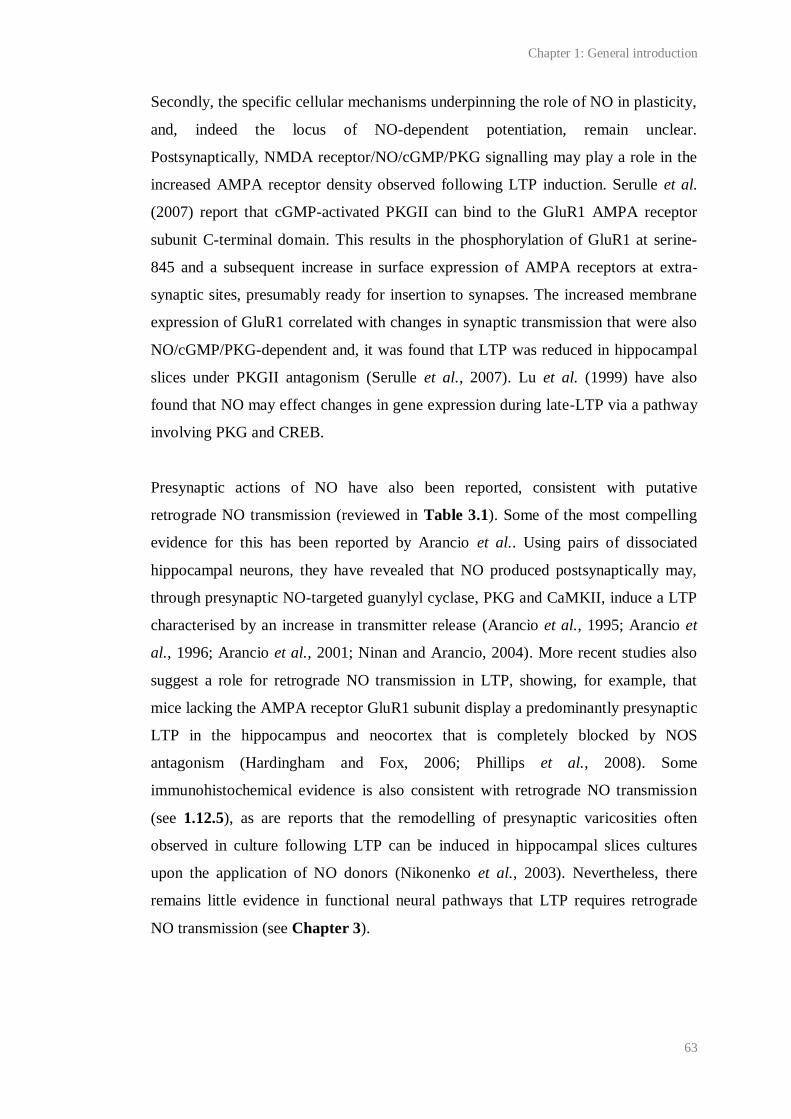
Chapter 1: General introduction
63
Secondly, the specific cellular mechanisms underpinning the role of NO in plasticity,
and, indeed the locus of NO-dependent potentiation, remain unclear.
Postsynaptically, NMDA receptor/NO/cGMP/PKG signalling may play a role in the
increased AMPA receptor density observed following LTP induction. Serulle et al.
(2007) report that cGMP-activated PKGII can bind to the GluR1 AMPA receptor
subunit C-terminal domain. This results in the phosphorylation of GluR1 at serine-
845 and a subsequent increase in surface expression of AMPA receptors at extra-
synaptic sites, presumably ready for insertion to synapses. The increased membrane
expression of GluR1 correlated with changes in synaptic transmission that were also
NO/cGMP/PKG-dependent and, it was found that LTP was reduced in hippocampal
slices under PKGII antagonism (Serulle et al., 2007). Lu et al. (1999) have also
found that NO may effect changes in gene expression during late-LTP via a pathway
involving PKG and CREB.
Presynaptic actions of NO have also been reported, consistent with putative
retrograde NO transmission (reviewed in Table 3.1). Some of the most compelling
evidence for this has been reported by Arancio et al.. Using pairs of dissociated
hippocampal neurons, they have revealed that NO produced postsynaptically may,
through presynaptic NO-targeted guanylyl cyclase, PKG and CaMKII, induce a LTP
characterised by an increase in transmitter release (Arancio et al., 1995; Arancio et
al., 1996; Arancio et al., 2001; Ninan and Arancio, 2004). More recent studies also
suggest a role for retrograde NO transmission in LTP, showing, for example, that
mice lacking the AMPA receptor GluR1 subunit display a predominantly presynaptic
LTP in the hippocampus and neocortex that is completely blocked by NOS
antagonism (Hardingham and Fox, 2006; Phillips et al., 2008). Some
immunohistochemical evidence is also consistent with retrograde NO transmission
(see 1.12.5), as are reports that the remodelling of presynaptic varicosities often
observed in culture following LTP can be induced in hippocampal slices cultures
upon the application of NO donors (Nikonenko et al., 2003). Nevertheless, there
remains little evidence in functional neural pathways that LTP requires retrograde
NO transmission (see Chapter 3).
Chapter 1: General introduction
64
Figure 1.12 NMDA receptor-dependent LTP induction and possible expression through NO. 1.
Glutamate released from the presynaptic terminal. 2. Postsynaptic AMPA receptors activate,
depolarising the cell. 3. Depolarisation summates sufficiently to relieve NMDA receptor channels of
Mg2. 4. NMDA receptors permit Ca2+ influx which activates nNOS and NO synthesis. 5. NO may
contribute to postsynaptic and/or presynaptic LTP mechanisms by activation of guanylyl cyclase and
cGMP accumulation. Presynaptic mechanisms require retrograde NO transmission (blue arrows).
1.10.4 Long-term depression and NO
Reciprocal in nature to LTP is LTD, a persistent decrease in synaptic efficacy that
can be induced by low frequency stimulation (for example, 100 stimuli at 1 Hz).
NMDA receptor-dependent LTD has been described in the hippocampus at CA1
synapses (Bliss et al., 2007), but this form of plasticity has been best characterised at
Purkinje cell synapses in the cerebellum. At these synapses, LTD is induced by the
repeated, low frequency activation of climbing fibre inputs, just after parallel fibre
inputs. It is hypothesised that the climbing fibre input acts as an error signal and
attenuates inappropriate parallel fibre input to Purkinje cells (Ito, 2001).
Interestingly, NO, produced in parallel fibres or interneurons upon NMDA receptor
activity, appears to play a critical role in cerebellar LTD (reviewed by Garthwaite,
2008). Studies have shown that NO causes an increase in cGMP in Purkinje cells,
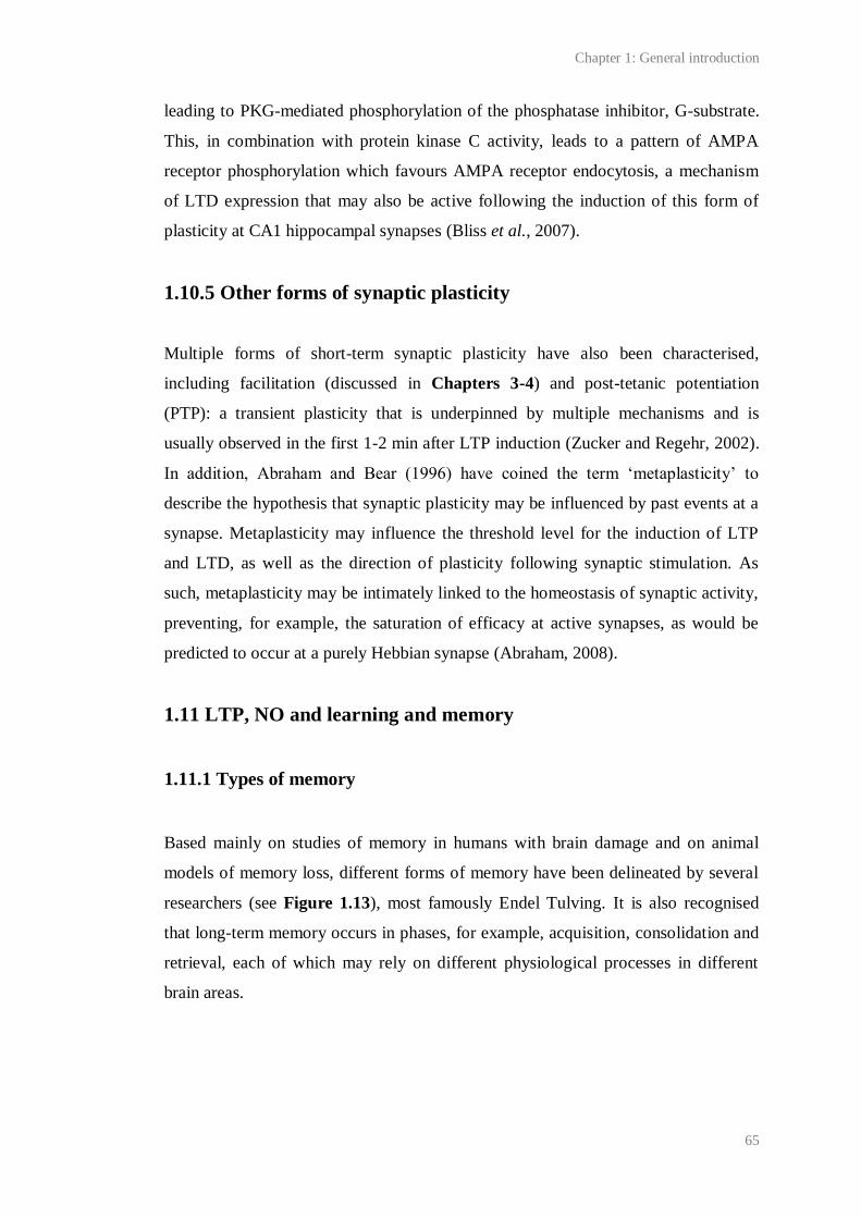
Chapter 1: General introduction
65
leading to PKG-mediated phosphorylation of the phosphatase inhibitor, G-substrate.
This, in combination with protein kinase C activity, leads to a pattern of AMPA
receptor phosphorylation which favours AMPA receptor endocytosis, a mechanism
of LTD expression that may also be active following the induction of this form of
plasticity at CA1 hippocampal synapses (Bliss et al., 2007).
1.10.5 Other forms of synaptic plasticity
Multiple forms of short-term synaptic plasticity have also been characterised,
including facilitation (discussed in Chapters 3-4) and post-tetanic potentiation
(PTP): a transient plasticity that is underpinned by multiple mechanisms and is
usually observed in the first 1-2 min after LTP induction (Zucker and Regehr, 2002).
In addition, Abraham and Bear (1996) have coined the term ‘metaplasticity’ to
describe the hypothesis that synaptic plasticity may be influenced by past events at a
synapse. Metaplasticity may influence the threshold level for the induction of LTP
and LTD, as well as the direction of plasticity following synaptic stimulation. As
such, metaplasticity may be intimately linked to the homeostasis of synaptic activity,
preventing, for example, the saturation of efficacy at active synapses, as would be
predicted to occur at a purely Hebbian synapse (Abraham, 2008).
1.11 LTP, NO and learning and memory
1.11.1 Types of memory
Based mainly on studies of memory in humans with brain damage and on animal
models of memory loss, different forms of memory have been delineated by several
researchers (see Figure 1.13), most famously Endel Tulving. It is also recognised
that long-term memory occurs in phases, for example, acquisition, consolidation and
retrieval, each of which may rely on different physiological processes in different
brain areas.
Chapter 1: General introduction
66
Figure 1.13 Current taxonomy of memory. Declarative memory is conscious. Non-declarative
memory is non-conscious. Episodic memory refers to memory for events. Semantic memory refers to
memory for facts. Taken from Bird and Burgess (2008).Adapted by kind permission of Nature
Publishing Group.
1.11.2 LTP and memory
The initial report of LTP by Bliss and Lomo (1973), coupled with studies showing
that it was input-specific, associative and persistent, provided the first demonstration
of a Hebbian synaptic plasticity. The Hebbian characteristics of LTP have led many
researchers to view LTP as a correlate of learning and memory, although this is
highly debated.
In 2000, Martin et al. formalised the ‘synaptic plasticity and memory’ (SPM)
hypothesis. This proposes that activity dependent synaptic plasticity occurs during
normal brain activity at synapses necessary for learning and memory, and is
necessary and sufficient for the storage of information. Martin et al. (2000) described
four criteria that must be met of a synaptic plasticity sufficient to explain memory.
Based on these criteria, Bliss et al. (2007) have posed four main questions that must
be answered to test whether LTP is a correlate of learning and memory: 1) are the
mechanisms underlying LTP correlated with those underlying learning and
memory?; 2) does learning induce LTP?; 3) do changes in synaptic weights
subsequent to learning cause forgetting?; 4) does LTP induction generate a memory
without animals having gone through learning?
Chapter 1: General introduction
67
A wealth of studies have been directed to answering the first question, and suggest
that, in general, the mechanisms underlying LTP of synapses are correlated with
those underlying learning and memory in tasks dependent on the same brain area.
Early studies showed that factors such as ageing and stress impact LTP and learning
and memory in a similar manner (reviewed by Lynch, 2004). Manipulations that
attenuate or augment LTP were also shown to similarly affect the ability of animals
to learn and remember some behavioural tasks. For example, Morris et al. (1986)
found that the NMDA antagonist, D/L-AP5, inhibited LTP at perforant path-granule
cell synapses in vivo and learning in the Morris water maze, a task that is
hippocampus-dependent (Morris et al., 1982). Morris et al.’s findings have since
been supported by numerous studies (for example, Tsien et al., 1996; Tang et al.,
1999). Additionally, other LTP ‘players’ appear to be required for learning and
memory (reviewed by Lynch, 2004). For example, Giese et al. (1998) have found
that, in accordance with multiple other reports (for example Silva et al., 1992a; Silva
et al., 1992b), mice lacking functional αCaMKII, a kinase heavily associated with
LTP (Lisman et al., 2002), display impaired hippocampal LTP and Morris water
maze learning. It should also be noted that a positive correlation between LTP and
learning and memory has been observed outside the hippocampus (reviewed by
Lynch, 2004). For example, in the amygdala, manipulations, including the blockade
of the NMDA/NO/cGMP pathway, that have been found to inhibit LTP at synapses
between the lateral amygdala and auditory thalamus have also been found to
attenuate auditory fear conditioning (for example, Bauer et al., 2002; Ota et al.,
2008), an amygdala-dependent task (Goosens and Maren, 2001)
In further support of the SPM hypothesis, and consistent with the second question (is
learning associated with the induction of LTP?), stimuli that enhance subsequent
learning, such as environmental enrichment and training in behavioural tasks, have
been found to enhance field EPSPs (fEPSPs), neurotransmitter release and cause
various biochemical changes associated with LTP, such as increased AMPA receptor
density at synapses, in brain areas including the visual cortex, motor cortex and
amygdala (reviewed by Lynch, 2004; Bliss et al., 2007). In the hippocampus, two
key studies have been performed by Gruart et al. (2006) and Whitlock et al. (2006)
who used arrays of electrodes implanted in area CA1 to measure synaptic efficacy

Chapter 1: General introduction
68
during learning. Gruart et al. (2006) found that, in mice during trace eye blink
conditioning, the amplitude of evoked fEPSPs steadily increased in a manner that
was NMDA receptor-dependent. Whitlock et al. (2006) found that at a minority of
electrodes, fEPSPs were significantly and persistently increased in rats following
one-trial inhibitory avoidance learning. Strikingly, this increase occluded subsequent
LTP.
Accordant with the third question (do changes in synaptic plasticity caused by LTP
induction cause forgetting, i.e. by altering synaptic weights?), Brun et al. (2001) have
found that high frequency stimulation applied to the dentate gyrus of rats through
implanted electrodes blocked prior memory for the Morris water maze (i.e. induced
retrograde amnesia). Interestingly, high frequency stimulation had no effect when
applied in the presence of a NMDA antagonist, 3-(2-carboxypiperazin-4-yl)propyl-1-
phosphonic acid, suggesting that the retrograde amnesia may have been caused by a
NMDA receptor-dependent change in synaptic weights.
These data are broadly in favour of the SPM hypothesis, although it should be noted
that there are several pieces of evidence that are inconsistent with the theory. For
example, whereas most forms of LTP are NMDA receptor-dependent, the NMDA
antagonist, D/L-AP5 seems only to attenuate spatial learning in task-naïve animals,
suggesting that the relationship between LTP and learning is not direct (Bannerman
et al., 1995; although see Chapter 5 for examples of NMDA receptor-independent
LTP). Additionally, some, but not all (Moser et al., 1998), studies have found no
effect of manipulations designed to saturate LTP on subsequent learning (Bliss and
Richter-Levin, 1993). These inconsistencies perhaps reflect the possibility that
learning and memory require the activity of multiple neural pathways involved in
networks spanning several brain areas. Indeed many researchers agree that the view
that ‘LTP equals memory’ is too simplistic (reviewed by Lynch et al., 1983; Bliss et
al., 2007).
The use of LTP induction protocols that are unlikely to represent natural patterns of
neuronal activity have also raised doubts over the relevance of the mechanisms
underlying LTP to learning, although induction protocols patterned after the theta
Chapter 1: General introduction
69
rhythm, which can be observed in the hippocampus using electroencephalography
during exploratory behaviour, or involving ‘primed bursts’ are efficient in generating
LTP (for example, Morgan and Teyler, 2001). The idea that synaptic plasticity, as
modelled by LTP, is involved in memory is further complicated by the fact that it has
not been possible to link LTP-like synaptic plasticity to a certain stage or type of
memory. The role of other forms of plasticity such as depotentiation and LTD are
also unclear, and a major outstanding question is whether synaptic plasticity in the
hippocampus must persist for as long as a memory.
Finally, it should be noted that synaptic plasticity has been proposed to better model
physiological processes that are related to, but distinct from learning and memory,
such as attention (Shors and Matzel, 1997), and that there have been no direct-tests
of fourth prediction of the SPM hypothesis (does LTP induction generate a memory
without animals having gone through learning?). Furthermore, it is unclear how
plasticity at an individual synapse might affect the activity of a network.
Nevertheless, studies, for example, those by Whitlock et al. (2006) and Gruart et al.
(2006) are compelling. Therefore, while it cannot be said that ‘LTP equal’s memory’,
or that LTP in the form induced in the laboratory exists naturally, the mechanisms
underlying LTP appear to be the best molecular correlates of learning and memory
currently amenable to study in the laboratory. Additionally, an appreciation of LTP
mechanisms could have wide implications for our understanding of all basic
principles of plastic synaptic transmission, especially if neurons make use of all the
means of synaptic plasticity available to them (Bliss et al., 2007).
1.11.3 NO and learning and memory
If LTP is a correlate of learning, and NO is required for some forms of LTP, then it is
predicted that NO is necessary for at least some forms of learning. In support of this
prediction, several studies suggest that a role for NO in learning and memory has
been evolutionarily conserved. In invertebrates such as the sea slug, Aplysia
californica, the pond snail, Lymnaea stagnalis, and honey bee, Apis mellifera, for
example, NO appears to be required for associational learning during classical
conditioning. In particular, tasks involving olfactory or appetitive cues seem
Chapter 1: General introduction
70
particularly sensitive to NOS inhibition (reviewed by Susswein et al., 2004). For
example, application of the NOS inhibitor, L-NAME, during and after aversive
appetitive conditioning in Aplysia has been found to block short- and long-term
memory for the conditioned stimulus. In contrast, L-NAME had no effect on
spontaneous feeding, and no effect when applied one min after training. The authors
therefore concluded that NO was required during the acquisition but not
consolidation of the memory (Katzoff et al., 2002).
In mammals, NOS and NO-targeted guanylyl cyclase inhibition has also been found
to attenuate learning during hippocampus-dependent tasks. For example,
intraperitoneal injection of the NOS inhibitor, L-NNA, was found by Bohme et al.
(1993) to block the ability of rats to learn in a radial arm maze (which is
hippocampus-associated) and a social recognition test involving olfactory memory.
CA1 LTP was blocked in slices from rats that received injections of the NOS
inhibitor. Similarly, Majlessi et al. (2008) have found that rats treated with L-NAME
via a cannula implanted near the CA1 region of the hippocampus were impaired in
the Morris water maze. Specifically, escape latency and travelled distance were
increased whereas the number of entries into the quadrant containing the platform
decreased. No effects on motivation or sensorimotor coordination were observed and
the inhibition could be reversed by the co-application of L-arginine, which may have
outcompeted L-NAME for binding to NOS, as has been shown to occur in
hippocampal slices (East and Garthwaite, 1991). In addition, 3-(5-hydroxymethyl-2-
furyl)-1-benzyl-indazole, a compound that sensitises NO-targeted guanylyl cyclase to
NO (Ko et al., 1994) and also blocks PDEs (Galle et al., 1999), has been found to
enhance CA1 LTP via a mechanism involving the NO/cGMP/PKG pathway and
shorten the escape latency of mice from the Morris water maze (Chien et al., 2003;
Chien et al., 2005).
It should be noted that some forms of learning and memory in tasks associated with
other brain areas also appear to be NOS-dependent, for example, NOS inhibition has
been found to impair cerebellum-dependent eye blink conditioning in rabbits
(Chapman et al., 1992) and amygdala-dependent place conditioning (Zarrindast et
al., 2002). ‘Natural’ forms of learning, such as olfactory learning of a ewe for her

Chapter 1: General introduction
71
lamb, have also been found to require NO/cGMP signalling (reviewed by Susswein
et al., 2004).
As with the role of NO in LTP, more work is necessary to elucidate the contribution
of NO to learning and memory. Reported effects of NO on learning and memory
phenomena are so diverse that NO has not been associated with a particular stage or
form of memory (reviewed by Susswein et al., 2004). Additionally, and consistent
with the role of NO in LTP, several groups have found no effect of NO on learning
in tasks which other groups have found to be NOS-dependent, for example, the
Morris water maze (Bannerman et al., 1994a; Blokland et al., 1999). The species and
strain of the animal under study, the behaviour being tested and crucially the training
paradigm may dictate whether NO is required. These factors also appear to regulate
the requirement of learning and memory during behavioural tasks for other LTP
‘players’. For example, the requirement of mice for functional αCaMKII to learn an
inhibitory avoidance task appears to be dependent on the number of training trials
given (Irvine et al., 2005).
1.11.4 The hippocampus and memory
As discussed above, the archetype of LTP occurs in the hippocampus. If LTP is a
correlate of learning and memory, then the SPM hypothesis dictates that the
hippocampus should be required for these phenomena. Consistent with this, theories
of hippocampal function have moved away from early hypotheses suggesting that it
is involved in olfaction, attention or emotion and towards a role in memory
(Andersen et al., 2007).
Probably the best known evidence for a role of the hippocampus in memory came
from a case study of a patient, named HM, by William Scoville and Brenda Milner
(1957). HM had ~ two thirds of his hippocampus, as well as other areas of the
hippocampal formation, removed in a surgery aimed to treat his epilepsy. The
operation successfully alleviated his condition, but at the expense of his ability to
form new long-term declarative memories. In accordance with the current taxonomy
of memory (Figure 1.11), HM’s short-term and non-declarative (for example
procedural or skills) memory remained intact (reviewed by Corkin, 2002 ).
Chapter 1: General introduction
72
Since Scoville and Milner’s study, multidisciplinary evidence has combined to assert
the consensus that the hippocampus is required for declarative memory. Several
theories of the specific role of the hippocampus have been developed. One of the
first, named ‘the declarative theory’, proposes that, in concert with other areas of the
medial temporal lobe, the hippocampus is required for the acquisition of all
declarative memory but that, after some period, these are consolidated to the
neocortex, explaining why old memories are often spared following hippocampal
damage (as in HM). More elaborate theories suggest that the hippocampus is also the
site for the long-term storage of episodic memories, and/or that the hippocampus is
involved in the acquisition of episodic, but not semantic, memory. Another set of
theories, for example, ‘the relational theory’, propose that, during acquisition and
recollection, the hippocampus allows the association of information, such as the
contextual details of an event, that are initially processed and later stored in different
neocortical areas.
An extension of the relational theory is ‘the cognitive-map theory’, which proposes
that the major role of the hippocampus is to construct and store an allocentric
representation of an environment in order to enable navigation through it. It is
thought that the cognitive map may arise via the acquisition, and subsequent retrieval
of spatiotemporal associations (reviewed by Lynch, 2004; Bird and Burgess, 2008).
This theory was borne of findings that humans and animals with hippocampal
damage have problems in navigation, for example during the Morris water maze (for
example, Morris et al., 1982), and from the discovery by O’Keefe and Dostrovsky in
1971 that pyramidal neurons are place cells- cells which fire when an animal is in or
imagining to be in a specific location in an environment (reviewed by O'Keefe,
2007).
1.12 The hippocampus
The hippocampus (Greek for sea horse) was first named by Aranzi (1587) because of
its resemblance to the fish. The laminar structure of the hippocampus (see 1.12.2)
facilitates the study of neurons and synaptic connections within it using extracellular
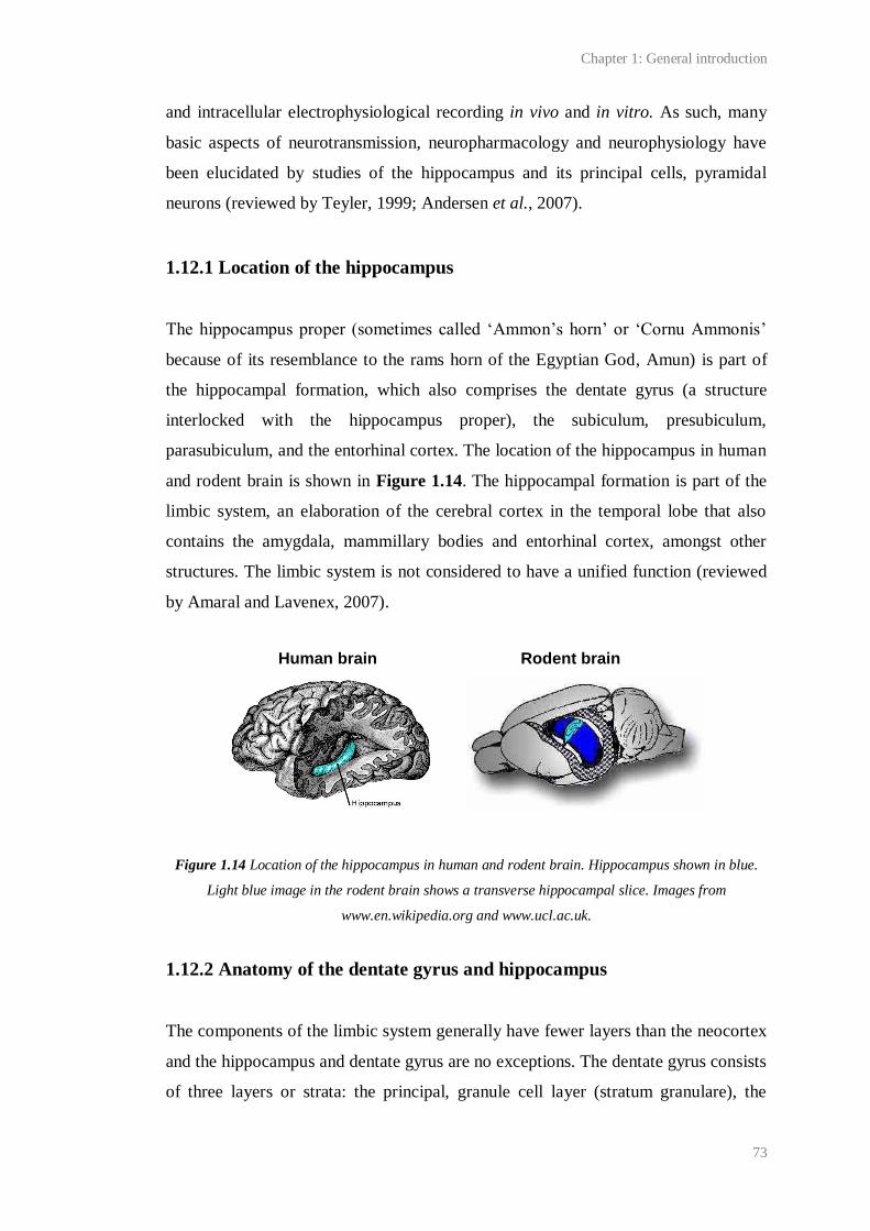
Chapter 1: General introduction
73
and intracellular electrophysiological recording in vivo and in vitro. As such, many
basic aspects of neurotransmission, neuropharmacology and neurophysiology have
been elucidated by studies of the hippocampus and its principal cells, pyramidal
neurons (reviewed by Teyler, 1999; Andersen et al., 2007).
1.12.1 Location of the hippocampus
The hippocampus proper (sometimes called ‘Ammon’s horn’ or ‘Cornu Ammonis’
because of its resemblance to the rams horn of the Egyptian God, Amun) is part of
the hippocampal formation, which also comprises the dentate gyrus (a structure
interlocked with the hippocampus proper), the subiculum, presubiculum,
parasubiculum, and the entorhinal cortex. The location of the hippocampus in human
and rodent brain is shown in Figure 1.14. The hippocampal formation is part of the
limbic system, an elaboration of the cerebral cortex in the temporal lobe that also
contains the amygdala, mammillary bodies and entorhinal cortex, amongst other
structures. The limbic system is not considered to have a unified function (reviewed
by Amaral and Lavenex, 2007).
Figure 1.14 Location of the hippocampus in human and rodent brain. Hippocampus shown in blue.
Light blue image in the rodent brain shows a transverse hippocampal slice. Images from
www.en.wikipedia.org and www.ucl.ac.uk.
1.12.2 Anatomy of the dentate gyrus and hippocampus
The components of the limbic system generally have fewer layers than the neocortex
and the hippocampus and dentate gyrus are no exceptions. The dentate gyrus consists
of three layers or strata: the principal, granule cell layer (stratum granulare), the
Human brain Rodent brain
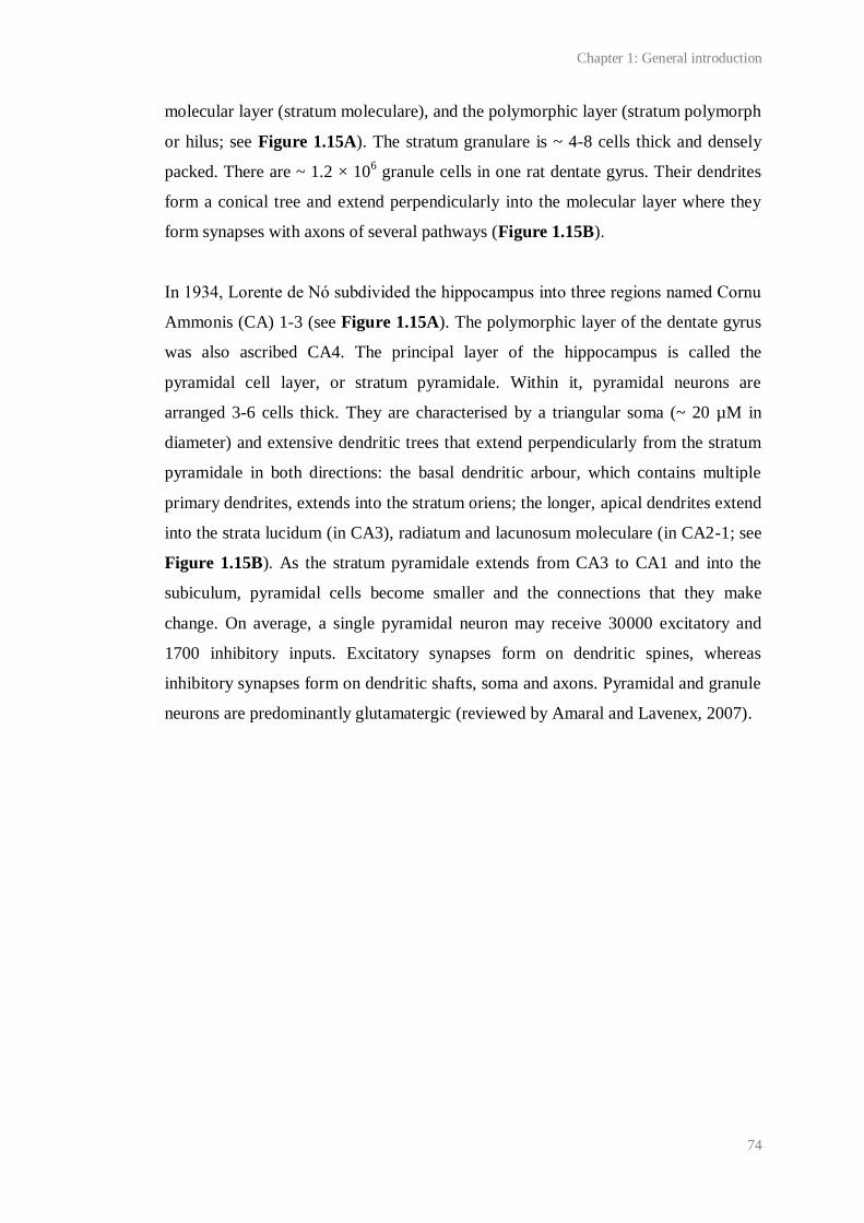
Chapter 1: General introduction
74
molecular layer (stratum moleculare), and the polymorphic layer (stratum polymorph
or hilus; see Figure 1.15A). The stratum granulare is ~ 4-8 cells thick and densely
packed. There are ~ 1.2 × 10⁶ granule cells in one rat dentate gyrus. Their dendrites
form a conical tree and extend perpendicularly into the molecular layer where they
form synapses with axons of several pathways (Figure 1.15B).
In 1934, Lorente de Nό subdivided the hippocampus into three regions named Cornu
Ammonis (CA) 1-3 (see Figure 1.15A). The polymorphic layer of the dentate gyrus
was also ascribed CA4. The principal layer of the hippocampus is called the
pyramidal cell layer, or stratum pyramidale. Within it, pyramidal neurons are
arranged 3-6 cells thick. They are characterised by a triangular soma (~ 20 µM in
diameter) and extensive dendritic trees that extend perpendicularly from the stratum
pyramidale in both directions: the basal dendritic arbour, which contains multiple
primary dendrites, extends into the stratum oriens; the longer, apical dendrites extend
into the strata lucidum (in CA3), radiatum and lacunosum moleculare (in CA2-1; see
Figure 1.15B). As the stratum pyramidale extends from CA3 to CA1 and into the
subiculum, pyramidal cells become smaller and the connections that they make
change. On average, a single pyramidal neuron may receive 30000 excitatory and
1700 inhibitory inputs. Excitatory synapses form on dendritic spines, whereas
inhibitory synapses form on dendritic shafts, soma and axons. Pyramidal and granule
neurons are predominantly glutamatergic (reviewed by Amaral and Lavenex, 2007).
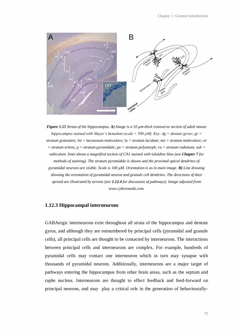
Chapter 1: General introduction
75
Figure 1.15 Strata of the hippocampus. A) Image is a 10 µm-thick transverse section of adult mouse
hippocampus stained with Mayer’s hemalum (scale = 500 µM). Key: dg = dentate gyrus; gr =
stratum granulare; lm = lacunosum moleculare; lu = stratum lucidum; mo = stratum moleculare; or
= stratum oriens; p = stratum pyramidale; po = stratum polymorph; ra = stratum radiatum; sub =
subiculum. Inset shows a magnified section of CA1 stained with toluidine blue (see Chapter 7 for
methods of staining). The stratum pyramidale is shown and the proximal apical dendrites of
pyramidal neurons are visible. Scale is 100 µM. Orientation is as in main image. B) Line drawing
showing the orientation of pyramidal neuron and granule cell dendrites. The directions of their
spread are illustrated by arrows (see 1.12.4 for discussion of pathways). Image adjusted from
www.cyberounds.com.
1.12.3 Hippocampal interneurons
GABAergic interneurons exist throughout all strata of the hippocampus and dentate
gyrus, and although they are outnumbered by principal cells (pyramidal and granule
cells), all principal cells are thought to be contacted by interneurons. The interactions
between principal cells and interneurons are complex. For example, hundreds of
pyramidal cells may contact one interneuron which in turn may synapse with
thousands of pyramidal neurons. Additionally, interneurons are a major target of
pathways entering the hippocampus from other brain areas, such as the septum and
raphe nucleus. Interneurons are thought to effect feedback and feed-forward on
principal neurons, and may play a critical role in the generation of behaviourally-
A B

Chapter 1: General introduction
76
cued oscillatory activity in the hippocampus, such as theta rhythm (reviewed by
Freund and Buzsaki, 1996).
Multiple subtypes of hippocampal interneuron have been discovered. Each have
different locations within the hippocampus, morphology, connections,
electrophysiology, pharmacology and immunocytochemistry. This diversity has been
illustrated by the finding that, in area CA1 alone, at least 16 types of interneuron
have been delineated (Parra et al., 1998). Interneurons with cell bodies in the
pyramidal cell layer have been classified into four groups on the basis of their
synaptic targets. These are: 1) axo-axonic or chandelier cells, which each contact the
axon initial segment of over 1000 pyramidal cells and regulate action potential
initiation; 2) basket cells, which innervate and receive excitatory input from as many
as 1000-plus pyramidal cells; 3) bistratified cells, which synapse onto apical and
basal pyramidal cell dendrites; 4) radial trilaminar cells, which span the entire radius
of pyramidal cell dendrites. Interneuron specific interneurons also occur throughout
all hippocampal strata. Their axons terminate only on other interneurons (reviewed
by Freund and Buzsaki, 1996; Amaral and Lavenex, 2007).
1.12.4 Connections in the hippocampus
The major input to the hippocampus is from the entorhinal cortex. This brain area
forms an interface between the hippocampus and neocortex. It receives, and is
thought to integrate, highly processed, multimodal sensory information from multiple
areas of the cortex, especially the associational, perirhinal and parahippocampal
cortices, as well as other brain areas such as the thalamus. It is thought to be required
for declarative memory, in particular spatial memory, and grid and head direction
cells, which may be required for spatial memory, have been found within it
(reviewed by Bird and Burgess, 2008).
A major input to the hippocampus from the entorhinal cortex occurs from pyramidal
neurons in layer II via the perforant path, which perforates the subiculum and forms
connections with granule cell dendrites (Figure 1.16). The perforant path may also
contact GABA-positive interneurons in the molecular layer and apical dendrites of
Chapter 1: General introduction
77
CA3 pyramidal neurons. Additionally, neurons from layer III of the entorhinal cortex
project to CA1 neurons and the subiculum via the temporoammonic pathway.
Within the hippocampus, a trisynaptic circuit exists which is thought to conduct a
unidirectional flow of information from CA3 to CA1 and the subiculum (Figure
1.16). First, the granule cells give rise to distinctive unmyelinated axons, named
mossy fibres because they display varicosities (called mossy fibre expansions) along
their entire length. These extend into the polymorphic cell layer, where they synapse
with GABAergic interneurons, and then enter the stratum lucidum in CA3, where
they make large glutamatergic synapses with CA3 pyramidal neurons. A single
mossy fibre may contact a dozen pyramidal neurons, and make ~ 30 contacts with
each of them. Each CA3 pyramidal neuron may receive input from more than 50
granule cells. From CA3 and CA2 pyramidal neurons, information may then be
passed to CA1 via the Schaffer collaterals residing in the strata radiatum and oriens.
Each CA3 pyramidal neuron may contact multiple CA1 neurons. Each CA1 neuron
might be innervated by over 5000 CA3 cells.
From CA1 pyramidal cells, connections extend into the oriens and alveus and on to
the deep layers of the entorhinal cortex via the subiculum. The deep layers of the
entorhinal cortex then send outputs back to the cortex. Through this pattern of
connections, it is thought that relatively unprocessed information entering the
entorhinal cortex from multiple cortical areas traverses the entire hippocampus, may
be processed and perhaps associated somehow, and then returned back to the cortex,
perhaps for long-term storage (reviewed by Bird and Burgess, 2008; Neves et al.,
2008).
Finally, it should be noted that there are also multiple connections between areas of
the hippocampus other than those noted above. Within CA3 and CA2 (but not CA1),
for example, there are multiple recurrent (associational) connections, as well as
connections from the contralateral CA3 and CA2 (commissural connections).
Modifications of recurrent connections in area CA3 during the acquisition of
information are central to a key computational mode of hippocampal function
proposed by Marr (1971). Additionally, Marr suggests that the reactivation of some
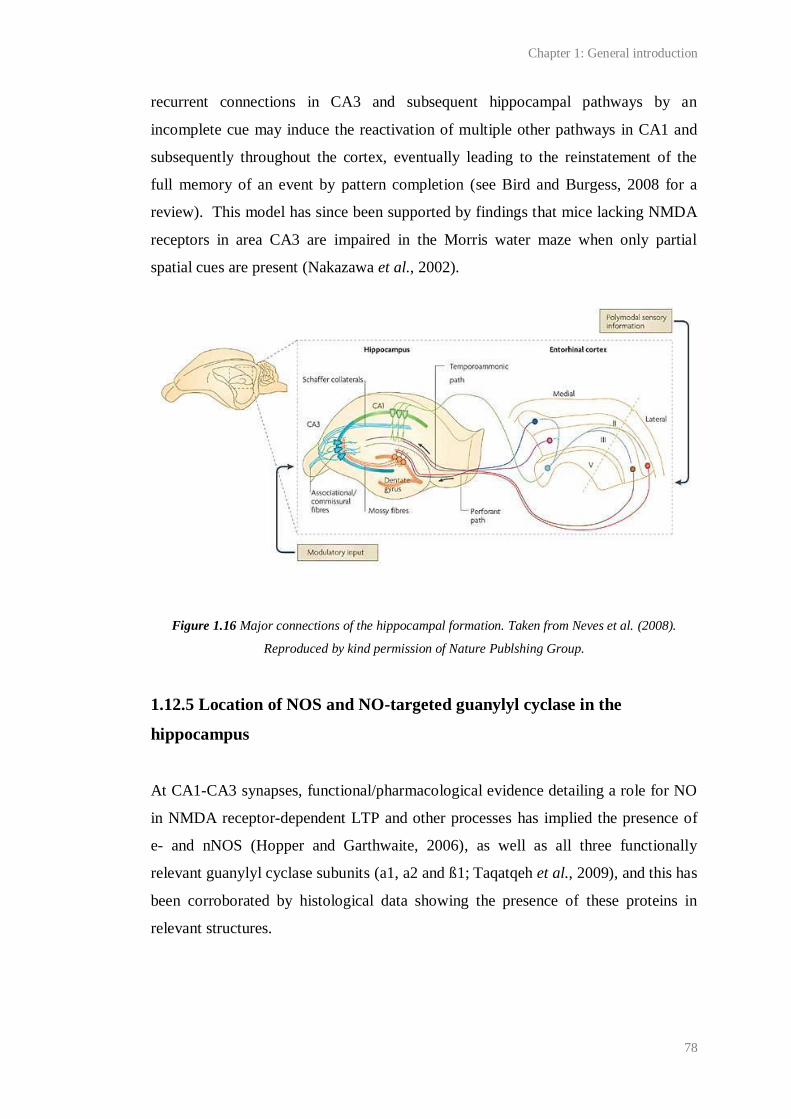
Chapter 1: General introduction
78
recurrent connections in CA3 and subsequent hippocampal pathways by an
incomplete cue may induce the reactivation of multiple other pathways in CA1 and
subsequently throughout the cortex, eventually leading to the reinstatement of the
full memory of an event by pattern completion (see Bird and Burgess, 2008 for a
review). This model has since been supported by findings that mice lacking NMDA
receptors in area CA3 are impaired in the Morris water maze when only partial
spatial cues are present (Nakazawa et al., 2002).
Figure 1.16 Major connections of the hippocampal formation. Taken from Neves et al. (2008).
Reproduced by kind permission of Nature Publshing Group.
1.12.5 Location of NOS and NO-targeted guanylyl cyclase in the
hippocampus
At CA1-CA3 synapses, functional/pharmacological evidence detailing a role for NO
in NMDA receptor-dependent LTP and other processes has implied the presence of
e- and nNOS (Hopper and Garthwaite, 2006), as well as all three functionally
relevant guanylyl cyclase subunits (a1, a2 and ß1; Taqatqeh et al., 2009), and this has
been corroborated by histological data showing the presence of these proteins in
relevant structures.
Chapter 1: General introduction
79
Initial histological studies found nNOS to be primarily located in the stratum
granulare of the dentate gyrus, in the neuropil of the dentate molecular layer and
stratum radiatum of Ammon’s horn, as well as in scattered cells, presumably
interneurons, throughout CA1 (see Figure 1.15 for key to anatomy; Bredt et al.,
1991a; Vincent and Kimura, 1992; Valtschanoff et al., 1993; Dun et al., 1994; Lin
and Totterdell, 1998). Since then, immunostaining and electron microscopy
following relatively weak fixation of tissues (0.5- 1 % paraformaldehyde; PFA) has
also revealed nNOS protein in the cytoplasm of pyramidal cell soma and at synapses
throughout the stratum radiatum where it may contribute to synaptic transmission
and/or plasticity (Wendland et al., 1994; Gonzalez-Hernandez et al., 1996; Burette et
al., 2002). This distribution of nNOS has since been confirmed by the isolation of
nNOS mRNA from dissociated CA1 pyramidal neurons (Chiang et al., 1994). The
increase in stained structures following immunohistochemistry for nNOS using
relatively weakly fixed tissue may reflect better preservation of nNOS epitopes or
improved access of the antibody to the protein due to a reduction in aldehyde cross-
linking of proteins, for example, in the PSD.
Unsurprisingly, eNOS is found throughout the hippocampal vasculature (Blackshaw
et al., 2003). As mentioned above (1.2.2), some groups have also reported eNOS
staining in pyramidal neurons in rodent (Dinerman et al., 1994; O'Dell et al., 1994)
and human (Doyle and Slater, 1997) hippocampus, although attempts to replicate
these results have failed. Rather, several studies (for example Chiang et al., 1994)
support the current consensus that eNOS expression is restricted to blood vessels.
An initial study of the location of guanylyl cyclase in hippocampus using in situ
hybridisation showed message for the protein throughout the strata pyramidale and
granulare (Matsuoka et al., 1992). Later, mRNA for all three functionally-relevant
subunits of the NO-targeted guanylyl cyclase, α1, α2 and β1, was shown to be
present in the developing and adult rat hippocampus (Gibb and Garthwaite, 2001;
Mergia et al., 2003). In contrast to other brain areas in which the amount of mRNA
for each of the α subunits is approximately equal, Mergia et al. (2003) have found
that, in the hippocampus, mRNA for the α2 subunit is significantly more abundant
than message for the α1 subunit. Using in situ hybridisation, Gibb and Garthwaite
Chapter 1: General introduction
80
(2001) detected an abundance of mRNA for the α2 and β1 subunits in the stratum
pyramidale, but it was unclear whether the α1 subunit was also present in pyramidal
cells or in the surrounding strata radiatum and oriens (Gibb and Garthwaite, 2001).
At CA3-CA1 synapses, functional evidence detailing both post- (for example,
Serulle et al., 2007) and presynaptic (for example, Phillips et al., 2008) effects of NO
following the induction of LTP has been corroborated by a complimentary
distribution of guanylyl cyclase and nNOS either side of the synapse (Burette et al.,
2002). Using immunohistochemistry optimised to detect synaptic proteins, Burette et
al. (2002) have shown that nNOS and the guanylyl cyclase β1 subunit preferentially
associate with each other at a subpopulation of synapses (< 10 %) within CA1. In
support of putative retrograde NO transmission, post-embedding immunogold
electron microscopy revealed nNOS within the PSD of asymmetric axospinous
synapses and in close spatial proximity to presynaptic guanylyl cyclase β1 located
within axon terminals. Additionally, Burette et al. (2002) imply that a minority of
postsynaptic densities were positive for guanylyl cyclase β1 and presynaptic
varicosities positive for nNOS, suggesting that anterograde and/or intracellular NO
transmission may occur. The location of the NO-targeted guanylyl cyclase α subunits
is the topic of Chapter 5.
1.13 General Aim
Interest in the role of NO in LTP is largely rooted in the hypothesis that NO,
synthesised by nNOS upon NMDA receptor channel opening, acts as a retrograde
messenger during NMDA receptor-dependent LTP (reviewed by Feil and Kleppisch,
2008). In this way, NO might account for presynaptic effects of LTP. Although
evidence for a presynaptic effect of NO during LTP has been yielded from studies of
synapses between dissociated hippocampal pyramidal neurons (Arancio et al., 1995;
Arancio et al., 1996; Arancio et al., 2001), and many researchers describe NO as a
retrograde messenger, there is little unambiguous evidence for retrograde NO
transmission or a presynaptic effect of NO following LTP induction at synapses in
intact tissues (reviewed in Table 3.1). The first aim of the project was therefore to
isolate the NO-dependent component of NMDA receptor-dependent LTP at CA3-
Chapter 1: General introduction
81
CA1 synapses in intact tissues (transverse hippocampal slices) and to test whether it
was characterised by a persistent increase in neurotransmitter release using changes
in the magnitude of PPF of CA1 fEPSPs as an indicator of presynaptic efficacy (see
Chapter 3).
Related to this, the second aim was to investigate the effect of NO on
neurotransmitter release at CA1 synapses under basal stimulation (i.e. stimulation
causing no observable persistent plasticity; see Chapter 4). The major reason was
that mice lacking the NO-targeted guanylyl cyclase α1 subunit had recently been
found to exhibit reduced CA1 PPF under basal conditions, consistent with tonic
facilitation of neurotransmitter release at wild-type CA1 synapses (Taqatqeh et al.,
2009). We hypothesised that if NO was found to regulate neurotransmitter release at
CA1 synapses under basal conditions, then the isoform responsible might be eNOS,
because a low-level, activity-dependent, endothelium-derived NO tone exists in the
hippocampus (Chetkovich et al., 1993; Hopper and Garthwaite, 2006).
A third aim related to whether the role of NO in CA1 LTP is strictly NMDA
receptor-dependent (see Chapter 5). Although nNOS is thought to be preferentially
activated by NMDA receptor opening, we noticed that the properties of a NMDA
receptor-independent CA1 LTP (reviewed by Teyler et al., 1994) were similar to
NO-dependent LTP. Therefore, we tested the involvement of NO in the NMDA
receptor-independent CA1 LTP.
Finally, we wanted to investigate the location of the NO-targeted guanylyl cyclase in
the hippocampus. Of specific interest was the location of the cyclases’ α subunits
(Chapter 7), because it had been recently suggested that the α1β1 and α2β1 isoforms
of the cyclase have distinct roles in LTP (Taqatqeh et al., 2009).
Chapter 2:
General materials and methods
Chapter 2: General materials and methods
83
2.1 Materials
2.1.1 Pharmacological compounds
The pharmacological compounds/ peptides that were used are listed in Table 2.1.
Unless otherwise stated, compounds were prepared such that the final concentration
of the solvent applied in vitro was no more than 1:100, or 1:1000 for DMSO. See 2.5
Key to Suppliers for supplier details.
Compound/
Peptide
Chemical name and primary reason for
use*
Solvent Supplier
Acetylcholine
chloride (ACh)
2-acetyloxyethyl(trimethyl)azanium chloride
(Cholinergic agonist)
H2O Sigma
ω-Agatoxin IVA -
(P/Q-type VGCC inhibitor)
H2O Alomone
D-AP5 (2R)-2-amino-5-phosphonopentanoic acid
(NMDA antagonist)
NaOH Tocris
L-Arginine (2S)-2-amino-5-
(diaminomethylideneamino)pentanoic acid
(NOS substrate)
aCSF Sigma
BAY 60-7550 2-[(3,4-dimethoxyphenyl)methyl]-7-[(1R)-1-
hydroxyethyl]-4-phenylbutyl]-5-methyl-imidazo[5,1-
f][1,2,4]triazin-4(1H)-one
(PDE 2 inhibitor)
DMSO Cayman
Cadmium sulphate cadmium trisulphate octahydrate
(VGCC antagonist (non-selective))
H2O Sigma
2-Chloroadenosine (2R,3R,4S,5R)-2-(6-amino-2-chloropurin-9-yl)-5-
(hydroxymethyl)oxolane-3,4-diol
(Adenosine receptor agonist)
aCSF Sigma
CNQX disodium disodium 6-cyano-7-nitroquinoxaline-2,3-diolate
(AMPA/kainate receptor inhibitor)
DMSO Tocris
ω-Conotoxin GVIA -
(N-type VGCC inhibitor)
H2O Sigma
EGTA 2-[2-[2-[2-
[bis(carboxymethyl)amino]ethoxy]ethoxy]ethyl-
(carboxymethyl)amino]acetic acid
NaOH Sigma
Chapter 2: General materials and methods
84
(Ca2+ chelator)
Forskolin [(3R,4aR,5S,6S,6aS,10S,10aR,10bS)-3-ethenyl-
6,10,10b-trihydroxy-3,4a,7,7,10a-pentamethyl-1-oxo-
5,6,6a,8,9,10-hexahydro-2H-benzo[f]chromen-5-
yl]acetate
(Adenylyl cyclase agonist)
DMSO Tocris
FPL 64176 methyl 4-(2-benzylbenzoyl)-2,5-dimethyl-1H-
pyrrole-3-carboxylate
(L-type VGCC current enhancer)
DMSO Tocris
FX-4053.3HCl 6-[(3R,4R)-4-(2-[2,2-difluoro-2-(3-
fluorophenyl)ethyl]aminoethoxy)pyrrolidin-3-
yl]methyl-4-methylpyridin-2-amine trihydrochloride
(nNOS inhibitor)
DMSO Prof.
Richard
Silverman
Gadolinium (III)
chloride
Trichlorogadolinium hexahydrate
(Transient receptor potential channel antagonist)
DMSO Sigma
IBMX 1-methyl-3-(2-methylpropyl)-7H-purine-2,6-dione
(PDE inhibitor (non-selective))
DMSO Sigma
JK-5.3HCl 6-[(3R,4R)-4-(2-[2-(3-chloro-5-fluorophenyl)-2,2-
difluoroethyl]aminoethoxy)pyrrolidin-3-yl]methyl-
4-methylpyridin-2-amine trihydrochloride
(Proposed nNOS inhibitor)
DMSO Prof.
Richard
Silverman
S-MCPG 4-[(2S)-2-amino-1-hydroxy-1-oxopropan-2-
yl]benzoic acid
(Metabotropic glutamate receptor inhibitor)
NaOH Tocris
(+)-MK-801
maleate
(+)-5-methyl-10,11- dihydro-5H-
dibenzo[a,d]cyclohepten-5,10-imine maleate
(NMDA receptor open channel blocker)
H2O Tocris
NBQX disodium 2,3-Dioxo-6-nitro-1,2,3,4-
tetrahydrobenzo[f]quinoxaline -7-sulfonamide
disodium
(AMPA/kainate receptor inhibitor)
DMSO Tocris
Nifedipine dimethyl 2,6-dimethyl-4-(2-nitrophenyl)-1,4-
dihydropyridine-3,5-dicarboxylate
(L-type VGCC inhibitor)
DMSO Tocris
NMDA (2R)-2-(methylamino)butanedioic acid
(NMDA receptor agonist)
NaOH Tocris
L-NNA 2-amino-5-[[amino(nitramido)methylidene]amino]
pentanoic acid
(NOS inhibitor (non-selective))
HCl Tocris
Chapter 2: General materials and methods
85
LY 341495 2-[(1S,2S)-2-carboxycyclopropyl]-3-(9H-xanthen-9-
yl)-D-alanine
(Metabotropic glutamate receptor inhibitor)
NaOH Tocris
Nickel(II) chloride Dichloronickel
(T/R-type VGCC inhibitor)
H2O Sigma
ODQ [1,2,4]oxadiazolo[4,3-a]quinoxalin-1-one
(NO-targeted guanylyl cyclase antagonist)
DMSO Sigma
PAPA/ NONOate N-[3-aminopropyl(propyl)amino]-N-hydroxynitrous
amide
(NO donor)
NaOH Enzo
Thapsigargin (3S,3aR,4S,6S,6aR,7S,8S,9bS)-6-(acetyloxy)-4-
(butyryloxy)-3,3a-dihydroxy-3,6,9-trimethyl-8--2-
oxo-2,3,3a,4,5,6,6a,7,8,9b-decahydroazuleno[4,5-
b]furan-7-yl octanoate
(Sarco/endoplasmic reticulum Ca2+ ATPase
inhibitor)
DMSO VWR
TTX (citrate free) (4R,4aR,5R,6S,7S,8S,8aR,10S,12S)-2-
azaniumylidene-4,6,8,12-tetrahydroxy-6-
(hydroximethyl)-2,3,4,4a,5,6,7,8-octahydro-1H-
8a,10-methano-5,7-(epoxymethanooxy)quinazolin-
10-olate
(Na2+ channel blocker)
C2H4O2 Latoxan
L-VNIO.HCl N5-(1-imino-3-butenyl)-L-ornithine,
monohydrochloride
(nNOS inhibitor)
DMSO Enzo
1400-W.2HCl N'-[[3 (aminomethyl)phenyl]methyl]ethanimidamide
dihydrochloride
(i/nNOS inhibitor)
H2O Enzo
Table 2.1 Pharmacological compounds used.*Secondary actions of each compound are discussed in
the text where appropriate.
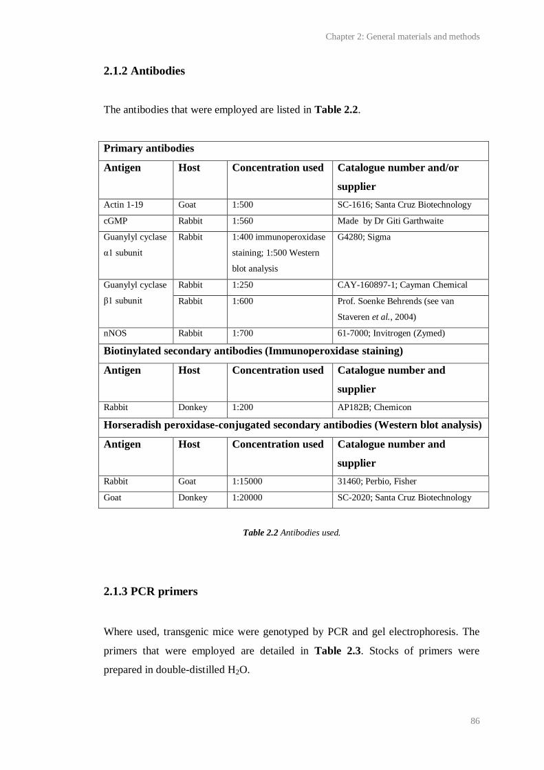
Chapter 2: General materials and methods
86
2.1.2 Antibodies
The antibodies that were employed are listed in Table 2.2.
Primary antibodies
Antigen Host Concentration used Catalogue number and/or
supplier
Actin 1-19 Goat 1:500 SC-1616; Santa Cruz Biotechnology
cGMP Rabbit 1:560 Made by Dr Giti Garthwaite
Guanylyl cyclase
α1 subunit
Rabbit 1:400 immunoperoxidase
staining; 1:500 Western
blot analysis
G4280; Sigma
Guanylyl cyclase
β1 subunit
Rabbit 1:250 CAY-160897-1; Cayman Chemical
Rabbit 1:600 Prof. Soenke Behrends (see van
Staveren et al., 2004)
nNOS Rabbit 1:700 61-7000; Invitrogen (Zymed)
Biotinylated secondary antibodies (Immunoperoxidase staining)
Antigen Host Concentration used Catalogue number and
supplier
Rabbit Donkey 1:200 AP182B; Chemicon
Horseradish peroxidase-conjugated secondary antibodies (Western blot analysis)
Antigen Host Concentration used Catalogue number and
supplier
Rabbit Goat 1:15000 31460; Perbio, Fisher
Goat Donkey 1:20000 SC-2020; Santa Cruz Biotechnology
Table 2.2 Antibodies used.
2.1.3 PCR primers
Where used, transgenic mice were genotyped by PCR and gel electrophoresis. The
primers that were employed are detailed in Table 2.3. Stocks of primers were
prepared in double-distilled H2O.
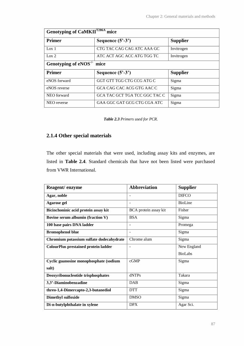
Chapter 2: General materials and methods
87
Genotyping of CaMKIIT286A
mice
Primer Sequence (5’-3’) Supplier
Lox 1 CTG TAC CAG CAG ATC AAA GC Invitrogen
Lox 2 ATC ACT AGC ACC ATG TGG TC Invitrogen
Genotyping of eNOS-/-
mice
Primer Sequence (5’-3’) Supplier
eNOS forward GGT GTT TGG CTG CCG ATG C Sigma
eNOS reverse GCA CAG CAC ACG GTG AAC C Sigma
NEO forward GCA TAC GCT TGA TCC GGC TAC C Sigma
NEO reverse GAA GGC GAT GCG CTG CGA ATC Sigma
Table 2.3 Primers used for PCR.
2.1.4 Other special materials
The other special materials that were used, including assay kits and enzymes, are
listed in Table 2.4. Standard chemicals that have not been listed were purchased
from VWR International.
Reagent/ enzyme Abbreviation Supplier
Agar, noble - DIFCO
Agarose gel - BioLine
Bicinchoninic acid protein assay kit BCA protein assay kit Fisher
Bovine serum albumin (fraction V) BSA Sigma
100 base pairs DNA ladder - Promega
Bromophenol blue - Sigma
Chromium potassium sulfate dodecahydrate Chrome alum Sigma
ColourPlus prestained protein ladder - New England
BioLabs
Cyclic guanosine monophosphate (sodium
salt)
cGMP Sigma
Deoxyribonucleotide trisphosphates dNTPs Takara
3,3’-Diaminobenzadine DAB Sigma
threo-1,4-Dimercapto-2,3-butanediol DTT Sigma
Dimethyl sulfoxide DMSO Sigma
Di-n-butylphthalate in xylene DPX Agar Sci.

Chapter 2: General materials and methods
88
Disodium ethylenediaminetetraacetate
dehydrate
EDTA Sigma
Donkey serum - Millipore
Ethidium bromide - Sigma
Gelatine - Sigma
Glycerol - Sigma
GoTaq hot start DNA polymerase - Promega
Halt protease inhibitor cocktail - Fisher
High performance chemiluminescence film - GE Healthcare
Hydrogen peroxide in H2O H2O2 Sigma
Hyperladder 1 - BioLine
Mayer’s hemalum - Merck
Sodium hydroxide NaOH Sigma
Optimal cutting temperature embedding
medium
OCT Fisher
Paraformaldehyde PFA Sigma
PCR buffer (10 x) - Invitrogen
Peroxidase suppressor - Fisher
Phosphate buffered saline PBS Sigma
Pico Fluor 40 scintillant - PerkinElmer
Polyvinylidene fluoride membrane PVDF membrane Millipore
Polyvinylpyrrolidone PVP Sigma
Potassium methyl sulphate KMeSO4 Fisher
Proteinase K from Tritirachium album - Sigma
Ready gels - Bio-Rad
Restore Western blot stripping buffer - Fisher
Sodium azide - Sigma
Sodium dodecyl sulphate SDS Sigma
SuperSignal west pico chemiluminescent
substrate
- Fisher
Taq DNA polymerase (recombinant) - Invitrogen
Toluidine blue - Sigma
Tris base, acetic acid and EDTA buffer TAE Sigma
Trisma base Tris Sigma
Tritium-labelled cGMP [3H]-cGMP GE Healthcare
Triton X-100 - Sigma
Tween 20 - Sigma
Vectastain avidin biotin complex ABC Vector

Chapter 2: General materials and methods
89
Table 2.4 Special chemicals, reagents and enzymes used.
2.1.5 Key to suppliers
Agar Sci. Agar Scientific Ltd., Essex, UK.
Alomone Alomone Labs Ltd., Jerusalem, Israel.
BioLine BioLine Ltd., London, UK.
Bio-Rad Bio-Rad Laboratories Ltd., Hertfordshire, UK.
Cayman IDS Ltd. (Cayman Chemical), Tyne and Wear, UK.
Chemicon Chemicon Europe Ltd., Hampshire, UK.
DIFCO BD, Oxford, UK.
Dr Giti Garthwaite Dr Giti Garthwaite, UCL, London, UK.
Enzo Enzo Life Sciences Ltd., Exeter, UK.
Fisher Fisher Scientific, Leicestershire, UK.
GE Healthcare GE Healthcare, Bucks, UK.
Invitrogen Invitrogen Ltd., Paisley, UK.
Latoxan Latoxan Laboratories, Valence, France.
Merck MSD, Hertfordshire, UK.
Millipore Millipore (UK) Ltd., Watford, UK
New England BioLabs New England BioLabs (UK) Ltd., Herts, UK.
PerkinElmer PerkinElmer, Cambridge, UK.
Prof. Richard Silverman Prof. Richard Silverman, Northwestern University, Chicago, USA.
Prof. Soenke Behrends Prof. Soenke Behrends, Technische Universität Braunschweig,
Braunschweig, Germany.
Promega Promega Corporation UK, Southampton, UK.
Santa Cruz Biotechnology Santa Cruz Biotechnology, Inc., Heidelberg, Germany
Sigma Sigma-Aldrich Company Ltd., Dorset, UK.
Takara Takara Bio. Inc., Shiga, Japan.
Tocris Tocris Cookson Ltd., Bristol, UK.
Vector Vector Labs Ltd., Peterborough, UK.
VWR VWR International Ltd., Leicestershire, UK.
Table 2.5 Suppliers of materials
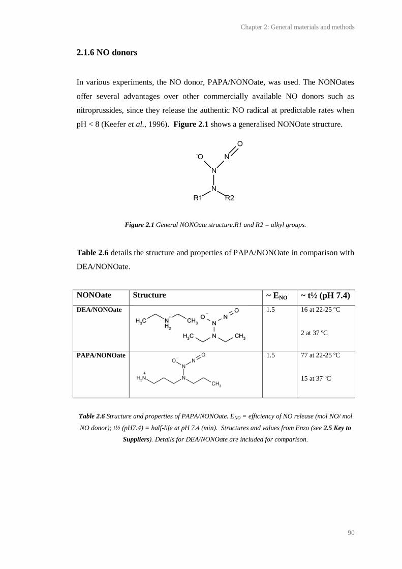
Chapter 2: General materials and methods
90
2.1.6 NO donors
In various experiments, the NO donor, PAPA/NONOate, was used. The NONOates
offer several advantages over other commercially available NO donors such as
nitroprussides, since they release the authentic NO radical at predictable rates when
pH < 8 (Keefer et al., 1996). Figure 2.1 shows a generalised NONOate structure.
Figure 2.1 General NONOate structure.R1 and R2 = alkyl groups.
Table 2.6 details the structure and properties of PAPA/NONOate in comparison with
DEA/NONOate.
NONOate Structure ~ ENO ~ t½ (pH 7.4)
DEA/NONOate
1.5 16 at 22-25 ºC
2 at 37 ºC
PAPA/NONOate
1.5 77 at 22-25 ºC
15 at 37 ºC
Table 2.6 Structure and properties of PAPA/NONOate. ENO = efficiency of NO release (mol NO/ mol
NO donor); t½ (pH7.4) = half-life at pH 7.4 (min). Structures and values from Enzo (see 2.5 Key to
Suppliers). Details for DEA/NONOate are included for comparison.

Chapter 2: General materials and methods
91
PAPA/NONOate was used since it was predicted to release NO over a time course
well-suited to the experiments, causing rapid (~ 2 min) accumulation of NO in
oxygenated buffer to a concentration that would remain ~ stable for several minutes
under the prevailing conditions (pH 7.4, 30 ± 1 ºC; Figure 2.2).
Figure 2.2 Predicted profile of NO release from two NONOates at 30 ºC, pH 7.4. t½ = 8 min for
DEA/NONOate; 45 min for PAPA/NONOate. PAPA/NONOate is predicted to release NO slower than
DEA/NONOate, resulting in a smaller accumulation of NO that remains roughly stable over 10 min.
See below for details on how to calculate the release profile.
To prevent decomposition prior to application in vitro, PAPA/NONOate was
prepared freshly on the day of each experiment as a 10 mM stock in 10 mM NaOH
(pH > 12) and kept on ice until immediately before use. It should be noted that
during the application of PAPA/NONOate to tissues, there will undoubtedly be
uncontrolled losses of NO that have not been accounted for in calculating the above
profile of NO release. These losses will occur due to evaporation from in vitro
solutions and through diffusion out of tubing used in tissue perfusion systems
(unpublished observations made in the laboratory). Precise estimates of the
concentration of NO in a bath that will permeate a tissue are also not possible, partly
because the proportion of PAPA/NONOate that will release NO within a tissue, as
opposed to in the bathing solution, is unknown.
0 2 4 6 8 100 2 4 6 8 10
0.0
0.5
1.0
1.5
2.0
Time (min)
[NO
] (
M)
Time (min)
1 M
3 M
10 M
30 M
DEA/NONOate PAPA/NONOate
Chapter 2: General materials and methods
92
Calculating NO release from a NONOate at a given time
The NO release profiles shown in Figure 2.2 were calculated using a ~ t½ at 30 °C
of 8 and 45 min for DEA/NONOate and PAPA/NONOate respectively and an
accepted model of NO release from a NONOate (Schmidt et al., 1997). The model
assumes that in oxygenated solution at pH 7.4: 1) NONOates decay exponentially; 2)
the primary route of removal of the resulting NO is via autoxidation. Therefore, the
concentration of NO released by a NONOate (CNO) at a given time (t) in oxygenated
solution at pH 7.4 can be calculated as the difference in the rate of NONOate decay
and the autoxidation of NO:
where initial conditions are:
cD(0) = c0 and cNO(0) = 0
and:
= –k1cD(t)
and:
t = time (s)
cNO(t) = concentration of NO at time t (M)
cD(t) = concentration of donor at time t (M)
c0 = initial concentration of donor (M)
eNO = mol of NO per mol donor
O2 = concentration of oxygen (M); estimated at 1 mM in carbogenated aCSF
k1 = rate constant for donor decomposition (s-1
)
k2 = rate constant for NO oxidation (M-2
s-1
); estimated at 13.6x10⁶
2.1.1 General solutions
aCSF was composed of (in mM) 120 NaCl, 2.5 KCl, 1.3 MgCl2, 1 NaH2PO4, 26
NaHCO3, 10 D-glucose, 2 CaCl2, equilibrated with 95% O2/5% CO2 to pH 7.4 at 30
ºC.
2
221 )()()( tcOketcktcdt
dNONODNO
dt
tdcD )(
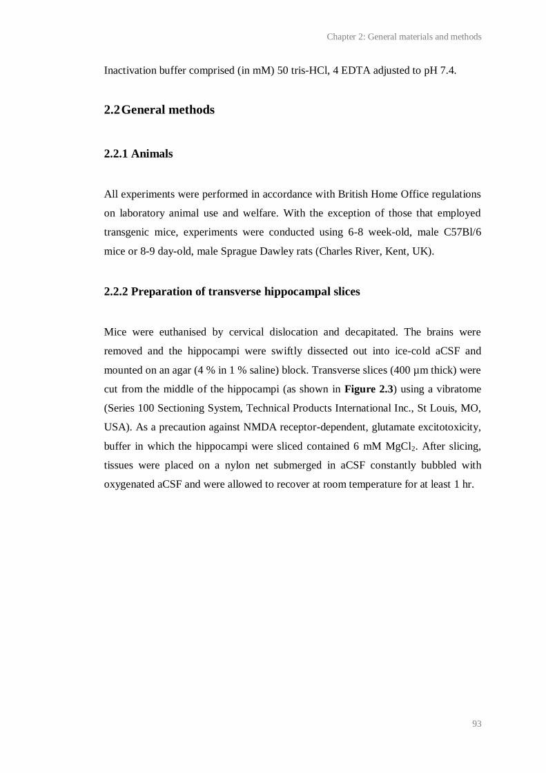
Chapter 2: General materials and methods
93
Inactivation buffer comprised (in mM) 50 tris-HCl, 4 EDTA adjusted to pH 7.4.
2.2 General methods
2.2.1 Animals
All experiments were performed in accordance with British Home Office regulations
on laboratory animal use and welfare. With the exception of those that employed
transgenic mice, experiments were conducted using 6-8 week-old, male C57Bl/6
mice or 8-9 day-old, male Sprague Dawley rats (Charles River, Kent, UK).
2.2.2 Preparation of transverse hippocampal slices
Mice were euthanised by cervical dislocation and decapitated. The brains were
removed and the hippocampi were swiftly dissected out into ice-cold aCSF and
mounted on an agar (4 % in 1 % saline) block. Transverse slices (400 µm thick) were
cut from the middle of the hippocampi (as shown in Figure 2.3) using a vibratome
(Series 100 Sectioning System, Technical Products International Inc., St Louis, MO,
USA). As a precaution against NMDA receptor-dependent, glutamate excitotoxicity,
buffer in which the hippocampi were sliced contained 6 mM MgCl2. After slicing,
tissues were placed on a nylon net submerged in aCSF constantly bubbled with
oxygenated aCSF and were allowed to recover at room temperature for at least 1 hr.
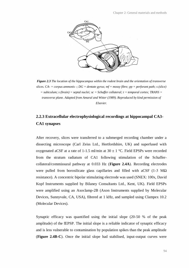
Chapter 2: General materials and methods
94
Figure 2.3 The location of the hippocampus within the rodent brain and the orientation of transverse
slices. CA- = corpus ammonis -; DG = dentate gyrus; mf = mossy fibre; pp = perforant path; s (slice)
= subiculum; s (brain) = septal nuclei; sc = Schaffer collateral; t = temporal cortex; TRANS =
transverse plane. Adapted from Amaral and Witter (1989). Reproduced by kind permission of
Elsevier.
2.2.3 Extracellular electrophysiological recordings at hippocampal CA3-
CA1 synapses
After recovery, slices were transferred to a submerged recording chamber under a
dissecting microscope (Carl Zeiss Ltd., Hertfordshire, UK) and superfused with
oxygenated aCSF at a rate of 1-1.5 ml/min at 30 ± 1 °C. Field EPSPs were recorded
from the stratum radiatum of CA1 following stimulation of the Schaffer-
collateral/commissural pathway at 0.033 Hz (Figure 2.4A). Recording electrodes
were pulled from borosilicate glass capillaries and filled with aCSF (1-3 MΩ
resistance). A concentric bipolar stimulating electrode was used (SNEX: 100x, David
Kopf Instruments supplied by Bilaney Consultants Ltd., Kent, UK). Field EPSPs
were amplified using an Axoclamp-2B (Axon Instruments supplied by Molecular
Devices, Sunnyvale, CA, USA), filtered at 1 kHz, and sampled using Clampex 10.2
(Molecular Devices).
Synaptic efficacy was quantified using the initial slope (20-50 % of the peak
amplitude) of the fEPSP. The initial slope is a reliable indicator of synaptic efficacy
and is less vulnerable to contamination by population spikes than the peak amplitude
(Figure 2.4B-C). Once the initial slope had stabilised, input-output curves were
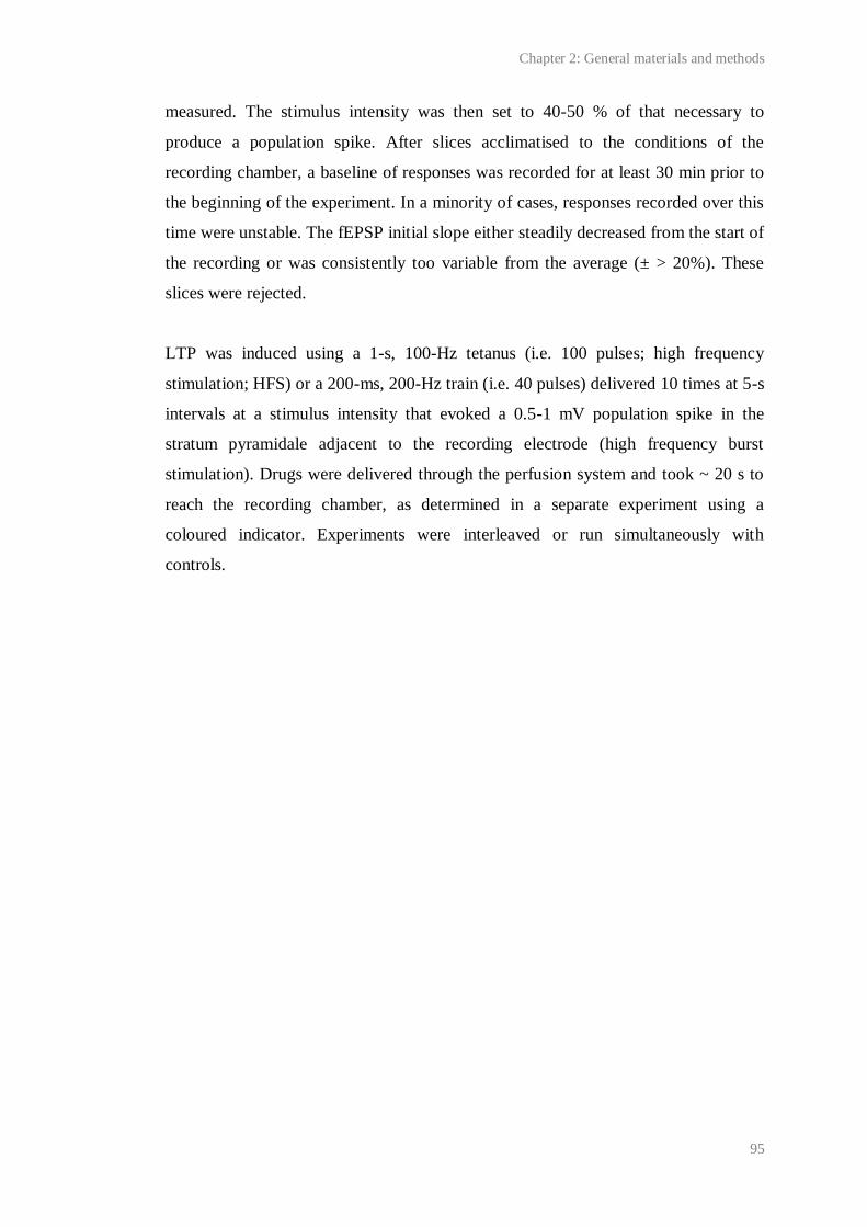
Chapter 2: General materials and methods
95
measured. The stimulus intensity was then set to 40-50 % of that necessary to
produce a population spike. After slices acclimatised to the conditions of the
recording chamber, a baseline of responses was recorded for at least 30 min prior to
the beginning of the experiment. In a minority of cases, responses recorded over this
time were unstable. The fEPSP initial slope either steadily decreased from the start of
the recording or was consistently too variable from the average (± > 20%). These
slices were rejected.
LTP was induced using a 1-s, 100-Hz tetanus (i.e. 100 pulses; high frequency
stimulation; HFS) or a 200-ms, 200-Hz train (i.e. 40 pulses) delivered 10 times at 5-s
intervals at a stimulus intensity that evoked a 0.5-1 mV population spike in the
stratum pyramidale adjacent to the recording electrode (high frequency burst
stimulation). Drugs were delivered through the perfusion system and took ~ 20 s to
reach the recording chamber, as determined in a separate experiment using a
coloured indicator. Experiments were interleaved or run simultaneously with
controls.
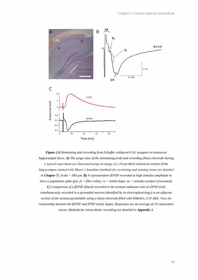
Chapter 2: General materials and methods
96
Figure 2.4 Stimulating and recording from Schaffer collateral-CA1 synapses in transverse
hippocampal slices. A) The target sites of the stimulating (red) and recording (blue) electrode during
a typical experiment are illustrated using an image of a 10 μm-thick transverse section of the
hippocampus stained with Mayer’s hemalum (methods for sectioning and staining tissue are detailed
in Chapter 7). Scale = 500 µm. B) A representative fEPSP recorded at high stimulus amplitude to
show a population spike (ps). fv = fibre volley; is = initial slope; sa = stimulus artefact (truncated).
C) Comparison of a fEPSP (black) recorded in the stratum radiatum with an EPSP (red)
simultaneously recorded in a pyramidal neuron (identified by its electrophysiology) in an adjacent
section of the stratum pyramidale using a sharp electrode filled with KMeSO4 (120 MΩ). Note the
relationship between the fEPSP and EPSP initial slopes. Responses are an average of 10 consecutive
traces. Methods for intracellular recording are detailed in Appendix 1.
Po
ten
tial
(mV
)
Time (ms)

Chapter 2: General materials and methods
97
2.2.4 Protein measurement
Intact tissues were individually sonicated in inactivation buffer. The protein content
of the resulting homogenates, or of lysates prepared for Western blot analysis, was
measured using the bicinchoninic acid (BCA) method and a series of bovine serum
albumin (BSA) standards (0-100 µg/ml inactivation buffer). 10 µl of each sample or
BSA standard were dispensed in triplicate into a 96-well plate. 200 µl BCA reagent
was added to each well and the plates were incubated for 30 min at 37 ºC to allow for
the assay reaction to occur. The assay reaction comprises two steps: 1) Cu2+
is
reduced to Cu+ by protein in the sample/standard; 2) Cu
+ chelates the BCA to form a
purple, water soluble complex that exhibits a strong absorbance at 562 nm. After
this, the plates were allowed to cool to room temperature and absorbance was
measured at 562 nm using a Spectra Max 250 spectrometer (Molecular Devices,
California, USA). As shown in Figure 2.5, the magnitude of the absorbance was
dependent on the concentration of the starting protein and, over the concentration
range of the BSA standards, was linear. From this, the protein content of each sample
was quantified.
Figure 2.5 An example of absorbance at 562 nm by a series of BSA standards (0-100 µg/ml
inactivation buffer) following the BCA protein assay. Data are means of triplicate measurements ±
SE. The inset shows the linear function used to fit the data, where a = intercept, b = slope. The
0 20 40 60 80 100
0.1
0.2
0.3
0.4
0.5
Me
an
Ab
so
rba
nce
(a
u)
BSA Concentration (g/ml)
Equation y = a + b*x
Residual Sum of Squares
1.65789E-6
Adj. R-Square 0.99596
Value Standard Error
Intercept 0.06701 0.00641
Slope 0.00438 1.13856E-4
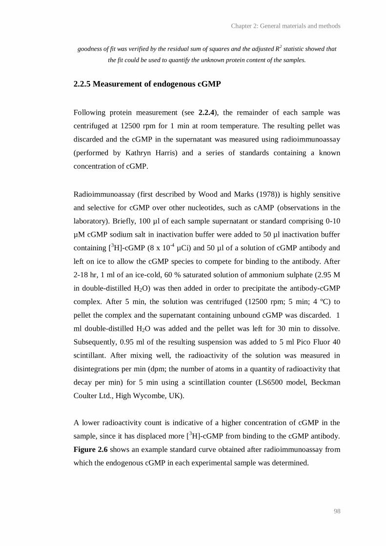
Chapter 2: General materials and methods
98
goodness of fit was verified by the residual sum of squares and the adjusted R2 statistic showed that
the fit could be used to quantify the unknown protein content of the samples.
2.2.5 Measurement of endogenous cGMP
Following protein measurement (see 2.2.4), the remainder of each sample was
centrifuged at 12500 rpm for 1 min at room temperature. The resulting pellet was
discarded and the cGMP in the supernatant was measured using radioimmunoassay
(performed by Kathryn Harris) and a series of standards containing a known
concentration of cGMP.
Radioimmunoassay (first described by Wood and Marks (1978)) is highly sensitive
and selective for cGMP over other nucleotides, such as cAMP (observations in the
laboratory). Briefly, 100 µl of each sample supernatant or standard comprising 0-10
µM cGMP sodium salt in inactivation buffer were added to 50 µl inactivation buffer
containing [3H]-cGMP (8 x 10
-4 µCi) and 50 µl of a solution of cGMP antibody and
left on ice to allow the cGMP species to compete for binding to the antibody. After
2-18 hr, 1 ml of an ice-cold, 60 % saturated solution of ammonium sulphate (2.95 M
in double-distilled H2O) was then added in order to precipitate the antibody-cGMP
complex. After 5 min, the solution was centrifuged (12500 rpm; 5 min; 4 ºC) to
pellet the complex and the supernatant containing unbound cGMP was discarded. 1
ml double-distilled H2O was added and the pellet was left for 30 min to dissolve.
Subsequently, 0.95 ml of the resulting suspension was added to 5 ml Pico Fluor 40
scintillant. After mixing well, the radioactivity of the solution was measured in
disintegrations per min (dpm; the number of atoms in a quantity of radioactivity that
decay per min) for 5 min using a scintillation counter (LS6500 model, Beckman
Coulter Ltd., High Wycombe, UK).
A lower radioactivity count is indicative of a higher concentration of cGMP in the
sample, since it has displaced more [3H]-cGMP from binding to the cGMP antibody.
Figure 2.6 shows an example standard curve obtained after radioimmunoassay from
which the endogenous cGMP in each experimental sample was determined.
Chapter 2: General materials and methods
99
Figure 2.6 An example standard curve obtained by radioimmunoassay. The inset shows the equation
used to fit the data where y = dpm, A1 and A2 = minimum and maximum dpm, p = slope, x = pmol
cGMP/standard, x0= pmol cGMP at which half the displacement has occurred. Note that the first data
point on the abscissa is actually zero. The adjusted R2 statistic showed that the fit was good and could
be used to quantify the endogenous cGMP in each experimental sample processed at the same time.
2.2.6 Genotyping by PCR and agarose gel electrophoresis
DNA extraction
Approximately 5 mm of tail or 0.2 mm2
ear were removed from each mouse and
added to 100 μl of tail lysis buffer which contained 10 mM tris (pH 8), 100 mM
NaCl, 10 mM EDTA (pH 8), 1 mM Ca2+
acetate (pH 7.5) and 100 μg/ml proteinase
K (~ 0.5-1.3 units) prepared in 50 % glycerol, 0.5 % SDS. This was then warmed to
55 ºC to provide optimal conditions for enzymatic digestion. After at least 24 hr the
lysates were vortexed to destroy any hard tissue and then diluted 1/10 with double-
distilled H2O.
1E-8 1E-7 1E-6 1E-5 1E-4 1E-3 0.01 0.1 1 10 100 1000
0
500
1000
1500
2000
2500
3000
Ra
dio
activity (
dp
m)
cGMP (pmol/standard)
Model Logistic
Equationy = A2 + (A1-A2)/(1 + (x/x0)^p)
Adj. R-Square0.99372
Value Standard Error
A1 2925.15296 52.01229
A2 159.85498 50.78524
x0 1.16187 0.08305
p 1.07218 0.06395
Chapter 2: General materials and methods
100
PCR
DNA from each lysate was amplified for electrophoresis using hot-start PCR. Unless
otherwise stated, 25 μl reactions were set up in 200 μl PCR tubes which contained:
0.2 µM primers, 200 µM deoxyribonucleotides, 0.5 U recombinant Taq DNA
polymerase (Invitrogen, Paisley, UK) and 1 μl of genomic DNA lysate (obtained as
above) in 1 x PCR buffer (Invitrogen).
PCR reactions were carried out in a MWG-Biotech Primus 96 plus (Ebersberg,
Germany). Samples were denatured at 93 ºC for 2 min. 35 PCR cycles were then
performed, each composed of: 30 s at 93 ºC, during which samples are denatured, 30
s at 58 ºC, in which primers anneal to the sample, and 30 s at 72 ºC for DNA
extension. Samples were then left at 72 ºC for 10 min to allow for the completion of
DNA extension. Each PCR included a negative control, in which the DNA sample
was replaced with double-distilled H2O, and a positive control including DNA of a
known genotype. PCR products were stored at 4 ºC until further use.
Agarose gel electrophoresis
After PCR, the products were mixed with 5 µl loading buffer containing ficol and
dyed with bromophenol. Unless otherwise stated, 5 µl of each product were then
electrophoresed in 2 % agarose gel in a tank containing 1 x TAE and 5 µg/ml
ethidium bromide for a minimum of 45 min at 80 V. DNA bands were visualised
under UV light using a Chemi-imager 4400 v 5.1 (Alpha Innotech, California, USA).
2.2.7 Statistics and data analysis
Statistics were performed using GraphPad InStat 3 software (GraphPad Software
Inc., California, USA). The significance level was 0.05. Where necessary, data were
fit with a logistic or exponential decay model using Origin Pro 8.1 (OriginLab
Corporation, Massachusetts, USA). The equations used are given below.
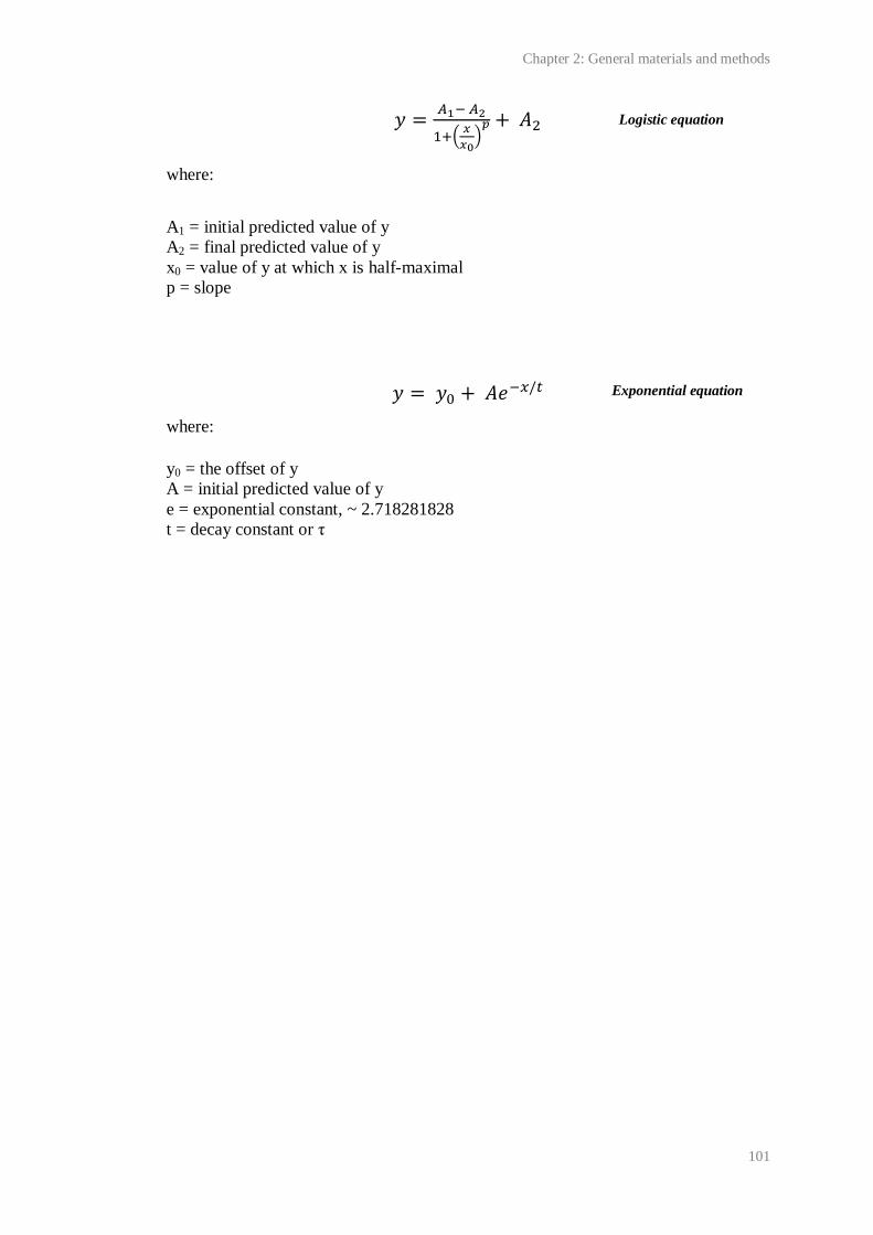
Chapter 2: General materials and methods
101
where:
A1 = initial predicted value of y
A2 = final predicted value of y
x0 = value of y at which x is half-maximal
p = slope
where:
y0 = the offset of y
A = initial predicted value of y
e = exponential constant, ~ 2.718281828
t = decay constant or τ
Logistic equation
Exponential equation
Chapter 3:
NO-induced, long-lasting potentiation at hippocampal CA1
synapses
Chapter 3: NO-induced, long-lasting potentiation at hippocampal CA1 synapses
103
3.1 Introduction
LTP is a long-lasting (> 1 hr), activity-dependent increase in the efficacy of a
synapse, typically with Hebbian characteristics and therefore, intensely studied as a
correlate of information processing and storage in the nervous system (see Chapter
1). Since Bliss and Lomo (1973) reported a persistent (up to 10 hr) enhancement in
synaptic activity following brief tetanic stimulation of hippocampal perforant path-
granule cell synapses in anesthetised rabbits, LTP has been studied throughout the
mammalian nervous system, and in several species, including humans (Cooke and
Bliss, 2006).
The archetype of LTP occurs at Schaffer-collateral/commissural-CA1 synapses in the
hippocampus. Often, this is induced using high frequency stimulation (HFS; 1-s,
100-Hz tetanus), although it has been generated by multitude other electrical and
chemical stimuli in vitro and in vivo (Bliss et al., 2007), and by learning in rodents
(Whitlock et al., 2006). Its induction is dependent upon postsynaptic Ca2+
influx
(Lynch et al., 1983; Malenka et al., 1988) and, typically, NMDA receptor activation
(Collingridge et al., 1983a; Collingridge et al., 1983b; Malenka, 1991), thereby
explaining why LTP is associative and input specific. Its expression is thought to rely
upon multiple, pre- and postsynaptic mechanisms (Malenka and Bear, 2004; Bliss et
al., 2007). However, the details of these expression mechanisms and the conditions
under which they are recruited are, to varying extents, unclear. In particular, the
retrograde messenger presumably required to reconcile the postsynaptic induction
with presynaptic expression of NMDA receptor-dependent CA1 LTP, is, despite
intense and long-lasting interest, yet to be conclusively identified.
Amongst several candidate retrograde messengers, such as CO and arachidonic acid
(reviewed by Tao and Poo, 2001), NO has, arguably, received the most attention
since there appears to be a particularly good match between the properties of NO
signals necessary for LTP and the requirements of a retrograde messenger (see Table
3.1 and Chapter 1 for general discussion of the role of NO in LTP ).
Chapter 3: NO-induced, long-lasting potentiation at hippocampal CA1 synapses
104
Requirements of a
retrograde transmitter
active in LTP
Evidence consistent with retrograde NO
transmission in NMDA receptor-dependent CA1
LTP
The machinery for
synthesis/release of the
transmitter is located
postsynaptically
Immunohistochemistry with electron microscopy on hippocampal
sections has shown that nNOS is expressed post- (and pre-)
synaptically (Burette et al., 2002; Szabadits et al., 2007).
LTP-inducing stimuli elicit
synthesis of the transmitter
NMDA has been found to induce NO synthesis in hippocampal
slices (see Chapter 5) and in vivo (Luo and Vincent, 1994). Tetanic
stimulation (three, 1-s, 100-Hz trains) sufficient for LTP has been
shown to elicit NO synthesis in slices (Chetkovich et al., 1993).
Postsynaptic injection of
inhibitors that block the
synthesis of the transmitter
inhibit LTP
In dissociated hippocampal neurons: post- but not presynaptic
injections of the NOS inhibitor, L-NMMA, found to block tetanus
(three 50-Hz, 2-s trains at 20 s intervals, Mg2+-free solution)-
evoked LTP (Arancio et al., 1996).
In slices: postsynaptic L-NMMA and L-NNA shown to block
pairing-induced LTP (Schuman and Madison, 1991; O'Dell et al.,
1991).
Extracellular scavengers of the
transmitter inhibit presynaptic
LTP
In dissociated hippocampal neurons: oxymyoglobin, an
extracellular NO scavenger, blocked potentiation evoked by a weak
tetanus paired with photolysis of caged NO in the postsynaptic
neuron, but had no effect on potentiation when caged NO was
presynaptic (Arancio et al., 1996).
In slices: haemoglobin blocked pairing-induced, NO-dependent
LTP (Schuman and Madison, 1991; O'Dell et al., 1991).
The transmitter affects a
presynaptic target
Immunohistochemistry with electron microscopy on hippocampal
sections indicates that NO-targeted guanylyl cyclase is expressed
pre- (and post-) synaptically (Burette et al., 2002; Szabadits et al.,
2007).
Presynaptic injections of
inhibitors of the presynaptic
target inhibit LTP.
Not-tested.
The retrograde transmitter and
its presynaptic target cause an
increase in the probability of
neurotransmitter release and/or
number of active release sites
In dissociated hippocampal neurons:
exogenous NO (5-10 nM) or 8-Br-cGMP (50-100 µM) found
to elicit a persistent increase in the frequency, independent of
the amplitude, of miniature excitatory postsynaptic currents
(O'Dell et al., 1991; EPSCs; Arancio et al., 1995).
activation of the NMDA receptor/NO/cGMP pathway shown to
Chapter 3: NO-induced, long-lasting potentiation at hippocampal CA1 synapses
105
enhance neurotransmitter vesicle recycling (Micheva et al.,
2001; Micheva et al., 2003).
LTP found to cause a rapid increase in presynaptic protein
clusters via the NMDA receptor/NO/cGMP pathway (Ninan
and Arancio, 2004; Wang et al., 2005).
In slices:
the NO donors, hydroxylamine and sodium nitroprusside,
caused an increase in the efflux of noradrenaline and ACh from
un-stimulated hippocampal slices (Lonart et al., 1992).
theta burst stimulation-induced LTP in slices from mice
lacking the AMPA receptor GluR1 subunit found to be
presynaptic (as assessed using PPF) and abolished by NOS
antagonism (Phillips et al., 2008).
a decrease in PPF (consistent with an increase in
neurotransmitter release) measured 2 min after HFS-induced
LTP was observed in wild-type slices but not in slices from
mice lacking the NO-targeted guanylyl cyclase α1 subunit
(Taqatqeh et al., 2009).
endogenous NO was necessary for the rapid 10-30 min)
NMDA receptor-dependent remodelling of presynaptic
varicosities in area CA1 of hippocampal slice cultures
following theta burst stimulation (Nikonenko et al., 2003).
Exogenous retrograde
transmitter induces or
facilitates presynaptic LTP
In dissociated hippocampal neurons:
NO (< 60 s, 10 nM) paired with a weak tetanus (50-Hz, 0.5-s)
during NMDA receptor blockade produced a persistent
potentiation that was blocked by pre- but not postsynaptic
injection of oxymyoglobin (Arancio et al., 1996).
pre- but not postsynaptic cGMP or PKG I paired with a weak
tetanus produced a similar potentiation (Arancio et al., 1995;
Arancio et al., 2001).
In hippocampal slices: see discussion below.
Table 3.1 Evidence consistent with retrograde NO transmission during NMDA receptor-dependent
LTP at CA1 synapses. The table summarises some of the main requirements of a retrograde
transmitter responsible for facilitating neurotransmitter release during NMDA receptor-dependent
LTP in area CA1, and some of the key NO research consistent with each requirement. The list of
requirements has been adapted from Bliss et al. (2007).
Chapter 3: NO-induced, long-lasting potentiation at hippocampal CA1 synapses
106
Given the above, NO has become widely described as a retrograde messenger during
LTP (for example, Feil and Kleppisch, 2008). However, three aspects of research on
the role of NO in LTP should not be overlooked when considering putative
retrograde NO transmission during synaptic plasticity. First, the results of some
studies of the role of NO in LTP are discordant with retrograde NO transmission.
Most notably, several groups have observed NO-independent LTP at CA1 synapses
(for example, Cummings et al., 1994; Bannerman et al., 1994b; Phillips et al., 2008,
and see the discussion in Chapter 5).
Second, some of the evidence consistent with retrograde NO transmission is also
consistent with a postsynaptic effect of NO in LTP. For example, the blockade of
pairing-induced LTP by postsynaptic injection of NOS inhibitors reported by
Schuman and Madison (1991) and O’Dell et al. (1991) could have resulted if NO
acts as an intracellular transmitter. Evidence consistent with intracellular NO
transmission shows that NOS and NO-targeted guanylyl cyclase co-localise in some
CA1 dendrites (Burette et al., 2002). The inhibition of the LTP described by
Schuman and Madison (1991) and O’Dell et al. (1991) by extracellular NO
scavengers is also consistent with intracellular NO signalling because an efficient
extracellular scavenger will draw NO out of a cell by keeping the NO concentration
gradient across the membrane steep (Garthwaite, 2008).
Third, most evidence in favour of a presynaptic effect of NO following long-lasting
plasticity has been generated using pairs of dissociated hippocampal pyramidal
neurons (as shown in Table 3.1). The use of dissociated hippocampal neurons offers
several advantages over using intact tissues, such as transverse slices, in which
presynaptic terminals are inaccessible. Some of the most compelling research has
been performed by Arancio et al. who have reported that NO produced
postsynaptically is, through presynaptic cGMP and PKG I, critical for NMDA
receptor-dependent LTP (Arancio et al., 1995; Arancio et al., 1996; Arancio et al.,
2001). In accordance with these findings, NO/cGMP signalling at synapses between
dissociated hippocampal neurons has been positively linked to neurotransmitter
release (see Table 3.1). However, the relevance of these data to the role of NO in
LTP at synapses in intact tissues is unclear. It has, for example, been speculated that

Chapter 3: NO-induced, long-lasting potentiation at hippocampal CA1 synapses
107
differences in the developmental stage of neurons maintained in slice preparations
versus dissociated cultures may cause differences in LTP expression (Enoki et al.,
2009). Additionally, eNOS, which is expressed only in blood vessels (Stanarius et
al., 1997; Blackshaw et al., 2003; Chan et al., 2004) and, therefore, was unlikely to
be present in the neuronal cultures used by Arancio et al., has recently been
positively linked to neurotransmitter release at CA1 synapses under conditions of
basal stimulation (Chapter 4 and Neitz et al., 2011) and is required for some forms
of CA1 LTP (Hopper and Garthwaite, 2006). Therefore, it is possible that the
presynaptic effect of NO during LTP at synapses between dissociated neurons was
favoured because: 1) the NO necessary for the potentiation had compensated for a
lack of eNOS; 2) the probability of neurotransmitter release and/or number of active
release sites present at synapses between the dissociated neurons was unnaturally low
during baseline recording. Indeed, it has been reported that an increase in the
probability of neurotransmitter release and/or number of active release sites at CA1
synapses following LTP is more likely if the initial probability and/or number are
low (Schulz et al., 1994).
In more intact tissues, some evidence consistent with a positive link between NO and
neurotransmitter release during LTP at CA1 synapses has been reported. For
example, if NO acts as a retrograde messenger to cause presynaptic changes
following NMDA receptor-dependent LTP induction, one prediction is that
exogenous NO should partially overcome the inhibitory effect of NMDA receptor
blockade on potentiation and induce a presynaptic component of LTP (the last
requirement in Table 3.1). Using hippocampal slices, three different groups have
provided evidence consistent with part of this prediction (Zhuo et al., 1993; Malen
and Chapman, 1997; Bon and Garthwaite, 2003). They have shown that a sub-
threshold induction protocol (for example, 0.1-s, 50-Hz), unable to produce LTP
alone, could, when delivered in the presence of exogenous NO, produce a significant
and long-lasting potentiation that was independent of NMDA-receptor activation.
Zhuo et al. (1993) showed that this potentiation was pathway specific and, consistent
with a subsequent study by Bon and Garthwaite (2003), that it was also activity-
dependent, insensitive to NMDA receptor-antagonism and occluded tetanus-induced
LTP. Later it was also reported that a potentiation with similar properties, which also
Chapter 3: NO-induced, long-lasting potentiation at hippocampal CA1 synapses
108
occluded subsequent LTP, could be induced at CA1 synapses by pairing of one of
two cGMP analogues with a weak, sub-threshold tetanus (Zhuo et al., 1994).
These data are consistent with a presynaptic effect of NO and are regarded as the
major evidence in favour of NO as a retrograde messenger at CA1 synapses in intact
tissues (for example, Hawkins et al., 1994). They also concur with the data collected
from dissociated cells by Arancio et al. (see above) and with more recent work on
CA1 LTP in slices from transgenic mice (see penultimate row of Table 3.1).
However, the data presented by Bon and Garthwaite (2003), Malen and Chapman
(1997) and Zhuo et al. (1993) are also consistent with a postsynaptic NO-induced
potentiation, and tests of this possibility are missing in the literature. Furthermore,
the potentiations observed by Zhuo et al. (1993) and Bon and Garthwaite (2003)
could only be induced when exogenous NO was paired with simultaneous synaptic
activity. Indeed, there are no examples of potentiation by exogenous NO under
physiological conditions in the absence of high frequency synaptic stimulation. This
suggests that simultaneous synaptic activity is required for the NO-dependent
component of LTP. Since the potentiations observed by Zhuo et al. (1993), Malen
and Chapman (1997) and Bon and Garthwaite (2003) were induced using a sub-
threshold induction protocol, a different complement of LTP mechanisms may have
been activated from those recruited by the induction protocols typically used in
studies of hippocampal LTP and perhaps by natural stimuli causing synaptic
potentiation in vivo. Surprisingly, given the proposed role of NO in NMDA receptor-
dependent LTP, tests of the prediction that exogenous NO, paired with a standard
induction protocol, should restore the NO-dependent component of LTP when
NMDA receptors are blocked have not been reported.
3.2 Aim
Initial characterisation of LTP at Schaffer-collateral/commissural-CA1 synapses in
hippocampal slices under our conditions found it to be NMDA receptor- and NO-
dependent (data shown below). Given the above, we therefore aimed to test the
prediction that exogenous NO paired with a standard LTP induction protocol (a 1-s,
100-Hz tetanus or high frequency stimulation; HFS) during NMDA receptor
Chapter 3: NO-induced, long-lasting potentiation at hippocampal CA1 synapses
109
blockade would restore a persistent NO-dependent component of LTP. In this way
we sought to isolate the long-lasting, NO-dependent component of LTP from other
mechanisms of LTP expression and to test its locus (pre and/or postsynaptic) at
synapses in functional neural pathways.
3.3 Methods
3.3.1 Animals
Experiments were conducted using 6-8 week-old male C57Bl/6 mice (Charles River,
Margate, UK). Transgenic mice incapable of αCaMKII autophosphorylation
(aCaMKIIT286A
; Giese et al., 1998) were obtained as heterozygotes in a
129sv/C57Bl/6 background and bred. Siblings were not mated with each other. In
preparation for experiments, pups were genotyped, heterozygotes were euthanised
and an appropriate number of male wild-type and homozygote mice were kept. On
the day of each experiment, a second person selected either a homozygote or wild-
type mouse for use. The experimenter was left blind to genotype until after all
experiments and data analysis were completed. Mice were used at 6-9-weeks old.
3.3.2 Transverse hippocampal slice preparation and extracellular
electrophysiology
Transverse hippocampal slices were prepared and extracellular electrophysiological
recordings of activity at CA3-CA1 synapses were made as described in Chapter
2.2.2-3. LTP was induced using high-frequency stimulation (HFS; a 1-s, 100-Hz
tetanus). PPF (reviewed by Zucker and Regehr, 2002; Bliss et al., 2007) was induced
at 0.033 Hz using pairs of stimuli separated by various intervals (called the inter-
stimulus interval (ISI)), which ranged from 10 to 400 ms. The magnitude of PPF was
measured using the mean paired-pulse ratio (PPR; calculated by the initial slope of
the second fEPSP/the initial slope of the first fEPSP) of ~ 10 consecutive pairs of
fEPSPs.
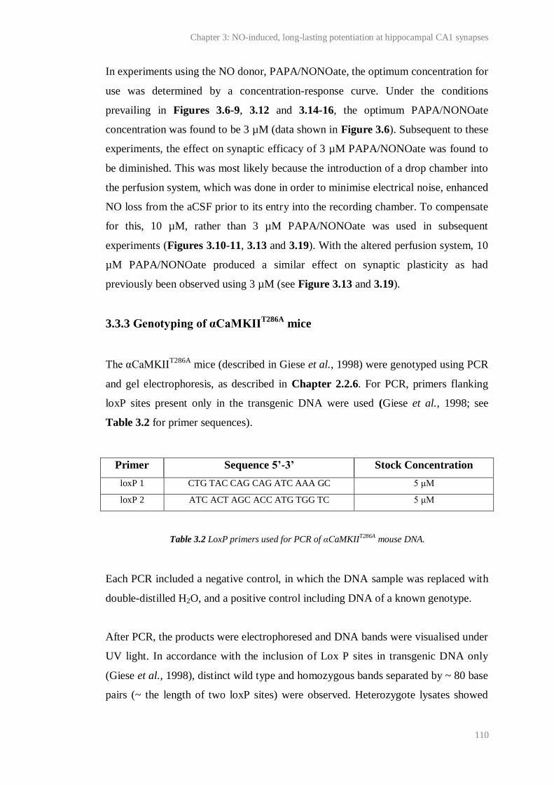
Chapter 3: NO-induced, long-lasting potentiation at hippocampal CA1 synapses
110
In experiments using the NO donor, PAPA/NONOate, the optimum concentration for
use was determined by a concentration-response curve. Under the conditions
prevailing in Figures 3.6-9, 3.12 and 3.14-16, the optimum PAPA/NONOate
concentration was found to be 3 µM (data shown in Figure 3.6). Subsequent to these
experiments, the effect on synaptic efficacy of 3 µM PAPA/NONOate was found to
be diminished. This was most likely because the introduction of a drop chamber into
the perfusion system, which was done in order to minimise electrical noise, enhanced
NO loss from the aCSF prior to its entry into the recording chamber. To compensate
for this, 10 µM, rather than 3 µM PAPA/NONOate was used in subsequent
experiments (Figures 3.10-11, 3.13 and 3.19). With the altered perfusion system, 10
µM PAPA/NONOate produced a similar effect on synaptic plasticity as had
previously been observed using 3 µM (see Figure 3.13 and 3.19).
3.3.3 Genotyping of αCaMKIIT286A
mice
The αCaMKIIT286A
mice (described in Giese et al., 1998) were genotyped using PCR
and gel electrophoresis, as described in Chapter 2.2.6. For PCR, primers flanking
loxP sites present only in the transgenic DNA were used (Giese et al., 1998; see
Table 3.2 for primer sequences).
Primer Sequence 5’-3’ Stock Concentration
loxP 1 CTG TAC CAG CAG ATC AAA GC 5 μM
loxP 2 ATC ACT AGC ACC ATG TGG TC 5 μM
Each PCR included a negative control, in which the DNA sample was replaced with
double-distilled H2O, and a positive control including DNA of a known genotype.
After PCR, the products were electrophoresed and DNA bands were visualised under
UV light. In accordance with the inclusion of Lox P sites in transgenic DNA only
(Giese et al., 1998), distinct wild type and homozygous bands separated by ~ 80 base
pairs (~ the length of two loxP sites) were observed. Heterozygote lysates showed
Table 3.2 LoxP primers used for PCR of αCaMKIIT286A mouse DNA.
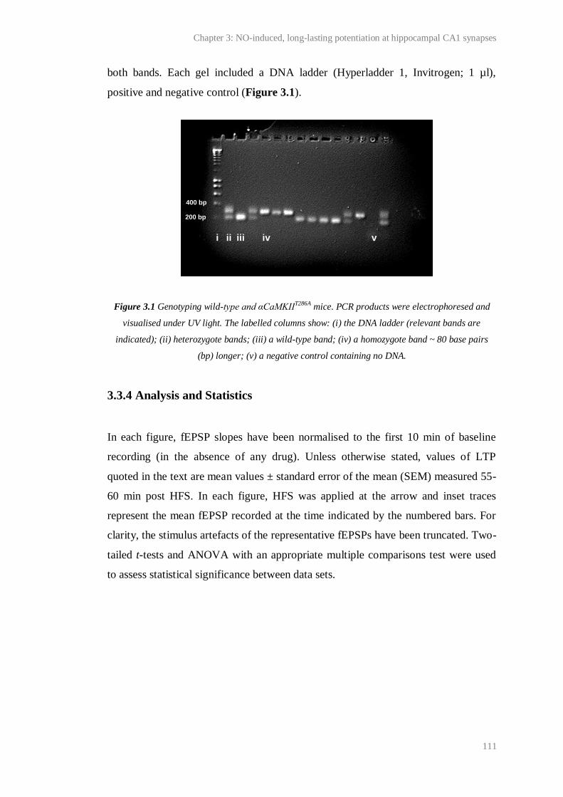
Chapter 3: NO-induced, long-lasting potentiation at hippocampal CA1 synapses
111
both bands. Each gel included a DNA ladder (Hyperladder 1, Invitrogen; 1 µl),
positive and negative control (Figure 3.1).
Figure 3.1 Genotyping wild-type and αCaMKIIT286A mice. PCR products were electrophoresed and
visualised under UV light. The labelled columns show: (i) the DNA ladder (relevant bands are
indicated); (ii) heterozygote bands; (iii) a wild-type band; (iv) a homozygote band ~ 80 base pairs
(bp) longer; (v) a negative control containing no DNA.
3.3.4 Analysis and Statistics
In each figure, fEPSP slopes have been normalised to the first 10 min of baseline
recording (in the absence of any drug). Unless otherwise stated, values of LTP
quoted in the text are mean values ± standard error of the mean (SEM) measured 55-
60 min post HFS. In each figure, HFS was applied at the arrow and inset traces
represent the mean fEPSP recorded at the time indicated by the numbered bars. For
clarity, the stimulus artefacts of the representative fEPSPs have been truncated. Two-
tailed t-tests and ANOVA with an appropriate multiple comparisons test were used
to assess statistical significance between data sets.
400 bp
200 bp
i ii iii iv v
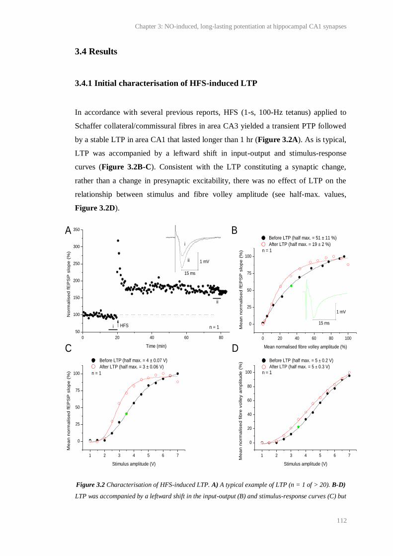
Chapter 3: NO-induced, long-lasting potentiation at hippocampal CA1 synapses
112
3.4 Results
3.4.1 Initial characterisation of HFS-induced LTP
In accordance with several previous reports, HFS (1-s, 100-Hz tetanus) applied to
Schaffer collateral/commissural fibres in area CA3 yielded a transient PTP followed
by a stable LTP in area CA1 that lasted longer than 1 hr (Figure 3.2A). As is typical,
LTP was accompanied by a leftward shift in input-output and stimulus-response
curves (Figure 3.2B-C). Consistent with the LTP constituting a synaptic change,
rather than a change in presynaptic excitability, there was no effect of LTP on the
relationship between stimulus and fibre volley amplitude (see half-max. values,
Figure 3.2D).
Figure 3.2 Characterisation of HFS-induced LTP. A) A typical example of LTP (n = 1 of > 20). B-D)
LTP was accompanied by a leftward shift in the input-output (B) and stimulus-response curves (C) but
1 2 3 4 5 6 7
0
25
50
75
100
0 20 40 60 80
50
100
150
200
250
300
350
1 2 3 4 5 6 7
0
20
40
60
80
100
0 20 40 60 80 100
0
25
50
75
100
Before LTP (half max. = 51 ± 11 %)
After LTP (half max. = 19 ± 2 %)M
ea
n n
orm
alise
d f
EP
SP
slo
pe
(%
)
Mean normalised fibre volley amplitude (%)
A B
C D Before LTP (half max. = 4 ± 0.07 V)
After LTP (half max. = 3 ± 0.06 V)
Me
an
no
rma
lise
d f
EP
SP
slo
pe
(%
)
Stimulus amplitude (V)
n = 1n = 1
n = 1
ii
i
i
ii
No
rma
lise
d f
EP
SP
slo
pe
(%
)
Time (min)
HFS n = 1
15 ms
1 mV
15 ms
1 mV
Before LTP (half max. = 5 ± 0.2 V)
After LTP (half max. = 5 ± 0.3 V)
Me
an
no
rma
lise
d f
ibre
vo
lle
y a
mp
litu
de
(%
)
Stimulus amplitude (V)
Chapter 3: NO-induced, long-lasting potentiation at hippocampal CA1 synapses
113
there was no effect of LTP on the relationship between stimulus amplitude and mean normalised fibre
volley amplitude (see half-max. values). Data were recorded 20 min prior to, and 60 min after HFS.
Each point is a mean of 5-10 consecutive fEPSPs measured from one typical slice. In B-C, data has
been normalised to the maximum mean value in each data set. In D, data have been normalised to the
maximum value recorded after LTP. The half-max. values were determined using the logistic fits
shown (adjusted R2 > 0.99; see Chapter 2 for logistic equation). The data coloured in green were
measured from the mean fEPSP shown in B. The fibre volley amplitude was measured as the negative
peak relative to the baseline.
Under certain conditions, LTP at CA1 synapses can be NMDA receptor- (Grover and
Teyler, 1990) and NO-independent (Cummings et al., 1994; Phillips et al., 2008).
Therefore, we tested the involvement of NMDA receptors and NO in LTP under our
conditions.
In agreement with the majority of previous findings at CA3-CA1 synapses (for
example, Collingridge et al., 1983a; Collingridge et al., 1983b; Malenka, 1991), the
NMDA antagonist, D-AP5 (50 µM, applied 10 min prior to and 5 min after HFS),
reversibly blocked HFS-induced LTP: following HFS in the presence of D-AP5, the
initial fEPSP was unchanged compared to the last 5 min of baseline recording,
although a second HFS, delivered ~ 1 hr after washing the antagonist, yielded
significant LTP (Figure 3.3A). There was no significant effect of D-AP5 on baseline
transmission (Figure 3.3A and Figure 3.3B).
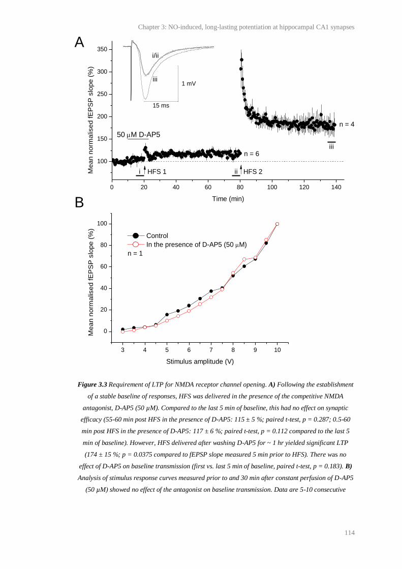
Chapter 3: NO-induced, long-lasting potentiation at hippocampal CA1 synapses
114
Figure 3.3 Requirement of LTP for NMDA receptor channel opening. A) Following the establishment
of a stable baseline of responses, HFS was delivered in the presence of the competitive NMDA
antagonist, D-AP5 (50 µM). Compared to the last 5 min of baseline, this had no effect on synaptic
efficacy (55-60 min post HFS in the presence of D-AP5: 115 ± 5 %; paired t-test, p = 0.287; 0.5-60
min post HFS in the presence of D-AP5: 117 ± 6 %; paired t-test, p = 0.112 compared to the last 5
min of baseline). However, HFS delivered after washing D-AP5 for ~ 1 hr yielded significant LTP
(174 ± 15 %; p = 0.0375 compared to fEPSP slope measured 5 min prior to HFS). There was no
effect of D-AP5 on baseline transmission (first vs. last 5 min of baseline, paired t-test, p = 0.183). B)
Analysis of stimulus response curves measured prior to and 30 min after constant perfusion of D-AP5
(50 µM) showed no effect of the antagonist on baseline transmission. Data are 5-10 consecutive
0 20 40 60 80 100 120 140
100
150
200
250
300
350
3 4 5 6 7 8 9 10
0
20
40
60
80
100
iii
i/ii
iii
iiMe
an
no
rma
lise
d f
EP
SP
slo
pe
(%
)
Time (min)
50 M D-AP5
i HFS 1 HFS 2
n = 6
n = 4
15 ms
1 mVM
ea
n n
orm
alis
ed
fE
PS
P s
lop
e (
%)
Stimulus amplitude (V)
Control
In the presence of D-AP5 (50 M)
A
B
n = 1

Chapter 3: NO-induced, long-lasting potentiation at hippocampal CA1 synapses
115
fEPSPs measured from one slice and are normalised to the maximum mean value recorded in each
data set.
The involvement of endogenous NO in HFS-induced LTP was tested using the non-
selective NOS antagonist, L-NNA (100 µM, applied 30 min prior to, and at least 30
min after HFS). Consistent with previous reports (for example, Hopper and
Garthwaite, 2006; Taqatqeh et al., 2009), HFS in the presence of L-NNA resulted in
a steadily declining potentiation. One hr after HFS, this potentiation was significantly
smaller than the LTP generated in un-treated, interleaved controls, although the
initial slope was significantly augmented from the baseline. There was no effect of
L-NNA on baseline transmission (Figure 3.4A and Figure 3.4B).
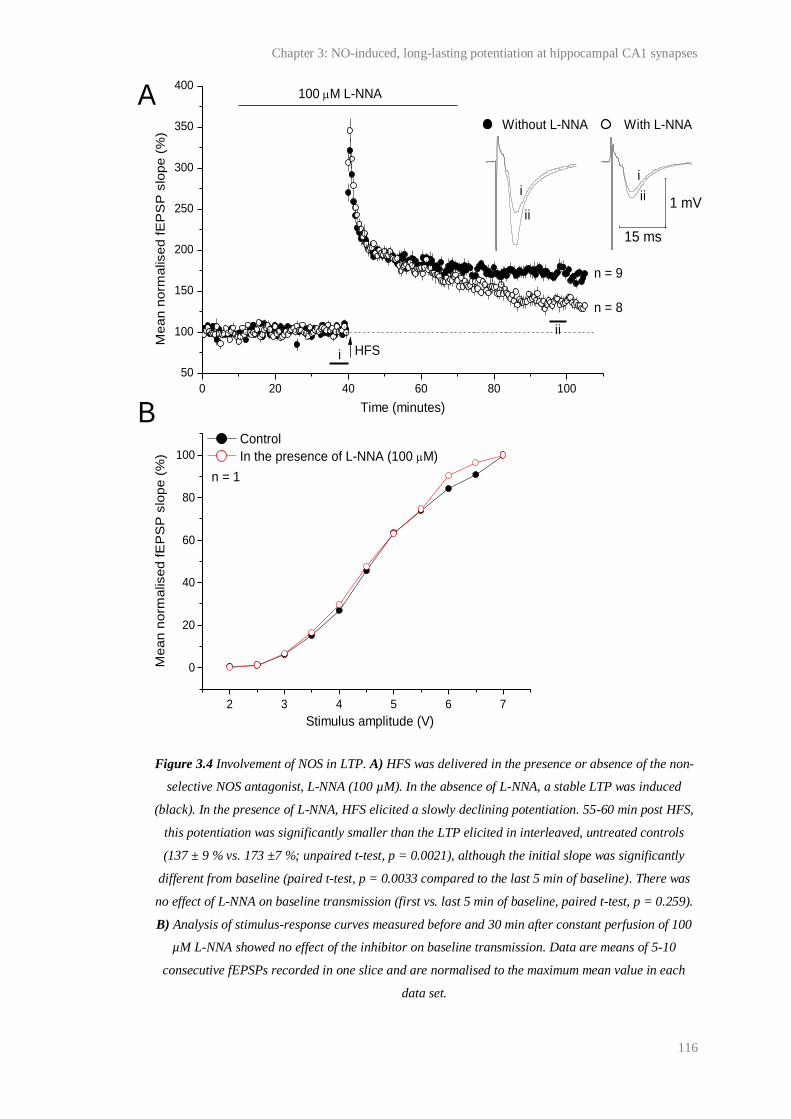
Chapter 3: NO-induced, long-lasting potentiation at hippocampal CA1 synapses
116
Figure 3.4 Involvement of NOS in LTP. A) HFS was delivered in the presence or absence of the non-
selective NOS antagonist, L-NNA (100 µM). In the absence of L-NNA, a stable LTP was induced
(black). In the presence of L-NNA, HFS elicited a slowly declining potentiation. 55-60 min post HFS,
this potentiation was significantly smaller than the LTP elicited in interleaved, untreated controls
(137 ± 9 % vs. 173 ±7 %; unpaired t-test, p = 0.0021), although the initial slope was significantly
different from baseline (paired t-test, p = 0.0033 compared to the last 5 min of baseline). There was
no effect of L-NNA on baseline transmission (first vs. last 5 min of baseline, paired t-test, p = 0.259).
B) Analysis of stimulus-response curves measured before and 30 min after constant perfusion of 100
µM L-NNA showed no effect of the inhibitor on baseline transmission. Data are means of 5-10
consecutive fEPSPs recorded in one slice and are normalised to the maximum mean value in each
data set.
0 20 40 60 80 100
50
100
150
200
250
300
350
400
2 3 4 5 6 7
0
20
40
60
80
100
ii
i
ii
i
i
Stimulus amplitude (V)Stimulus intensity (V)
Me
an
no
rma
lise
d f
EP
SP
slo
pe
(%
)
Without L-NNA With L-NNA
Me
an
no
rma
lise
d f
EP
SP
slo
pe
(%
)
Time (minutes)
100 M L-NNA
ii
HFS
n = 9
n = 8
15 ms
1 mV
Control
In the presence of L-NNA (100 M)
A
B
n = 1

Chapter 3: NO-induced, long-lasting potentiation at hippocampal CA1 synapses
117
The effect of the NOS substrate, L-arginine, on LTP was also investigated. L-
arginine is present in the cerebrospinal fluid of adult rats (50-80 µM; Takasugi et al.,
2003) and humans (20 µM; Martens-Lobenhoffer et al., 2007). However, the amino
acid is not normally added to solutions used in vitro, raising the possibility that the
concentration of L-arginine might limit NO synthesis and therefore NO-mediated
plasticity in hippocampal slices. Previously it has been reported that NMDA-induced
cGMP accumulation is augmented in rat hippocampal and cerebellar slices pre-
incubated with L-arginine (Garthwaite et al., 1989; East and Garthwaite, 1991), but
there are no reports of the effect of L-arginine on NO-dependent LTP in vitro.
To test the effect of L-arginine on our LTP, the amino acid (50 µM) was applied 30
min before HFS and remained present for the duration of the experiment. Compared
to un-treated interleaved controls, there was no significant effect of L-arginine on the
magnitude of LTP or on baseline transmission (Figure 3.5).
Figure 3.5 Dependency of LTP on extracellular L-arginine. L-arginine (50 µM) was applied 30 min
prior to HFS and for the remainder of the experiment. Compared to the LTP induced in interleaved,
untreated controls, the magnitude of LTP induced in slices perfused with L-arginine was not
significantly altered (with L-arginine (grey): 155 ± 4 %; without L-arginine (black): 170 ± 7 %;
0 20 40 60 80 100
50
100
150
200
250
300
350
iiii
ii
ii
Without L-arginine With L-arginine
Mea
n n
orm
alis
ed
fE
PS
P s
lop
e (
%)
Time (min)
50 M L-arginine
i
n = 9
n = 4
15 ms
1 mV

Chapter 3: NO-induced, long-lasting potentiation at hippocampal CA1 synapses
118
unpaired t-test, p = 0.109). There was also no significant effect of L-arginine on baseline
transmission (mean fEPSP slope over the first vs. last 5 min of baseline transmission = 101 ± 0.7 %
vs. 101 ± 4 %; paired t-test, p = 0.871). Note that control data is the same as that shown in Figure 3.4
but has been shown again for ease of comparison.
3.4.2 Effect of pairing exogenous NO with HFS during NMDA receptor
blockade
Under our conditions, LTP was NMDA receptor- (Figure 3.3) and NO-dependent
(Figure 3.4). Therefore the hypothesis that pairing exogenous NO with HFS during
NMDA receptor blockade should restore the NO-dependent component of LTP was
valid and could be tested.
For this purpose, the NO donor, PAPA/NONOate was used. As previously described
(see Chapter 2.1.6), PAPA/NONOate releases NO in a predictable manner (t½ = 15
min at 37 ºC in oxygenated aCSF, pH 7.4; Keefer et al., 1996). Since the
concentration of NO generated at a synapse during LTP is unknown, the effects of
different concentrations of PAPA/NONOate, co-applied with the D-AP5 during HFS,
were investigated. Tests of each concentration were interleaved with each other and
with control experiments in which: 1) HFS was delivered alone (as a positive control
for LTP induction and expression); 2) HFS was delivered in the presence of D-AP5
only (to control for the NMDA receptor-dependency of LTP; data have been shown
in Figure 3.3 and are shown again for ease of comparison).
As above, HFS delivered alone yielded a significant LTP (data shown in grey in
Figure 3.6). As shown previously (Figure 3.3), and again here for ease of
comparison, LTP was blocked by the NMDA antagonist, D-AP5 (data shown in
black in Figure 3.6). Consistent with our prediction, application of 3 µM
PAPA/NONOate in the presence of 50 µM D-AP5 and HFS yielded a long-lasting
potentiation (data shown in blue Figure 3.6A). This ‘NO-induced potentiation’ was
slow to develop (half-maximal value = 18 ± 2 min; red line in Figure 3.6A) but
reached a magnitude comparable to that observed in control slices that received HFS
alone (compare blue and grey in Figure 3.6A).
Chapter 3: NO-induced, long-lasting potentiation at hippocampal CA1 synapses
119
The magnitude of the NO-induced potentiation was dependent on the concentration
of PAPA/NONOate. Concentrations lower than 3 µM failed to give rise to a
significant potentiation in fEPSP slope, as did concentrations above 3 µM (ANOVA
with one factor Dunnett’s test, p < 0.05 compared to the fEPSP slope measured after
HFS in the presence of D-AP5 alone; Figure 3.6B-G).

Chapter 3: NO-induced, long-lasting potentiation at hippocampal CA1 synapses
120
Figure 3.6 Effect of PAPA/NONOate application during HFS and NMDA receptor blockade on
synaptic efficacy. A) HFS yielded a significant LTP (grey; 170 ± 5 %; paired t-test = 4.50 × 10⁻⁶
compared to the last 5 min of baseline; n = 8) that was blocked by the NMDA antagonist, D-AP5 (50
0 20 40 60 80
50
100
150
200
300
350
0 20 40 60 80
50
100
150
200
300
350
0 20 40 60 80
50
100
150
200
300
350
1 10
0 20 40 60 80
50
100
150
200
300
350
0 20 40 60 80
50
100
150
200
300
350
0 20 40 60 80
50
100
150
200
300
350
50 M D-AP50.3 M PAPA/NONOate
HFS
A B
3 M PAPA/NONOate
50 M D-AP5 HFS
HFS and D-AP5
HFS, D-AP5 and PAPA
Logistic, half-time = 18 ± 2 min
C D
HFS
Time (min) Time (min)
Time (min) Time (min)
Time (min)
Me
an
no
rm.
fEP
SP
slo
pe
(%
) 30 M PAPA/NONOate
Me
an
no
rm.
fEP
SP
slo
pe
(%
)
Time (min)
50 M D-AP5
HFS
(6) **
(5)
(2)
(3)(4)(4)
(3)
[PAPA/NONoate] (M)
55-60 min post HFS
** compared to D-AP5
90
100
110
120
130
140
150
160
170
D-AP5
G
Me
an
no
rm.
fEP
SP
slo
pe
(%
)
1 M PAPA/NONOate
Me
an
no
rm.
fEP
SP
slo
pe
(%
)
50 M D-AP5
HFS
10 M PAPA/NONOate 50 M D-AP5
HFS
E F60 M PAPA/NONOate
50 M D-AP5
HFS

Chapter 3: NO-induced, long-lasting potentiation at hippocampal CA1 synapses
121
µM; black; 115 ± 5 %; paired t-test, p = 0.287 compared to the last 5 min of baseline recording; n =
6; note that these data have been previously shown in Figure 3.3). Application of 3 μM
PAPA/NONOate during HFS in the presence of D-AP5 led to a significant, long-lasting potentiation
in fEPSP slope (blue; 150 ± 8 %; one factor ANOVA with Dunnett’s test, p < 0.01 compared to the
fEPSP slope measured following HFS in the presence of D-AP5 alone (black)). This was slowly-rising
(red; half-maximal value = 18 ± 2 min as estimated using a logistic fit of the data 3 min after HFS;
adjusted R2 = 0.664; see Chapter 2 for fit details) but reached a magnitude comparable to control
HFS-induced LTP (grey; unpaired t-test; p = 0.0545). B-F) Pairing of HFS and D-AP5 with lower
(0.3-1 µM; B-C) or higher (10-30 µM; D-F) concentrations of PAPA/NONOate had no significant
effect on synaptic efficacy (one factor ANOVA with Dunnett’s test, p > 0.05 compared to the fEPSP
following HFS in the presence of D-AP5 alone). Black and grey data are the same as in panel A but
have been shown repeatedly for ease of comparison. G) Summary showing the magnitude of NO-
induced potentiation 55-60 min following HFS as a function of [PAPA/NONOate]. Numbers above
points are n values; ** = one factor ANOVA with Dunnett’s test, p < 0.01 compared to HFS in the
presence of D-AP5 alone.
3.4.3 Characterisation of NO-induced potentiation
The NO-induced potentiation (generated using 3 µM PAPA/NONOate) was
consistent with the possibility that exogenous NO had bypassed the requirement for
NMDA receptor activity during LTP induction and prompted the expression of the
NO-dependent component of HFS-induced LTP. To test this and begin to
characterise the NO-induced potentiation, we investigated the relationship between
the NO-induced potentiation and standard HFS-induced LTP.
Interaction between the NO-induced potentiation and HFS-induced LTP
It was hypothesised that, if the NO-induced potentiation relied upon the same
mechanisms as the NO-dependent component of HFS-induced LTP, then these forms
of plasticity would be non-additive. To test this postulate, a second HFS was
delivered to slices at the end of the experiments shown in Figure 3.6 (i.e. 1 hr after
pre-treatment with D-AP5, PAPA/NONOate and HFS). After pre-treatment with 3
µM PAPA/NONOate, which generated significant NO-induced potentiation (see
Figure 3.6A and 3.7A), a second HFS (labelled HFS 2 in Figure 3.7A) generated
significant LTP, of an apparently remarkably high magnitude (255 ± 11 %; Figure

Chapter 3: NO-induced, long-lasting potentiation at hippocampal CA1 synapses
122
3.7A). The magnitude of the HFS-induced LTP appeared to be related to the
magnitude of the preceding NO-induced potentiation (Figure 3.7B-D).
Figure 3.7 Impact of the NO-induced potentiation on subsequent HFS-induced LTP. A) Slices
received HFS in the presence of D-AP5 alone (black) or HFS in the presence of D-AP5 and 3 µM
PAPA/NONOate, which generated a significant NO-induced potentiation (blue; same data as in
Figure 3.6A). One hr later, all slices received a second HFS (labelled HFS 2). In both conditions, this
resulted in significant LTP (paired t-tests, p < 0.05 compared to the initial slope measured 5 min
prior to HFS). In slices that had undergone NO-induced potentiation, the LTP was of a remarkably
high magnitude (unpaired t-test, p = 0.0162 compared to LTP in control slices after D-AP5 wash-out
(black)). B- C) Subsequent to treatment with HFS, D-AP5 and lower (0.3 µM; B) or higher (30 µM;
C) concentrations of PAPA/NONOate, HFS-induced LTP was not significantly different to that
observed in control slices after D-AP5 wash-out (one factor ANOVA with Dunnett’s test, p > 0.05). D)
The magnitude of the NO-induced potentiation (measured 55-60 min post HFS 1) and the magnitude
of subsequent HFS-induced LTP (measured 55-60 min post HFS 2) were similarly dependent on
0 20 40 60 80 100 120 140
50
100
150
200
250
300
350
400
450
0 20 40 60 80 100 120 140
50
100
150
200
250
300
350
400
450
1 10 100
0 20 40 60 80 100 120 140
50
100
150
200
250
300
350
400
450
50 M D-AP5
0.3 M PAPA
15 ms
1 mV
iiii
Time (min) Time (min)
Time (min)
50 M D-AP5
30 M PAPA
15 ms
1 mV
55-60 min post HFS 2
(6) (2)(3)
(3)
(5)(4)
(4)
[PAPA/NO] (M)
(6)
*
D-AP5 alone
55-60 min post HFS 1
HFS 1 HFS 2
* compared to LTP in D-AP5 controls
ii
i
i
ii i/ii
iii
iii
iiiiii
iii
iii
iii
iii
iii
ii
i/ii
i/ii
i/ii
i
i
D-AP5 D-AP5 and PAPA/NONOate
D-AP5 D-AP5 and PAPA/NONOate
Me
an
no
rma
lise
d f
EP
SP
slo
pe
(%
)
Me
an
no
rma
lise
d f
EP
SP
slo
pe
(%
)
Me
an
no
rma
lise
d f
EP
SP
slo
pe
(%
)
D-AP5 D-AP5 and PAPA/NONOate
Me
an
no
rma
lise
d f
EP
SP
slo
pe
(%
)
50 M D-AP5
3 M PAPA
HFS 1 HFS 2i
A B
C D
15 ms
1 mV
80
100
120
140
160
180
200
220
240
260
280
Chapter 3: NO-induced, long-lasting potentiation at hippocampal CA1 synapses
123
[PAPA/NONOate]. Numbers by points are n; * = one factor ANOVA with Dunnett’s test, p < 0.05
compared to the magnitude of HFS-induced LTP in control slices after D-AP5 wash-out (see black
bar).
Relative to the initial slope measured 5 min prior to HFS 2, the amount of LTP
yielded by HFS subsequent to the NO-induced potentiation (generated using 3 µM
PAPA/NONOate) was not significantly different from the amount yielded by HFS
subsequent to pre-treatment with D-AP5 alone (unpaired t-test, p = 0.0516; see
Figure 3.7A). This suggested that the NO-induced potentiation was additive with
subsequent HFS-induced LTP.
Further analysis of the relationship between the magnitude of the NO-induced
potentiation generated using 3 µM PAPA/NONOate and subsequent HFS-induced
LTP was also consistent with a purely additive interaction: the magnitude of the NO-
induced potentiation (generated using 3 µM PAPA/NONOate) was significantly
positively correlated with the magnitude of subsequent HFS-induced LTP (Figure
3.8, red); there was no significant correlation between the NO-induced potentiation
and subsequent LTP when the latter was normalised to the former (blue); the mean
magnitude of HFS-induced LTP, once normalised to the magnitude of the NO-
induced potentiation, was not significantly different from the magnitude of control,
HFS-induced LTP (grey bar vs. blue mean).
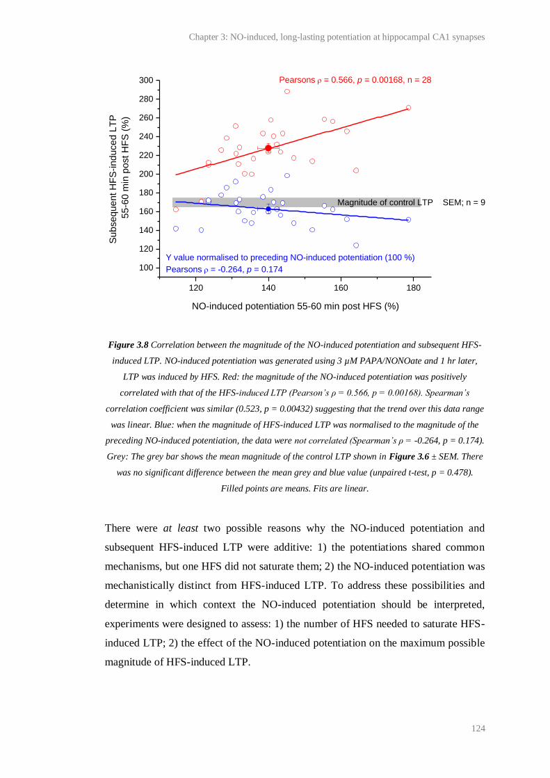
Chapter 3: NO-induced, long-lasting potentiation at hippocampal CA1 synapses
124
Figure 3.8 Correlation between the magnitude of the NO-induced potentiation and subsequent HFS-
induced LTP. NO-induced potentiation was generated using 3 µM PAPA/NONOate and 1 hr later,
LTP was induced by HFS. Red: the magnitude of the NO-induced potentiation was positively
correlated with that of the HFS-induced LTP (Pearson’s ρ = 0.566, p = 0.00168). Spearman’s
correlation coefficient was similar (0.523, p = 0.00432) suggesting that the trend over this data range
was linear. Blue: when the magnitude of HFS-induced LTP was normalised to the magnitude of the
preceding NO-induced potentiation, the data were not correlated (Spearman’s ρ = -0.264, p = 0.174).
Grey: The grey bar shows the mean magnitude of the control LTP shown in Figure 3.6 ± SEM. There
was no significant difference between the mean grey and blue value (unpaired t-test, p = 0.478).
Filled points are means. Fits are linear.
There were at least two possible reasons why the NO-induced potentiation and
subsequent HFS-induced LTP were additive: 1) the potentiations shared common
mechanisms, but one HFS did not saturate them; 2) the NO-induced potentiation was
mechanistically distinct from HFS-induced LTP. To address these possibilities and
determine in which context the NO-induced potentiation should be interpreted,
experiments were designed to assess: 1) the number of HFS needed to saturate HFS-
induced LTP; 2) the effect of the NO-induced potentiation on the maximum possible
magnitude of HFS-induced LTP.
120 140 160 180
100
120
140
160
180
200
220
240
260
280
300
Y value normalised to preceding NO-induced potentiation (100 %)
Pearsons = -0.264, p = 0.174
Su
bse
qu
en
t H
FS
-in
du
ce
d L
TP
5
5-6
0 m
in p
ost H
FS
(%
)
NO-induced potentiation 55-60 min post HFS (%)
Pearsons = 0.566, p = 0.00168, n = 28
Magnitude of control LTP SEM; n = 9
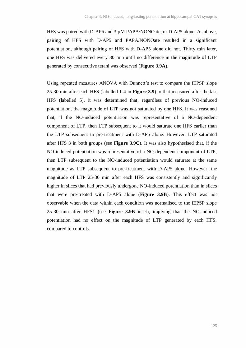
Chapter 3: NO-induced, long-lasting potentiation at hippocampal CA1 synapses
125
HFS was paired with D-AP5 and 3 µM PAPA/NONOate, or D-AP5 alone. As above,
pairing of HFS with D-AP5 and PAPA/NONOate resulted in a significant
potentiation, although pairing of HFS with D-AP5 alone did not. Thirty min later,
one HFS was delivered every 30 min until no difference in the magnitude of LTP
generated by consecutive tetani was observed (Figure 3.9A).
Using repeated measures ANOVA with Dunnett’s test to compare the fEPSP slope
25-30 min after each HFS (labelled 1-4 in Figure 3.9) to that measured after the last
HFS (labelled 5), it was determined that, regardless of previous NO-induced
potentiation, the magnitude of LTP was not saturated by one HFS. It was reasoned
that, if the NO-induced potentiation was representative of a NO-dependent
component of LTP, then LTP subsequent to it would saturate one HFS earlier than
the LTP subsequent to pre-treatment with D-AP5 alone. However, LTP saturated
after HFS 3 in both groups (see Figure 3.9C). It was also hypothesised that, if the
NO-induced potentiation was representative of a NO-dependent component of LTP,
then LTP subsequent to the NO-induced potentiation would saturate at the same
magnitude as LTP subsequent to pre-treatment with D-AP5 alone. However, the
magnitude of LTP 25-30 min after each HFS was consistently and significantly
higher in slices that had previously undergone NO-induced potentiation than in slices
that were pre-treated with D-AP5 alone (Figure 3.9B). This effect was not
observable when the data within each condition was normalised to the fEPSP slope
25-30 min after HFS1 (see Figure 3.9B inset), implying that the NO-induced
potentiation had no effect on the magnitude of LTP generated by each HFS,
compared to controls.
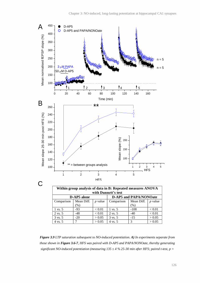
Chapter 3: NO-induced, long-lasting potentiation at hippocampal CA1 synapses
126
Within group analysis of data in B: Repeated measures ANOVA
with Dunnett’s test
D-AP5 alone D-AP5 and PAPA/NONOate Comparison Mean Diff.
(%)
p value Comparison Mean Diff.
(%)
p value
1 vs. 5 -93 < 0.01 1 vs. 5 -108 < 0.01
2 vs. 5 -48 < 0.01 2 vs. 5 -40 < 0.01
3 vs. 5 -20 > 0.05 3 vs. 5 -15 > 0.05
4 vs. 5 7 > 0.05 4 vs. 5 3 > 0.05
Figure 3.9 LTP saturation subsequent to NO-induced potentiation. A) In experiments separate from
those shown in Figure 3.6-7, HFS was paired with D-AP5 and PAPA/NONOate, thereby generating
significant NO-induced potentiation (measuring 135 ± 4 % 25-30 min after HFS; paired t-test, p =
0 20 40 60 80 100 120 140 160
100
150
200
250
300
350
400
450
1 2 3 4 5
100
120
140
160
180
200
220
240
260
1 2 3 4 5
100
150
200
D-AP5
D-AP5 and PAPA/NONOate
Me
an
no
rma
lise
d f
EP
SP
slo
pe
(%
)
Time (min)
50 M D-AP5
3 M PAPA
1 2 3 4 5
n = 5
n = 5
Me
an
slo
pe
25
-30
min
po
st
HF
S (
%)
HFS
A
B **
** = between groups analysis
Me
an
slo
pe
(%
)
HFS
C

Chapter 3: NO-induced, long-lasting potentiation at hippocampal CA1 synapses
127
0.00401 compared to the last 5 min of baseline), or D-AP5 alone, which had no significant effect on
the fEPSP slope (111 ± 5 % 25-30 min after HFS; p = 0.103 compared to the last 5 min of baseline).
30 min later a series of HFS were delivered to slices (one every 30 min; HFS 2-5) until the magnitude
of LTP was saturated (see comparison 4 vs. 5 in C). B) The mean fEPSP slope recorded 25-30 min
following each HFS (1-5) is plotted. ** = the magnitude of LTP measured 25-30 min after each HFS
was significantly higher in slices that had undergone NO-induced potentiation than in slices pre-
treated with D-AP5 alone (two factor ANOVA with repeated measures across HFS, p = 0.0007).
Inset: within each group, the data have been normalised to the value recorded after HFS 1. C) Within
each group, the magnitude of LTP yielded after each HFS is compared to that after the last HFS (HFS
5) using repeated measures ANOVA with Dunnett’s test. Within both groups, LTP was saturated after
HFS 3.
The above results were discordant with the possibility that the NO-induced
potentiation and subsequent LTP were additive because one HFS was not saturating
LTP. Rather, they were consistent with the possibility that the NO-induced
potentiation and subsequent LTP were mechanistically distinct. To test this, a series
of HFS were delivered to slices (one every 30 min; see HFS 1-5 in Figure 3.10A)
until there was no observable effect of consecutive tetani on the magnitude of LTP.
As above, repeated measures ANOVA with Dunnett’s test used to compare the
magnitude of LTP 25-30 min following each HFS (labelled 1-4) to that recorded
after the final tetanus (labelled 5) showed that 3 tetani were required to saturate the
magnitude of LTP (see Figure 3.10B). Subsequently, HFS was delivered in the
presence of PAPA/NONOate (see HFS 6 in Figure 3.10). If the NO-induced
potentiation was mechanistically distinct from HFS-induced LTP, then pairing of
exogenous NO with HFS was predicted to further increase the fEPSP slope.
However, there was no significant effect of pairing HFS with PAPA/NONOate
(Figure 3.10B).
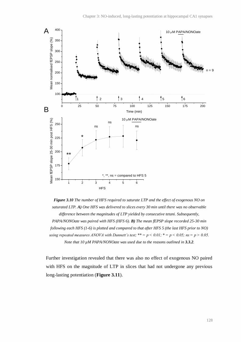
Chapter 3: NO-induced, long-lasting potentiation at hippocampal CA1 synapses
128
Figure 3.10 The number of HFS required to saturate LTP and the effect of exogenous NO on
saturated LTP. A) One HFS was delivered to slices every 30 min until there was no observable
difference between the magnitudes of LTP yielded by consecutive tetani. Subsequently,
PAPA/NONOate was paired with HFS (HFS 6). B) The mean fEPSP slope recorded 25-30 min
following each HFS (1-6) is plotted and compared to that after HFS 5 (the last HFS prior to NO)
using repeated measures ANOVA with Dunnett’s test; ** = p < 0.01; * = p < 0.05; ns = p > 0.05.
Note that 10 µM PAPA/NONOate was used due to the reasons outlined in 3.3.2.
Further investigation revealed that there was also no effect of exogenous NO paired
with HFS on the magnitude of LTP in slices that had not undergone any previous
long-lasting potentiation (Figure 3.11).
0 25 50 75 100 125 150 175 200
100
150
200
250
300
350
400
1 2 3 4 5 6150
175
200
225
250
10 M PAPA/NONOate
Me
an
no
rma
lise
d f
EP
SP
slo
pe
(%
)
Time (min)
10 M PAPA/NONOate
1 2 3 4 5 6
n = 9
ns
ns
*
Me
an
fE
PS
P s
lop
e 2
5-3
0 m
in p
ost
HF
S (
%)
HFS
**
ns
*, **, ns = compared to HFS 5
A
B
Chapter 3: NO-induced, long-lasting potentiation at hippocampal CA1 synapses
129
Figure 3.11 Effect of exogenous NO on control LTP. Slices received HFS, or HFS in the presence of
PAPA/NONOate. PAPA/NONOate had no significant effect on the magnitude of the resulting LTP
(HFS alone = 178 ± 8 %; HFS and PAPA/NONOate = 166 ± 6 %; unpaired t test, p = 0.199). Two
concentrations of PAPA/NONOate were tested due to the reasons discussed in 3.3.2.
Mechanisms underlying the NO-induced potentiation
The NO-induced potentiation was additive with subsequent HFS-induced LTP
(Figure 3.7-9) and this could not be explained by the inability of one HFS to saturate
LTP (Figure 3.9). These findings were discordant with the hypothesis that
exogenous NO paired with a standard LTP induction protocol during NMDA
receptor blockade would restore a persistent NO-dependent component of LTP and
suggested that the NO-induced potentiation was mechanistically distinct from LTP.
However, exogenous NO paired with HFS had no significant effect on the magnitude
of the resulting potentiation (Figure 3.10-11). Possible explanations for this are
detailed below (see 3.5 Discussion). To further investigate whether the NO-induced
potentiation was dependent upon similar mechanisms as NMDA receptor-and NO-
dependent LTP, key properties of the NO-induced potentiation were investigated.
0 20 40 60 80
50
100
150
200
250
300
350
iiii
ii
ii
Without PAPA With PAPA
Me
an
no
rma
lise
d fE
PS
P s
lop
e (
%)
Time (min)
3-10 M PAPA/NONOate
i
n = 5-6
15 ms
1 mV
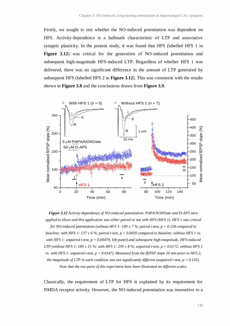
Chapter 3: NO-induced, long-lasting potentiation at hippocampal CA1 synapses
130
Firstly, we sought to test whether the NO-induced potentiation was dependent on
HFS. Activity-dependence is a hallmark characteristic of LTP and associative
synaptic plasticity. In the present study, it was found that HFS (labelled HFS 1 in
Figure 3.12) was critical for the generation of NO-induced potentiation and
subsequent high-magnitude HFS-induced LTP. Regardless of whether HFS 1 was
delivered, there was no significant difference in the amount of LTP generated by
subsequent HFS (labelled HFS 2 in Figure 3.12). This was consistent with the results
shown in Figure 3.8 and the conclusions drawn from Figure 3.9.
Figure 3.12 Activity-dependence of NO-induced potentiation. PAPA/NONOate and D-AP5 were
applied to slices and this application was either paired or not with HFS (HFS 1). HFS 1 was critical
for NO-induced potentiation (without HFS 1: 109 ± 7 %; paired t-test, p = 0.158 compared to
baseline; with HFS 1: 137 ± 6 %; paired t-test, p = 0.0039 compared to baseline; without HFS 1 vs.
with HFS 1: unpaired t-test, p = 0.00479; left panel) and subsequent high-magnitude, HFS-induced
LTP (without HFS 1: 189 ± 15 %; with HFS 1: 239 ± 8 %; unpaired t-test, p = 0.0172; without HFS 1
vs. with HFS 1: unpaired t-test, p = 0.0147). Measured from the fEPSP slope 10 min prior to HFS 2,
the magnitude of LTP in each condition was not significantly different (unpaired t-test, p = 0.155).
Note that the two parts of this experiment have been illustrated on different scales.
Classically, the requirement of LTP for HFS is explained by its requirement for
NMDA receptor activity. However, the NO-induced potentiation was insensitive to a
0 20 40 60 80
50
100
150
200
250
80 100 120 140
50
100
150
200
250
300
350
400
450
ii
With HFS 1 (n = 5) Without HFS 1 (n = 7)
Me
an
no
rma
lise
d fE
PS
P s
lop
e (
%)
Time (min)
50 M D-AP5
3 M PAPA/NONOate
HFS 1 HFS 2
i
iii
iii
ii ii
i i
iii
ii
Me
an
no
rma
lise
d fE
PS
P s
lop
e (
%)
Time (min)
15 ms
1 mV
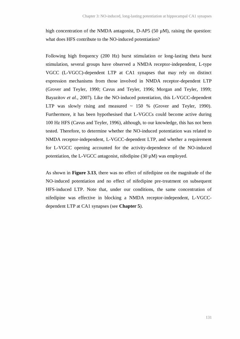
Chapter 3: NO-induced, long-lasting potentiation at hippocampal CA1 synapses
131
high concentration of the NMDA antagonist, D-AP5 (50 µM), raising the question:
what does HFS contribute to the NO-induced potentiation?
Following high frequency (200 Hz) burst stimulation or long-lasting theta burst
stimulation, several groups have observed a NMDA receptor-independent, L-type
VGCC (L-VGCC)-dependent LTP at CA1 synapses that may rely on distinct
expression mechanisms from those involved in NMDA receptor-dependent LTP
(Grover and Teyler, 1990; Cavus and Teyler, 1996; Morgan and Teyler, 1999;
Bayazitov et al., 2007). Like the NO-induced potentiation, this L-VGCC-dependent
LTP was slowly rising and measured ~ 150 % (Grover and Teyler, 1990).
Furthermore, it has been hypothesised that L-VGCCs could become active during
100 Hz HFS (Cavus and Teyler, 1996), although, to our knowledge, this has not been
tested. Therefore, to determine whether the NO-induced potentiation was related to
NMDA receptor-independent, L-VGCC-dependent LTP, and whether a requirement
for L-VGCC opening accounted for the activity-dependence of the NO-induced
potentiation, the L-VGCC antagonist, nifedipine (30 µM) was employed.
As shown in Figure 3.13, there was no effect of nifedipine on the magnitude of the
NO-induced potentiation and no effect of nifedipine pre-treatment on subsequent
HFS-induced LTP. Note that, under our conditions, the same concentration of
nifedipine was effective in blocking a NMDA receptor-independent, L-VGCC-
dependent LTP at CA1 synapses (see Chapter 5).
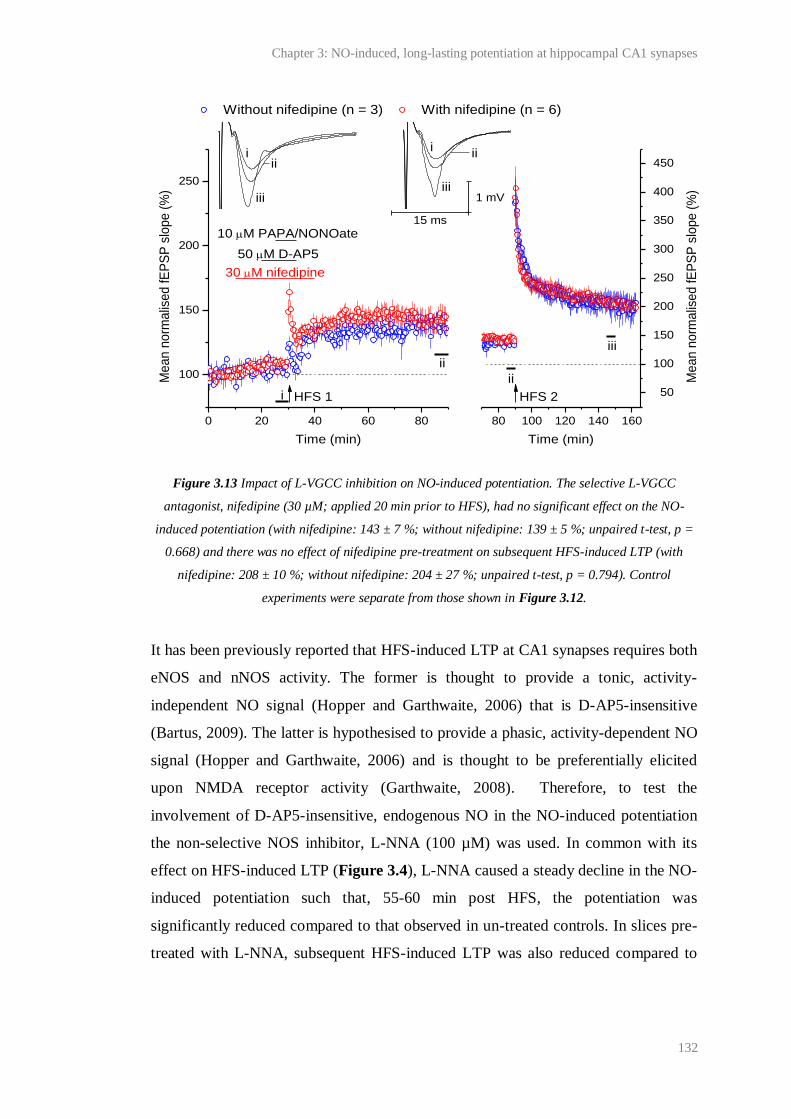
Chapter 3: NO-induced, long-lasting potentiation at hippocampal CA1 synapses
132
Figure 3.13 Impact of L-VGCC inhibition on NO-induced potentiation. The selective L-VGCC
antagonist, nifedipine (30 µM; applied 20 min prior to HFS), had no significant effect on the NO-
induced potentiation (with nifedipine: 143 ± 7 %; without nifedipine: 139 ± 5 %; unpaired t-test, p =
0.668) and there was no effect of nifedipine pre-treatment on subsequent HFS-induced LTP (with
nifedipine: 208 ± 10 %; without nifedipine: 204 ± 27 %; unpaired t-test, p = 0.794). Control
experiments were separate from those shown in Figure 3.12.
It has been previously reported that HFS-induced LTP at CA1 synapses requires both
eNOS and nNOS activity. The former is thought to provide a tonic, activity-
independent NO signal (Hopper and Garthwaite, 2006) that is D-AP5-insensitive
(Bartus, 2009). The latter is hypothesised to provide a phasic, activity-dependent NO
signal (Hopper and Garthwaite, 2006) and is thought to be preferentially elicited
upon NMDA receptor activity (Garthwaite, 2008). Therefore, to test the
involvement of D-AP5-insensitive, endogenous NO in the NO-induced potentiation
the non-selective NOS inhibitor, L-NNA (100 µM) was used. In common with its
effect on HFS-induced LTP (Figure 3.4), L-NNA caused a steady decline in the NO-
induced potentiation such that, 55-60 min post HFS, the potentiation was
significantly reduced compared to that observed in un-treated controls. In slices pre-
treated with L-NNA, subsequent HFS-induced LTP was also reduced compared to
80 100 120 140 160
50
100
150
200
250
300
350
400
450
0 20 40 60 80
100
150
200
250
ii
ii
iii
i
Me
an
no
rma
lise
d fE
PS
P s
lop
e (
%)
Time (min)
HFS 1 HFS 2
ii
ii
iii
iii
ii
Without nifedipine (n = 3) With nifedipine (n = 6)
Me
an
no
rma
lise
d fE
PS
P s
lop
e (
%)
Time (min)
30 M nifedipine
50 M D-AP5
10 M PAPA/NONOate15 ms
1 mV
Chapter 3: NO-induced, long-lasting potentiation at hippocampal CA1 synapses
133
that observed in un-treated controls (Figure 3.14). It is unclear whether this was a
consequence of reduced NO-induced potentiation, or incomplete L-NNA wash-out.
Figure 3.14 Requirement of NO-induced potentiation for NOS. The non-selective NOS inhibitor, L-
NNA (100 µM), was applied to slices 30 min prior to and after HFS. This caused a steady decline in
the NO-induced potentiation such that, 55-60 min post HFS it was significantly reduced compared to
interleaved, un-treated controls (118 ± 8 % vs. 148 ± 11 %; unpaired t-test, p= 0.0383). In slices pre-
treated with L-NNA, subsequent HFS-induced LTP was also significantly reduced compared to that in
un-treated controls (160 ± 11 % vs. 238 ± 19 %; p = 0.0049).
Physiological NO signal transduction is thought to occur via guanylyl cyclase
activation and cGMP accumulation (Garthwaite, 2008). Consistent with this
mechanism, NO-dependent CA1 LTP has been found to be significantly inhibited by
the NO-targeted guanylyl cyclase antagonist, ODQ (for example, Boulton et al.,
1995; Lu et al., 1999; Bon and Garthwaite, 2003). Accordingly, we sought to test the
requirement of the NO-induced potentiation for NO-targeted guanylyl cyclase
activity. In common with the effect of L-NNA, ODQ (10 µM) caused a significant
decline in the expression of the NO-induced potentiation, and in slices pre-treated
with ODQ, the magnitude of subsequent HFS-induced LTP was also significantly
80 100 120 140 160
50
100
150
200
250
300
350
400
450
500
550
0 20 40 60 80 100
50
100
150
200
250
i
ii
i
ii
iii
Me
an
no
rma
lise
d fE
PS
P s
lop
e (
%)
Time (min)
iii
iii
Without L-NNA (n= 5) With L-NNA (n = 5)
Me
an
no
rma
lise
d fE
PS
P s
lop
e (
%)
Time (min)
100 M L-NNA
50M D-AP5
3 M PAPA/NONOate
iii
HFS 1 HFS 2
15 ms
1 mV
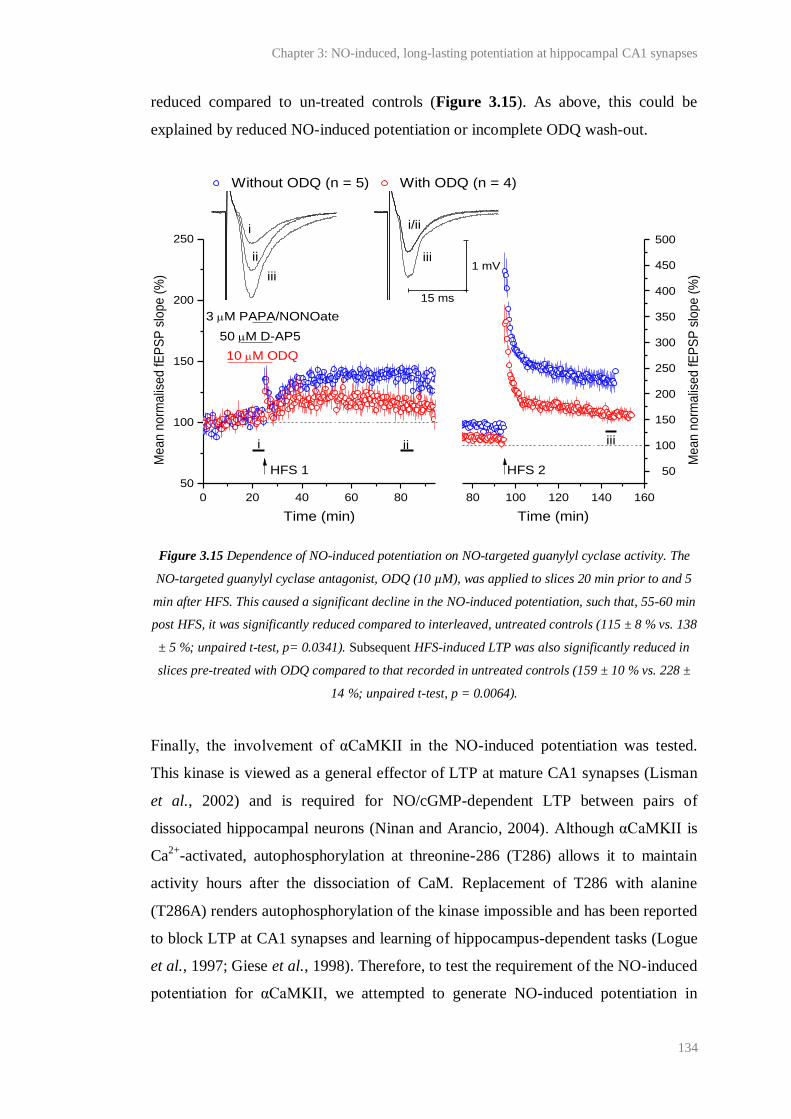
Chapter 3: NO-induced, long-lasting potentiation at hippocampal CA1 synapses
134
reduced compared to un-treated controls (Figure 3.15). As above, this could be
explained by reduced NO-induced potentiation or incomplete ODQ wash-out.
Figure 3.15 Dependence of NO-induced potentiation on NO-targeted guanylyl cyclase activity. The
NO-targeted guanylyl cyclase antagonist, ODQ (10 µM), was applied to slices 20 min prior to and 5
min after HFS. This caused a significant decline in the NO-induced potentiation, such that, 55-60 min
post HFS, it was significantly reduced compared to interleaved, untreated controls (115 ± 8 % vs. 138
± 5 %; unpaired t-test, p= 0.0341). Subsequent HFS-induced LTP was also significantly reduced in
slices pre-treated with ODQ compared to that recorded in untreated controls (159 ± 10 % vs. 228 ±
14 %; unpaired t-test, p = 0.0064).
Finally, the involvement of αCaMKII in the NO-induced potentiation was tested.
This kinase is viewed as a general effector of LTP at mature CA1 synapses (Lisman
et al., 2002) and is required for NO/cGMP-dependent LTP between pairs of
dissociated hippocampal neurons (Ninan and Arancio, 2004). Although αCaMKII is
Ca2+
-activated, autophosphorylation at threonine-286 (T286) allows it to maintain
activity hours after the dissociation of CaM. Replacement of T286 with alanine
(T286A) renders autophosphorylation of the kinase impossible and has been reported
to block LTP at CA1 synapses and learning of hippocampus-dependent tasks (Logue
et al., 1997; Giese et al., 1998). Therefore, to test the requirement of the NO-induced
potentiation for αCaMKII, we attempted to generate NO-induced potentiation in
0 20 40 60 80
50
100
150
200
250
80 100 120 140 160
50
100
150
200
250
300
350
400
450
500
Without ODQ (n = 5) With ODQ (n = 4)
Me
an
no
rma
lise
d fE
PS
P s
lop
e (
%)
Time (min)
10 M ODQ
50 M D-AP5
3 M PAPA/NONOate
iii
iii
iii
HFS 1 HFS 2
15 ms
1 mV
i i/ii
ii
Me
an
no
rma
lise
d fE
PS
P s
lop
e (
%)
Time (min)
iii
Chapter 3: NO-induced, long-lasting potentiation at hippocampal CA1 synapses
135
slices prepared from αCaMKIIT286A
homozygote and wild-type mice (described by
Giese et al., 1998).
At CA1 synapses in slices prepared from wild-type littermates, significant NO-
induced potentiation was observed. However, at synapses in slices from
αCaMKIIT286A
homozygote mice, neither the NO-induced potentiation nor
subsequent HFS-induced LTP could be induced (Figure 3.16A).
Compared to the magnitude typically observed in C57Bl/6 mice, a relative decrease
in the magnitude of the NO-induced potentiation and subsequent HFS-induced LTP
was apparent in the wild-type mice (NO-induced potentiation in wild-type data set:
130 ± 4 %; subsequent LTP: 183 ± 10 %; n = 5). However, the magnitude of NO-
induced potentiation was identical to the degree of HFS-induced LTP observed in a
pilot experiment using 1 wild-type littermate (130 %; Figure 3.16B). The relative
decrease may, therefore, be inherent to these mice and caused, perhaps, by their
background.

Chapter 3: NO-induced, long-lasting potentiation at hippocampal CA1 synapses
136
Figure 3.16 Involvement of αCaMKII in NO-induced potentiation. A) Neither the NO-induced
potentiation, nor subsequent HFS-induced LTP could be induced in αCaMKIIT286A homozygote mice
(NO-induced potentiation: 99 ± 8 %; paired t-test, p = 0.461 compared to baseline; subsequent HFS-
induced LTP: 101 ± 9 %; p = 0.367 compared to baseline). However, both forms of potentiation
could be observed in wild-type littermates (NO-induced potentiation: 130 ± 4 %; paired t-test, p =
0.0006 compared to baseline; HFS-induced LTP: 183 ± 10 %; paired t-test, p = 0247 compared to the
last 5 min of the NO-induced potentiation). B) HFS-induced LTP in a wild-type littermate (130 %).
3.4.4 Synaptic locus of the NO-induced potentiation
Although the precise relationship between the NO-induced potentiation and HFS-
induced LTP was unclear (Figure 3.8-11), initial characterisation of the NO-induced
80 100 120 140
50
100
150
200
250
300
350
400
450
500
0 20 40 60 80
50
100
150
200
250
0 10 20 30 40 50 60 70 80 90
80
100
120
140
160
180
200
iiiiii
Me
an
no
rma
lise
d f
EP
SP
slo
pe
(%
)
Time (min)
A
B
iiii
iii
ii/iii
Wild type littermates (n = 5) CAMKIIT286A
(n = 6)
Me
an
no
rma
lise
d f
EP
SP
slo
pe
(%
)
Time (min)
50 M D-AP5
3 M PAPA/NONOate
HFS 1 HFS 2
15 ms
1 mV
No
rma
lise
d f
EP
SP
slo
pe
(%
)
Time (min)
HFS n = 1
Chapter 3: NO-induced, long-lasting potentiation at hippocampal CA1 synapses
137
potentiation showed that it had properties in common with at least some forms of
LTP. For example, the NO-induced potentiation required HFS (Figure 3.12),
endogenous NO (Figure 3.14), NO-targeted guanylyl-cyclase activity (Figure 3.15),
and functional αCaMKII (Figure 3.16). Given the hypothesis that the NO acts as a
retrograde messenger during LTP (see 3.1 Introduction), we therefore sought to test
the synaptic locus (pre- or postsynaptic) of the NO-induced potentiation.
To this end, the effect of the NO-induced potentiation on PPF, at CA1 synapses was
investigated. PPF, which can be elicited when a pair of stimuli are delivered to
synapses in quick succession (typically with an ISI < 500 ms), is characterised by a
transient increase in synaptic efficacy (for example, see Figure 3.17). Although
some doubts exist over the synaptic locus of PPF (reviewed by Bliss et al., 2007), the
facilitation can be explained by a change in presynaptic efficacy caused, for example,
by residual Ca2+
in the axon bouton (Wu and Saggau, 1994b) or a related
mechanism, such as the saturation of a presynaptic Ca2+
buffer (Rozov et al., 2001).
Therefore changes in PPF are often used to diagnose a presynaptic change in efficacy
following LTP induction. Specifically, an increase in the probability of
neurotransmitter release following LTP is thought to reduce the scope for a
subsequent increase during PPF and, therefore, cause a decrease in the magnitude of
PPF (measured using the PPR; the initial slope of the second fEPSP/the initial slope
of the first fEPSP; Zucker and Regehr, 2002; Bliss et al., 2007).
Initial characterisation of PPF
Before testing the effect of the NO-induced potentiation on the magnitude of PPF at
CA1 synapses, we sought to characterise PPF under our conditions and determine
whether statistically significant, evoked changes in the PPR could be detected. In
accordance with previous reports (Creager et al., 1980), initial studies showed that
PPF of CA1 fEPSPs could be consistently generated using ISIs of 10-400 ms (Figure
3.17). The magnitude of PPF (determined using the PPR) was dependent on the ISI
used, and the relationship was remarkably similar to that previously described at
CA1 synapses in rat hippocampal slices (Creager et al., 1980; Dunwiddie and Haas,
1985) and in awake C57/Bl6 mice (Fontinha et al., 2009).

Chapter 3: NO-induced, long-lasting potentiation at hippocampal CA1 synapses
138
Figure 3.17 PPF of CA1 fEPSPs. PPF of CA1 fEPSPs could be consistently elicited by stimulating
the Schaffer collateral/commissural fibres twice in quick succession (ISI = 10-400 ms). The
magnitude of PPF (determined using the PPR) was dependent on the ISI: the PPR increased from 10
ms to peak at 25 ms and then decreased exponentially over the range of 25-400 ms. The PPR was
half-maximal at 81 ± 6 s (see τ; see Chapter 2 for exponential equation; adjusted R2 = 0.993). In
individual experiments, PPRs were measured from the mean of 10 consecutive pairs of fEPSPs (an
example is inset).
To determine whether, at a given ISI, statistically significant, evoked changes in PPF
could be detected under our conditions, we measured alterations in the PPR in
response to 2 compounds, 2-Cl-adenosine and forskolin, known to have opposite
effects on the magnitude of PPF at CA1 synapses. To avoid ceiling effects on any
changes in the PPR, the ISI was set to 100 ms, since this ISI was found to elicit ~
half-maximal PPF in initial experiments (see τ in Figure 3.17).
In accordance with previous studies of the effect of adenosine on PPF at CA1
synapses in adult rat hippocampal slices (Dunwiddie and Haas, 1985; Dumas and
Foster, 1998), bath application of 2-Cl-adenosine (0.5 µM) elicited a depression of
fEPSP 1 and, to a lesser extent, fEPSP 2 (Figure 3.18A), causing a significant
increase of the mean PPR from baseline (Figure 3.18C). As shown in Figure 3.18B,
the increase in PPR was observed in all the slices tested.
0 200 400
1.2
1.4
1.6
1.8
2.0
2.2
2.4
Exponential fit, = 81 ± 6 s
Me
an
PP
R (
arb
itra
ry u
nits)
ISI (ms)
n = 5-19
50 ms
0.5 mV
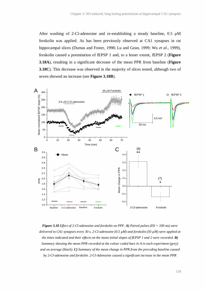
Chapter 3: NO-induced, long-lasting potentiation at hippocampal CA1 synapses
139
After washing of 2-Cl-adenosine and re-establishing a steady baseline, 0.5 µM
forskolin was applied. As has been previously observed at CA1 synapses in rat
hippocampal slices (Dumas and Foster, 1998; Lu and Gean, 1999; Wu et al., 1999),
forskolin caused a potentiation of fEPSP 1 and, to a lesser extent, fEPSP 2 (Figure
3.18A), resulting in a significant decrease of the mean PPR from baseline (Figure
3.18C). This decrease was observed in the majority of slices tested, although two of
seven showed an increase (see Figure 3.18B).
Figure 3.18 Effect of 2-Cl-adenosine and forskolin on PPF. A) Paired pulses (ISI = 100 ms) were
delivered to CA1 synapses every 30 s. 2-Cl-adenosine (0.5 µM) and forskolin (50 µM) were applied at
the times indicated and their effects on the mean initial slopes of fEPSP 1 and 2 were recorded. B)
Summary showing the mean PPR recorded at the colour coded bars in A in each experiment (grey)
and on average (black). C) Summary of the mean change in PPR from the preceding baseline caused
by 2-Cl-adenosine and forskolin. 2-Cl-Adenosine caused a significant increase in the mean PPR
Baseline 2-Cl-adenosine Wash Forskolin
1.0
1.2
1.4
1.6
1.8
2.0
2.2
2.4
2.6
2.8
3.0
0 10 20 30 40 50 60 70
0
50
100
150
200
250
300
50 ms
0.5 mV
PP
R
Mean
A
B C
Baseline
(7)
fEPSP 1 fEPSP 2
Me
an
no
rma
lise
d fE
PS
P s
lop
e (
%)
Time (min)
0.5 M 2-Cl-adenosine
50 M Forskolin
(8)
2-Cl-adenosine Forskolin
-0.2
-0.1
0.0
0.1
0.2
0.3
0.4
*
Me
an
ch
an
ge
in
PP
R
**
Chapter 3: NO-induced, long-lasting potentiation at hippocampal CA1 synapses
140
(mean change (red minus black in A and B): 0.31 ± 0.08; ** one factor t-test, p = 0.00482 compared
to zero). Forskolin induced a significant decrease in the mean PPR (mean change (green minus blue
in A and B): 0.14 ± 0.05; * one factor t-test, p = 0.0436 compared to zero change). Numbers above
bars are n.
Effect of HFS-induced LTP and the NO-induced potentiation on the
magnitude of PPF
The above results suggested that significant bidirectional changes in PPF could be
detected under our conditions. Therefore, the effects of the NO-induced potentiation
and subsequent HFS-induced LTP on PPF were determined. For the reasons outlined
in 3.3.2, 10 µM PAPA/NONOate was used to generate a significant NO-induced
potentiation, which measured 138 ± 5 %. Subsequent LTP was 222 ± 11 %.
Additionally, experiments were performed to control for the effect of HFS paired
with D-AP5 on PPF. In these experiments, HFS in the presence of D-AP5 alone had
no significant effect on fEPSP slope, although HFS subsequent to D-AP5 wash-out
generated significant LTP (Figure 3.19A). PPF was induced at the coloured bars
shown in Figure 3.19A. As above, the ISI was set to 100 ms.
As shown in grey in Figure 3.19B, the effects of all treatments on PPF varied
between experiments. On average, there was no significant change in the mean PPR
from baseline after pairing D-AP5 with HFS, or after LTP induced subsequent to D-
AP5 wash-out. Compared to the change in PPR following HFS in the presence of D-
AP5 alone, there was no significant effect of the NO-induced potentiation on PPR.
Compared to the change in PPR following LTP in slices pre-treated with D-AP5
alone, LTP subsequent to the NO-induced potentiation also had no effect on mean
PPR (Figure 3.19C).
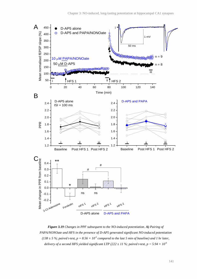
Chapter 3: NO-induced, long-lasting potentiation at hippocampal CA1 synapses
141
Figure 3.19 Changes in PPF subsequent to the NO-induced potentiation. A) Pairing of
PAPA/NONOate and HFS in the presence of D-AP5 generated significant NO-induced potentiation
(138 ± 5 %; paired t-test, p = 8.56 × 10⁻⁵ compared to the last 5 min of baseline) and 1 hr later,
delivery of a second HFS yielded significant LTP (222 ± 11 %; paired t-test, p = 5.94 × 10⁻⁶
0 20 40 60 80 100 120 140
50
100
150
200
250
300
350
400
450
Baseline Post HFS 1 Post HFS 2
1.2
1.4
1.6
1.8
2.0
2.2
2.4
Baseline Post HFS 1 Post HFS 2
1.2
1.4
1.6
1.8
2.0
2.2
2.4
i
ii
D-AP5 alone
D-AP5 and PAPA/NONOate
Me
an
no
rma
lise
d f
EP
SP
slo
pe
(%
)
Time (min)
50 M D-AP5
10 M PAPA/NONOate
HFS 1 HFS 2i
iii
n = 9
n = 8
PP
R
D-AP5 alone D-AP5 and PAPA
i ii iii i ii iii
2-Cl-Adenosine
ForskolinHFS 1
HFS 2HFS 1
HFS 2
-0.2
-0.1
0.0
0.1
0.2
0.3
0.4
nsns
Me
an
ch
an
ge
in
PP
R f
rom
ba
se
line
**
*
D-AP5 alone D-AP5 and PAPA
ISI = 100 ms
A
B
C
#
#
50 ms
1 mV

Chapter 3: NO-induced, long-lasting potentiation at hippocampal CA1 synapses
142
compared to the last 5 min of NO-induced potentiation). HFS in the presence of D-AP5 alone
generated no significant potentiation (107 ± 5 %; paired t-test, p = 0.312 compared to the last 5 min
of baseline), although after washing D-AP5, a significant LTP was generated (163 ± 8 %; paired t-
test, p = 7.08 × 10⁻⁶ compared to fEPSP slope 5 min prior to HFS). Paired stimuli (ISI = 100 ms)
were delivered at the bars number i-iii. B) The magnitudes of the PPF elicited at the time points
indicated by the numbered bars in A in individual experiments (grey) and on average (black or blue)
are plotted. C) Summary showing the average change in PPR following HFS 1 or 2 (see panel A) in
slices treated with D-AP5 or D-AP5 and PAPA/NONOate compared to baseline. The average changes
in PPR following treatment with 0.5 µM 2-Cl-adenosine or 50 µM forskolin (ISI = 100 ms; see
Figure 3.17) are shown again for comparison. Statistics: one factor t-tests compared to zero change:
** = p < 0.01, * = p < 0.05, ns= p > 0.05 compared to zero; #: unpaired t-test, p > 0.05.
Finally, to place the above results into context, the effect of HFS-induced LTP on
PPF was evaluated. In these experiments, LTP measured 179 ± 6 % (Figure 3.20A).
As shown in Figure 3.20B, the effect of HFS-induced LTP on PPF when the ISI was
100 ms was very variable (PPR increased in 4 slices and decreased in 6 slices) and
on average, there was no significant change. Similar results were obtained when the
ISI was 25 or 200 ms (Figure 3.20C).
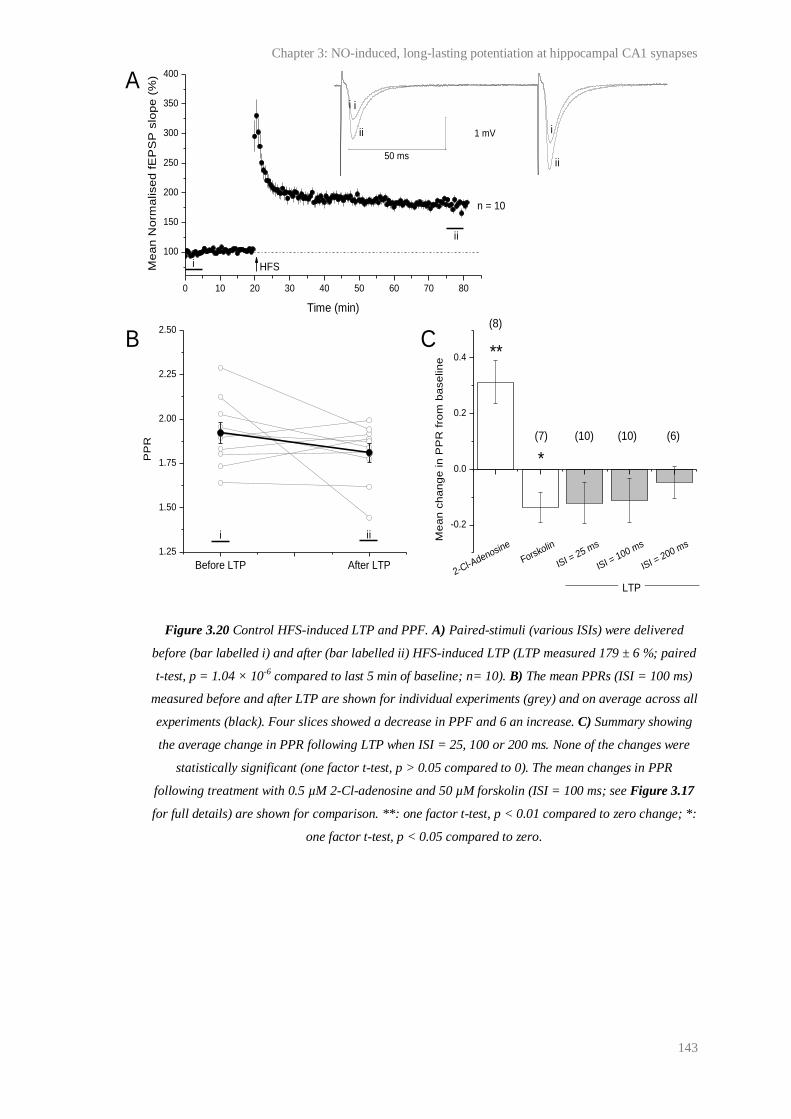
Chapter 3: NO-induced, long-lasting potentiation at hippocampal CA1 synapses
143
Figure 3.20 Control HFS-induced LTP and PPF. A) Paired-stimuli (various ISIs) were delivered
before (bar labelled i) and after (bar labelled ii) HFS-induced LTP (LTP measured 179 ± 6 %; paired
t-test, p = 1.04 × 10⁻⁶ compared to last 5 min of baseline; n= 10). B) The mean PPRs (ISI = 100 ms)
measured before and after LTP are shown for individual experiments (grey) and on average across all
experiments (black). Four slices showed a decrease in PPF and 6 an increase. C) Summary showing
the average change in PPR following LTP when ISI = 25, 100 or 200 ms. None of the changes were
statistically significant (one factor t-test, p > 0.05 compared to 0). The mean changes in PPR
following treatment with 0.5 µM 2-Cl-adenosine and 50 µM forskolin (ISI = 100 ms; see Figure 3.17
for full details) are shown for comparison. **: one factor t-test, p < 0.01 compared to zero change; *:
one factor t-test, p < 0.05 compared to zero.
Before LTP After LTP
1.25
1.50
1.75
2.00
2.25
2.50
0 10 20 30 40 50 60 70 80
100
150
200
250
300
350
400
PP
R
i ii
50 ms
1 mV
ii
i
Me
an
No
rma
lise
d f
EP
SP
slo
pe
(%
)
Time (min)
i
ii
i
ii
i
HFS
n = 10
2-Cl-Adenosine
Forskolin
ISI = 25 m
s
ISI = 100 m
s
ISI = 200 m
s
-0.2
0.0
0.2
0.4
(7) (10) (10) (6)
*
Me
an
ch
an
ge
in
PP
R f
rom
ba
se
lin
e
LTP
**
A
B C(8)
Chapter 3: NO-induced, long-lasting potentiation at hippocampal CA1 synapses
144
3.5 Discussion
Under our experimental conditions, LTP at CA1 synapses in hippocampal slices was,
in accordance with several previous reports (Collingridge et al., 1983a; Collingridge
et al., 1983b; Schuman and Madison, 1991; Bohme et al., 1991; Malenka, 1991; Son
et al., 1996; Hopper and Garthwaite, 2006), NMDA receptor- (Figure 3.3) and NO-
dependent (Figure 3.4). The aim of this study was to test the prediction that
exogenous NO, paired with a standard LTP induction protocol during NMDA
receptor blockade would restore a persistent NO-dependent component of NMDA
receptor-dependent LTP. In this way, we sought to isolate the long-lasting NO-
dependent component of LTP from other LTP expression mechanisms (such as those
observed > 1 hr post HFS in the presence of L-NNA; Figure 3.4) and test its locus
(pre and/or postsynaptic).
3.5.1 NO-induced potentiation
We report that pairing of the NO donor, PAPA/NONOate, with HFS of Schaffer
collateral/commissural fibres in the presence of a NMDA receptor antagonist yielded
a long-lasting potentiation of CA1 fEPSPs that we have called ‘NO-induced
potentiation’. Surprisingly, given the proposed role of NO in NMDA receptor-
dependent LTP, tests of the effect of pairing NO with a standard LTP induction
protocol on synaptic efficacy have not been previously reported. Multiple groups
have, reported a long-lasting potentiation of activity at CA1 synapses after a sub-
threshold tetanus was paired with exogenous NO (Zhuo et al., 1993; Malen and
Chapman, 1997; Bon and Garthwaite, 2003). In common with this potentiation, and
consistent with the effect of the NOS inhibitor, L-NNA, on HFS-induced LTP
(Figure 3.4), the NO-induced potentiation that we observed was slowly rising (half-
max. ~ 18 min) and, could reach a magnitude similar to HFS-induced LTP (Figure
3.6).
Characterisation of the NO-induced potentiation revealed that it shared other
properties with the potentiation induced by Bon and Garthwaite (2003) and Zhuo et
al. (1993), as well as with NO-dependent LTP in general. Firstly, it was found that
Chapter 3: NO-induced, long-lasting potentiation at hippocampal CA1 synapses
145
the NO-induced potentiation was HFS-dependent and presumably, therefore,
pathway-specific (Figure 3.12). This would serve to restrict the effects of the
exogenous NO to synapses that are active at the same time, thereby allowing NO to
participate in Hebbian signalling, rather than as a simple volume transmitter. The
dependence of the NO-induced potentiation on HFS suggests that it is dependent
upon NMDA receptor-independent, as well as NMDA receptor-dependent
mechanisms. Activity presumably provides a pre- and/or postsynaptic signal that
primes or tags synapses for potentiation, perhaps by activating a signalling cascade
convergent with the NO-cGMP pathway. The signalling cascade that converges with
the NO-cGMP pathway might involve CaMKII (Figure 3.16), or alternatively,
CaMKII may be downstream of NO-cGMP during NO-induced potentiation, as has
been suggested by Ninan and Arancio (2004). The signal provided by the HFS
appears to be independent of L-VGCC activation (Figure 3.13), suggesting that the
NO-induced potentiation is distinct from the slowly-rising, NMDA receptor-
independent, L-VGCC-dependent LTP that has been described at CA1 synapses
(reviewed by Teyler et al., 1995). Other possible sources of Ca2+
that may be
required for the signal include other VGCCs, metabotropic glutamate receptors
and/or intracellular Ca2+
stores, all of which have been implicated in LTP (reviewed
by Bliss et al., 2007).
Secondly, the NO-induced potentiation was attenuated by NOS antagonism (Figure
3.14). This finding was consistent with LTP at CA1 synapses requiring a phasic,
neuronal, and tonic, NMDA receptor-independent, endothelial NO signal (Hopper
and Garthwaite, 2006; Bartus, 2009).
Third, the NO-induced potentiation was inhibited by NO-targeted guanylyl cyclase-
inhibition (Figure 3.15). This finding was consistent with the current consensus that
physiological NO signal transduction is achieved by NO-targeted guanylyl cyclase
activation (reviewed by, Garthwaite, 2008) and with previous reports that NO-
dependent LTP is transduced through NO-targeted guanylyl cyclase (for example,
Boulton et al., 1995; Lu et al., 1999; Bon and Garthwaite, 2003).
Chapter 3: NO-induced, long-lasting potentiation at hippocampal CA1 synapses
146
Fourth, the NO-induced potentiation and subsequent HFS-induced LTP were
abolished in αCaMKIIT286A
mice (Figure 3.16). This kinase constitutes ~ 1-2 % of
total brain protein and co-localises with NMDA receptors at CA1 synapses.
Although the kinase is Ca2+
-activated, autophosphorylation at threonine-286 allows it
to maintain activity hours after the dissociation of CaM. The ability of the kinase for
autophosphorylation has led to the hypothesis that αCaMKII may play multiple roles
in LTP, firstly as a transducer for the Ca2+
signal required during LTP induction and
secondly as a persistent mechanism for LTP expression (Lisman et al., 2002).
Replacement of threonine-286 with alanine (T286A) renders autophosphorylation of
the kinase impossible, blocks LTP at CA1 synapses (Giese et al., 1998; Cooke et al.,
2006) and delays hippocampus-dependent maze learning (Jarrard, 1993; Giese et al.,
1998; Irvine et al., 2005), even though the enzyme’s CaM-dependent activity
remains intact (Giese et al., 1998). In combination with other evidence (reviewed by
Lisman et al., 2002), these findings have led to the view that αCaMKII is a critical
for NMDA receptor-dependent LTP. It should be noted that some exceptions to this
apply (Cooke et al., 2006), and it could be argued that the deficit in LTP observed in
αCaMKIIT286A
mice is due to secondary effects of the point mutation, since the
nervous system has developed without the functional protein. Indeed αCaMKII is
required for developmental processes including dendrite morphogenesis and
stabilisation (Lisman et al., 2002). Nevertheless, the original characterisation of the
αCaMKIIT286A
mice argues against a major effect on synaptic transmission in CA1
(Giese et al., 1998). Therefore, the lack of NO-induced potentiation in slices from
αCaMKIIT286A
mice suggests that the plasticity is dependent on signalling pathways
typically required for ‘classical’ NMDA receptor-dependent LTP. Interestingly, a
study performed using dissociated neurons has suggested that CaMKII may act
downstream of NO-cGMP during LTP (Ninan and Arancio, 2004).
The data discussed above are consistent with the hypothesis that pairing of
exogenous NO with HFS during NMDA receptor-antagonism should restore the NO-
dependent component of LTP, and suggest that the NO-induced potentiation is a
useful model for the NO-dependent component of LTP, in isolation of other LTP
expression mechanisms. Importantly, the data conflict with (but do not rule out) the
possibility that the NO-induced potentiation represents a general change in neuronal

Chapter 3: NO-induced, long-lasting potentiation at hippocampal CA1 synapses
147
excitability. However, in stark contrast with the potentiation yielded by Zhou et al.
(1993) and Bon and Garthwaite (2003) after pairing exogenous NO with a sub-
threshold tetanus, the NO-induced potentiation that we observed did not occlude
subsequent LTP (Figure 3.7-9), raising the possibility that the NO-induced
potentiation was distinct from the NO-dependent component of LTP, and that pairing
of exogenous NO with HFS during NMDA receptor blockade had resulted in more
than a simple rescue of the NO-dependent component of LTP.
3.5.2 Relationship between the NO-induced potentiation and HFS-
induced LTP
The magnitude of the NO-induced potentiation and of subsequent HFS-induced LTP
showed a similar biphasic pattern of dependence upon the concentration of
PAPA/NONOate (Figure 3.7). A diffusion-inactivation model suggests that
application of 3 µM PAPA/NONOate would, under the conditions used, give a
concentration in the recording bath of ~ 0.4 µM NO 5 min following application (i.e.
during HFS) that would remain stable until wash-out began at 8 min (see Figure
2.2). Studies in cerebellar slices estimate that this would give rise to ~ 1 nM NO on
average in the slice (Hall and Garthwaite, 2006), close to the hypothesised
physiological NO concentration at active synapses (Garthwaite, 2008) and in excess
of that necessary to cause a physiologically relevant rise in cGMP (see Chapter 1:
General introduction). Considering that the NO-induced potentiation generated
using 3 µM PAPA/NONOate was blocked by ODQ, the increase in magnitude from
0.3 to 3 µM PAPA/NONOate likely reflects increasing NO-targeted guanylyl cyclase
stimulation. The decrease in the NO-induced potentiation following the application
of higher concentrations (from 3 to 60 µM) could reflect several factors, including
guanylyl cyclase de-sensitisation or an increase in PDE 2 or 5 activity through cGMP
feedback (Garthwaite, 2008). It could be argued that the decrease in magnitude might
reflect a pathological effect of high NO concentrations, for example on
mitochondrial respiration, as NO competes with O2 for cytochrome C oxidase.
However, contrary to expectations if metabolism were being inhibited, there was no
effect of any NO concentration applied on baseline transmission and it is unlikely
Chapter 3: NO-induced, long-lasting potentiation at hippocampal CA1 synapses
148
that the concentration of NO generated would be able to compete with physiological
O2 concentrations for binding to cytochrome oxidase C (Bellamy et al., 2002).
Analysis of the relationship between the NO-induced potentiation and subsequent
HFS-induced LTP showed that the phenomena were additive (Figure 3.8). As
discussed in the results section, two main explanations for this effect arose: 1) HFS
was not saturating for LTP; 2) the NO/HFS-induced potentiation was mechanistically
distinct from HFS-induced LTP. Upon testing of these possibilities, it was found that
multiple HFS were necessary to saturate LTP under our conditions (Figure 3.9).
However, this result could not account for the failure of the NO-induced potentiation
to occlude subsequent LTP, since the NO-induced potentiation had no significant
effect on the number of HFS required to saturate subsequent LTP. Furthermore, the
NO-induced potentiation offset the ceiling magnitude of subsequent HFS-induced
LTP (Figure 3.9). Rather, the data presented in Figure 3.9 were consistent with the
phenomena being mechanistically distinct.
If the NO-induced potentiation and LTP were mechanistically distinct, it was
predicted that pairing of exogenous NO with HFS would result in a potentiation
higher in magnitude than control, HFS-induced LTP. However, tests of this
possibility revealed no such effect (Figure 3.10-11). These results were hard to unite
with the finding that the NO-induced potentiation was additive with subsequent LTP,
and with the conclusion that the NO-induced potentiation was distinct from LTP.
Precedent for such a relationship between a slowly rising potentiation and LTP has
previously been reported at CA1 synapses in vivo (Li et al., 2007). In this case, a
slowly rising potentiation elicited by metabotropic acetylcholine antagonists was
found to be additive with subsequent LTP, but occluded if LTP was induced first.
The antagonist-induced potentiation was later found to rely upon PKA and PKMζ
(Hayes et al., 2008), but, an explanation for its relationship with tetanus-evoked LTP
was not provided.
Several possible reasons for the relationship between the NO-induced potentiation
and LTP were considered. A working scheme for HFS-induced LTP that
Chapter 3: NO-induced, long-lasting potentiation at hippocampal CA1 synapses
149
accommodates all the data is illustrated in Figure 3.21. Consistent with current
views of LTP expression (Lynch, 2004; Malenka and Bear, 2004; Bliss et al., 2007),
the scheme relies upon the assumption that HFS activates multiple signalling
cascades that act in concert to generate ‘classical’ LTP. Within the scheme, rapid
onset LTP is unstable and decays to baseline over time. Rapid onset LTP is NO-
independent but NMDA receptor-dependent. In accordance with the typical effect of
NOS inhibition on LTP (for example see Figure 3.4), the NO-dependent component
of LTP is proposed to be slowly-rising, becoming maximal > 40 min post HFS. This
time-course matches that of the NO-induced potentiation, which within the
boundaries of this scheme, can be interpreted as representative of the NO-dependent
component of HFS-induced LTP. Consistent with results found on pairing of a LTP
induction protocol with exogenous NO during NMDA receptor-blockade, the NO-
induced potentiation would be generated by activating part of the pathway labelled
‘c’ and all of pathway labelled ‘d’ (and ‘a’) in Figure 3.21. The results of our tests of
the properties of the NO-induced potentiation (Figures 3.12-16) support the
possibility that the NO-induced potentiation is representative of the NO-dependent
component of LTP.
The expression of both the rapid onset and slowly-rising phases of LTP shown in the
scheme rely upon HFS-induced, NMDA receptor-independent signals. This reliance
potentially explains why inhibitors of certain NMDA receptor-independent
mechanisms inhibit LTP in a manner similar to NMDA antagonists (reviewed by
Bliss et al., 2007), and why the NO-induced potentiation necessitates HFS (Figure
3.12). Importantly, the pathways contributing to the rapid onset phase of LTP
(labelled ‘a’ and ‘b’ in Figure 3.20) would determine the amplitude generated by one
HFS, as well as the maximum ceiling. This hypothesis accommodates an additive
interaction between the NO-induced potentiation and subsequent LTP (Figure 3.9),
because induction of the NO-induced potentiation would not involve the expression
of rapid onset LTP. The hypothesis also explains why exogenous NO paired with
HFS fails to effect the magnitude of the resulting LTP (because rapid onset LTP
dictates the LTP amplitude; Figure 3.10-11).
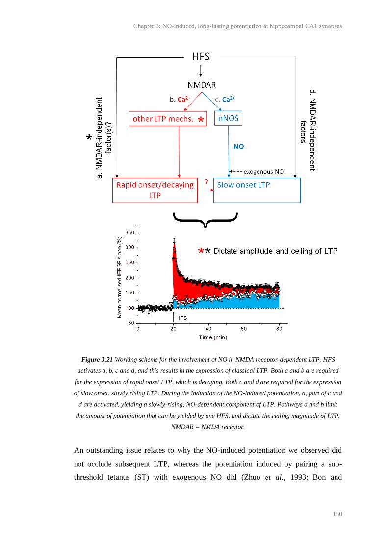
Chapter 3: NO-induced, long-lasting potentiation at hippocampal CA1 synapses
150
Figure 3.21 Working scheme for the involvement of NO in NMDA receptor-dependent LTP. HFS
activates a, b, c and d, and this results in the expression of classical LTP. Both a and b are required
for the expression of rapid onset LTP, which is decaying. Both c and d are required for the expression
of slow onset, slowly rising LTP. During the induction of the NO-induced potentiation, a, part of c and
d are activated, yielding a slowly-rising, NO-dependent component of LTP. Pathways a and b limit
the amount of potentiation that can be yielded by one HFS, and dictate the ceiling magnitude of LTP.
NMDAR = NMDA receptor.
An outstanding issue relates to why the NO-induced potentiation we observed did
not occlude subsequent LTP, whereas the potentiation induced by pairing a sub-
threshold tetanus (ST) with exogenous NO did (Zhuo et al., 1993; Bon and
Chapter 3: NO-induced, long-lasting potentiation at hippocampal CA1 synapses
151
Garthwaite, 2003). On this point, it is worth noting that even if the NO-induced
potentiation characterised in the present study were representative of the NO-
dependent component of HFS-induced LTP, we would not expect it to occlude
further HFS-induced LTP, because one HFS was not saturating of LTP under our
conditions (Figure 3.9-10). Given the scheme presented in Figure 3.21, a tentative
explanation for the results obtained using the ST is that this stimulus failed to
activate pathway ‘a’. Pathway ‘a’ could act like a switch for subsequent HFS-
induced LTP and determine the magnitude of LTP generated by one HFS. HFS
subsequent to pairing exogenous NO with the sub-threshold tetanus would activate
pathway ‘a’ for the first time, enabling the expression of rapid onset LTP. This rapid
onset LTP would be equal in magnitude to the potentiation induced by the ST and
exogenous NO. Since rapid onset LTP would determine the magnitude of
potentiation that can be yielded by one HFS, no further slow onset LTP would be
induced, giving the appearance that the potentiation induced by the weak threshold
paired with exogenous NO had occluded subsequent HFS-induced LTP.
It should be noted that the scheme outlined in Figure 3.21 is one of several possible
explanations for our data. However, with the exception of a role for eNOS in LTP,
the scheme does accommodate all of the data presented in this chapter and in the key
literature on the role of NO in NMDA receptor-dependent LTP. Therefore, it may be
a useful guide for further tests of the role of NO in the NO-induced potentiation and
LTP.
3.5.3 Synaptic locus of the NO-induced potentiation
Given that NO is a putative retrograde messenger during LTP, but that there is little
evidence for this at wild-type synapses in intact tissues (see Table 3.1), we sought to
test the locus (pre- or postsynaptic) of the NO-induced potentiation, a presynaptic
effect being consistent with retrograde NO transmission. To do this, the effect of the
NO-induced potentiation on PPF, a standard indicator of presynaptic efficacy, was
determined. Initially, we sought to measure changes in the PPF of pyramidal neuron
EPSCs, using intracellular sharp electrodes, as well as CA1 fEPSPs, because: 1) it
was unclear whether significant changes in fEPSP PPF would be detectable; 2)
Chapter 3: NO-induced, long-lasting potentiation at hippocampal CA1 synapses
152
intracellular recordings are more amenable than extracellular recordings to other
measurements of presynaptic efficacy, such as changes in the coefficient of variation
of postsynaptic responses (discussed by Korn and Faber, 1991). However, ~ 50 % of
intracellular recordings were too short-lasting to allow for experiments to be
completed (< 20 min), and although LTP was consistently observed using adjacent
field electrodes, ~ 50 % of recorded neurons were incapable of LTP (induction and
expression; see Appendix 1). One explanation for the lack of LTP in this proportion
of cells was that not all pyramidal neurons contribute to the LTP of nearby fEPSPs,
and therefore, we conducted experiments using field electrodes only.
To determine whether statistically significant evoked changes in the magnitude of
fEPSP PPF could be detected under our conditions, control experiments using 2-Cl-
adenosine or the adenylyl cyclase agonist, forskolin, were conducted. Each
compound was found to cause a statistically significant change in fEPSP PPF (ISI =
100 ms; Figure 3.18). The directions of the changes were consistent with previous
findings of the effects of adenosine and forskolin on PPF at CA1 (Dunwiddie and
Haas, 1985; Dumas and Foster, 1998; Lu and Gean, 1999; Wu et al., 1999) and other
synapses (Kahle and Cotman, 1993; Chen and Regehr, 1997; Chen and Roper, 2003),
and on other measures of presynaptic efficacy such as mEPSC frequency (Wu and
Saggau, 1994a; Sokolova et al., 2006; Bender et al., 2009). The changes in PPF that
were under observed our conditions were accompanied by changes in the slope of the
first fEPSP that were similar in magnitude to the NO-induced potentiation.
Therefore, it was reasoned that the locus of the NO-induced potentiation could be
investigated by its effect on fEPSP PPF.
Contrary to predictions based on work using dissociated hippocampal neurons, which
has shown a primarily presynaptic effect of NO following LTP (Arancio et al., 1995;
Arancio et al., 1996; Arancio et al., 2001), we observed no significant change in PPF
following the NO-induced potentiation (Figure 3.19). Considering the results of our
control experiments, which showed that a significant change in PPF could be
detected under our conditions following ~ 50 % changes in fEPSP initial slope
(Figure 3.18), there were two different explanations for the lack of a significant
change in PPF following the NO-induced potentiation: 1) the potentiation was

Chapter 3: NO-induced, long-lasting potentiation at hippocampal CA1 synapses
153
primarily postsynaptic (see Chapter 1: General introduction for examples of
postsynaptic NO-dependent plasticity); 2) the NO-induced potentiation relied upon
coordinated pre- and postsynaptic effects which yielded no net observable change in
the probability of presynaptic neurotransmitter release and/or number of release sites.
Given current estimates of the spread of NO through tissues (see Chapter 1:
General introduction), NO is well-placed to affect plasticity at both sides of the
synapse, and could do so almost simultaneously. Consistent with NO acting on both
sides of the synapse, studies of LTP at synapses between dissociated hippocampal
neurons have shown that the activation of the NO/cGMP/PKG pathway elicits a
rapid (within 1-10 min) increase in pre- and postsynaptic clusters (Antonova et al.,
2001; Wang et al., 2005). Nikonenko et al. (2003) also report that theta burst
stimulation of Schaffer collateral/commissural fibres in hippocampal slice cultures
yields NO-dependent synaptogenesis in area CA1 with a time-course similar to that
of the NO-induced potentiation (occurring within 10-30 min).
It should also be noted that our PPF analysis raises the issue that the role of NO in
LTP at synapses between dissociated neurons may not be directly applicable to the
role of NO in LTP at synapses in intact tissues. As discussed in 3.1 Introduction, a
lack of eNOS in neuronal cultures may give rise to differences in NO-dependent
synaptic plasticity at synapses between dissociated cells, compared to plasticity at
cells in intact tissues.
Finally, the lack of a significant change in PPF following HFS-induced LTP (Figure
3.20), though consistent with some other studies of the effect of CA1 LTP on PPF
(Schulz et al., 1994), and suggestive of the NO-induced potentiation being
representative of the NO-dependent component of LTP, is discordant with the effect
of NO-dependent CA1 LTP on PPF reported by some groups (Taqatqeh et al., 2009).
The difference between our results and those of other groups could arise from subtly
different experimental conditions or from the use of different protocols to induce
LTP (HFS vs. TBS). Indeed, the induction protocols used to generate LTP are known
to affect the expression mechanisms recruited (Raymond, 2007). Alternatively, the
difference could reflect the different time points relative to LTP induction at which
PPF is sampled. Taqatqeh et al. (2009) sampled PPF 2 min post TBS, whereas we
Chapter 3: NO-induced, long-lasting potentiation at hippocampal CA1 synapses
154
tested for persistent presynaptic effects by measuring PPF 55-60 post HFS. On this
point, it should be noted that the after-effects of PTP, which is distinct from LTP
(Zucker and Regehr, 2002) and lasted ~ 2 min under Taqatqeh et al.’s conditions,
may have confounded their PPF analysis.
3.6 Conclusion
Consistent with the proposed role for NO in NMDA receptor-dependent LTP,
pairing of exogenous NO with HFS in the presence of D-AP5 was found to elicit an
enduring potentiation of activity at CA1 synapses. This NO-induced potentiation
shared several properties with classical, control LTP and with a potentiation yielded
at CA1 synapses by pairing a sub-threshold tetanus with exogenous NO (Bon and
Garthwaite, 2003; Zhuo et al. 1993). However, in contrast with the plasticity
observed by Bon and Garthwaite (2003) and Zhuo et al. (1993), the NO-induced
potentiation that we observed was additive with LTP. This raised the possibility that
the NO-induced potentiation might be distinct from the NO-dependent component of
LTP. Unfortunately, our data leave this possibility open. However, our data led to the
generation of a scheme (Figure 3.21) for NO-dependent, HFS-induced LTP that
accommodates the current literature on the role of NO in LTP, and may be a useful
guide for the generation of further testable hypotheses. The scheme assumes that the
NO-induced potentiation is representative of the NO-dependent component of LTP,
and if correct, this implies that the NO-induced potentiation is a useful correlate for
persistent NO-dependent potentiation in isolation of other LTP expression
mechanisms.
The mechanisms underlying the NO-induced potentiation, and the NO-dependent
component of LTP, require further investigation. The slow rise time of the NO-
induced potentiation, as well as other forms of NO-dependent plasticity (for example,
Bon and Garthwaite, 2003; Phillips et al., 2008), and the slow effect of NOS
inhibition on HFS-induced LTP, are consistent with an expression mechanism that is
slow to take effect. As outlined above, one reported outcome of NO-dependent
plasticity that has an appropriate time-course is the synthesis of new synapses. This,

Chapter 3: NO-induced, long-lasting potentiation at hippocampal CA1 synapses
155
or a combination of changes on both sides of the synapse, could explain the lack of
effect of the NO-induced potentiation, and HFS-induced LTP, on PPF.
Chapter 4:
Modulation of basal synaptic efficacy by NO in area CA1 of
the hippocampus
Chapter 4: Modulation of basal synaptic efficacy by NO in area CA1 of the hippocampus
157
4.1 Introduction
NO signalling has been linked to the expression of activity-dependent synaptic
plasticity (see Chapter 3). Most research has focused on the role of NO in NMDA
receptor-dependent LTP at CA1 synapses in the hippocampus, a plasticity that is
usually induced by high frequency (100 Hz) or theta burst stimulation (Bliss et al.,
2007). In contrast, the potential for endogenous NO to modulate the efficacy of
‘naive’ synapses (i.e. where no attempt has been made to induce long-lasting
plasticity) undergoing ‘basal’ stimulation (i.e. that causes no observable change in
synaptic efficacy) has received little attention.
Recently, it was reported by Taqatqeh et al. (2009) that the magnitude of PPF (ISI =
20-50 ms) of EPSCs at ‘naive’ CA1 synapses undergoing ‘basal’ stimulation (0.033
Hz) is larger in hippocampal slices from mice lacking the NO-targeted guanylyl
cyclase α1 subunit than in slices from wild-type mice. As discussed in the previous
chapter, PPF can be explained by an increase in the probability of neurotransmitter
release (reviewed by Zucker and Regehr, 2002; Bliss et al., 2007) caused by residual
axonal Ca2+
(Wu and Saggau, 1994b) or a related mechanism, such as the saturation
of a Ca2+
buffer (Rozov et al., 2001). Therefore, one interpretation of the finding
made by Taqatqeh et al. (2007) is that, at wild-type synapses under basal conditions,
there is less scope for an increase in the probability of neurotransmitter release
during PPF because neurotransmitter release is tonically facilitated by a NO/α1-
containing NO targeted guanylyl cyclase/cGMP pathway.
Consistent with this, direct measurements of the efflux of various neurotransmitters,
such as glutamate, GABA and dopamine, from brain areas including the
hypothalamus and corpus striatum have been reported to be subject to increase or
decrease by exogenous NO or NOS inhibitors, respectively (reviewed by Prast and
Philippu, 2001). Regarding the hippocampus, the NO donors, sodium nitroprusside
(10-30 mM) and hydroxylamine (1-300 µM), have been found to increase the basal
efflux of radiolabelled noradrenaline and ACh from slices (Lonart et al., 1992). Brief
applications of more physiological concentrations of exogenous NO (5-10 nM;
O'Dell et al., 1991) or a cGMP analogue (50-100 µM 8-Br-cGMP; Arancio et al.,
Chapter 4: Modulation of basal synaptic efficacy by NO in area CA1 of the hippocampus
158
1995) to dissociated hippocampal neurons have been also been found to cause an
increase in the frequency, independent of the amplitude, of miniature EPSCs,
consistent with an increase in the efficacy of neurotransmitter release (Bliss et al.,
2007).
The mechanisms underlying NO-dependent or -induced increases in neurotransmitter
release are unclear. However, some research links NO to the modulation of resting
neuronal excitability in brain areas including the visual cortex and hypothalamus by
the modulation of targets such as voltage-gated K+, HCN and CNG channels. This
may shape the presynaptic action potential and thereby effect neurotransmitter
release (reviewed by Garthwaite, 2008; Steinert et al., 2010). Long-term regulation
of basal neurotransmitter release by NO might also occur via the regulation of the
number of functional release sites, since it has also been reported that application of
an NO donor (150 µM DETA/NONOate, 2 days) or cGMP analogue, 8-Br-cGMP (5
mM, 2 days), to hippocampal slice cultures causes an increase in the proportion of
multiply-innervated spines in area CA1. Conversely, chronic NOS inhibition (using
200 µM L-NAME, 2 days) was found to reduce synapse density (Nikonenko et al.,
2008). Alternatively, the mechanisms may be similar to those underlying NO-
dependent, activity-dependent increases in neurotransmitter release (see previous
chapter, Table 3.1).
The notion that basal synaptic efficacy might be facilitated by endogenous NO raises
questions over the isoform(s) of NOS involved. It is unclear whether nNOS, which is
thought to be preferentially activated by NMDA receptor opening (Garthwaite,
2008), is spontaneously active at synapses or whether it can become activated during
basal stimulation such as that used by Taqatqeh et al. (2009; 0.033 Hz). However, it
is well-known that eNOS can become Ca2+
- and consequently activity-independent
upon phosphorylation by kinases, most notably protein kinase Akt (Fulton et al.,
2001). Although endothelial NOS is expressed solely in blood vessels (Stanarius et
al., 1997; Blackshaw et al., 2003; Chan et al., 2004), it is estimated that central
neurons are ~ 25 µm (equivalent to ~ one cell diameter) at most from a capillary
(Pawlik et al., 1981). Early work on the physiology of endothelium-derived NO was
focused on its synthesis in endothelial cells upon innervations by cholinergic nerves
Chapter 4: Modulation of basal synaptic efficacy by NO in area CA1 of the hippocampus
159
and subsequent diffusion to smooth muscle to affect vasodilation (Furchgott and
Martin, 1985). However, a study using optic nerve has now set a precedent for NO
signalling from blood vessels to nerves in the CNS (Garthwaite et al., 2006), the
effect being the tonic depolarisation of axons. Moreover, an activity-independent,
eNOS-dependent low-level NO tone has been discovered in the hippocampus and
found to influence the expression of LTP in area CA1. These findings have generated
the hypothesis that eNOS may prime synapses for activity-induced plasticity (Hopper
and Garthwaite, 2006), perhaps, we speculate, by providing a prerequisite level of
synaptic efficacy.
4.1 Aim
The possibility that endogenous NO might regulate the basal efficacy of CA1
synapses is by itself intriguing and may also have far-reaching implications for our
understanding of the role of NO in activity-dependent plasticity. Nevertheless,
adequate tests of the effect of endogenous NO on basal synaptic efficacy in the
hippocampus are lacking. Musleh et al. (1993) have reported that the non-selective
NOS inhibitor, N-methyl-L-arginine (L-NMA; 125 µM), has no effect on the
magnitude of PPF at CA1 synapses in adult rat hippocampal slices. Yet it is known
that L-NMA can become hydroxylated in situ and metabolised by NOS, resulting in
the formation of amino acid products, including L-arginine, several of which are
capable of activating NOS (Olken and Marletta, 1993).
Given the above, we investigated whether endogenous NO could modulate the
efficacy of basal neurotransmission at CA1 synapses in intact tissues (containing
both e- and nNOS), and, if so, aimed to test the potential involvement of each NOS
isoform present.

Chapter 4: Modulation of basal synaptic efficacy by NO in area CA1 of the hippocampus
160
4.2 Methods
4.2.1 Animals
Unless otherwise stated, 6-8-week-old male C57Bl/6 mice were used (Charles River,
Margate, UK). Male, 6-9-week-old, 129sv/C57Bl/6 transgenic mice lacking
functional eNOS (eNOS-/-
; Huang et al., 1995) were kindly provided by Dr Adrian
Hobbs (UCL, London, UK).
4.2.2 Transverse hippocampal slice preparation and electrophysiology
Transverse hippocampal slices were prepared as detailed in Chapter 2.2.2.
Electrophysiological activity at Schaffer collateral/commissural-CA1 synapses was
recorded extracellularly using the methods described in Chapter 2.2.3. Pairs of
stimuli were delivered to Schaffer collateral/commissural fibres at 0.033 Hz at a
stimulus intensity set to 40-50 % of that required to elicit a population spike. The ISI
was 10 or 100 ms. PPF of CA1 fEPSPs was measured using the PPR (the initial
slope of fEPSP 2/the initial slope of fEPSP 1; Figure 4.1). Drugs were delivered
through the perfusion system. In all figures, fEPSP slopes have been normalised to
the mean slope of fEPSP 1 during the first 10 min of baseline recording shown (in
the absence of any drugs). In each experiment, baseline measurements of PPR were
made over 5 min immediately prior to the application of drugs. To avoid sample bias,
PPR in the presence of L-NNA, ODQ and D-AP5 was measured 30-35, 15-20 and
10-15 min respectively following their application. These times were chosen based
on the duration that each drug was applied to slices prior to HFS in the experiments
shown in the previous chapter.
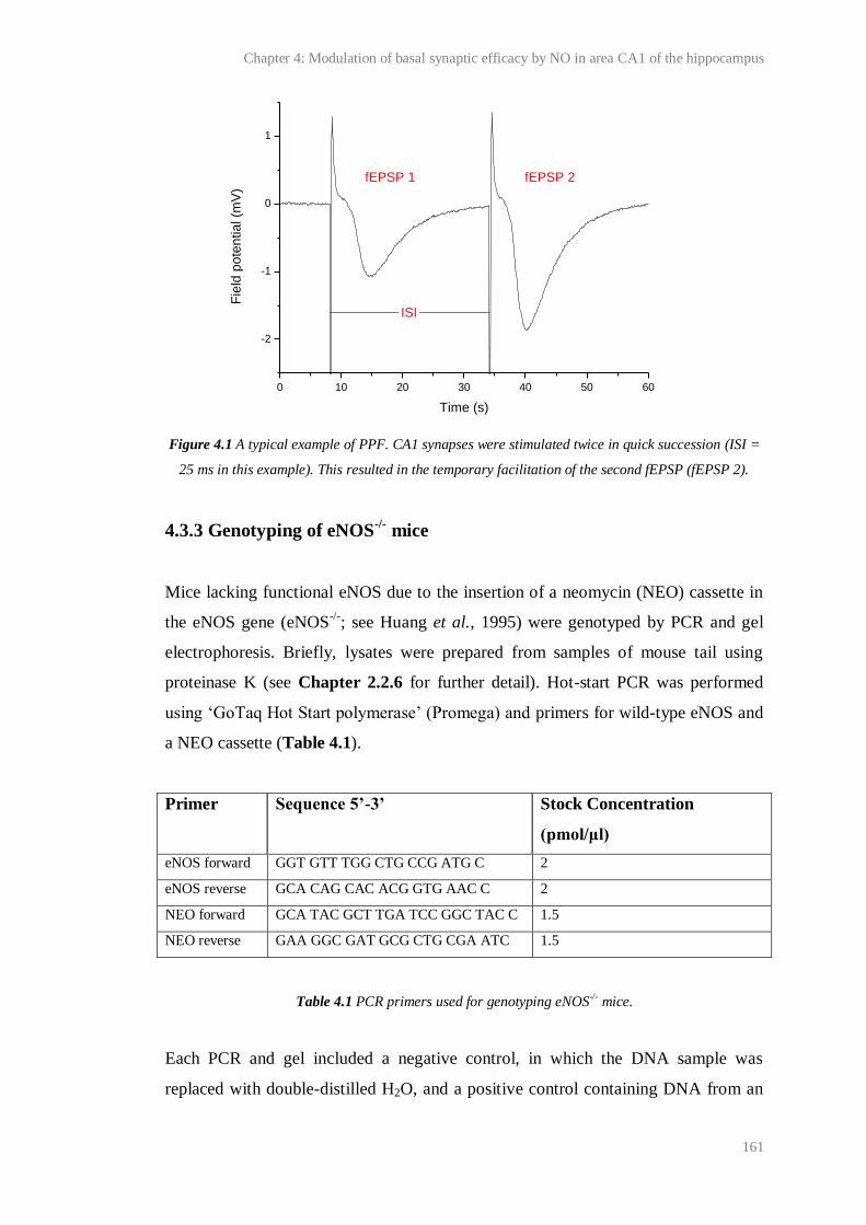
Chapter 4: Modulation of basal synaptic efficacy by NO in area CA1 of the hippocampus
161
Figure 4.1 A typical example of PPF. CA1 synapses were stimulated twice in quick succession (ISI =
25 ms in this example). This resulted in the temporary facilitation of the second fEPSP (fEPSP 2).
4.3.3 Genotyping of eNOS-/-
mice
Mice lacking functional eNOS due to the insertion of a neomycin (NEO) cassette in
the eNOS gene (eNOS-/-
; see Huang et al., 1995) were genotyped by PCR and gel
electrophoresis. Briefly, lysates were prepared from samples of mouse tail using
proteinase K (see Chapter 2.2.6 for further detail). Hot-start PCR was performed
using ‘GoTaq Hot Start polymerase’ (Promega) and primers for wild-type eNOS and
a NEO cassette (Table 4.1).
Primer Sequence 5’-3’ Stock Concentration
(pmol/μl)
eNOS forward GGT GTT TGG CTG CCG ATG C 2
eNOS reverse GCA CAG CAC ACG GTG AAC C 2
NEO forward GCA TAC GCT TGA TCC GGC TAC C 1.5
NEO reverse GAA GGC GAT GCG CTG CGA ATC 1.5
Each PCR and gel included a negative control, in which the DNA sample was
replaced with double-distilled H2O, and a positive control containing DNA from an
Table 4.1 PCR primers used for genotyping eNOS-/- mice.
0 10 20 30 40 50 60
-2
-1
0
1
Fie
ld p
ote
ntia
l (m
V)
Time (s)
ISI
fEPSP 1 fEPSP 2
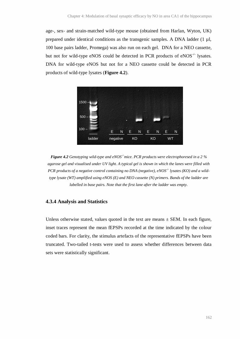
Chapter 4: Modulation of basal synaptic efficacy by NO in area CA1 of the hippocampus
162
age-, sex- and strain-matched wild-type mouse (obtained from Harlan, Wyton, UK)
prepared under identical conditions as the transgenic samples. A DNA ladder (1 µl,
100 base pairs ladder, Promega) was also run on each gel. DNA for a NEO cassette,
but not for wild-type eNOS could be detected in PCR products of eNOS-/-
lysates.
DNA for wild-type eNOS but not for a NEO cassette could be detected in PCR
products of wild-type lysates (Figure 4.2).
Figure 4.2 Genotyping wild-type and eNOS-/-mice. PCR products were electrophoresed in a 2 %
agarose gel and visualised under UV light. A typical gel is shown in which the lanes were filled with
PCR products of a negative control containing no DNA (negative), eNOS-/- lysates (KO) and a wild-
type lysate (WT) amplified using eNOS (E) and NEO cassette (N) primers. Bands of the ladder are
labelled in base pairs. Note that the first lane after the ladder was empty.
4.3.4 Analysis and Statistics
Unless otherwise stated, values quoted in the text are means ± SEM. In each figure,
inset traces represent the mean fEPSPs recorded at the time indicated by the colour
coded bars. For clarity, the stimulus artefacts of the representative fEPSPs have been
truncated. Two-tailed t-tests were used to assess whether differences between data
sets were statistically significant.
1500 – 2
500 –
100 –
100 -
E N E N E N E N
ladder negative KO KO WT
Chapter 4: Modulation of basal synaptic efficacy by NO in area CA1 of the hippocampus
163
Results
4.4.1 Effect of endogenous NO on PPF at naive CA1 synapses
To test the hypothesis that NO modulates the efficacy of naive CA1 synapses during
basal stimulation, we investigated the effect of endogenous NO on the magnitude of
PPF of CA1 fEPSPs in hippocampal slices. It is predicted that, following any
manipulation of synaptic efficacy that results in an increase in the probability of
neurotransmitter release, the scope for further increase during PPF will be reduced,
and, therefore, a decrease in the magnitude of PPF will be observed (measured using
the PPR; the initial slope of the second fEPSP/ the initial slope of the first fEPSP;
Zucker and Regehr, 2002; Bliss et al., 2007). The results of previous experiments
showed that statistically significant, evoked changes in PPF could be detected under
our conditions (see experiments using 2-Cl-adenosine and forskolin, Figure 3.18).
Pairs of stimuli were delivered to slices at 0.033 Hz and the effect of the non-
selective NOS inhibitor, L-NNA (100 µM, 30 min), on the magnitude of the resulting
PPF (measured using the PPR) was tested (see Figure 4.3A for the time course of
experiments). In order to determine whether any changes in the PPR were consistent
when PPF was induced using different ISIs, and because the magnitude of basal PPF
reported in mice lacking the α1 NO-targeted guanylyl cyclase subunit deviated most
from wild-type PPF at short ISIs (< 50 ms; Taqatqeh et al. 2010), experiments were
conducted using an ISI of 100 ms (as in the experiments detailed in Figure 3.18) and
10 ms.
When PPF was continuously elicited at 0.033 Hz, the PPR was stable and there was
no effect on the initial slope of fEPSP 1 (see baseline in Figure 4.3A). Application of
the non-selective NOS inhibitor, L-NNA, caused a significant increase in the mean
PPR (measured 30-35 min after the application of L-NNA) relative from that
measured over the last 5 min of baseline prior to L-NNA application (Figure 4.3B-
D). This was observed whether the ISI was 100 or 10 ms (change in PPF when ISI
was 10 ms: 0.19 ± 0.04; one factor t-test, p = 0.00144 compared to zero change; n =
10; change in PPF when ISI was 100 ms: 0.12 ± 0.05; one factor t-test, p = 0.0487
Chapter 4: Modulation of basal synaptic efficacy by NO in area CA1 of the hippocampus
164
compared to zero change; n =10). This increase in PPR was observed in the majority
of individual experiments, although a decrease was observed in 1 of 10 slices when
the ISI was 10 ms (Figure 4.3B), and two of ten slices when the ISI was 100 ms
(Figure 4.3C).
To assess whether the effect of L-NNA could be reversed by exogenous NO, the NO
donor, PAPA/NONOate (10 µM, 10 min) was co-applied with L-NNA. On average,
the PPR measured in the presence of PAPA/NONOate and L-NNA was not
significantly different from that measured in the presence of L-NNA alone (Figure
4.3B-D; ISI = 10 ms: paired t-test, p = 0.708; ISI = 100 ms: paired t-test, p = 0.537).

Chapter 4: Modulation of basal synaptic efficacy by NO in area CA1 of the hippocampus
165
Figure 4.3 Involvement of endogenous NO in basal PPF. A) The time-course of a typical experiment
is illustrated. Pairs of stimuli (ISI = 10 or 100 ms) were delivered to slices every 30 s and the initial
slopes of fEPSP 1 and 2 (upper panel) and the PPR (lower panel) were continuously monitored. The
general NOS inhibitor, L-NNA (100 µM), and the NO donor, PAPA/NONOate (10 µM) were applied
at the times indicated. The red dashed lines in each figure show the mean fEPSP slope or PPR
recorded prior to L-NNA application. B-C) The mean PPRs recorded at the times indicated by the
colour-coded bars in A during individual experiments (grey) and on average (black) are plotted (B:
Baseline LNNA + PAPA
1.4
1.6
1.8
2.0
2.2
2.4
2.6
2.8
3.0
Baseline LNNA + PAPA LNNA
0.8
1.0
1.2
1.4
1.6
1.8
2.0
2.2
2.4
0 20 40 60 80 100
1.0
1.5
2.0
2.5
0 20 40 60 80 100
0
50
100
150
200
250
0 10 20 30 40 50
0
50
100
150
200
250
300
1 2
0.5
1.0
1.5
2.0P
PR
Mean
ISI = 100 ms
PP
R
Mean
ISI = 10 ms
(panel A)
**, * = compared to zero
2-Cl-aden. Forskolin LNNA + PAPA LNNA LNNA + PAPA-0.2
-0.1
0.0
0.1
0.2
0.3
0.4
*** ** *
*
Me
an
ch
an
ge
in P
PR
fro
m b
ase
line **
ISI = 10 ms ISI = 100 ms
A B
C
D
100 M L-NNA
10 M PAPA/NONOate
Adjacent average (8 points)
PP
R
Time (min)
fEPSP 1 fEPSP 2
No
rma
lise
d f
EP
SP
slo
pe
(%
)
Time (min)
100 M L-NNA
10 M PAPA/NONOate
ISI = 10 ms
15 ms
1 mV
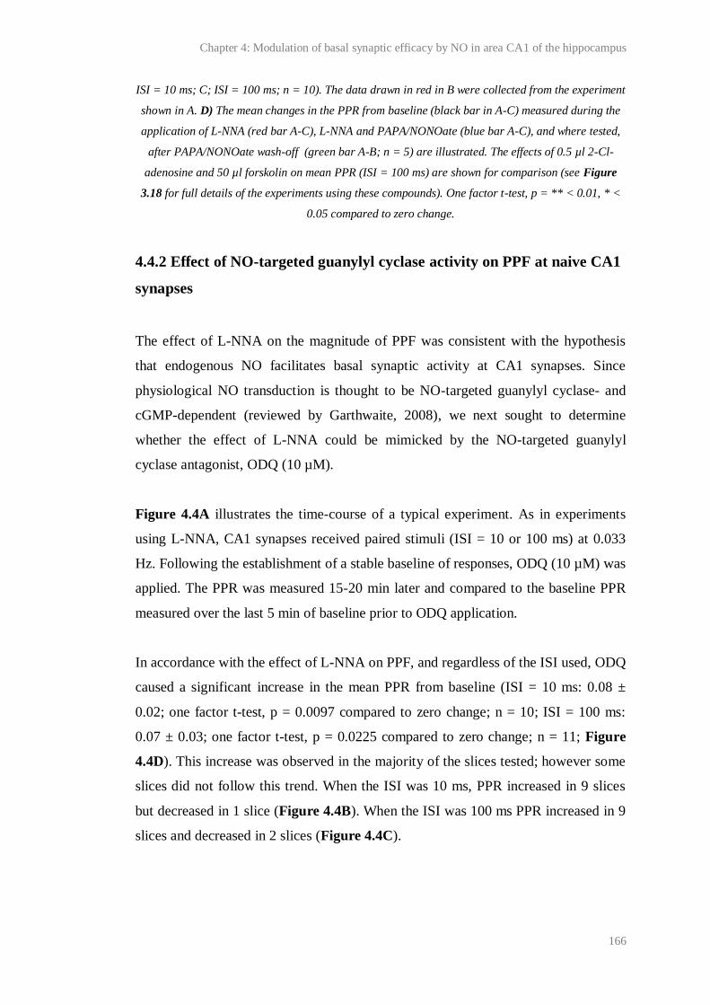
Chapter 4: Modulation of basal synaptic efficacy by NO in area CA1 of the hippocampus
166
ISI = 10 ms; C; ISI = 100 ms; n = 10). The data drawn in red in B were collected from the experiment
shown in A. D) The mean changes in the PPR from baseline (black bar in A-C) measured during the
application of L-NNA (red bar A-C), L-NNA and PAPA/NONOate (blue bar A-C), and where tested,
after PAPA/NONOate wash-off (green bar A-B; n = 5) are illustrated. The effects of 0.5 µl 2-Cl-
adenosine and 50 µl forskolin on mean PPR (ISI = 100 ms) are shown for comparison (see Figure
3.18 for full details of the experiments using these compounds). One factor t-test, p = ** < 0.01, * <
0.05 compared to zero change.
4.4.2 Effect of NO-targeted guanylyl cyclase activity on PPF at naive CA1
synapses
The effect of L-NNA on the magnitude of PPF was consistent with the hypothesis
that endogenous NO facilitates basal synaptic activity at CA1 synapses. Since
physiological NO transduction is thought to be NO-targeted guanylyl cyclase- and
cGMP-dependent (reviewed by Garthwaite, 2008), we next sought to determine
whether the effect of L-NNA could be mimicked by the NO-targeted guanylyl
cyclase antagonist, ODQ (10 µM).
Figure 4.4A illustrates the time-course of a typical experiment. As in experiments
using L-NNA, CA1 synapses received paired stimuli (ISI = 10 or 100 ms) at 0.033
Hz. Following the establishment of a stable baseline of responses, ODQ (10 µM) was
applied. The PPR was measured 15-20 min later and compared to the baseline PPR
measured over the last 5 min of baseline prior to ODQ application.
In accordance with the effect of L-NNA on PPF, and regardless of the ISI used, ODQ
caused a significant increase in the mean PPR from baseline (ISI = 10 ms: 0.08 ±
0.02; one factor t-test, p = 0.0097 compared to zero change; n = 10; ISI = 100 ms:
0.07 ± 0.03; one factor t-test, p = 0.0225 compared to zero change; n = 11; Figure
4.4D). This increase was observed in the majority of the slices tested; however some
slices did not follow this trend. When the ISI was 10 ms, PPR increased in 9 slices
but decreased in 1 slice (Figure 4.4B). When the ISI was 100 ms PPR increased in 9
slices and decreased in 2 slices (Figure 4.4C).

Chapter 4: Modulation of basal synaptic efficacy by NO in area CA1 of the hippocampus
167
It should be noted that the increase in mean PPR from baseline caused by ODQ was
smaller than that caused by L-NNA (Figures 4.4D), and, when the ISI was 10 ms,
this trend was statistically significant (ISI = 10 ms: unpaired t-test, p = 0.0303; n =
10; ISI = 100 ms: unpaired t-test, p = 0.375; n = 10-11).
Figure 4.4 Contribution of NO-targeted guanylyl cyclase to basal PPF. A) The time-course of a
typical experiment is shown. Paired stimuli (ISI = 10 or 100 ms) were delivered to slices every 30 s
and the initial slopes of fEPSP 1 and 2 (upper panel) and the PPR (lower panel) were continuously
recorded. The NO-targeted guanylyl cyclase antagonist, ODQ (10 µM) was applied at the time
Baseline ODQ
0.8
1.0
1.2
1.4
1.6
1.8
2.0
2.2
0 10 20 30 40 50
1.0
1.5
2.0
2.5
0 10 20 30 40 50
0
50
100
150
200
250
Baseline ODQ
0.8
1.0
1.2
1.4
1.6
1.8
2.0
2.2
PP
R
Mean
ISI = 10 ms
Adjacent average (8 points)
PP
R
Time (min)
10 M ODQ
fEPSP 1
fEPSP 2
No
rma
lise
d f
EP
SP
slo
pe
(%
)
Time (min)
10 M ODQ
ISI = 100 ms
ISI = 100 ms
PP
R
Mean
(panel A)
50 ms
1 mV
L-NNA ODQ L-NNA ODQ
0.0
0.1
0.2
0.3
†
*
*
**
**
Me
an
ch
an
ge
in
PP
R f
rom
ba
se
line
ISI = 10 ms ISI = 100 ms
A B
C
D
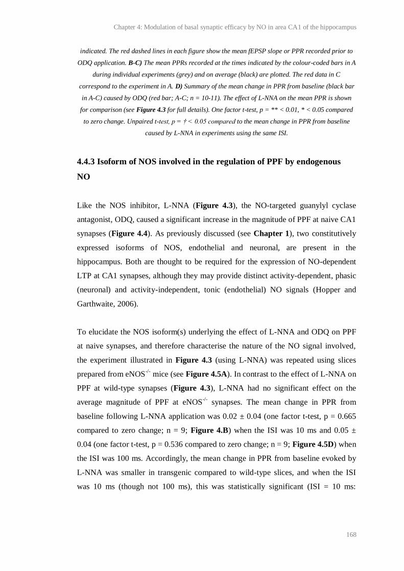
Chapter 4: Modulation of basal synaptic efficacy by NO in area CA1 of the hippocampus
168
indicated. The red dashed lines in each figure show the mean fEPSP slope or PPR recorded prior to
ODQ application. B-C) The mean PPRs recorded at the times indicated by the colour-coded bars in A
during individual experiments (grey) and on average (black) are plotted. The red data in C
correspond to the experiment in A. D) Summary of the mean change in PPR from baseline (black bar
in A-C) caused by ODQ (red bar; A-C; n = 10-11). The effect of L-NNA on the mean PPR is shown
for comparison (see Figure 4.3 for full details). One factor t-test, p = ** < 0.01, * < 0.05 compared
to zero change. Unpaired t-test, p = † < 0.05 compared to the mean change in PPR from baseline
caused by L-NNA in experiments using the same ISI.
4.4.3 Isoform of NOS involved in the regulation of PPF by endogenous
NO
Like the NOS inhibitor, L-NNA (Figure 4.3), the NO-targeted guanylyl cyclase
antagonist, ODQ, caused a significant increase in the magnitude of PPF at naive CA1
synapses (Figure 4.4). As previously discussed (see Chapter 1), two constitutively
expressed isoforms of NOS, endothelial and neuronal, are present in the
hippocampus. Both are thought to be required for the expression of NO-dependent
LTP at CA1 synapses, although they may provide distinct activity-dependent, phasic
(neuronal) and activity-independent, tonic (endothelial) NO signals (Hopper and
Garthwaite, 2006).
To elucidate the NOS isoform(s) underlying the effect of L-NNA and ODQ on PPF
at naive synapses, and therefore characterise the nature of the NO signal involved,
the experiment illustrated in Figure 4.3 (using L-NNA) was repeated using slices
prepared from eNOS-/-
mice (see Figure 4.5A). In contrast to the effect of L-NNA on
PPF at wild-type synapses (Figure 4.3), L-NNA had no significant effect on the
average magnitude of PPF at eNOS-/-
synapses. The mean change in PPR from
baseline following L-NNA application was 0.02 ± 0.04 (one factor t-test, p = 0.665
compared to zero change; n = 9; Figure 4.B) when the ISI was 10 ms and 0.05 ±
0.04 (one factor t-test, p = 0.536 compared to zero change; n = 9; Figure 4.5D) when
the ISI was 100 ms. Accordingly, the mean change in PPR from baseline evoked by
L-NNA was smaller in transgenic compared to wild-type slices, and when the ISI
was 10 ms (though not 100 ms), this was statistically significant (ISI = 10 ms:

Chapter 4: Modulation of basal synaptic efficacy by NO in area CA1 of the hippocampus
169
unpaired t-test, p = 0.00897; n = 9-10: ISI = 100 ms: unpaired t-test, p = 0.0993; n =
9-10; Figure 4.5D).
Figure 4.5 Effect of NOS inhibition on the magnitude of PPF in slices from eNOS -/- mice. A) The
time-course of a typical experiment is shown. Paired stimuli (ISI = 10 or 100 ms) were delivered to
slices every 30 s and the initial slopes of fEPSP 1 and 2 (upper panel) and the PPR (lower panel)
were continuously monitored. The general NOS inhibitor, L-NNA (10 µM) was applied at the time
Baseline L-NNA
0.6
0.8
1.0
1.2
1.4
1.6
1.8
2.0
2.2
Baseline L-NNA
0.8
1.0
1.2
1.4
1.6
1.8
2.0
2.2
2.4
0 10 20 30 40 50
0
50
100
150
200
250
0 10 20 30 40 50
1.0
1.5
2.0
2.5
Wild-type eNOS-/- Wild-type eNOS -/-
0.0
0.1
0.2
0.3
ISI = 100 msMe
an
ch
an
ge
in
PP
R f
rom
ba
se
line
ISI = 10 ms
A B
C
D
PP
R
Mean
ISI = 10 ms
(panel A)
ISI = 100 msP
PR
Mean
ISI = 10 ms
fEPSP 1
fEPSP 2
No
rma
lise
d f
EP
SP
slo
pe
(%
)
Time (min)
100 M L-NNA
Adjacent average (8 points)
PP
R
Time (min)
100 M L-NNA
†
***
10 ms
1 mV

Chapter 4: Modulation of basal synaptic efficacy by NO in area CA1 of the hippocampus
170
indicated. The red dashed lines in each figure show the mean fEPSP slope or PPR recorded prior to
L-NNA wash-on. B-C) The mean PPRs recorded at the times indicated by the colour-coded bars in A
during individual experiments (grey) and on average (black) are plotted. The red data in B were
collected during the experiment shown in A. D) Summary of the mean change in PPR from baseline
(black bar in A-C) caused by L-NNA (red bar; A-C; n = 9) in eNOS-/- mice, and, for comparison, wild-
type C57Bl/6 mice (see Figure 4.3). One factor t-test, p = ** < 0.01, * < 0.05 compared to zero
change. Unpaired t-test, p = † < 0.01 compared to the change in wild-type mice in experiments using
the same ISI.
The above findings were consistent with eNOS providing the entire NO responsible
for the regulation of basal PPF at naive CA1 synapses. However, it should be noted
that, in contrast with the proposed role of eNOS in regulating basal PPF, the mean
basal (baseline) PPR was significantly lower in slices from eNOS-/-
compared to
wild-type mice when the ISI was 100 ms (baseline PPR in eNOS-/-
mice vs. baseline
PPR shown in Figure 4.3-4: unpaired t test, p = 0.0042), although not when the ISI
was 10 ms (unpaired t-test, p = 0.140). This could be explained by natural variation,
or if a compensatory mechanism also responsible for the regulation of basal PPF
were acting at synapses in slices from eNOS-/-
mice. Alternatively the discrepancy in
PPF between slices might be explained by strain differences between the eNOS-/-
and
wild-type mice. Unfortunately, the prevailing lack of eNOS inhibitors (Alderton et
al., 2001) meant that the role of eNOS could not be directly tested in wild-type mice.
Likewise, no reliable means of testing the effect of nNOS on PPF are available
because the best characterised nNOS-deficient mice express active splice variants in
the hippocampus (Eliasson et al., 1997), and our recent results show that supposed
nNOS inhibitors are inadequately selective to be of use diagnostically (Chapter 6).
Therefore, based on the assumption that, in brain, nNOS is preferentially stimulated
by NMDA receptor activity (Garthwaite, 2008), we used the NMDA receptor
antagonist, D-AP5, to test the involvement of the NMDA receptor-nNOS pathway in
modulating basal neurotransmitter PPF (see Figure 4.6A).
As shown in Figure 4.6, D-AP5 (50 µM; pre-applied for 10 min) had no significant
effect on the average magnitude of PPF when the ISI was 10 ms (-0.07 ± 0.09; one
factor t-test, p = 0.928 compared to zero change; n = 9; Figure 4.6D) or 100 ms
(0.12 ± 0.10; one factor t-test, p = 0.264 compared to zero; n = 8; Figure 4.6D).

Chapter 4: Modulation of basal synaptic efficacy by NO in area CA1 of the hippocampus
171
Figure 4.6 NMDA receptor-dependency of basal PPF in wild-type mice. A) The time-course of a
typical experiment is shown. Paired stimuli (ISI = 10 or 100 ms) were delivered to slices every 30 s
and the initial slopes of fEPSP 1 and 2 (upper panel) and the PPR (lower panel) were continuously
recorded. The NMDA receptor antagonist, D-AP5 (50 µM) was applied at the time indicated. The red
dashed lines in each figure show the mean fEPSP slope or PPR recorded prior to perfusion with D-
AP5. B-C) The mean PPRs recorded at the times indicated by the colour-coded bars in A during
individual experiments (grey) and on average (black) are plotted. The red data in C are from the
0 10 20 30 40 50
0
50
100
150
200
250
0 10 20 30 40 50
1.0
1.5
2.0
2.5
Baseline D-AP5
1.4
1.6
1.8
2.0
2.2
2.4
2.6
2.8
3.0
Baseline D-AP5
0.6
0.8
1.0
1.2
1.4
1.6
1.8
2.0
2.2
fEPSP 1
fEPSP 2
No
rma
lise
d f
EP
SP
slo
pe
(%
)
Time (min)
ISI = 100 ms
50 M D-AP5
Adjacent average (8 points)
50 M D-AP5
PP
R
Time (min)
50 ms
0.5 mV
PP
R
Mean
ISI = 100 ms
(panel A)
ISI = 10 ms
PP
R
Mean
10 ms 100 ms-0.2
-0.1
0.0
0.1
0.2
Me
an
ch
an
ge
in P
PR
fro
m b
ase
line
50 M D-AP5
A B
C
D
Chapter 4: Modulation of basal synaptic efficacy by NO in area CA1 of the hippocampus
172
experiment in A. D) Summary of the mean change in PPR from baseline (black bar in A-C) caused by
D-AP5 (red bar in A-C; n =8- 9).
4.3 Discussion
Under many, though not all conditions, NO is required for the expression of long-
lasting, activity-dependent plasticity, in particular NMDA receptor-dependent LTP at
CA1 synapses (Garthwaite, 2008). Some studies of the effect of exogenous NO or
NO-targeted guanylyl cyclase α1 subunit knock-out on neurotransmitter release from
hippocampal neurons have also suggested that endogenous NO, via cGMP, might
regulate the basal efficacy of CA1 synapses, through the up-regulation of
neurotransmitter release (O'Dell et al., 1991; Lonart et al., 1992; Arancio et al.,
1995; Taqatqeh et al., 2009).
Here, we report that superfusion of hippocampal slices with the NOS or NO-targeted
guanylyl cyclase antagonists, L-NNA (100 µM) or ODQ (10 µM), caused a
significant increase in PPF of fEPSPs in area CA1 (Figure 4.3-4). The increase in
PPF could not be mimicked by inhibition of NMDA receptors, suggesting that the
NMDA receptor-nNOS pathway (Figure 4.6) was not involved in the basal
regulation of PPF. However, the effect of L-NNA was abolished in slices lacking
functional eNOS (Figure 4.5). This finding was against the possibility that the rise in
PPF observed in experiments using L-NNA and ODQ was the effect of a general
increase in basal PPF over time (for example, caused by baseline drift).
These findings are in accordance with the facilitation of neurotransmitter release at
naive CA1 synapses under basal conditions by endogenous NO/cGMP and, although
tests of the involvement of each NOS isoform present were limited, with the
conclusion that eNOS provides all the NO necessary for the regulation of basal
synaptic efficacy. (Note that our data cannot exclude the influence of NMDA
receptor-independent nNOS activity). It was found that L-NNA and ODQ caused an
increase in PPF in C57/Bl6 mice when the ISI was both short (10 ms) and long (100
ms; Figure 4.3-4). Because the duration of inhibitory postsynaptic potentials (IPSPs)
measured at CA1 synapses under our conditions using sharp intracellular electrodes
Chapter 4: Modulation of basal synaptic efficacy by NO in area CA1 of the hippocampus
173
was < 100 ms (for an example trace see Appendix 1), it seems unlikely that the
changes in PPF caused by L-NNA and ODQ were the result of changes in the
inhibitory input to pyramidal neurons. However, GABAergic transmission was not
blocked in these studies and therefore, endogenous NO might have been acting at
inhibitory and/or excitatory synapses on pyramidal cells to regulate neurotransmitter
release under basal conditions. It should be noted that at CA1 synapses in slices from
mice lacking the NO-targeted guanylyl cyclase α1 subunit, PPF was significantly
increased from that recorded in slices from wild-type mice only when the ISI was <
50 ms (Taqatqeh et al., 2009). However, an insignificant increase in PPF was
observed by Taqatqeh et al. when the ISI was 100 ms and it is possible that if paired
statistical analysis were permitted, this would become a significant increase.
At the time of this investigation, Neitz et al. (2011) published the results of a series
of experiments also aimed at determining the effect of endogenous NO on basal
synaptic efficacy at naive CA1 synapses in hippocampal slices. They also focused on
the potential presynaptic effects of NO. In accordance with the effects of L-NNA and
ODQ on the magnitude of PPF observed in the present study, Neitz et al. (2011)
found that the frequency, but not the amplitude, of AMPA receptor-mediated
miniature EPSCs was reduced in area CA1 of mice lacking the NO-targeted guanylyl
cyclase α1 subunit. The reduction in frequency was reversed by application of the
cGMP analogue, 8-Br-PET-cGMP (100 µM), and replicated in slices from wild-type
mice upon L-NNA or ODQ application. Concordant effects of NO-targeted guanylyl
cyclase α1 subunit knock-out and ODQ on the frequency of minimally evoked
EPSCs and magnitude of EPSC PPF were also found. The effects on miniature EPSC
frequency were replicated by the HCN channel blocker, ZD7288 (10 µM),
suggesting that NO, via cGMP and the regulation of Ih might facilitate
neurotransmitter release by enhancing axon depolarisation, as in the optic nerve
(Garthwaite et al., 2006).
To address the question of which NOS isoform was responsible for maintaining a
reduced PPF at synapses in slices from wild-type mice, Neitz et al. (2011) used the
supposedly isoform selective nNOS inhibitor, L-VNIO (0.1 µM). They found no
effect of 0.1 µM L-VNIO on miniature EPSC frequency and therefore, concluded
Chapter 4: Modulation of basal synaptic efficacy by NO in area CA1 of the hippocampus
174
that eNOS, but not nNOS, was responsible for the modulation of basal synaptic
efficacy in area CA1. While our data support this conclusion, our recent findings
strongly suggest that L-VNIO was completely ineffective in inhibiting NOS at the
concentration used by Neitz et al. (Chapter 6).
Apart from the experiments using L-VNIO, the results of Neitz et al. (2011) clearly
add strength to the conclusions that can be drawn from our study, in which the effect
of endogenous NO on only one indicator of synaptic efficacy was assessed.
Nevertheless, there are at least three points that need to be addressed to clarify the
effect of endogenous NO on basal synaptic efficacy at naive CA1 synapses.
Firstly, in our study, the mean change in PPR from baseline caused by ODQ was
significantly smaller than that caused by L-NNA when the ISI was 10 ms, and
therefore experiments are necessary to elucidate the source of this difference (Figure
4.4). It is unlikely that this reflects a difference between the potencies of L-NNA and
ODQ, since the concentrations of each inhibitor that were used (100 µM and 10 µM,
respectively) are considered to be supra-maximal for enzyme inhibition.
Furthermore, we have found that both inhibitors abolish cGMP accumulation in our
hippocampal slices in response to maximal doses of NMDA (see Chapter 5). It also
seems unlikely that the difference reflects the involvement of guanylyl-cyclase-
independent NO signalling, since almost all physiological NO signals are transduced
by cGMP (Garthwaite, 2008). Rather, the most parsimonious explanation is natural
variation, especially considering that Neitz et al. (2011) report that the effects of L-
NNA and ODQ on miniature EPSC frequency were comparable and that, as is
typical of evoked changes in PPR (for example Schulz et al., 1995), the amplitude of
the change in PPF that we observed was small and highly variable. This may also
explain why eNOS knock-out caused a significant change in PPR compared to wild-
type PPR when the ISI was 10, but not 100 ms (Figure 4.3).
Secondly, the finding that the NO donor, PAPA/NONOate (10 µM, applied for 10
min), could not reverse the effect of L-NNA on PPF (Figure 4.3) is seemingly at
odds with the facilitation of basal synaptic activity by endogenous NO. It seems
unlikely that the effect of L-NNA on PPF can be attributed by an effect other than
Chapter 4: Modulation of basal synaptic efficacy by NO in area CA1 of the hippocampus
175
NOS inhibition, since no secondary targets of L-NNA are known (Alderton et al.,
2001) and, consistent with the effect of ODQ on PPF, NMDA-induced cGMP
accumulation in hippocampal slices treated with L-NNA can be reconstituted by
exogenous NO (Bartus, 2009). Therefore, investigations are necessary to determine
whether a longer application and/or different concentration of exogenous NO is
required to reverse the effect of L-NNA on PPF. Regarding this possibility, it is
interesting to note that the NO-induced potentiation described in the previous chapter
was induced during NMDA receptor (and therefore presumably nNOS) antagonism
but was inhibited by L-NNA and ODQ (see Figures 3.6 and 3.13-14), suggesting
that the exogenous NO used to induce it (3 or 10 µM PAPA/NONOate, applied for 8
min in total) was not sufficient to mimic completely the endogenous NO profile.
Finally, it should be also be noted that as with most parameters used to define the
locus of changes in synaptic efficacy, including those used by Neitz et al. (2011),
evidence provided by PPF analysis is suggestive rather than conclusive. Typically,
changes in the magnitude of PPF are explained by changes in the probability of
presynaptic release and/or number of release sites (reviewed by Zucker and Regehr,
2002; Bliss et al., 2007), and all the data presented in this study, as well as the
studies discussed above, are consistent with NO acting on a presynaptic target to
effect the modulation of basal synaptic efficacy. Furthermore, immunohistochemistry
and in situ hybridisation studies suggest that the α1 NO-targeted guanylyl cyclase
subunit is presynaptic in area CA1 (Szabadits et al., 2007; although see Chapter 7).
However, a change in postsynaptic efficacy could account for our results, as well as
those of the above studies, if, for example, NOS inhibition caused the internalisation
of postsynaptic receptors and this was favoured at synapses with a high probability of
release, as during LTD at CA1 synapses (Bliss et al., 2007). On this point, it should
be noted that HCN channels appear to be predominantly postsynaptic in area CA1
(Notomi and Shigemoto, 2004).
4.4 Conclusion
Clearly, further experiments are necessary to clarify the effect of endogenous NO on
the efficacy of naïve CA1 synapses, especially because effect sizes yielded from PPF

Chapter 4: Modulation of basal synaptic efficacy by NO in area CA1 of the hippocampus
176
analysis are typically small and highly variable. Certain caveats of the data also limit
the strength of the conclusions that can be drawn from our study, for example, the
inability of exogenous NO to reverse the effect of L-NNA on PPR. However, the
potential role of eNOS and NO in the modulation of basal synaptic efficacy in area
CA1 does extend the finding that NO derived from blood vessels influences axons in
the optic nerve (Garthwaite et al., 2006), and suggests a novel role for the eNOS-
derived NO tone discovered in the hippocampus (Chetkovich et al., 1993; Hopper
and Garthwaite, 2006). As discussed above, it has been hypothesised that eNOS
primes CA1 synapses for LTP (Hopper and Garthwaite, 2006) and it is conceivable
that this could occur via the setting of a prerequisite, basal level of neurotransmitter
release (or postsynaptic quantal amplitude). Indeed, homeostatic mechanisms are
thought to be critical in maintaining the ability of synapses and networks to be both
up- and down-regulated in response to varying stimuli (Pozo and Goda, 2010), and
the potential involvement of eNOS in this links NO to metaplasticity (reviewed by
Abraham, 2008).
The requirement of basal synaptic efficacy and LTP expression for eNOS in area
CA1 complicates the interpretation of NO-dependent LTP between dissociated
pyramidal neurons, since blood vessels are unlikely to be present in neuron cultures.
This lack of eNOS could explain why Arancio et al. (1995; 1996; 2001) found that
NO-dependent LTP at synapses between pairs of dissociated neurons was
presynaptic, whereas the NO-induced potentiation described in the previous chapter
was not accompanied by a persistent decrease in PPF. For example, it is possible that
the increase in transmitter release that was observed by Arancio et al. was caused
because the NO necessary for the LTP had compensated for a lack of eNOS, or
because the baseline probability of neurotransmitter release was unnaturally low.
Indeed, it is well-known that the magnitudes of PPF recorded under basal conditions
and following LTP are inversely correlated (Schulz et al., 1995). Accordingly,
Serulle et al. (2007) have found that neither NOS nor NO-targeted guanylyl cyclase
antagonism alters the magnitude of PPF in dissociated hippocampal neurons.
Finally, it should be noted that endothelium-derived NO is unlikely to be the only
mechanism capable of regulating PPF at CA1 synapses, since baseline PPF (in the
Chapter 4: Modulation of basal synaptic efficacy by NO in area CA1 of the hippocampus
177
absence of any drugs) was not significantly increased in eNOS-/-
slices (Figure 4.5).
In fact, baseline PPF was significantly decreased in slices from eNOS-/-
compared to
wild-type mice when the ISI was 100 ms. If eNOS is truly responsible for the
regulation of basal synaptic efficacy at CA1 synapses, it is also likely to affect
synaptic transmission at other synapses, since eNOS and guanylyl cyclase are
expressed throughout the brain (; eNOS: Seidel et al., 1997; Stanarius et al., 1997;
Topel et al., 1998; Demas et al., 1999; NO-targeted guanylyl cyclase: Matsuoka et
al., 1992; Gibb and Garthwaite, 2001; Ding et al., 2004). It is tempting to speculate
that, in a situation where NO derived from blood vessels influences neurons and vice
versa (reviewed by Garthwaite, 2008), changes in blood flow, basal neuronal
efficacy and the probability of plasticity might be coordinated. As well as the
physiological implications of this, it is conceivable that disordered NO signalling
might contribute to pathologies characterised by anomalous network or synaptic
efficacy, such as hippocampal epileptogenesis, which may result from the increase in
neuronal excitability of CA3 neurons capable of generating recurrent bursts of
activity (Walker et al., 2007), or central sensitisation (a processes likened to LTP) of
noiceceptive synapses in the spine (Ji et al., 2003).

Chapter 5:
NO and NMDA receptor-independent, L-type voltage-gated
Ca2+
channel-dependent LTP
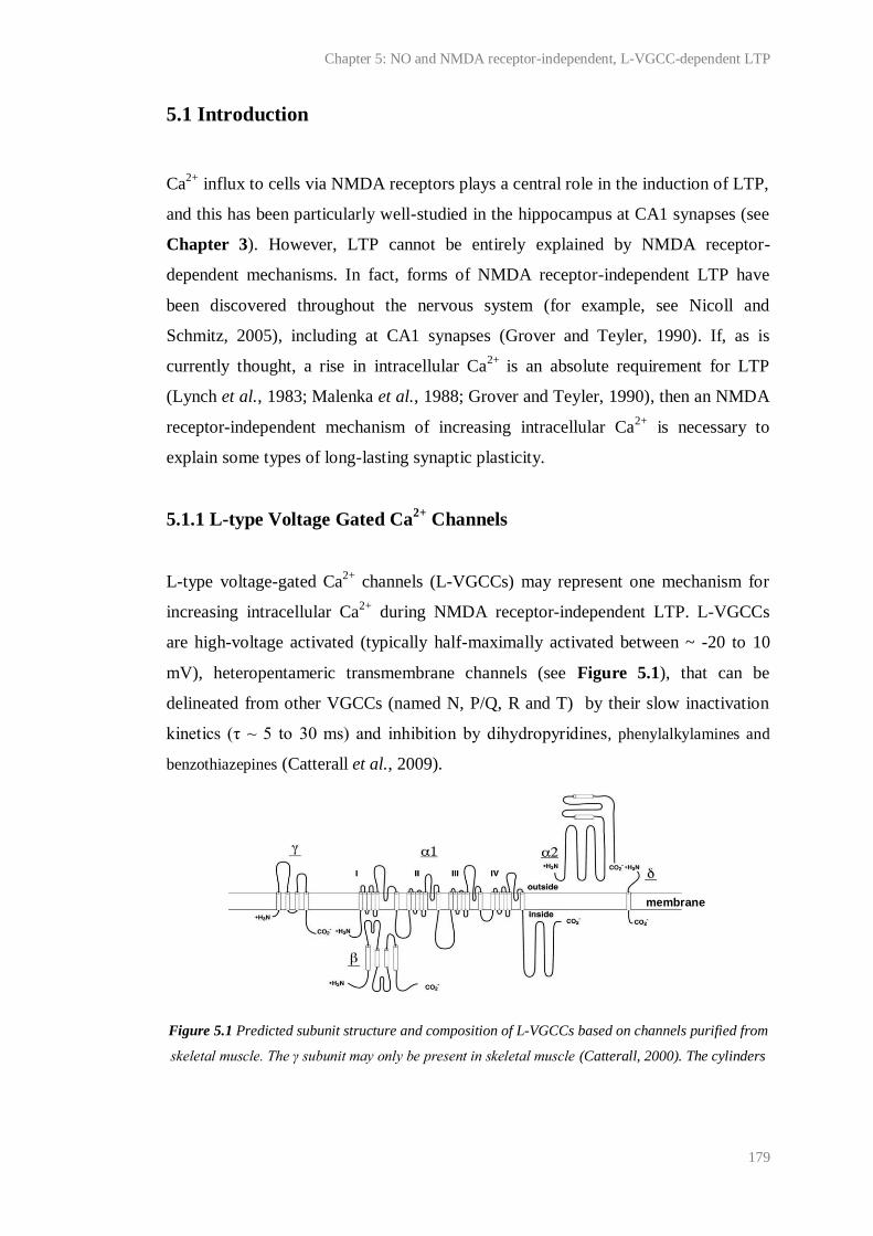
Chapter 5: NO and NMDA receptor-independent, L-VGCC-dependent LTP
179
5.1 Introduction
Ca2+
influx to cells via NMDA receptors plays a central role in the induction of LTP,
and this has been particularly well-studied in the hippocampus at CA1 synapses (see
Chapter 3). However, LTP cannot be entirely explained by NMDA receptor-
dependent mechanisms. In fact, forms of NMDA receptor-independent LTP have
been discovered throughout the nervous system (for example, see Nicoll and
Schmitz, 2005), including at CA1 synapses (Grover and Teyler, 1990). If, as is
currently thought, a rise in intracellular Ca2+
is an absolute requirement for LTP
(Lynch et al., 1983; Malenka et al., 1988; Grover and Teyler, 1990), then an NMDA
receptor-independent mechanism of increasing intracellular Ca2+
is necessary to
explain some types of long-lasting synaptic plasticity.
5.1.1 L-type Voltage Gated Ca2+
Channels
L-type voltage-gated Ca2+
channels (L-VGCCs) may represent one mechanism for
increasing intracellular Ca2+
during NMDA receptor-independent LTP. L-VGCCs
are high-voltage activated (typically half-maximally activated between ~ -20 to 10
mV), heteropentameric transmembrane channels (see Figure 5.1), that can be
delineated from other VGCCs (named N, P/Q, R and T) by their slow inactivation
kinetics (τ ~ 5 to 30 ms) and inhibition by dihydropyridines, phenylalkylamines and
benzothiazepines (Catterall et al., 2009).
Figure 5.1 Predicted subunit structure and composition of L-VGCCs based on channels purified from
skeletal muscle. The γ subunit may only be present in skeletal muscle (Catterall, 2000). The cylinders
membrane
Chapter 5: NO and NMDA receptor-independent, L-VGCC-dependent LTP
180
depict predicted α-helices and lengths of lines approximate lengths of polypeptide segments. Diagram
taken from Catterall et al. (2005). Reproduced by kind permission of ASPET journals.
The α1 L-VGCC subunit forms the pore of the channel and contains the voltage
sensor, the selectivity filter and antagonist binding sites. On its own, the α1 subunit is
capable of forming a functional channel (Perez-Reyes et al., 1989; Striessnig, 1999),
although other subunits (see Figure 5.1) may influence its cell surface targeting,
electrophysiological properties and stability of the channel.
So far, four subtypes of the α1 subunit have been discovered (Cav1.1-1.4; Table 5.1).
These subtypes vary in their electrophysiological properties (kinetics and voltage-
dependency of activation and inactivation and channel conductance), tissue
distribution and physiology. This diversity is reflected in the myriad physiological
processes and pathologies that L-VGCCs have been implicated in, which range from
smooth muscle contraction, blood pressure regulation and hypertension (Ozawa et
al., 2006) to neurotransmitter release and the modulation of neuronal excitability
(Lacinova et al., 2008), pain sensitisation (Park and Luo, 2010), psychiatric disorders
(Casamassima et al., 2010), Alzheimer’s disease (Anekonda et al., 2011) and
epilepsy (N'Gouemo et al., 2010).

Chapter 5: NO and NMDA receptor-independent, L-VGCC-dependent LTP
181
Class Antagonists Subtype Subtype localisation Subtype function(s)
L
Dihydropyridines;
phenylalkylamines;
benzothiazepines
Cav1.1 Skeletal muscle Muscle contraction
Cav1.2 Cardiac and smooth
muscle; endocrine cells;
neurons (soma and
dendrites)
Muscle contraction, hormone
secretion, neurotransmission,
gene transcription, synaptic
plasticity, learning, memory
Cav1.3 Cardiac muscle;
pacemaker cells;
endocrine cells; cochlear
hair cells; neurons (soma
and dendrites)
Smooth muscle contraction,
hormone secretion,
neurotransmission
Cav1.4 Retinal rod and bipolar
cells; mast cells; adrenal
gland
Neurotransmission
N
ω-conotoxins Cav2.2 Neurons (axons and
dendrites);
neuroendocrine cells
Neurotransmission, hormone
release
P/Q
ω-agatoxins Cav2.1 Neurons (axons and
dendrites);
neuroendocrine cells
Neurotransmission, hormone
release
R SNX-482 Cav2.3 Neurons (soma and
dendrites)
Neurotransmission
T Ni2+
Flunarizine
Mibefradil
Ethosuximide
Cav3.1 Neurons (soma and
dendrites); cardiac and
smooth muscle
Pacemaking, repetitive firing.
Cav3.2 Neurons (soma and
dendrites); cardiac and
smooth muscle
Pacemaking, repetitive firing.
Cav3.3 Neurons (soma and
dendrites)
Pacemaking, repetitive firing.
Table 5.1 Summary of the pharmacological properties, tissue distribution and function of L-VGCC
subtypes. Different subtypes contain different α1 subunits. Information for N-, P/Q-, R- and T-type
VGCCs are given for comparison. Like L-VGCCs, N-, P/Q- and R-VGCCs are high voltage activated.
T-type VGCCs are low voltage activated. Adapted from Catterall et al. (2009).
In the hippocampus, where radioligand binding has shown the Cav1.2 subtype to
predominate (Clark et al., 2003), L-VGCC subunits appear to be densely expressed
Chapter 5: NO and NMDA receptor-independent, L-VGCC-dependent LTP
182
in the membrane of pyramidal cell soma and at the base of apical dendrites. Subunits
have also been detected histologically in granule cells, interneurons and at synapses
in more distal dendrites of pyramidal neurons, particularly in area CA3 (Ahlijanian et
al., 1990; Westenbroek et al., 1990; Hell et al., 1993). They may be activated by
back-propagating somatic action potentials and/or EPSPs (Mermelstein et al., 2000),
and, like NMDA receptors, have been demonstrated to cause significant intra-
dendritic Ca2+
influx to pyramidal neurons. Fura-2 measurements in hippocampal
slices, for example, show that L-VGCCs contribute ~ 30 % of the whole-cell Ca2+
current during spiking (Regehr and Tank, 1992; Christie et al., 1995) and this is in
agreement with estimates made using electrophysiological recordings from
dissociated pyramidal neurons (Mermelstein et al., 2000). Following step
depolarisations to negative voltages (-30 mV) designed to approximate the effect of
an EPSP, the contribution of L-VGCCs to the total Ca2+
current was found to
increase to ~ 50 % (Mermelstein et al., 2000). L-VGCC activity is also thought to
contribute to the resting intracellular Ca2+
concentration of pyramidal neurons
(Magee et al., 1996), neuronal excitability in response to depolarising pulses
(Lacinova et al., 2008), and has been implicated in hippocampal synaptic plasticity,
learning and memory (see below).
5.1.2 L-VGCC-dependent LTP at CA1 synapses
Following 200 Hz- or prolonged theta burst stimulation of hippocampal Schaffer
collaterals in vitro, a high magnitude ‘compound’ LTP has been observed at CA1
synapses that is composed of two pharmacologically-separable components: one
blocked by NMDA receptor-antagonism, the other independent of NMDA receptor
blockade but attenuated by L-VGCC inhibitors (Grover and Teyler, 1990; Grover
and Teyler, 1994; Cavus and Teyler, 1996; Morgan and Teyler, 2001). Similar
findings have been made in vivo (Morgan and Teyler, 1999), and L-VGCC inhibitors
have also been reported to attenuate spike timing-dependent potentiation (Fuenzalida
et al., 2010), as well as CA1 LTP induced by various other stimuli (Aniksztejn and
Ben-Ari, 1991; Kato et al., 1993; Huang and Malenka, 1993; Impey et al., 1996).
Chapter 5: NO and NMDA receptor-independent, L-VGCC-dependent LTP
183
Like NMDA receptor-dependent LTP, L-VGCC-dependent LTP has been reported to
be input-specific, blocked by Ca2+
chelators (Grover and Teyler, 1992), induced
postsynaptically and yet expressed on both sides of the synapse (Grover, 1998;
Bayazitov et al., 2007). While some studies indicate that the expression mechanisms
underlying L-VGCC-dependent LTP may be shared with NMDA receptor-dependent
LTP (for example, Little et al., 1995), several indicate that L-VGCC- and NMDA
receptor-dependent LTP are, at least in part, mechanistically distinct. For example,
using synapto-pHlourin to continuously monitor presynaptic vesicle recycling in area
CA1, it was found by Bayazitov et al. (2007) that the L-VGCC-dependent
component of compound LTP induced using 200 Hz burst stimulation was slow to
start (t½ ~ 35 min), partly presynaptic and long-lasting (of ~ maximal magnitude at
least 3 hr post induction). Conversely, the NMDA receptor-dependent component
was found to be almost immediate, largely postsynaptic and, although clearly evident
3 hr post-induction, was not of maximal magnitude over this time.
The difference in the onset of NMDA receptor- and L-VGCC-dependent LTP has
also been observed electrophysiologically (for example, Grover and Teyler, 1990)
and the distinction between their synaptic loci is supported by studies using FM-143
to monitor presynaptic plasticity (Zakharenko et al., 2001; Zakharenko et al., 2003).
Using a spatially-restricted brain-derived neurotrophic factor (BDNF) knock-out
mouse, Zakharenko et al. (2003) also identified BDNF release from CA3 neurons as
critical for L-VGCC-dependent, but not NMDA receptor-dependent LTP at CA1
synapses. This was in accordance with a previous report showing that the expression
of the BDNF receptor, tyrosine kinase B (TrkB), is up-regulated following L-VGCC,
but not NMDA receptor-dependent LTP (Teyler et al., 1994), and has since been
supported by evidence that TrkB is necessary for 200 Hz burst stimulation-induced
LTP (Gruart et al., 2007).
The persistent expression of L-VGCC-dependent but not NMDA-receptor dependent
LTP noted by Bayazitov et al. (2007) also accords with other data showing that L-
VGCCs, but not NMDA receptors, are necessary for CA1 late-LTP and cAMP
response element (CRE)-mediated β-galactosidase expression induced by multiple
100-Hz tetani in hippocampal slices (Impey et al., 1996). L-VGCC activation has

Chapter 5: NO and NMDA receptor-independent, L-VGCC-dependent LTP
184
also been reported to be critical for activity-dependent gene expression in cortical
neurons (Murphy et al., 1991) and glutamate-induced, CRE-regulated transcription
in hippocampal neurons (Bading et al., 1993). Indeed, several authors have
hypothesised that the dense expression of L-type channels at the base of dendrites
makes them likely candidates for the transduction of distal dendritic activity to the
soma where alterations in gene expression may occur (for example, Westenbroek et
al., 1990). Further, the requirement of late-LTP for L-VGCC’s may explain why
more stringent induction protocols than those needed for NMDA receptor-dependent
early-LTP are necessary for its induction and expression.
5.1.3 Physiological relevance of L-VGCC-dependent LTP
In the past, it has been suggested that, because L-VGCCs are typically high voltage-
activated (Catterall et al., 2009), the natural occurrence of L-VGCC-dependent long-
lasting plasticity may be restricted to pathological conditions such as epileptogenesis
during which cell excitability may become supra-physiological (Huang and Malenka,
1993). Since then, however, L-VGCCs have been shown to account for a significant
proportion (30-50 %) of the whole cell Ca2+
current of pyramidal neurons following
stimulation designed to mimic dendritic EPSPs, as well as spikes (Regehr and Tank,
1992; Christie et al., 1995; Mermelstein et al., 2000) and it has been ventured by
Morgan and Teyler (1999) that the activation requirements for L-VGCC-dependent
long-lasting plasticity in vitro may be met in nature by short, aperiodic, high-
frequency (200 Hz), high-magnitude (1-3 mV) sharp waves (population EPSPs)
produced by Schaffer collaterals that project onto CA1 neurons and interneurons.
Furthermore, pharmacological, electrophysiological and behavioural evidence has
combined to show that L-VGCC and NMDA receptor dependent plasticity may have
distinct functions, modulating different aspects of memory. For example, it has been
reported that, while systemic injection of the NMDA receptor antagonist, MK-801,
inhibits rats’ ability to acquire memory of an eight-arm radial maze, which is a
hippocampus-dependent task (Jarrard, 1993), the L-VGCC inhibitor, verapamil, and
not MK-801, inhibits long term (7-10 days) retention of the memory (Borroni et al.,
2000; Woodside et al., 2004). Injection with both drugs during the task training
blocks all acquisition and retention.
Chapter 5: NO and NMDA receptor-independent, L-VGCC-dependent LTP
185
It should be noted that other studies in the hippocampus have failed to find an effect
of L-VGCC inhibition on memory retention/consolidation, and that some research
has found L-VGCC inhibition to enhance spatial memory formation, although these
effects are most common when learning and memory is already compromised
(reveiwed by Casamassima et al., 2010). Research surrounding the issue of whether
fear extinction in amygdala is L-VGCC dependent has also highlighted the problems
of interpreting data following systemic injection of L-VGCC inhibitors, which can
have multiple indirect effects on learning and memory through, for example, actions
on locomotor activity and cerebral blood flow (Schafe, 2008). Nevertheless,
temporally-restricted knock-out of Cav1.2 in the hippocampus and neocortex has
been reported to result in a selective loss of NMDA receptor-independent LTP at
Schaffer collateral/commissural-CA1 synapses and a gross impairment in
performance of spatial learning (maze) tasks (Kleppisch et al., 2004; Moosmang et
al., 2005a; Moosmang et al., 2005b). Deletion of Cav1.3, which accounts for ~ 20 %
of L-VGCCs present in the hippocampus, has not been found to effect NMDA
receptor-independent LTP at CA1 synapses (Clark et al., 2003), although it has been
reported to attenuate LTP in the lateral amygdala and to severely limit the
consolidation of fear conditioning (McKinney and Murphy, 2006; McKinney et al.,
2009).
5.1.4 NO and L-VGCC-dependent LTP
Previously, it was noted that L-VGCC-dependent LTP follows a similar time course
to HFS-dependent, NO-induced potentiation (see Chapter 3) and to other slowly-
rising forms of NO-dependent LTP observed at CA1 synapses. Using nifedipine (30
µM), it was found that the HFS-dependent, NO-induced potentiation did not rely on
L-VGCCs, either for induction or expression. However, the involvement of NO in L-
VGCC-dependent LTP at CA1 synapses remained untested.
As a freely diffusible and putative retrograde transmitter, the involvement of NO in
L-VGCC-dependent LTP could reconcile previous results showing that L-VGCC-
dependent LTP is postsynaptically induced (Grover, 1998), yet, at least in part,
presynaptically expressed (Bayazitov et al., 2007). Indeed it has been previously

Chapter 5: NO and NMDA receptor-independent, L-VGCC-dependent LTP
186
hypothesised that a retrograde messenger must be necessary for L-VGCC-dependent
LTP (Bayazitov et al., 2007). Furthermore, NO-dependent LTP has been shown to
rely on similar mechanisms to L-VGCC-dependent LTP. For example, NO/cGMP is
also necessary for late-LTP and activity-induced CREB phosphorylation at CA1
synapses (Lu et al., 1999). Evidence that NO- and BDNF-dependent LTP rely on
common mechanisms has also recently been reported (Lessmann et al., 2011).
5.2 Aim
Given the above, we sought to determine whether NO is necessary for L-VGCC-
dependent LTP, with the aim to better understand NO- and L-VGCC-dependent
synaptic plasticity.
5.3 Methods
5.3.1 Animals
Unless otherwise stated, 6-9-week-old, male, C57Bl/6 mice were used (Charles
River, Margate, UK). In some experiments, male, 6-9 week-old, 129sv/C57Bl/6 mice
lacking a functional eNOS gene (eNOS-/-
; Huang et al., 1995) were used. These were
kindly provided by Dr Adrian Hobbs (UCL, London, UK). Age-, sex- and strain-
matched wild-type mice obtained from Harlan (Wyton, UK) were used as controls. It
was requested from the supplier that these animals were not part of the subpopulation
of Harlan C57Bl/6 mice known to lack α-synuclein (Specht and Schoepfer, 2001),
which is necessary for efficient transmitter release in several brain areas and for
some forms of LTP (Abeliovich et al., 2000; Liu et al., 2004; Liu et al., 2007).
5.3.2 Transverse hippocampal slice preparation and extracellular
electrophysiology
As has been detailed in Chapter 2.2.2-2.2.3, transverse hippocampal slices were cut
using a vibratome and fEPSPs were recorded from the stratum radiatum of CA1
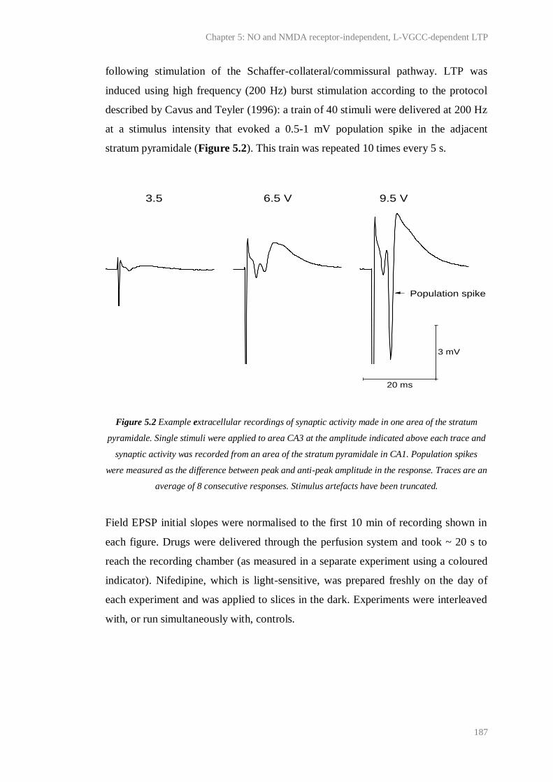
Chapter 5: NO and NMDA receptor-independent, L-VGCC-dependent LTP
187
following stimulation of the Schaffer-collateral/commissural pathway. LTP was
induced using high frequency (200 Hz) burst stimulation according to the protocol
described by Cavus and Teyler (1996): a train of 40 stimuli were delivered at 200 Hz
at a stimulus intensity that evoked a 0.5-1 mV population spike in the adjacent
stratum pyramidale (Figure 5.2). This train was repeated 10 times every 5 s.
Figure 5.2 Example extracellular recordings of synaptic activity made in one area of the stratum
pyramidale. Single stimuli were applied to area CA3 at the amplitude indicated above each trace and
synaptic activity was recorded from an area of the stratum pyramidale in CA1. Population spikes
were measured as the difference between peak and anti-peak amplitude in the response. Traces are an
average of 8 consecutive responses. Stimulus artefacts have been truncated.
Field EPSP initial slopes were normalised to the first 10 min of recording shown in
each figure. Drugs were delivered through the perfusion system and took ~ 20 s to
reach the recording chamber (as measured in a separate experiment using a coloured
indicator). Nifedipine, which is light-sensitive, was prepared freshly on the day of
each experiment and was applied to slices in the dark. Experiments were interleaved
with, or run simultaneously with, controls.
3.5 6.5 V 9.5 V
Population spike
20 ms
3 mV
Chapter 5: NO and NMDA receptor-independent, L-VGCC-dependent LTP
188
5.3.3 Measuring cGMP production in hippocampal slices
Hippocampal slices from multiple animals were randomly assigned to flasks of
oxygenated aCSF (30 ºC) held in a shaking water-bath. NOS dependent-cGMP
production was stimulated by submerging the slices in high K+ (30-122.5 mM) aCSF
for 5 min. Standard aCSF contained 2.5 mM K+ (see Chapter 2). The concentration
of Na+ in high K
+ aCSF was lowered to maintain osmolarity. After stimulation, slices
were individually inactivated by submersion in 200 µl boiling buffer containing 50
mM tris-HCl and 4 mM EDTA (pH 7.4) for ~ 30 min and then sonicated. The cGMP
content of the resulting solution was measured by radioimmunoassay and protein
using the BCA method (see Chapter 2 for methods).
In order that endogenous cGMP generation could be detected, all slices were
incubated with an inhibitor of PDE 2 (BAY 60-7500, 1 µM, 30 min), which is the
main PDE responsible for cGMP breakdown in the hippocampus (van Staveren et
al., 2001; Suvarna and O'Donnell, 2002; van Staveren et al., 2003), prior to
submersion in high K+, low Na
+ aCSF. TTX (1 µM, 35 min) was also pre-applied to
prevent network activity upon stimulation, and D-AP5 (100 µM, 35 min) to isolate
the NMDA receptor-independent response. In every experiment, un-stimulated/basal
and NMDA-induced cGMP levels were measured so that the consistency of cGMP
accumulation could be monitored. NMDA was applied for 2 min at 100 µM in the
absence of D-AP5.
5.3.4 Estimating membrane potential (Vm) as a function of extracellular
K+
concentration
The effect of extracellular K+
concentration ([K+]o) on Vm was estimated using the
Goldman-Hodgkin-Katz equation:
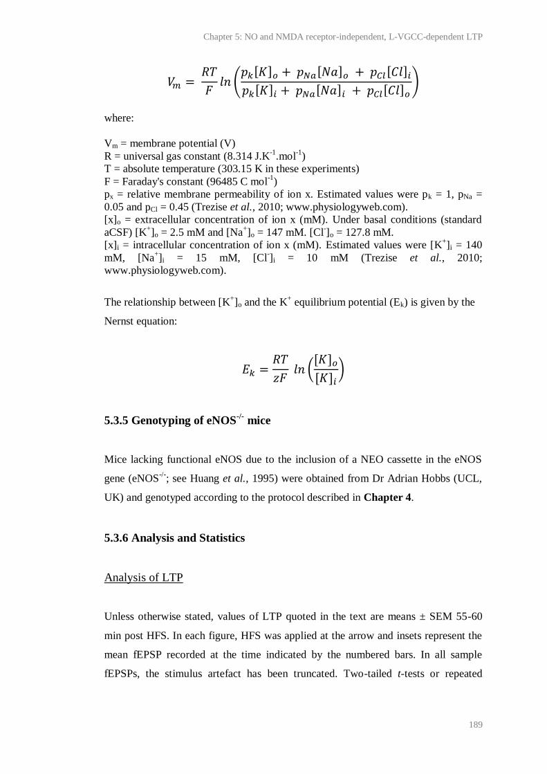
Chapter 5: NO and NMDA receptor-independent, L-VGCC-dependent LTP
189
where:
Vm = membrane potential (V)
R = universal gas constant (8.314 J.K-1
.mol-1
)
T = absolute temperature (303.15 K in these experiments)
F = Faraday's constant (96485 C mol-1
)
px = relative membrane permeability of ion x. Estimated values were pk = 1, pNa =
0.05 and pCl = 0.45 (Trezise et al., 2010; www.physiologyweb.com).
[x]o = extracellular concentration of ion x (mM). Under basal conditions (standard
aCSF) [K+]o = 2.5 mM and [Na
+]o = 147 mM. [Cl
-]o = 127.8 mM.
[x]i = intracellular concentration of ion x (mM). Estimated values were [K+]i = 140
mM, [Na+]i = 15 mM, [Cl
-]i = 10 mM (Trezise et al., 2010;
www.physiologyweb.com).
The relationship between [K+]o and the K
+ equilibrium potential (Ek) is given by the
Nernst equation:
5.3.5 Genotyping of eNOS-/-
mice
Mice lacking functional eNOS due to the inclusion of a NEO cassette in the eNOS
gene (eNOS-/-
; see Huang et al., 1995) were obtained from Dr Adrian Hobbs (UCL,
UK) and genotyped according to the protocol described in Chapter 4.
5.3.6 Analysis and Statistics
Analysis of LTP
Unless otherwise stated, values of LTP quoted in the text are means ± SEM 55-60
min post HFS. In each figure, HFS was applied at the arrow and insets represent the
mean fEPSP recorded at the time indicated by the numbered bars. In all sample
fEPSPs, the stimulus artefact has been truncated. Two-tailed t-tests or repeated
Chapter 5: NO and NMDA receptor-independent, L-VGCC-dependent LTP
190
measures, one-factor ANOVA with Bonferroni test were used to assess statistical
significance between data sets 55-60 min post HFS.
Analysis of NOS activity assays
Values in the text are means ± SEM. Statistical significance was assessed using one-
factor ANOVA with Dunnett’s test or unpaired t-tests.
5.4 Results
5.4.1 Characterisation of LTP induced by high frequency (200 Hz) burst
stimulation
As was found originally by Grover and Teyler (1990), LTP could be reliably induced
at CA1 synapses by 200 Hz burst stimulation (Figure 5.3). Consistent with the
original observation, the LTP was of high magnitude (200 ± 13 %; n = 3). As
expected of a compound LTP, no further significant potentiation could be yielded by
subsequent 200 Hz burst stimulation or HFS (100-Hz, 1-s), suggesting that the
limiting factor(s) for the magnitude of this plasticity was saturated.
Unlike LTP induced by HFS, or the LTP originally observed by Grover and Teyler
(1990) and Cavus and Teyler (1996) using 200 Hz burst stimulation, the LTP
induced by 200 Hz burst stimulation was not preceded by PTP. Rather, a short-
lasting (1-2 min) post-tetanic depression of the initial slope measuring ~ 50 % from
baseline was consistently observed (Figure 5.3-4). Consequently, the potentiation
was more reminiscent of the slow onset (10-15 min to maximum slope) LTP induced
by 200 Hz burst stimulation in vivo by Morgan and Teyler (1999). Since it is known
that high frequency burst stimulation can induce a transient (lasting ~ 5 min)
heterosynaptic depression of synaptic activity in area CA1 (Grover and Teyler,
1992), it is conceivable that in this study, small methodological differences led to the
induction of a stronger depression than that induced by Grover and Teyler (1990) and
that this masked PTP. This would explain the biphasic shape of the potentiation
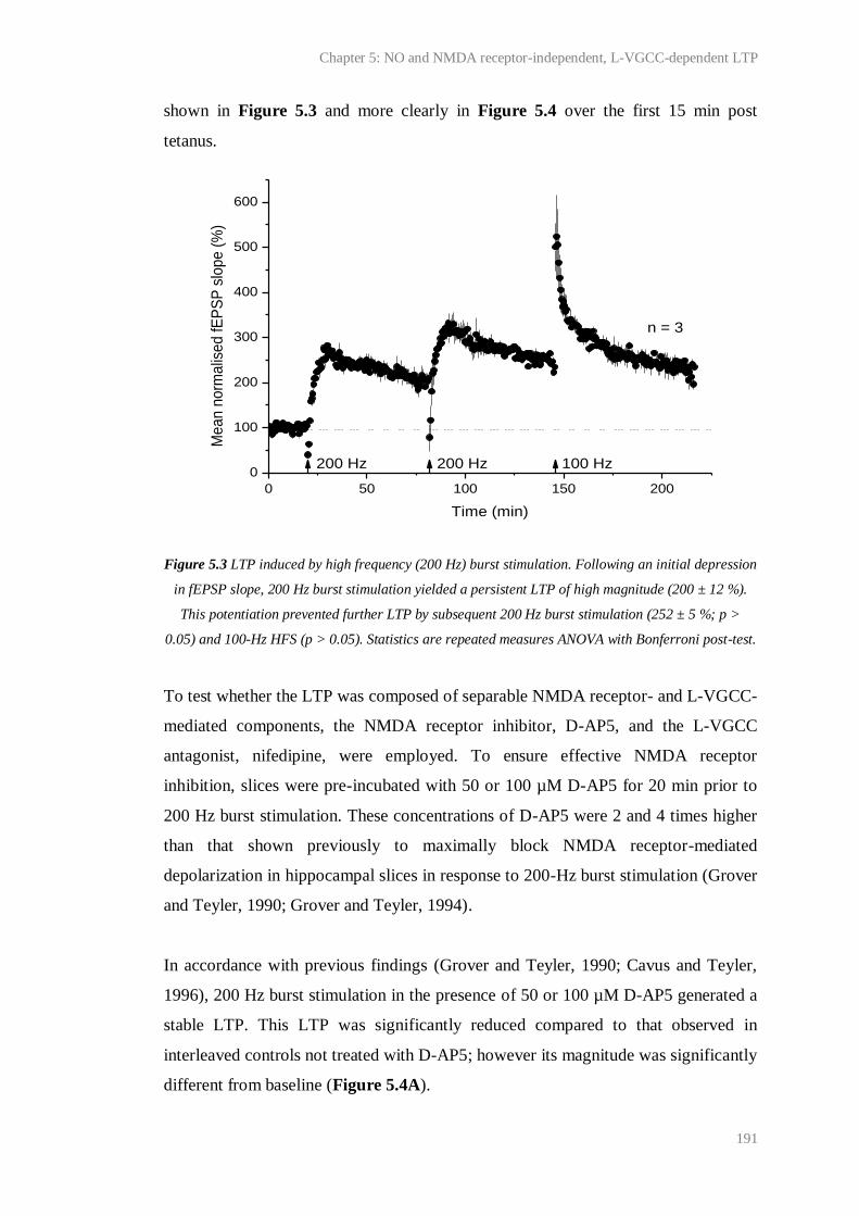
Chapter 5: NO and NMDA receptor-independent, L-VGCC-dependent LTP
191
shown in Figure 5.3 and more clearly in Figure 5.4 over the first 15 min post
tetanus.
Figure 5.3 LTP induced by high frequency (200 Hz) burst stimulation. Following an initial depression
in fEPSP slope, 200 Hz burst stimulation yielded a persistent LTP of high magnitude (200 ± 12 %).
This potentiation prevented further LTP by subsequent 200 Hz burst stimulation (252 ± 5 %; p >
0.05) and 100-Hz HFS (p > 0.05). Statistics are repeated measures ANOVA with Bonferroni post-test.
To test whether the LTP was composed of separable NMDA receptor- and L-VGCC-
mediated components, the NMDA receptor inhibitor, D-AP5, and the L-VGCC
antagonist, nifedipine, were employed. To ensure effective NMDA receptor
inhibition, slices were pre-incubated with 50 or 100 µM D-AP5 for 20 min prior to
200 Hz burst stimulation. These concentrations of D-AP5 were 2 and 4 times higher
than that shown previously to maximally block NMDA receptor-mediated
depolarization in hippocampal slices in response to 200-Hz burst stimulation (Grover
and Teyler, 1990; Grover and Teyler, 1994).
In accordance with previous findings (Grover and Teyler, 1990; Cavus and Teyler,
1996), 200 Hz burst stimulation in the presence of 50 or 100 µM D-AP5 generated a
stable LTP. This LTP was significantly reduced compared to that observed in
interleaved controls not treated with D-AP5; however its magnitude was significantly
different from baseline (Figure 5.4A).
0 50 100 150 200
0
100
200
300
400
500
600M
ea
n n
orm
alis
ed
fE
PS
P s
lop
e (
%)
Time (min)
200 Hz 200 Hz 100 Hz
n = 3

Chapter 5: NO and NMDA receptor-independent, L-VGCC-dependent LTP
192
Figure 5.4 Effect of NMDA receptor and L-VGCC inhibition on LTP induced by 200 Hz burst
stimulation. A) In the presence of the NMDA receptor antagonist, D-AP5 (50 or 100 µM), 200 Hz
burst stimulation produced a stable, but significantly reduced LTP (unfilled circles) compared to
interleaved untreated controls (50 µM D-AP5: 149 ± 5 %; n = 3; 100 µM D-AP5: 166 ± 7 %; n = 2;
no D-AP5: 200 ± 13 %; unpaired t-test, 50 and 100 µM D-AP5 (which were not significantly different
from each other: unpaired t-test, p = 0.137), vs. no D-AP5, p = 0.009). This potentiation was
significantly different from baseline, however (paired t-test, p = 1.91 × 10⁻⁴ compared to the last 5
0 20 40 60 80 100
20
40
60
80
100
120
140
160
180
200
220ii
iiii
ii
D-AP5 D-AP5 and nifedipine
Mean n
orm
alis
ed fE
PS
P s
lope (
%)
Time (min)
30 M nifedipine
50 or 100 M D-AP5
Exponential fit, = 57 ± 2 min
n = 5
n = 5
i
15 ms
1 mV
0 10 20 30 40 50 60 70
0
50
100
150
200
250
300 iiii
ii
Without D-AP5 With D-AP5
Mean n
orm
alis
ed fE
PS
P s
lope (
%)
Time (min)
50 or 100 M D-AP5
n = 3
n = 5
i
ii
15 ms
1 mV
A B B
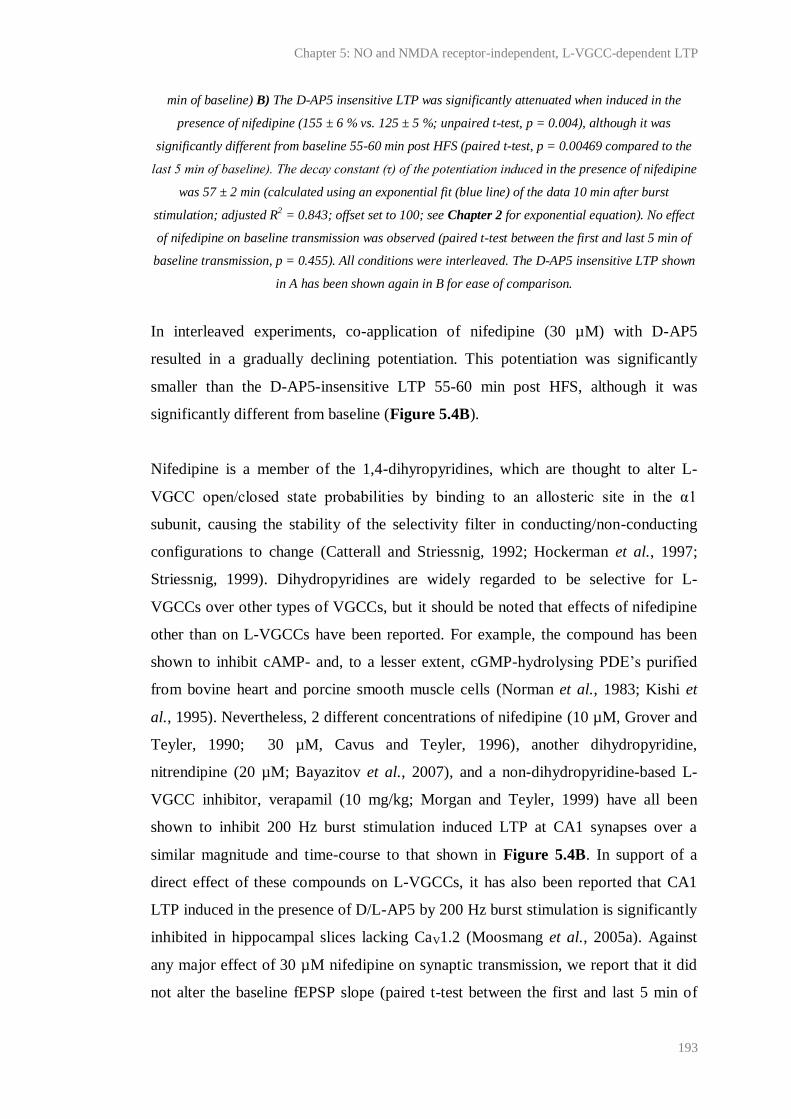
Chapter 5: NO and NMDA receptor-independent, L-VGCC-dependent LTP
193
min of baseline) B) The D-AP5 insensitive LTP was significantly attenuated when induced in the
presence of nifedipine (155 ± 6 % vs. 125 ± 5 %; unpaired t-test, p = 0.004), although it was
significantly different from baseline 55-60 min post HFS (paired t-test, p = 0.00469 compared to the
last 5 min of baseline). The decay constant (τ) of the potentiation induced in the presence of nifedipine
was 57 ± 2 min (calculated using an exponential fit (blue line) of the data 10 min after burst
stimulation; adjusted R2 = 0.843; offset set to 100; see Chapter 2 for exponential equation). No effect
of nifedipine on baseline transmission was observed (paired t-test between the first and last 5 min of
baseline transmission, p = 0.455). All conditions were interleaved. The D-AP5 insensitive LTP shown
in A has been shown again in B for ease of comparison.
In interleaved experiments, co-application of nifedipine (30 µM) with D-AP5
resulted in a gradually declining potentiation. This potentiation was significantly
smaller than the D-AP5-insensitive LTP 55-60 min post HFS, although it was
significantly different from baseline (Figure 5.4B).
Nifedipine is a member of the 1,4-dihyropyridines, which are thought to alter L-
VGCC open/closed state probabilities by binding to an allosteric site in the α1
subunit, causing the stability of the selectivity filter in conducting/non-conducting
configurations to change (Catterall and Striessnig, 1992; Hockerman et al., 1997;
Striessnig, 1999). Dihydropyridines are widely regarded to be selective for L-
VGCCs over other types of VGCCs, but it should be noted that effects of nifedipine
other than on L-VGCCs have been reported. For example, the compound has been
shown to inhibit cAMP- and, to a lesser extent, cGMP-hydrolysing PDE’s purified
from bovine heart and porcine smooth muscle cells (Norman et al., 1983; Kishi et
al., 1995). Nevertheless, 2 different concentrations of nifedipine (10 µM, Grover and
Teyler, 1990; 30 µM, Cavus and Teyler, 1996), another dihydropyridine,
nitrendipine (20 µM; Bayazitov et al., 2007), and a non-dihydropyridine-based L-
VGCC inhibitor, verapamil (10 mg/kg; Morgan and Teyler, 1999) have all been
shown to inhibit 200 Hz burst stimulation induced LTP at CA1 synapses over a
similar magnitude and time-course to that shown in Figure 5.4B. In support of a
direct effect of these compounds on L-VGCCs, it has also been reported that CA1
LTP induced in the presence of D/L-AP5 by 200 Hz burst stimulation is significantly
inhibited in hippocampal slices lacking CaV1.2 (Moosmang et al., 2005a). Against
any major effect of 30 µM nifedipine on synaptic transmission, we report that it did
not alter the baseline fEPSP slope (paired t-test between the first and last 5 min of

Chapter 5: NO and NMDA receptor-independent, L-VGCC-dependent LTP
194
baseline recording, p = 0.455; n = 5). Therefore, it was concluded that LTP induced
by 200 Hz burst stimulation could be dissociated into a NMDA receptor-dependent
and a NMDA receptor-independent, L-VGCC-dependent component, and the role of
NO in the latter could be tested.
5.4.2 Contribution of NO to NMDA receptor-independent, L-VGCC-
dependent LTP
To test whether NO was required for the L-VGCC-dependent, NMDA receptor-
independent LTP, the non-selective NOS inhibitor, L-NNA (100 µM), was used
(Figure 5.5). Compared to the LTP observed following 200 Hz burst stimulation in
the presence of D-AP5, the potentiation induced in the presence of L-NNA and D-
AP5 was unstable and significantly reduced 55-60 min following induction.
Although it was significantly different from baseline 55-60 min post HFS, it
appeared to be declining further.
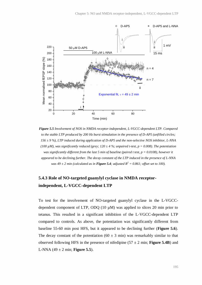
Chapter 5: NO and NMDA receptor-independent, L-VGCC-dependent LTP
195
Figure 5.5 Involvement of NOS in NMDA receptor-independent, L-VGCC-dependent LTP. Compared
to the stable LTP produced by 200 Hz burst stimulation in the presence of D-AP5 (unfilled circles;
156 ± 9 %), LTP induced during application of D-AP5 and the non-selective NOS inhibitor, L-NNA
(100 µM), was significantly reduced (grey; 120 ± 4 %; unpaired t-test, p = 0.008). The potentiation
was significantly different from the last 5 min of baseline (paired t test, p = 0.0108), however it
appeared to be declining further. The decay constant of the LTP induced in the presence of L-NNA
was 49 ± 2 min (calculated as in Figure 5.4; adjusted R2 = 0.861; offset set to 100).
5.4.3 Role of NO-targeted guanylyl cyclase in NMDA receptor-
independent, L-VGCC-dependent LTP
To test for the involvement of NO-targeted guanylyl cyclase in the L-VGCC-
dependent component of LTP, ODQ (10 µM) was applied to slices 20 min prior to
tetanus. This resulted in a significant inhibition of the L-VGCC-dependent LTP
compared to controls. As above, the potentiation was significantly different from
baseline 55-60 min post HFS, but it appeared to be declining further (Figure 5.6).
The decay constant of the potentiation (60 ± 3 min) was remarkably similar to that
observed following HFS in the presence of nifedipine (57 ± 2 min; Figure 5.4B) and
L-NNA (49 ± 2 min; Figure 5.5).
0 20 40 60 80
20
40
60
80
100
120
140
160
180
200
220 iiii
ii
ii
D-AP5 D-AP5 and L-NNA
50 M D-AP5
Mean n
orm
alis
ed fE
PS
P s
lope (
%)
Time (min)
100 M L-NNA
Exponential fit, = 49 ± 2 min
n = 4
n = 7
i
15 ms
1 mV
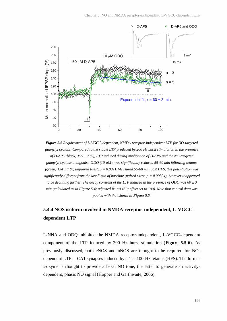
Chapter 5: NO and NMDA receptor-independent, L-VGCC-dependent LTP
196
Figure 5.6 Requirement of L-VGCC-dependent, NMDA receptor-independent LTP for NO-targeted
guanylyl cyclase. Compared to the stable LTP produced by 200 Hz burst stimulation in the presence
of D-AP5 (black; 155 ± 7 %), LTP induced during application of D-AP5 and the NO-targeted
guanylyl cyclase antagonist, ODQ (10 µM), was significantly reduced 55-60 min following tetanus
(green; 134 ± 7 %; unpaired t-test, p = 0.031). Measured 55-60 min post HFS, this potentiation was
significantly different from the last 5 min of baseline (paired t-test, p = 0.00304), however it appeared
to be declining further. The decay constant of the LTP induced in the presence of ODQ was 60 ± 3
min (calculated as in Figure 5.4; adjusted R2 =0.450; offset set to 100). Note that control data was
pooled with that shown in Figure 5.5.
5.4.4 NOS isoform involved in NMDA receptor-independent, L-VGCC-
dependent LTP
L-NNA and ODQ inhibited the NMDA receptor-independent, L-VGCC-dependent
component of the LTP induced by 200 Hz burst stimulation (Figure 5.5-6). As
previously discussed, both eNOS and nNOS are thought to be required for NO-
dependent LTP at CA1 synapses induced by a 1-s. 100-Hz tetanus (HFS). The former
isozyme is thought to provide a basal NO tone, the latter to generate an activity-
dependent, phasic NO signal (Hopper and Garthwaite, 2006).
0 20 40 60 80 100
20
40
60
80
100
120
140
160
180
200
220 i
i
ii
ii
D-AP5 D-AP5 and ODQ
Me
an
no
rma
lise
d fE
PS
P s
lop
e (
%)
Time (min)
50 M D-AP5
10 M ODQ
Exponential fit, = 60 ± 3 min
n = 8
n = 5
i
ii
15 ms
1 mV

Chapter 5: NO and NMDA receptor-independent, L-VGCC-dependent LTP
197
To elucidate the NOS isoform(s) required for NMDA receptor-independent, L-
VGCC-dependent LTP, and therefore begin to characterise the nature of the NO
signal involved, eNOS-/-
mice were used. Neither nNOS inhibitors nor nNOS knock-
out mice were used due to reasons outlined in Chapter 6.
We observed no significant difference between the NMDA receptor-independent, L-
VGCC-dependent LTP induced in slices from eNOS-/-
or age-, sex- and strain-
matched wild-type mice (Figure 5.7A). However, in interleaved experiments, LTP in
eNOS-/-
mice was significantly reduced by L-NNA (Figure 5.7B).

Chapter 5: NO and NMDA receptor-independent, L-VGCC-dependent LTP
198
Figure 5.7 NMDA receptor-independent, L-VGCC-dependent LTP in eNOS-/- mice. A) No significant
difference was detected between the LTP induced in slices from eNOS-/- and matched wild-type mice
(148 ± 6 % vs. 141 ± 5 % respectively; unpaired t-test, p = 0.374). B) The non-specific NOS inhibitor,
L-NNA, significantly attenuated LTP in eNOS-/- slices (124 ± 6 % vs. 148 ± 6 %; unpaired t-test, p =
0.021). All conditions were interleaved.
0 20 40 60 80 100
20
40
60
80
100
120
140
160
180
15 ms
1 mV
iiii
ii
ii
Wild-type eNOS-/-
Me
an
no
rma
lise
d fE
PS
P s
lop
e (
%)
Time (min)
50 M D-AP5
n = 7-9
i
0 20 40 60 80 100
20
40
60
80
100
120
140
160
180
iiii
ii
i
i
eNOS-/-
eNOS-/-
and L-NNA
Me
an
no
rma
lise
d fE
PS
P s
lop
e (
%)
Time (min)
50 M D-AP5
100 M L-NNA
n = 9
n = 6
i 15 ms
1 mV
A B
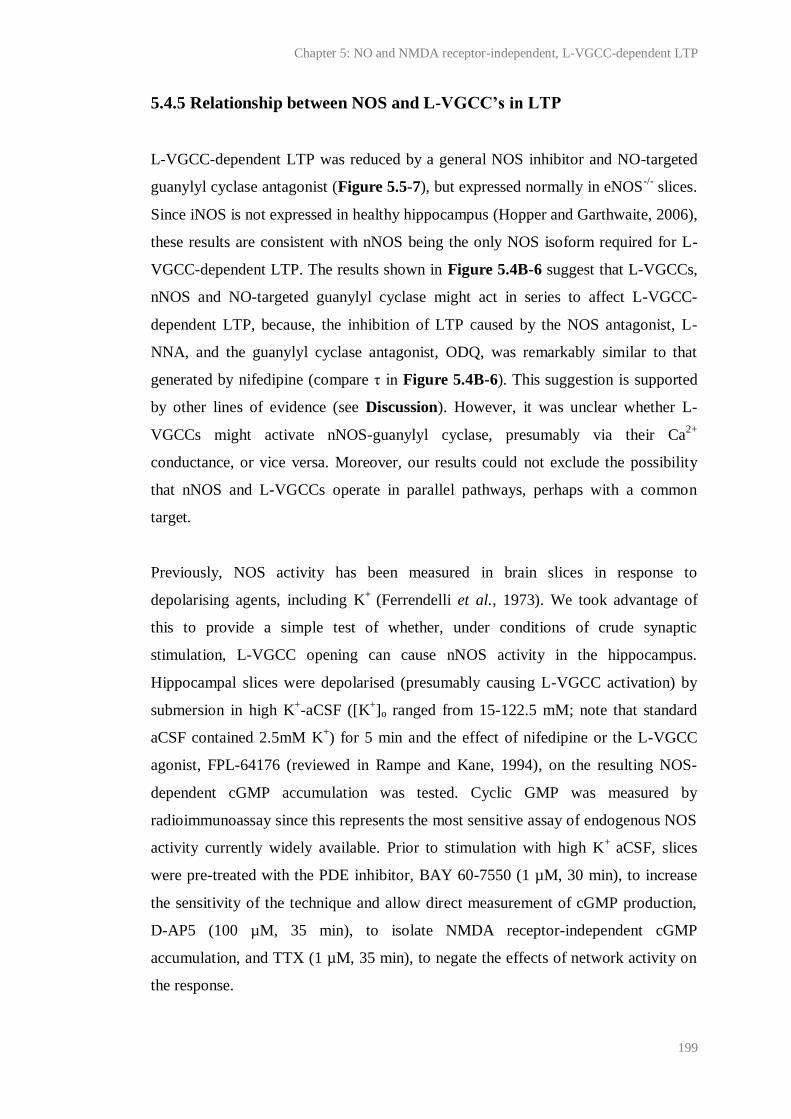
Chapter 5: NO and NMDA receptor-independent, L-VGCC-dependent LTP
199
5.4.5 Relationship between NOS and L-VGCC’s in LTP
L-VGCC-dependent LTP was reduced by a general NOS inhibitor and NO-targeted
guanylyl cyclase antagonist (Figure 5.5-7), but expressed normally in eNOS-/-
slices.
Since iNOS is not expressed in healthy hippocampus (Hopper and Garthwaite, 2006),
these results are consistent with nNOS being the only NOS isoform required for L-
VGCC-dependent LTP. The results shown in Figure 5.4B-6 suggest that L-VGCCs,
nNOS and NO-targeted guanylyl cyclase might act in series to affect L-VGCC-
dependent LTP, because, the inhibition of LTP caused by the NOS antagonist, L-
NNA, and the guanylyl cyclase antagonist, ODQ, was remarkably similar to that
generated by nifedipine (compare τ in Figure 5.4B-6). This suggestion is supported
by other lines of evidence (see Discussion). However, it was unclear whether L-
VGCCs might activate nNOS-guanylyl cyclase, presumably via their Ca2+
conductance, or vice versa. Moreover, our results could not exclude the possibility
that nNOS and L-VGCCs operate in parallel pathways, perhaps with a common
target.
Previously, NOS activity has been measured in brain slices in response to
depolarising agents, including K+
(Ferrendelli et al., 1973). We took advantage of
this to provide a simple test of whether, under conditions of crude synaptic
stimulation, L-VGCC opening can cause nNOS activity in the hippocampus.
Hippocampal slices were depolarised (presumably causing L-VGCC activation) by
submersion in high K+-aCSF ([K
+]o ranged from 15-122.5 mM; note that standard
aCSF contained 2.5mM K+) for 5 min and the effect of nifedipine or the L-VGCC
agonist, FPL-64176 (reviewed in Rampe and Kane, 1994), on the resulting NOS-
dependent cGMP accumulation was tested. Cyclic GMP was measured by
radioimmunoassay since this represents the most sensitive assay of endogenous NOS
activity currently widely available. Prior to stimulation with high K+
aCSF, slices
were pre-treated with the PDE inhibitor, BAY 60-7550 (1 µM, 30 min), to increase
the sensitivity of the technique and allow direct measurement of cGMP production,
D-AP5 (100 µM, 35 min), to isolate NMDA receptor-independent cGMP
accumulation, and TTX (1 µM, 35 min), to negate the effects of network activity on
the response.
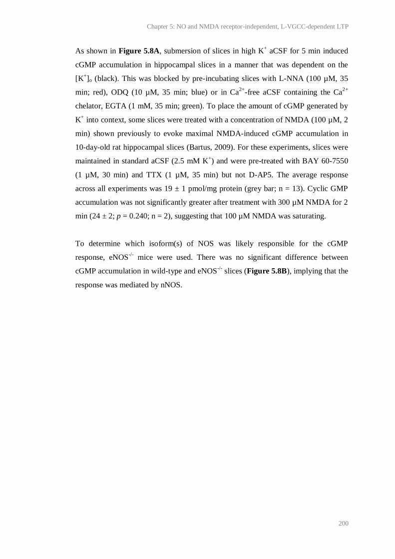
Chapter 5: NO and NMDA receptor-independent, L-VGCC-dependent LTP
200
As shown in Figure 5.8A, submersion of slices in high K+ aCSF for 5 min induced
cGMP accumulation in hippocampal slices in a manner that was dependent on the
[K+]o (black). This was blocked by pre-incubating slices with L-NNA (100 µM, 35
min; red), ODQ (10 µM, 35 min; blue) or in Ca2+
-free aCSF containing the Ca2+
chelator, EGTA (1 mM, 35 min; green). To place the amount of cGMP generated by
K+ into context, some slices were treated with a concentration of NMDA (100 µM, 2
min) shown previously to evoke maximal NMDA-induced cGMP accumulation in
10-day-old rat hippocampal slices (Bartus, 2009). For these experiments, slices were
maintained in standard aCSF (2.5 mM K+) and were pre-treated with BAY 60-7550
(1 µM, 30 min) and TTX (1 µM, 35 min) but not D-AP5. The average response
across all experiments was 19 ± 1 pmol/mg protein (grey bar; n = 13). Cyclic GMP
accumulation was not significantly greater after treatment with 300 µM NMDA for 2
min (24 ± 2; p = 0.240; n = 2), suggesting that 100 µM NMDA was saturating.
To determine which isoform(s) of NOS was likely responsible for the cGMP
response, eNOS-/-
mice were used. There was no significant difference between
cGMP accumulation in wild-type and eNOS-/-
slices (Figure 5.8B), implying that the
response was mediated by nNOS.

Chapter 5: NO and NMDA receptor-independent, L-VGCC-dependent LTP
201
Figure 5.8 K+-induced cGMP accumulation in hippocampal slices. A) In slices incubated in standard
aCSF (2.5 mM K) and pre-treated with BAY 60-7550, and TTX, NMDA (100 µM, 2 min) generated 19
± 1 pmol cGMP/mg protein on average (grey bar). In slices pre-treated with BAY 60-7550, TTX and
D-AP5, K+ induced cGMP accumulation in a concentration-dependent manner (black) that was well
described by a logistic equation (R2 =0.997; see Chapter 2 for logistic equation). Slices treated with
the highest concentration of K+ (122.5 mM) produced significantly more cGMP than those treated
with NMDA (ANOVA with Dunnett’s test, p < 0.01). Where tested, K+-induced cGMP accumulation
0 20 40 60 80 100 120 140
K+ (logistic fit; EC
50 = 61 ± 4 pmol/mg protein; n = 3-14)
K+ and 100 M L-NNA (n = 3-4)
K+ and 10 M ODQ (n = 4)
K+ and Ca
2+ free aCSF + 1 mM EGTA (n = 4)
[K+]O (mM)
A
0
5
10
15
20
25
30
35
Mean c
GM
P (
pm
ol/m
g p
rote
in)
100 M NMDA
n = 13
0
5
10
15
20
25
30
Me
an
cG
MP
(p
mo
l/m
g p
rote
in)
Wild-type
eNOS-/-
n = 3-4
+ L-NNA
Basal 122.5 mM K+
NMDA
B
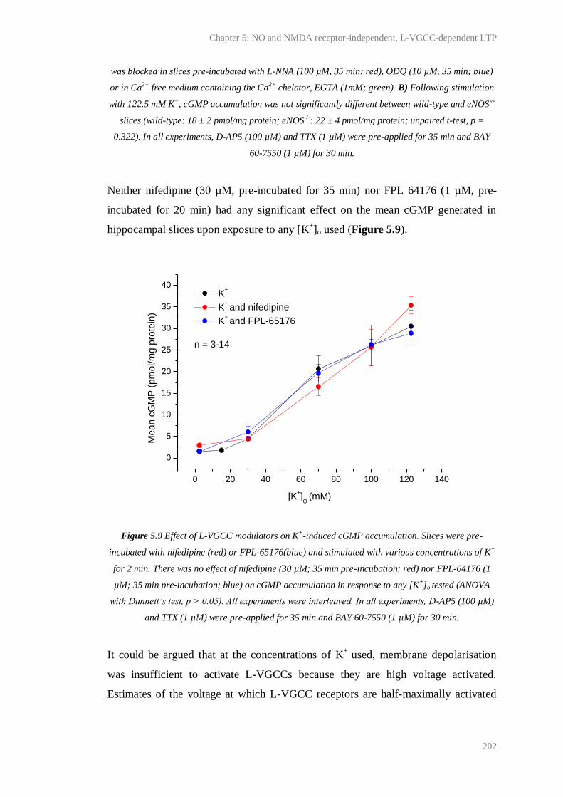
Chapter 5: NO and NMDA receptor-independent, L-VGCC-dependent LTP
202
was blocked in slices pre-incubated with L-NNA (100 µM, 35 min; red), ODQ (10 µM, 35 min; blue)
or in Ca2+ free medium containing the Ca2+ chelator, EGTA (1mM; green). B) Following stimulation
with 122.5 mM K+, cGMP accumulation was not significantly different between wild-type and eNOS-/-
slices (wild-type: 18 ± 2 pmol/mg protein; eNOS-/-: 22 ± 4 pmol/mg protein; unpaired t-test, p =
0.322). In all experiments, D-AP5 (100 µM) and TTX (1 µM) were pre-applied for 35 min and BAY
60-7550 (1 µM) for 30 min.
Neither nifedipine (30 µM, pre-incubated for 35 min) nor FPL 64176 (1 µM, pre-
incubated for 20 min) had any significant effect on the mean cGMP generated in
hippocampal slices upon exposure to any [K+]o used (Figure 5.9).
Figure 5.9 Effect of L-VGCC modulators on K+-induced cGMP accumulation. Slices were pre-
incubated with nifedipine (red) or FPL-65176(blue) and stimulated with various concentrations of K+
for 2 min. There was no effect of nifedipine (30 µM; 35 min pre-incubation; red) nor FPL-64176 (1
µM; 35 min pre-incubation; blue) on cGMP accumulation in response to any [K+]o tested (ANOVA
with Dunnett’s test, p > 0.05). All experiments were interleaved. In all experiments, D-AP5 (100 µM)
and TTX (1 µM) were pre-applied for 35 min and BAY 60-7550 (1 µM) for 30 min.
It could be argued that at the concentrations of K+
used, membrane depolarisation
was insufficient to activate L-VGCCs because they are high voltage activated.
Estimates of the voltage at which L-VGCC receptors are half-maximally activated
0 20 40 60 80 100 120 140
0
5
10
15
20
25
30
35
40 K
+
K+ and nifedipine
K+ and FPL-65176
n = 3-14
Me
an
cG
MP
(p
mo
l/m
g p
rote
in)
[K+]O
(mM)
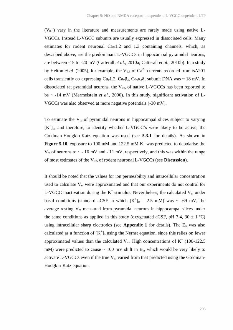
Chapter 5: NO and NMDA receptor-independent, L-VGCC-dependent LTP
203
(V0.5) vary in the literature and measurements are rarely made using native L-
VGCCs. Instead L-VGCC subunits are usually expressed in dissociated cells. Many
estimates for rodent neuronal CaV1.2 and 1.3 containing channels, which, as
described above, are the predominant L-VGCCs in hippocampal pyramidal neurons,
are between -15 to -20 mV (Catterall et al., 2010a; Catterall et al., 2010b). In a study
by Helton et al. (2005), for example, the V0.5 of Ca2+
currents recorded from tsA201
cells transiently co-expressing Cav1.2, Cavβ3, Cavα2δ1 subunit DNA was ~ 18 mV. In
dissociated rat pyramidal neurons, the V0.5 of native L-VGCCs has been reported to
be ~ -14 mV (Mermelstein et al., 2000). In this study, significant activation of L-
VGCCs was also observed at more negative potentials (-30 mV).
To estimate the Vm of pyramidal neurons in hippocampal slices subject to varying
[K+]o, and therefore, to identify whether L-VGCC’s were likely to be active, the
Goldman-Hodgkin-Katz equation was used (see 5.3.1 for details). As shown in
Figure 5.10, exposure to 100 mM and 122.5 mM K+ was predicted to depolarise the
Vm of neurons to ~ - 16 mV and - 11 mV, respectively, and this was within the range
of most estimates of the V0.5 of rodent neuronal L-VGCCs (see Discussion).
It should be noted that the values for ion permeability and intracellular concentration
used to calculate Vm were approximated and that our experiments do not control for
L-VGCC inactivation during the K+ stimulus. Nevertheless, the calculated Vm under
basal conditions (standard aCSF in which [K+]o = 2.5 mM) was ~ -69 mV, the
average resting Vm measured from pyramidal neurons in hippocampal slices under
the same conditions as applied in this study (oxygenated aCSF, pH 7.4, 30 ± 1 ºC)
using intracellular sharp electrodes (see Appendix 1 for details). The Ek was also
calculated as a function of [K+]o using the Nernst equation, since this relies on fewer
approximated values than the calculated Vm. High concentrations of K+ (100-122.5
mM) were predicted to cause ~ 100 mV shift in Ek, which would be very likely to
activate L-VGCCs even if the true Vm varied from that predicted using the Goldman-
Hodgkin-Katz equation.
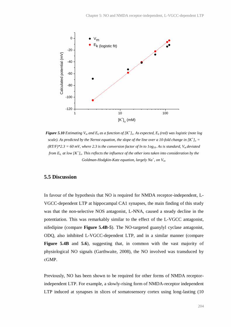
Chapter 5: NO and NMDA receptor-independent, L-VGCC-dependent LTP
204
Figure 5.10 Estimating Vm and Ek as a function of [K+]o. As expected, Ek (red) was logistic (note log
scale). As predicted by the Nernst equation, the slope of the line over a 10-fold change in [K+]o =
(RT/F)*2.3 = 60 mV, where 2.3 is the conversion factor of ln to 1og10. As is standard, Vm deviated
from Ek, at low [K+]o. This reflects the influence of the other ions taken into consideration by the
Goldman-Hodgkin-Katz equation, largely Na+, on Vm.
5.5 Discussion
In favour of the hypothesis that NO is required for NMDA receptor-independent, L-
VGCC-dependent LTP at hippocampal CA1 synapses, the main finding of this study
was that the non-selective NOS antagonist, L-NNA, caused a steady decline in the
potentiation. This was remarkably similar to the effect of the L-VGCC antagonist,
nifedipine (compare Figure 5.4B-5). The NO-targeted guanylyl cyclase antagonist,
ODQ, also inhibited L-VGCC-dependent LTP, and in a similar manner (compare
Figure 5.4B and 5.6), suggesting that, in common with the vast majority of
physiological NO signals (Garthwaite, 2008), the NO involved was transduced by
cGMP.
Previously, NO has been shown to be required for other forms of NMDA receptor-
independent LTP. For example, a slowly-rising form of NMDA-receptor independent
LTP induced at synapses in slices of somatosensory cortex using long-lasting (10
1 10 100
-120
-100
-80
-60
-40
-20
0 Vm
Ek (logistic fit)
Ca
lcu
late
d p
ote
ntia
l (m
V)
[K+]O (mM)

Chapter 5: NO and NMDA receptor-independent, L-VGCC-dependent LTP
205
min), 100-Hz burst stimulation has been reported to be blocked by the NOS
antagonist, L-NAME, and in eNOS-/-
mice (Haul et al., 1999). It has also been
reported that LTP induced in the presence of D-AP5 at synapses between the dentate
gyrus mossy fibres and the proximal-apical dendrites of CA3 pyramidal neurons
using a 1-s, 100-Hz tetanus is reduced in eNOS-/-
mice (Doreulee et al., 2001;
although see Nicolarakis et al., 1994). However, to our knowledge, this is the first
report that endogenous NO is necessary for NMDA receptor-independent and L-
VGCC-dependent LTP at CA1 synapses.
Following a 100-Hz, 1-s tetanus (HFS), LTP is, in our hands, NMDA receptor-
dependent (Chapter 3). This LTP likely requires both nNOS and eNOS (Hopper and
Garthwaite 2006). Endothelial NOS, which after phosphorylation can become
tonically active (Fulton et al., 2001), is thought to provide a continuous, low-level
NO tone which may prime synapses for potentiation (Hopper and Garthwaite, 2006),
perhaps by modulating basal levels of presynaptic transmitter release at CA1
synapses (Neitz et al., 2011 and see Chapter 4). Neuronal NOS, on the other hand,
is thought to produce a phasic, activity-dependent signal necessary for hippocampal
LTP induction (Hopper and Garthwaite, 2006).
To characterise the NO signal necessary for the L-VGCC-dependent LTP, the NOS
isoform(s) required was determined. Given previous evidence, it seemed reasonable
that both sources of NO could be involved in L-VGCC-dependent LTP: NMDA
receptor-independent nNOS activity has been previously reported (see below) and
NMDA-receptor-independent forms of LTP at mossy fibre synapses (Doreulee et al.,
2001) and in the somatosensory cortex (Haul et al., 1999) are reportedly reduced in
eNOS-/-
mice. However, our experiments using eNOS-/-
mice favoured the conclusion
that nNOS was the only source of the NO needed for L-VGCC-dependent LTP
(Figure 5.7). Note that, unfortunately, selective nNOS inhibitors were unavailable
for use (see Chapter 6) and mice lacking active splice variants in the hippocampus
could not be obtained within the time constraints of this project. Taken together with
previous findings (Hopper and Garthwaite, 2006), the lack of effect of eNOS knock-
out on the L-VGCC-dependent LTP immediately suggested that the NO signal
involved in the LTP was activity-dependent, rather than tonic, and synthesised in

Chapter 5: NO and NMDA receptor-independent, L-VGCC-dependent LTP
206
neurons, rather than endothelial cells. The lack of effect also implies that eNOS and
its effects on neurotransmission, for example, through the tonic modulation of
neurotransmitter release onto CA1 cells (Chapter 4 and Neitz et al., 2011), are not
necessary for all forms of CA1 LTP. Furthermore, the result provides important
support for the role of nNOS in LTP, which has been disputed by findings that nNOS
knock-out mice are capable of normal NOS-inhibitor sensitive LTP at CA1 synapses
(O'Dell et al., 1994; although it should be noted that the mice involved may have
expressed active nNOS splice variants; Eliasson et al., 1997), and that nNOS
inhibitors shown previously to attenuate CA1 LTP (Hopper and Garthwaite, 2006),
are not useably selective for nNOS over eNOS (see Chapter 6).
As discussed above (see 5.1 Introduction), NO-, and L-VGCC-dependent forms of
LTP at CA1 synapses have previously been shown to rely on similar mechanisms,
and both NO and L-VGCCs have been found to modulate activity-dependent changes
associated with gene expression, learning and memory in the hippocampus. It is also
interesting to note that an interaction between NO and L-VGCC’s during long-lasting
synaptic activity, and perhaps learning and memory, may extend beyond CA1
synapses because L-VGCCs and NO have been found to be necessary for some
forms of LTP in the lateral amygdala, as well as amygdala-dependent learning, such
as auditory fear conditioning (Weisskopf et al., 1999; Bauer et al., 2002; Ota et al.,
2008). However, the critical role of NO in L-VGCC-dependent LTP was, to some
extent, unexpected and this was for two main reasons.
Firstly, several groups have long-hypothesised that LTP induction protocols using
high stimulus intensities favour the expression of NO-independent LTP (Haley et al.,
1993; Chetkovich et al., 1993; O'Dell et al., 1994; Wilson et al., 1999), thus
explaining why some forms of LTP are independent of NO (for example, Cummings
et al., 1994; Bannerman et al., 1994b; Phillips et al., 2008). This hypothesis has,
arguably, received more attention than theories that other factors, such as the
temperature at which the experiments were performed (Williams et al., 1993), or the
age and strain of the animals used (Holscher, 2002), determine whether NO is
involved in LTP. However, we report clear evidence that NO-dependent LTP can be
induced using an induction protocol delivered at relatively high stimulus intensities

Chapter 5: NO and NMDA receptor-independent, L-VGCC-dependent LTP
207
(≥ 1 V higher than that typically used to evoke the baseline fEPSP (see 5.3 Methods
for details)). Our data suggest therefore suggest that some other variable(s) dictate
the involvement of NO, or another factor capable of compensating for NO, in LTP
and that the above hypothesis should be reconsidered. Furthermore, our results are
accordant with more recent findings that postsynaptic somatic action potentials, the
generation of which would be favoured by high vs. low intensity stimulation (and
may also be necessary for L-VGCC activation), are necessary for NO-dependent
LTP at CA1 synapses (Phillips et al., 2008).
Secondly, physiological nNOS activation is thought to be achieved preferentially
through NMDA receptors. It is well known that nNOS can bind the NMDA receptor
subunit, NR2B, via PSD-95 (Christopherson et al., 1999) and in situ hybridisation
studies show that message for nNOS is co-localised with mRNA for PSD-95
throughout the brain (Brenman et al., 1996). In the hippocampus, immunofluorescent
staining has shown that nNOS, NR2B and PSD-95 co-localise in pyramidal cell
dendritic spines (Burette et al., 2002). NMDA has been shown to increase NO
production in the hippocampus in vivo (Luo and Vincent, 1994) and in vitro (Figure
5.8A), and this has been shown to be nNOS- (Huang et al., 1993) and PSD-95-
dependent (Sattler et al., 1999). A simple explanation for the requirement of L-
VGCC-dependent LTP for nNOS, therefore, was incomplete antagonism of NMDA
receptors during 200 Hz burst stimulation. Nevertheless, evidence presented by
Grover and Teyler (1990, 1994) strongly suggests that glutamate released during this
tetanus would be insufficient to overcome the concentrations of D-AP5 (50-100 µM)
used throughout this study. For example, they report that, in the presence of 50 µM
D/L-AP5 (equivalent to 25 µM D-AP5), 95 % of NMDA receptors are blocked
during 200 Hz burst stimulation, and that raising the concentration of D/L-AP5 to
200 µM yields no further inhibition of NMDA receptors, nor any inhibition of L-
VGCC-dependent LTP (Grover and Teyler, 1990). Therefore, the finding that NO
was required for L-VGCC-dependent, NMDA receptor-independent LTP implies
that, following 200 Hz burst stimulation, nNOS was activated by a source of Ca2+
influx other than NMDA receptors.
Chapter 5: NO and NMDA receptor-independent, L-VGCC-dependent LTP
208
Several lines of evidence suggest that L-VGCCs might be responsible for nNOS
activation during L-VGCC-dependent CA1 LTP. First, histological studies have
shown that nNOS and L-VGCCs may co-localise in pyramidal cell soma, at the base
of apical dendrites and in interneurons (Westenbroek et al., 1990; Ahlijanian et al.,
1990; Hell et al., 1993; Wendland et al., 1994; Gonzalez-Hernandez et al., 1996;
Burette et al., 2002). Interestingly, some histological and functional data also shows
that CaV1.2 L-VGCCs are expressed in the postsynaptic densities of more distal
pyramidal cell dendrites where they may lie in close proximity to NMDA receptors,
and therefore, probably nNOS (Hell et al., 1996). Second, as assessed using Fura-2
in hippocampal slices (Regehr and Tank, 1992; Christie et al., 1995) and whole cell
recordings of dissociated pyramidal neurons (Mermelstein et al., 2000), L-VGCCs
have been reported to cause significant (~ 30-50 % of the total) Ca2+
influx into
pyramidal cell soma and dendrites during spikes and ramp depolarisations (to -30
mV) designed to approximate the effects of EPSPs. Third, as mentioned above, LTP
induced in the presence of nifedipine, L-NNA and ODQ was characterised by a
similar decay constant (Figure 5.4B-6). Fourth, L-VGCCs have previously been
reported to activate nNOS in the nervous system. In the PNS, for example, in the
enteric nervous system, Ca2+
influx via VGCCs, including L-VGCCs, is well-known
to trigger NOS activation, leading to NANC transmission and smooth muscle
relaxation (reviewed by Vincent, 2010). In primary cultures of mouse cortical
neurons, in which non-neuronal cells constituted only ~ 5 % of the total, K+ (25-50
mM for 10-15 min) induced NOS-dependent, presumably nNOS-dependent, cGMP
accumulation that was insensitive to TTX, MK-801 and CNQX but was attenuated
by the L-VGCC antagonists, 4-(4-fluorophenyl)-2-methyl-6-(5-
piperidinopentyloxy)pyrimidine hydrochloride (NS-7; Suma et al., 1997; Tatsumi et
al., 1998) and nifedipine (Oka et al., 1999). Furthermore, the L-VGCC agonist, BAY
K-8644, concentration-dependently increased K+-induced cGMP accumulation and
this was also attenuated by NS-7 and nifedipine (Oka et al., 1999).
Nevertheless, it should be noted that the results presented in Figure 5.4B-6 are
equally consistent with two other possibilities, both consistent with another,
unknown source of Ca2+
influx to neurons being responsible for stimulating nNOS.
The first possibility is that NO could have activated L-VGCCs, as has been shown in

Chapter 5: NO and NMDA receptor-independent, L-VGCC-dependent LTP
209
various biological systems, including the cardiac system (reviewed by Striessnig,
1999). In human atrial myocytes, for example, picomolar concentrations of the NO
donor, 3-morpholinosydnonimine (SIN-1), and cGMP (500 nM) have been reported
to increase L-VGCC-mediated currents, most likely via an effect on cGMP-inhibited
PDE 3, leading to cAMP accumulation, PKA activation, L-VGCC phosphorylation
and an increase in the open state probability of the channels (Kirstein et al., 1995).
The second possibility is that L-VGCCs and NO acted in parallel pathways,
separated for example, by intracellular compartmentalisation, but with a common
target necessary for LTP expression. In support of this, L-VGCC-dependent LTP is
thought to require sources of Ca2+
influx to neurons other than L-VGCCs, such as
metabotropic glutamate receptors (Little et al., 1995). Furthermore, Kullmann et al.
(1992) have shown that, although repeated depolarising pulses applied to CA1
neurons in hippocampal slices elicit a D-AP5-insensitive, nifedipine-sensitive
increase in pyramidal cell EPSPs and EPSCs, this potentiation is transient (decaying
back to baseline within ~ 30 min) and requires coincident synaptic activity for
persistent expression. These findings suggest that another factor is required for L-
VGCC-dependent LTP. This factor could be responsible for nNOS activation, and
may also lend L-VGCC-dependent LTP its input-specificity, which, because L-
VGCCs are most densely clustered in the pyramidal cell soma and at the base of
apical dendrites, is difficult to explain.
Using a crude stimulus (2.5 – 122.5 mM K+, 5 min) to depolarise hippocampal slices
and activate L-VGCCs, we found no evidence that L-VGCCs activate nNOS (Figure
5.9). The Goldman-Hodgkin-Katz equation predicts that the Vm of neurons exposed
to the highest concentrations of K+ used was close to or above the V0.5 of the L-
VGCC’s present (Figure 5.10), suggesting that L-VGCCs were active under our
conditions. Furthermore, cGMP generation was quantified using the cGMP
radioimmunoassay, which provides the most sensitive measure of NOS activity
currently widely available. Therefore at present, it is reasonable to assume that, in the
hippocampus, L-VGCC-dependent, K+-induced nNOS activation is minimal or does
not occur. Of course, this cannot rule out the possibility that during 200 Hz burst
stimulation, L-VGCCs do activate nNOS. However, without any indication that L-
Chapter 5: NO and NMDA receptor-independent, L-VGCC-dependent LTP
210
VGCCs can activate nNOS in the hippocampus, the relationship between L-VGCCs
and nNOS in L-VGCC-dependent LTP remains an open question.
5.6 Conclusion
NO/cGMP appears to play a diverse role in hippocampal synaptic plasticity. In
Chapter 4 it was shown that, under our conditions, NMDA receptor-dependent LTP
at CA1 synapses requires endogenous NO. In addition to this, the data presented in
Figures 5.4-6 suggest for the first time that NO/cGMP is also necessary for L-
VGCC-dependent, NMDA receptor-independent CA1 LTP. Consistent with the
results of experiments using eNOS-/-
mice (Figure 5.7) and the reported lack of iNOS
in healthy hippocampus (Hopper and Garthwaite, 2006), nNOS may be the sole
isoform of the NO required for L-VGCC-dependent LTP, implying that a NMDA
receptor-independent mechanism, most likely involving an increase in intracellular
Ca2+
, is capable of activating nNOS during synaptic stimulation (Figure 5.7). This
could hold considerable influence over NO physiology and pathology.
Taking advantage of previous findings that K+ induces NOS-dependent cGMP
accumulation in hippocampal slices, and a highly sensitive assay for cGMP
detection, experiments were performed to ascertain whether, under crude stimulation,
L-VGCCs activate nNOS. Under these conditions, significant NMDA receptor-
independent, NOS-dependent cGMP accumulation was recorded (Figure 5.8),
consistent with the suggestion that nNOS can become activated by an unknown,
NMDA receptor-independent mechanism(s). No evidence for L-VGCC-induced
nNOS activity was found (Figure 5.9). This was not in favour of L-VGCC-induced
nNOS activation during 200 Hz burst stimulation, although it must be emphasised
that it is unclear how comparable the neuronal consequences of K+ and tetanic
stimulation are. Experiments to identify the source(s) of Ca2+
influx responsible for
K+-induced cGMP-accumulation have been performed (see Appendix 1), but were
inconclusive. Interestingly, a cGMP sensor with picomolar sensitivity has been
developed very recently (Batchelor et al., 2010) and future application of this to
hippocampal slices could allow L-VGCC-dependent NO synthesis to be re-
investigated with improved sensitivity and following 200 Hz burst stimulation.

Chapter 5: NO and NMDA receptor-independent, L-VGCC-dependent LTP
211
Finally, it is tempting to speculate that NO- and L-VGCC-dependent signalling
pathways activated during LTP may converge on pathways linked to gene expression
and/or BDNF release (see 5.2 Aim). Concerning the former, NMDA receptor-
independent NO synthesis during conditions also activating L-VGCCs could explain
the discrepancy in the literature as to why NMDA receptors and NO are necessary
for early-LTP induced by a 1-s, 100-Hz tetanus (HFS), but only the latter is required
for late-LTP and some associated changes in gene expression induced by multiple
HFS (see 5.1 Introduction).
212
Chapter 6:
Evaluation of nNOS inhibitors using intact tissues

Chapter 6: Evaluation of nNOS inhibitors using intact tissues
213
6.1 Introduction
This study resulted from attempts to identify the isoform of NOS involved in the
NO-dependent plasticity’s described in Chapter 4-5 using nNOS inhibitors. The use
of NOS inhibitors represents a major strategy for investigating NO physiology, as
has been demonstrated throughout this thesis, and is also an attractive means of
treating pathologies hypothesised to involve NO overproduction by NOS. These
pathologies include disorders of the nervous system thought to involve nNOS, such
as stroke and Parkinson’s disease, and inflammatory diseases considered to involve
iNOS, such as asthma and arthritis (reviewed in Gross and Wolin, 1995; Hobbs et
al., 1999; Vallance and Leiper, 2002).
The first series of NOS inhibitors to be developed were L-arginine derivatives such
as L-NMMA, L-NNA (see Table 6.1) and its prodrug, L-NAME. These compete
with L-arginine for binding to NOS and are non-selective for any NOS isozyme.
They are actively transported into cells and typically have IC50 values in the low
micromolar range (Alderton et al., 2001).
Early on, L-NNA and L-NMMA were reported to block eNOS-dependent, ACh-
induced relaxation of rabbit aorta, which could be restored upon application of L-
arginine or exogenous NO (Moore et al., 1990). L-NMMA was also shown to dose-
dependently increase blood pressure in two patients with septic shock who had failed
to respond to conventional treatment. L-NAME was reported to have a similar effect
in one of the patients (Petros et al., 1991). Since then, the L-arginine-based inhibitors
have been shown to inhibit various NO-dependent phenomena in different tissues
and to benefit patients with other disorders involving NO overproduction, such as
chronic tension-type headache (Ashina et al., 1999b) and migraine (Lassen et al.,
1997), which may result from central sensitisation of noiceceptive synapses by
overactive nNOS (Ashina et al., 1999a).
However, the interpretation of research using non-selective NOS inhibitors is limited
because all three isoforms of NOS may be simultaneously active in one tissue. The
therapeutic applications of non-selective NOS inhibitors are also hampered for the

Chapter 6: Evaluation of nNOS inhibitors using intact tissues
214
same reason. For example, non-selective NOS inhibitors cannot be used to treat
asthma, which may be augmented by excessive iNOS activity, because eNOS
inhibition causes vasoconstriction in the airways (Hansel et al., 2003). Likewise,
nNOS and eNOS inhibition appear to have opposing consequences for tissue damage
after ischemic insult (Huang et al., 1994; Huang et al., 1995; Huang et al., 1996).
To avoid these problems, NOS inhibitors selective for one isozyme are necessary,
but, unfortunately, their development has been demanding (Alderton et al., 2001).
The two most selective and potent nNOS inhibitors currently commercially available
are L-VNIO (or Vinyl L-NIO; Babu and Griffith, 2008) and 1400-W (Garvey et al.,
1997; see Table 6.1). Both are competitive with L-arginine for binding to NOS.
1400-W is foremost known as a potent iNOS inhibitor, although it is also used as a
nNOS inhibitor in healthy tissues, since in isolated enzyme assays it has been
reported to exhibit significant selectivity for nNOS over eNOS (Table 6.1), and
endogenous iNOS expression usually requires immune challenge (Kroncke et al.,
1998). Indeed, L-VNIO (0.1 μM) and 1400-W (1 μM) have been reported to block
NMDA-induced cGMP accumulation in adult rat hippocampal slices, which is well-
accepted to be nNOS-dependent (Huang et al., 1993), and NO-dependent LTP at
CA1-CA3 synapses in adult mouse hippocampal slices. The latter could be restored
upon application of exogenous NO. At the same concentrations, neither compound
affected cGMP accumulation in rat aortic rings upon stimulation with ACh (Hopper
and Garthwaite, 2006), which is eNOS-dependent (Furchgott and Zawadzki, 1980;
Huang et al., 1995).
Since these findings were published, L-VNIO and 1400-W have been used at the
above concentrations (0.1 μM and 1 μM, respectively) to test for the involvement of
nNOS in the physiology of various tissues, including hippocampus, cortex and
cerebellum (for example, Hall and Attwell, 2008; Taqatqeh et al., 2009; Romberg et
al., 2009; Neitz et al., 2011). However, recent findings indicate that their potency
and selectivity may vary in different tissue preparations. First, it was reported that
10-fold higher concentrations of L-VNIO were required to block NMDA-induced
cGMP accumulation in immature rat hippocampal slices and that this concentration
also inhibited ACh-induced cGMP accumulation in aortic rings (Bartus, 2009).

Chapter 6: Evaluation of nNOS inhibitors using intact tissues
215
Second, tests of the NOS isoform(s) involved in the modulation of basal
neurotransmitter release (Chapter 4) and expression of NMDA receptor-independent
LTP (Chapter 5) at CA1 synapses led us to the finding that 0.1 µM L-VNIO and 1
µM 1400-W have no effect on cGMP accumulation in slices of adult mouse
hippocampus (see Figure 6.1 and 6.3 below).
6.2 Aim
Given the diverse roles of nNOS in the mammalian CNS, the wealth of research
being directed to understand these roles, and the potential therapeutic benefits of
nNOS inhibition, alternative inhibitors to L-VNIO and 1400-W that are potent and
selective for nNOS across different tissues are highly desirable.
Work in the laboratory of Professor R.B. Silverman (Northwestern University,
Chicago, USA) has produced a series of pyrrolidine (C4H9N)-based compounds
shown to potently inhibit nNOS in cell-free and cell-based assays with unrivalled
selectivity over e- and iNOS (Ji et al., 2009; Lawton et al., 2009; Xue et al., 2010a;
Xue et al., 2010b). One of the most recently developed compounds, described in Xue
et al. (2010a) and called FX-5043 here, has a Ki (calculated using an IC50 measured
in an isolated enzyme assay) of 80 nM, and is ~ 780- and ~ 650-fold selective over
eNOS and iNOS, respectively (as calculated using Ki values; see Table 6.1 for
structure).
Concerns over the bioavailability of other pyrrolidine based nNOS inhibitors have
been raised (Lawton et al., 2009; Xue et al., 2010a). However, unlike its parent
compound (Compound 1 in Table 6.1), which, at physiological pH, is predicted to be
mostly (> 85 % as assessed using MarvinSketch 5.4.1.1, ChemAxon Kft., Budapest,
Hungary) charged at both NH groups, FX-5043 is predicted to be mostly
monocationic, because the CF2 group, which is electron withdrawing, is expected to
lower the pKa of the adjacent NH (Xue et al., 2010a). As a result, FX-5043 has been
found to cross membranes ~ 2.5-fold better than its parent compound (calculated by
comparing ratios of IC50 values measured using cell-free and cell-based assays; Xue
et al., 2010a) and retain high potency for rat nNOS over-expressed in HEK T293
Chapter 6: Evaluation of nNOS inhibitors using intact tissues
216
cells (~ IC50 determined after 8 hr pre-incubation in the presence of a Ca2+
ionophore and 10 µM L-arginine = 19 µM; Xue et al., 2010a; see Fang and
Silverman, 2009 for further details of the cell-based assay). Therefore, we aimed to
test the potential of the compound as a selective nNOS inhibitor using intact tissues.
A second potential nNOS inhibitor, JK-5 (see Table 6.1), with a structure based
upon FX-5043 was also tested.
Chapter 6: Evaluation of nNOS inhibitors using intact tissues
217
Compound name and structure ~ Ki (µM) ~ Selectivity
nNOS eNOS iNOS e/n i/n e/i
L-NNAi
0.015 0.039 4.4 2.6 293.3 0.009
L-VNIOii
0.1 12 60 120 600 0.2
1400-Wiii
2 50 0.007 25 0.004 > 7000
Compound 1iv
0.015 31 9.5 > 2000 633.3 3.3
FX-4053iv
0.08 62 52 775 650 1.2
JK-5v
- - - - - -
Table 6.1 Summary of NOS inhibitors discussed. Structures were taken from suppliers. For L-NNA,
note that if the groups highlighted by the red box were replaced with NH2, the structure would show
Chapter 6: Evaluation of nNOS inhibitors using intact tissues
218
L-arginine. Ki data were taken from: i Furfine et al. (1993) and Garvey et al. (1994); ii Babu and
Griffith (1998); iii Garvey et al. (1997); iv Xue et al. (2010a). v No published Ki is available. Selectivity
was calculated as the ratio of Ki values.
6.3 Methods
6.3.1 Animals
Studies of the effect of L-VNIO and 1400-W on nNOS activity in hippocampus were
performed using male 6-8 week-old C57/Bl6 mice from Charles River (Margate,
UK). For all other experiments, male, 9-10 day-old Sprague Dawley rats (Charles
River) were used.
6.3.2 Preparation of transverse hippocampal slices
Hippocampal slices, 400 µm-thick, were cut using the methods described in Chapter
2.2.2.
6.3.3 Preparation of cerebellar slices and aortic rings
Sprague Dawley rat pups were culled and decapitated. The cerebella were removed
from the brains and 400 µm-thick sagittal slices were cut using a McIlwain tissue
chopper (Campden Instruments, Loughborough, UK). In one experiment, aortic rings
were prepared by Prof. John Garthwaite (UCL, London, UK). The aorta was exposed
and the perivascular connective tissue was removed. The thoracic portion was
dissected out into ice-cold Krebs solution containing (in mM) 120 NaCl, 2 KCl, 26
NaHCO3, 1.19 MgSO4, 1.18 KH2PO4, 11 D-glucose, 2 CaCl2, equilibrated with 95 %
O2/ 5 % CO2 (pH 7.4). Any remaining blood was washed away and 2-3 mm-wide
rings were cut using a razor blade. Tissues were then recovered in oxygenated Krebs
solution at 37ºC for at least 1 hr.
Chapter 6: Evaluation of nNOS inhibitors using intact tissues
219
6.3.4 Extracellular electrophysiological recordings at CA3-CA1 synapses
Extracellular electrophysiological recordings were made at CA3-CA1 synapses using
the methods detailed in Chapter 5.3.3.
6.3.5 cGMP measurement
Hippocampal slices were randomly assigned to flasks of oxygenated aCSF (30ºC)
and cerebellar slices and aortic rings to flasks of oxygenated Krebs solution (37ºC),
all held in a shaking water-bath. Neuronal NOS dependent-cGMP production was
stimulated in brain slices by exposure to NMDA (100 µM, 2 min). ACh (10 µM, 1
min exposure) was used to stimulate eNOS in aortic rings. In order that a significant
accumulation of cGMP could be detected, hippocampal slices were pre-treated with
the PDE 2 inhibitor, BAY 60-7550 (1 µM; 10 min pre-incubation) and aortic rings
with the non-selective inhibitor, IBMX (1 mM; 20 min pre-incubation). PDE
inhibition was not necessary to detect cGMP in cerebellar slices. Details of all other
drugs applied are given in the text.
Immediately after stimulation (within 30 s), tissues were individually inactivated by
submersion in 200 µl boiling buffer containing 50 mM tris-HCl and 4 mM EDTA
(pH 7.4). Cyclic GMP was measured by radioimmunoassay and protein using the
BCA method (see Chapter 2.2.4-5 for methods). In every experiment, un-stimulated
cGMP levels and the effect of the non-selective NOS inhibitor, L-NNA (100 µM,
minimum 20 min pre-incubation) on the stimulated cGMP response were measured.
6.3.6 Liquid chromatography-mass spectrometry
Liquid chromatography-mass spectrometry was performed by Dr. Matthew Gooding
(UCL, London, UK). Buffer for mass spectrometry contained formic acid, which
added 1 proton to each sample compound.
Chapter 6: Evaluation of nNOS inhibitors using intact tissues
220
6.3.7 Analysis and Statistics
Analysis of LTP
Values of LTP quoted in the text are means ± SEM 55-60 min post HFS. Statistical
significance was assessed using two-tailed unpaired t-tests.
Analysis of NOS activity assays
Values in the text are means ± SEM. To assess statistical significance between data
sets, two-tailed t-tests or one-factor ANOVA with Tukey-Kramer or Dunnett’s
multiple comparisons test were used.
6.4 Results
6.4.1 Effect of L-VNIO on NMDA-induced cGMP accumulation in adult
mouse hippocampal slices
Hopper and Garthwaite (2006) determined that 0.1 µM L-VNIO and 1 µM 1400-W
completely inhibit nNOS in adult rat hippocampal slices by measuring their effect on
NMDA-induced cGMP accumulation. This is well-accepted to depend upon nNOS,
since, in the hippocampus, Ca2+
-dependent NO synthesis has been shown to be
absent in nNOS-deficient mice (Huang et al., 1993). They also showed that LTP at
CA1 synapses in adult mouse hippocampal slices was blocked after 15 min
incubation with 0.1 µM L-VNIO.
In adult mouse hippocampal slices, it was therefore expected that L-VNIO would
inhibit a form of CA1 LTP that was NO-dependent but eNOS-independent (see
Chapter 5 for details of the LTP). However, pre-incubation of slices for 15 min with
0.1 µM of the compound had no effect on the potentiation (Figure 6.1A).

Chapter 6: Evaluation of nNOS inhibitors using intact tissues
221
To control for the lack of effect of 0.1 µM L-VNIO on the LTP, its effect on NMDA-
induced cGMP accumulation was tested. In slices pre-incubated with the PDE2
inhibitor, BAY-60 7550 (1 µM, 10 min), NMDA (100 µM, 2 min exposure)
generated a significant accumulation of cGMP compared to the amount of cGMP
measured in basal/un-stimulated slices. This response was abolished in slices pre-
treated with 100 µM L-NNA for 25 min, but unaffected by L-VNIO (Figure 6.1B).
Figure 6.1 Effect of L-VNIO on NMDA-induced cGMP accumulation and nNOS-dependent LTP in
adult mouse hippocampal slices. A) There was no significant effect of 0.1 µM L-VNIO on nNOS-
dependent LTP at CA1 synapses (unpaired t-test, p = 0.928). High frequency burst stimulation was
0 20 40 60 80
0
50
100
150
200
Basal + L-VNIO + L-NNA
0
5
10
15
20
6
6
Me
an
cG
MP
(p
mo
l/m
g p
rote
in)
NMDA
BAY 60-7550
3
6
iiii
ii
ii
0.1 M L-VNIO
D-AP5 (n = 6) D-AP5 and L-VNIO (n = 6)
Me
an
no
rma
lise
d f
EP
SP
slo
pe
(%
)
Time (min)
50 M D-AP5A
B
i
15 ms
1 mV
Chapter 6: Evaluation of nNOS inhibitors using intact tissues
222
applied at the arrow. The insets represent the mean fEPSP recorded at the time indicated by the
numbered bars. The stimulus artefacts have been truncated. B) In slices pre-treated with 10 µM BAY-
60 7550, NMDA (100 µM, 2 min exposure) induced a significant accumulation of cGMP from basal
levels (p < 0.05). This was inhibited by pre-treatment with 100 µM L-NNA (p > 0.05 compared to
basal cGMP) but not with 0.1 µM L-VNIO (p > 0.05 compared to NMDA-induced cGMP). Pre-
incubation times for BAY-60 7550, L-NNA and L-VNIO were 10, 25 and 15 min, respectively.
Statistics are ANOVA with Tukey-Kramer. Numbers in bars are n values.
One simple explanation for the lack of effect of L-VNIO on NMDA-induced cGMP
accumulation and nNOS-dependent LTP was that the compound was not authentic.
Therefore a sample was processed for liquid chromatography-mass spectrometry. As
shown in Figure 6.2, the compound appeared to be undegraded and free of major
impurities.
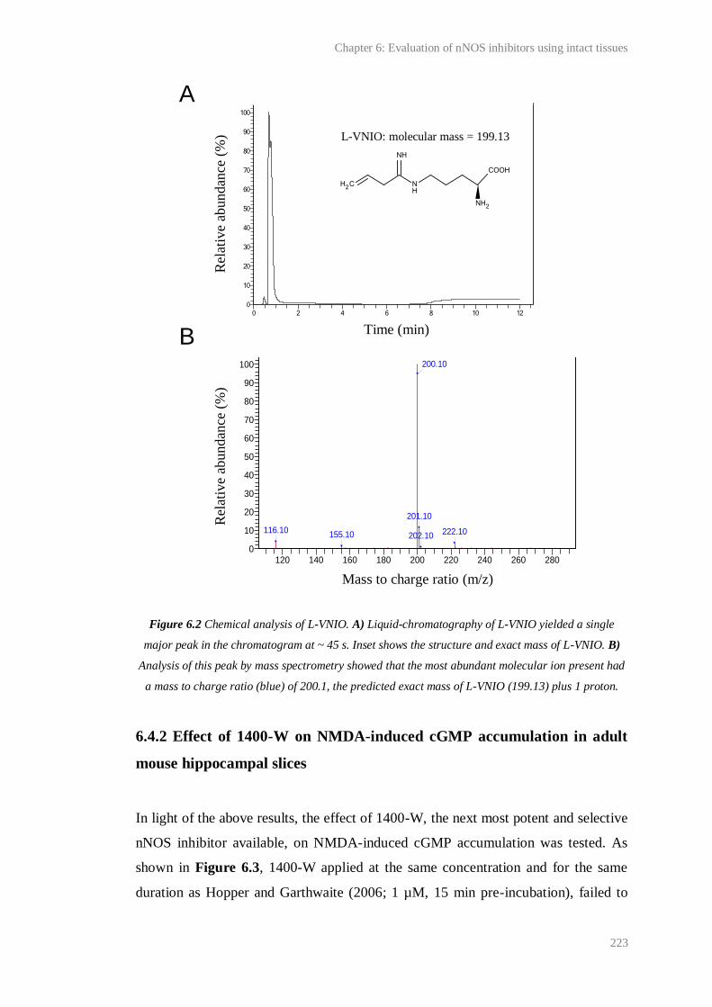
Chapter 6: Evaluation of nNOS inhibitors using intact tissues
223
Figure 6.2 Chemical analysis of L-VNIO. A) Liquid-chromatography of L-VNIO yielded a single
major peak in the chromatogram at ~ 45 s. Inset shows the structure and exact mass of L-VNIO. B)
Analysis of this peak by mass spectrometry showed that the most abundant molecular ion present had
a mass to charge ratio (blue) of 200.1, the predicted exact mass of L-VNIO (199.13) plus 1 proton.
6.4.2 Effect of 1400-W on NMDA-induced cGMP accumulation in adult
mouse hippocampal slices
In light of the above results, the effect of 1400-W, the next most potent and selective
nNOS inhibitor available, on NMDA-induced cGMP accumulation was tested. As
shown in Figure 6.3, 1400-W applied at the same concentration and for the same
duration as Hopper and Garthwaite (2006; 1 µM, 15 min pre-incubation), failed to
Rel
ativ
e ab
undan
ce (
%)
116.10155.10
201.10
202.10 222.10
200.10
0
10
20
30
40
50
60
70
80
90
100
120 140 160 180 200 220 240 260 280
Peak 1 MSD1 TIC, MS File(+) 1.3E6
%
m/z
0
10
20
30
40
50
60
70
80
90
100
0 2 4 6 8 10 12
DAD1 A, Sig=250,100 Ref=off 1E3
%
Time(minute)
Rel
ativ
e ab
undan
ce (
%)
Time (min)
Mass to charge ratio (m/z)
A
B
C
L-VNIO: molecular mass = 199.13
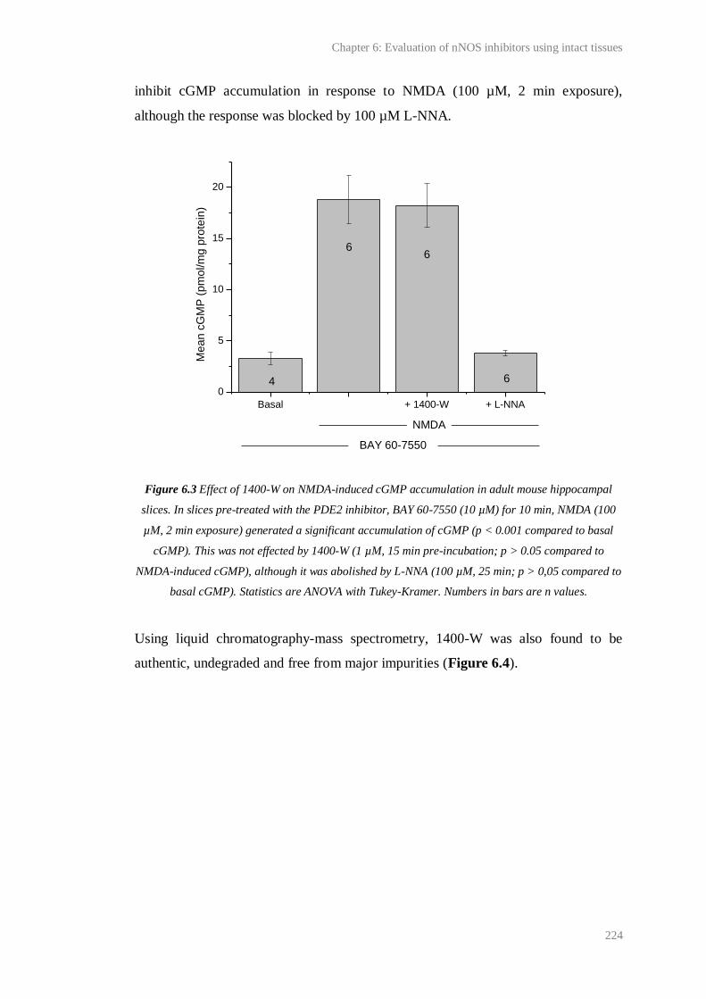
Chapter 6: Evaluation of nNOS inhibitors using intact tissues
224
inhibit cGMP accumulation in response to NMDA (100 µM, 2 min exposure),
although the response was blocked by 100 µM L-NNA.
Figure 6.3 Effect of 1400-W on NMDA-induced cGMP accumulation in adult mouse hippocampal
slices. In slices pre-treated with the PDE2 inhibitor, BAY 60-7550 (10 µM) for 10 min, NMDA (100
µM, 2 min exposure) generated a significant accumulation of cGMP (p < 0.001 compared to basal
cGMP). This was not effected by 1400-W (1 µM, 15 min pre-incubation; p > 0.05 compared to
NMDA-induced cGMP), although it was abolished by L-NNA (100 µM, 25 min; p > 0,05 compared to
basal cGMP). Statistics are ANOVA with Tukey-Kramer. Numbers in bars are n values.
Using liquid chromatography-mass spectrometry, 1400-W was also found to be
authentic, undegraded and free from major impurities (Figure 6.4).
Basal + 1400-W + L-NNA
0
5
10
15
20
6
66
Me
an
cG
MP
(p
mo
l/m
g p
rote
in)
NMDA
BAY 60-7550
4
Rel
ativ
e ab
undan
ce
(%)
Rel
ativ
e ab
undan
ce
(%)
Time (min)
Mass to charge ratio (m/z)
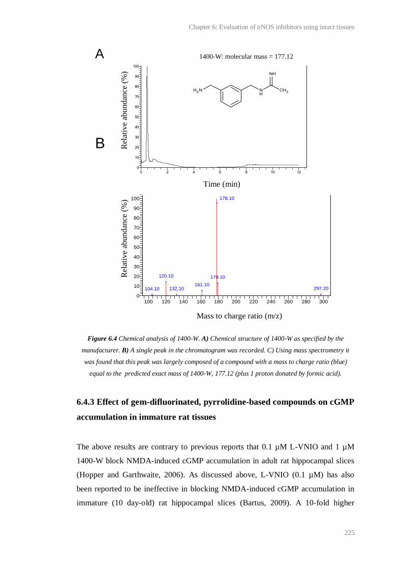
Chapter 6: Evaluation of nNOS inhibitors using intact tissues
225
Figure 6.4 Chemical analysis of 1400-W. A) Chemical structure of 1400-W as specified by the
manufacturer. B) A single peak in the chromatogram was recorded. C) Using mass spectrometry it
was found that this peak was largely composed of a compound with a mass to charge ratio (blue)
equal to the predicted exact mass of 1400-W, 177.12 (plus 1 proton donated by formic acid).
6.4.3 Effect of gem-difluorinated, pyrrolidine-based compounds on cGMP
accumulation in immature rat tissues
The above results are contrary to previous reports that 0.1 µM L-VNIO and 1 µM
1400-W block NMDA-induced cGMP accumulation in adult rat hippocampal slices
(Hopper and Garthwaite, 2006). As discussed above, L-VNIO (0.1 µM) has also
been reported to be ineffective in blocking NMDA-induced cGMP accumulation in
immature (10 day-old) rat hippocampal slices (Bartus, 2009). A 10-fold higher
Mass to charge ratio (m/z)
104.10
120.10
132.10161.10
179.10
297.20
178.10
0
10
20
30
40
50
60
70
80
90
100
100 120 140 160 180 200 220 240 260 280 300
Peak 1 MSD1 TIC, MS File(+) 0.33E7
%
m/z
0
10
20
30
40
50
60
70
80
90
100
0 2 4 6 8 10 12
DAD1 A, Sig=250,100 Ref=off 0.47E3
%
Time(minute)
1400-W: molecular mass = 177.12 A
B
Rel
ativ
e ab
undan
ce (
%)
Rel
ativ
e ab
undan
ce (
%)
Time (min)
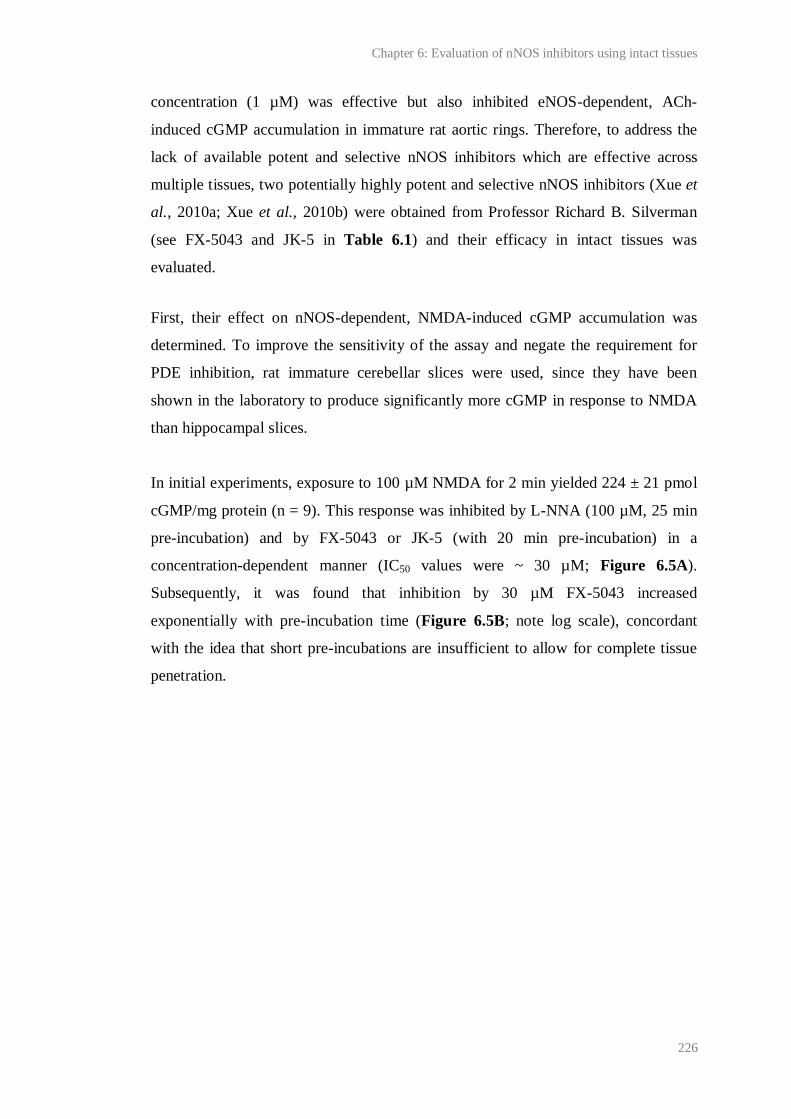
Chapter 6: Evaluation of nNOS inhibitors using intact tissues
226
concentration (1 µM) was effective but also inhibited eNOS-dependent, ACh-
induced cGMP accumulation in immature rat aortic rings. Therefore, to address the
lack of available potent and selective nNOS inhibitors which are effective across
multiple tissues, two potentially highly potent and selective nNOS inhibitors (Xue et
al., 2010a; Xue et al., 2010b) were obtained from Professor Richard B. Silverman
(see FX-5043 and JK-5 in Table 6.1) and their efficacy in intact tissues was
evaluated.
First, their effect on nNOS-dependent, NMDA-induced cGMP accumulation was
determined. To improve the sensitivity of the assay and negate the requirement for
PDE inhibition, rat immature cerebellar slices were used, since they have been
shown in the laboratory to produce significantly more cGMP in response to NMDA
than hippocampal slices.
In initial experiments, exposure to 100 µM NMDA for 2 min yielded 224 ± 21 pmol
cGMP/mg protein (n = 9). This response was inhibited by L-NNA (100 µM, 25 min
pre-incubation) and by FX-5043 or JK-5 (with 20 min pre-incubation) in a
concentration-dependent manner (IC50 values were ~ 30 µM; Figure 6.5A).
Subsequently, it was found that inhibition by 30 µM FX-5043 increased
exponentially with pre-incubation time (Figure 6.5B; note log scale), concordant
with the idea that short pre-incubations are insufficient to allow for complete tissue
penetration.
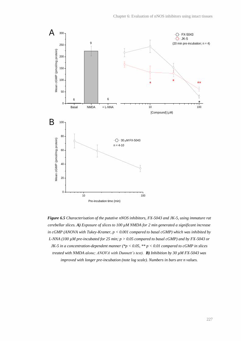
Chapter 6: Evaluation of nNOS inhibitors using intact tissues
227
Figure 6.5 Characterisation of the putative nNOS inhibitors, FX-5043 and JK-5, using immature rat
cerebellar slices. A) Exposure of slices to 100 µM NMDA for 2 min generated a significant increase
in cGMP (ANOVA with Tukey-Kramer, p < 0.001 compared to basal cGMP) which was inhibited by
L-NNA (100 µM pre-incubated for 25 min; p > 0.05 compared to basal cGMP) and by FX-5043 or
JK-5 in a concentration-dependent manner (*p < 0.05, ** p < 0.01 compared to cGMP in slices
treated with NMDA alone; ANOVA with Dunnett’s test). B) Inhibition by 30 µM FX-5043 was
improved with longer pre-incubation (note log scale). Numbers in bars are n values.
10 100
10 100
0
20
40
60
80
100
Basal NMDA + L-NNA
0
50
100
150
200
250
300
6
n = 4-10
Me
an
cG
MP
(p
mo
l/m
g p
rote
in)
6
9
*
***
FX-5043
JK-5
[Compound] (M)
*
(20 min pre-incubation; n = 4)
30 M FX-5043
Me
an
cG
MP
(p
mo
l/m
g p
rote
in)
Pre-incubation time (min)
A
B
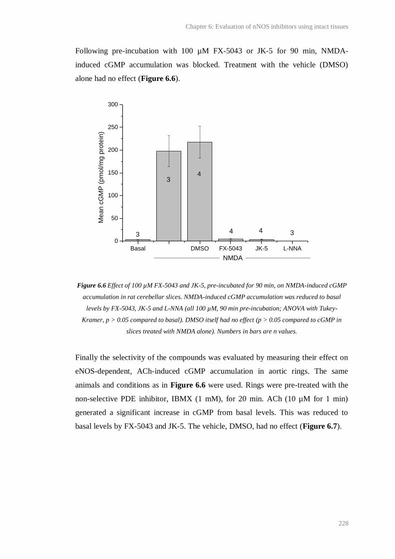
Chapter 6: Evaluation of nNOS inhibitors using intact tissues
228
Following pre-incubation with 100 µM FX-5043 or JK-5 for 90 min, NMDA-
induced cGMP accumulation was blocked. Treatment with the vehicle (DMSO)
alone had no effect (Figure 6.6).
Figure 6.6 Effect of 100 µM FX-5043 and JK-5, pre-incubated for 90 min, on NMDA-induced cGMP
accumulation in rat cerebellar slices. NMDA-induced cGMP accumulation was reduced to basal
levels by FX-5043, JK-5 and L-NNA (all 100 µM, 90 min pre-incubation; ANOVA with Tukey-
Kramer, p > 0.05 compared to basal). DMSO itself had no effect (p > 0.05 compared to cGMP in
slices treated with NMDA alone). Numbers in bars are n values.
Finally the selectivity of the compounds was evaluated by measuring their effect on
eNOS-dependent, ACh-induced cGMP accumulation in aortic rings. The same
animals and conditions as in Figure 6.6 were used. Rings were pre-treated with the
non-selective PDE inhibitor, IBMX (1 mM), for 20 min. ACh (10 µM for 1 min)
generated a significant increase in cGMP from basal levels. This was reduced to
basal levels by FX-5043 and JK-5. The vehicle, DMSO, had no effect (Figure 6.7).
Basal DMSO FX-5043 JK-5 L-NNA
0
50
100
150
200
250
300
44
4
3
3
Me
an
cG
MP
(p
mo
l/m
g p
rote
in)
NMDA
3

Chapter 6: Evaluation of nNOS inhibitors using intact tissues
229
Figure 6.7 Effect of 100 µM FX-5043 and JK-5, pre-incubated for 90 min, on ACh-induced cGMP
accumulation in rat aortic rings. Aortic rings were pre-treated with 1 mM IBMX for 20 min. Animals
and conditions were the same as Figure 6.6. ACh-induced significant cGMP accumulation from
basal levels (ANOVA with Tukey-Kramer, p < 0.001). This was reduced to basal levels by FX-5043,
JK-5 and L-NNA (all 100 µM, 90 min pre-incubation; p > 0.05 compared to basal/un-stimulated
levels). DMSO alone had no effect (p > 0.05 compared to cGMP in rings treated with ACh alone).
6.5 Discussion
Since the initial characterisation of the effect of L-VNIO and 1400-W on NMDA-
induced cGMP accumulation and CA1 LTP in adult rat and mouse hippocampus,
respectively (Hopper and Garthwaite, 2006), L-VNIO and 1400-W have been widely
used at standard concentrations (0.1 µM and 1 µM, respectively) to affect selective
nNOS inhibition in various tissues (for example, Hall and Attwell, 2008; Taqatqeh et
al., 2009; Romberg et al., 2009; Neitz et al., 2011). In one case, NO-dependent
synaptic plasticity at CA1 synapses in adult mouse hippocampal slices that was
insensitive to 0.1 µM L-VNIO was assumed to be nNOS-independent (Neitz et al.,
2011). However, in accordance with previous findings in immature rat hippocampus
(Bartus, 2009), one of the main conclusions of this study was that neither L-VNIO
Basal DMSO FX-5043 JK-5 L-NNA
0
200
400
600
800
Me
an
cG
MP
(p
mo
l/m
g p
rote
in)
ACh
IBMX
n = 4
Chapter 6: Evaluation of nNOS inhibitors using intact tissues
230
(0.1 µM) nor 1400-W (1 µM) are at all effective in inhibiting NMDA-induced cGMP
accumulation in adult mouse hippocampal slices (Figure 6.1 and 6.3).
There could be several reasons for the discrepancy between our findings and those of
Hopper and Garthwaite (2006). Given that L-VNIO and 1400-W are competitive for
binding to nNOS with L-arginine (Garvey et al., 1997; Babu and Griffith, 1998), one
explanation could be that the concentration of the amino acid varies between
different tissues and tissue preparations. This is conceivable since L-arginine, which
can be as concentrated as 80 µM in the cerebrospinal fluid of 8-week old rats
(Takasugi et al., 2003), is not normally included in solutions used to incubate tissues
in vitro. Other reasons are likely to be related to the bioavailability of the compounds
or their affinity for NOS in different tissues from different species. Factors
underlying differences in bioavailability could include tissue and cell permeability,
metabolism and transport of compounds. Differences in the compounds’ affinity for
NOS could arise, for example, through phosphorylation of the enzyme. The only
clear methodological difference between the experiments performed by us and
Hopper and Garthwaite (2006) to measure the effect of L-VNIO and 1400-W on
NMDA-induced cGMP accumulation is the PDE 2 inhibitor used (BAY 60-7550 vs.
EHNA). Experiments are planned to test whether these factors may account for the
differences in potency of the inhibitors.
Regardless of the explanation, L-VNIO and 1400-W are clearly not ideal nNOS
inhibitors and should not be used to determine the role of nNOS in biological
phenomena without the appropriate tissue-specific controls for their potency. In light
of the above results, published data on the effect of L-VNIO and 1400-W on
biological phenomena should perhaps be re-interpreted and in future, the ‘inhibitors’
should be used cautiously, if at all.
The selectivity of other established nNOS inhibitors is also in doubt. For example, at
the isolated enzyme level, the compound, N-propyl-L-arginine (NPA), reportedly
displays high selectivity for nNOS over iNOS (3158-fold difference) and eNOS
(149-fold difference) (Zhang et al., 1997), but this has not been confirmed in vivo
(Gowda et al., 2004). 7-nitroindazole (7-NI) is also widely used as a selective nNOS
Chapter 6: Evaluation of nNOS inhibitors using intact tissues
231
inhibitor in vitro and in vivo, although it has been reported to be completely non-
selective at the isolated enzyme level (Alderton et al., 2001). Its apparent selectivity
in intact tissues could be explained if it accumulates preferentially in neurons rather
than endothelial cells. Nevertheless, selective nNOS inhibition should ideally be
achieved by the differential action of compounds on NOS isozymes, rather than
through differences in compound bioavailability.
To address the lack of potent and selective nNOS inhibitors effective across multiple
tissue preparations, we sought to evaluate two, new, gem-difluorinated, pyrrolidine-
based compounds (FX-5043 and JK-5). As discussed above, FX-5043 has been
shown to potently inhibit nNOS with unrivalled selectivity over both e- and iNOS in
isolated enzyme and cell-based assays (Xue et al., 2010a). However, under the
conditions necessary to effect complete inhibition of nNOS-dependent, NMDA-
induced cGMP accumulation in adult rat cerebellar slices (100 µM, 90 min pre-
incubation), we found that FX-5043 and JK-5 blocked eNOS-dependent, ACh-
induced cGMP accumulation in adult rat aortic rings (Figure 6.6 and 6.7).
One possible explanation for the apparent lack of selectivity for nNOS over eNOS in
intact tissues was that they also inhibit NO-targeted guanylyl cyclase. This could
have explained why FX-5043 was observed to selectively inhibit nNOS over eNOS
and iNOS using a cell-based assay, because this assay relied upon the measurement
of the accumulation of nitrite, a metabolite of NO, in the medium (Fang and
Silverman, 2009). However, tests of this possibility have since shown that the
compounds have no significant effect on DEA/NONOate-induced cGMP production
by purified bovine NO-targeted guanylyl cyclase (Prof. John Garthwaite,
unpublished work).
Another reason for the apparent lack of selectivity of the compounds in intact tissues
could be that they preferentially accumulate in endothelial cells over neurons to the
extent that, at the concentration necessary to affect nNOS inhibition, the
concentration in endothelial cells is so high that it inhibits eNOS. Given the reported
Ki values of the compound (Xue et al., 2010a), the difference in concentration
between endothelial and neuronal cells would probably have to be ~ an order of
Chapter 6: Evaluation of nNOS inhibitors using intact tissues
232
magnitude. Also, it cannot be ruled out that the compounds have some other, perhaps
toxic, secondary effect on intact tissues, leading to a decrease in cGMP production.
Irrespective of the explanation, the main conclusion to be drawn from the data is that,
while FX-5043 and JK-5 might be useful for selectively inhibiting nNOS at the level
of the isolated enzyme or cell-based assay, their application to intact tissues is not
appropriate. Since both compounds had a relatively high IC50 in cerebellar slices (~
30 µM following 20 min pre-incubation; Figure 6.5A), require long pre-incubation
times to be effective, likely due to poor tissue penetration, and may have unknown
secondary effects, they should also not be used as non-selective NOS inhibitors over
L-arginine-based compounds such as L-NNA, which have IC50 values in the low
micromolar range (see above for details), are actively transported into cells and have
no known secondary effects in tissues (Alderton et al., 2001).
Recently a symmetric, double headed aminopyridine has been developed that shows
lower potency and selectivity for nNOS but increased cell permeability compared to
the gem-difluorinated compounds used here (Xue et al., 2011). This, as well as the
above data, could be useful in directing the development of more compounds with
high potency and selectivity for nNOS in its native state. Meanwhile, the results of
studies using ‘selective’ nNOS inhibitors currently widely available should be
interpreted with caution.
Chapter 7:
The location of NO-targeted guanylyl cyclase in adult
mouse hippocampus

Chapter 7: The location of NO-targeted guanylyl cyclase in adult mouse hippocampus
234
7.1 Introduction
As discussed previously (see Chapter 1), functional evidence has implicated
NO/cGMP signalling to be active in multiple brain regions in several species in
which it may contribute to the generation of diverse phenomena (Garthwaite, 2008).
Accordingly, studies employing in situ hybridisation and immunohistochemistry
have suggested the presence of NOS and NO-targeted guanylyl cyclase throughout
the developing and mature rodent brain, albeit at varying levels in different regions
(nNOS; Vincent and Kimura, 1992; Rodrigo et al., 1994; eNOS ; Seidel et al., 1997 ;
Stanarius et al., 1997; Topel et al., 1998; Demas et al., 1999; NO-targeted guanylyl
cyclase; Matsuoka et al., 1992; Gibb and Garthwaite, 2001; Ding et al., 2004).
Moreover, NADPH diaphorase histochemistry for NOS in rat brain has shown a
remarkably coincident distribution with structures immunopositive for cGMP
following in vivo perfusion of the NO donor, sodium nitroprusside (Southam and
Garthwaite, 1993), consistent with functional evidence that guanylyl cyclase is the
primary target of endogenous NO.
In the hippocampus, functional studies suggest that eNOS, nNOS and all three
functionally relevant guanylyl cyclase subunits, α1, α2 and β1, are present in area
CA1 (Chapters 3-6; Hopper and Garthwaite, 2006; Taqatqeh et al., 2009). This has
largely been corroborated by histological data (see Chapter 1); however, the precise
location of the guanylyl cyclase α1 subunit has recently come under question.
Studies employing NO-targeted guanylyl cyclase α1- and α2- lacking mice
(NOGCα1-/-
and NOGCα2-/-
, respectively) have shown that both the α1β1 and α2β1
isoforms are necessary for LTP in the visual cortex (Haghikia et al., 2007) and
hippocampus (Taqatqeh et al., 2009). In the hippocampus, Taqatqeh et al. (2009)
report that under basal conditions, and following LTP, PPF at CA1 synapses in
NOGCα1-/-
mice is increased compared to wild-type mice and recently, evidence in
favour of α1-dependent tonic modulation of transmitter release at CA1 synapses has
been extended (Neitz et al. 2011 and Chapter 4). The results of these studies
consistently imply that the α1β1 isoform is located presynaptically and is responsible
for transducing NO-dependent changes in transmitter release onto CA1 neurons.
Chapter 7: The location of NO-targeted guanylyl cyclase in adult mouse hippocampus
235
Conversely, knocking-out the α2 subunit appears to have no effect on PPF or other
measures of presynaptic transmitter release, such as miniature EPSC frequency
(Taqatqeh et al., 2009; Neitz et al., 2011). One interpretation of this is that the α2β1
isoform is postsynaptic.
Changes in PPF at CA1 synapses following plasticity are typically interpreted as a
result of changes in presynaptic transmitter release occurring directly at Schaffer-
collateral axon terminals. Much research has been directed to understanding these
presynaptic forms of LTP and the involvement of NO in them (Arancio et al., 1995;
Arancio et al., 1996; Arancio et al., 2001). However, there is no unambiguous
evidence that the α1 subunit is present at CA3-CA1 synapses. In a report by Gibb
and Garthwaite (2001), low magnification images of hippocampus labelled for α1
mRNA showed message for the subunit to be located in the stratum radiatum, but it
was unclear whether the subunit was in pyramidal neurons or interneurons. Rather, a
key study using in situ hybridization and immunohistochemistry in conjunction with
electron microscopy showed that α2 (which was co-localised with β1) was expressed
only in pyramidal cells and α1 (also co-localised with β1) only in interneurons. These
interneurons, the majority of which were parvalbumin- or cholecystokinin-positive
and therefore probably basket cells, formed synapses on CA1 pyramidal cell soma,
dendrites and axon initial segments. Neuronal NOS was found to be localised to the
PSD of these synapses, suggesting that GABAergic inputs to CA1 pyramidal neurons
may be retrogradely modulated by NO-targeted α1β1 guanylyl cyclase activity
(Szabadits et al., 2007). This distribution of NOS and α1β1 has since been confirmed
in the developing hippocampus (Cserep et al., 2011).
Taken together, the studies discussed above suggest that NO regulates transmitter
release at the GABAergic, rather than glutamatergic, inputs to CA1 pyramidal
neurons. Consistent with this, NO has been shown to modulate GABAergic
transmission in several brain areas (for a review see Garthwaite, 2008). In the ventral
tegmental area, for example, NO produced by dopaminergic neurons upon NMDA
receptor activation has been found to retrogradely modulate GABA release from
nearby inhibitory axon terminals, resulting in a heterosynaptic long-term potentiation
of GABAA-mediated synaptic transmission (Nugent et al., 2007). In the
Chapter 7: The location of NO-targeted guanylyl cyclase in adult mouse hippocampus
236
hippocampus, NO may act in conjunction with endocannabinoids as a retrograde
messenger to depress GABAergic IPSCs (Makara et al., 2007). Depression of
GABAergic transmission in area CA1 by NO via the NO-targeted α1β1 guanylyl
cyclase isoform has also recently been reported to occur in slices from young (p5-p8)
mice (Cserep et al., 2011). However, the notion that apparent changes in transmitter
release following LTP may be due to the effect of NO at GABAergic rather than
glutamatergic synapses is at odds with findings that NO, produced postsynaptically
may, through cGMP and PKG, induce a presynaptic LTP characterised by an
increase in transmitter release between pairs of pyramidal neurons (identified by
electrophysiological properties) in dissociated cell culture (Arancio et al., 1995;
Arancio et al., 1996; Arancio et al., 2001).
7.2 Aim
The pattern of α1 expression reported by Szabadits et al. (2007) was consistent
following immunofluorescent, immunoperoxidase and in situ hybridization
techniques in tissue fixed with 4 % PFA and accorded with immunostaining for α1 in
sections of immature hippocampus, also fixed with 4 % PFA (Cserep et al., 2011).
However, it is known that the distribution of proteins as determined by histochemical
methods can be significantly altered by the conditions under which the tissues are
processed. Several groups have reported immunostaining for nNOS and the guanylyl
cyclase β1 subunit in pyramidal neurons in hippocampus fixed with 1 % PFA but not
≥ 4 % PFA (Wendland et al., 1994; Gonzalez-Hernandez et al., 1996; Burette et al.,
2002). It has been hypothesised that aldehyde cross-linking of proteins following
strong fixation protocols may mask antibody epitopes and/or reduce the access of the
antibody to its target protein. The latter circumstance may be particularly common
when target proteins are located in PSD’s or presynaptic varicosities. Therefore, we
sought to re-investigate the distribution of α1 in the hippocampus, using weakly
fixed tissue (1 % PFA). The aim was to either confirm the work of Szabadits et al.
(2007), or to uncover new structures, perhaps pyramidal neurons, immunopositive
for the NO-targeted guanylyl cyclase α1 subunit.
Chapter 7: The location of NO-targeted guanylyl cyclase in adult mouse hippocampus
237
7.3 Methods
7.3.1 Animals
Unless otherwise stated, adult (8 week-old), male, C57/Bl6 mice were used. For
some experiments, adult (2 8-9 week-old, 1 19 week-old), male, C57/Bl6/SV129
mice lacking the NO-receptor guanylyl cyclase α1 subunit (NOGCα1-/-
) were used.
These were kindly provided by Dr Adrian Hobbs (UCL, London, UK). Male 8-9
week old wild-type siblings were used as controls. Rat brain lysate was prepared
from an 8 day-old, male Sprague Dawley pup. All work was conducted in
accordance with British Home Office regulations on laboratory animal use and
welfare.
7.3.2 General Solutions
Phosphate buffer (PB; 0.2 M) was composed of 19 % 0.2 M NaH2PO4.2H2O and 81
% 0.2 M Na2HPO4.2H2O in double-distilled H2O adjusted to pH 7.4.
Tris-buffered saline (TBS) comprised 48.4 mM trisma base and 150.2 mM NaCl in
double-distilled H2O equilibrated to pH 7.6 with concentrated HCl.
TBS-triton (TBS-T) additionally included 0.1 % triton X-100.
TBS-tween comprised TBS plus 0.05 % tween 20.
Tissue-lysis buffer consisted of 60 mM tris-HCl (pH 6.8), 1 % SDS, 1 % tween 20
and 1 x Halt protease inhibitor cocktail in double-distilled H2O.
Chapter 7: The location of NO-targeted guanylyl cyclase in adult mouse hippocampus
238
7.3.3 Immunohistochemistry
Preparation of transverse hippocampal sections
Mice were euthanized by cervical dislocation and decapitated. The hippocampi were
either left in the brain during fixing and sectioning, or swiftly dissected out and
sliced as described in Chapter 2: General materials and methods. In the former
case, the brains were removed from the skulls and submerged in freshly prepared,
ice-cold 1 % PFA prepared in 0.1 M PB and adjusted to pH 7.4. The anterior and
posterior ends of the brains were cut (leaving the middle of the hippocampi intact)
using a razor blade and the hemispheres were separated and fixed for 2 hr in 1 or 4 %
PFA on a rocker at 4 ºC. In the latter case, slices were left to recover in oxygenated
aCSF for 1 hr before being fixed for 1 hr using 1 % PFA on a rocker at room
temperature. Just prior to fixing of tissue from NOGCα1-/-
and wild-type mice, the
cerebellum was removed for Western blot analysis (see below for methods).
After fixing, tissues were washed with 0.1 M PB (4 times, 5 min) and cryoprotected
with sucrose in 0.1 M PB (5 % sucrose for 4 h, then 20 % overnight, then 30 % for 4
hr and finally 50:50 30 % sucrose: OCT for 2 hr, all at 4 ºC). They were then washed
and embedded in OCT and swiftly frozen on dry ice made extra cold with
isopentane. Tissue was stored at -80 ºC prior to sectioning.
For sectioning, the embedded tissue was mounted on a cutting chuck using OCT. 10
μm transverse hippocampal sections were made on 0.05 % chrome alum/ 0.5 %
gelatine-coated slides using a cryostat (Model OTF, Bright Instruments Co Ltd.,
UK). During this process, some slides were stained with toluidine blue in order that
the hippocampus proper could be visualised and the cutting plane adjusted as
necessary. 1 % toluidine blue was prepared as a stock solution in 1 % borate. Before
use, this was diluted 1/10 in tap H2O and filtered. Slides were dried over a flame,
washed with filtered toluidine blue for approximately 30 s, rinsed with tap H2O,
dried and mounted in di-N-butyle phthalate in xylene (DPX).
Slides were stored at -20 ºC prior to immunohistochemistry.
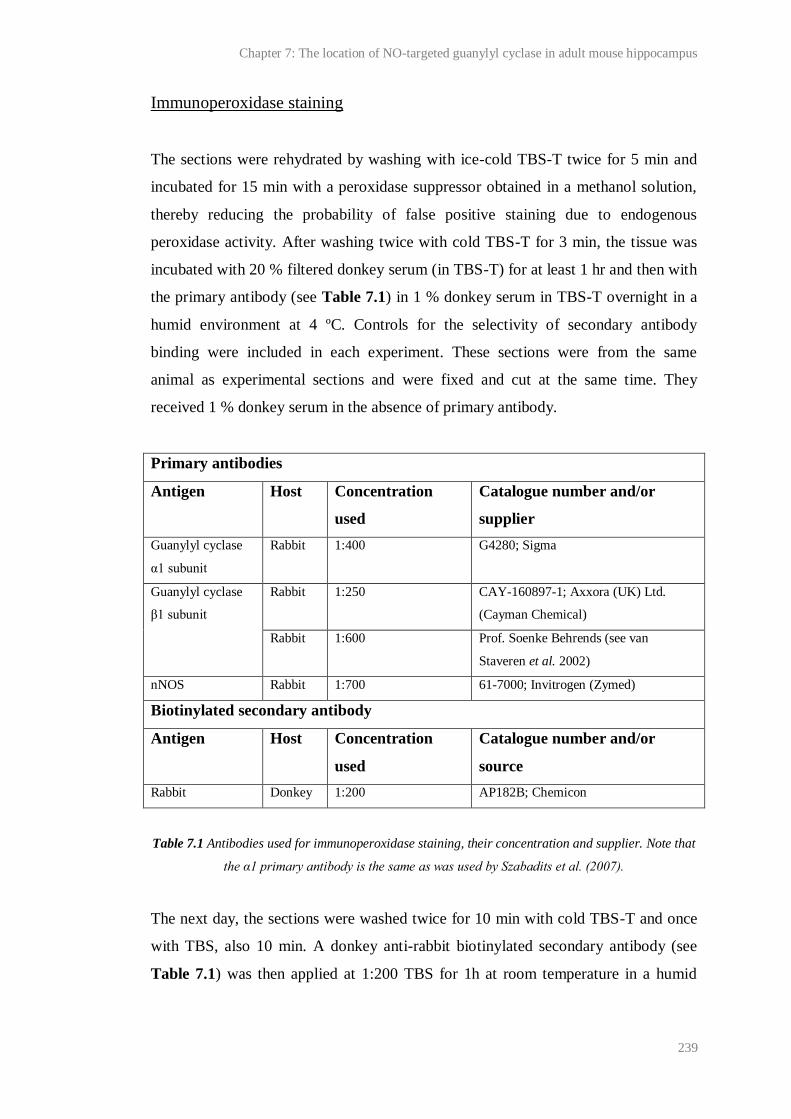
Chapter 7: The location of NO-targeted guanylyl cyclase in adult mouse hippocampus
239
Immunoperoxidase staining
The sections were rehydrated by washing with ice-cold TBS-T twice for 5 min and
incubated for 15 min with a peroxidase suppressor obtained in a methanol solution,
thereby reducing the probability of false positive staining due to endogenous
peroxidase activity. After washing twice with cold TBS-T for 3 min, the tissue was
incubated with 20 % filtered donkey serum (in TBS-T) for at least 1 hr and then with
the primary antibody (see Table 7.1) in 1 % donkey serum in TBS-T overnight in a
humid environment at 4 ºC. Controls for the selectivity of secondary antibody
binding were included in each experiment. These sections were from the same
animal as experimental sections and were fixed and cut at the same time. They
received 1 % donkey serum in the absence of primary antibody.
Primary antibodies
Antigen Host Concentration
used
Catalogue number and/or
supplier
Guanylyl cyclase
α1 subunit
Rabbit 1:400 G4280; Sigma
Guanylyl cyclase
β1 subunit
Rabbit 1:250 CAY-160897-1; Axxora (UK) Ltd.
(Cayman Chemical)
Rabbit 1:600 Prof. Soenke Behrends (see van
Staveren et al. 2002)
nNOS Rabbit 1:700 61-7000; Invitrogen (Zymed)
Biotinylated secondary antibody
Antigen Host Concentration
used
Catalogue number and/or
source
Rabbit Donkey 1:200 AP182B; Chemicon
Table 7.1 Antibodies used for immunoperoxidase staining, their concentration and supplier. Note that
the α1 primary antibody is the same as was used by Szabadits et al. (2007).
The next day, the sections were washed twice for 10 min with cold TBS-T and once
with TBS, also 10 min. A donkey anti-rabbit biotinylated secondary antibody (see
Table 7.1) was then applied at 1:200 TBS for 1h at room temperature in a humid
Chapter 7: The location of NO-targeted guanylyl cyclase in adult mouse hippocampus
240
environment. Slides were then washed (3 times for 10 min in ice-cold TBS),
incubated with Vectastain avidin biotin complex for 45 min at room temperature,
stained for 4 min with 0.05% 3,3'-diaminobenzidine in TBS and then counterstained
with Mayer’s haemalum solution. Mayer’s haemalum was applied for 2-10 s and the
slides were then rinsed in tap H2O for 25-30 s. After washing slides in double-
distilled H2O, they were left to dry overnight and then mounted in DPX. Slides were
photographed using an upright microscope (Leitz, UK) fitted with a digital camera
(Q-imaging, Canada).
Analysis
Experimental and control sections were photographed under the same conditions.
Images were adjusted for brightness and contrast using Adobe Photoshop (Adobe,
USA)/ Microsoft PowerPoint (Microsoft, USA).
All images are representative of at least two sections from at least two animals.
Images of NOGCα1-/-
tissue are representative of sections from three animals. All
NOGCα1-/-
mice were genotyped by Richard Burt (UCL). Additionally, we confirmed
the phenotype of the NOGCα1-/-
mouse from which the sections imaged in this report
were taken using Western blot analysis (see below).
7.3.4 Western Blotting
Tissue homogenisation and protein preparation
Animals were culled by cervical dislocation and decapitation. The head was
immediately submerged in ice-cold PBS to cool the brain and remove excess blood.
The brain was quickly removed to an ice-cold surface upon which the cerebellum
was isolated and the hemispheres separated from each other. Each portion was
transferred to a separate mortar containing 2 ml ice-cold tissue-lysis buffer. Tissue
was smoothed using a glass pestle and the homogenate removed to tubes on ice. To
further prevent protein degradation by proteases, the homogenates were heated for 10
min at 70 °C. They were then sheared using a 27 G needle to remove any outstanding
Chapter 7: The location of NO-targeted guanylyl cyclase in adult mouse hippocampus
241
solid tissue and centrifuged for 10 min at 13,000 rpm (5 °C). The resulting
supernatant, containing solubilised proteins, was removed and the pellet was
discarded. Finally, the protein concentrations of the supernatants were measured
using the BCA method, as described in Chapter 2: General materials and
methods.
Sodium dodecyl sulphate polyacrylamide gel electrophoresis (SDS PAGE)
For SDS PAGE, 50 μg of sample protein was loaded per lane. Samples were brought
to the same volume using tissue-lysis buffer (minus protease inhibitors). Then gel
loading buffer, comprising 416 mM SDS, ~ 0.9 mM bromophenol blue, 778 mM
dithiothreitol, 3 % 2 M tris (pH 6.8) and 47 % glycerol prepared in double-distilled
H2O, was added 1 in 3 to give a final volume of 13.3 µl.
SDS PAGE was run for 35 min at 200 V. A Mini-PROTEAN 3 Cell and 4-15 %
Ready Gel (both Bio-Rad Laboratories, Hertfordshire, UK) were used. Running
buffer contained (in mM): 25 tris; 192 glycine and 3.5 SDS. All 13.3 μl of sample
were loaded per lane and one lane per gel contained 5 μl New England BioLabs
ColourPlus prestained protein ladder.
Gel to membrane protein transfer
Following SDS PAGE, the gel was equilibrated for 30 min in transfer buffer
additionally containing 0.2 % triton X-100. Transfer buffer comprised, in mM: 25
trisma base; 192 glycine; 3.5 SDS and 2.5 % methanol. Immediately before use, a
polyvinylidene fluoride membrane was submerged in methanol for 30 s, rinsed with
double-distilled H2O and soaked in transfer buffer. A submerged transfer method
was implemented at 0.38 A constant for 2 hr at 4 °C using the Amersham Bioscience
(GE Healthcare Life Sciences, Buckinghamshire, UK) TE 22 Mighty Small Tank
Transfer Unit.
Chapter 7: The location of NO-targeted guanylyl cyclase in adult mouse hippocampus
242
Membrane probing
After transfer, the membrane was rinsed in double-distilled H2O, then TBS and
submerged in blocking buffer containing 3 % skimmed milk, 0.2 % tween 20, 0.5 %
polyvinylpyrrolidone, and 1.5 % sodium azide in TBS. After 1 hr the membrane was
washed for 5 min with TBS and the primary antibodies (see Table 7.2), diluted 1:500
in 50 % blocking buffer in TBS, were applied for 1 hr. The membrane was then
washed 3 times for 5 min in TBS-tween, once for 5 min in TBS and submerged in an
appropriate horseradish peroxidase-conjugated secondary antibody (Table 7.2)
diluted in 50 % blocking buffer in TBS. After 1 hr, the membrane was washed 3
times in TBS-tween and once in TBS (all 5 min) and then blotted to remove excess
solution. All treatments/washes were performed at room temperature on a shaking
platform.
Antibody binding was visualised using the Thermo Scientific SuperSignal West Pico
Chemiluminescent Substrate system. Luminol/enhancer solution and peroxide
solution were mixed in equal measures and applied to the membrane for 5 min at
room temperature on a shaker. The membrane was then blotted and covered in a
plastic film. Luminescence was developed after exposure to High Performance
Chemiluminescence Film for 5-20 min.
In order to control for protein loading, some membranes were stripped of guanylyl
cyclase antibodies and probed for actin. Membranes were stored at room
temperature. As necessary, they were re-hydrated in methanol, washed in double-
distilled H2O once for 2 min and then in TBS for 5 min. Thermo Scientific Restore
Western Blot Stripping Buffer was applied for 10 min and membranes were rinsed
for 5 min with TBS. The primary and secondary actin antibodies (see Table 7.2)
were then applied following the method detailed above. Due to high background
staining, the membranes were then washed copiously in TBS-tween (4 x 5 min; 5 x
15 min at room temperature). Antibody binding was exposed as above except that the
membrane was exposed to the photographic film for 5-30 s.
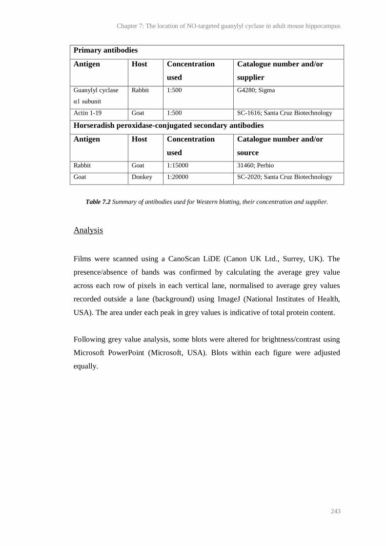
Chapter 7: The location of NO-targeted guanylyl cyclase in adult mouse hippocampus
243
Primary antibodies
Antigen Host Concentration
used
Catalogue number and/or
supplier
Guanylyl cyclase
α1 subunit
Rabbit 1:500 G4280; Sigma
Actin 1-19 Goat 1:500 SC-1616; Santa Cruz Biotechnology
Horseradish peroxidase-conjugated secondary antibodies
Antigen Host Concentration
used
Catalogue number and/or
source
Rabbit Goat 1:15000 31460; Perbio
Goat Donkey 1:20000 SC-2020; Santa Cruz Biotechnology
Table 7.2 Summary of antibodies used for Western blotting, their concentration and supplier.
Analysis
Films were scanned using a CanoScan LiDE (Canon UK Ltd., Surrey, UK). The
presence/absence of bands was confirmed by calculating the average grey value
across each row of pixels in each vertical lane, normalised to average grey values
recorded outside a lane (background) using ImageJ (National Institutes of Health,
USA). The area under each peak in grey values is indicative of total protein content.
Following grey value analysis, some blots were altered for brightness/contrast using
Microsoft PowerPoint (Microsoft, USA). Blots within each figure were adjusted
equally.
Chapter 7: The location of NO-targeted guanylyl cyclase in adult mouse hippocampus
244
7.4 Results
7.4.1 Location of the NO-targeted guanylyl cyclase α1 subunit in adult
mouse hippocampus
Evaluation using tissue fixed with 4 % PFA
Before testing the hypothesis that immunostaining of weakly fixed (1 % PFA)
hippocampal sections would reveal the NO-targeted guanylyl cyclase α1 subunit to
be present in pyramidal neurons, the distribution of the protein was evaluated by
immunoperoxidase staining of sections fixed using 4 % PFA. After
immunofluorescence and immunoperoxidase staining of hippocampus fixed with 4 %
PFA using the same α1 primary antibody as employed in this study, Szabadits et al.
(2007) found that only interneurons were immunopositive for α1. In accordance with
this result, we found that cells outside the stratum pyramidale, presumably
interneurons and/or glia, were most intensely stained (Figure 7.1 C, E). However, in
the stratum pyramidale, cell soma and fibres resembling apical dendrites of
pyramidal neurons were faintly immunopositive (Figure 7.1 D, E, and F). Although
interneurons are found throughout this layer, the morphology and number of
immunopositive cell soma favoured their classification as pyramidal neurons.
The neuropil in every strata was also densely stained compared to that in control
sections not treated with the primary antibody (Figure 7.1 A, B), indicating that a
wealth of α1-containing fibres run through or terminate in the hippocampus.
Particularly prominent were fibres running throughout the stratum polymorph of the
dentate gyrus and the stratum lucidum (Figure 7.1 A, F). Given their position, it is
likely that these were mossy fibres (labelled mf in Figure 7.1 A, F; Amaral and
Witter, 1989) which, interestingly, have also been reported to be strongly
immunoreactive for cGMP hydrolysing-PDE 2 (Stephenson et al., 2009) and subject
to NO-dependent LTP at synapses with CA3 neurons (Doreulee et al., 2001;
although see Nicolarakis et al., 1994). As shown in Figure 7.1 B and G no non-
selective secondary antibody binding was observed.

Chapter 7: The location of NO-targeted guanylyl cyclase in adult mouse hippocampus
245
Figure 7.1 Immunoperoxidase staining for the NO-targeted guanylyl cyclase α1 subunit in transverse
hippocampal sections fixed with 4 % PFA. A) The hippocampus major. Two juxtaposed images. B)
Control for secondary antibody binding in A. mf = possible mossy fibres. C-F) Higher magnification
images of a second section stained for α1 showing an area of the stratum oriens (so; C), stratum
pyramidale (sp; D) and stratum radiatum (sp; E) of CA1, and an area of CA3/stratum lucidum (F). G)
Control for secondary antibody binding in C-F. Unfilled arrows (D, E) indicate possible apical
Chapter 7: The location of NO-targeted guanylyl cyclase in adult mouse hippocampus
246
dendrites of pyramidal neurons. Control sections were not treated with the primary antibody. Scale
bar in A: 500 µm, A-B; bar in C: 100 µm, C-G. Filled arrows (C, E) indicate interneurons.
Evaluation using tissue fixed with 1 % PFA
In accordance with the distribution of staining observed in Figure 7.1, cells scattered
outside the stratum pyramidale and fibres running throughout the dentate gyrus and
stratum lucidum were strongly stained after identical treatment with tissue fixed with
1 % PFA (Figure 7.2 A, C). Unlike the staining observed in tissues fixed with 4 %
PFA, however, and in favour with the hypothesis that pyramidal neurons express α1,
cell soma and proximal apical dendrites in stratum pyramidale throughout Ammon’s
horn were very strongly immunopositive (Figure 7.2 C). Whether the rest of the
dendritic arbour was also immunopositive, as is suggested by functional evidence
implicating the α1β1 guanylyl cyclase in synaptic plasticity (Taqatqeh et al., 2009;
Neitz et al., 2011), was unclear because the dendrites of pyramidal neurons become
rapidly thinner as they deviate from the apical fibre (Routh et al., 2009), and using
light microscopy, are usually indistinguishable from the rest of the neuropil.
Blood vessels throughout the hippocampus and subiculum were also immunopositive
(Figure 7.2 A, C). This was not observed following staining of tissue fixed with 4 %
PFA, but was consistent with functional evidence implicating NO-targeted guanylyl
cyclase α1β1 heterodimers in smooth muscle relaxation and vasodilation (Mergia et
al., 2006; Nimmegeers et al., 2007). Moreover, some immunohistochemical data
collected using a α1β1 antibody also shows guanylyl cyclase to be present in
endothelial, as well as smooth muscle cells surrounding blood vessels (Zhan et al.,
1999; Jarry et al., 2003).
Some scattered cells, for example, in the stratum radiatum in Figure 7.2 C were
immunonegative. Some non-selective staining was observed in the fimbrium and
alveus surrounding the hippocampus and, to a lesser extent, in the lacunosum
moleculare, (Figure 7.2 B).
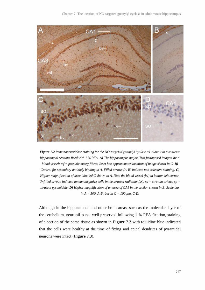
Chapter 7: The location of NO-targeted guanylyl cyclase in adult mouse hippocampus
247
Figure 7.2 Immunoperoxidase staining for the NO-targeted guanylyl cyclase α1 subunit in transverse
hippocampal sections fixed with 1 % PFA. A) The hippocampus major. Two juxtaposed images. bv =
blood vessel; mf = possible mossy fibres. Inset box approximates location of image shown in C. B)
Control for secondary antibody binding in A. Filled arrows (A-B) indicate non-selective staining. C)
Higher magnification of area labelled C shown in A. Note the blood vessel (bv) in bottom left corner.
Unfilled arrows indicate immunonegative cells in the stratum radiatum (sr). so = stratum oriens; sp =
stratum pyramidale. D) Higher magnification of an area of CA1 in the section shown in B. Scale bar
in A = 500, A-B; bar in C = 100 µm, C-D.
Although in the hippocampus and other brain areas, such as the molecular layer of
the cerebellum, neuropil is not well preserved following 1 % PFA fixation, staining
of a section of the same tissue as shown in Figure 7.2 with toluidine blue indicated
that the cells were healthy at the time of fixing and apical dendrites of pyramidal
neurons were intact (Figure 7.3).

Chapter 7: The location of NO-targeted guanylyl cyclase in adult mouse hippocampus
248
Figure 7.3 Toluidine blue staining of a section of the same tissue as shown in Figure 7.2. A) CA3. B)
CA1. sl = stratum lucidum; so = stratum oriens; sp = stratum pyramidale; sr = stratum radiatum.
Scale bar in A: 100 µm, A-B.
7.4.2 Location of the NO-targeted guanylyl cyclase β1 subunit and nNOS
in adult mouse hippocampus
In favour of the hypothesis that pyramidal cells express α1, Figure 7.2 shows for the
first time that, in tissues fixed with 1 % PFA, hippocampal pyramidal neurons can be
immunostained for the NO-targeted guanylyl cyclase α1 subunit using standard
methods.
In immature granule cells of the developing cerebellum, mRNA for the NO-targeted
α2 subunit has been reported to be expressed without the β1 subunit (Gibb and
Garthwaite, 2001). Intense signals for β1 subunit mRNA have also been detected in
brain areas apparently with relatively little or no mRNA for either of the α subunits
(Mergia et al., 2003; Krumenacker et al., 2006; Pifarre et al., 2007). Since
heterodimerisation of subunits is required for guanylyl cyclase catalytic activity,
these data, if correct, suggest that individual NO-targeted guanylyl cyclase subunits
may function in ways other than to transduce NO signals via cGMP.
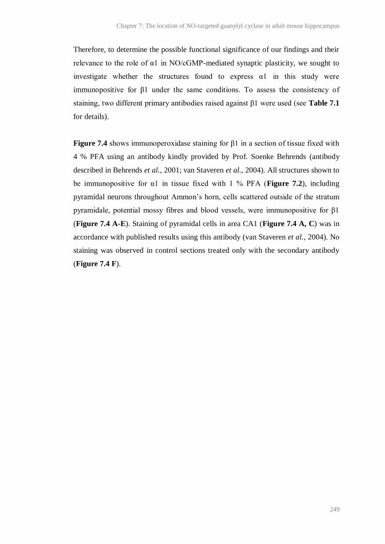
Chapter 7: The location of NO-targeted guanylyl cyclase in adult mouse hippocampus
249
Therefore, to determine the possible functional significance of our findings and their
relevance to the role of α1 in NO/cGMP-mediated synaptic plasticity, we sought to
investigate whether the structures found to express α1 in this study were
immunopositive for β1 under the same conditions. To assess the consistency of
staining, two different primary antibodies raised against β1 were used (see Table 7.1
for details).
Figure 7.4 shows immunoperoxidase staining for β1 in a section of tissue fixed with
4 % PFA using an antibody kindly provided by Prof. Soenke Behrends (antibody
described in Behrends et al., 2001; van Staveren et al., 2004). All structures shown to
be immunopositive for α1 in tissue fixed with 1 % PFA (Figure 7.2), including
pyramidal neurons throughout Ammon’s horn, cells scattered outside of the stratum
pyramidale, potential mossy fibres and blood vessels, were immunopositive for β1
(Figure 7.4 A-E). Staining of pyramidal cells in area CA1 (Figure 7.4 A, C) was in
accordance with published results using this antibody (van Staveren et al., 2004). No
staining was observed in control sections treated only with the secondary antibody
(Figure 7.4 F).
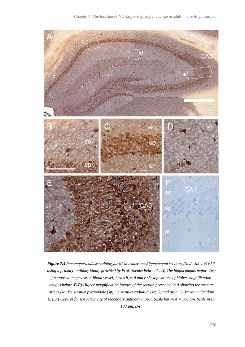
Chapter 7: The location of NO-targeted guanylyl cyclase in adult mouse hippocampus
250
Figure 7.4 Immunoperoxidase staining for β1 in transverse hippocampal sections fixed with 4 % PFA
using a primary antibody kindly provided by Prof. Soenke Behrends. A) The hippocampus major. Two
juxtaposed images. bv = blood vessel; boxes b, c, d and e show positions of higher magnification
images below. B-E) Higher magnification images of the section presented in A showing the stratum
oriens (so; B), stratum pyramidale (sp; C), stratum radiatum (sr; D) and area CA3/stratum lucidum
(E). F) Control for the selectivity of secondary antibody in A-E. Scale bar in A = 500 µm. Scale in B:
100 µm, B-F.
Chapter 7: The location of NO-targeted guanylyl cyclase in adult mouse hippocampus
251
The above results therefore suggested that all the structures that were found to be
immunopositive for α1 in tissue fixed with 1 % PFA are capable of forming
functional α1β1-containing guanylyl cyclase, and this was supported by
immunoperoxidase staining of sections fixed with 1 % PFA using a β1 antibody
supplied by Cayman (see Table 7.1 for antibody details). As shown in Figure 7.5,
staining with this antibody was observed in the stratum pyramidale, blood vessels
and a proportion of cells scattered throughout the hippocampus. As described by
Burette et al. (2002), pyramidal cell staining was inhibited by strong (4 % PFA)
tissue fixation.
Unlike the staining observed in Figure 7.4, fibres surrounding CA3 in the stratum
lucidum were only very weakly immunopositive. This could have resulted from a
difference in the overall efficacy of the two β1 antibodies used, because, in the
cerebellum, staining for β1 using the Cayman antibody is weaker than staining
observed using the antibody provided by Prof. Behrends (unpublished observations,
Giti Garthwaite and Kathryn Harris, UCL, London, UK).
Some non-selective staining was observed in the alveus and fimbrium surrounding
the hippocampus and, to a lesser extent, in the lacunosum moleculare (Figure 7.5 B).
Figure 7.5 Immunoperoxidase staining for β1 in transverse hippocampal sections fixed with 4 % PFA
using a primary antibody obtained from Cayman. A) The hippocampus major. Two juxtaposed
images. bv = blood vessel. B) Control for secondary antibody binding in B in which the primary
Chapter 7: The location of NO-targeted guanylyl cyclase in adult mouse hippocampus
252
antibody was omitted. Filled arrows (A-B) indicate non-selective staining. Scale bar in A: 500 µm, A-
B.
Further to the functional significance of the above findings, and in accordance with
previous results (Wendland et al., 1994; Gonzalez-Hernandez et al., 1996; Burette et
al., 2002), immunoperoxidase staining for nNOS suggested that it was present
throughout the hippocampus and in a distribution complimentary to that of α1 and β1
(Figure 7.6). Immunopositive structures included cells and fibres in the stratum
oriens, radiatum and pyramidale (Figure 7.6 C-E). As was previously found
(Wendland et al., 1994; Gonzalez-Hernandez et al., 1996; Burette et al., 2002),
staining of pyramidal neurons was dependent on weak tissue fixation (1 vs. 4 %
PFA). As expected, no staining of blood vessels was detected.
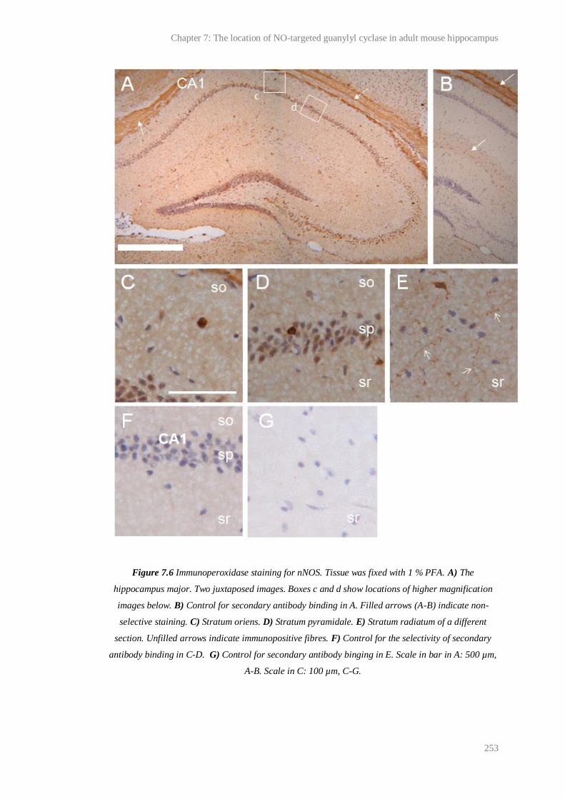
Chapter 7: The location of NO-targeted guanylyl cyclase in adult mouse hippocampus
253
Figure 7.6 Immunoperoxidase staining for nNOS. Tissue was fixed with 1 % PFA. A) The
hippocampus major. Two juxtaposed images. Boxes c and d show locations of higher magnification
images below. B) Control for secondary antibody binding in A. Filled arrows (A-B) indicate non-
selective staining. C) Stratum oriens. D) Stratum pyramidale. E) Stratum radiatum of a different
section. Unfilled arrows indicate immunopositive fibres. F) Control for the selectivity of secondary
antibody binding in C-D. G) Control for secondary antibody binging in E. Scale in bar in A: 500 µm,
A-B. Scale in C: 100 µm, C-G.

Chapter 7: The location of NO-targeted guanylyl cyclase in adult mouse hippocampus
254
7.4.3 Selectivity of the α1 primary antibody for α1 protein
As would typically be expected if the immunoperoxidase staining shown in Figure
7.2 was representative of the true expression pattern of the protein in vivo, all the
immunopositive structures also stained for β1 and were in close proximity to those
that stained for nNOS (Figure 7.2-6). However, the selectivity of the α1 antibody
required testing because: 1) stringent-tests of its selectivity are absent in the
literature, and tests should be performed under identical conditions to those in Figure
7.2; 2) other antibodies thought to selectively bind the α1 subunit have been found to
bind the α2 subunit (Ding et al., 2004), which would also be expected to be found in
β1-containing structures close to nNOS; 3) the distribution of α1 staining observed in
this study was strikingly different to that observed previously by Szabadits et al.
(2007).
Therefore, Western blot analysis was performed. In order that weak, non-selective
antibody binding might be detected, films were exposed until the strongest bands
began to saturate. As shown in Figure 7.7, only one main antigen, which migrated at
the molecular weight of rodent α1 (~ 80 kDa; Kamisaki et al., 1986), was detected by
the α1 antibody after incubation with blots of mouse and rat forebrain and
cerebellum lysates.
Figure 7.7 Western blots of rat and mouse cerebellum (cb) and forebrain (fb) lysates for α1. The first
three films were exposed for 2, 5 and 10 min (left to right) to the membrane and show that the α1
antibody detected only one antigen which migrated at an apparent molecular weight (MW) of ~80
MW Mouse Rat
cb fb cb fbkDa
230 -
100 -
80 -
25 -
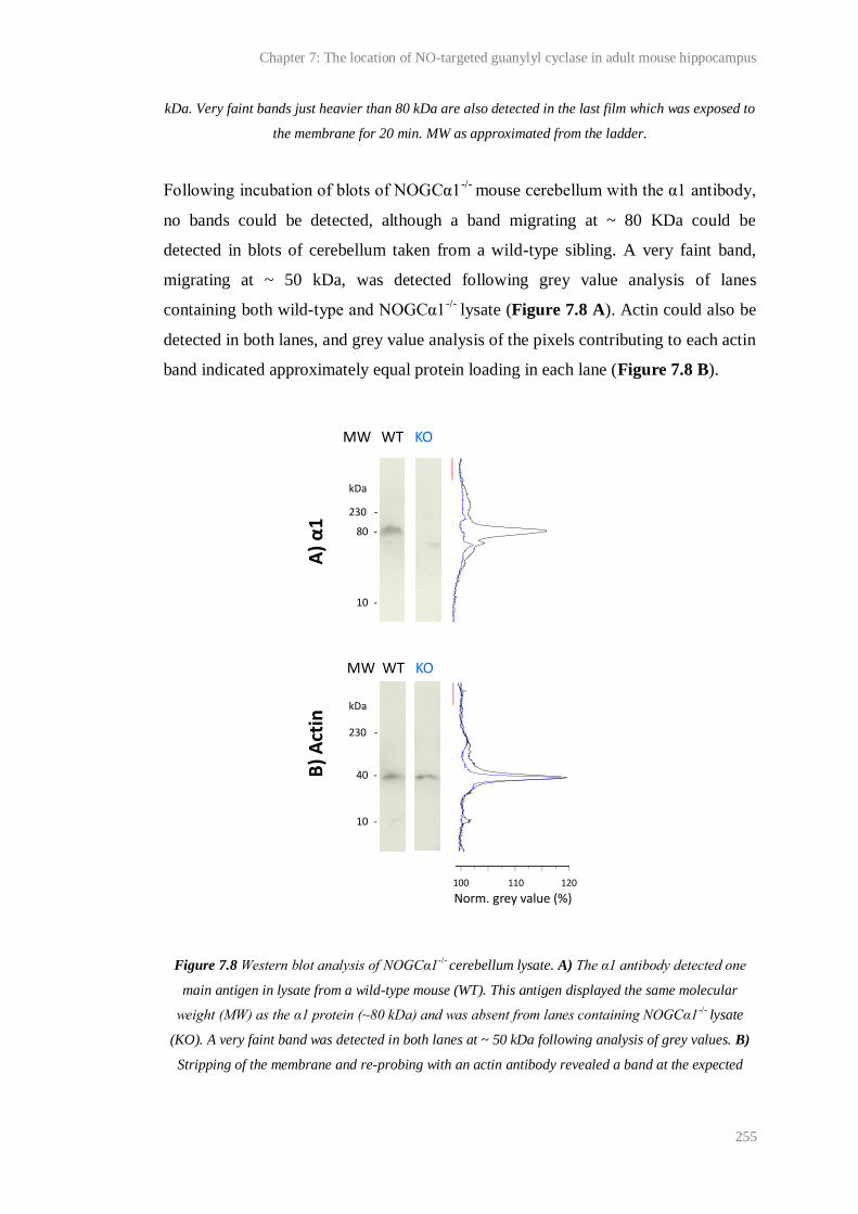
Chapter 7: The location of NO-targeted guanylyl cyclase in adult mouse hippocampus
255
kDa. Very faint bands just heavier than 80 kDa are also detected in the last film which was exposed to
the membrane for 20 min. MW as approximated from the ladder.
Following incubation of blots of NOGCα1-/-
mouse cerebellum with the α1 antibody,
no bands could be detected, although a band migrating at ~ 80 KDa could be
detected in blots of cerebellum taken from a wild-type sibling. A very faint band,
migrating at ~ 50 kDa, was detected following grey value analysis of lanes
containing both wild-type and NOGCα1-/-
lysate (Figure 7.8 A). Actin could also be
detected in both lanes, and grey value analysis of the pixels contributing to each actin
band indicated approximately equal protein loading in each lane (Figure 7.8 B).
Figure 7.8 Western blot analysis of NOGCα1-/- cerebellum lysate. A) The α1 antibody detected one
main antigen in lysate from a wild-type mouse (WT). This antigen displayed the same molecular
weight (MW) as the α1 protein (~80 kDa) and was absent from lanes containing NOGCα1-/- lysate
(KO). A very faint band was detected in both lanes at ~ 50 kDa following analysis of grey values. B)
Stripping of the membrane and re-probing with an actin antibody revealed a band at the expected
100 110 120
kDa
230 -
40 -
10 -
MW WT KO
MW WT KO
kDa
230 -
80 -
10 -
A) α
1B
) A
ctin
Norm. grey value (%)
Chapter 7: The location of NO-targeted guanylyl cyclase in adult mouse hippocampus
256
molecular weight (~ 40 kDa) in both the WT and KO lanes. Analysis of grey values was indicative of
approximately equal protein loading in each lane. Red bar in each panel indicates area of membrane
to which grey values were normalised. Similar results were obtained following analysis of NOGCα1-/-
forebrain lysate.
Under the conditions of Western blot, therefore, the α1 antibody displayed high
selectivity for the α1 protein. However, immunoperoxidase staining of sections of
NOGCα1-/-
and wild-type hippocampus fixed with 4 % PFA was identical (Figure
7.9). All structures that had previously been identified as strongly immunopositive
for α1 in tissue from C57/Bl6 mice were intensely stained in sections from NOGCα1-
/- mice, including cells outside the stratum pyramidale and fibres throughout the
stratum polymorph, lucidum and the rest of the neuropil. Weak staining of pyramidal
neurons was also observed. As before, blood vessels were immunonegative.
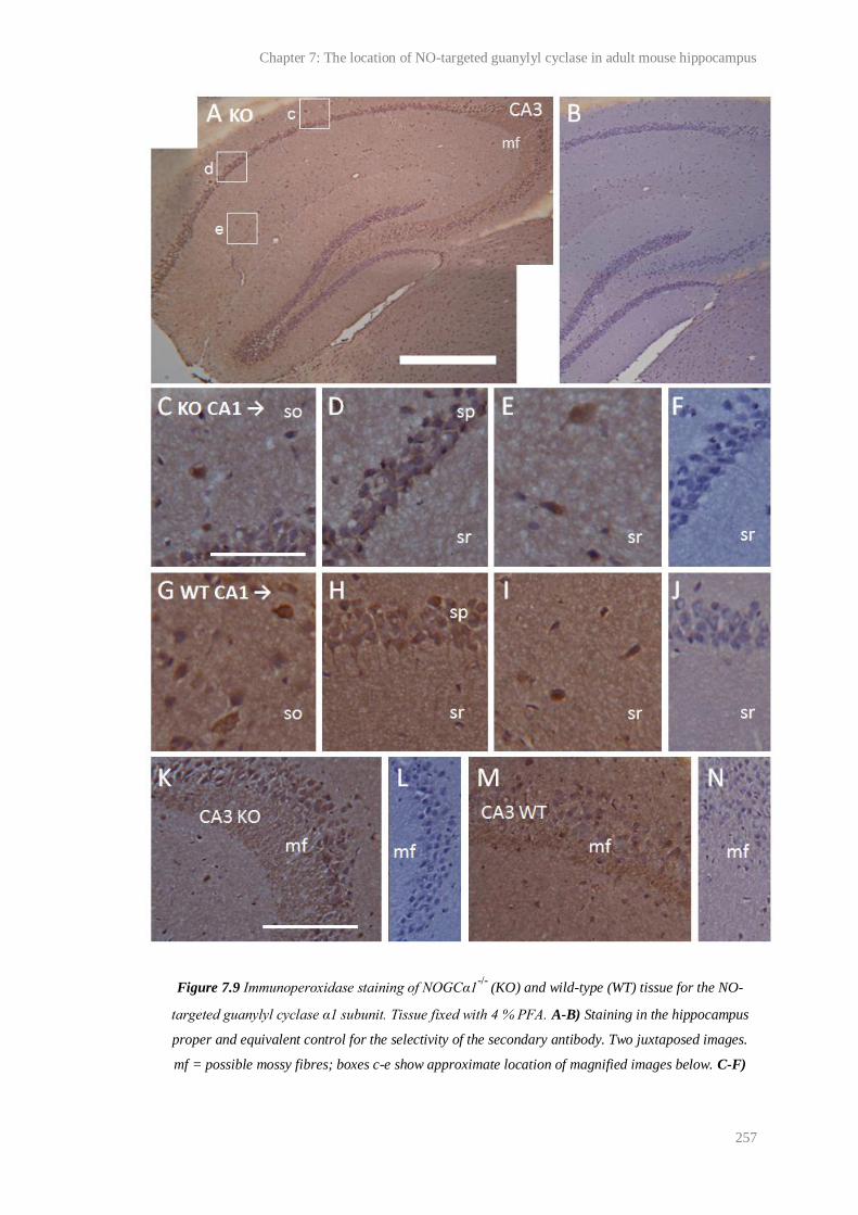
Chapter 7: The location of NO-targeted guanylyl cyclase in adult mouse hippocampus
257
Figure 7.9 Immunoperoxidase staining of NOGCα1-/-
(KO) and wild-type (WT) tissue for the NO-
targeted guanylyl cyclase α1 subunit. Tissue fixed with 4 % PFA. A-B) Staining in the hippocampus
proper and equivalent control for the selectivity of the secondary antibody. Two juxtaposed images.
mf = possible mossy fibres; boxes c-e show approximate location of magnified images below. C-F)
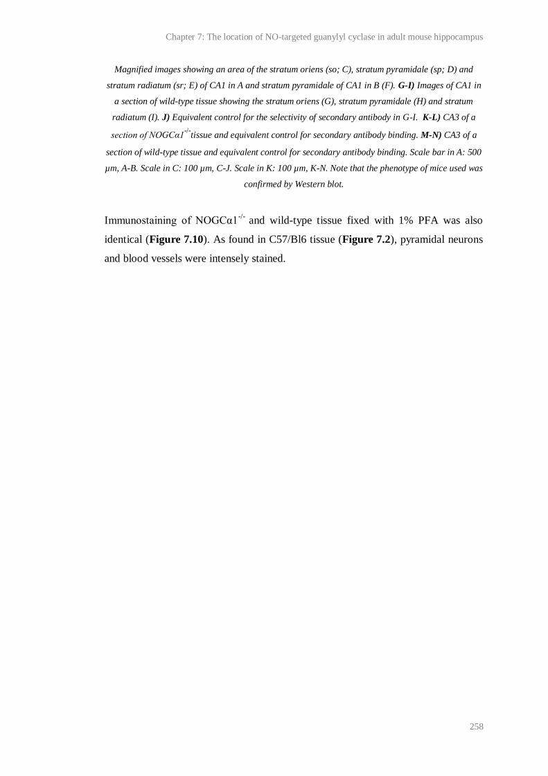
Chapter 7: The location of NO-targeted guanylyl cyclase in adult mouse hippocampus
258
Magnified images showing an area of the stratum oriens (so; C), stratum pyramidale (sp; D) and
stratum radiatum (sr; E) of CA1 in A and stratum pyramidale of CA1 in B (F). G-I) Images of CA1 in
a section of wild-type tissue showing the stratum oriens (G), stratum pyramidale (H) and stratum
radiatum (I). J) Equivalent control for the selectivity of secondary antibody in G-I. K-L) CA3 of a
section of NOGCα1-/-
tissue and equivalent control for secondary antibody binding. M-N) CA3 of a
section of wild-type tissue and equivalent control for secondary antibody binding. Scale bar in A: 500
µm, A-B. Scale in C: 100 µm, C-J. Scale in K: 100 µm, K-N. Note that the phenotype of mice used was
confirmed by Western blot.
Immunostaining of NOGCα1-/-
and wild-type tissue fixed with 1% PFA was also
identical (Figure 7.10). As found in C57/Bl6 tissue (Figure 7.2), pyramidal neurons
and blood vessels were intensely stained.

Chapter 7: The location of NO-targeted guanylyl cyclase in adult mouse hippocampus
259
Figure 7.10 Immunoperoxidase staining of NOGCα1-/-
(KO) and wild-type (WT) tissue for the NO-
targeted guanylyl cyclase α1 subunit. Tissue fixed with 1 % PFA. A-C) Images showing an area of the
stratum oriens (so; A), stratum pyramidale (sp; B) and stratum radiatum (sr; C) of CA1 in a section of
NOGCα1-/-
tissue. bv = blood vessel. D) Equivalent control for secondary antibody binding in A-C. E-
F) Images of CA1 in a section of wild-type tissue showing the stratum oriens (E), stratum pyramidale
(F) and stratum radiatum (G). H) Equivalent control for the selectivity of secondary antibody in E-H.
I-J) CA3 of a section of NOGCα1-/-
tissue and equivalent control for secondary antibody binding. K)
CA3 of a section of wild-type tissue. Scale bar in for A: 100 µm, A-H. Scale in I: 100 µm, I-K. Note
that the phenotype of mice used was confirmed by Western blot.
Clear staining for α1 in the cells and neuropil of the cortex was also observed in
tissue from NOGCα1-/-
mice, whether fixed with 4 % PFA (Figure 7.11) and 1 %
PFA (not shown).
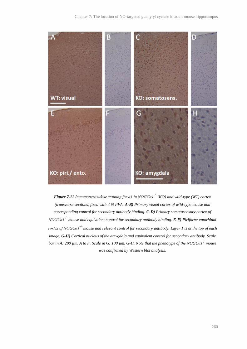
Chapter 7: The location of NO-targeted guanylyl cyclase in adult mouse hippocampus
260
Figure 7.11 Immunoperoxidase staining for α1 in NOGCα1-/-
(KO) and wild-type (WT) cortex
(transverse sections) fixed with 4 % PFA. A-B) Primary visual cortex of wild-type mouse and
corresponding control for secondary antibody binding. C-D) Primary somatosensory cortex of
NOGCα1-/-
mouse and equivalent control for secondary antibody binding. E-F) Piriform/ entorhinal
cortex of NOGCα1-/-
mouse and relevant control for secondary antibody. Layer 1 is at the top of each
image. G-H) Cortical nucleus of the amygdala and equivalent control for secondary antibody. Scale
bar in A: 200 µm, A to F. Scale in G: 100 µm, G-H. Note that the phenotype of the NOGCα1-/- mouse
was confirmed by Western blot analysis.

Chapter 7: The location of NO-targeted guanylyl cyclase in adult mouse hippocampus
261
7.4.4 Results summary
The above results are summarised in Table 7.3.
Structure
Antibody
α1 β1
(Behrends)
β1
(Cayman)
nNOS
WT KO
4 % 1 % 4 % 1 % 4/1 % 1 % 1 %
Pyramidal
neurons
+ +++ + +++ +++ ++ ++
Interneurons +++ +++ +++ +++ +++ +++ +++
Granule cells + + + + + + +
Mossy fibres ++ ++ ++ ++ ++ + -
Neuropil + + + + + + +
Blood vessels - +++ - +++ +++ ++ -
Table 7.3 Summary of results. The relative intensity of staining in each of the structures listed (as
judged by the experimenter) is indicated. Key: - = immunonegative/no staining; + =
weak/inconsistent staining; ++ = moderate staining; +++ = strong/intense staining. WT = wild-type
tissue; KO = NOGCα1-/- tissue. Percentages indicate the concentration of PFA used to fix tissues.
7.5 Discussion
Given the role of NO/cGMP signalling in hippocampal physiology, and the
possibility that NO may operate as a retrograde, anterograde and/or intracellular
transmitter (Garthwaite, 2008), the precise location of NO sources and targets in the
hippocampus is of great interest. As discussed above (see 7.1 Introduction), the
location of the NO-targeted guanylyl cyclase α1 subunit is contentious. Using
immunohistochemical techniques, the subunit has been found in interneurons in the
adult (Szabadits et al. 2007) and developing (Cserep et al., 2011) hippocampus,
suggesting that, if functional studies are correct (Taqatqeh et al., 2009; Neitz et al.,
2011), presynaptic effects of NO may be restricted to synapses involving these cells.

Chapter 7: The location of NO-targeted guanylyl cyclase in adult mouse hippocampus
262
Szabadits et al. (2007) and Cserep et al. (2011) used tissues fixed with 4 % PFA to
perform their immunohistochemical studies but it has been reported that this
concentration of PFA inhibits immunostaining of hippocampal pyramidal neurons for
synaptic proteins including nNOS and the β1 NO-targeted guanylyl cyclase subunit
(Wendland et al., 1994; Gonzalez-Hernandez et al., 1996; Burette et al., 2002).
Possible explanations for this inhibition are that high concentrations of PFA (4% vs.
1 %) mask antibody epitopes and/or restrict the access of antibodies to their target
proteins by excessively cross-linking proteins. In PSD’s and presynaptic varicosities,
where protein density is high and nNOS, β1 and α1 are expected to reside, the latter
circumstance may be particularly likely. Therefore we reinvestigated the distribution
of α1 in the hippocampus by immunoperoxidase staining of weakly fixed (1 % PFA)
tissue to see whether evidence for the α1 subunit in pyramidal neurons had been
overlooked.
In accordance with Szabadits et al. (2007) and Cserep et al. (2011), we found that
cells scattered outside of the stratum pyramidale, presumably interneurons and/or
glia, were most intensely immunopositive for α1 after staining of tissues fixed with 4
% PFA. The same antibody as employed by Szabadits et al. (2007) and Cserep et al.
(2011) was used. However, pyramidal cell soma and apical dendrites throughout
Ammon’s horn were also weakly immunopositive (Figure 7.1) and, in accordance
with the notion that pyramidal neurons must be weakly fixed to be immunostained
for some synaptic proteins, staining of these cells was intensified after identical
treatment of tissues fixed with 1 % PFA.
Considering that pyramidal neurons were also found to be immunopositive for the β1
NO-targeted guanylyl cyclase subunit and nNOS (Figure 7.4-6), and in light of the
studies by Taqatqeh et al. (2009) and Cserep et al. (2011), the distribution of α1
staining that we observed in tissues fixed with 1 % PFA suggested that α1β1-
dependent NO-induced modulation of transmitter release might occur at CA1-CA3
synapses. NO-regulated neurotransmitter release at CA1-CA3 synapses is consistent
with the effect of NO at synapses between pairs of dissociated pyramidal neurons
during LTP (Arancio et al., 1995; Arancio et al., 1996; Arancio et al., 2001).

Chapter 7: The location of NO-targeted guanylyl cyclase in adult mouse hippocampus
263
The distributions of staining for α1, β1 and nNOS observed in tissues fixed with 1 %
PFA (see Figure 7.2-7.6) were remarkably similar. This was in favour of the α1
antibody selectively binding protein for the α1 subunit, and therefore, with the
distribution of α1 protein observed in tissues fixed with 1 % PFA being
physiologically relevant. Indeed, these possibilities were supported by several other
observations.
First, blood vessels were found to be intensely immunopositive for α1 following
staining of tissue fixed with 1 % but not 4 % PFA (Figure 7.1-2). This was
consistent with immunohistochemical and functional evidence suggesting the
presence of α1β1 in the smooth muscle of blood vessels, such as arteries (Mergia et
al., 2006; Nimmegeers et al., 2007). Notably, smaller blood vessels, such as those
that were found to be immunopositive in this study, also contain smooth muscle (for
example, venules measure on average ~ 20 µM; Germann and Stanfield, 2002).
Moreover, endothelial cells in kidney and lung have also been reported to express
α1β1 (Zhan et al., 1999; Jarry et al., 2003). Second, and in apparent discord with the
notion that all structures in the hippocampus were non-selectively stained, some cells
scattered outside the stratum pyramidale, for example, in the stratum granulare, were
immunonegative for α1 in tissues fixed with 1 % PFA (Figure 7.2). Third, and most
importantly, Western blot analysis showed that, in tissue lysates prepared from
C57/Bl6 mice, the α1 antibody detected only one main antigen which had the same
molecular weight as the α1 subunit (~ 80 kDa; Kamisaki et al., 1986) and was absent
from lysates prepared from NOGCα1-/-
tissue (Figure 7.7-7.8).
However, immunoperoxidase staining of sections from mice lacking the α1 subunit,
as confirmed by Western blotting, revealed that, at least under our conditions, the α1
antibody was not selective for the α1 protein. As shown in Figure’s 7.9-10, all the
structures found to be immunopositive after staining of tissues prepared from
C57/Bl6 or NOGCα1-/-
wild-type mice were also immunopositive in sections
prepared from NOGCα1-/-
, regardless of fixing with 1 or 4 % PFA. Similarly,
immunostaining was prevalent throughout sections of cortex prepared from
NOGCα1-/-
mice (Figure 7.11).
Chapter 7: The location of NO-targeted guanylyl cyclase in adult mouse hippocampus
264
Since the protein sequence for the α1 antibody epitope is not present anywhere else
in the mouse proteome (as assessed using the Basic Local Alignment Search Tool
(BLAST); www.ncbi.nlm.nih.gov/genome/seq/BlastGen/BlastGen.cgi?taxid=10090l;
accessed 20/01/2011) it is hard to make a firm hypothesis as to what the α1 antibody
was binding to. The distribution of staining for α1 was remarkably coincident with
the distribution of staining for β1 (Figure 7.2 and 7.3), so it could be hypothesised
that the α1 antibody binds the β1 subunit. Alternatively, it could be speculated that
the α1 antibody recognises the α2 subunit, which weighs ~ 82 kDa ( ß1 weights ~ 70
KDa; Kamisaki et al., 1986; Harteneck et al., 1991), because following Western
blotting of lysates of rat forebrain and cerebellum for α1, faint bands surrounding the
major signal at ~ 80 kDa could be detected after a long (20 min) exposure of the
blotting membrane to film (Figure 7.6). This would also explain why the β1
antibodies yielded similar distributions of immunoperoxidase staining to the α1
antibody. Notably, another antibody thought to selectively bind the α1 subunit has
been found to bind protein for the α2 subunit (Ding et al., 2004). One way to test the
possibility that the α1 antibody bound protein for the α2 or β1 protein would be to
stain tissues prepared from animals deficient in these subunits. However, it is should
be noted that the α1 subunit may have bound a protein unrelated to guanylyl cyclase.
Consistent with this, grey analysis of images of Western blots probed with the α1
antibody revealed a very faint signal in both NOGCα1-/-
and wild-type lysates that
was unlikely to be a guanylyl cyclase subunit (~ 50 kDa; Figure 7.7).
The conditions used for immunohistochemistry vary widely between groups of
experimenters and therefore, it cannot be said that other observations made using the
α1 antibody (for example, Szabadits et al., 2007; Fukutani et al., 2009; Wilson and
Garthwaite, 2010; Cserep et al., 2011), are artefactual. Indeed, the lack of any major
signal following Western blotting of NOGCα1-/-
tissue with the α1 antibody shows
that the antibody does selectively bind the α1 subunit under some conditions.
Furthermore, in the study by Szabadits et al. (2007), the reported distributions of α1
and α2 subunit mRNA, as assessed using in situ hybridisation, were distinct from
each other, and the distribution of α1 mRNA was complementary with the
distribution of the α1 protein, as judged by immunofluorescence and
Chapter 7: The location of NO-targeted guanylyl cyclase in adult mouse hippocampus
265
immunoperoxidase, consistent with the distribution of immunostaining for α1
reflecting the true expression pattern of this subunit.
Unfortunately, indicators of selective binding like those included in the study by
Szabadits et al. (2007) are absent in other studies using the α1 antibody and stringent,
relevant-tests of the antibody’s selectivity are, inexplicably, missing in the literature.
Therefore, the present work clearly complicates the interpretation of the results of
published studies employing the antibody. (It should be noted that adequate controls
for the selectivity of both of the β1 antibodies used here are also lacking and, until
they are provided, the precise location of β1 in the hippocampus will also remain
uncertain). To clarify the location of the α1 subunit in hippocampus, future work
must determine the conditions, if any, under which the α1 antibody is selective for its
target. Interestingly, precedent for selective antibody binding under the conditions of
Western blotting but not conventional immunoperoxidase staining exists in the
literature (Watanabe et al., 1998). Watanabe et al. (1998) found that the selective
binding (as assessed using knock-out tissue) of primary antibodies for the NMDA
receptor subunits NR1 and NR2A and B in 4% PFA-fixed sections of mouse brain,
including the hippocampus, was dependent on the pre-treatment of sections with
pepsin, which facilitated the access of very low, previously sub-threshold
concentrations of the antibodies to their targets. The resulting pattern of staining was
drastically different to that observed in pepsin-untreated sections using higher
antibody concentrations that were non-specific. In fact, they accorded better with
expectations of the distribution of a synaptic protein (since, for example, dendritic
staining in the hippocampus became stronger than staining of cell soma) and with the
distribution of the NMDA receptor subunits’ mRNA, as assessed by in situ
hybridisation. Similar results have been reported following the combined use of
pepsin and very low concentrations of antibodies to stain for other synaptic proteins,
such as PSD-95 (Fukaya and Watanabe, 2000), and it is noteworthy that the
distribution of the non-specific staining for NMDA receptor subunits observed in
pepsin un-treated sections by Watanabe et al. (1998) was remarkably similar to the
distribution of staining that we observed following immunoperoxidase staining for
the NO-targeted guanylyl cyclase α1 subunit in 1 % PFA-fixed tissue. Therefore
Chapter 7: The location of NO-targeted guanylyl cyclase in adult mouse hippocampus
266
pepsin treatment could represent a strategy for obtaining selective binding of the NO-
targeted guanylyl cyclase α1 antibody.
If no conditions for selective antibody binding are found, the development of an
alternative antibody with higher selectivity for the α1 protein will be desirable.
Considering that functional evidence implies that presynaptic effects of NO are α1-
dependent (Taqatqeh et al., 2009; Neitz et al., 2011), the true location of α1, whether
in interneurons and/or pyramidal neurons will be very interesting and could be of
critical importance to researchers seeking to understand NO/cGMP-dependent
plasticity.

Chapter 8:
Summary
Chapter 8: Summary
268
8.1 NO and NMDA receptor-dependent LTP
It is well accepted that various forms of synaptic plasticity require NO. Brain areas in
which NO-dependent forms of LTP have been studied include the hippocampus (for
example, Son et al., 1996; Doreulee et al., 2001; Hopper and Garthwaite, 2006), the
neocortex (for example, Haul et al., 1999; Hardingham and Fox, 2006; Haghikia et
al., 2007) and amygdala (Watanabe et al., 1995). At CA1 synapses, where LTP is
archetypal, both endothelium-derived and neuronal NO signals are thought be
required for stable NMDA receptor- and NO-dependent LTP (Son et al., 1996;
Hopper and Garthwaite, 2006).
8.1.1 Role of nNOS
Given the physical link between nNOS and NMDA receptors in neurons, and that
NO is a putative retrograde transmitter at synapses, thoughts on the role of NO in
LTP usually centre around its generation upon NMDA receptor channel opening and
action on presynaptic targets (Feil and Kleppisch, 2008; Garthwaite, 2008).
Compelling evidence for retrograde NO transmission has been provided by studies of
LTP at synapses between dissociated hippocampal neurons. However, there are few
examples of presynaptic effects of NO (consistent with retrograde transmission)
during LTP at synapses in intact, wild-type tissues (see Table 3.1).
Given the above, the study presented in Chapter 3 was designed to test the
prediction that exogenous NO, when paired with HFS, would restore the NO-
dependent component of NMDA receptor-dependent LTP at CA1 synapses when
NMDA receptors were blocked. In this way we aimed to isolate the NO-dependent
component and test its locus (pre- or postsynaptic). Consistent with the prediction,
we report for the first time that the NO donor, PAPA/NONOate, generated a
persistent enhancement of CA1 fEPSPs when paired with HFS in the presence of the
NMDA antagonist, D-AP5. In accordance with the typical effect of NOS inhibition
on NMDA receptor-dependent LTP (see Figure 3.4), this NO-induced potentiation
was of slow onset but reached a magnitude similar to HFS-induced LTP. Further
characterisation of this NO-induced potentiation was consistent with it being
Chapter 8: Summary
269
representative of a NO-dependent component of LTP (although see Chapter 3 for a
broader discussion), suggesting that the potentiation may be useful tool for the study
of long-lasting NO-dependent potentiation in isolation of other mechanisms
underlying LTP.
The hypothesis that NO acts as a retrograde messenger during LTP at synapses in
intact tissues, in conjunction with studies of LTP at synapses between dissociated
cells, predicted that the NO-induced potentiation would be presynaptic. However, we
found no evidence for this. While our data cannot rule out a presynaptic effect of NO
during HFS-induced and other types of LTP, including those that may occur
naturally in vivo, they are not consistent with this possibility. Furthermore, much of
the evidence in favour of a presynaptic effect of NO during LTP in intact tissues is
also consistent with NO acting postsynaptically. Additionally, the data imply that
conclusions drawn from studying synapses between dissociated neurons may not be
applicable to synapses in intact tissues (perhaps due to a lack of eNOS in the former
preparation; see below).
In the future, fluorescent indicators of presynaptic efficacy (reviewed by Blundon
and Zakharenko, 2008) could be used to further test the possible presynaptic effect of
NO during LTP at synapses in intact tissues. At present, we conclude that an action
on both sides of the synapse is most parsimonious with the current data on the role of
NO/cGMP in LTP (see Garthwaite, 2008; Feil and Kleppisch, 2008 for a review).
Such a role for NO in LTP may enable it to coordinate synaptic plasticity across the
synapse; a potentially important action of which few other molecules would be
capable.
8.1.2 Role of eNOS
The NO-induced potentiation detailed in Chapter 3 was facilitated by endogenous
NO. Based on the current literature (Hopper and Garthwaite, 2006), the endogenous
NO required was probably endothelium-derived. Endothelial NOS has been reported
to generate a low-level, activity-independent NO tone in optic nerve and
hippocampus. In optic nerve, this NO signal causes the tonic depolarisation of axons
Chapter 8: Summary
270
via a pathway involving HCN channels (Garthwaite et al., 2006). In hippocampus,
the NO tone has been postulated to prime synapses for LTP (Hopper and Garthwaite,
2006), but, prior to the present study, it had no defined role.
The data presented in Chapter 4 imply that eNOS activity elicits the tonic
facilitation of neurotransmitter release at CA1 synapses under conditions of basal
stimulation (i.e. stimulation causing no observable change in synaptic efficacy).
Although our data are limited, our conclusion is consistent with a recent study by
Neitz et al. (2011), and proposes a novel role for the endothelium-derived, activity-
independent NO tone in the hippocampus. The consequences of such a role for eNOS
may be of critical importance to NO-dependent plasticity, since factors that influence
neurotransmitter release during LTP induction likely influence LTP expression.
Indeed, it is noteworthy that the effect of (e)NOS inhibition on PPF under basal
conditions was observed when the ISI was 10 ms- the same ISI as occurs during
HFS. We hypothesise that the facilitation of neurotransmitter release at CA1
synapses by eNOS may regulate the stimulus threshold for the induction of stable
LTP, thus explaining why some forms of LTP require endothelium-derived NO (for
example, Haul et al., 1999; Son et al., 1996; Hopper and Garthwaite, 2006). Since
eNOS activity is subject to dynamic regulation (Chapter 1), it is, given the above
hypothesis, also tempting to speculate that endothelium-derived NO may contribute
to metaplasticity. On this point it is interesting to note that exercise, which causes an
increase in eNOS activity in the cardiovascular system and brain, probably via the
shear stress of endothelial cells (reviewed by Walther et al., 2004; Faraci, 2006), has
been proposed by those studying adult neurogenesis in the dentate gyrus, LTP and
learning in spatial tasks to prime the hippocampus for plasticity as animals move
through environments and increase their capacity for experience (Kempermann,
2002; Kempermann et al., 2010).
Recently, it has been reported that the regulation of neurotransmitter release
under basal conditions by NO (eNOS) requires the α1β1 isoform of NO-targeted
guanylyl cyclase (Taqatqeh et al., 2009; Neitz et al., 2011). Message and protein
for the α1 NO-targeted guanylyl cyclase has been detected in hippocampal
interneurons but not pyramidal cells (Szabadits et al., 2007; Cserep et al., 2011).

Chapter 8: Summary
271
Using conditions that have been reported to enhance the staining of other
presumed synaptic proteins in fibres and principal cells (Burette et al., 2002;
Wendland et al., 1994; Gonzalez-Hernandez et al., 1996), we made attempts to
determine whether the NO-targeted guanylyl cyclase α1 subunit was also present
in hippocampal pyramidal neurons, as effects of NO on neurotransmitter release
at synapses between pairs of dissociated pyramidal neurons would suggest (see
Table 3.1). Under these conditions, immunoperoxidase staining for the α1
subunit was detected in pyramidal neurons (Chapter 7). However, the staining
was found to be entirely non-specific based on the use of α1-null mice.
Importantly, interneuron staining, regardless of the conditions used, was also
non-specific. Although the α1 antibody that we used is the most commonly
employed for immunohistochemistry, no other stringent tests of its specificity
have been reported. The specificity of antibodies used for the detection of the β1
subunit is also unclear and tests of this are hampered by a lack of viable β1-
deficient mice. Given the wealth of physiological processes and pathologies
thought to involve NO/cGMP, and the intriguing possibility that pre- and
postsynaptic effects of NO might be differentially transduced by the α1- and α2-
containing NO-targeted guanylyl cyclase isoforms, further, controlled tests of the
location of NO-targeted guanylyl cyclase are required (see Chapter 7 for a
strategy).
8.2 NO and NMDA receptor-independent LTP
The data presented in Chapter 5 show for the first time that a form of NMDA
receptor-independent, L-VGCC-dependent LTP at CA1 synapses requires NO, in this
case, apparently generated solely by nNOS. This finding challenges the view that
neuronal NO is preferentially synthesised by NMDA receptor channel opening. L-
VGCC-dependent forms of synaptic plasticity have been correlated with learning and
memory (Borroni et al., 2000; Kleppisch et al., 2004; Woodside et al., 2004;
Moosmang et al., 2005a), and may be particularly important for changes in gene
expression during synaptic plasticity (Murphy et al., 1991; Bading et al., 1993;
Impey et al., 1996). Therefore, our data indicate that a potentially important role for
NO in the regulation of synaptic efficacy may have been previously overlooked.
Chapter 8: Summary
272
Neuronal NOS activation during NMDA receptor-independent, L-VGCC-dependent
LTP likely results from a rise in intradendritic Ca2+
. Although we were unable to
identify a potential source of Ca2+
responsible for nNOS activation during L-VGCC-
dependent LTP (Chapter 5 and Appendix 2), the use of a new biosensor with
unrivalled sensitivity to NO (described in Batchelor et al., 2010; Wood et al., 2011)
may, in the future, enable the elucidation of the signalling pathway(s) responsible.
The role of nNOS in L-VGCC-dependent LTP was deduced from studies using a
non-selective NOS inhibitor (L-NNA) in conjunction with eNOS-deficient mice.
Ideally, the role of nNOS would also have been tested using a selective inhibitor.
Unfortunately, the study described in Chapter 5 led to the discovery that the most
potent nNOS inhibitors (L-VNIO and 1400-W) currently available, were, at the
standard concentrations for use (0.1 µM and 1 µM, respectively), ineffective in
blocking NMDA-induced, NOS-dependent cGMP synthesis in adult mouse
hippocampal slices (see Chapter 6). Prior to our discovery, similar findings were
made using slices of immature mouse hippocampus (Bartus, 2009). Our data were
particularly surprising since the original characterisation of L-VNIO (0.1 µM) and
1400-W (1 µM) as selective nNOS inhibitors in intact tissues was done using adult
hippocampus (Hopper and Garthwaite, 2006). Taken together, the data presented in
Chapter 6 and by Bartus (2009) suggest that the compounds, which, have been
widely used at the above concentrations, are inadequately selective for nNOS over
eNOS to be of use diagnostically. Experiments to address the difference between the
results of the present study and those of Hopper and Garthwaite (2006) are in
progress. Since a wealth of studies using these compounds lack appropriate controls
for selective nNOS inhibition (for example, Neitz et al., 2011), the related data
should be re-interpreted.
To address the lack of useable nNOS inhibitors, we tested another compound (FX-
5043), shown previously to inhibit nNOS with unrivalled potency and selectivity in
cell-free and cell-based assays (Xue et al., 2010a). In testament to the challenges that
face that the development of selective NOS inhibitors (reviewed by Alderton et al.,
2001), neither FX-5043, nor a similar compound, JK-5, were found to be appropriate
for use in intact tissues (Chapter 5). Therefore more work is needed to develop

Chapter 8: Summary
273
selective nNOS inhibitors for research, and potentially clinical (see Gross and Wolin,
1995; Hobbs et al., 1999; Vallance and Leiper, 2002), use.
8.3 Some general outstanding issues
The data presented here add support to some key hypotheses of the role of NO in
LTP, and show novel roles for NO in synaptic plasticity. Additionally, a scheme for
NMDA receptor and NO-dependent, HFS-induced LTP laid out in Figure 3.21 may
be a useful foundation for further tests of the role of NO in LTP.
Overall, the results are consistent with the view that physiological NO signals are
transduced by NO-targeted guanylyl cyclase and cGMP accumulation, but the
mechanisms downstream of NO/cGMP in synaptic plasticity largely remain to be
elucidated (see Chapter 1 for review). The definition of mechanisms downstream of
NO/cGMP during LTP may help to explain how two NO/cGMP signals
(endothelium-derived and neuronal) act upon one synapse simultaneously. Based on
recent work by others (Taqatqeh et al., 2009; Neitz et al., 2011), it is tempting to
speculate that NO signals produced by eNOS and nNOS may differentially target the
α1β1 and α2β1 cyclases, and that these may be confined to different sides of the
synapse. If eNOS and nNOS act on the same sides of the synapse, but have distinct
roles, the compartmentalisation of NO/cGMP signals by PDEs may be important, as
may be the involvement of downstream effectors with different sensitiveness to
cGMP, such as PKGI and PKGII (see Chapter 1).
There are, of course, many alternatives to the ‘one synapse’ hypothesis for NO action
in LTP. Immunohistochemical evidence, for example, suggests that astrocytes
respond to NO in multiple brain areas (de Vente et al., 1998). Interestingly astrocytes
have low PDE activity, and are therefore highly sensitive to NO (Garthwaite, 2005).
Although little is known about the physiological significance of possible signals
conveyed to astrocytes by NO, astrocytes are well-placed to regulate synaptic
plasticity. Actions that may influence LTP include: the regulation of synaptogenesis;
neurotransmitter uptake; and the release of gliotransmitters (reviewed by Barker and
Ullian, 2010). Many of these actions could conceivably be influenced by changes in

Chapter 8: Summary
274
astrocyte morphology and, considering the potential role of NO in structural synaptic
plasticity (Nikonenko et al., 2003; Nikonenko et al., 2008), it is interesting that the
NO pathway has been found to regulate the morphology of rat cerebellar astroglia in
culture (Boran and Garcia, 2007), in this case via cGMP/PKG, and astrocyte-like
cells in hypothalamus (De Seranno et al., 2004).
Additionally, an emerging focus of research with relevance to the involvement of
NO in synaptic plasticity is on the role of NO in GABAergic transmission at
interneuron synapses. It has long been recognised that NO regulates the release of
GABA at synapses in various brain regions (reviewed by Prast and Philippu,
2001). In the ventral tegmental area (Nugent et al., 2007) and the hippocampus
(Makara et al., 2007; Cserep et al., 2011), evidence consistent with the activity-
dependent synthesis of NO in pyramidal neurons and subsequent down-
regulation of GABAergic transmission via retrograde transmission has been
reported (see Chapter 7, 7.1 for discussion). In the hippocampus, it is tempting
to speculate that such an NO-mediated depolarisation-induced suppression of
inhibition would influence the LTP induction threshold at pyramidal cell
synapses. The effects of NO on GABAergic transmission in the developing
hippocampus appeared to be regulated by the α1β1-containing guanylyl cyclase
(Cserep et al., 2011), which has recently been identified as presynaptic and
tonically active on the basis of functional evidence (Taqatqeh et al., 2009; Neitz
et al., 2011; Chapter 4).
Excitingly, the future use of a new biosensor able to detect picomolar concentrations
of NO/cGMP in real-time (Batchelor et al., 2010; Wood et al., 2011) may enable a
multitude of questions regarding the physiology of NO and its role in synaptic
plasticity to be answered. If the biosensor can be transfected into hippocampal cells,
it may permit direct tests of questions as fundamental as whether or not neuronal NO
signals generated during LTP are synapse specific (see above) and/or subject to
potentiation themselves.

Appendix 1:
Intracellular recording of synaptic activity in area CA1 using sharp
electrodes
Appendix 1: Intracellular recording of synaptic activity in area CA1 using sharp electrodes
276
We investigated the effects of HFS-induced LTP and the NO-induced potentiation
described in Chapter 3 on PPF in individual pyramidal neurons in area CA1 in
hippocampal slices using standard methods for intracellular electrophysiological
recording with sharp electrodes. Sharp electrode recording offered three major
advantages over patch-clamp recording. First, sharp electrode recordings can be
maintained for longer than patch-clamp recordings because sharp electrodes confer
experiments with greater physical and biochemical stability. Second, and in
accordance with the first advantage, sharp electrode recording has less effect on the
postsynaptic cell cytoplasm. Third, sharp electrodes can be advanced into the slice to
the depth where field recordings are typically made and away from tissue damage
caused by slicing. This point was especially important since the concentration of
bath-applied exogenous NO that penetrates a brain slice is known to decline steeply
as a function of depth into the slice (Hall and Garthwaite, 2006).
Intracellular sharp electrodes, ~ 70-120 MΩ when filled with 2-3 M KMeSO4, were
placed within the stratum pyramidale in area CA1, adjacent to an extracellular
recording electrode in the stratum radiatum (Figure 9.1; see Chapter 2 for details of
field electrode recording). Pyramidal neurons were identified by their
electrophysiological properties, including their resting Vm (~ - 69 mV), action
potential threshold (~ 50 mV), slow after-hyperpolarisation (AHP) and spike
frequency adaptation (Figure 9.1B-C; see Spruston and McBain, 2007 for
comparison). Presumed pyramidal cell EPSPs were largely inhibited by CNQX (10
µM). A slower, CNQX-resistant, D-AP5 (50 µM)-sensitive EPSP could be observed
on raising the stimulation amplitude. These findings were consistent with the
synaptic activation of AMPA/kainate and NMDA receptors (Figure 9.1C).
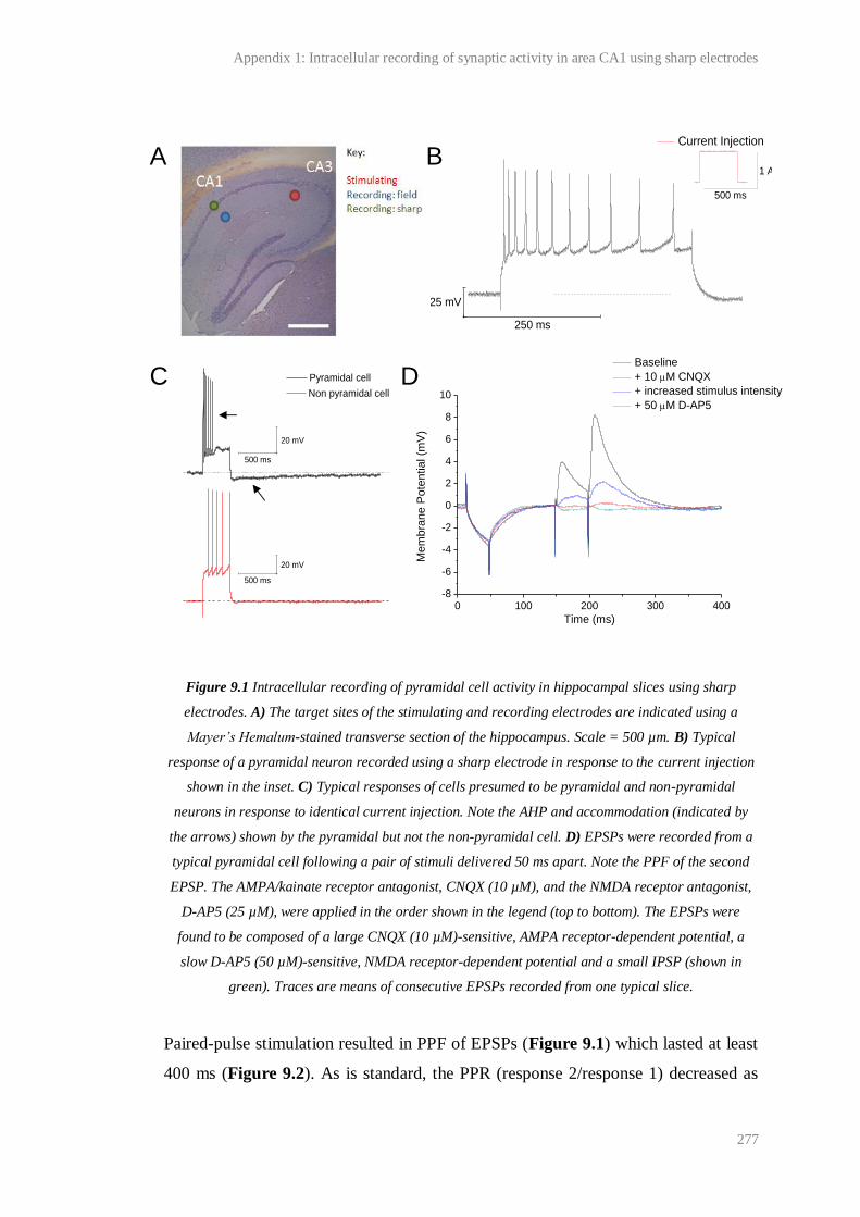
Appendix 1: Intracellular recording of synaptic activity in area CA1 using sharp electrodes
277
Figure 9.1 Intracellular recording of pyramidal cell activity in hippocampal slices using sharp
electrodes. A) The target sites of the stimulating and recording electrodes are indicated using a
Mayer’s Hemalum-stained transverse section of the hippocampus. Scale = 500 µm. B) Typical
response of a pyramidal neuron recorded using a sharp electrode in response to the current injection
shown in the inset. C) Typical responses of cells presumed to be pyramidal and non-pyramidal
neurons in response to identical current injection. Note the AHP and accommodation (indicated by
the arrows) shown by the pyramidal but not the non-pyramidal cell. D) EPSPs were recorded from a
typical pyramidal cell following a pair of stimuli delivered 50 ms apart. Note the PPF of the second
EPSP. The AMPA/kainate receptor antagonist, CNQX (10 µM), and the NMDA receptor antagonist,
D-AP5 (25 µM), were applied in the order shown in the legend (top to bottom). The EPSPs were
found to be composed of a large CNQX (10 µM)-sensitive, AMPA receptor-dependent potential, a
slow D-AP5 (50 µM)-sensitive, NMDA receptor-dependent potential and a small IPSP (shown in
green). Traces are means of consecutive EPSPs recorded from one typical slice.
Paired-pulse stimulation resulted in PPF of EPSPs (Figure 9.1) which lasted at least
400 ms (Figure 9.2). As is standard, the PPR (response 2/response 1) decreased as
250 ms
25 mV
Current Injection
500 ms
1 A
0 100 200 300 400-8
-6
-4
-2
0
2
4
6
8
10
Me
mb
ran
e P
ote
ntia
l (m
V)
Time (ms)
Baseline
+ 10 M CNQX
+ increased stimulus intensity
+ 50 M D-AP5 Non pyramidal cell
500 ms
20 mV
Pyramidal cell
500 ms
20 mV
A B
C D
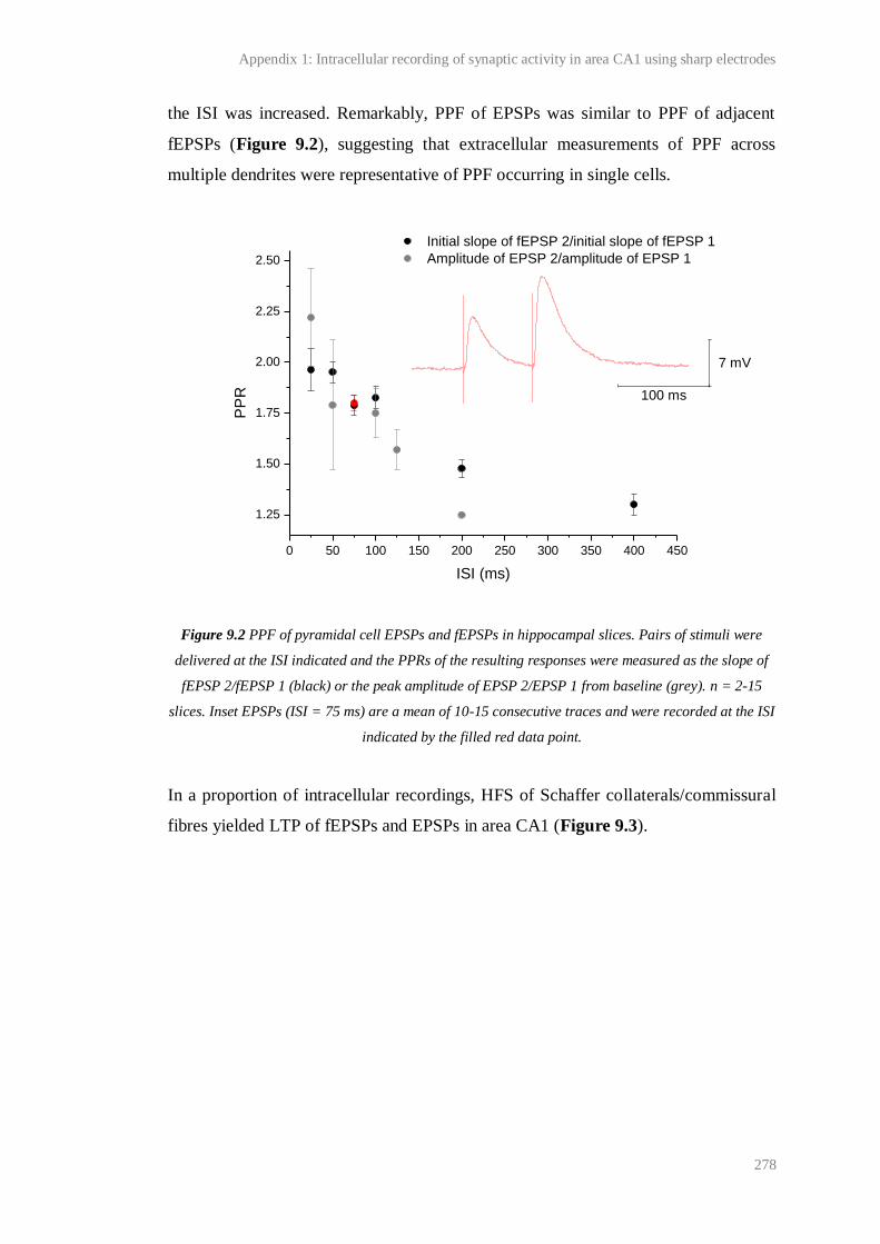
Appendix 1: Intracellular recording of synaptic activity in area CA1 using sharp electrodes
278
the ISI was increased. Remarkably, PPF of EPSPs was similar to PPF of adjacent
fEPSPs (Figure 9.2), suggesting that extracellular measurements of PPF across
multiple dendrites were representative of PPF occurring in single cells.
Figure 9.2 PPF of pyramidal cell EPSPs and fEPSPs in hippocampal slices. Pairs of stimuli were
delivered at the ISI indicated and the PPRs of the resulting responses were measured as the slope of
fEPSP 2/fEPSP 1 (black) or the peak amplitude of EPSP 2/EPSP 1 from baseline (grey). n = 2-15
slices. Inset EPSPs (ISI = 75 ms) are a mean of 10-15 consecutive traces and were recorded at the ISI
indicated by the filled red data point.
In a proportion of intracellular recordings, HFS of Schaffer collaterals/commissural
fibres yielded LTP of fEPSPs and EPSPs in area CA1 (Figure 9.3).
0 50 100 150 200 250 300 350 400 450
1.25
1.50
1.75
2.00
2.25
2.50
Initial slope of fEPSP 2/initial slope of fEPSP 1
Amplitude of EPSP 2/amplitude of EPSP 1
PP
R
ISI (ms)
100 ms
7 mV

Appendix 1: Intracellular recording of synaptic activity in area CA1 using sharp electrodes
279
Figure 9.3 LTP of adjacent fEPSPs and pyramidal neuron EPSPs. A typical example of CA1 LTP
induced by HFS (1-s, 100-Hz tetanus) of Schaffer collaterals/commissural fibres and recorded using
an extracellular field electrode in the stratum radiatum (black) and an adjacent sharp electrode
within a presumed pyramidal neuron in the stratum pyramidale (grey). The data recorded
intracellularly has been truncated since the cell fired during PTP. Sample traces are the mean
response recorded at the times indicated by the colour-coded bars.
However, ~ 50 % of recordings could not be maintained for the duration required,
and ~ 50 % of neurons from which stable recordings were made were unable to
support LTP (induction and expression). The lack of stable LTP could have resulted
from a combination of factors, including poor slice or neuron health, instability of the
intracellular electrode or the possibility that not all neurons adjacent to the field
electrode contributed to LTP of the fEPSP. In support of the latter possibility, studies
of LTP and LTD at CA1 synapses in transverse hippocampal slices and organotypic
hippocampal slice cultures suggest that a small proportion of CA1 synapses do not
exhibit synaptic plasticity (Petersen et al., 1998; Debanne et al., 1999). We favour
this possibility as an explanation for the lack of LTP because, in all cases, the fEPSP
appeared to be healthy and could be potentiated, baseline EPSPs were maintained
despite the lack of LTP, and PTP was not observed in cells that failed to potentiate.
0 5 10 15 20 25 30 35 40
0
50
100
150
200
250
300
350
400
450
500
Normalised fEPSP slope Normalised EPSP amplitude
No
rma
lise
d f
EP
SP
slo
pe
/ E
PS
P a
mp
litu
de
(%
)
Time (min)
HFS
200 ms
10 mV
10 ms
0.5 mV
Appendix 1: Intracellular recording of synaptic activity in area CA1 using sharp electrodes
280
Given the above, extracellular recording was taken as the sole approach to the
investigation of the effect of LTP and the NO-induced potentiation on PPF. To
determine whether statistically significant changes in PPF of fEPSP could be
detected, experiments using two compounds, 2-Cl-adenosine and forskolin, shown
previously to affect opposite changes in PPF were performed (see Chapter 3).
Appendix 2:
Mechanism of K+-induced, NOS-dependent cGMP
accumulation in hippocampus
Appendix 2: Mechanism of K+-induced, NOS-dependent cGMP accumulation in hippocampus
282
Neuronal NOS is thought to be preferentially activated by NMDA receptor channel
opening. However, nNOS activity appears to be critical for the expression of NMDA
receptor-independent, L-VGCC-dependent LTP at CA1 synapses in the
hippocampus. As shown in Chapter 5, exposure of hippocampal slices to high
concentrations of extracellular K+ ([K
+]o) elicited D-AP5-insensitive, nNOS-
dependent cGMP accumulation in a concentration-dependent manner (Figure 5.8).
Following stimulation with the highest [K+]o tested (122.5 mM), cGMP
accumulation was significantly greater than that induced by a maximal concentration
of NMDA, implying that if the mechanism(s) underlying the K+-induced cGMP
response were active following more physiological stimuli, they could hold
considerable influence over NO physiology and/or pathology in the hippocampus,
including during synaptic plasticity and conditions of excitotoxicity. Therefore a
preliminary set of experiments aimed at identifying the mechanism(s) underlying the
K+-induced cGMP accumulation in hippocampal slices were performed.
Hippocampal slices were stimulated with 122.5 mM K+ for 5 min. In all experiments,
slices were pre-incubated with D-AP5 (100 µM, 35 min), to block NMDA receptor-
dependent cGMP accumulation, TTX (1 µM, 35 min), to block network activity, and
BAY 60-7550 (1µM, 30 min), to inhibit the major PDE present in the hippocampus
(PDE 2; van Staveren et al., 2001; Suvarna and O'Donnell, 2002; van Staveren et al.,
2003), thus increasing the sensitivity of the measurement of cGMP production (see
Chapter 5 for full methods). In interleaved experiments slices were pre-incubated
for 35 min with one of the inhibitors listed in Table 10.1, or with thapsigargin (10
µM, 100 min). Statistical significance was tested using one-factor ANOVA with
Dunnett’s test.

Appendix 2: Mechanism of K+-induced, NOS-dependent cGMP accumulation in hippocampus
283
Inhibitor Conc.
(µM)
Primary
target
Reason for use
NBQX
disodium
10 AMPA/
kainate
receptors
AMPA reportedly induces NOS-dependent cGMP production in
rat cerebellar slices (Southam et al., 1991; Okada, 1992) and
conversion of radiolabelled L-arginine to citrulline in adult rat
hippocampus in vivo (Bhardwaj et al., 1997b).
LY 341495 100 All mGluRs mGluR agonism shown to induce NOS-dependent cGMP
generation in rat cerebellar slices (Okada, 1992), and conversion
of radiolabelled L-arginine to citrulline in adult rat hippocampus
in vivo (Bhardwaj et al., 1997a), likely via Ca2+ release from
InsP3-sensitive intracellular stores.
S-MCPG 500 Group I/II
mGluRs
Shown to block NMDA receptor-independent, L-VGCC-
dependent LTP at CA1 synapses in adult hippocampal slices
(Little et al., 1995). Also see reason for using LY 341495.
(+)- MK-801 10 NMDA
receptors
NMDA causes NO synthesis in the hippocampus in vitro
(Chapter 5) and in vivo (Luo and Vincent, 1994).
Cadmium
sulphate
200 All VGCCs See reasons for using ω-agatoxin IVA, ω-conotoxin GVIA and
nickel (II) chloride.
ω-conotoxin
GVIA
1 N-VGCCs N-VGCCs necessary for some forms of NO-dependent, NANC
transmission and smooth muscle contraction in the PNS
(reviewed in Vincent, 2010).
Nickel (II)
chloride
50 R/T-VGCCs Reported to inhibit un-stimulated, basal NOS-dependent cGMP
accumulation in immature rat hippocampal (Bartus, 2009).
ω-agatoxin
IVA
1 P/Q-VGCCs Shown to attenuate K+-induced, NOS-dependent cGMP
generation in isolated mouse cortical neurons (Tatsumi et al.,
1998) and conversion of L-arginine to L-citrulline in rat
cerebrocortical slices (Alagarsamy et al., 1994).
Gadolinium
(III) chloride
30 Some TRP
channels
Reportedly inhibits un-stimulated, basal NOS-dependent cGMP
accumulation in immature rat hippocampal slices (Bartus,
2009).
Table 10.1 Inhibitors used to identify the molecular mechanism of K+-induced, nNOS-dependent
cGMP accumulation in hippocampal slices. The names, concentrations used, primary targets and
reasons for use of each antagonist are given. All compounds were pre-applied for 35 min.
Abbreviations: mGluR = metabotropic glutamate receptor; IP3 =inositol trisphosphate; NANC =
non-adrenergic, non-cholinergic; TRP = transient receptor potential. Note that many of the
compounds used affect secondary targets
Appendix 2: Mechanism of K+-induced, NOS-dependent cGMP accumulation in hippocampus
284
As shown above (Figure 5.8) and again in Figure 10.1 for ease of comparison,
application of 122.5 mM K+ for 5 min caused a significant increase in cGMP
accumulation relative to that recorded in un-stimulated slices. This rise in cGMP was
blocked by the NOS antagonist, L-NNA, the NO-targeted guanylyl cyclase
antagonist, ODQ, and was unaltered in slices from mice lacking eNOS, suggesting
the response was mediated by the nNOS/NO/cGMP pathway. The response was also
significantly attenuated in slices incubated in Ca2+
-free aCSF containing the
membrane impermeable Ca2+
chelator, EGTA, consistent with NOS being
Ca2+
/CaM-dependent (Alderton et al., 2001) and implying that Ca2+
influx across the
neuronal membrane was required for the cGMP response, either to directly activate
nNOS or to maintain intracellular Ca2+
stores necessary for indirect nNOS activation.
Since high [K+]o would depolarise neurons, the above results were consistent with
nNOS activation by the voltage-gated entry of Ca2+
into cells, and therefore, the
effect of VGCC inhibition (various inhibitors, 35 min) on the K+-induced cGMP
accumulation was tested. It was found that the general Ca2+
channel antagonist,
cadmium sulphate (200 µM; Cd2+
) significantly reduced the cGMP response to K+,
but neither the R/T VGCC inhibitor, Ni2+
(50 µM), nor the highly selective toxins,
ω-conotoxin GVIA (1 µM) and ω-agatoxin IVA (1 µM), which block N and P/Q
VGCCs respectively, had any significant effect on the K+-induced cGMP rise
(Figure 10.1; note that the effect of L-VGCC inhibition on the cGMP response was
tested in Chapter 5 using nifedipine).
To try to determine what other mechanism(s) could render the response to high [K+]o
dependent on extracellular Ca2+
, we tested whether the cGMP response required
glutamate receptor activity because glutamate release would be expected to be Ca2+
-
dependent. To test the possibility that K+-induced glutamate release had overcome
the competitive blockade of NMDA receptors by the D-AP5 present in our
experiments, leading to NMDA receptor-dependent nNOS activation, slices were
pre-incubated with the non-competitive, use-dependent NMDA receptor-antagonist,
(+)-MK-801 (10 µM, 35 min), in conjunction with D-AP5 (100 µM, 35 min). This
had no significant effect on the mean cGMP response (Figure 10.1). Neither did pre-
incubation of slices with the AMPA/kainate inhibitor, NBQX (10 µM), or the
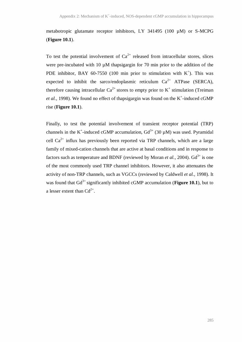
Appendix 2: Mechanism of K+-induced, NOS-dependent cGMP accumulation in hippocampus
285
metabotropic glutamate receptor inhibitors, LY 341495 (100 µM) or S-MCPG
(Figure 10.1).
To test the potential involvement of Ca2+
released from intracellular stores, slices
were pre-incubated with 10 µM thapsigargin for 70 min prior to the addition of the
PDE inhibitor, BAY 60-7550 (100 min prior to stimulation with K+). This was
expected to inhibit the sarco/endoplasmic reticulum Ca2+
ATPase (SERCA),
therefore causing intracellular Ca2+
stores to empty prior to K+ stimulation (Treiman
et al., 1998). We found no effect of thapsigargin was found on the K+-induced cGMP
rise (Figure 10.1).
Finally, to test the potential involvement of transient receptor potential (TRP)
channels in the K+-induced cGMP accumulation, Gd
3+ (30 µM) was used. Pyramidal
cell Ca2+
influx has previously been reported via TRP channels, which are a large
family of mixed-cation channels that are active at basal conditions and in response to
factors such as temperature and BDNF (reviewed by Moran et al., 2004). Gd3+
is one
of the most commonly used TRP channel inhibitors. However, it also attenuates the
activity of non-TRP channels, such as VGCCs (reviewed by Caldwell et al., 1998). It
was found that Gd3+
significantly inhibited cGMP accumulation (Figure 10.1), but to
a lesser extent than Cd2+
.
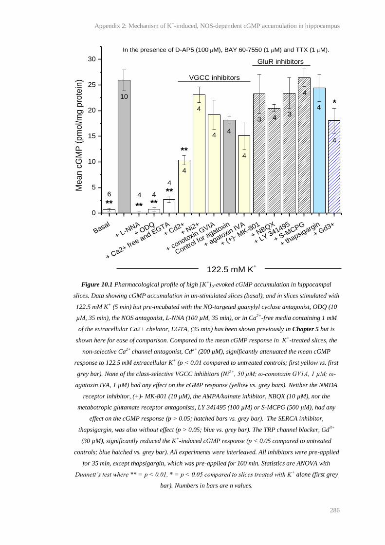
Appendix 2: Mechanism of K+-induced, NOS-dependent cGMP accumulation in hippocampus
286
Figure 10.1 Pharmacological profile of high [K+]o-evoked cGMP accumulation in hippocampal
slices. Data showing cGMP accumulation in un-stimulated slices (basal), and in slices stimulated with
122.5 mM K+ (5 min) but pre-incubated with the NO-targeted guanylyl cyclase antagonist, ODQ (10
µM, 35 min), the NOS antagonist, L-NNA (100 µM, 35 min), or in Ca2+-free media containing 1 mM
of the extracellular Ca2+ chelator, EGTA, (35 min) has been shown previously in Chapter 5 but is
shown here for ease of comparison. Compared to the mean cGMP response in K+-treated slices, the
non-selective Ca2+ channel antagonist, Cd2+ (200 µM), significantly attenuated the mean cGMP
response to 122.5 mM extracellular K+ (p < 0.01 compared to untreated controls; first yellow vs. first
grey bar). None of the class-selective VGCC inhibitors (Ni2+, 50 µM; ω-conotoxin GV1A, 1 µM; ω-
agatoxin IVA, 1 µM) had any effect on the cGMP response (yellow vs. grey bars). Neither the NMDA
receptor inhibitor, (+)- MK-801 (10 µM), the AMPA/kainate inhibitor, NBQX (10 µM), nor the
metabotropic glutamate receptor antagonists, LY 341495 (100 µM) or S-MCPG (500 µM), had any
effect on the cGMP response (p > 0.05; hatched bars vs. grey bar). The SERCA inhibitor,
thapsigargin, was also without effect (p > 0.05; blue vs. grey bar). The TRP channel blocker, Gd3+
(30 µM), significantly reduced the K+-induced cGMP response (p < 0.05 compared to untreated
controls; blue hatched vs. grey bar). All experiments were interleaved. All inhibitors were pre-applied
for 35 min, except thapsigargin, which was pre-applied for 100 min. Statistics are ANOVA with
Dunnett’s test where ** = p < 0.01, * = p < 0.05 compared to slices treated with K+ alone (first grey
bar). Numbers in bars are n values.
Basal
+ L-NNA
+ ODQ
+ Ca2+ fre
e and EGTA
+ Cd2+
+ Ni2+
+ conotoxin GVIA
Control fo
r agatoxin
+ agatoxin IVA
+ (+)- M
K-801
+ NBQX
+ LY 341495
+ S-MCPG
+ thapsigargin
+ Gd3+
0
5
10
15
20
25
30
43
4
4
4
4
4
4
44
GluR inhibitors
*
**
******
Me
an
cG
MP
(p
mo
l/m
g p
rote
in)
122.5 mM K+
**
In the presence of D-AP5 (100 M), BAY 60-7550 (1 M) and TTX (1 M).
VGCC inhibitors
10
4
4
3
4
6
Appendix 2: Mechanism of K+-induced, NOS-dependent cGMP accumulation in hippocampus
287
In summary, the results presented here and in Chapter 5 show that significant
NMDA receptor-independent, nNOS-dependent cGMP accumulation is possible in
the hippocampus and requires a rise in intracellular Ca2+
. This finding is accordant
with a role for NO in NMDA receptor-independent CA1 LTP. Of the inhibitors used
to identify the source of the rise in intracellular Ca2+
responsible for the K+-induced
cGMP accumulation, Cd2+
most potently inhibited the NMDA receptor-independent,
K+-induced cGMP response. However, a residual Cd
2+-insensitive cGMP
accumulation was observed. This might represent incomplete Ca2+
channel inhibition
during depolarisation (Thevenod and Jones, 1992), an inadequate Cd2+
concentration
for complete Ca2+
channel block, or a Cd2+
-induced increase in intracellular Ca2+
caused, for example, by SERCA inhibition and the release of Ca2+
from intracellular
stores (Visser et al., 1993). Previous work in the laboratory has shown that 200 µM
Cd2+
does not inhibit exogenous NO-induced cGMP production in immature
hippocampal slices (Bartus, 2009), ruling out the possibility that the inhibitory effect
we observed was due to a direct effect of Cd2+
on the NO/cGMP pathway, and
against a toxic influence of Cd2+
(Li et al., 2000).
No selective VGCC inhibitor could account for the effect of Cd2+
on the cGMP
response, suggesting that the compound was acting on a Ca2+
channel whose
involvement was not-tested, or a combination of Ca2+
channels activated by the
relatively crude stimulus. Perhaps because Ca2+
is such a ubiquitous signalling
molecule, it is not completely surprising that significant NO-induced cGMP
accumulation can occur in neurons independent of NMDA receptor activation and
that a single source of Ca2+
necessary for the K+-induced, nNOS-cGMP
accumulation could not be isolated in these experiments. Taking into account the
non-physiological nature of the stimulation, the lack of selective pharmacological
tools and the potential complexity of investigating a combinatorial effect of Ca2+
on
NOS, we did not make any further attempts to isolate the source of Ca2+
responsible
for the K+-induced cGMP response.
288
References
Abeliovich A, Schmitz Y, Farinas I, Choi-Lundberg D, Ho WH, Castillo PE, Shinsky N, Verdugo JM,
Armanini M, Ryan A, Hynes M, Phillips H, Sulzer D, Rosenthal A (2000) Mice lacking alpha-
synuclein display functional deficits in the nigrostriatal dopamine system. Neuron 25: 239-252.
Abraham WC (2008) Metaplasticity: tuning synapses and networks for plasticity. Nat. Rev. Neurosci.
9: 387.
Abraham WC, Logan B, Greenwood JM, Dragunow M (2002) Induction and experience-dependent
consolidation of stable long-term potentiation lasting months in the hippocampus. J. Neurosci 22:
9626-9634.
Ahlijanian MK, Westenbroek RE, Catterall WA (1990) Subunit structure and localization of
dihydropyridine-sensitive calcium channels in mammalian brain, spinal cord, and retina. Neuron 4:
819-832.
Alagarsamy S, Lonart G, Johnson KM (1994) The role of P-type calcium channels in the
depolarization-induced activation of nitric oxide synthase in frontal cortex. J. Neurochem. 62: 400-
403.
Alderton WK, Cooper CE, Knowles RG (2001) Nitric oxide synthases: structure, function and
inhibition. Biochem. J. 357: 593-615.
Amaral D, Lavenex P (2007) Hippocampal neuroanatomy. In: The hippocampus book (Andersen P, Morris R, Amaral D, Bliss T.V.P., O'Keefe J, eds), pp 37-114. Oxford: Oxford University Press.
Amaral DG, Witter MP (1989) The three-dimensional organization of the hippocampal formation: A
review of anatomical data. Neuroscience 31: 571-591.
Andersen P, Morris R, Amaral D, Bliss T.V.P., O'Keefe J (2007) Historical perspective: proposed
functions, biological characteristics, and neurobiological models of the hippocampus. In: The
hippocampus book (Andersen P, Morris R, Amaral D, Bliss TVP, O'Keefe J, eds), pp 9-36. Oxford:
Oxford University Press.
Andersen P, Sundberg SH, Sveen O, Wigstrom H (1977) Specific long-lasting potentiation of synaptic
transmission in hippocampal slices. Nature 266: 736-737.
Anekonda TS, Quinn JF, Harris C, Frahler K, Wadsworth TL, Woltjer RL (2011) L-type voltage-
gated calcium channel blockade with isradipine as a therapeutic strategy for Alzheimer's disease. Neurobiol. Dis. 41: 62-70.
Aniksztejn L, Ben-Ari Y (1991) Novel form of long-term potentiation produced by a K+ channel
blocker in the hippocampus. Nature 349: 67-69.
Antonova I, Arancio O, Trillat AC, Wang HG, Zablow L, Udo H, Kandel ER, Hawkins RD (2001)
Rapid increase in clusters of presynaptic proteins at onset of long-lasting potentiation. Science 294:
1547-1550.
Arancio O, Kandel ER, Hawkins RD (1995) Activity-dependent long-term enhancement of transmitter
release by presynaptic 3',5'-cyclic GMP in cultured hippocampal neurons. Nature 376: 74-80.
Arancio O, Antonova I, Gambaryan S, Lohmann SM, Wood JS, Lawrence DS, Hawkins RD (2001)
Presynaptic role of cGMP-dependent protein kinase during long-lasting potentiation. J. Neurosci. 21:
143-149.
289
Arancio O, Kiebler M, Lee CJ, Lev-Ram V, Tsien RY, Kandel ER, Hawkins RD (1996) Nitric oxide
acts directly in the presynaptic neuron to produce long-term potentiation in cultured hippocampal
neurons. Cell 87: 1025-1035.
Arnold WP, Mittal CK, Katsuki S, Murad F (1977) Nitric oxide activates guanylate cyclase and
increases guanosine 3':5'-cyclic monophosphate levels in various tissue preparations. Proc. Natl.
Acad. Sci. U. S. A 74: 3203-3207.
Ashina M, Bendtsen L, Jensen R, Lassen LH, Sakai F, Olesen J (1999a) Possible mechanisms of
action of nitric oxide synthase inhibitors in chronic tension-type headache. Brain 122 ( Pt 9): 1629-
1635.
Ashina M, Lassen LH, Bendtsen L, Jensen R, Olesen J (1999b) Effect of inhibition of nitric oxide
synthase on chronic tension-type headache: a randomised crossover trial. Lancet 353: 287-289.
Babu BR, Griffith OW (1998) N5-(1-Imino-3-butenyl)-L-ornithine. A neuronal isoform selective
mechanism-based inactivator of nitric oxide synthase. J. Biol. Chem. 273: 8882-8889.
Bading H, Ginty DD, Greenberg ME (1993) Regulation of gene expression in hippocampal neurons
by distinct calcium signaling pathways. Science 260: 181-186.
Bannerman DM, Chapman PF, Kelly PA, Butcher SP, Morris RG (1994a) Inhibition of nitric oxide
synthase does not impair spatial learning. J. Neurosci 14: 7404-7414.
Bannerman DM, Chapman PF, Kelly PA, Butcher SP, Morris RG (1994b) Inhibition of nitric oxide
synthase does not prevent the induction of long-term potentiation in vivo. J. Neurosci 14: 7415-7425.
Bannerman DM, Good MA, Butcher SP, Ramsay M, Morris RG (1995) Distinct components of
spatial learning revealed by prior training and NMDA receptor blockade. Nature 378: 182-186.
Barker AJ, Ullian EM (2010) Astrocytes and synaptic plasticity. Neuroscientist 16: 40-50.
Bartus K (2009) Nitric oxide-mediated cGMP signal transduction in the central nervous system. PhD
Thesis, University College London, London, UK.
Batchelor AM, Bartus K, Reynell C, Constantinou S, Halvey EJ, Held KF, Dostmann WR, Vernon J,
Garthwaite J (2010) Exquisite sensitivity to subsecond, picomolar nitric oxide transients conferred on
cells by guanylyl cyclase-coupled receptors. Proc. Natl. Acad. Sci. U. S. A 107: 22060-22065.
Bauer EP, Schafe GE, LeDoux JE (2002) NMDA receptors and L-type voltage-gated calcium
channels contribute to long-term potentiation and different components of fear memory formation in
the lateral amygdala. J. Neurosci. 22: 5239-5249.
Bayazitov IT, Richardson RJ, Fricke RG, Zakharenko SS (2007) Slow presynaptic and fast
postsynaptic components of compound long-term potentiation. J. Neurosci. 27: 11510-11521.
Baylor D (1996) How photons start vision. Proc. Natl. Acad. Sci. U. S. A 93: 560-565.
Behrends S, Kempfert J, Mietens A, Koglin M, Scholz H, Middendorff R (2001) Developmental changes of nitric oxide-sensitive guanylyl cyclase expression in pulmonary arteries. Biochem.
Biophys. Res. Commun. 283: 883-887.
Bekkers JM, Stevens CF (1990) Presynaptic mechanism for long-term potentiation in the
hippocampus. Nature 346: 724-729.
Bellamy TC, Garthwaite J (2002) The receptor-like properties of nitric oxide-activated soluble
guanylyl cyclase in intact cells. Mol. Cell Biochem. 230: 165-176.
290
Bellamy TC, Griffiths C, Garthwaite J (2002) Differential sensitivity of guanylyl cyclase and
mitochondrial respiration to nitric oxide measured using clamped concentrations. J. Biol. Chem. 277:
31801-31807.
Bender AT, Beavo JA (2006) Cyclic nucleotide phosphodiesterases: molecular regulation to clinical
use. Pharmacol. Rev. 58: 488-520.
Bender VA, Pugh JR, Jahr CE (2009) Presynaptically expressed long-term potentiation increases
multivesicular release at parallel fiber synapses. J. Neurosci. 29: 10974-10978.
Bhardwaj A, Northington FJ, Martin LJ, Hanley DF, Traystman RJ, Koehler RC (1997a)
Characterization of metabotropic glutamate receptor-mediated nitric oxide production in vivo. J.
Cereb. Blood Flow Metab. 17: 153-160.
Bhardwaj A, Northington FJ, Ichord RN, Hanley DF, Traystman RJ, Koehler RC (1997b) Characterization of ionotropic glutamate receptor mediated nitric oxide production in vivo in rats.
Stroke 28: 850-857.
Biel M, Wahl-Schott C, Michalakis S, Zong X (2009) Hyperpolarization-activated cation channels:
from genes to function. Physiol Rev. 89: 847-885.
Bird CM, Burgess N (2008) The hippocampus and memory: insights from spatial processing. Nat.
Rev. Neurosci. 9: 182-194.
Blackshaw S, Eliasson MJL, Sawa A, Watkins CC, Krug D, Gupta A, Arai T, Ferrante RJ, Snyder SH
(2003) Species, strain and developmental variations in hippocampal neuronal and endothelial nitric
oxide synthase clarify discrepancies in nitric oxide-dependent synaptic plasticity. Neuroscience 119:
979-990.
Bliss TVP, Collingridge GL, Morris RG (2007) Synaptic plasticity in the hippocampus. In: The hippocampus book. (Andersen P, Morris RG, Amaral D, Bliss T.V.P., O'Keefe J, eds), pp 343-474.
New York: Oxford University Press, Inc.
Bliss TVP, Gardner-Medwin AR (1973) Long-lasting potentiation of synaptic transmission in the
dentate area of the unanaestetized rabbit following stimulation of the perforant path. J. Physiol. 232:
357-374.
Bliss TVP, Lomo T (1973) Long-lasting potentiation of synaptic transmission in the dentate area of
the anaesthatised rabbit following stimulation of the perforant path. J. Physiol. (Lond. ) 232: 331-356.
Bliss TVP, Richter-Levin G (1993) Spatial learning and the saturation of long-term potentiation.
Hippocampus 3: 123-125.
Blokland A, de VJ, Prickaerts J, Honig W, Markerink-Van IM, Steinbusch H (1999) Local inhibition
of hippocampal nitric oxide synthase does not impair place learning in the Morris water escape task in
rats. Eur. J. Neurosci 11: 223-232.
Blundon JA, Zakharenko SS (2008) Dissecting the components of long-term potentiation.
Neuroscientist. 14: 598-608.
Boehning D, Snyder SH (2003) Novel neural modulators. Annu. Rev. Neurosci. 26: 105-131.
Boess FG, Hendrix M, van der Staay FJ, Erb C, Schreiber R, van SW, de VJ, Prickaerts J, Blokland
A, Koenig G (2004) Inhibition of phosphodiesterase 2 increases neuronal cGMP, synaptic plasticity
and memory performance. Neuropharmacology 47: 1081-1092.
Bohme GA, Bon C, Lemaire M, Reibaud M, Piot O, Stutzmann JM, Doble A, Blanchard JC (1993)
Altered synaptic plasticity and memory formation in nitric oxide synthase inhibitor-treated rats. Proc.
Natl. Acad. Sci. U. S. A 90: 9191-9194.
291
Bohme GA, Bon C, Stutzmann JM, Doble A, Blanchard JC (1991) Possible involvement of nitric
oxide in long-term potentiation. Eur. J. Pharmacol. 199: 379-381.
Bon CL, Garthwaite J (2001) Exogenous nitric oxide causes potentiation of hippocampal synaptic
transmission during low-frequency stimulation via the endogenous nitric oxide-cGMP pathway. Eur.
J. Neurosci. 14: 585-594.
Bon CL, Garthwaite J (2003) On the role of nitric oxide in hippocampal long-term potentiation. J.
Neurosci. 23: 1941-1948.
Bonigk W, Altenhofen W, Muller F, Dose A, Illing M, Molday RS, Kaupp UB (1993) Rod and cone
photoreceptor cells express distinct genes for cGMP-gated channels. Neuron 10: 865-877.
Boran MS, Garcia A (2007) The cyclic GMP-protein kinase G pathway regulates cytoskeleton
dynamics and motility in astrocytes. J. Neurochem. 102: 216-230.
Borroni AM, Fichtenholtz II, Woodside BL, Teyler TJ (2000) Role of voltage-dependent calcium
channel long-term potentiation (LTP) and NMDA LTP in spatial memory. J. Neurosci. 20: 9272-
9276.
Boulton CL, Southam E, Garthwaite J (1995) Nitric oxide-dependent long-term potentiation is
blocked by a specific inhibitor of soluble guanylyl cyclase. Neuroscience 69: 699-703.
Bredt DS, Glatt CE, Hwang PM, Fotuhi M, Dawson TM, Snyder SH (1991a) Nitric oxide synthase
protein and mRNA are discretely localized in neuronal populations of the mammalian CNS together
with NADPH diaphorase. Neuron 7: 615-624.
Bredt DS, Hwang PM, Glatt CE, Lowenstein C, Reed RR, Snyder SH (1991b) Cloned and expressed
nitric oxide synthase structurally resembles cytochrome P-450 reductase. Nature 351: 714-718.
Bredt DS, Hwang PM, Snyder SH (1990) Localization of nitric oxide synthase indicating a neural role for nitric oxide. Nature 347: 768-770.
Bredt DS, Snyder SH (1990) Isolation of nitric oxide synthetase, a calmodulin-requiring enzyme.
Proc. Natl. Acad. Sci. U. S. A 87: 682-685.
Brenman JE, Chao DS, Gee SH, McGee AW, Craven SE, Santillano DR, Wu Z, Huang F, Xia H,
Peters MF, Froehner SC, Bredt DS (1996) Interaction of nitric oxide synthase with the postsynaptic
density protein PSD-95 and alpha1-syntrophin mediated by PDZ domains. Cell 84: 757-767.
Brun VH, Ytterbo K, Morris RG, Moser MB, Moser EI (2001) Retrograde amnesia for spatial
memory induced by NMDA receptor-mediated long-term potentiation. J. Neurosci. 21: 356-362.
Brune B, Ullrich V (1987) Inhibition of platelet aggregation by carbon monoxide is mediated by
activation of guanylate cyclase. Mol. Pharmacol. 32: 497-504.
Bucci M, Gratton JP, Rudic RD, Acevedo L, Roviezzo F, Cirino G, Sessa WC (2000) In vivo delivery
of the caveolin-1 scaffolding domain inhibits nitric oxide synthesis and reduces inflammation. Nat. Med. 6: 1362-1367.
Buechler WA, Nakane M, Murad F (1991) Expression of soluble guanylate cyclase activity requires
both enzyme subunits. Biochem. Biophys. Res. Commun. 174: 351-357.
Burette A, Zabel U, Weinberg RJ, Schmidt HH, Valtschanoff JG (2002) Synaptic localization of nitric
oxide synthase and soluble guanylyl cyclase in the hippocampus. J. Neurosci. 22: 8961-8970.
Caldwell RA, Clemo HF, Baumgarten CM (1998) Using gadolinium to identify stretch-activated
channels: technical considerations. Am. J. Physiol. 275: C619-C621.

292
Casamassima F, Hay AC, Benedetti A, Lattanzi L, Cassano GB, Perlis RH (2010) L-type calcium
channels and psychiatric disorders: A brief review. Am. J. Med. Genet. B Neuropsychiatr. Genet.
153B: 1373-1390.
Catterall WA (2000) Structure and regulation of voltage-gated Ca2+ channels. Annu. Rev. Cell Dev.
Biol. 16: 521-555.
Catterall WA, Perez-Reyes E, Snutch TP, Striessnig J (2009) Voltage-gated calcium channels,
introductory chapter. IUPHAR database.
Catterall WA, Perez-Reyes E, Snutch TP, Striessnig J (2005) International Union of Pharmacology.
XLVIII. Nomenclature and structure-function relationships of voltage-gated calcium channels.
Pharmacol. Rev. 57: 411-425.
Catterall WA, Perez-Reyes E, Snutch TP, Striessnig J (2010a) Voltage-Gated Calcium Channels:
Cav1.3. IUPHAR database.
Catterall WA, Perez-Reyes E, Snutch TP, Striessnig J (2010b) Voltage-Gated Calcium
Channels:Cav1.2. IUPHAR database.
Catterall WA, Striessnig J (1992) Receptor sites for Ca2+ channel antagonists. Trends Pharmacol. Sci.
13: 256-262.
Cavus I, Teyler T (1996) Two forms of long-term potentiation in area CA1 activate different signal
transduction cascades. J. Neurophysiol. 76: 3038-3047.
Chan Y, Fish JE, D'Abreo C, Lin S, Robb GB, Teichert AM, Karantzoulis-Fegaras F, Keightley A,
Steer BM, Marsden PA (2004) The cell-specific expression of endothelial nitric-oxide synthase: a role
for DNA methylation. J. Biol. Chem. 279: 35087-35100.
Chapman PF, Atkins CM, Allen MT, Haley JE, Steinmetz JE (1992) Inhibition of nitric oxide synthesis impairs two different forms of learning. NeuroReport 3: 567-570.
Chen C, Regehr WG (1997) The mechanism of cAMP-mediated enhancement at a cerebellar synapse.
J. Neurosci. 17: 8687-8694.
Chen HX, Roper SN (2003) PKA and PKC enhance excitatory synaptic transmission in human
dentate gyrus. J. Neurophysiol. 89: 2482-2488.
Chetkovich DM, Klann E, Sweatt JD (1993) Nitric oxide synthase-independent long-term potentiation
in area CA1 of hippocampus. NeuroReport 4: 919-922.
Chiang LW, Schweizer FE, Tsien RW, Schulman H (1994) Nitric oxide synthase expression in single
hippocampal neurons. Brain Res. Mol Brain Res. 27: 183-188.
Chien WL, Liang KC, Teng CM, Kuo SC, Lee FY, Fu WM (2003) Enhancement of long-term
potentiation by a potent nitric oxide-guanylyl cyclase activator, 3-(5-hydroxymethyl-2-furyl)-1-benzyl-indazole. Mol. Pharmacol. 63: 1322-1328.
Chien WL, Liang KC, Teng CM, Kuo SC, Lee FY, Fu WM (2005) Enhancement of learning
behaviour by a potent nitric oxide-guanylate cyclase activator YC-1. Eur. J. Neurosci. 21: 1679-1688.
Christie BR, Eliot LS, Ito K, Miyakawa H, Johnston D (1995) Different Ca2+
channels in soma and
dendrites of hippocampal pyramidal neurons mediate spike-induced Ca2+ influx. J. Neurophysiol. 73:
2553-2557.
293
Christopherson KS, Hillier BJ, Lim WA, Bredt DS (1999) PSD-95 assembles a ternary complex with
the N-methyl-D-aspartic acid receptor and a bivalent neuronal NO synthase PDZ domain. J. Biol.
Chem. 274: 27467-27473.
Clark NC, Nagano N, Kuenzi FM, Jarolimek W, Huber I, Walter D, Wietzorrek G, Boyce S,
Kullmann DM, Striessnig J, Seabrook GR (2003) Neurological phenotype and synaptic function in
mice lacking the Cav1.3 α subunit of neuronal L-type voltage-dependent Ca2+ channels. Neuroscience
120: 435-442.
Clutton-Brock J (1967) Two cases of poisoning by contamination of nitrous oxide with higher oxides
of nitrogen during anaesthesia. Br. J. Anaesth. 39: 388-392.
Collingridge GL, Kehl SJ, McLennan H (1983a) Excitatory amino acids in synaptic transmission in
the Schaffer collateral-commissural pathway of the rat hippocampus. J. Physiol. 334: 33-46.
Collingridge GL, Kehl SJ, McLennan H (1983b) The antagonism of amino acid-induced excitations of
rat hippocampal CA1 neurones in vitro. J. Physiol. 334: 19-31.
Contestabile A (2000) Roles of NMDA receptor activity and nitric oxide production in brain
development. Brain Res. Rev. 32: 476-509.
Cooke SF, Bliss TVP (2006) Plasticity in the human central nervous system. Brain 129: 1659-1673.
Cooke SF, Wu J, Plattner F, Errington M, Rowan M, Peters M, Hirano A, Bradshaw KD, Anwyl R,
Bliss TVP, Giese KP (2006) Autophosphorylation of αCaMKII is not a general requirement for
NMDA receptor-dependent LTP in the adult mouse. J. Physiol. 574: 805-818.
Corbin JD, Turko IV, Beasley A, Francis SH (2000) Phosphorylation of phosphodiesterase-5 by
cyclic nucleotide-dependent protein kinase alters its catalytic and allosteric cGMP-binding activities.
Eur. J. Biochem. 267: 2760-2767.
Corkin S (2002) What's new with the amnesic patient H.M.? Nat. Rev. Neuro. 3: 153-160.
Craven KB, Zagotta WN (2006) CNG and HCN channels: two peas, one pod. Annu. Rev. Physiol 68:
375-401.
Craven PA, DeRubertis FR (1978) Restoration of the responsiveness of purified guanylate cyclase to
nitrosoguanidine, nitric oxide, and related activators by heme and hemeproteins. Evidence for
involvement of the paramagnetic nitrosyl-heme complex in enzyme activation. J. Biol. Chem. 253:
8433-8443.
Creager R, Dunwiddie T, Lynch G (1980) Paired-pulse and frequency facilitation in the CA1 region
of the in vitro rat hippocampus. J. Physiol. 299: 409-424.
Cserep C, Szonyi A, Veres JM, Nemeth B, Szabadits E, de VJ, Hajos N, Freund TF, Nyiri G (2011)
Nitric oxide signaling modulates synaptic transmission during early postnatal development. Cereb.
Cortex. 9: 2065-2074.
Cukkemane A, Seifert R, Kaupp UB (2011) Cooperative and uncooperative cyclic-nucleotide-gated
ion channels. Trends Biochem. Sci. 36: 55-64.
Cummings JA, Nicola SM, Malenka RC (1994) Induction in the rat hippocampus of long-term
potentiation (LTP) and long-term depression (LTD) in the presence of a nitric oxide synthase
inhibitor. Neurosci. Lett. 176: 110-114.
Daff S (2010) NO synthase: structures and mechanisms. Nitric Oxide 23: 1-11.
294
Daff S, Sagami I, Shimizu T (1999) The 42-amino acid insert in the FMN domain of neuronal nitric-
oxide synthase exerts control over Ca2+/calmodulin-dependent electron transfer. J. Biol. Chem. 274:
30589-30595.
Davies SN, Collingridge GL (1989) Role of excitatory amino acid receptors in synaptic transmission
in area CA1 of rat hippocampus. Proc. R. Soc. Lond B Biol. Sci. 236: 373-384.
Davy H (1800) Researches clinical and philosophical, chiefly concerning nitroous oxide or
dephlogisticated nitrous air. Volume published for J Johnson, London by Biggs and Cottle, Bristol.
Dawson TM, Snyder SH (1994) Gases as biological messengers: nitric oxide and carbon monoxide in
the brain. J. Neurosci. 14: 5147-5159.
De Seranno S, Estrella C, Loyens A, Cornea A, Ojeda SR, Beauvillain JC, Prevot V (2004) Vascular
endothelial cells promote acute plasticity in ependymoglial cells of the neuroendocrine brain. J. Neurosci. 24: 10353-10363.
de Vente J, Asan E, Gambaryan S, Markerink-Van IM, Axer H, Gallatz K, Lohmann SM, Palkovits M
(2001) Localization of cGMP-dependent protein kinase type II in rat brain. Neuroscience 108: 27-49.
de Vente J, Hopkins DA, Markerink-Van IM, Emson PC, Schmidt HH, Steinbusch HW (1998)
Distribution of nitric oxide synthase and nitric oxide-receptive, cyclic GMP-producing structures in
the rat brain. Neuroscience 87: 207-241.
Debanne D, Gahwiler BH, Thompson SM (1999) Heterogeneity of synaptic plasticity at unitary CA3-
CA1 and CA3-CA3 connections in rat hippocampal slice cultures. J. Neurosci. 19: 10664-10671.
Dedio J, Konig P, Wohlfart P, Schroeder C, Kummer W, Muller-Esterl W (2001) NOSIP, a novel
modulator of endothelial nitric oxide synthase activity. FASEB J. 15: 79-89.
Degerman E, Belfrage P, Manganiello VC (1997) Structure, localization, and regulation of cGMP-inhibited phosphodiesterase (PDE3). J. Biol. Chem. 272: 6823-6826.
Deguchi T, Yoshioka M (1982) L-Arginine identified as an endogenous activator for soluble
guanylate cyclase from neuroblastoma cells. J. Biol. Chem. 257: 10147-10151.
Demas GE, Kriegsfeld LJ, Blackshaw S, Huang P, Gammie SC, Nelson RJ, Snyder SH (1999)
Elimination of aggressive behavior in male mice lacking endothelial nitric oxide synthase. J.
Neurosci. 19: RC30.
Detre JA, Nairn AC, Aswad DW, Greengard P (1984) Localization in mammalian brain of G-
substrate, a specific substrate for guanosine 3',5'-cyclic monophosphate-dependent protein kinase. J.
Neurosci. 4: 2843-2849.
Dhallan RS, Yau KW, Schrader KA, Reed RR (1990) Primary structure and functional expression of a
cyclic nucleotide-activated channel from olfactory neurons. Nature 347: 184-187.
Dinerman JL, Dawson TM, Schell MJ, Snowman A, Snyder SH (1994) Endothelial nitric oxide synthase localized to hippocampal pyramidal cells: implications for synaptic plasticity. Proc. Natl.
Acad. Sci. U. S. A 91: 4214-4218.
Ding JD, Burette A, Nedvetsky PI, Schmidt HH, Weinberg RJ (2004) Distribution of soluble guanylyl
cyclase in the rat brain. J. Comp. Neurol. 472: 437-448.
Dityatev AE, Bolshakov VY (2005) Amygdala, long-term potentiation, and fear conditioning.
Neuroscientist 11: 75-88.
Dolphin AC, Errington ML, Bliss TV (1982) Long-term potentiation of the perforant path in vivo is
associated with increased glutamate release. Nature 297: 496-498.

295
Doreulee N, Brown RE, Yanovsky Y, Godecke A, Schrader J, Haas HL (2001) Defective
hippocampal mossy fiber long-term potentiation in endothelial nitric oxide synthase knockout mice.
Synapse 41: 191-194.
Doreulee N, Sergeeva OA, Yanovsky Y, Chepkova AN, Selbach O, Godecke A, Schrader J, Haas HL
(2003) Cortico-striatal synaptic plasticity in endothelial nitric oxide synthase deficient mice. Brain
Res. 964: 159-163.
Doyle CA, Slater P (1997) Localization of neuronal and endothelial nitric oxide synthase isoforms in
human hippocampus. Neuroscience 76: 387-395.
Drab M, Verkade P, Elger M, Kasper M, Lohn M, Lauterbach B, Menne J, Lindschau C, Mende F,
Luft FC, Schedl A, Haller H, Kurzchalia TV (2001) Loss of caveolae, vascular dysfunction, and
pulmonary defects in caveolin-1 gene-disrupted mice. Science 293: 2449-2452.
Dreyer J, Schleicher M, Tappe A, Schilling K, Kuner T, Kusumawidijaja G, Muller-Esterl W, Oess S,
Kuner R (2004) Nitric oxide synthase (NOS)-interacting protein interacts with neuronal NOS and
regulates its distribution and activity. J. Neurosci. 24: 10454-10465.
Dudzinski DM, Igarashi J, Greif D, Michel T (2006) The regulation and pharmacology of endothelial
nitric oxide synthase. Annu. Rev. Pharmacol. Toxicol. 46: 235-276.
Dumas TC, Foster TC (1998) Late developmental changes in the ability of adenosine A1 receptors to
regulate synaptic transmission in the hippocampus. Brain Res. Dev. Brain Res. 105: 137-139.
Dun NJ, Dun SL, Wong RK, Forstermann U (1994) Colocalization of nitric oxide synthase and
somatostatin immunoreactivity in rat dentate hilar neurons. Proc. Natl. Acad. Sci. U. S. A 91: 2955-
2959.
Dunwiddie TV, Haas HL (1985) Adenosine increases synaptic facilitation in the in vitro rat hippocampus: evidence for a presynaptic site of action. J. Physiol. 369: 365-377.
East SJ, Garthwaite J (1991) NMDA receptor activation in rat hippocampus induces cyclic GMP
formation through the L-arginine-nitric oxide pathway. Neurosci. Lett. 123: 17-19.
el-Husseini AE, Bladen C, Vincent SR (1995) Molecular characterization of a type II cyclic GMP-
dependent protein kinase expressed in the rat brain. J. Neurochem. 64: 2814-2817.
Eliasson MJ, Blackshaw S, Schell MJ, Snyder SH (1997) Neuronal nitric oxide synthase alternatively
spliced forms: prominent functional localizations in the brain. Proc. Natl. Acad. Sci. U. S. A 94: 3396-
3401.
Enoki R, Hu YL, Hamilton D, Fine A (2009) Expression of long-term plasticity at individual synapses
in hippocampus is graded, bidirectional, and mainly presynaptic: optical quantal analysis. Neuron 62:
242-253.
Erusalimsky JD, Moncada S (2007) Nitric oxide and mitochondrial signaling: from physiology to pathophysiology. Arterioscler. Thromb. Vasc. Biol. 27: 2524-2531.
Fang J, Silverman RB (2009) A cellular model for screening neuronal nitric oxide synthase inhibitors.
Anal. Biochem. 390: 74-78.
Faraci FM (2006) Protecting the brain with eNOS: run for your life. Circ. Res. 99: 1029-1030.
Feelisch M (1998) The use of nitric oxide donors in pharmacological studies. Naunyn Schmiedebergs
Arch. Pharmacol. 358: 113-122.
Feil R, Hofmann F, Kleppisch T (2005a) Function of cGMP-dependent protein kinases in the nervous
system. Rev. Neurosci. 16: 23-41.
296
Feil R, Kleppisch T (2008) NO/cGMP-dependent modulation of synaptic transmission. Handb. Exp.
Pharmacol. 184: 529-560.
Feil S, Zimmermann P, Knorn A, Brummer S, Schlossmann J, Hofmann F, Feil R (2005b)
Distribution of cGMP-dependent protein kinase type I and its isoforms in the mouse brain and retina.
Neuroscience 135: 863-868.
Feldman D (2009) Synaptic mechanisms for plasticity in neocortex. Annu. Rev Neurosci. 32: 33-55.
Feron O, Saldana F, Michel JB, Michel T (1998) The endothelial nitric-oxide synthase-caveolin
regulatory cycle. J. Biol. Chem. 273: 3125-3128.
Ferrendelli JA, Chang MM, Kinscherf DA (1974) Elevation of cyclic GMP levels in central nervous
system by excitatory and inhibitory amino acids. J. Neurochem. 22: 535-540.
Ferrendelli JA, Kinscherf DA, Chang MM (1973) Regulation of levels of guanosine cyclic 3',5'-monophosphate in the central nervous system: effects of depolarizing agents. Mol. Pharmacol 9: 445-
454.
Ferrendelli JA, Rubin EH, Kinscherf DA (1976) Influence of divalent cations on regulation of cyclic
GMP and cyclic AMP levels in brain tissue. J. Neurochem. 26: 741-748.
Ferrero R, Rodriguez-Pascual F, Miras-Portugal MT, Torres M (2000) Nitric oxide-sensitive guanylyl
cyclase activity inhibition through cyclic GMP-dependent dephosphorylation. J. Neurochem. 75:
2029-2039.
Fesenko EE, Kolesnikov SS, Lyubarsky AL (1985) Induction by cyclic GMP of cationic conductance
in plasma membrane of retinal rod outer segment. Nature 313: 310-313.
Fischmeister R, Castro L, Abi-Gerges A, Rochais F, Vandecasteele G (2005) Species- and tissue-
dependent effects of NO and cyclic GMP on cardiac ion channels. Comp. Biochem. Physiol A Mol. Integr. Physiol. 142: 136-143.
Foerster J, Harteneck C, Malkewitz J, Schultz G, Koesling D (1996) A functional heme-binding site
of soluble guanylyl cyclase requires intact N-termini of alpha 1 and beta 1 subunits. Eur. J. Biochem.
240: 380-386.
Fontinha BM, Delgado-Garcia JM, Madronal N, Ribeiro JA, Sebastiao AM, Gruart A (2009)
Adenosine A(2A) receptor modulation of hippocampal CA3-CA1 synapse plasticity during
associative learning in behaving mice. Neuropsychopharmacology 34: 1865-1874.
Francis SH, Busch JL, Corbin JD, Sibley D (2010) cGMP-dependent protein kinases and cGMP
phosphodiesterases in nitric oxide and cGMP action. Pharmacol. Rev. 62: 525-563.
Freund TF, Buzsaki G (1996) Interneurons of the hippocampus. Hippocampus 6: 347-470.
Friebe A, Mergia E, Dangel O, Lange A, Koesling D (2007) Fatal gastrointestinal obstruction and
hypertension in mice lacking nitric oxide-sensitive guanylyl cyclase. Proc. Natl. Acad. Sci. U. S. A 104: 7699-7704.
Fuenzalida M, Fernandez de SD, Couve A, Buno W (2010) Role of AMPA and NMDA receptors and
back-propagating action potentials in spike timing-dependent plasticity. J. Neurophysiol. 103: 47-54.
Fukaya M, Watanabe M (2000) Improved immunohistochemical detection of postsynaptically located
PSD-95/SAP90 protein family by protease section pretreatment: a study in the adult mouse brain. J.
Comp. Neurol. 426: 572-586.
Fukutani T, Iino S, Nojyo Y (2009) The expression of soluble guanylate cyclase in the vasculature of
rat skeletal muscle. Arch. Histol. Cytol. 72: 117-126.
297
Fulton D, Gratton JP, Sessa WC (2001) Post-translational control of endothelial nitric oxide synthase:
why isn't calcium/calmodulin enough? J. Pharmacol. Exp. Ther. 299: 818-824.
Furchgott RF, Martin W (1985) Interactions of endothelial cells and smooth muscle cells of arteries.
Chest 88: 210S-213S.
Furchgott RF, Zawadzki JV (1980) The obligatory role of endothelial cells in the relaxation of arterial
smooth muscle by acetylcholine. Nature 288: 373-376.
Furfine ES, Harmon MF, Paith JE, Garvey EP (1993) Selective inhibition of constitutive nitric oxide
synthase by L-NG-nitroarginine. Biochemistry 32: 8512-8517.
Galle J, Zabel U, Hubner U, Hatzelmann A, Wagner B, Wanner C, Schmidt HH (1999) Effects of the
soluble guanylyl cyclase activator, YC-1, on vascular tone, cyclic GMP levels and phosphodiesterase
activity. Br. J. Pharmacol. 127: 195-203.
Gamm DM, Francis SH, Angelotti TP, Corbin JD, Uhler MD (1995) The type II isoform of cGMP-
dependent protein kinase is dimeric and possesses regulatory and catalytic properties distinct from the
type I isoforms. J. Biol. Chem. 270: 27380-27388.
Garcia-Cardena G, Fan R, Shah V, Sorrentino R, Cirino G, Papapetropoulos A, Sessa WC (1998)
Dynamic activation of endothelial nitric oxide synthase by Hsp90. Nature 392: 821-824.
Garcia-Cardena G, Martasek P, Masters BS, Skidd PM, Couet J, Li S, Lisanti MP, Sessa WC (1997)
Dissecting the interaction between nitric oxide synthase (NOS) and caveolin. Functional significance
of the nos caveolin binding domain in vivo. J. Biol. Chem. 272: 25437-25440.
Garcia-Cardena G, Oh P, Liu J, Schnitzer JE, Sessa WC (1996) Targeting of nitric oxide synthase to
endothelial cell caveolae via palmitoylation: implications for nitric oxide signaling. Proc. Natl. Acad.
Sci. U. S. A 93: 6448-6453.
Garthwaite G, Bartus K, Malcolm D, Goodwin D, Kollb-Sielecka M, Dooldeniya C, Garthwaite J
(2006) Signaling from blood vessels to CNS axons through nitric oxide. J. Neurosci. 26: 7730-7740.
Garthwaite J (1985) Cellular uptake disguises action of L-glutamate on N-methyl-D-aspartate
receptors. With an appendix: diffusion of transported amino acids into brain slices. Br. J. Pharmacol.
85: 297-307.
Garthwaite J (2005) Dynamics of cellular NO-cGMP signaling. Front Biosci. 10: 1868-1880.
Garthwaite J (2007) Neuronal nitric oxide synthase and the serotonin transporter get harmonious.
Proc. Natl. Acad. Sci. U. S. A 104: 7739-7740.
Garthwaite J (2008) Concepts of neural nitric oxide-mediated transmission. Eur. J. Neurosci. 11:
2783-2802.
Garthwaite J, Charles SL, Chess Williams R (1988) Endothelium-derived relaxing factor release on
activation of NMDA receptors suggests role as intercellular messenger in the brain. Nature 336: 385-388.
Garthwaite J, Garthwaite G (1987) Cellular origins of cyclic GMP responses to excitatory amino acid
receptor agonists in rat cerebellum in vitro. J. Neurochem. 48: 29-39.
Garthwaite J, Garthwaite G, Palmer RM, Moncada S (1989) NMDA receptor activation induces nitric
oxide synthesis from arginine in rat brain slices. Eur. J. Pharmacol. 172: 413-416.
Garthwaite J, Southam E, Boulton CL, Nielsen EB, Schmidt K, Mayer B (1995) Potent and selective
inhibition of nitric oxide-sensitive guanylyl cyclase by 1H-[1,2,4]oxadiazolo[4,3-a]quinoxalin-1-one.
Mol. Pharmacol. 48: 184-188.
298
Garvey EP, Oplinger JA, Furfine ES, Kiff RJ, Laszlo F, Whittle BJ, Knowles RG (1997) 1400W is a
slow, tight binding, and highly selective inhibitor of inducible nitric-oxide synthase in vitro and in
vivo. J. Biol. Chem. 272: 4959-4963.
Garvey EP, Tuttle JV, Covington K, Merrill BM, Wood ER, Baylis SA, Charles IG (1994)
Purification and characterization of the constitutive nitric oxide synthase from human placenta. Arch.
Biochem. Biophys. 311: 235-241.
Germann WJ, Stanfield CL (2002) Principles of human physiology. Pearson Education Inc., San
Fransico, CA.
Gerzer R, Bohme E, Hofmann F, Schultz G (1981) Soluble guanylate cyclase purified from bovine
lung contains heme and copper. FEBS Lett. 132: 71-74.
Gibb BJ, Garthwaite J (2001) Subunits of the nitric oxide receptor, soluble guanylyl cyclase, expressed in rat brain. Eur. J. Neurosci 13: 539-544.
Gibb BJ, Wykes V, Garthwaite J (2003) Properties of NO-activated guanylyl cyclases expressed in
cells. Br. J. Pharmacol. 139: 1032-1040.
Giese KP, Fedorov NB, Filipkowski RK, Silva AJ (1998) Autophosphorylation at Thr286 of the
αCalcium-Calmodulin Kinase II in LTP and Learning. Science 279: 870-873.
Golderer G, Werner ER, Leitner S, Grobner P, Werner-Felmayer G (2001) Nitric oxide synthase is
induced in sporulation of Physarum polycephalum. Genes Dev. 15: 1299-1309.
Gonzalez-Hernandez T, Perez de la Cruz MA, Mantolan-Sarmiento B (1996) Histochemical and
immunohistochemical detection of neurons that produce nitric oxide: effect of different fixative
parameters and immunoreactivity against non-neuronal NOS antisera. J. Histochem. Cytochem. 44:
1399-1413.
Goosens KA, Maren S (2001) Contextual and auditory fear conditioning are mediated by the lateral,
basal, and central amygdaloid nuclei in rats. Learn. Mem. 8: 148-155.
Gowda C, Toomayan GA, Qi WN, Chen LE, Cai Y, Allen DM, Seaber AV, Urbaniak JR (2004) The
effects of NΩ-propyl-L-arginine on reperfusion injury of skeletal muscle. Nitric Oxide 11: 17-24.
Gratton JP, Fontana J, O'Connor DS, Garcia-Cardena G, McCabe TJ, Sessa WC (2000) Reconstitution
of an endothelial nitric-oxide synthase (eNOS), hsp90, and caveolin-1 complex in vitro. Evidence that
hsp90 facilitates calmodulin stimulated displacement of eNOS from caveolin-1. J. Biol. Chem. 275:
22268-22272.
Green LC, Ruiz de LK, Wagner DA, Rand W, Istfan N, Young VR, Tannenbaum SR (1981a) Nitrate
biosynthesis in man. Proc. Natl. Acad. Sci. U. S. A 78: 7764-7768.
Green LC, Tannenbaum SR, Goldman P (1981b) Nitrate synthesis in the germfree and conventional
rat. Science 212: 56-58.
Gross SS, Wolin MS (1995) Nitric oxide: pathophysiological mechanisms. Annu. Rev. Physiol. 57:
737-769.
Grover LM (1998) Evidence for postsynaptic induction and expression of NMDA receptor
independent LTP. J. Neurophysiol. 79: 1167-1182.
Grover LM, Teyler TJ (1990) Two components of long-term potentiation induced by different patterns
of afferent activation. Nature 347: 477-479.
299
Grover LM, Teyler TJ (1992) N-methyl-d-aspartate receptor-independent long-term potentiation in
area CA1 of rat hippocampus: Input-specific induction and preclusion in a non-tetanized pathway.
Neuroscience 49: 7-11.
Grover LM, Teyler TJ (1994) Activation of NMDA receptors in hippocampal area CA1 by low and
high frequency orthodromic stimulation and their contribution to induction of long-term potentiation.
Synapse 16: 66-75.
Gruart A, Munoz MD, Delgado-Garcia JM (2006) Involvement of the CA3-CA1 synapse in the
acquisition of associative learning in behaving mice. J. Neurosci. 26: 1077-1087.
Gruart A, Sciarretta C, Valenzuela-Harrington M, Delgado-Garcia JM, Minichiello L (2007) Mutation
at the TrkB PLCγ-docking site affects hippocampal LTP and associative learning in conscious mice.
Learn. Mem. 14: 54-62.
Gruetter CA, Barry BK, McNamara DB, Kadowitz PJ, Ignarro LJ (1980a) Coronary arterial relaxation
and guanylate cyclase activation by cigarette smoke, N'-nitrosonornicotine and nitric oxide. J.
Pharmacol. Exp. Ther. 214: 9-15.
Gruetter DY, Gruetter CA, Barry BK, Baricos WH, Hyman AL, Kadowitz PJ, Ignarro LJ (1980b)
Activation of coronary arterial guanylate cyclase by nitric oxide, nitroprusside, and nitrosoguanidine--
inhibition by calcium, lanthanum, and other cations, enhancement by thiols. Biochem. Pharmacol. 29:
2943-2950.
Gupta G, Azam M, Yang L, Danziger RS (1997) The beta2 subunit inhibits stimulation of the
alpha1/beta1 form of soluble guanylyl cyclase by nitric oxide. Potential relevance to regulation of
blood pressure. J. Clin. Invest 100: 1488-1492.
Hada J, Kaku T, Jiang MH, Morimoto K, Hayashi Y (2003) Inhibition of high K+-evoked gamma-aminobutyric acid release by sodium nitroprusside in rat hippocampus. Eur. J. Pharmacol. 467: 119-
123.
Haghikia A, Mergia E, Friebe A, Eysel UT, Koesling D, Mittmann T (2007) Long-term potentiation
in the visual cortex requires both nitric oxide receptor guanylyl cyclases. J. Neurosci. 27: 818-823.
Haley JE, Malen PL, Chapman PF (1993) Nitric oxide synthase inhibitors block long-term
potentiation induced by weak but not strong tetanic stimulation at physiological brain temperatures in
rat hippocampal slices. Neurosci. Lett. 160: 85-88.
Hall CN, Attwell D (2008) Assessing the physiological concentration and targets of nitric oxide in
brain tissue. J. Physiol. 586: 3597-3615.
Hall CN, Garthwaite J (2006) Inactivation of nitric oxide by rat cerebellar slices. J. Physiol. 577: 549-
567.
Hall CN, Garthwaite J (2009) What is the real physiological NO concentration in vivo? Nitric Oxide 21: 92-103.
Hansel TT, Kharitonov SA, Donnelly LE, Erin EM, Currie MG, Moore WM, Manning PT, Recker
DP, Barnes PJ (2003) A selective inhibitor of inducible nitric oxide synthase inhibits exhaled breath
nitric oxide in healthy volunteers and asthmatics. FASEB J. 17: 1298-1300.
Hardingham N, Fox K (2006) The role of nitric oxide and GluR1 in presynaptic and postsynaptic
components of neocortical potentiation. J. Neurosci. 26: 7395-7404.
Harteneck C, Wedel B, Koesling D, Malkewitz J, Bohme E, Schultz G (1991) Molecular cloning and
expression of a new alpha-subunit of soluble guanylyl cyclase. Interchangeability of the alpha-
subunits of the enzyme. FEBS Lett. 292: 217-222.
300
Haul S, Godecke A, Schrader J, Haas HL, Luhmann HJ (1999) Impairment of neocortical long-term
potentiation in mice deficient of endothelial nitric oxide synthase. J. Neurophysiol. 81: 494-497.
Hawkins RD, Zhuo M, Arancio O (1994) Nitric oxide and carbon monoxide as possible retrograde
messengers in hippocampal long-term potentiation. J. Neurobiol. 25: 652-665.
Hayes J, Li S, Anwyl R, Rowan MJ (2008) A role for protein kinase A and protein kinase M zeta in
muscarinic acetylcholine receptor-initiated persistent synaptic enhancement in rat hippocampus in
vivo. Neuroscience 151: 604-612.
Hebb DO (1949) The organisation of behaviour. New York: Wiley.
Hegesh E, Shiloah J (1982) Blood nitrates and infantile methemoglobinemia. Clin. Chim. Acta 125:
107-115.
Hell JW, Westenbroek RE, Breeze LJ, Wang KK, Chavkin C, Catterall WA (1996) N-methyl-D-aspartate receptor-induced proteolytic conversion of postsynaptic class C L-type calcium channels in
hippocampal neurons. Proc. Natl. Acad. Sci. U. S. A 93: 3362-3367.
Hell JW, Westenbroek RE, Warner C, Ahlijanian MK, Prystay W, Gilbert MM, Snutch TP, Catterall
WA (1993) Identification and differential subcellular localization of the neuronal class C and class D
L-type calcium channel alpha 1 subunits. J. Cell Biol. 123: 949-962.
Helton TD, Xu W, Lipscombe D (2005) Neuronal L-type calcium channels open quickly and are
inhibited slowly. J. Neurosci. 25: 10247-10251.
Hibbs JB, Jr., Taintor RR, Vavrin Z, Rachlin EM (1988) Nitric oxide: a cytotoxic activated
macrophage effector molecule. Biochem. Biophys. Res. Commun. 157: 87-94.
Hibbs JB, Jr., Vavrin Z, Taintor RR (1987) L-arginine is required for expression of the activated
macrophage effector mechanism causing selective metabolic inhibition in target cells. J. Immunol. 138: 550-565.
Hobbs AJ, Higgs A, Moncada S (1999) Inhibition of nitric oxide synthase as a potential therapeutic
target. Annu. Rev. Pharmacol. Toxicol. 39: 191-220.
Hockerman GH, Peterson BZ, Johnson BD, Catterall WA (1997) Molecular determinants of drug
binding and action on L-type calcium channels. Annu. Rev. Pharmacol. Toxicol. 37: 361-396.
Holscher C (2002) Different strains of rats show different sensitivity to block of long-term
potentiation by nitric oxide synthase inhibitors. Eur. J. Pharmacol 457: 99-106.
Hopper R, Lancaster B, Garthwaite J (2004) On the regulation of NMDA receptors by nitric oxide.
Eur. J. Neurosci. 19: 1675-1682.
Hopper RA, Garthwaite J (2006) Tonic and phasic nitric oxide signals in hippocampal long-term
potentiation. J. Neurosci. 26: 11513-11521.
Huang PL, Dawson TM, Bredt DS, Snyder SH, Fishman MC (1993) Targeted disruption of the neuronal nitric oxide synthase gene. Cell 75: 1273-1286.
Huang PL, Huang Z, Mashimo H, Bloch KD, Moskowitz MA, Bevan JA, Fishman MC (1995)
Hypertension in mice lacking the gene for endothelial nitric oxide synthase. Nature 377: 239-242.
Huang YY, Malenka RC (1993) Examination of TEA-induced synaptic enhancement in area CA1 of
the hippocampus: the role of voltage-dependent Ca2+ channels in the induction of LTP. J. Neurosci.
13: 568-576.
301
Huang Z, Huang PL, Ma J, Meng W, Ayata C, Fishman MC, Moskowitz MA (1996) Enlarged infarcts
in endothelial nitric oxide synthase knockout mice are attenuated by nitro-L-arginine. J. Cereb. Blood
Flow Metab. 16: 981-987.
Huang Z, Huang PL, Panahian N, Dalkara T, Fishman MC, Moskowitz MA (1994) Effects of cerebral
ischemia in mice deficient in neuronal nitric oxide synthase. Science 265:1883-1885.
Ignarro LJ, Buga GM, Wood KS, Byrns RE, Chaudhuri G (1987) Endothelium-derived relaxing factor
produced and released from artery and vein is nitric oxide. Proc. Natl. Acad. Sci. U. S. A 84: 9265-
9269.
Ignarro LJ, Lippton H, Edwards JC, Baricos WH, Hyman AL, Kadowitz PJ, Gruetter CA (1981)
Mechanism of vascular smooth muscle relaxation by organic nitrates, nitrites, nitroprusside and nitric oxide: evidence for the involvement of S-nitrosothiols as active intermediates. J. Pharmacol. Exp.
Ther. 218: 739-749.
Impey S, Mark M, Villacres EC, Poser S, Chavkin C, Storm DR (1996) Induction of CRE-mediated
gene expression by stimuli that generate long-lasting LTP in area CA1 of the hippocampus. Neuron
16: 973-982.
Irvine EE, Vernon J, Giese KP (2005) AlphaCaMKII autophosphorylation contributes to rapid
learning but is not necessary for memory. Nat. Neurosci. 8: 411-412.
Ito M (2001) Cerebellar long-term depression: characterization, signal transduction, and functional
roles. Physiol. Rev. 81: 1143-1195.
Iyengar R, Stuehr DJ, Marletta MA (1987) Macrophage synthesis of nitrite, nitrate, and N-
nitrosamines: precursors and role of the respiratory burst. Proc. Natl. Acad. Sci. U. S. A 84: 6369-6373.
Jacoby S, Sims RE, Hartell NA (2001) Nitric oxide is required for the induction and heterosynaptic
spread of long-term potentiation in rat cerebellar slices. J. Physiol. 535: 825-839.
Jaffrey SR, Benfenati F, Snowman AM, Czernik AJ, Snyder SH (2002) Neuronal nitric-oxide
synthase localization mediated by a ternary complex with synapsin and CAPON. Proc. Natl. Acad.
Sci. U. S. A 99: 3199-3204.
Jaffrey SR, Snowman AM, Eliasson MJ, Cohen NA, Snyder SH (1998) CAPON: a protein associated
with neuronal nitric oxide synthase that regulates its interactions with PSD95. Neuron 20: 115-124.
Jarrard LE (1993) On the role of the hippocampus in learning and memory in the rat. Behav. Neural
Biol. 60: 9-26.
Jarry A, Renaudin K, Denis MG, Robard M, Buffin-Meyer B, Karam G, Buzelin F, Paris H, Laboisse
CL, Vallette G (2003) Expression of NOS1 and soluble guanylyl cyclase by human kidney epithelial cells: morphological evidence for an autocrine/paracrine action of nitric oxide. Kidney Int. 64: 170-
180.
Ji H, Li H, Martasek P, Roman LJ, Poulos TL, Silverman RB (2009) Discovery of highly potent and
selective inhibitors of neuronal nitric oxide synthase by fragment hopping. J. Med. Chem. 52: 779-
797.
Ji RR, Kohno T, Moore KA, Woolf CJ (2003) Central sensitization and LTP: do pain and memory
share similar mechanisms? Trends Neurosci. 26: 696-705.
Kahle JS, Cotman CW (1993) Adenosine, L-AP4, and baclofen modulation of paired-pulse
potentiation in the dentate gyrus: interstimulus interval-dependent pharmacology. Exp. Brain Res. 94:
97-104.
302
Kamisaki Y, Saheki S, Nakane M, Palmieri JA, Kuno T, Chang BY, Waldman SA, Murad F (1986)
Soluble guanylate cyclase from rat lung exists as a heterodimer. J. Biol. Chem. 261: 7236-7241.
Kandel ER, O'Dell TJ (1992) Are adult learning mechanisms also used for development? Science 258:
243-245.
Kantor DB, Lanzrein M, Stary SJ, Sandoval GM, Smith WB, Sullivan BM, Davidson N, Schuman
EM (1996) A role for endothelial NO synthase in LTP revealed by adenovirus-mediated inhibition
and rescue. Science 274: 1744-1748.
Kato K, Clifford DB, Zorumski CF (1993) Long-term potentiation during whole-cell recording in rat
hippocampal slices. Neuroscience 53: 39-47.
Katsuki S, Arnold W, Mittal C, Murad F (1977a) Stimulation of guanylate cyclase by sodium
nitroprusside, nitroglycerin and nitric oxide in various tissue preparations and comparison to the effects of sodium azide and hydroxylamine. J. Cyclic. Nucleotide. Res. 3: 23-35.
Katsuki S, Arnold WP, Murad F (1977b) Effects of sodium nitroprusside, nitroglycerin, and sodium
azide on levels of cyclic nucleotides and mechanical activity of various tissues. J. Cyclic. Nucleotide.
Res. 3: 239-247.
Katzoff A, Ben-Gedalya T, Susswein AJ (2002) Nitric oxide is necessary for multiple memory
processes after learning that a food is inedible in aplysia. J. Neurosci. 22: 9581-9594.
Kaupp UB, Seifert R (2002) Cyclic nucleotide-gated ion channels. Physiol. Rev. 82: 769-824.
Kazerounian S, Pitari GM, Ruiz-Stewart I, Schulz S, Waldman SA (2002) Nitric oxide activation of
soluble guanylyl cyclase reveals high and low affinity sites that mediate allosteric inhibition by
calcium. Biochemistry 41: 3396-3404.
Keefer LK, Nims RW, Davies KM, Wink DA (1996) "NONOates" (1-substituted diazen-1-ium-1,2-diolates) as nitric oxide donors: convenient nitric oxide dosage forms. Methods Enzymol. 268: 281-
293.
Keilhoff G, Seidel B, Noack H, Tischmeyer W, Stanek D, Wolf G (1996) Patterns of nitric oxide
synthase at the messenger RNA and protein levels during early rat brain development. Neuroscience
75: 1193-1201.
Kempermann G (2002) Why new neurons? Possible functions for adult hippocampal neurogenesis. J.
Neurosci. 22: 635-638.
Kempermann G, Fabel K, Ehninger D, Babu H, Leal-Galicia P, Garthe A, Wolf SA (2010) Why and
how physical activity promotes experience-induced brain plasticity. Front Neurosci. 4: 189.
Kerchner GA, Nicoll RA (2008) Silent synapses and the emergence of a postsynaptic mechanism for
LTP. Nat. Rev. Neurosci. 9: 813-825.
Kessels HW, Malinow R (2009) Synaptic AMPA receptor plasticity and behavior. Neuron 61: 340-350.
Kim HY, Kim SJ, Kim J, Oh SB, Cho H, Jung SJ (2005) Effect of nitric oxide on hyperpolarization-
activated current in substantia gelatinosa neurons of rats. Biochem. Biophys. Res. Commun. 338:
1648-1653.
Kirstein M, Rivet-Bastide M, Hatem S, Benardeau A, Mercadier JJ, Fischmeister R (1995) Nitric
oxide regulates the calcium current in isolated human atrial myocytes. J. Clin. Invest. 95: 794-802.
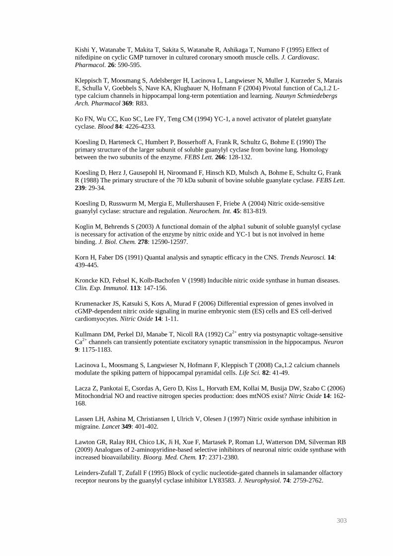
303
Kishi Y, Watanabe T, Makita T, Sakita S, Watanabe R, Ashikaga T, Numano F (1995) Effect of
nifedipine on cyclic GMP turnover in cultured coronary smooth muscle cells. J. Cardiovasc.
Pharmacol. 26: 590-595.
Kleppisch T, Moosmang S, Adelsberger H, Lacinova L, Langwieser N, Muller J, Kurzeder S, Marais
E, Schulla V, Goebbels S, Nave KA, Klugbauer N, Hofmann F (2004) Pivotal function of Cav1.2 L-
type calcium channels in hippocampal long-term potentiation and learning. Naunyn Schmiedebergs
Arch. Pharmacol 369: R83.
Ko FN, Wu CC, Kuo SC, Lee FY, Teng CM (1994) YC-1, a novel activator of platelet guanylate
cyclase. Blood 84: 4226-4233.
Koesling D, Harteneck C, Humbert P, Bosserhoff A, Frank R, Schultz G, Bohme E (1990) The
primary structure of the larger subunit of soluble guanylyl cyclase from bovine lung. Homology between the two subunits of the enzyme. FEBS Lett. 266: 128-132.
Koesling D, Herz J, Gausepohl H, Niroomand F, Hinsch KD, Mulsch A, Bohme E, Schultz G, Frank
R (1988) The primary structure of the 70 kDa subunit of bovine soluble guanylate cyclase. FEBS Lett.
239: 29-34.
Koesling D, Russwurm M, Mergia E, Mullershausen F, Friebe A (2004) Nitric oxide-sensitive
guanylyl cyclase: structure and regulation. Neurochem. Int. 45: 813-819.
Koglin M, Behrends S (2003) A functional domain of the alpha1 subunit of soluble guanylyl cyclase
is necessary for activation of the enzyme by nitric oxide and YC-1 but is not involved in heme
binding. J. Biol. Chem. 278: 12590-12597.
Korn H, Faber DS (1991) Quantal analysis and synaptic efficacy in the CNS. Trends Neurosci. 14:
439-445.
Kroncke KD, Fehsel K, Kolb-Bachofen V (1998) Inducible nitric oxide synthase in human diseases.
Clin. Exp. Immunol. 113: 147-156.
Krumenacker JS, Katsuki S, Kots A, Murad F (2006) Differential expression of genes involved in
cGMP-dependent nitric oxide signaling in murine embryonic stem (ES) cells and ES cell-derived
cardiomyocytes. Nitric Oxide 14: 1-11.
Kullmann DM, Perkel DJ, Manabe T, Nicoll RA (1992) Ca2+ entry via postsynaptic voltage-sensitive
Ca2+ channels can transiently potentiate excitatory synaptic transmission in the hippocampus. Neuron
9: 1175-1183.
Lacinova L, Moosmang S, Langwieser N, Hofmann F, Kleppisch T (2008) Cav1.2 calcium channels
modulate the spiking pattern of hippocampal pyramidal cells. Life Sci. 82: 41-49.
Lacza Z, Pankotai E, Csordas A, Gero D, Kiss L, Horvath EM, Kollai M, Busija DW, Szabo C (2006)
Mitochondrial NO and reactive nitrogen species production: does mtNOS exist? Nitric Oxide 14: 162-168.
Lassen LH, Ashina M, Christiansen I, Ulrich V, Olesen J (1997) Nitric oxide synthase inhibition in
migraine. Lancet 349: 401-402.
Lawton GR, Ralay RH, Chico LK, Ji H, Xue F, Martasek P, Roman LJ, Watterson DM, Silverman RB
(2009) Analogues of 2-aminopyridine-based selective inhibitors of neuronal nitric oxide synthase with
increased bioavailability. Bioorg. Med. Chem. 17: 2371-2380.
Leinders-Zufall T, Zufall F (1995) Block of cyclic nucleotide-gated channels in salamander olfactory
receptor neurons by the guanylyl cyclase inhibitor LY83583. J. Neurophysiol. 74: 2759-2762.
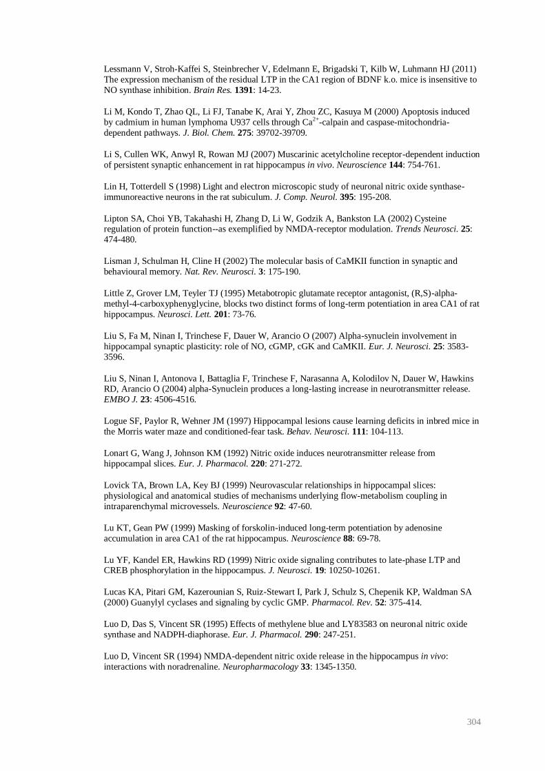
304
Lessmann V, Stroh-Kaffei S, Steinbrecher V, Edelmann E, Brigadski T, Kilb W, Luhmann HJ (2011)
The expression mechanism of the residual LTP in the CA1 region of BDNF k.o. mice is insensitive to
NO synthase inhibition. Brain Res. 1391: 14-23.
Li M, Kondo T, Zhao QL, Li FJ, Tanabe K, Arai Y, Zhou ZC, Kasuya M (2000) Apoptosis induced
by cadmium in human lymphoma U937 cells through Ca2+-calpain and caspase-mitochondria-
dependent pathways. J. Biol. Chem. 275: 39702-39709.
Li S, Cullen WK, Anwyl R, Rowan MJ (2007) Muscarinic acetylcholine receptor-dependent induction
of persistent synaptic enhancement in rat hippocampus in vivo. Neuroscience 144: 754-761.
Lin H, Totterdell S (1998) Light and electron microscopic study of neuronal nitric oxide synthase-
immunoreactive neurons in the rat subiculum. J. Comp. Neurol. 395: 195-208.
Lipton SA, Choi YB, Takahashi H, Zhang D, Li W, Godzik A, Bankston LA (2002) Cysteine regulation of protein function--as exemplified by NMDA-receptor modulation. Trends Neurosci. 25:
474-480.
Lisman J, Schulman H, Cline H (2002) The molecular basis of CaMKII function in synaptic and
behavioural memory. Nat. Rev. Neurosci. 3: 175-190.
Little Z, Grover LM, Teyler TJ (1995) Metabotropic glutamate receptor antagonist, (R,S)-alpha-
methyl-4-carboxyphenyglycine, blocks two distinct forms of long-term potentiation in area CA1 of rat
hippocampus. Neurosci. Lett. 201: 73-76.
Liu S, Fa M, Ninan I, Trinchese F, Dauer W, Arancio O (2007) Alpha-synuclein involvement in
hippocampal synaptic plasticity: role of NO, cGMP, cGK and CaMKII. Eur. J. Neurosci. 25: 3583-
3596.
Liu S, Ninan I, Antonova I, Battaglia F, Trinchese F, Narasanna A, Kolodilov N, Dauer W, Hawkins RD, Arancio O (2004) alpha-Synuclein produces a long-lasting increase in neurotransmitter release.
EMBO J. 23: 4506-4516.
Logue SF, Paylor R, Wehner JM (1997) Hippocampal lesions cause learning deficits in inbred mice in
the Morris water maze and conditioned-fear task. Behav. Neurosci. 111: 104-113.
Lonart G, Wang J, Johnson KM (1992) Nitric oxide induces neurotransmitter release from
hippocampal slices. Eur. J. Pharmacol. 220: 271-272.
Lovick TA, Brown LA, Key BJ (1999) Neurovascular relationships in hippocampal slices:
physiological and anatomical studies of mechanisms underlying flow-metabolism coupling in
intraparenchymal microvessels. Neuroscience 92: 47-60.
Lu KT, Gean PW (1999) Masking of forskolin-induced long-term potentiation by adenosine
accumulation in area CA1 of the rat hippocampus. Neuroscience 88: 69-78.
Lu YF, Kandel ER, Hawkins RD (1999) Nitric oxide signaling contributes to late-phase LTP and CREB phosphorylation in the hippocampus. J. Neurosci. 19: 10250-10261.
Lucas KA, Pitari GM, Kazerounian S, Ruiz-Stewart I, Park J, Schulz S, Chepenik KP, Waldman SA
(2000) Guanylyl cyclases and signaling by cyclic GMP. Pharmacol. Rev. 52: 375-414.
Luo D, Das S, Vincent SR (1995) Effects of methylene blue and LY83583 on neuronal nitric oxide
synthase and NADPH-diaphorase. Eur. J. Pharmacol. 290: 247-251.
Luo D, Vincent SR (1994) NMDA-dependent nitric oxide release in the hippocampus in vivo:
interactions with noradrenaline. Neuropharmacology 33: 1345-1350.

305
Lynch G, Larson J, Kelso S, Barrionuevo G, Schottler F (1983) Intracellular injections of EGTA
block induction of hippocampal long-term potentiation. Nature 305: 719-721.
Lynch MA (2004) Long-term potentiation and memory. Physiol Rev 84: 87-136.
MacMicking J, Xie QW, Nathan C (1997) Nitric oxide and macrophage function. Annu. Rev.
Immunol. 15: 323-350.
Magee JC, Avery RB, Christie BR, Johnston D (1996) Dihydropyridine-sensitive, voltage-gated Ca2+
channels contribute to the resting intracellular Ca2+ concentration of hippocampal CA1 pyramidal
neurons. J. Neurophysiol. 76: 3460-3470.
Makara JK, Katona I, Nyiri G, Nemeth B, Ledent C, Watanabe M, de VJ, Freund TF, Hajos N (2007)
Involvement of nitric oxide in depolarization-induced suppression of inhibition in hippocampal
pyramidal cells during activation of cholinergic receptors. J. Neurosci. 27: 10211-10222.
Malen PL, Chapman PF (1997) Nitric oxide facilitates long-term potentiation, but not long-term
depression. J. Neurosci. 17: 2645-2651.
Malenka RC, Kauer JA, Zucker RS, Nicoll RA (1988) Postsynaptic calcium is sufficient for
potentiation of hippocampal synaptic transmission. Science 242: 81-84.
Malenka RC (1991) Postsynaptic factors control the duration of synaptic enhancement in area CA1 of
the hippocampus. Neuron 6: 53-60.
Malenka RC, Bear MF (2004) LTP and LTD: an embarrassment of riches. Neuron 44: 5-21.
Marsh N, Marsh A (2000) A short history of nitroglycerine and nitric oxide in pharmacology and
physiology. Clin. Exp. Pharm. Physiol. 27: 313-319.
Martens-Lobenhoffer J, Sulyok E, Czeiter E, Buki A, Kohl J, Firsching R, Troger U, Bode-Boger SM
(2007) Determination of cerebrospinal fluid concentrations of arginine and dimethylarginines in patients with subarachnoid haemorrhage. J. Neurosci. Methods 164: 155-160.
Martin SJ, Grimwood PD, Morris RG (2000) Synaptic plasticity and memory: an evaluation of the
hypothesis. Annu. Rev. Neurosci. 23: 649-711.
Massberg S, Sausbier M, Klatt P, Bauer M, Pfeifer A, Siess W, Fassler R, Ruth P, Krombach F,
Hofmann F (1999) Increased adhesion and aggregation of platelets lacking cyclic guanosine 3',5'-
monophosphate kinase I. J. Exp. Med. 189: 1255-1264.
Matsuoka I, Giuili G, Poyard M, Stengel D, Parma J, Guellaen G, Hanoune J (1992) Localization of
adenylyl and guanylyl cyclase in rat brain by in situ hybridization: comparison with calmodulin
mRNA distribution. J. Neurosci 12: 3350-3360.
Mayer B, Brunner F, Schmidt K (1993) Novel actions of methylene blue. Eur. Heart J. 14 Suppl I:
22-26.
McKinney BC, Murphy GG (2006) The L-Type voltage-gated calcium channel Cav1.3 mediates consolidation, but not extinction, of contextually conditioned fear in mice. Learn. Mem. 13: 584-589.
McKinney BC, Sze W, Lee B, Murphy GG (2009) Impaired long-term potentiation and enhanced
neuronal excitability in the amygdala of Cav1.3 knockout mice. Neurobiol. Learn. Mem. 92: 519-528.
McNaughton BL, Douglas RM, Goddard GV (1978) Synaptic enhancement in fascia dentata:
cooperativity among coactive afferents. Brain Res. 157: 277-293.
Mergia E, Friebe A, Dangel O, Russwurm M, Koesling D (2006) Spare guanylyl cyclase NO
receptors ensure high NO sensitivity in the vascular system. J. Clin. Invest. 116: 1731-1737.
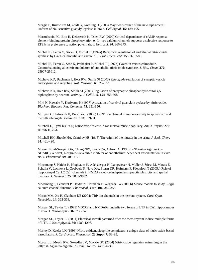
306
Mergia E, Russwurm M, Zoidl G, Koesling D (2003) Major occurrence of the new alpha2beta1
isoform of NO-sensitive guanylyl cyclase in brain. Cell Signal. 15: 189-195.
Mermelstein PG, Bito H, Deisseroth K, Tsien RW (2000) Critical dependence of cAMP response
element-binding protein phosphorylation on L-type calcium channels supports a selective response to
EPSPs in preference to action potentials. J. Neurosci. 20: 266-273.
Michel JB, Feron O, Sacks D, Michel T (1997a) Reciprocal regulation of endothelial nitric-oxide
synthase by Ca2+-calmodulin and caveolin. J. Biol. Chem. 272: 15583-15586.
Michel JB, Feron O, Sase K, Prabhakar P, Michel T (1997b) Caveolin versus calmodulin.
Counterbalancing allosteric modulators of endothelial nitric oxide synthase. J. Biol. Chem. 272:
25907-25912.
Micheva KD, Buchanan J, Holz RW, Smith SJ (2003) Retrograde regulation of synaptic vesicle endocytosis and recycling. Nat. Neurosci. 6: 925-932.
Micheva KD, Holz RW, Smith SJ (2001) Regulation of presynaptic phosphatidylinositol 4,5-
biphosphate by neuronal activity. J. Cell Biol. 154: 355-368.
Miki N, Kawabe Y, Kuriyama K (1977) Activation of cerebral guanylate cyclase by nitric oxide.
Biochem. Biophys. Res. Commun. 75: 851-856.
Milligan CJ, Edwards IJ, Deuchars J (2006) HCN1 ion channel immunoreactivity in spinal cord and
medulla oblongata. Brain Res. 1081: 79-91.
Mitchell D, Tyml K (1996) Nitric oxide release in rat skeletal muscle capillary. Am. J. Physiol 270:
H1696-H1703.
Mitchell HH, Shonle HA, Grindley HS (1916) The origin of the nitrates in the urine. J. Biol. Chem.
24: 461-490.
Moore PK, al-Swayeh OA, Chong NW, Evans RA, Gibson A (1990) L-NG-nitro arginine (L-
NOARG), a novel, L-arginine-reversible inhibitor of endothelium-dependent vasodilatation in vitro.
Br. J. Pharmacol. 99: 408-412.
Moosmang S, Haider N, Klugbauer N, Adelsberger H, Langwieser N, Muller J, Stiess M, Marais E,
Schulla V, Lacinova L, Goebbels S, Nave KA, Storm DR, Hofmann F, Kleppisch T (2005a) Role of
hippocampal Cav1.2 Ca2+ channels in NMDA receptor-independent synaptic plasticity and spatial
memory. J. Neurosci. 25: 9883-9892.
Moosmang S, Lenhardt P, Haider N, Hofmann F, Wegener JW (2005b) Mouse models to study L-type
calcium channel function. Pharmacol. Ther. 106: 347-355.
Moran MM, Xu H, Clapham DE (2004) TRP ion channels in the nervous system. Curr. Opin.
Neurobiol. 14: 362-369.
Morgan SL, Teyler TJ (1999) VDCCs and NMDARs underlie two forms of LTP in CA1 hippocampus in vivo. J. Neurophysiol. 82: 736-740.
Morgan SL, Teyler TJ (2001) Electrical stimuli patterned after the theta-rhythm induce multiple forms
of LTP. J. Neurophysiol. 86: 1289-1296.
Morley D, Keefer LK (1993) Nitric oxide/nucleophile complexes: a unique class of nitric oxide-based
vasodilators. J. Cardiovasc. Pharmacol. 22 Suppl 7: S3-S9.
Moroz LL, Meech RW, Sweedler JV, Mackie GO (2004) Nitric oxide regulates swimming in the
jellyfish Aglantha digitale. J. Comp. Neurol. 471: 26-36.
307
Morris RG, Anderson E, Lynch GS, Baudry M (1986) Selective impairment of learning and blockade
of long-term potentiation by an N-methyl-D-aspartate receptor antagonist, AP5. Nature 319: 774-776.
Morris RG, Garrud P, Rawlins JN, O'Keefe J (1982) Place navigation impaired in rats with
hippocampal lesions. Nature 297: 681-683.
Moser EI, Krobert KA, Moser MB, Morris RG (1998) Impaired spatial learning after saturation of
long-term potentiation. Science 281: 2038-2042.
Murphy TH, Worley PF, Baraban JM (1991) L-type voltage-sensitive calcium channels mediate
synaptic activation of immediate early genes. Neuron 7: 625-635.
Murthy KS (2001) Activation of phosphodiesterase 5 and inhibition of guanylate cyclase by cGMP-
dependent protein kinase in smooth muscle. Biochem. J. 360: 199-208.
Musleh WY, Shahi K, Baudry M (1993) Further studies concerning the role of nitric oxide in LTP induction and maintenance. Synapse 13: 370-375.
N'Gouemo P, Yasuda R, Faingold CL (2010) Seizure susceptibility is associated with altered protein
expression of voltage-gated calcium channel subunits in inferior colliculus neurons of the genetically
epilepsy-prone rat. Brain Res. 1308: 153-157.
Nakane M, Arai K, Saheki S, Kuno T, Buechler W, Murad F (1990) Molecular cloning and expression
of cDNAs coding for soluble guanylate cyclase from rat lung. J. Biol. Chem. 265: 16841-16845.
Nakane M, Saheki S, Kuno T, Ishii K, Murad F (1988) Molecular cloning of a cDNA coding for 70
kilodalton subunit of soluble guanylate cyclase from rat lung. Biochem. Biophys. Res. Commun. 157:
1139-1147.
Nakazawa K, Quirk MC, Chitwood RA, Watanabe M, Yeckel MF, Sun LD, Kato A, Carr CA,
Johnston D, Wilson MA, Tonegawa S (2002) Requirement for hippocampal CA3 NMDA receptors in associative memory recall. Science 297: 211-218.
Nedvetsky PI, Sessa WC, Schmidt HH (2002) There's NO binding like NOS binding: protein-protein
interactions in NO/cGMP signaling. Proc. Natl. Acad. Sci. U. S. A 99: 16510-16512.
Neitz A, Mergia E, Eysel UT, Koesling D, Mittmann T (2011) Presynaptic nitric oxide/cGMP
facilitates glutamate release via hyperpolarization-activated cyclic nucleotide-gated channels in the
hippocampus. Eur. J. Neurosci. 9: 1611-1621.
Neves G, Cooke SF, Bliss TV (2008) Synaptic plasticity, memory and the hippocampus: a neural
network approach to causality. Nat. Rev. Neurosci. 9: 65-75.
Nicolarakis PJ, Lin YQ, Bennett MR (1994) Effect of nitric oxide synthase inhibition on long-term
potentiation at associational-commissural and mossy fibre synapses on CA3 pyramidal neurones. Br.
J. Pharmacol. 111: 521-524.
Nicoll RA (2003) Expression mechanisms underlying long-term potentiation: a postsynaptic view. Philos. Trans. R. Soc. Lond B Biol. Sci. 358: 721-726.
Nicoll RA, Schmitz D (2005) Synaptic plasticity at hippocampal mossy fibre synapses. Nat. Rev.
Neurosci. 6: 863-876.
Nikonenko I, Boda B, Steen S, Knott G, Welker E, Muller D (2008) PSD-95 promotes synaptogenesis
and multiinnervated spine formation through nitric oxide signaling. J. Cell Biol. 183: 1115-1127.
Nikonenko I, Jourdain P, Muller D (2003) Presynaptic remodeling contributes to activity-dependent
synaptogenesis. J. Neurosci. 23: 8498-8505.
308
Nimmegeers S, Sips P, Buys E, Brouckaert P, Van d, V (2007) Functional role of the soluble guanylyl
cyclase α1 subunit in vascular smooth muscle relaxation. Cardiovasc. Res. 76: 149-159.
Ninan I, Arancio O (2004) Presynaptic CaMKII is necessary for synaptic plasticity in cultured
hippocampal neurons. Neuron 42: 129-141.
Norman JA, Ansell J, Phillips MA (1983) Dihydropyridine Ca2+ entry blockers selectively inhibit
peak I cAMP phosphodiesterase. Eur. J. Pharmacol. 93: 107-112.
Notomi T, Shigemoto R (2004) Immunohistochemical localization of Ih channel subunits, HCN1-4, in
the rat brain. J. Comp Neurol. 471: 241-276.
Nugent FS, Penick EC, Kauer JA (2007) Opioids block long-term potentiation of inhibitory synapses.
Nature 446: 1086-1090.
O'Dell TJ, Hawkins RD, Kandel ER, Arancio O (1991) Tests of the roles of two diffusible substances in long-term potentiation: evidence for nitric oxide as a possible early retrograde messenger. Proc.
Natl. Acad. Sci. U. S. A 88: 11285-11289.
O'Dell TJ, Huang PL, Dawson TM, Dinerman JL, Snyder SH, Kandel ER, Fishman MC (1994)
Endothelial NOS and the blockade of LTP by NOS inhibitors in mice lacking neuronal NOS. Science
265: 542-546.
O'Keefe J (2007) Hippocampal neurophysiology in the behaving animal. In: The hippocampus book
(Andersen P, Morris R, Amaral D, Bliss TVP, O'Keefe J, eds), pp 475-548. Oxford: Oxford
University Press.
Oka M, Itoh Y, Ukai Y, Kimura K (1999) Blockade by NS-7, a neuroprotective compound, of both L-
type and P/Q- type Ca2+ channels involving depolarization-stimulated nitric oxide synthase activity in
primary neuronal culture. J. Neurochem. 72: 1315-1322.
Okada D (1992) Two pathways of cyclic GMP production through glutamate receptor-mediated nitric
oxide synthesis. J. Neurochem. 59: 1203-1210.
Olken NM, Marletta MA (1993) NG-methyl-L-arginine functions as an alternate substrate and
mechanism-based inhibitor of nitric oxide synthase. Biochemistry 32: 9677-9685.
Ota KT, Pierre VJ, Ploski JE, Queen K, Schafe GE (2008) The NO-cGMP-PKG signaling pathway
regulates synaptic plasticity and fear memory consolidation in the lateral amygdala via activation of
ERK/MAP kinase. Learn. Mem. 15: 792-805.
Ozawa Y, Hayashi K, Kobori H (2006) New generation calcium channel blockers in hypertensive
treatment. Curr. Hypertens. Rev. 2: 103-111.
Palmer RM, Ferrige AG, Moncada S (1987) Nitric oxide release accounts for the biological activity of
endothelium-derived relaxing factor. Nature 327: 524-526.
Pape HC, Mager R (1992) Nitric oxide controls oscillatory activity in thalamocortical neurons. Neuron 9: 441-448.
Parent A, Schrader K, Munger SD, Reed RR, Linden DJ, Ronnett GV (1998) Synaptic transmission
and hippocampal long-term potentiation in olfactory cyclic nucleotide-gated channel type 1 null
mouse. J. Neurophysiol. 79: 3295-3301.
Park JF, Luo ZD (2010) Calcium channel functions in pain processing. Channels (Austin) 4: 510-517.
Park JH, Straub VA, O'Shea M (1998) Anterograde signaling by nitric oxide: characterization and in
vitro reconstitution of an identified nitrergic synapse. J. Neurosci. 18: 5463-5476.
309
Parra P, Gulyas AI, Miles R (1998) How many subtypes of inhibitory cells in the hippocampus?
Neuron 20: 983-993.
Pawlik G, Rackl A, Bing RJ (1981) Quantitative capillary topography and blood flow in the cerebral
cortex of cats: an in vivo microscopic study. Brain Res. 208: 35-58.
Perez-Reyes E, Kim HS, Lacerda AE, Horne W, Wei XY, Rampe D, Campbell KP, Brown AM,
Birnbaumer L (1989) Induction of calcium currents by the expression of the alpha 1-subunit of the
dihydropyridine receptor from skeletal muscle. Nature 340: 233-236.
Petersen CC, Malenka RC, Nicoll RA, Hopfield JJ (1998) All-or-none potentiation at CA3-CA1
synapses. Proc. Natl. Acad. Sci. U. S. A 95: 4732-4737.
Petros A, Bennett D, Vallance P (1991) Effect of nitric oxide synthase inhibitors on hypotension in
patients with septic shock. Lancet 338: 1557-1558.
Phillips KG, Hardingham NR, Fox K (2008) Postsynaptic action potentials are required for nitric-
oxide-dependent long-term potentiation in CA1 neurons of adult GluR1 knock-out and wild-type
mice. J. Neurosci. 28: 14031-14041.
Pifarre P, Garcia A, Mengod G (2007) Species differences in the localization of soluble guanylyl
cyclase subunits in monkey and rat brain. J. Comp. Neurol. 500: 942-957.
Pigott B, Garthwaite J (2009) Activity-dependent long-term potentiation produced by exogenous
nitric oxide during NMDA receptor blockade in hippocampal slices. Proc. Physiol. Soc. 15: Abstract
C109.
Poss KD, Thomas MJ, Ebralidze AK, O'Dell TJ, Tonegawa S (1995) Hippocampal long-term
potentiation is normal in heme oxygenase-2 mutant mice. Neuron 15: 867-873.
Potter LR (2011) Guanylyl cyclase structure, function and regulation. Cell Signal. 23: 1921-1926.
Pozo K, Goda Y (2010) Unraveling mechanisms of homeostatic synaptic plasticity. Neuron 66: 337-
351.
Prast H, Philippu A (2001) Nitric oxide as modulator of neuronal function. Prog. Neurobiol. 64: 51-
68.
Priestley J (1775) Experiments and observations on different kinds of air. Volume published by W.
Bowyer and J. Nichols for J. Johnson, London.
Pyriochou A, Papapetropoulos A (2005) Soluble guanylyl cyclase: more secrets revealed. Cell Signal.
17: 407-413.
Rameau GA, Chiu LY, Ziff EB (2004) Bidirectional regulation of neuronal nitric-oxide synthase
phosphorylation at serine 847 by the N-methyl-D-aspartate receptor. J. Biol. Chem. 279: 14307-
14314.
Rameau GA, Tukey DS, Garcin-Hosfield ED, Titcombe RF, Misra C, Khatri L, Getzoff ED, Ziff EB (2007) Biphasic coupling of neuronal nitric oxide synthase phosphorylation to the NMDA receptor
regulates AMPA receptor trafficking and neuronal cell death. J. Neurosci. 27: 3445-3455.
Rampe D, Kane M (1994) Activators of voltage-dependent L-type calcium channels. Drug Dev. Res.
33: 344-363.
Rapoport RM, Draznin MB, Murad F (1983) Endothelium-dependent vasodilator-and
nitrovasodilator-induced relaxation may be mediated through cyclic GMP formation and cyclic GMP-
dependent protein phosphorylation. Trans. Assoc. Am. Physicians 96: 19-30.
310
Raymond CR (2007) LTP forms 1, 2 and 3: different mechanisms for the "long" in long-term
potentiation. Trends Neurosci. 30: 167-175.
Razani B, Woodman SE, Lisanti MP (2002) Caveolae: from cell biology to animal physiology.
Pharmacol. Rev. 54: 431-467.
Regehr WG, Tank DW (1992) Calcium concentration dynamics produced by synaptic activation of
CA1 hippocampal pyramidal cells. J. Neurosci. 12: 4202-4223.
Robello M, Amico C, Bucossi G, Cupello A, Rapallino MV, Thellung S (1996) Nitric oxide and
GABAA receptor function in the rat cerebral cortex and cerebellar granule cells. Neuroscience 74: 99-
105.
Rodrigo J, Springall DR, Uttenthal O, Bentura ML, badia-Molina F, Riveros-Moreno V, Martinez-
Murillo R, Polak JM, Moncada S (1994) Localization of nitric oxide synthase in the adult rat brain. Philos. Trans. R. Soc. Lond B Biol. Sci. 345: 175-221.
Rodriguez-Crespo I, Straub W, Gavilanes F, Ortiz de Montellano PR (1998) Binding of dynein light
chain (PIN) to neuronal nitric oxide synthase in the absence of inhibition. Arch. Biochem. Biophys.
359: 297-304.
Romberg C, Raffel J, Martin L, Sprengel R, Seeburg PH, Rawlins JN, Bannerman DM, Paulsen O
(2009) Induction and expression of GluA1 (GluR-A)-independent LTP in the hippocampus. Eur. J.
Neurosci. 29: 1141-1152.
Routh BN, Johnston D, Harris K, Chitwood RA (2009) Anatomical and electrophysiological
comparison of CA1 pyramidal neurons of the rat and mouse. J. Neurophysiol. 102: 2288-2302.
Roy B, Garthwaite J (2006) Nitric oxide activation of guanylyl cyclase in cells revisited. Proc. Natl.
Acad. Sci. U. S. A 103: 12185-12190.
Rozov A, Burnashev N, Sakmann B, Neher E (2001) Transmitter release modulation by intracellular
Ca2+ buffers in facilitating and depressing nerve terminals of pyramidal cells in layer 2/3 of the rat
neocortex indicates a target cell-specific difference in presynaptic calcium dynamics. J. Physiol. 531:
807-826.
Ruiz-Stewart I, Tiyyagura SR, Lin JE, Kazerounian S, Pitari GM, Schulz S, Martin E, Murad F,
Waldman SA (2004) Guanylyl cyclase is an ATP sensor coupling nitric oxide signaling to cell
metabolism. Proc. Natl. Acad. Sci. U. S. A 101: 37-42.
Russwurm M, Behrends S, Harteneck C, Koesling D (1998) Functional properties of a naturally
occuring isoform of soluble guanylyl cyclase. Biochem. J. 335: 125-130.
Russwurm M, Wittau N, Koesling D (2001) Guanylyl cyclase/PSD-95 interaction: targeting of the
nitric oxide-sensitive α2β1 guanylyl cyclase to synaptic membranes. J. Biol. Chem. 276: 44647-
44652.
Sato M, Hida N, Umezawa Y (2005) Imaging the nanomolar range of nitric oxide with an amplifier-
coupled fluorescent indicator in living cells. Proc. Natl. Acad. Sci. U. S. A 102: 14515-14520.
Sato M, Nakajima T, Goto M, Umezawa Y (2006) Cell-based indicator to visualize picomolar
dynamics of nitric oxide release from living cells. Anal. Chem. 78: 8175-8182.
Sattler R, Xiong Z, Lu WY, Hafner M, MacDonald JF, Tymianski M (1999) Specific coupling of
NMDA receptor activation to nitric oxide neurotoxicity by PSD-95 protein. Science 284: 1845-1848.
Savchenko A, Barnes S, Kramer RH (1997) Cyclic-nucleotide-gated channels mediate synaptic
feedback by nitric oxide. Nature 390: 694-698.
311
Schafe GE (2008) Rethinking the role of L-type voltage-gated calcium channels in fear memory
extinction. Learn. Mem. 15: 324-325.
Schlossmann J, Ammendola A, Ashman K, Zong X, Huber A, Neubauer G, Wang GX, Allescher HD,
Korth M, Wilm M, Hofmann F, Ruth P (2000) Regulation of intracellular calcium by a signalling
complex of IRAG, IP3 receptor and cGMP kinase Ibeta. Nature 404: 197-201.
Schlossmann J, Feil R, Hofmann F (2005) Insights into cGMP signalling derived from cGMP kinase
knockout mice. Front Biosci. 10: 1279-1289.
Schmachtenberg O, Diaz J, Bacigalupo J (2003) NO activates the olfactory cyclic nucleotide-gated
conductance independent from cGMP in isolated rat olfactory receptor neurons. Brain Res. 980: 146-
150.
Schmidt K, Desch W, Klatt P, Kukovetz WR, Mayer B (1997) Release of nitric oxide from donors with known half-life: a mathematical model for calculating nitric oxide concentrations in aerobic
solutions. Naunyn Schmiedebergs Arch. Pharmacol. 355: 457-462.
Schrammel A, Behrends S, Schmidt K, Koesling D, Mayer B (1996) Characterization of 1H-
[1,2,4]oxadiazolo[4,3-a]quinoxalin-1-one as a heme-site inhibitor of nitric oxide-sensitive guanylyl
cyclase. Mol. Pharmacol. 50: 1-5.
Schulz PE, Cook EP, Johnston D (1994) Changes in paired-pulse facilitation suggest presynaptic
involvement in long-term potentiation. J. Neurosci. 14: 5325-5337.
Schulz PE, Cook EP, Johnston D (1995) Using paired-pulse facilitation to probe the mechanisms for
long-term potentiation (LTP). J. Physiol. Paris 89: 3-9.
Schulz S, Chinkers M, Garbers DL (1989) The guanylate cyclase/receptor family of proteins. FASEB
J. 3: 2026-2035.
Schuman EM, Madison DV (1991) A requirement for the intercellular messenger nitric oxide in long-
term potentiation. Science 254: 1503-1506.
Seidel B, Stanarius A, Wolf G (1997) Differential expression of neuronal and endothelial nitric oxide
synthase in blood vessels of the rat brain. Neurosci. Lett. 239: 109-112.
Senter PD, Eckstein F, Mulsch A, Bohme E (1983) The stereochemical course of the reaction
catalyzed by soluble bovine lung guanylate cyclase. J. Biol. Chem. 258: 6741-6745.
Serulle Y, Zhang S, Ninan I, Puzzo D, McCarthy M, Khatri L, Arancio O, Ziff EB (2007) A GluR1-
cGKII interaction regulates AMPA receptor trafficking. Neuron 56: 670-688.
Shors TJ, Matzel LD (1997) Long-term potentiation: what's learning got to do with it? Behav. Brain
Sci. 20: 597-614.
Silva AJ, Paylor R, Wehner JM, Tonegawa S (1992a) Impaired spatial learning in alpha-calcium-
calmodulin kinase II mutant mice. Science 257: 206-211.
Silva AJ, Stevens CF, Tonegawa S, Wang Y (1992b) Deficient hippocampal long-term potentiation in
alpha-calcium-calmodulin kinase II mutant mice. Science 257: 201-206.
Sokolova IV, Lester HA, Davidson N (2006) Postsynaptic Mechanisms Are Essential for Forskolin-
Induced Potentiation of Synaptic Transmission. J. Neurophysiol 95: 2570-2579.
Son H, Hawkins RD, Martin K, Kiebler M, Huang PL, Fishman MC, Kandel ER (1996) Long-term
potentiation is reduced in mice that are doubly mutant in endothelial and neuronal nitric oxide
synthase. Cell 87: 1015-1023.

312
Song Y, Zweier JL, Xia Y (2001) Heat-shock protein 90 augments neuronal nitric oxide synthase
activity by enhancing Ca2+/calmodulin binding. Biochem. J. 355: 357-360.
Southam E, East SJ, Garthwaite J (1991) Excitatory amino acid receptors coupled to the nitric
oxide/cyclic GMP pathway in rat cerebellum during development. J. Neurochem. 56: 2072-2081.
Southam E, Garthwaite J (1993) The nitric oxide-cyclic GMP signalling pathway in rat brain.
Neuropharmacology 32: 1267-1277.
Southam E, Morris R, Garthwaite J (1992) Sources and targets of nitric oxide in rat cerebellum.
Neurosci. Lett. 137: 241-244.
Specht CG, Schoepfer R (2001) Deletion of the alpha-synuclein locus in a subpopulation of C57BL/6J
inbred mice. BMC. Neurosci. 2: 11.
Spruston N, McBain C (2007) Structural and functional properties of hippocampal neurons. In: The hippocampus book. (Andersen P, Morris RG, Amaral D, Bliss TVP., O'Keefe J, eds), pp 133-201.
Oxford: Oxford University Press.
Stanarius A, Topel I, Schulz S, Noack H, Wolf G (1997) Immunocytochemistry of endothelial nitric
oxide synthase in the rat brain: a light and electron microscopical study using the tyramide signal
amplification technique. Acta Histochem. 99: 411-429.
Steinert JR, Chernova T, Forsythe ID (2010) Nitric oxide signaling in brain function, dysfunction, and
dementia. Neuroscientist 16: 435-452.
Stephenson DT, Coskran TM, Wilhelms MB, Adamowicz WO, O'Donnell MM, Muravnick KB,
Menniti FS, Kleiman RJ, Morton D (2009) Immunohistochemical localization of phosphodiesterase
2A in multiple mammalian species. J. Histochem. Cytochem. 57: 933-949.
Stone JR, Marletta MA (1994) Soluble guanylate cyclase from bovine lung: activation with nitric oxide and carbon monoxide and spectral characterization of the ferrous and ferric states. Biochemistry
33: 5636-5640.
Straub VA, Grant J, O'Shea M, Benjamin PR (2007) Modulation of serotonergic neurotransmission by
nitric oxide. J. Neurophysiol. 97: 1088-1099.
Striessnig J (1999) Pharmacology, structure and function of cardiac L-type Ca2+ channels. Cell
Physiol. Biochem. 9: 242-269.
Stuehr DJ (1999) Mammalian nitric oxide synthases. Biochim. Biophys. Acta 1411: 217-230.
Stuehr DJ, Marletta MA (1985) Mammalian nitrate biosynthesis: mouse macrophages produce nitrite
and nitrate in response to Escherichia coli lipopolysaccharide. Proc. Natl. Acad. Sci. U. S. A 82: 7738-
7742.
Suma C, Hayashi S, Ukai Y, Yoshikuni Y, Kimura K (1997) Na+ and high-voltage-activated Ca2+
channel blocking actions of NS-7, a novel neuroprotective agent, in NG108-15 cells. Eur. J. Pharmacol. 336: 283-290.
Sunahara RK, Beuve A, Tesmer JJ, Sprang SR, Garbers DL, Gilman AG (1998) Exchange of
substrate and inhibitor specificities between adenylyl and guanylyl cyclases. J. Biol. Chem. 273:
16332-16338.
Susswein AJ, Katzoff A, Miller N, Hurwitz I (2004) Nitric oxide and memory. Neuroscientist 10:
153-162.

313
Suvarna NU, O'Donnell JM (2002) Hydrolysis of N-methyl-D-aspartate receptor-stimulated cAMP
and cGMP by PDE4 and PDE2 phosphodiesterases in primary neuronal cultures of rat cerebral cortex
and hippocampus. J. Pharmacol. Exp. Ther. 302: 249-256.
Szabadits E, Cserep C, Ludanyi A, Katona I, Gracia-Llanes J, Freund TF, Nyiri G (2007)
Hippocampal GABAergic synapses possess the molecular machinery for retrograde nitric oxide
signaling. J. Neurosci. 27: 8101-8111.
Takahashi S, Mendelsohn ME (2003a) Calmodulin-dependent and -independent activation of
endothelial nitric-oxide synthase by heat shock protein 90. J. Biol. Chem. 278: 9339-9344.
Takahashi S, Mendelsohn ME (2003b) Synergistic activation of endothelial nitric-oxide synthase
(eNOS) by HSP90 and Akt: calcium-independent eNOS activation involves formation of an HSP90-
Akt-CaM-bound eNOS complex. J. Biol. Chem. 278: 30821-30827.
Takasugi Y, Kawata K, Okuda T, Koga Y, Mizuguchi N, Yamanaka S, Watanabe S (2003) Strain
differences to effects of aging on concentrations of amino acids in cerebrospinal fluid between
Sprague Dawley rat and Wistar Kyoto rat. Exp. Anim. 52: 429-432.
Tang YP, Shimizu E, Dube GR, Rampon C, Kerchner GA, Zhuo M, Liu G, Tsien JZ (1999) Genetic
enhancement of learning and memory in mice. Nature 401: 63-69.
Tao HW, Poo M (2001) Retrograde signaling at central synapses. Proc. Natl. Acad. Sci. U. S. A 98:
11009-11015.
Taqatqeh F, Mergia E, Neitz A, Eysel UT, Koesling D, Mittmann T (2009) More than a retrograde
messenger: nitric oxide needs two cGMP pathways to induce hippocampal long-term potentiation. J.
Neurosci. 29: 9944-9350.
Tatsumi S, Itoh Y, Ma FH, Higashira H, Ukai Y, Yoshikuni Y, Kimura K (1998) Inhibition of depolarization-induced nitric oxide synthase activation by NS-7, a phenylpyrimidine derivative, in
primary neuronal culture. J. Neurochem. 70: 59-65.
Teyler TJ, Cavus I, Coussens C (1995) Synaptic plasticity in the hippocampal slice: functional
consequences. J. Neurosc. Methods 59: 11-17.
Teyler TJ, Cavus I, Coussens C, DiScenna P, Grover L, Lee YP, Little Z (1994) Multideterminant role
of calcium in hippocampal synaptic plasticity. Hippocampus 4: 623-634.
Teyler TJ (1999) Use of brain slices to study long-term potentiation and depression as examples of
synaptic plasticity. Methods 18: 109-116.
Thevenod F, Jones SW (1992) Cadmium block of calcium current in frog sympathetic neurons.
Biophys. J. 63: 162-168.
Thomas MK, Francis SH, Corbin JD (1990) Substrate- and kinase-directed regulation of
phosphorylation of a cGMP-binding phosphodiesterase by cGMP. J. Biol. Chem. 265: 14971-14978.
Topel I, Stanarius A, Wolf G (1998) Distribution of the endothelial constitutive nitric oxide synthase
in the developing rat brain: an immunohistochemical study. Brain Res. 788: 43-48.
Treiman M, Caspersen C, Christensen SB (1998) A tool coming of age: thapsigargin as an inhibitor of
sarco-endoplasmic reticulum Ca2+-ATPases. Trends Pharmacol. Sci. 19: 131-135.
Trezise D, Dale T, Main M (2010) Ion channels: principles, terminology and methodology. In: Ion
channels from structure to function (Kew J, Davies C, eds), pp 3-17. Oxford: Oxford University Press.
Tsien JZ, Huerta PT, Tonegawa S (1996) The essential role of hippocampal CA1 NMDA receptor-
dependent synaptic plasticity in spatial memory. Cell 87: 1327-1338.
314
Vaandrager AB, Ehlert EM, Jarchau T, Lohmann SM, de Jonge HR (1996) N-terminal myristoylation
is required for membrane localization of cGMP-dependent protein kinase type II. J. Biol. Chem. 271:
7025-7029.
Vallance P, Leiper J (2002) Blocking NO synthesis: how, where and why? Nat. Rev. Drug Discov. 1:
939-950.
Valtschanoff JG, Weinberg RJ, Kharazia VN, Nakane M, Schmidt HHHW (1993) Neurons in rat
hippocampus that synthesize nitric-oxide. J. Comp. Neurol. 331: 111-121.
van Staveren WC, Markerink-Van IM, Steinbusch HW, de VJ (2001) The effects of
phosphodiesterase inhibition on cyclic GMP and cyclic AMP accumulation in the hippocampus of the
rat. Brain Res. 888: 275-286.
van Staveren WC, Steinbusch HW, Markerink-Van IM, Behrends S, de VJ (2004) Species differences in the localization of cGMP-producing and NO-responsive elements in the mouse and rat
hippocampus using cGMP immunocytochemistry. Eur. J. Neurosci. 19: 2155-2168.
van Staveren WC, Steinbusch HW, Markerink-Van IM, Repaske DR, Goy MF, Kotera J, Omori K,
Beavo JA, de VJ (2003) mRNA expression patterns of the cGMP-hydrolyzing phosphodiesterases
types 2, 5, and 9 during development of the rat brain. J. Comp. Neurol. 467: 566-580.
Venema RC, Venema VJ, Ju H, Harris MB, Snead C, Jilling T, Dimitropoulou C, Maragoudakis ME,
Catravas JD (2003) Novel complexes of guanylate cyclase with heat shock protein 90 and nitric oxide
synthase. Am. J. Physiol Heart Circ. Physiol 285: H669-H678.
Verma A, Hirsch DJ, Glatt CE, Ronnett GV, Snyder SH (1993) Carbon monoxide: a putative neural
messenger. Science 259: 381-384.
Vincent SR (2010) Nitric oxide neurons and neurotransmission. Prog. Neurobiol. 90: 246-255.
Vincent SR, Kimura H (1992) Histochemical mapping of nitric oxide synthase in the rat brain.
Neuroscience 46: 755-784.
Visser GJ, Peters PH, Theuvenet AP (1993) Cadmium ion is a non-competitive inhibitor of red cell
Ca2+-ATPase activity. Biochem. Biophys. Acta 1152: 26-34.
Wagner DA, Young VR, Tannenbaum SR (1983) Mammalian nitrate biosynthesis: incorporation of
15NH3 into nitrate is enhanced by endotoxin treatment. Proc. Natl. Acad. Sci. U. S. A 80: 4518-4521.
Walker M, Chan D, Thom M (2007) Hippocampus in human diseases. In: The hippocampus book
(Andersen P, Morris RG, Amaral D, Bliss TVP, O'Keefe J, eds), pp 769-812. Oxford: Oxford
University Press.
Walther C, Gielen S, Hambrecht R (2004) The effect of exercise training on endothelial function in
cardiovascular disease in humans. Exerc. Sport Sci. Rev. 32: 129-134.
Wang HG, Lu FM, Jin I, Udo H, Kandel ER, de VJ, Walter U, Lohmann SM, Hawkins RD, Antonova I (2005) Presynaptic and postsynaptic roles of NO, cGK, and RhoA in long-lasting potentiation and
aggregation of synaptic proteins. Neuron 45: 389-403.
Wang LY, Salter MW, MacDonald JF (1991) Regulation of kainate receptors by cAMP-dependent
protein kinase and phosphatases. Science 253: 1132-1135.
Watanabe M, Fukaya M, Sakimura K, Manabe T, Mishina M, Inoue Y (1998) Selective scarcity of
NMDA receptor channel subunits in the stratum lucidum (mossy fibre-recipient layer) of the mouse
hippocampal CA3 subfield. Eur. J. Neurosci. 10: 478-487.
315
Watanabe Y, Saito H, Abe K (1995) Nitric oxide is involved in long-term potentiation in the medial
but not lateral amygdala neuron synapses in vitro. Brain Res. 688: 233-236.
Wedel B, Harteneck C, Foerster J, Friebe A, Schultz G, Koesling D (1995) Functional domains of
soluble guanylyl cyclase. J. Biol. Chem. 270: 24871-24875.
Wedel B, Humbert P, Harteneck C, Foerster J, Malkewitz J, Bohme E, Schultz G, Koesling D (1994)
Mutation of His-105 in the beta 1 subunit yields a nitric oxide-insensitive form of soluble guanylyl
cyclase. Proc. Natl. Acad. Sci. U. S. A 91: 2592-2596.
Weisskopf MG, Bauer EP, LeDoux JE (1999) L-type voltage-gated calcium channels mediate
NMDA-independent associative long-term potentiation at thalamic input synapses to the amygdala. J.
Neurosci. 19: 10512-10519.
Wendland B, Schweizer FE, Ryan TA, Nakane M, Murad F, Scheller RH, Tsien RW (1994) Existence of nitric oxide synthase in rat hippocampal pyramidal cells. Proc. Natl. Acad. Sci. U. S. A 91: 2151-
2155.
Westenbroek RE, Ahlijanian MK, Catterall WA (1990) Clustering of L-type Ca2+ channels at the base
of major dendrites in hippocampal pyramidal neurons. Nature 347: 281-284.
Whitlock JR, Heynen AJ, Shuler MG, Bear MF (2006) Learning induces long-term potentiation in the
hippocampus. Science 313: 1093-1097.
Wigstrom H, Gustafsson B, Huang YY, Abraham WC (1986) Hippocampal long-term potentiation is
induced by pairing single afferent volleys with intracellularly injected depolarizing current pulses.
Acta. Physiol. Scand. 126: 317-319.
Williams JH, Li YG, Nayak A, Errington ML, Murphy KP, Bliss TV (1993) The suppression of long-
term potentiation in rat hippocampus by inhibitors of nitric oxide synthase is temperature and age dependent. Neuron 11: 877-884.
Wilson EM, Chinkers M (1995) Identification of sequences mediating guanylyl cyclase dimerization.
Biochemistry 34: 4696-4701.
Wilson GW, Garthwaite J (2010) Hyperpolarization-activated ion channels as targets for nitric oxide
signalling in deep cerebellar nuclei. Eur. J. Neurosci. 31: 1935-1945.
Wilson RI, Godecke A, Brown RE, Schrader J, Haas HL (1999) Mice deficient in endothelial nitric
oxide synthase exhibit a selective deficit in hippocampal long-term potentiation. Neuroscience 90:
1157-1165.
Wojtaszek P (2000) Nitric oxide in plants. To NO or not to NO. Phytochemistry 54: 1-4.
Wolf ME (2003) LTP may trigger addiction. Mol. Interv. 3: 248-252.
Wood KC, Batchelor AM, Bartus K, Harris KL, Garthwaite G, Vernon J, Garthwaite J (2011)
Picomolar nitric oxide signals from central neurons recorded using ultrasensitive detector cells. J. Biol. Chem. Epub, DOI: 10.1074/jbc.M111.289777.
Wood PJ, Marks V (1978) Direct measurement of cGMP in blood plasma and urine by
radioimmunoassay. Ann. Clin. Biochem. 15: 25-30.
Wood PL, Emmett MR, Rao TS, Cler J, Mick S, Iyengar S (1990) Inhibition of nitric oxide synthase
blocks N-methyl-D-aspartate-, quisqualate-, kainate-, harmaline-, and pentylenetetrazole-dependent
increases in cerebellar cyclic GMP in vivo. J. Neurochem. 55: 346-348.
316
Woodside BL, Borroni AM, Hammonds MD, Teyler TJ (2004) NMDA receptors and voltage-
dependent calcium channels mediate different aspects of acquisition and retention of a spatial memory
task. Neurobiol. Learn. Mem. 81: 105-114.
Wu HH, Williams CV, McLoon SC (1994) Involvement of nitric oxide in the elimination of a
transient retinotectal projection in development. Science 265: 1593-1596.
Wu LG, Saggau P (1994a) Adenosine inhibits evoked synaptic transmission primarily by reducing
presynaptic calcium influx in area CA1 of hippocampus. Neuron 12: 1139-1148.
Wu LG, Saggau P (1994b) Presynaptic calcium is increased during normal synaptic transmission and
paired-pulse facilitation, but not in long-term potentiation in area CA1 of hippocampus. J. Neurosci.
14: 645-654.
Wu SP, Lu KT, Chang WC, Gean PW (1999) Involvement of mitogen-activated protein kinase in hippocampal long-term potentiation. J. Biomed. Sci. 6: 409-417.
Xue F, Fang J, Delker SL, Li H, Martasek P, Roman LJ, Poulos TL, Silverman RB (2011) Symmetric
double-headed aminopyridines, a novel strategy for potent and membrane-permeable inhibitors of
neuronal nitric oxide synthase. J. Med. Chem. 54: 2039-2048.
Xue F, Fang J, Lewis WW, Martsek P, Roman LJ, Silverman RB (2010a) Potent and selective neuronal nitric oxide synthase inhibitors with improved cellular permeability. Bioorganic Medicinal
Chem. Lett. 20: 554-557.
Xue F, Li H, Delker SL, Fang J, Marta¦üsek P, Roman LJ, Poulos TL, Silverman RB (2010b) Potent,
Highly Selective, and Orally Bioavailable Gem-Difluorinated Monocationic Inhibitors of Neuronal
Nitric Oxide Synthase. J. Ameri. Chem. Society 132: 14229-14238.
Xue L, Farrugia G, Miller SM, Ferris CD, Snyder SH, Szurszewski JH (2000) Carbon monoxide and
nitric oxide as coneurotransmitters in the enteric nervous system: evidence from genomic deletion of
biosynthetic enzymes. Proc. Natl. Acad. Sci. U. S. A 97: 1851-1855.
Zabel U, Kleinschnitz C, Oh P, Nedvetsky P, Smolenski A, Muller H, Kronich P, Kugler P, Walter U,
Schnitzer JE, Schmidt HH (2002) Calcium-dependent membrane association sensitizes soluble
guanylyl cyclase to nitric oxide. Nat. Cell Biol. 4: 307-311.
Zabel U, Weeger M, La M, Schmidt HH (1998) Human soluble guanylate cyclase: functional
expression and revised isoenzyme family. Biochem. J. 335 ( Pt 1): 51-57.
Zakharenko SS, Patterson SL, Dragatsis I, Zeitlin SO, Siegelbaum SA, Kandel ER, Morozov A (2003)
Presynaptic BDNF required for a presynaptic but not postsynaptic component of LTP at hippocampal
CA1-CA3 synapses. Neuron 39: 975-990.
Zakharenko SS, Zablow L, Siegelbaum SA (2001) Visualization of changes in presynaptic function
during long-term synaptic plasticity. Nat. Neurosci. 4: 711-717.
Zarrindast MR, Karami M, Sepehri H, Sahraei H (2002) Influence of nitric oxide on morphine-induced conditioned place preference in the rat central amygdala. Eur. J. Pharmacol. 453: 81-89.
Zhan X, Li D, Johns RA (1999) Immunohistochemical evidence for the NO cGMP signaling pathway
in respiratory ciliated epithelia of rat. J. Histochem. Cytochem. 47: 1369-1374.
Zhang HQ, Fast W, Marletta MA, Martasek P, Silverman RB (1997) Potent and selective inhibition of
neuronal nitric oxide synthase by N omega-propyl-L-arginine. J. Med. Chem. 40: 3869-3870.
Zhang Y, Hogg N (2005) S-Nitrosothiols: cellular formation and transport. Free Radic. Biol. Med. 38:
831-838.

317
Zhuo M, Hu Y, Schultz C, Kandel ER, Hawkins RD (1994) Role of guanylyl cyclase and cGMP-
dependent protein kinase in long-term potentiation. Nature 368: 635-639.
Zhuo M, Small SA, Kandel ER, Hawkins RD (1993) Nitric oxide and carbon monoxide produce
activity-dependent long-term synaptic enhancement in hippocampus. Science 260: 1946-1950.
Zucker RS, Regehr WG (2002) Short-term synaptic plasticity. Annu. Rev Physiol 64: 355-405.