Embed Size (px)
Citation preview
32003report on zoonotic agents in belgiumworking group on foodborne infections and intoxications
• Federal Agency for the Safety of the Food Chain (FASFC)
• Scienti� c Institute of Public Health (WIV-ISP)
• Veterinary and Agrochemical Research Centre (CODA-CERVA)

• Federal Agency for the Safety of the Food Chain (FASFC)
• Scienti� c Institute of Public Health (WIV-ISP)
• Veterinary and Agrochemical Research Centre (CODA-CERVA)
3report on zoonotic agents in belgium in
report on zoonotic agents in belgium in
2003
report on zoonotic agents in belgium in
report on zoonotic agents in belgium in
4

5report on zoonotic agents in belgium in
PrefaceIn February 2003, the Veterinary and Agrochemical Research centre and the Scienti� c Institute of Public Health published
for the � rst time the report on zoonotic agents in Belgium, with data available from 2002. Since professionals involved in
feed production and control, animal health, food safety and human infections, both from public and private organisations
from Belgium and from abroad responded encouragingly to this � rst publication, a new edition was prepared. As was the
case for the � rst edition, the basis for this booklet was the o( cial Belgian “Trends and Sources” document that was trans-
mitted to the European Commission in June 2004.
Almost all data, mainly from o( cial monitoring programmes or laboratory � ndings, from primary production, from food
and human (reference) laboratories available today in Belgium can be found in this report. Therefore, we are convinced
that the � gures presented here are useful for the professional reader. In addition, some general information on the
selected zoonotic infections, on the monitoring programmes themselves and on the relevant laboratory methodology
are given, which makes the document even more useful, also for those with a more general interest in animal and human
infections.
As can be concluded from the number of collaborators, this compilation is the combined e3 ort of many people, laborato-
ries and institutions. We therefore explicitly express our gratitude to those who made this publication possible, not in the
least the Federal Agency for the Safety of the Food Chain and the di3 erent National Reference Laboratories.
We wish the reader a pleasant time reading this second edition of the report on zoonotic agents.
The working group on zoonosis, foodborne infections and intoxications
• Table of Contents
• Introduction
• Belgian Reference Laboratories for Zoonotic Agents
6 report on zoonotic agents in belgium in
Table of contents
Preface Table of Contents Introduction
Susceptible human population Susceptible animal populations
Zoonotic tuberculosis (Mycobacterium bovis) Mycobacterium bovis in cattle 16
Mycobacterium in other animals 17
Mycobacterium bovis in humans 17
Zoonotic brucellosis Brucellosis in cattle 20
Brucellosis in sheep 21
Brucellosis in pigs 21
Brucellosis in humans 21
Salmonellosis Salmonella in animal feed 24
Salmonella in poultry 24
Salmonella in pigeons 27
Salmonella in pigs 27
Salmonella in cattle 28
Antimicrobial resistance in strains from living animals 29
Salmonella in food (meat and meat products) 32
Salmonella in humans 36
Antimicrobial restistance of human isolates 37
Trichinella Trichinella in food animals 42
Rabies Rabies in animals 46
Campylobacteriosis Campylobacter in food 50
Campylobacter in humans 51
Echinococcosis
Listeriosis Listeria monocytogenes in food 56
Listeria monocytogenes in humans 57
Yersinia enterocolitica Yersinia enterocolitica in food 60
Yersiniosis in humans 60
Verotoxin producing Escherichia coli Verotoxin producing Escherichia coli in cattle 64
Escherichia coli O157 in food 64
Verotoxinogenic Escherichia coli in humans 65
Cysticercosis
Sarcosporidiosis and Toxoplasmosis
Foodborne outbreaks in humans Causative agents 72
Source of the foodborne outbreaks 72
7report on zoonotic agents in belgium in
Introduction
This publication is based on the o0 cial “Trends and Sources” document that was transmitted to the European Commission in
June 2004 and contains data from the year 2003. The submission of the o0 cial report is according to European Directive 92/117/
EEC concerning measures for protection against speci8 ed zoonoses and speci8 ed zoonotic agents in animals and products of
animal origin in order to prevent outbreaks of food-borne infections and intoxications.
The report on zoonotic agents in Belgium lists the reference laboratories active in the 8 elds of feedstu; s, primary production
and foods and some human reference laboratories. In addition, some plain 8 gures on the human population in Belgium and
on the animal populations are provided, as well as the number of animals slaughtered during 2003.
In addition to the bare listing of the available data, some general information on the clinical aspects of the zoonotic infection,
the route of infection and some feasible recommendations are clari8 ed. For each pathogenic agent the same information, if
relevant, is presented, e.g. if vaccination is allowed, whether a monitoring is conducted, or what laboratory methodology is
used. Finally, the brochure summarises the evolution of the main zoonotic agents among animals and in foodstu; s.
Most of the data in this report are from the following sources:
• Federal Agency for the Safety of the Food Chain (FASFC) ;
• National Reference Laboratory for Food Microbiology (NRLFM);
• Scienti8 c Institute of Public Health (WIV - ISP);
• Veterinary and Agrochemical Research Centre (CODA - CERVA).
This report was co-ordinated by K. Dierick (WIV - ISP), Y. Gha8 r (NRLFM), H. Imberechts (CODA - CERVA) and M. Jouret (FASFC),
and, with the collaborative help of (alphabetical order):
• J. - M. Collard, National Reference Laboratory for Salmonella and Shigella, Scienti8 c Institute of Public Health,
Bacteriology Section;

8 report on zoonotic agents in belgium in
• F. Costy, National Reference Laboratory for Rabies, Scienti8 c Institute of Public Health, Pasteur Institute Department;
• P. Butaye, Nationa Reference Laboratory for Salmonella, Antibiotic Resistance, CODA-CERVA
• G. Daube, National Reference Laboratory for Food Microbiology for the FASFC, Faculty of Veterinary Medicine,
Université de Liège;
• J. De Borghrave and P. Dorny, National Reference Laboratory for Trichinella and Cysticercus,
ITG-Diergeneeskunde Antwerpen;
• P. Dechamps and Ph. Dodion, Federal Agency for the Safety of the Food Chain, Control Division;
• K. De Schrijver, Ministry of the Flemisch Community, Dept Hygiene and Health Inspection;
• L. De Zutter, Laboratory of Food Microbiology, Faculty of Veterinary Medicine, Universiteit Gent;
• G. Duco; re and S. Quoilin, Epidemiology Section, Scienti8 c Institute of Public Health;
• J. Godfroid, K. Walravens and M. Govaerts, Veterinary and Agrochemical Research Centre, National Reference Laboratory for
Brucellosis, Laboratory of Bacterial Diseases and Immunology;
• M. Dauville-Dufaux, National Reference Laboratory for tuberculosis and mycobacterium, Scienti8 c Institute of Public Health,
Pasteur Institute Department;
• D. Pierard, National Reference Laboratory for Enterohemorrhagic Escherichia coli, AZ-VUB, Microbiology Section;
• C. Saegerman, Federal Agency for the Safety of the Food Chain, Control Policy Division, Scienti8 c Secretariat;
• L. Vanholme and J-P. Maudoux, Federal Agency for the Safety of the Food Chain, Control Policy Division;
• M. Yde, National Reference Laboratory for Listeria, Scienti8 c Institute of Public Health, Bacteriology Section.
9report on zoonotic agents in belgium in
Belgian reference laboratories for zoonotic agents
Zoonotic agent Contact Address E-mail
Brucella sp J.Godfroid
As from September :
K. Walravens
CODA - CERVA
Groeselenberg , Brussels
Escherichia coli VTEC and EHEC,
animal health
H. Imberechts CODA - CERVA
Groeselenberg , Brussels
Escherichia coli VTEC and EHEC,
public health
D. Pierard AZ-VUB, Microbiologie
Laarbeeklaan , Brussels
Food Microbiology G. Daube Université de Liège
Fac. de méd. vétérinaire, Bat. Bbis,
Sart Tilman, Liège
Listeria monocytogenes M.Yde WIV, Bacteriologie, J. Wijtmanstraat , Brussel [email protected]
Mycobacterium sp. M. Fauville-Dufaux
F. Portaels
As from September :
K. Walravens
ISP-Dept Institut Pasteur, Rue Engeland ,
Brussels
ITG-Mycobacteriologie
Nationalestraat , Antwerpen
CODA - CERVA, Groeselenberg , Brussels
Phage typing centre C. Godard ISP-Dept Institut Pasteur,
Rue Engeland , Brussels
Rabies F. Costy ISP-Dept Institut Pasteur
Rue Engeland , Brussels
Salmonella, public health J.M. Collard ISP-Bacteriologie, Rue J. Wijtsman , Brussels [email protected]
Salmonella, animal health H. Imberechts CODA-CERVA, Groeselenberg , Brussels [email protected]
Yersinia enterocolitica J. Verhaegen
G. Wauters
UZ-Leuven-Microbiol.Herestraat, , Leuven
UCL-ST-Luc , Av.Hippocrate , Brussels
Campylobacter G. Zissis
O. Vandenberg
CHU St.-Pierre, Microbiologie
Rue Haute, , Bruxelles
Clostridium botulinum M. Turneer ISP-Dept Institut Pasteur
rue Engeland, , Bruxelles
Trichinella and other zoonotic parasites J. de Borchgrave
P. Dorny
ITG-Diergeneeskunde
Nationalestraat, , Antwerpen

report on zoonotic agents in belgium in
general information
10
11report on zoonotic agents in belgium in
• Susceptible human population
• Susceptible animal populations
• Animals slaughtered in 2003
Susceptible human population
The total human population in Belgium: 10.355.844 on 1 January 2003 is shown in Table A.
Flanders Brussels Wallonia Belgium
Female .. . .. ..
- . . . ..
- .. . . ..
+ . . . ..
Male .. . .. ..
- . . . ..
- .. . . ..
+ . . . .
Total ..
- .. . . ..
- .. . .. ..
+ .. . . ..
Table A: Total human population in Belgium: on January . Source: National Institute for Statistics
12 report on zoonotic agents in belgium in
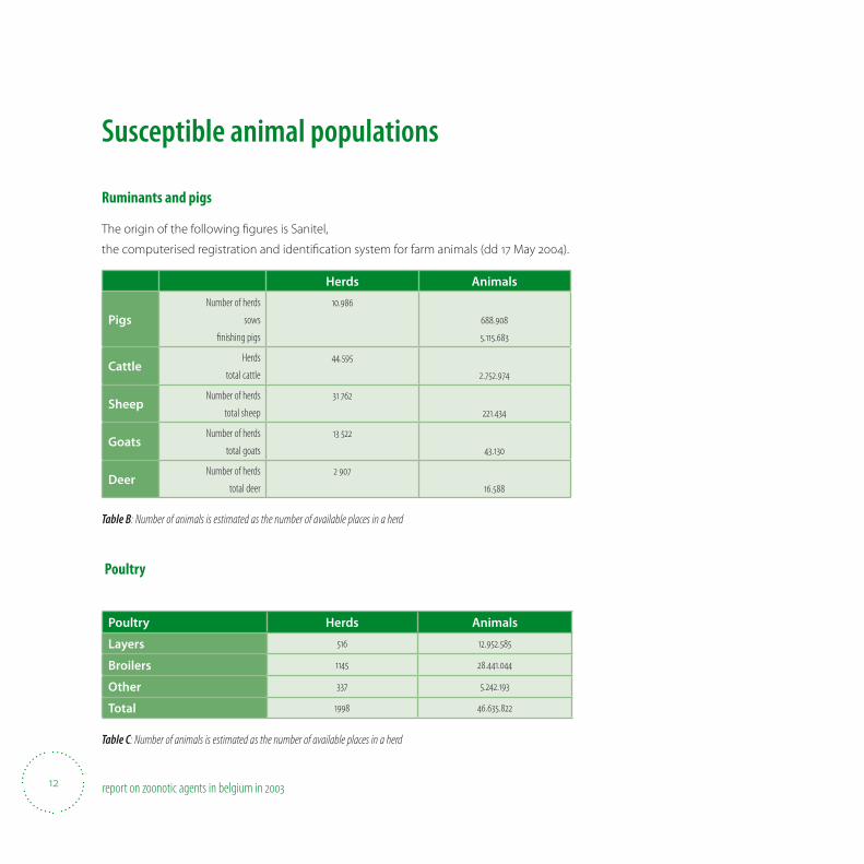
Susceptible animal populations
Ruminants and pigs
The origin of the following 8 gures is Sanitel,
the computerised registration and identi8 cation system for farm animals (dd 17 May 2004).
Herds Animals
PigsNumber of herds
sows
fi nishing pigs
.
.
..
CattleHerds
total cattle
.
..
Sheep Number of herds
total sheep
.
Goats Number of herds
total goats
.
Deer Number of herds
total deer
.
Table B: Number of animals is estimated as the number of available places in a herd
Poultry
Poultry Herds Animals
Layers ..
Broilers ..
Other ..
Total ..
Table C: Number of animals is estimated as the number of available places in a herd
13report on zoonotic agents in belgium in
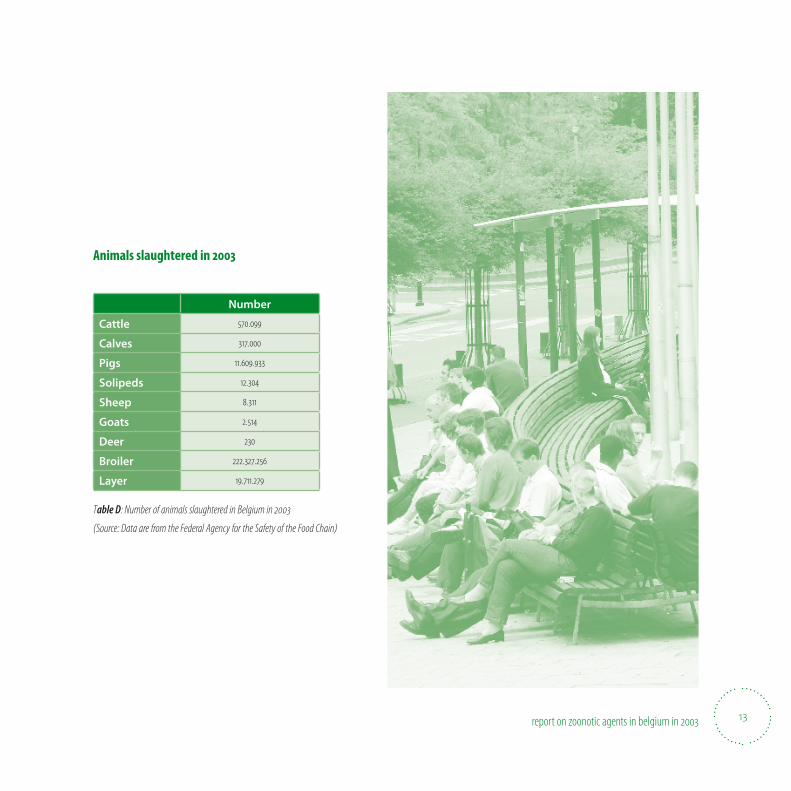
Animals slaughtered in
Number
Cattle .
Calves .
Pigs ..
Solipeds .
Sheep .
Goats .
Deer
Broiler ..
Layer ..
Table D: Number of animals slaughtered in Belgium in
(Source: Data are from the Federal Agency for the Safety of the Food Chain)
report on zoonotic agents in belgium in
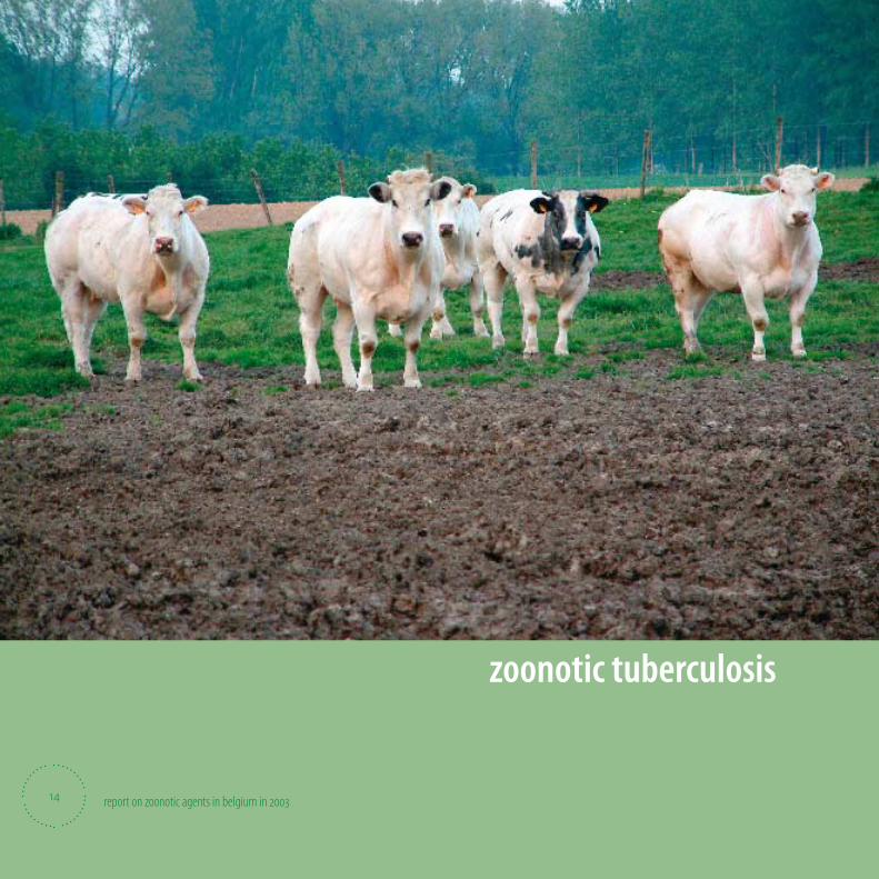
zoonotic tuberculosis
14
15report on zoonotic agents in belgium in
Zoonotic tuberculosis (Mycobacterium bovis)Tuberculosis in humans caused by M. bovis is rare. In regions where M. bovis infections in cattle are largely eliminated,
only few residual cases occur among elderly persons as a result of the reactivation of dormant M. bovis within old lesions,
and among migrants from high-prevalence countries. Agricultural workers may acquire infection by M. bovis by inhaling
cough aerosols from infected cattle and may subsequently develop typical pulmonary or genito-urinary tuberculosis.
Such patients may infect cattle through cough or urine, but evidence for human-to-human transmission is only limited.
In developing countries where M. bovis is largely prevalent among cattle, some studies reported that 3-6% of all diag-
nosed tuberculosis cases are due to M. bovis and that mostly young persons get infected by contaminated raw milk.
In human, the disease caused by M. bovis is clinically indistinguishable from that caused by M. tuberculosis. Pulmonary
tuberculosis is frequently observed but cervical lymphadenopathy, intestinal lesions, chronic skin tuberculosis and other
nonpulmonary forms are particularly common. In Belgium, 5 human cases of bovine tuberculosis were identi� ed in 2003.
Molecular typing of strains isolated from cattle and human cases is on going in order to evaluate the presence of similar
strains in both species.
Belgium is o( cially free from bovine tuberculosis since 25 June 2003 (Decision 2003/467/EC).
• Mycobacterium bovis in cattle
• Mycobacterium in other animals
• Mycobacterium in humans

16 report on zoonotic agents in belgium in
Mycobacterium bovis in cattleSurveillance system
The control of tuberculosis is based on European Directive 64/432/EEC, which is implemented and adapted in the national
legislation since 1963 and last adapted by Royal Decree of October 2002. The control implies skin testing of animals at the oc-
casion of trade and in the context of tracing contact animals. The Federal Agency for the Safety of the Food Chain is informed
about any doubtful or positive result of the skin test and may decide to re-examine (additional tests) the animals or to kill
them (test slaughter). Systematic post mortem examinations at the slaughterhouse are performed as well. In case a suspected
lesion is identi8 ed, a sample is sent to the reference laboratory for analysis. Consequently, if Mycobacterium bovis is isolated, all
animals in the herd of origin are skin tested, and a complete epidemiological investigation is made.
Isolation of M. bovis and biochemical testing is exclusively performed in the National Reference Laboratory where also
IFN-gamma and molecular typing by means of RFLP are done.
No vaccination is allowed in Belgium.
Epidemiological history and results of surveillance
Belgium is o0 cially free from bovine tuberculosis since 25 June 2003 (Decision 2003/467/EC). A total of 7 infected herds were
recognised in Belgium in 2003 (cumulative incidence over the year), which is comparable to the 10 herds noti8 ed in 2002.
Stamping out was done in 5 herds. A total of 409 animals reacted after tuberculination, which is less than the 799 reactors in
2002. This number corresponds to the intensive testing of infected and contact farms. In total 3 799 herds and 337 260 animals
were included in epidemiological investigations. The Federal Agency for the Safety of the Food Chain instructed the slaughter
of 1014 animals. During 2003, more than 950 000 € was spent on compensation.
At the end of December 2003, 44 588 herds (99.98%) with 2 752 327 animals (99,97%) were o0 cially free of tuberculosis.
Figures from the Federal Agency for the Safety of the Food Chain show that in abattoirs in 2003, 6 whole carcasses and 49 parts
were rejected on a total of 570 099. Not a single calf carcass was rejected (317 000 inspections).
17report on zoonotic agents in belgium in
Mycobacterium in other animalsDuring slaughter, the Federal Agency for the Safety of the Food Chain rejected 1 complete carcass and 87 partial carcasses of
pigs due to tuberculosis (more than 11 million inspections). No tuberculosis was registered in solipeds (mainly horses), sheep or
goats.
Mycobacterium bovis in humansIn 2003, 5 cases of M. bovis infection were detected in humans.

report on zoonotic agents in belgium in
brucellosis
18
19report on zoonotic agents in belgium in
Zoonotic brucellosis(Brucella melitensis, Brucella abortus, Brucella suis)
Bacteria of the genus Brucella may infect sheep, goats, cattle, deer, elk, pigs, dogs, and several other animals, where
they cause disease. Humans become infected by coming in contact with infected animals or with contaminated animal
products. Brucella infections in humans may cause a range of symptoms that are similar to that of E u and may include
fever, sweats, headaches, back pains, and physical weakness. Several infections of the central nervous systems or lining of
the heart may occur.
In the non-”O( cially Brucellosis Free” Mediterranean countries, the consumption of raw milk or raw cheese from sheep
and goats is thought to be the major source of contamination. In Northern European countries, besides some
occupational human cases of B. abortus infections, the majority of brucellosis cases are imported and are caused by
B. melitensis. In Belgium, less than 10 cases/year of imported B. melitensis infections have been reported over the past
few years. Last year no single case was reported. Belgian pigs and small ruminants are free of brucellosis and since the
publication of Decision 2003/467/EC in June 2003, Belgium is also o( cially free from bovine brucellosis. B. suis biovar 2 is
responsible for an enzootic brucellosis in wild boars (Sus scrofa) throughout continental EU, but is not considered to be an
important source of human brucellosis.
• Brucellosis in cattle
• Brucellosis in sheep
• Brucellosis in pigs
• Brucellosis in humans
20 report on zoonotic agents in belgium in
Brucellosis in cattleSurveillance system and methods used
Belgium is o0 cially free from bovine brucellosis since the 25 June 2003 (Decision 2003/467/EC). For this reason, the eradica-
tion programme has been changed in a surveillance programme. Beef cattle older than 2 years are monitored once every
three years by means of serological tests. The herds for serological examination are selected by geographical localisation. Dairy
cattle are checked at least 4 times a year via tank milk. Furthermore, all animals are serologically tested at trade. Each abortion
or premature birth in animals at risk is subject to compulsory noti8 cation to the Federal Agency for the Safety of the Food
Chain, and testing for brucellosis is obligatory. Aborting females should be kept in isolation until the results of the investigation
exclude Brucella infections. Pooled tank milk is examined by means of the milk ring test. For animals older than 2 years, serology
(i.e.–micro-agglutination as screening test; in case of a positive result, an indirect ELISA test is performed in parallel) is used if no
su0 cient milk ring tests are done (at least 4 ring tests a year). Bacteriological examination is done when serological and/or epi-
demiological suspicion is present. Allergic (brucellin) and IFN-gamma test may be carried out if serological cross-reactions are
suspected. These tests are performed by the Federal Agency for the Safety of the Food Chain in collaboration with the National
Reference Laboratory.
An animal is legally suspected of brucellosis in case of a positive ELISA. If, according to the epidemiology and the results of the
skin test, an animal or herd is found to be at risk, a bacteriological investigation always takes place. Hence, a brucellosis animal
is de8 ned as an animal in which Brucella has been isolated, and cattle herd is legally positive if one of its animals is bacteriologi-
cally positive for brucellosis.
Vaccination has been prohibited in Belgium since 1992.
Epidemiological history
The intensi8 ed bovine brucellosis control programme started in Belgium in 1988. In case of active brucellosis, i.e. excretion of
Brucella, the plan consisted in the culling of all animals of the infected herd (total depopulation), and was compensated for
based on the replacement value of the animals. The annual herd prevalence noti8 ed at the year end was 1,13% in 1988 and has
fallen below 0.01% since 1998. In March 2000, the last case of bovine brucellosis was identi8 ed. No infected herd was recog-
nised in Belgium in 2003. A total of 44 586 herds (99,98%) covering 2 820 258 animals (99,95%) were o0 cially free of brucellosis.
The Federal Agency for the Safety of the Food Chain instructed the slaughter of 58 animals during 2003, which is less than the
85 animals in 2002, 239 animals in 2001 and the 436 animals in 2000.
21report on zoonotic agents in belgium in
Brucellosis in sheepSurveillance system
Serum samples taken in the framework of national monitoring for Visna-Maedi and at export were examined for Brucella
melitensis speci8 c antibodies by means of ELISA. Positive samples were subsequently tested in Rose Bengal and in comple-
ment 8 xation test. A sample is classi8 ed as positive for brucellosis only if positive in all three tests. If this is the case, a skin test
should be performed on the seropositive animals and the congeners. A positive skin test leads to the bacteriological investiga-
tion of the animal. In 2001, 2002 and 2003 about 7 000 serum samples were tested at the National Reference Laboratory. In ad-
dition, 388 serum samples from sheep for export were analysed. The results con8 rmed those of previous years, i.e. the absence
of any epidemiological or bacteriological evidence of ovine brucellosis in Belgium.
Brucellosis in pigsSurveillance system and epidemiological history
Serological screening for Brucella is done for breeding pigs that are gathered (at a fair, for example), at arti8 cial insemination
centres and in animals intended for trade. The methods used are Rose Bengal, Slow Agglutination test according to Wright,
complement 8 xation test and ELISA. Bacteriological examination for Brucella and Yersinia is done in case of positive serology.
Regularly, false positive serological reactions are reported. These are due to a Yersinia enterocolitica O9 infection and are con-
8 rmed by Yersinia spp. isolation in the absence of Brucella spp. isolation. B. suis biovar 2 may be isolated from wild boars (Sus
scrofa). The infection seems to be enzootic in wild boar in Europe.
The domestic pig population is free of brucellosis (last Brucella isolation in pigs in Belgium was in 1969). It is interesting to note
that the O0 ce International des Epizooties (http://www.oie.int) considers that the value of any brucellosis serological test in
pigs is questionable.
Brucellosis in humansProbably due to the absence of a domestic animal reservoir of Brucella in Belgium, no single autochthon human brucellosis
case was reported in 2003. In addition, no case of imported B. melitensis infection was registered at the National Reference
Centre. It is helpful to note that B. suis biovar 2, circulating among wild boars, shows only limited pathogenicity for human, if
pathogenic at all.
report on zoonotic agents in belgium in
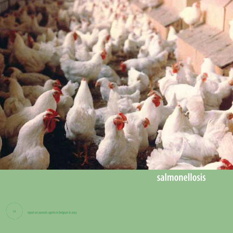
salmonellosis
22
23report on zoonotic agents in belgium in
SalmonellosisSalmonella is the major cause of bacterial food-borne infections, both in individuals and in communities. It may cause
gastro-intestinal infection with nausea, vomiting, abdominal cramps, diarrhoea and fever. In susceptible persons bacter-
aemia and septicaemia may occur. Often, food prepared with contaminated raw eggs, egg products or insu( ciently
heated poultry meat or pork are the source of the human Salmonella infection. Therefore, surveillance programmes that
timely detect Salmonella contaminations in the whole food chain (feed, living animals, slaughterhouses, cutting plants,
retail sector, restaurants) together with sanitary measures to reduce contamination are essential. Good hygienic practices
during food preparation in the kitchen and adequate refrigeration and heating also help to prevent Salmonella infections.
In 2003, a total number of 12 894 Salmonella cases were recorded in humans, which is 28% more than in 2002. This rise was due
to the signi� cant increase of serotype Enteritidis isolates that thus surpassed for the � rst time 70% of the total number of sero-
typed strains. The source of human S. Enteritidis infection was not clari� ed : serotype Enteritidis is mainly associated with poultry
(laying hens) and poultry products (especially eggs), but available � gures did not indicate a signi� cant rise at these parts of the
food chain. On the contrary, the increase in the serotype Virchow in poultry from 20 to about 30% was striking.
As for Salmonella contamination of meat, contamination of pork in retail cuts and of minced pork decreased, whereas contami-
nation of pig carcasses, broilers and spent hens remained at the same level of 2002. In pork as well as in pigs serotypes Typh-
imurium and Derby were predominant, whereas in poultry meat and in broilers, serotypes Virchow, Hadar and Paratyphi B were
mainly found. Minced beef was rarely contaminated; serotypes Dublin and Typhimurium were the most prevalent in this meat.
In animal, food and human Salmonella isolates, antibiotic resistance was most apparent against tetracycline (25 to 31%),
ampicillin (29%), sulfonamides (17 to 23%), nalidixic acid (24 to 29%) and the combination trimethoprim-sulfonamides (11
to 17%). Resistance to cefotaxime was found in 14% of the human Virchow isolates. The majority of S. Enteritidis isolates re-
mained susceptible to all antimicrobials tested. On the contrary, multi-resistance was commonly found in S. Typhimurium.
These isolates, from animals, food and humans, showed in 21 to 51% of cases resistance to ampicillin, chloramphenicol,
streptomycin, sulfonamides and tetracycline, a resistance pro� le frequently associated with phage type DT 104. Indeed,
73% of human S. Typhimurium isolates with this pentaresistance pro� le belong to phage type DT 104. Multi-resistance was
remarkable among isolates belonging to serotypes Hadar and Virchow.
• Salmonella in animal feed
• Salmonella in poultry
• Salmonella in pigeons
• Salmonella in pigs
• Salmonella in cattle
• Antibiotic resistance in strains from living animals
• Salmonella in food (meat and meat products)
• Antimicrobial resistance in strains isolated from meat
• Salmonella in humans
• Antimicrobial resistance of human isolates
24 report on zoonotic agents in belgium in
Salmonella in animal feedAn o0 cial monitoring for the detection of Salmonella in compound feedingstu; s and in raw materials was organised by the
Federal Agency for the Security of the Food Chain. The microbiological testing on 25g of sample was done at the CODA -
CERVA and the laboratory of FASFC Gembloux. In case of isolation of Salmonella in o0 cial samples no certi8 cation was pro-
vided by the FASFC.
A total of 519 raw feed materials and compound feedingstu; s were analysed. Six of these samples were found positive. The
serotypes were as follows: one Salmonella O3,10 in feed material of animal origin (8 sh meal), and 5 Salmonella in compound
feedingstu; s (1 S. Mbandaka in 8 nal product for cattle, 1 S. Thompson and 1 S. Brandenburg in 8 nal product for pigs,
2 S. Enteritidis in 8 nal product for poultry layers).
Salmonella in poultryA total of 846 Salmonella strains from poultry origin were serotyped at the CODA - CERVA in 2003, which is 24% more than in
2002 and somewhat the same number as in 2001 (n=890). Serotypes Virchow (30,0%), Enteritidis (15,6%), Typhimurium (9,0%)
and Agona (5,6%) were the most frequent.
From 403 isolates (47,6%) the production level was known. Among the breeder isolates four S. Enteritidis isolates (9,1% of
breeder strains) were detected, but 22,7% and 20,5% were serotypes Typhimurium and Virchow, respectively. Eleven isolates
from hatcheries were analysed, including 3 S. Typhimurium and 4 S. Virchow isolates. Almost 63% of layer isolates were serotype
Enteritidis, while also Typhimurium (10,4%) and to a lesser extent Virchow and Senftenberg (both 6,0%) were found. Two S. Gal-
linarum strains were identi8 ed on one of the herds that were found infected in 2002. As for broilers, especially Virchow (25,3%)
but also Typhimurium (9,6%), Agona and Paratyphi B (both 8,9%) and Enteritidis (6,8%) represented a large number of isolates.
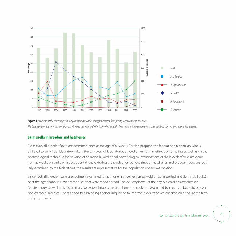
Figure A shows the evolution of the main Salmonella serotypes in poultry since 1992. The evolution of serotypes among
poultry isolates probably correctly represents the incidence of Salmonella infections in the sector due to the o0 cial monitoring
programmes. Serotype Enteritidis has increased somewhat as compared to 2002, whereas Typhimurium remained at the same
level, i.e. about 9%. Serotype Hadar is clearly of less importance among poultry, whereas the rise of serotype Virchow continues
steadily. The signi8 cant increase of Serotype Paratyphi B is noteworthy.
25report on zoonotic agents in belgium in
Figure A. Evolution of the percentages of the principal Salmonella serotypes isolated from poultry between and .
The bars represent the total number of poultry isolates per year, and refer to the right axis; the lines represent the percentage of each serotype per year and refer to the left axis.
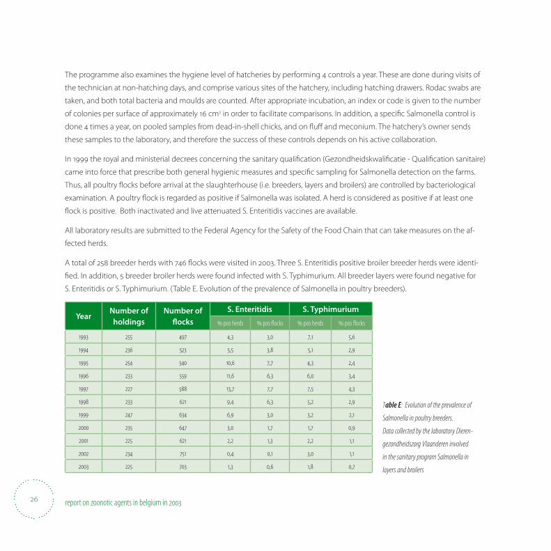
Salmonella in breeders and hatcheries
From 1993, all breeder S ocks are examined once at the age of 16 weeks. For this purpose, the federation’s technician who is
a0 liated to an o0 cial laboratory takes litter samples. All laboratories agreed on uniform methods of sampling, as well as on the
bacteriological technique for isolation of Salmonella. Additional bacteriological examinations of the breeder S ocks are done
from 22 weeks on and each subsequent 6 weeks during the production period. Since all hatcheries and breeder S ocks are regu-
larly examined by the federations, the results are representative for the population under investigation.
Since 1998 all breeder S ocks are routinely examined for Salmonella at delivery as day-old birds (imported and domestic S ocks),
or at the age of about 16 weeks for birds that were raised abroad. The delivery boxes of the day-old chickens are checked
(bacteriology) as well as living animals (serology). Imported reared hens and cocks are examined by means of bacteriology on
pooled faecal samples. Cocks added to a breeding S ock during laying to improve production are checked on arrival at the farm
in the same way.
0
10
20
30
40
50
60
70
80
90
1992 1993 1994 1995 1996 1997 1998 1999 2000 2001 2002 2003
Perc
enta
ge
0
200
400
600
800
1000
1200
Num
ber o
f iso
late
s
■ Total
■ S. Enteritidis
▲ S. Typhimurium
◆ S. Hadar
● S. Paratyphi B
■ S. Virchow
26 report on zoonotic agents in belgium in
The programme also examines the hygiene level of hatcheries by performing 4 controls a year. These are done during visits of
the technician at non-hatching days, and comprise various sites of the hatchery, including hatching drawers. Rodac swabs are
taken, and both total bacteria and moulds are counted. After appropriate incubation, an index or code is given to the number
of colonies per surface of approximately 16 cm2 in order to facilitate comparisons. In addition, a speci8 c Salmonella control is
done 4 times a year, on pooled samples from dead-in-shell chicks, and on S u; and meconium. The hatchery’s owner sends
these samples to the laboratory, and therefore the success of these controls depends on his active collaboration.
In 1999 the royal and ministerial decrees concerning the sanitary quali8 cation (Gezondheidskwali8 catie - Quali8 cation sanitaire)
came into force that prescribe both general hygienic measures and speci8 c sampling for Salmonella detection on the farms.
Thus, all poultry S ocks before arrival at the slaughterhouse (i.e. breeders, layers and broilers) are controlled by bacteriological
examination. A poultry S ock is regarded as positive if Salmonella was isolated. A herd is considered as positive if at least one
S ock is positive. Both inactivated and live attenuated S. Enteritidis vaccines are available.
All laboratory results are submitted to the Federal Agency for the Safety of the Food Chain that can take measures on the af-
fected herds.
A total of 258 breeder herds with 746 S ocks were visited in 2003. Three S. Enteritidis positive broiler breeder herds were identi-
8 ed. In addition, 5 breeder broiler herds were found infected with S. Typhimurium. All breeder layers were found negative for
S. Enteritidis or S. Typhimurium. (Table E. Evolution of the prevalence of Salmonella in poultry breeders).
YearNumber of
holdingsNumber of
3 ocksS. Enteritidis S. Typhimurium
pos herds pos fl ocks pos herds pos fl ocks
, , , ,
, , , ,
, , , ,
, , , ,
, , , ,
, , , ,
, , , ,
, , , ,
, , , ,
, , , ,
, , , ,
Table E: Evolution of the prevalence of
Salmonella in poultry breeders.
Data collected by the laboratory Dieren-
gezondheidszorg Vlaanderen involved
in the sanitary program Salmonella in
layers and broilers
27report on zoonotic agents in belgium in
There was no o0 cial surveillance system for layers. However, the industry is responsible for sampling at entrance (voluntary)
and before slaughter (compulsory). A poultry farm was regarded as positive if Salmonella was identi8 ed by bacteriology (layers
and broilers) or by serology (layers only).
Three vaccines were available, one inactivated and one attenuated S. Enteritidis vaccine and one attenuated S. Gallinarum vac-
cine only for layers.
Almost 63% of layer isolates were serotype Enteritidis, while also Typhimurium (10,4%) and Virchow and Senftenberg (both
6,0%) were frequently found. Two S. Gallinarum strains were identi8 ed on one of the herds that were found infected in 2002.
As for broilers, especially Virchow (25,3%) but also Typhimurium (9,6%), Agona and Paratyphi B (both 8,9%) and Enteritidis (6,8%)
represented a large number of isolates.
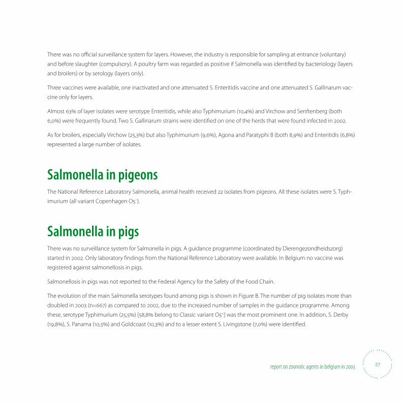
Salmonella in pigeons The National Reference Laboratory Salmonella, animal health received 22 isolates from pigeons. All these isolates were S. Typh-
imurium (all variant Copenhagen O5–).
Salmonella in pigsThere was no surveillance system for Salmonella in pigs. A guidance programme (coordinated by Dierengezondheidszorg)
started in 2002. Only laboratory 8 ndings from the National Reference Laboratory were available. In Belgium no vaccine was
registered against salmonellosis in pigs.
Salmonellosis in pigs was not reported to the Federal Agency for the Safety of the Food Chain.
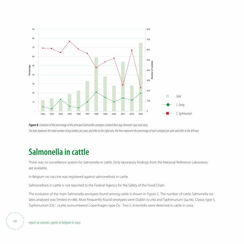
The evolution of the main Salmonella serotypes found among pigs is shown in Figure B. The number of pig isolates more than
doubled in 2003 (n=667) as compared to 2002, due to the increased number of samples in the guidance programme. Among
these, serotype Typhimurium (25,5%) [58,8% belong to Classic variant O5+] was the most prominent one. In addition, S. Derby
(19,8%), S. Panama (10,5%) and Goldcoast (10,3%) and to a lesser extent S. Livingstone (7,0%) were identi8 ed.
28 report on zoonotic agents in belgium in
-
10
20
30
40
50
60
70
80
90
1992 1993 1994 1995 1996 1997 1998 1999 2000 2001 2002 2003
Perc
enta
ge
0
100
200
300
400
500
600
700
800
Num
ber o
f iso
late
s
Figure B. Evolution of the percentage of the principal Salmonella serotypes isolated from pigs between and .
The bars represent the total number of pig isolates per year, and refer to the right axis; the lines represent the percentage of each serotype per year and refer to the left axis.
Salmonella in cattleThere was no surveillance system for Salmonella in cattle. Only laboratory 8 ndings from the National Reference Laboratory
are available.
In Belgium no vaccine was registered against salmonellosis in cattle.
Salmonellosis in cattle is not reported to the Federal Agency for the Safety of the Food Chain.
The evolution of the main Salmonella serotypes found among cattle is shown in Figure C. The number of cattle Salmonella iso-
lates analysed was limited (n=88). Most frequently found serotypes were Dublin (51,1%) and Typhimurium (34,1%). Classic type S.
Typhimurium (O5+, 73,3%) outnumbered Copenhagen type O5–. Two S. Enteritidis were detected in cattle in 2003.
■ Total
◆ S. Derby
■ S. Typhimurium
29report on zoonotic agents in belgium in
-
10
20
30
40
50
60
70
80
90
1992 1993 1994 1995 1996 1997 1998 1999 2000 2001 2002 2003
Perc
enta
ge
0
20
40
60
80
100
120
140
160
180
200
Num
ber o
f iso
late
s
Figure C. Evolution of the percentage of the principal Salmonella serotypes isolated from cattle between and .
The bars represent the total number of cattle isolates per year, and refer to the right axis; the lines represent the percentage of each serotype per year and refer to the left axis.
Antimicrobial resistance in strains isolated from living animalsData on antibiotic resistance of Salmonella strains from livestock came from the National Reference Laboratory for Salmonella,
animal health. A selection of Salmonella isolates sent to the National Reference Laboratory was also routinely analysed for
their resistance against antibiotic drugs by means of agar di; usion disks. The antimicrobial drugs tested are the beta-lactam
ampicillin (Ap), the cephalosporin ceftiofur, the aminoglycosides streptomycin (Sm), gentamicin and neomycin, tetracycline
(Tc), trimethoprim + sulfonamides (TSu) and sulfonamides (Su), the quinolones nalidixic acid (Nal) and enroS oxacin, and chlo-
ramphenicol (Cm) and Florfenicol (Ff ) In 2003 only selected serotypes were tested for antibiotic susceptibility, i.e. strains from
serotypes Agona, Dublin, Enteriditis, Hadar, Paratyphi B, Typhimurium and Virchow. Data refer to samples analysed in 2003.
Susceptibility tests were performed by the disk di; usion test, using Neo-Sensitabs (Results were accepted when results with
the QC strain were within the limits as proposed by Rosco).
■ Total
◆ S. Dublin
■ S. Typhimurium
30 report on zoonotic agents in belgium in
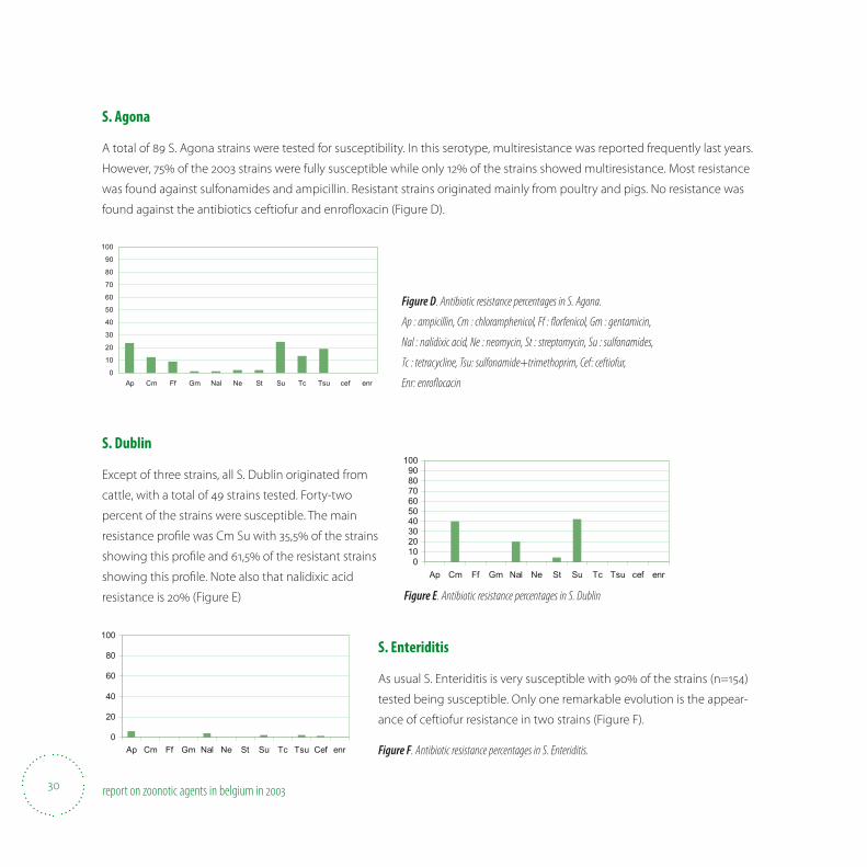
S. Agona
A total of 89 S. Agona strains were tested for susceptibility. In this serotype, multiresistance was reported frequently last years.
However, 75% of the 2003 strains were fully susceptible while only 12% of the strains showed multiresistance. Most resistance
was found against sulfonamides and ampicillin. Resistant strains originated mainly from poultry and pigs. No resistance was
found against the antibiotics ceftiofur and enroS oxacin (Figure D).
0
10
20
30
40
50
60
70
80
90
100
Ap Cm Ff Gm Nal Ne St Su Tc Tsu cef enr
Figure D. Antibiotic resistance percentages in S. Agona.
Ap : ampicillin, Cm : chloramphenicol, Ff : fl orfenicol, Gm : gentamicin,
Nal : nalidixic acid, Ne : neomycin, St : streptomycin, Su : sulfonamides,
Tc : tetracycline, Tsu: sulfonamide+trimethoprim, Cef: ceftiofur,
Enr: enrofl ocacin
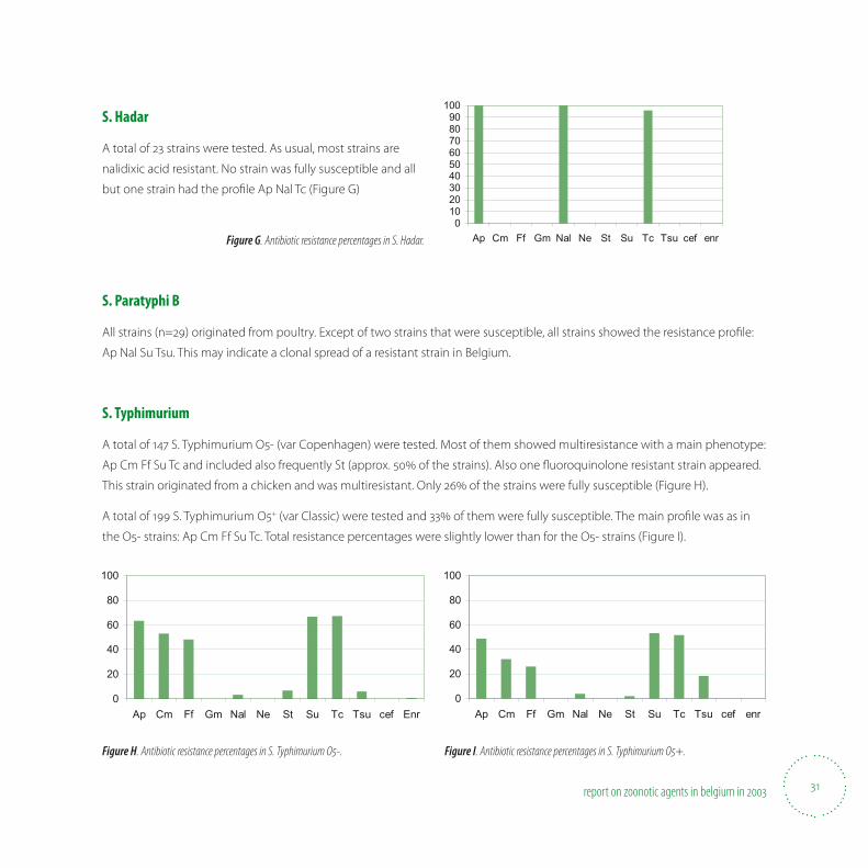
S. Dublin
Except of three strains, all S. Dublin originated from
cattle, with a total of 49 strains tested. Forty-two
percent of the strains were susceptible. The main
resistance pro8 le was Cm Su with 35,5% of the strains
showing this pro8 le and 61,5% of the resistant strains
showing this pro8 le. Note also that nalidixic acid
resistance is 20% (Figure E)
0102030405060708090
100
Ap Cm Ff Gm Nal Ne St Su Tc Tsu cef enr
0
20
40
60
80
100
Ap Cm Ff Gm Nal Ne St Su Tc Tsu Cef enr
S. Enteriditis
As usual S. Enteriditis is very susceptible with 90% of the strains (n=154)
tested being susceptible. Only one remarkable evolution is the appear-
ance of ceftiofur resistance in two strains (Figure F).
Figure E. Antibiotic resistance percentages in S. Dublin
Figure F. Antibiotic resistance percentages in S. Enteriditis.
31report on zoonotic agents in belgium in
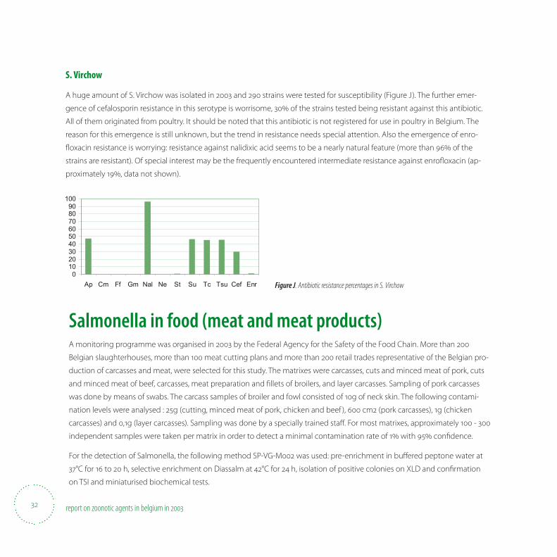
S. Hadar
A total of 23 strains were tested. As usual, most strains are
nalidixic acid resistant. No strain was fully susceptible and all
but one strain had the pro8 le Ap Nal Tc (Figure G)
0102030405060708090
100
Ap Cm Ff Gm Nal Ne St Su Tc Tsu cef enrFigure G. Antibiotic resistance percentages in S. Hadar.
S. Paratyphi B
All strains (n=29) originated from poultry. Except of two strains that were susceptible, all strains showed the resistance pro8 le:
Ap Nal Su Tsu. This may indicate a clonal spread of a resistant strain in Belgium.
S. Typhimurium
A total of 147 S. Typhimurium O5- (var Copenhagen) were tested. Most of them showed multiresistance with a main phenotype:
Ap Cm Ff Su Tc and included also frequently St (approx. 50% of the strains). Also one S uoroquinolone resistant strain appeared.
This strain originated from a chicken and was multiresistant. Only 26% of the strains were fully susceptible (Figure H).
A total of 199 S. Typhimurium O5+ (var Classic) were tested and 33% of them were fully susceptible. The main pro8 le was as in
the O5- strains: Ap Cm Ff Su Tc. Total resistance percentages were slightly lower than for the O5- strains (Figure I).
0
20
40
60
80
100
Ap Cm Ff Gm Nal Ne St Su Tc Tsu cef Enr0
20
40
60
80
100
Ap Cm Ff Gm Nal Ne St Su Tc Tsu cef enr
Figure H. Antibiotic resistance percentages in S. Typhimurium O-. Figure I. Antibiotic resistance percentages in S. Typhimurium O+.
32 report on zoonotic agents in belgium in
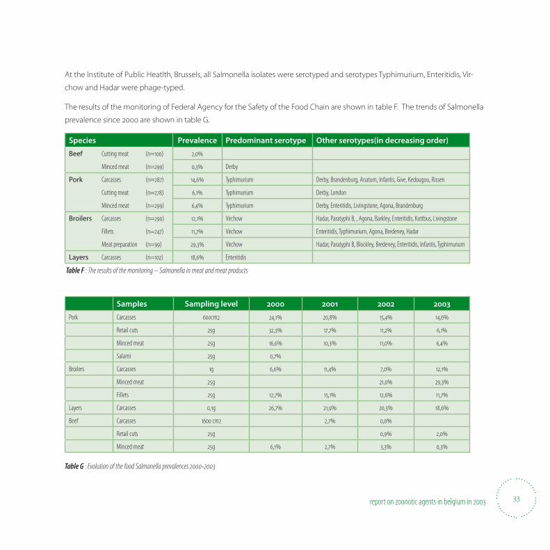
S. Virchow
A huge amount of S. Virchow was isolated in 2003 and 290 strains were tested for susceptibility (Figure J). The further emer-
gence of cefalosporin resistance in this serotype is worrisome, 30% of the strains tested being resistant against this antibiotic.
All of them originated from poultry. It should be noted that this antibiotic is not registered for use in poultry in Belgium. The
reason for this emergence is still unknown, but the trend in resistance needs special attention. Also the emergence of enro-
S oxacin resistance is worrying: resistance against nalidixic acid seems to be a nearly natural feature (more than 96% of the
strains are resistant). Of special interest may be the frequently encountered intermediate resistance against enroS oxacin (ap-
proximately 19%, data not shown).
0102030405060708090
100
Ap Cm Ff Gm Nal Ne St Su Tc Tsu Cef Enr Figure J. Antibiotic resistance percentages in S. Virchow
Salmonella in food (meat and meat products)A monitoring programme was organised in 2003 by the Federal Agency for the Safety of the Food Chain. More than 200
Belgian slaughterhouses, more than 100 meat cutting plans and more than 200 retail trades representative of the Belgian pro-
duction of carcasses and meat, were selected for this study. The matrixes were carcasses, cuts and minced meat of pork, cuts
and minced meat of beef, carcasses, meat preparation and 8 llets of broilers, and layer carcasses. Sampling of pork carcasses
was done by means of swabs. The carcass samples of broiler and fowl consisted of 10g of neck skin. The following contami-
nation levels were analysed : 25g (cutting, minced meat of pork, chicken and beef ), 600 cm2 (pork carcasses), 1g (chicken
carcasses) and 0,1g (layer carcasses). Sampling was done by a specially trained sta; . For most matrixes, approximately 100 - 300
independent samples were taken per matrix in order to detect a minimal contamination rate of 1% with 95% con8 dence.
For the detection of Salmonella, the following method SP-VG-M002 was used: pre-enrichment in bu; ered peptone water at
37°C for 16 to 20 h, selective enrichment on Diassalm at 42°C for 24 h, isolation of positive colonies on XLD and con8 rmation
on TSI and miniaturised biochemical tests.
33report on zoonotic agents in belgium in
At the Institute of Public Heatlth, Brussels, all Salmonella isolates were serotyped and serotypes Typhimurium, Enteritidis, Vir-
chow and Hadar were phage-typed.
The results of the monitoring of Federal Agency for the Safety of the Food Chain are shown in table F. The trends of Salmonella
prevalence since 2000 are shown in table G.
Species Prevalence Predominant serotype Other serotypes(in decreasing order)
Beef Cutting meat (n=) ,
Minced meat (n=) , Derby
Pork Carcasses (n=) , Typhimurium Derby, Brandenburg, Anatum, Infantis, Give, Kedougou, Rissen
Cutting meat (n=) , Typhimurium Derby, London
Minced meat (n=) , Typhimurium Derby, Enteritidis, Livingstone, Agona, Brandenburg
Broilers Carcasses (n=) , Virchow Hadar, Paratyphi B, , Agona, Barkley, Enteritidis, Kottbus, Livingstone
Fillets (n=) , Virchow Enteritidis, Typhimurium, Agona, Bredeney, Hadar
Meat preparation (n=) , Virchow Hadar, Paratyphi B, Blockley, Bredeney, Enteritidis, Infantis, Typhimurium
Layers Carcasses (n=) , Enteritidis
Table F : The results of the monitoring – Salmonella in meat and meat products
Samples Sampling level 2000 2001 2002 2003
Pork Carcasses cm , , , ,
Retail cuts g , , , ,
Minced meat g , , , ,
Salami g ,
Broilers Carcasses g , , , ,
Minced meat g , ,
Fillets g , , , ,
Layers Carcasses ,g , , , ,
Beef Carcasses cm , ,
Retail cuts g , ,
Minced meat g , , , ,
Table G : Evolution of the food Salmonella prevalences -
34 report on zoonotic agents in belgium in
Antibiotic resistance in strains isolated from meat and meat productsData on antibiotic resistance of the 178 Salmonella enterica strains isolated from food were tested for their antimicrobial
susceptibility. The disk di; usion method (Kirky-Bauer) was used following NCCLS recommendations at the Institute of Public
Health, (IPH) Food Section. The following antibiotics were tested: ampicillin (AMP), ceftriaxone (CTRX), chloramphenicol (CHL),
ciproS oxacin (CIP), kanamycin (KAN), nalidixic acid (NAL), streptomycin (STR), sulfonamides (SUL), tetracycline (TET), trimetho-
prim (TMP) and trimethoprim+sulfonamides (SXT). The results are shown in table H.
The level of resistance of Salmonella from broilers, spent hens, beef and pork is inS uenced by the serotype distribution in the
respective meat species. The presence of highly resistant serotypes as Hadar, Virchow, Paratyphi B and Typhimurium contrib-
utes mainly to the high resistance levels in some matrices.
In general, the highest resistance was found against ampicillin (29%), tetracycline (25%), sulfonamides (23%), trimethoprim-
sulfonamides (18%) and nalidixic acid (24%). No resistance was found against ciproS oxacin and kanamycin. Some isolates were
found resistant against ceftriaxone (2%), all from broilers. Overall resistance remained at the same level as in 2002 and 2001
except for nalidixic acid where an increase of resistance was noticed. No strains were found resistant to ciproS oxacin. The ap-
parition of ceftriaxone resistant strains in 2002, was con8 rmed in 2003.
Resistance data according to meat species
The highest resistance was found in Salmonellae isolated from broilers and meat preparations of chicken (n=58) (predominant
serotypes Virchow, Hadar, and Paratyphi B) : ampicillin (43% and 52% ), nalidixic acid (45% and 52%), sulfonamides (31% and 33%)
and trimethoprim (33% and 30%). 28% of the Salmonellae isolated from broilers and 23% isolated from chicken meat prepara-
tion were resistant to 5 antibiotics or more.
Pork isolates (n=78) (mainly S. Typhimurium, S. Derby and S. Brandenburg) showed a high resistance against tetracyclin (31%)
and to a less extent to sulfonamides (18%), streptomycin (17%) and ampicillin (15%). Salmonellae isolated from spent hens
belonged almost all to the serotype Enteritidis and showed very little resistance.
The few beef (n=4) isolates showed no resistance at all.
35report on zoonotic agents in belgium in
Resistance data of the most prevalent Salmonella serotypes
In total 29 S. Typhimurium strains were tested for their susceptibility. The overall resistance was high: 52% for tetracycline, 41%
for ampicillin : 34% for sulfonamides, 31% for streptomycin and 21% for chloramphenicol, trimethoprim and the combination
trimethoprim-sulfonamide. 31% of the strains were resistant to 5 antibiotics or more. No resistance was noticed against ceftriax-
one, ciproS oxacin or nalidixic acid.
All 18 S. Virchow strains were resistant against nalidixic acid (100%), and about 39% of the strains were resistant against ampicil-
lin, 33% against tetracycline, sulfonamides or trimethoprim-sulfonamides. Four strains (22%) were resistant against ceftriaxone,
all were isolated from broilers or chicken meat preparations. S.Virchow is the only serotype that presented resistant strains
against this antibiotic agent.
The majority of the 21 S. Enteritidis isolates tested were susceptible to all antimicrobials. Only one strain from a chicken 8 llet was
resistant to nalidixic acid, another strain from a spent hen was resistant to both nalidixic acid and ampicillin.
A total of 10 S. Agona isolates were tested : 70% were resistant to ampicillin, 60% to sulfonamides, 40% to trimethoprim and to
trimethoprim-sulfonamides, 30% to tetracycline and chloramphenicol and 20% to streptomycin. No resistance was observed
against ceftriaxone, nalidixic acid and ciproS oxacin.
S. Derby(n=24) showed a resistance of 25% against tetracycline, 17% against sulfonamides and streptomycin, and 8% against
trimethoprim and the combination trimethoprim-sulfonamides.
S. Paratyphi B (n=9) was 100% resistant to ampicillin and trimethoprim and 89 % to nalidixic acid.
No resistance was detected in the 16 strains of S. Bredeney that were tested.
Only 8 strains of S. Hadar were isolated in 2003. However all were resistant to ampicillin, nalidixic acid and tetracycline. Two
strains presented an additional resistance to streptomycin.
36 report on zoonotic agents in belgium in
Salmonella in humansData were obtained by a weekly updated surveillance system. In 2003, the National Reference Centre for Salmonella and
Shigella received human Salmonella isolates from 194 clinical laboratories. All isolates were serotyped by slide agglutination
with commercial antisera following the Kau; mann-White scheme. When necessary, additional biochemical tests were realized
to con8 rm the identi8 cation or to di; erentiate between the subspecies. Phage typing (Scienti8 c Institute of Public Health
– Pasteur Institute Dept)) and antimicrobial susceptibility testing were realized on isolates randomly sampled from the four
serotypes Enteritidis, Typhimurium, Hadar and Virchow. Two additional serotypes (Brandenburg and Derby) were also randomly
sampled and only tested for their antimicrobial susceptibility.
The aim of our national surveillance program (in collaboration with the Epidemiology Section of the Scienti8 c Institute of
Public Health) is to document the occurrence and trends of serovars, to detect local, regional, national or even international
outbreaks (in collaboration with the Enter-net network), to 8 nd and eliminate the source, and to suggest preventive actions
to the Federal Agency for the Safety of the Food Chain. Since 1987 a remarkable increase in the number of registered human
salmonellosis was monitored by the National Reference Centre, with a peak of 15,774 cases in 1999. This situation was chieS y
linked to the increase of Salmonella Enteritidis, the most important serotype in Belgium. From 1987 to 1999, the incidence of
laboratory-con8 rmed cases doubled to reach a value of 160/100.000 inhabitants in 1999.
Since then the total number of laboratory-con8 rmed cases fell to 14.088, 10.783 and 10.075 reports in 2000, 2001 and 2002,
respectively (Table H). In 2003, an increase in the total number of human salmonellosis was again recorded (28% more than in
2002). This resulted from the spectacular increase of the serotype Enteritidis in 2003 which exceeded for the 8 rst time 70% of
the total representativeness.
Typhimurium, the second serotype in importance, declined from 1999 until 2001 and then remained stable in ‘number of
isolates’ (although its representativeness decreased in 2003 due to the increase of Enteritidis).
37report on zoonotic agents in belgium in
1998 1999 2000 2001 2002 2003
S. Enteritidis , , , , , ,
S. Typhimurium , , , , , ,
S. Virchow , , , , , ,
S. Derby , , , , , ,
S. Brandenburg , , , , , ,
S. Hadar , , , , , ,
Others , , , , , ,
Total number
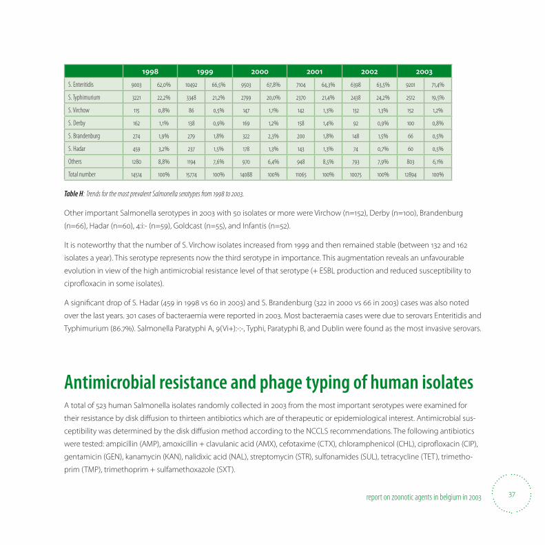
Table H : Trends for the most prevalent Salmonella serotypes from to .
Other important Salmonella serotypes in 2003 with 50 isolates or more were Virchow (n=152), Derby (n=100), Brandenburg
(n=66), Hadar (n=60), 4:i:- (n=59), Goldcast (n=55), and Infantis (n=52).
It is noteworthy that the number of S. Virchow isolates increased from 1999 and then remained stable (between 132 and 162
isolates a year). This serotype represents now the third serotype in importance. This augmentation reveals an unfavourable
evolution in view of the high antimicrobial resistance level of that serotype (+ ESBL production and reduced susceptibility to
ciproS oxacin in some isolates).
A signi8 cant drop of S. Hadar (459 in 1998 vs 60 in 2003) and S. Brandenburg (322 in 2000 vs 66 in 2003) cases was also noted
over the last years. 301 cases of bacteraemia were reported in 2003. Most bacteraemia cases were due to serovars Enteritidis and
Typhimurium (86.7%). Salmonella Paratyphi A, 9(Vi+):-:-, Typhi, Paratyphi B, and Dublin were found as the most invasive serovars.
Antimicrobial resistance and phage typing of human isolatesA total of 523 human Salmonella isolates randomly collected in 2003 from the most important serotypes were examined for
their resistance by disk di; usion to thirteen antibiotics which are of therapeutic or epidemiological interest. Antimicrobial sus-
ceptibility was determined by the disk di; usion method according to the NCCLS recommendations. The following antibiotics
were tested: ampicillin (AMP), amoxicillin + clavulanic acid (AMX), cefotaxime (CTX), chloramphenicol (CHL), ciproS oxacin (CIP),
gentamicin (GEN), kanamycin (KAN), nalidixic acid (NAL), streptomycin (STR), sulfonamides (SUL), tetracycline (TET), trimetho-
prim (TMP), trimethoprim + sulfamethoxazole (SXT).
38 report on zoonotic agents in belgium in
Resistance was mostly found against tetracycline (31.3%), ampicillin (29.9%), nalidixic acid (29.5%), and to a lesser extent against
streptomycin (20.6%), sulfonamides (17.2%) and trimethoprim + sulfamethoxazole (11.6%).
All S. Hadar isolates (n=42) were resistant to at least two antibiotis. The highest antibiotic resistance levels were observed for
this serotype. Resistance to tetracycline, nalidixic acid, ampicillin and streptomycin reached values from 71.4 up to 90.5% (Table
I). Simultaneous resistance to these four antibiotics was observed in 57% of these isolates. However, isolates from this serotype
remained fully sensitive to cefotaxime, chloramphenicol, and gentamicin.
S. Typhimurium (n=314) also showed a high level of antibiotic resistance with 29% of isolates resistant to four or more antimi-
crobial agents (de8 ned as multiresistance). Eighteen percent of the isolates were shown resistant to ampicillin, chlorampheni-
col, streptomycin, sulfonamides and tetracycline (R-type ACSSuT with or without additional resistances), of which 73% were of
de8 nitive phage type (DT)104. Three full resistances against ciproS oxacin were also detected in multiresistant S. Typhimurium
isolates (all three from phagetype 12/ad).
Multiresistance was also common in S. Virchow (n=44; 60% of the isolates in place of 29.7% in 2002). The highest incidence of
resistance was observed for nalidixic acid (86.4%). Resistances to ampicillin, tetracycline and to trimethoprim + sulfamethoxa-
zole were common (> 50%). Resistance to cefotaxime was found in 14% of the S. Virchow mutiresistant isolates.
In contrast, the vast majority of S. Enteritidis (95.9%), S. Brandenburg (93.5%) and S. Derby (93%) isolates remained sensitive to
all tested antibiotics.
Resistance patterns and levels in 2003 were generally the same than those in 2002 and 2001, except for the serotype Virchow for
which a signi8 cant increase of resistance against cefotaxime, tetracycline, sulfonamides, and trimethoprim + sulfamethoxazole
was observed (Table I). Co-trimoxazole resistance (trimethoprim + sulfamethoxazole) is also appearing in the serotype Hadar.
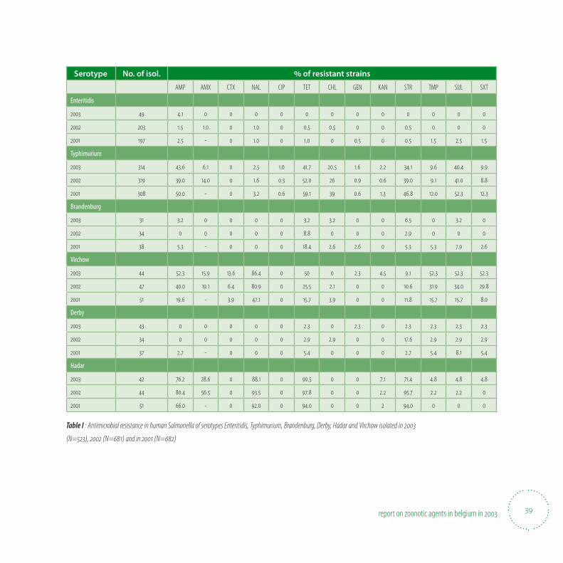
39report on zoonotic agents in belgium in
Serotype No. of isol. % of resistant strains
AMP AMX CTX NAL CIP TET CHL GEN KAN STR TMP SUL SXT
Enteritidis
.
. . . . . .
. - . . . . . . .
Typhimurium
. . . . . . . . . . . .
. . . . . . . . . . .
. - . . . . . . . . .
Brandenburg
. . . . .
. .
. - . . . . . . .
Virchow
. . . . . . . . . .
. . . . . . . . . .
. - . . . . . . . .
Derby
. . . . . .
. . . . . .
. - . . . . .
Hadar
. . . . . . . . .
. . . . . . . .
. - . . .
Table I : Antimicrobial resistance in human Salmonella of serotypes Enteritidis, Typhimurium, Brandenburg, Derby, Hadar and Virchow isolated in
(N=), (N=) and in (N=)
report on zoonotic agents in belgium in
trichinellosis
40
41report on zoonotic agents in belgium in
TrichinellaTrichinella is an intestinal parasite whose larvae can be present in the muscles of di3 erent animal species, and is trans-
ferred to humans by the consumption of contaminated raw or rare meat. Therefore, pork, wild boar and horsemeat are
examined before marketing, except when appropriately frozen.
Carcasses found positive for the presence of Trichinella are declared un� t for consumption. It is recommended to
travellers not to import raw meats of susceptible animals, e.g. sausages, bear; and not to consume meats of unknown
quality abroad.
Human pathology: after a 1 to 4 weeks incubation, trichinellosis can cause myalgia, fever, eosinophilia, facial oedema,
myocarditis. Trichinella has not been detected in carcasses of animal species produced for human consumption in
Belgium for years.
• Trichinella in food animals
42 report on zoonotic agents in belgium in
Trichinella in food animals
Surveillance system and methods used
Pig carcasses intended for export and all locally slaughtered horses and wild boars marketed were checked for Trichinella.
The examination is done by magnetic stirrer digestion of pooled 100 gram samples (1 g in case of pig, 5 g in case of boar and
horse) as described in Directive 77/96/EEC. Serology may be done in live pigs and in epidemiological studies on wildlife.
Noti8 cation to the Federal Agency for the Safety of the Food Chain is compulsory.
Results of the investigations in
A total of 10. 226.408 pigs, 12.304 solipeds (mainly horses) and 8.834 wild boars were examined. Not one sample was found posi-
tive for Trichinella.

43report on zoonotic agents in belgium in
report on zoonotic agents in belgium in
rabies
44
45report on zoonotic agents in belgium in
RabiesRabies is an acute viral encephalomyelitis of warm blooded animals (foxes, dogs, cats, bats …) including human beings.
The disease is caused by a Lyssavirus (8 genotypes), which is spread in the saliva of infected animals. In humans, the in-
ability to swallow liquids has given the disease the name of hydrophobia.
Infected animals pass on the infection especially through bites or scratches, or less frequently via the injured skin or mu-
cous membranes. The incubation period is usually from 4 to 8 weeks, but may range from 10 days to as long as one year
or more. If not treated, human rabies is almost always fatal. Administration of rabies post-exposure prophylaxis combin-
ing wound treatment, passive immunization and vaccination are e3 ective when appropriately applied. Pre-exposure
vaccination should be o3 ered to persons at risk, such as laboratory workers, veterinarians, animal handlers, international
travellers.
In July 2001, Belgium has obtained the o( cial status of rabies-free country according to the WHO recommendations. No
indigenous cases of human rabies have been reported since 1923 although cases imported from Africa are diagnosed from
time to time.
• Rabies in animals
46 report on zoonotic agents in belgium in
Rabies in animals
Surveillance system and methods used
Food animals with nervous symptoms are suspect for rabies and therefore these cases have to be noti8 ed to the veterinary of-
8 cer. Wildlife found dead or shot should also be transferred to the Federal Agency for the Safety of the Food Chain.
A; ected animals are killed, and their brain is examined by immunoS uorescence and virus cultivation in neuroblasts at the
National Reference Laboratory.
Vaccination policy
Vaccine baits (Raboral, Rhône-Mérieux) were dispersed for the vaccination of foxes. In April and October 2003, a zone of ap-
proximately 1 800 km2 along the German border was covered by spreading 32 000 baits by means of a helicopter (17.78 baits
per km2). Since there were no more cases of rabies for the last years, vaccination of foxes by baits will be stopped in 2004.
In the south of the country, below the rivers Sambre and Meuse, vaccination of dogs and cats is compulsory. In addition, all
pets staying on any Belgian public camping must be vaccinated.
Results of the investigations in
A total of 615 analyses were done at the National Reference Laboratory. The majority of samples originated from foxes (44%)
and cattle (41%). The high percentage for cattle is the consequence of the surveillance system for TSE in cattle: all suspected
cases were 8 rst examined for rabies. Rabies must be considered in the di; erential diagnosis of BSE, although the course of the
disease is usually shorter.
None of the samples was found positive. Since the last indigenously acquired case of rabies occurred in Belgium in a bovine in
July 1999, Belgium obtained the o0 cial status of rabies-free country in July 2001 according to the WHO recommendations.
47report on zoonotic agents in belgium in
report on zoonotic agents in belgium in
campylobacteriosis
48
49report on zoonotic agents in belgium in
CampylobacteriosisCampylobacter is a leading source of bacterial foodborne gastrointestinal diseases in humans in all parts of the world.
It can also cause postinfectious complications as Guillain-Barré syndrome.
In 80% of the cases, the infection route of campylobacteriosis is food, but domestic animals including pets are also
involved. The transmission of this pathogen to humans is mostly due to consumption of undercooked poultry, pork and
beef, unpasteurized milk, contaminated drinking water, or contacts with the faeces of infected pets. This chapter will focus
on Campylobacter jejuni and Campylobacter coli that are the main causes of enteritis in humans1.
The contamination of poultry carcasses and meat with Campylobacter are monitored since 2000 by the Federal Agency
for the Safety of the Food Chain. The rate of positive poultry samples is stable, but high. Chicken and layer meat have to
be well cooked and cross-contamination should be avoided during preparation.
• Campylobacter in food
• Campylobacter in humans
1 J.Hu and D.J. Kopecko. Campylobacter species. In: International handbook of foodborne pathogens. p181-198 Ed. M.D. Milliotis and J.W. Bier. Marcel Dekker, New York, 2003.
50 report on zoonotic agents in belgium in
Campylobacter in food
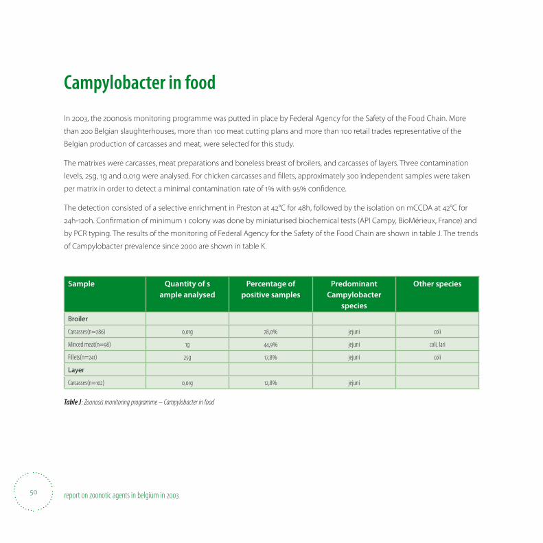
In 2003, the zoonosis monitoring programme was putted in place by Federal Agency for the Safety of the Food Chain. More
than 200 Belgian slaughterhouses, more than 100 meat cutting plans and more than 100 retail trades representative of the
Belgian production of carcasses and meat, were selected for this study.
The matrixes were carcasses, meat preparations and boneless breast of broilers, and carcasses of layers. Three contamination
levels, 25g, 1g and 0,01g were analysed. For chicken carcasses and 8 llets, approximately 300 independent samples were taken
per matrix in order to detect a minimal contamination rate of 1% with 95% con8 dence.
The detection consisted of a selective enrichment in Preston at 42°C for 48h, followed by the isolation on mCCDA at 42°C for
24h-120h. Con8 rmation of minimum 1 colony was done by miniaturised biochemical tests (API Campy, BioMérieux, France) and
by PCR typing. The results of the monitoring of Federal Agency for the Safety of the Food Chain are shown in table J. The trends
of Campylobacter prevalence since 2000 are shown in table K.
Sample Quantity of sample analysed
Percentage of positive samples
Predominant Campylobacter
species
Other species
Broiler
Carcasses(n=) ,g , jejuni coli
Minced meat(n=) g , jejuni coli, lari
Fillets(n=) g , jejuni coli
Layer
Carcasses(n=) ,g , jejuni
Table J : Zoonosis monitoring programme – Campylobacter in food
51report on zoonotic agents in belgium in
Sampling level 2000 2001 2002 2003
Broilers Carcasses ,g , , , ,
Fillets g , , , ,
Minced meat g , ,
Layers Carcasses ,g , , , ,
Table K : Evolution of the food Campylobacter prevalences -
Campylobacter in humans
Data were obtained from passive surveillance through sentinel laboratory results. All cases were updated weekly.
From 2000 to 2002, the number of diagnosed cases was stable and 71% of the cases were located in Flanders. In 2003, the
number of cases decreased by 11% and this reduction was observed in most districts of Flanders.
1996 1997 1998 1999 2000 2001 2002 2003
Number of isolates
Table L : Campylobacter in humans
report on zoonotic agents in belgium in
echinococcosis
52
53report on zoonotic agents in belgium in
Echinococcosis Echinococcosis is caused either by Echinococcus granulosus or Echinococcus multilocularis.
Echinococcus granulosus produces unilocular human hydatidosis. It is a small tapeworm (6 mm) that lives in the small
intestine of domestic and wild canids. Sheep and cattle serve as intermediate hosts for the infection. Humans acquire
infection by ingestion of typical taeniid eggs, which are excreted in the faeces of infected dogs. When eggs are ingested
by the intermediate hosts or by humans, the oncospheres liberated from the eggs migrate via the bloodstream to the
liver, lungs and other tissues to develop in hydatid cysts. Within the cyst brood capsules and protoscoleces develop. Each
protoscolex is a potentially infective organism for canids. Indigenous unilocular hydatidosis in man has been reported
in Belgium. Echinococcus multilocularis causes alveolar (multilocular) echinococcosis in humans. Foxes and dogs are the
de� nitive hosts of this parasite and small rodents the intermediate hosts. In the liver of rodents the invasive larval stage
has a multi-compartimented appearance containing many protoscoleces. Ingestion of the eggs by humans can result in
the development of invasive cysts in the liver.
In Belgium, the percentage of infected foxes varies with the region, with a decreasing rate from the South-East to the
North-West: e.g 33% in the Ardennes, 13% in the Condroz region and 2% in Flanders. The endemic region is situated under
the river Meuse, on the heights of the Ardennes.
Only six human cases of alveolar echinococcosis have been detected in Belgium since 1999, thanks to an e( cient
information campaign in wooded areas.
Surveillance system and results:
Post-mortem macroscopic examination is performed at
the slaughterhouse in the domestic intermediate hosts:
cattle, sheep, horses and pigs. Whole carcasses or parts
are rejected in case Echinococcus granulosus cysts were
found.
The following partial rejections were noted by the Federal
Agency for the Safety of the Food Chain in 2003: 200 cases
of adult cattle and 3 of sheep. Echinococcus granulosus
was not detected in calves, pigs, goats or wild boars.
report on zoonotic agents in belgium in
listeriosis
54
55report on zoonotic agents in belgium in
ListeriosisListeria monocytogenes has become a major concern for the food industry and public health authorities. Ingestion of
food contaminated with Listeria monocytogenes may cause either a serious invasive illness a3 ecting people with altered
or de� cient immune responses, or a non-invasive febrile gastro-enteritidis. Although the incidence of listeriosis is low,
the high mortality rate, which often reaches as high as 30-40%, requires early diagnosis and appropriate antimicrobial
therapy.
Listeriosis is transmitted to humans via contact with animals, cross-infection of foetus or newborn babies and foodborne
infection2. Listeria is ubiquitous and widely distributed in the environment (soil, vegetables, meat, milk, � sh). All food as-
sociated with Listeria monocytogenes outbreaks were consumed without further processing or after minimal heat treat-
ment, and many of them had a suitable environment for growth3.
The contamination of food in the Belgian surveillance plan of the Federal Agency for the Safety of the Food Chain is
stable, except for the chicken meat preparation (containing raw minced meat) that is very high in 2003. The number of hu-
man cases has almost doubled in comparison with 2002, but no large-scale listeriosis outbreak was reported.
General food hygiene rules are essential for the prevention of human listeriosis. As some persons are at high risk (preg-
nant women, immunocompromised people), they are advised not to eat certain categories of food with proven elevated
risk of Listeria monocytogenes contamination, such as unpasteurized milk and butter, soft cheeses and ice cream made
from unpasteurized milk, any soft cheese crust, smoked � sh, pâté, cooked ham, salami, cooked meat in jelly, raw minced
meat from beef, pork and poultry, steak tartar, raw � sh and shell� sh (oysters, mussels, shrimps), � sh, meat and surimi
salads, insu( ciently rinsed raw vegetables, unpeeled fruit.
• Listeria monocytogenes in food
• Listeria monocytogenes in humans
2 C. Bell and A. Kyriakides. Listeria. A practical approach to the organism and its control in foods. Blackie academic & profes-sional, London, 1998.
3 A.R. Datta. Listeria monocytogenes. In: International handbook of foodborne pathogens. p105-121 Ed. M.D. Milliotis and J.W. Bier. Marcel Dekker, New York, 2003.
56 report on zoonotic agents in belgium in
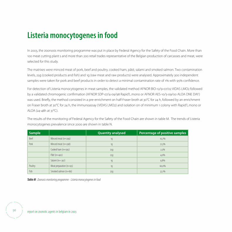
Listeria monocytogenes in food
In 2003, the zoonosis monitoring programme was put in place by Federal Agency for the Safety of the Food Chain. More than
100 meat cutting plant s and more than 200 retail trades representative of the Belgian production of carcasses and meat, were
selected for this study.
The matrixes were minced meat of pork, beef and poultry, cooked ham, pâté, salami and smoked salmon. Two contamination
levels, 25g (cooked products and 8 sh) and 1g (raw meat and raw products) were analysed. Approximately 300 independent
samples were taken for pork and beef products in order to detect a minimal contamination rate of 1% with 95% con8 dence.
For detection of Listeria monocytogenes in meat samples, the validated method AFNOR BIO-12/9-07/02 VIDAS LMO2 followed
by a validated chromogenic con8 rmation (AFNOR SDP-07/4-09/98 Rapid’L.mono or AFNOR AES-10/3-09/00 ALOA ONE DAY)
was used. BrieS y, the method consisted in a pre-enrichment on half-Fraser broth at 30°C for 24 h, followed by an enrichment
on Fraser broth at 30°C for 24 h, the immunoassay (VIDAS LMO2) and isolation on of minimum 1 colony with Rapid’L.mono or
ALOA (24-48h at 37°C).
The results of the monitoring of Federal Agency for the Safety of the Food Chain are shown in table M. The trends of Listeria
monocytogenes prevalence since 2000 are shown in table N.
Sample Quantity analysed Percentage of positive samples
Beef Minced meat (n=) g ,
Pork Minced meat (n=) g ,
Cooked ham (n=) g ,
Pâté (n=) g ,
Salami (n= ) g ,
Poultry Meat preparation (n=) g ,
Fish Smoked salmon (n=) g ,
Table M : Zoonosis monitoring programme - Listeria monocytogenes in food
57report on zoonotic agents in belgium in
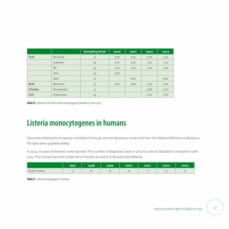
Sampling level 2000 2001 2002 2003
Pork Minced meat g , , , ,
Cooked ham g , , , ,
Pâté g , , , ,
Salami g ,
Salami g , ,
Beef Minced meat g , , , ,
Chicken Meat preparation g , ,
Fish Smoked salmon g , ,
Table N: Evolution of the food Listeria monocytogenes prevalences -
Listeria monocytogenes in humans
Data were obtained from passive surveillance through sentinel laboratory results and from the National Reference Laboratory.
All cases were updated weekly.
In 2003, 76 cases of listeriosis were reported. The number of diagnosed cases in 2003 has almost doubled in comparison with
2002. This increase has been observed in Flanders as well as in Brussels and Wallonia.
1997 1998 1999 2000 2001 2002 2003
Number of isolates
Table O : Listeria monocytogenes in humans
report on zoonotic agents in belgium in
yersiniosis
58
59report on zoonotic agents in belgium in
Yersinia enterocoliticaY. enterocolitica is a relatively infrequent cause of diarrhea and abdominal pain. Infection with Y. enterocolitica occurs most
often in young children. Common symptoms in children are fever, abdominal pain, and diarrhea, which is often bloody.
Symptoms typically develop 4 to 7 days after exposure and may last 1 to 3 weeks or longer. In older children and adults,
right-sided abdominal pain and fever may be the predominant symptoms, and may be confused with appendicitis. In a
small proportion of cases, complications such as skin rash, joint pains, or spread of bacteria to the bloodstream can occur.
Only a few strains of Y. enterocolitica cause illness in humans. The major animal reservoir for Y. enterocolitica strains that
cause human illness is pigs, but other strains are also found in many other animals including rodents, rabbits, sheep, cat-
tle, horses, dogs, and cats. In pigs, the bacteria are most likely to be found on the tonsils. Infection is most often acquired
by eating contaminated food, especially raw or undercooked pork products. Drinking contaminated unpasteurized milk
or untreated water can also transmit the infection4.
• Yersinia enterocolitica in food
• Yersinia enterocolitica in humans
4 U.S. Department of Health and Human Services, Centers for Disease Control and Prevention, http://www.cdc.gov/az.do
60 report on zoonotic agents in belgium in
Yersinia enterocolitica in food
Surveillance system
The FASFC organised a food surveillance of meat and meat products in 1997, which showed a very low prevalence of meat and
meat products from pork, beef and poultry.
No food surveillance programme was organised in 2003.
Yersiniosis in humans
Surveillance system
Data were obtained from passive surveillance through sentinel laboratory 8 ndings. All cases were updated on a weekly base.
Results of the investigations in and epidemiological evolution
The number of cases reported for human yersiniosis was 338 in 2003. As compared to 330 in 2002, 375 in 2001 and 507 cases in
2000, there is a clear stabilization in the number of infections reported in humans in Belgium.
61report on zoonotic agents in belgium in
report on zoonotic agents in belgium in
verotoxin producing escherichia coli (vtec)
62

63report on zoonotic agents in belgium in
Verotoxin producing Escherichia coliZoonotic verotoxin producing E. coli may cause life-threatening diseases in young children or in immunocompromised
or elderly people, i.e. hemorrhagic colitis, hemorrhagic uremic syndrome (HUS) and even death. E. coli O157 is the best
known and most studied VTEC. Cattle are often indicated as the principal reservoir of VTEC, but are not clinically a3 ected
by zoonotic VTEC infection. Infection of humans takes place via consumption of contaminated food, through contact with
contaminated water, or by direct transmission of VTEC from infected humans or animals. Therefore, prevention mainly
relies on hygienic measures.
In 2003, typical E. coli O157 (i.e. verotoxigenic, eaeA-positive) were found on cattle carcasses, in sliced and minced meat in
0.7%, 0.7% and 1.7% of the cases, respectively. Living cattle were not analysed in this o( cial monitoring programme.
As for VTEC infections in humans, approximately 40 verotoxin producing E. coli strains are annually analysed at the Na-
tional Reference Laboratory. About half of these belong to serogroup O157. Eight typical EHEC isolates were from children
between 10 months and 11 years old su3 ering from HUS: 5 belonged to serotype O157:H7, 2 to O157:H- and one to O145:H-.
Since only few clinical laboratories examine human stools for the presence of E. coli O157, the incidence of VTEC among
humans cannot be correctly estimated.
• Verotoxin producing Escherichia coli in cattle
• Escherichia coli O157 in food
• Verotoxinogenic Escherichia coli in humans
64 report on zoonotic agents in belgium in
Verotoxin producing Escherichia coli in cattleSurveillance systems and methods used
In case E. coli O157 was isolated from a carcass at the slaughterhouse (o0 cial zoonosis programme), the farm of origin was
traced back. Faecal samples were taken from twenty percent of the animals aged between 6 months and 2 years. In addition,
samples of the available feed and of dust were collected. Hygienic and management measures were imposed on these farms
during the period that the samples were analysed in the laboratory. The sale of not heat-treated milk or milk products was
forbidden.
The faecal, feed and dust samples were enriched in mTSB and treated by immunomagnetic separation. Subsequently, the sus-
pected colonies on CT-SMAC were latex agglutinated for the detection of E. coli O157. Con8 rmation of serotype (O group) was
done by means of slow tube agglutination after heating of the bacterial cultures. Virulence factors were determined by PCR
for toxin genes vt1 and vt2, and for eae (intimin) speci8 c sequences. A typical E. coli O157 isolate is de8 ned as a strain isolated
by immunomagnetic separation and O157 speci8 c agglutination and con8 rmed by PCR as vt2 and eae positive. An atypical E.
coli O157 had either no eae or vt gene. Laboratory 8 ndings are available on clinical E. coli strains sent to the National Reference
Laboratory for VTEC, animal health for analysis. A VTEC strain was identi8 ed as a VT1 or VT2 positive E. coli strain.
Results of the investigations in
Monitoring. In 2003 only 4 herds were sampled following identi8 cation of E. coli O157 on carcasses in the slaughterhouse. On
three herds E. coli O157 VT2 eae was isolated and on one herd E. coli O157 without vt (atypical EHEC). Of the 184 bovine E. coli
strains from clinical cases analysed in 2003 at the National Reference Laboratory, only 6 were VTEC. Of these, 5 were of patho-
type VT1 eae (known to be associated with diarrhea), and 1 was VT1.
Escherichia coli O in food Surveillance system and method used
In 2003, the zoonosis monitoring programme developed by the Federal Agency for the Safety of the Food Chain (FASFC) was
implemented in over 200 Belgian abattoirs, over 100 meat cutting plants and over 100 retail outlets representative of the Bel-
gian production of carcasses and meat. The samples tested were carcasses (1600cm2), cutting meat (25g) and minced meat of
beef (25g). Sta; of the FASFC was speci8 cally trained to obtain uniform results.
65report on zoonotic agents in belgium in
The detection method (SP-VG-MOO1) consisted of a pre-enrichment in mTSB + novobiocin at 42°C for 6 hours. After that,
enrichment was done in CT-Mac Conkey at 37°C for 16-18 hours and subsequent testing in the immunoassay O157 (VIDAS ECO,
bioMérieux). Further on followed a selective immunomagnetic enrichment (Dynabeads, Dynal or VIDAS ICE, bioMérieux) and
an isolation on sorbitol-Mac Conkey that was incubated at 42°C for 18 h. Isolation and con8 rmation were by means of agglu-
tination of latex particles (Oxoid). Suspected isolates were sent to the National Reference Laboratory for detection of genes
encoding virulence factors.
Results of the investigations in
The results of the monitoring of Federal Agency for the Safety of the Food Chain are shown in table P.
Sample Prevalence(95% con� dence interval)
Beef Carcasses (n=) , [, – ,]
Cutting meat (n=) , [, – ,]
Minced meat (n=) , [, – ,]
Table P : Zoonosis monitoring programme - E. coli O.
Verotoxinogenic Escherichia coli in humans Data were obtained from the National Reference Laboratory. In 2003, 41 typical EHEC isolates were identi8 ed, that is VTEC with
virulence gene eae (intimin) and with the gene encoding enterohemolysin (EHEC virulence plasmid). The majority of these
isolates (n=21) belonged to serogroup O157, and 18 were O157:H7. Eight typical EHEC isolates were from children between 10
months and 11 years old su; ering from HUS: 5 belonged to serotype O157:H7, 2 to O157:H- and one to O145:H-. In addition, 6
atypical EHEC isolates (without intimin or enterohemolysin genes) were characterised.
Fifteen of the 21 E. coli O157 strains produced only VT2 and 6 strains both VT1 and VT2. Fifteen of the 20 typical non-O157 EHEC
produced only VT1 and 3 only VT2 and 2 produced both VT1 and VT2. Three of the 6 atypical non-O157 isolates produced VT1,
one isolate VT2 and 2 produced both VT1 and VT2. During the last seven years, the number of isolates analysed annually by the
Reference Laboratory has been rather constant.
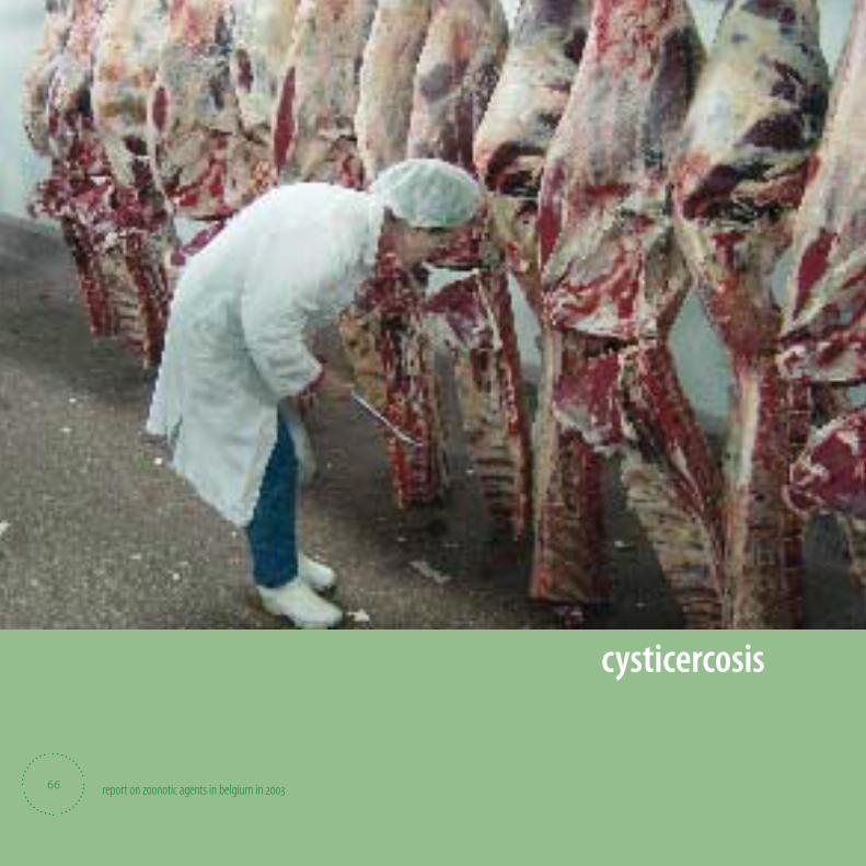
report on zoonotic agents in belgium in
cysticercosis
66
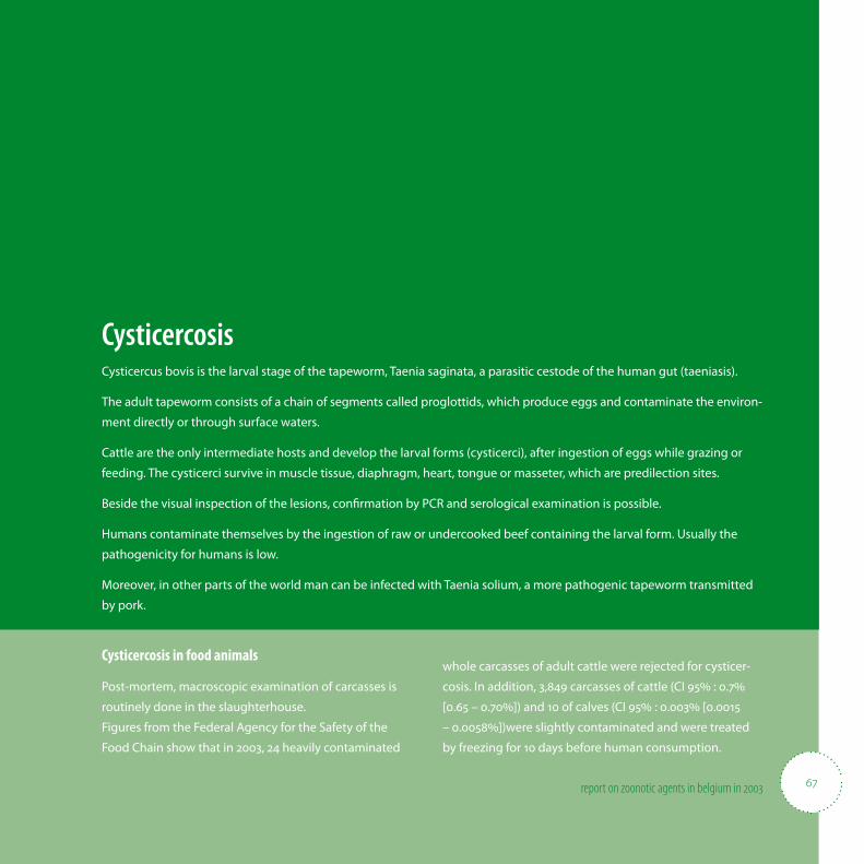
67report on zoonotic agents in belgium in
CysticercosisCysticercus bovis is the larval stage of the tapeworm, Taenia saginata, a parasitic cestode of the human gut (taeniasis).
The adult tapeworm consists of a chain of segments called proglottids, which produce eggs and contaminate the environ-
ment directly or through surface waters.
Cattle are the only intermediate hosts and develop the larval forms (cysticerci), after ingestion of eggs while grazing or
feeding. The cysticerci survive in muscle tissue, diaphragm, heart, tongue or masseter, which are predilection sites.
Beside the visual inspection of the lesions, con� rmation by PCR and serological examination is possible.
Humans contaminate themselves by the ingestion of raw or undercooked beef containing the larval form. Usually the
pathogenicity for humans is low.
Moreover, in other parts of the world man can be infected with Taenia solium, a more pathogenic tapeworm transmitted
by pork.
Cysticercosis in food animals
Post-mortem, macroscopic examination of carcasses is
routinely done in the slaughterhouse.
Figures from the Federal Agency for the Safety of the
Food Chain show that in 2003, 24 heavily contaminated
whole carcasses of adult cattle were rejected for cysticer-
cosis. In addition, 3,849 carcasses of cattle (CI 95% : 0.7%
[0.65 – 0.70%]) and 10 of calves (CI 95% : 0.003% [0.0015
– 0.0058%])were slightly contaminated and were treated
by freezing for 10 days before human consumption.
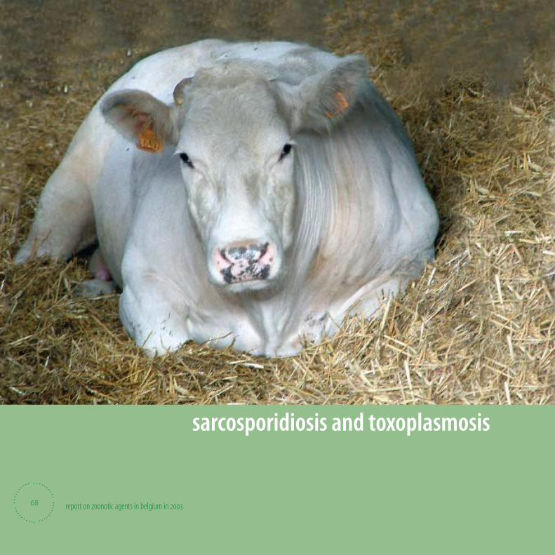
report on zoonotic agents in belgium in
sarcosporidiosis and toxoplasmosis
68
69report on zoonotic agents in belgium in
Sarcosporidiosis and ToxoplasmosisSarcocystis hominis (bovine as intermediate host), Sarcocystis suihominis, Toxoplasma gondii
Domestic carnivores are hosts of the adult stage of the protozoans Sarcocystis spp and Toxoplasma,
with many species as intermediate hosts.
Human can be a de� nitive (sarcosporidiosis) or intermediate host (toxoplasmosis) by ingestion of infected meat
or excreted oocysts and develop symptoms like diarrhoea, headache, eosinophilia, abortion, congenital disorder.
The majority of adult persons have acquired a degree of immunity to re-infection but can remain carrier for
toxoplasmosis, while for human sarcosporidiosis there is no immunity development.
The majority of grazing animals are inapparent carriers of tissue cysts. There is a need for suitable microsopic, serological
and molecular biological methods for the detection.
Surveillance system in food animals
Carcasses are partially or enterily condemned when lesions
of sarcosporidiosis or toxoplasmosis are apparent.
Number of partial rejections of cattle in 2003 because of
sarcosporidiosis lesions: 14.
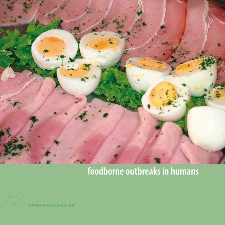
report on zoonotic agents in belgium in
foodborne outbreaks in humans
70
71report on zoonotic agents in belgium in
Foodborne outbreaks in humans During 2003, a total of 101 outbreaks of foodborne infections and intoxications were recorded in Belgium. Data came from
the Flemish Community, the Federal Agency for the Safety of the Food Chain, the French Community, the Brussels Capital
Region, the sentinel laboratories and the National Reference Laboratory for Salmonella and Shigella. More than 1293 peo-
ple were ill, at least 142 persons were hospitalised and 1 child died. However not all outbreaks were noti� ed and for many
outbreaks data are incomplete.
• Causative agents
• Source of the foodborne outbreaks
72 report on zoonotic agents in belgium in
Causative agents
62% of the outbreaks were due to Salmonella, with Salmonella Enteritidis as predominant serotype. Other serotypes involved
in the outbreaks were Dublin, Derby, Typhimurium, Virchow and 9: -: -. Not in every outbreak of Salmonella the serotype was
recorded.
B. cereus was the causative agent in 5% of the cases, twice in combination with Staphylococcus aureus and once in combina-
tion with Cl. perfringens. In one severe outbreak of B. cereus, 5 persons of a family had to be hospitalised with symptoms of
toxic shock after a meal of pasta salad and one child died.
Staphylococcus aureus was demonstrated in 3% of the cases. Other con8 rmed agents were Giardia (n=3), Cl. perfringens (n=1),
Campylobacter (n=1), Shigella (n=1). Canned apricots were responsible for a chemical intoxication (ethanol, aceton, hexanal).
In 22% of the outbreaks no causative agent could be identi8 ed.
Source of the foodborne outbreaks
In 43% of the outbreaks, preparations with raw eggs (Tiramisu, chocolate mousse, mashed potatoes prepared with raw eggs,
sabayon, mayonnaise, bavarois) were identi8 ed as the source of the illness. Meat or meat based products were responsible for
20% of the cases (beef, hamburger, chicken, pita and spaghetti sauce).
73report on zoonotic agents in belgium in
Causative agent Outbreaks Ill Died Hospitalised Sources
Salmonella - Preparations with raw eggs, meat
B. cereus Pasta salad, beef stew
S. aureus - Candy, milk
B. cereus and S. aureus - Pita, pasta salad
B. cereus and Cl. perfringens - Beef stew
Campylobacter - - Barbecue meat
Other - Giardia, Shigella, chemical substances, toxins, heat strike
Unknown -
Total
Table Q: Foodborne outbreaks in humans in Belgium in
Data from the Flemisch Community, the Federal Agency for the Safety of the Food Chain, the French Community, the Brussels-Capital Region,
the sentinel laboratories, and the National Reference Laboratory for Salmonella and Shigella.

74 report on zoonotic agents in belgium in
Tables & FiguresTable A: Total human population in Belgium 11
Table B: Number of ruminants and pigs 12
Table C: Number of poultry 12
Table D: Number of animals slaughtered in Belgium in 2003 13
Table E: Evolution of the prevalence of Salmonella in poultry breeders. 26
Table F: The results of the monitoring – Salmonella in meat and meat products 33
Table G: Evolution of the food Salmonella prevalences 2000-2003 33
Table H: Trends for the most prevalent Salmonella serotypes from 1998 to 2003. 37
Table I: Antimicrobial resistance in human Salmonella 39
Table J: Zoonosis monitoring programme – Campylobacter in food 50
Table K: Evolution of the food Campylobacter prevalences 2000-2003 51
Table L: Campylobacter in humans 51
Table M: Zoonosis monitoring programme - Listeria monocytogenes in food 56
Table N: Evolution of the food Listeria monocytogenes prevalences 2000-2003 57
Table O: Listeria monocytogenes in humans 57
Table P: Zoonosis monitoring programme - E. coli O157. 65
Table Q: Foodborne outbreaks in humans in Belgium in 2003 73
Figure A. Evolution of the percentages of Salmonella serotypes isolated from poultry between 1992 and 2003 25
Figure B. Evolution of the percentage of Salmonella serotypes isolated from pigs between 1992 and 2003 28
Figure C. Evolution of the percentage of Salmonella serotypes isolated from cattle between 1992 and 2003 29
Figure D. Antibiotic resistance percentages in S. Agona 30
Figure E. Antibiotic resistance percentages in S. Dublin 30
Figure F. Antibiotic resistance percentages in S. Enteriditis 30
Figure G. Antibiotic resistance percentages in S. Hadar 31
Figure H. Antibiotic resistance percentages in S. Typhimurium O5- 31
Figure I. Antibiotic resistance percentages in S. Typhimurium O5+ 31
Figure J. Antibiotic resistance percentages in S. Virchow 32

Editor in charge:
Piet Vanthemsche
FAVV-AFSCA (FASFC)
Simon Bolivarlaan 30
1000 Brussel
© FAVV-AFSCA 2005
Graphic design: FASFC Communication service (EDV&JG)
D/2005/

report on zoonotic agents in belgium in
• Federal Agency for the Safety of the Food Chain (FASFC)
• Scienti� c Institute of Public Health (WIV-ISP)
• Veterinary and Agrochemical Research Centre (CODA-CERVA)