Embed Size (px)
Citation preview

1
Evaluation of the Merlin MICRONAUT System for Rapid Direct
Susceptibility Testing of Gram-Positive Cocci and Gram-Negative
Bacilli from Positive Blood Cultures
Nele Wellinghausen*, Tim Pietzcker, Sven Poppert, Syron Belak, Nicole Fieser, Melanie 5
Bartel, and Andreas Essig
Institute of Medical Microbiology and Hygiene, University Hospital of Ulm, Germany
Short title: Direct susceptibility testing from blood cultures 10
Key words: susceptibility testing, blood culture, microtiter broth dilution,
*Corresponding author:
PD Dr. Nele Wellinghausen, MD 15
Institute of Medical Microbiology and Hygiene
University Hospital of Ulm
Ulm, Germany
Tel. +49-731-500 65316
Fax +49-731-500 65302 20
Email: [email protected]
ACCEPTED
Copyright © 2007, American Society for Microbiology and/or the Listed Authors/Institutions. All Rights Reserved.J. Clin. Microbiol. doi:10.1128/JCM.01856-06 JCM Accepts, published online ahead of print on 3 January 2007
on July 5, 2018 by guesthttp://jcm
.asm.org/
Dow
nloaded from

2
Abstract
Bloodstream infections are life-threatening conditions which require timely initiation of
appropriate antimicrobial therapy. We evaluated the automated Merlin MICRONAUT
system for rapid direct microtiter broth antimicrobial susceptibility testing (AST) of
Gram-positive cocci and Gram-negative bacilli from positive BACTEC 9240 blood 5
culture bottles in comparison to the standard method on the Merlin MICRONAUT
system. This prospective study was conducted under routine working conditions during a
9-month period. Altogether, 504 isolates and 11,819 organism-antibiotic combinations
from 409 patients were evaluable for comparison of direct and standard AST. Concerning
Gram-negative bacilli, direct and standard AST was evaluated in 110 isolates and MIC 10
agreement was found in 98.1% of 2,637 organism-antibiotic combinations. Category
(SIR) agreement was found in 99.0%, with 0.04% very major, 0.2% major, and 0.8%
minor errors. Concerning Gram-positive cocci, 373 isolates were evaluated and MIC
agreement was found in 95.6% of 8,951 organism-antibiotic combinations. SIR
agreement was found in 98.8%, with 0.3% very major, 0.4% major, and 0.5% minor 15
errors. Although the number of tested isolates was limited (n=33) direct AST of
streptococci was performed for the first time yielding promising results with a SIR
agreement 98.6% of 363 organism-antibiotic combinations. In conclusion, direct AST of
Gram-negative bacilli and Gram-positive cocci from positive blood cultures on the
MICRONAUT system is a reliable technique that allows omitting repeat testing of 20
subcultured isolates. Thereby, it reduces the time to result of blood culture testing and
may have a positive impact on patient care.
ACCEPTED
on July 5, 2018 by guesthttp://jcm
.asm.org/
Dow
nloaded from

3
Introduction
Bloodstream infections are life-threatening conditions which require timely initiation of
antimicrobial therapy. Inappropriate initial antimicrobial therapy of septic patients is
associated with adverse outcome (13,15,20). Automated blood culture systems that
monitor blood culture bottles continuously for bacterial growth minimize the time 5
necessary to detect positive blood cultures. Once bacterial growth is detected in blood
cultures, rapid identification and susceptibility testing of the isolate is an important task
for the clinical microbiology laboratory. Reducing the turnaround time of microbiological
analysis by use of automated systems can lead to a significant reduction of patient
morbidity, mortality, and costs (3,9,27). 10
While standard antimicrobial susceptibility testing of bacteria commonly involves pure
overnight subcultures, preparation of the inoculum for automated susceptibility testing
directly from the positive blood culture appears extremely attractive with respect to the
time to result. Thus, direct antimicrobial susceptibility testing from positive blood
cultures has been evaluated on many automated testing systems, like the BD PHOENIX 15
(BD, Heidelberg, Germany), the VITEK and VITEK 2 (BioMérieux, Nürtingen,
Germany), the Sensititre (Trek Diagnostics, West Lake, Ohio), and the MicroScan (Dade
Behring, Eschborn, Germany) system (4-6,12,14,18,21,24,26,28). In general, good
agreement between direct and standard susceptibility testing results were observed when
Gram-negative bacilli were tested, including both Enterobacteriaceae and Pseudomonas 20
species (4-6,12,14,18,21,24,26,28). Concerning direct testing of Gram-positive cocci
from blood cultures, only limited data from small studies are available for the VITEK, the
VITEK 2, the Sensititre, and the MicroScan system (5,6,8,18,26,29). A significantly
ACCEPTED
on July 5, 2018 by guesthttp://jcm
.asm.org/
Dow
nloaded from

4
higher rate of disagreement between direct and standard testing results was found
compared to the testing of Gram-negative bacilli. Reporting of a false susceptibility of
staphylococci against oxacillin and enterococci against various antibiotics (18,26) is a
major problem with enormous clinical relevance. Since Gram-positive cocci constitute
the majority of bloodstream infections (23,29), rapid and reliable automated susceptibility 5
testing of Gram-positive bacteria is highly desirable.
We evaluated the automated MICRONAUT system (Merlin, Bornheim-Hesel, Germany)
for rapid direct microtiter broth susceptibility testing of Gram-positive cocci and Gram-
negative bacilli from positive BACTEC blood culture bottles. The study was conducted
under routine working conditions in the clinical microbiological laboratory of the 10
University Hospital of Ulm during a 9-month period, including 850 positive blood
cultures.
ACCEPTED
on July 5, 2018 by guesthttp://jcm
.asm.org/
Dow
nloaded from

5
Materials and Methods
Samples. The study was conducted from July 2005 to March 2006 at the University
Hospital of Ulm, Germany, a 1,100-bed tertiary hospital which provides a full range of
medical and surgical services. The automated blood culture system BACTEC 9240 (BD)
with the culture bottles PLUS Aerobic/F, PLUS Anaerobic/F, and PLUS Pediatric is used 5
in the hospital. One blood culture consists of an aerobic and an anaerobic bottle or, in the
case of children, only of a pediatric bottle. All blood cultures that were detected positive
by the BACTEC system and that showed Gram-positive cocci or Gram-negative bacilli in
at least one bottle in the initial Gram stain were included in the study. If both the aerobic
and anaerobic bottle of one blood culture were detected positive and showed identical 10
Gram stain morphology, only the aerobic bottle was used for the study. Blood cultures
showing mixed growth in the initial Gram stain, i.e. more than one morphology of
bacteria in a single bottle, were excluded from the study. The study was conducted on
both weekdays and weekends. If the same species with an identical antimicrobial
susceptibility testing profile was detected in more than one blood culture within 14 days, 15
the direct susceptibility testing of the first isolate only was repeated by the standard
method and included in the final data analyses (see below).
Standard susceptibility testing. Standard testing of all isolates was performed from a
pure overnight subculture on the MICRONAUT system as recommended by the
manufacturer (Merlin). The MICRONAUT system is an automated microtiter broth 20
dilution susceptibility testing system that is distributed throughout Germany and Europe
in private and hospital-based laboratories. It is performed in 384-well microtiter plates. It
allows determination of real minimum inhibitory concentrations (MICs) for up to 25
ACCEPTED
on July 5, 2018 by guesthttp://jcm
.asm.org/
Dow
nloaded from

6
substances and testing of two bacterial isolates in one plate. Bacterial growth in the wells
is monitored photometrically at a wavelength of 620 nm and a density above the cut-off
value of the respective medium is interpreted as bacterial growth. Several colonies were
used to prepare a 0.5 McFarland suspension in 0.9% saline. For the testing of
staphylococci, enterococci, and micrococci, 100µl of the suspension were diluted with 15 5
ml Mueller-Hinton II broth (containing 0,25g/l phytagel, an agar substitute produced
from bacterial fermentation, Oxoid, Wesel, Germany) while for the testing of Gram-
negative bacilli 50µl of the suspension were diluted in 15 ml broth. The broth was
inoculated into Merlin MICRONAUT 384-well Gram-positive (GP plate) and Gram-
negative (GN plate) antimicrobial susceptibility testing plates, respectively, designed for 10
the German Network for Antimicrobial Resistance Surveillance (GENARS,
www.genars.de), by using the automated Merlin Sprint device. For testing of the majority
of antibiotics, the plates contain eight dilutions of the antibiotic, determining a real
minimal inhibitory concentration (MIC). Breakpoint testing was done with fusidic acid
and netilmicin on the GP plate and for aztreonam, cefotiam, mezlocillin, and netilmicin 15
on the GN plate. Inoculated plates were incubated for 18-24 hours at 36°C under ambient
air. For the testing of streptococci, 200µl of the suspension were diluted with 15 ml
Mueller-Hinton II broth (containing 0,25g/l phytagel and 200µl lysed horse blood). The
broth was inoculated into Merlin MICRONAUT 96-well testing plates for streptococci
(Strep plate) and plates were incubated for 18-24 hours at 36°C in a 5% CO2 atmosphere. 20
Reading of all plates was done with a photometer (Merlin) interpreting an optical density
>0.1 as growth. Obtained MIC values were interpreted with the advanced expert system
(AES) MCN-6 of Merlin MICRONAUT using the interpretation guidelines of the
ACCEPTED
on July 5, 2018 by guesthttp://jcm
.asm.org/
Dow
nloaded from

7
German Standardisation Institute (Deutsches Institut für Normung, DIN (7)) and
validated by a clinical microbiologist. A sheep blood agar was inoculated from all
McFarland suspensions used for susceptibility testing and incubated at 36°C for 18-24
hours in order to control for growth, mixed cultures and possible contamination.
Direct susceptibility testing. For direct testing, 8ml of the positive blood culture 5
medium were centrifuged at 130 g (800 rpm) for 10 min. The supernatant was transferred
into a new tube and centrifuged at 1800 g (3000 rpm) for 5 min. The resultant pellet was
diluted in sterile 0.9% saline to prepare a 0.5 McFarland suspension and the suspension
was processed as described above. The antimicrobial resistance testing panel was chosen
according to the result of the Gram stain prepared from the positive blood culture bottle. 10
For testing of Gram-positive cocci in clusters and Gram-positive diplococci and cocci in
short chains, suggestive of enterococci, the GP plate was used. If small Gram-positive
cocci in chains, suggestive of streptococci, were seen, the Strep plate was chosen. For
testing of Gram-negative bacilli the GN plate was used.
Identification of bacterial strains. Identification of all bacterial species apart from most 15
staphylococci was done by API immediately after obtaining pure subcultures (API 20
Strep, API Rapid ID 32 Strep, API 20 E, API 20 NE; BioMérieux, Germany). For
staphylococci, diagnosis was based on typical microscopy and morphology (color,
haemolysis etc.), positive catalase-reaction and growth on mannitol-salt-agar.
Staphylococcus aureus was differentiated from coagulase-negative staphylococci by 20
morphology and positive clumping factor (Slidex, BioMérieux). If differentiation was
ambiguous, aurease detection by RAPIDEC Staph (BioMérieux) and an API 20 Staph
was done. In all isolates where biochemical identification was ambiguous (n=5)
ACCEPTED
on July 5, 2018 by guesthttp://jcm
.asm.org/
Dow
nloaded from
8
sequencing of the complete 16S rRNA gene was performed as described previously
(17,1). All isolates included in the study were stored on microbanc tubes (Doenitz
ProLab, Augsburg, Germany) at -20°C.
Confirmative susceptibility testing in staphylococci. Identification of the staphylo-
coccal mecA gene by PCR was done as published earlier (25). Quinupristin/dalfopristin 5
(Synercid®) testing by E-test (Viva Diagnostika, Koeln, Germany) was done on Mueller-
Hinton agar (Heipha, Heidelberg, Germany) using a 0.5 McFarland suspension of the
respective strain. Plates were incubated in ambient air at 36°C for 24 hours.
Quality control. Quality control stains, including Staphylococcus aureus ATCC 29213,
methicillin-resistant Staphylococcus aureus ATCC 43300, Enterococcus faecalis ATCC 10
29212, Escherichia coli ATCC 25922, Escherichia coli ATCC 35218, Pseudomonas
aeruginosa ATCC 27853, Klebsiella pneumoniae ATCC 700603, and Enterococcus
faecium VanA positive (DSM 17050), were investigated daily (each strain 3 times a
week) by the standard procedure. In addition, precision of the standard method was
determined by measuring Staphylococcus aureus ATCC 29213, Enterococcus faecalis 15
ATCC 29212, and Escherichia coli ATCC 25922 in ten replicates from a single
McFarland suspension (data not shown). Differences exceeding a range of two twofold
dilutions of the MIC were observed with imipenem, ertapenem, and tobramycin.
Therefore, these antibiotics were not included in the data analysis. Precision of the direct
AST method was determined by investigation of ten blood cultures containing blood 20
from a healthy volunteer spiked with Escherichia coli ATCC 25922 and Pseudomonas
aeruginosa ATCC 27853. Differences exceeding a range of two twofold dilutions of the
ACCEPTED
on July 5, 2018 by guesthttp://jcm
.asm.org/
Dow
nloaded from

9
MIC and results beyond the given limits of the DIN (7) were only observed with
imipenem.
Data analysis. For each antibiotic test result, raw MIC values and validated interpretation
results (SIR) after AES validation were compared between direct testing and standard
testing. MIC agreement was defined as direct MIC being within one twofold dilution of 5
the standard MIC (11). Category (SIR) agreement was defined as concordance between
validated SIR interpretations. SIR discrepancies in test results that did in fact display
MIC agreement were also counted as SIR agreement in order to minimize method-
inherent artifacts, e.g. SIR discrepancies introduced by AES validation. Regarding
antibiotics with breakpoint testing, only fusidic acid (GP plate) and aztreonam (GN plate) 10
were included in the data analysis since artifacts in SIR validation introduced by the AES
could be excluded in these antibiotics. A very major error was defined as susceptible in
the direct testing and resistant in the standard testing, a major error was defined as
resistant in the direct testing and susceptible in the standard testing, and a minor error was
defined as all other discrepancies between direct and standard testing (11). 15
ACCEPTED
on July 5, 2018 by guesthttp://jcm
.asm.org/
Dow
nloaded from

10
Results
Study population
During the study period, direct antimicrobial susceptibility testing (AST) was done from
850 positive blood cultures, including 637 aerobic and 213 anaerobic bottles. Out of the
850 blood cultures, 146 were positive with Gram-negative rods (17.2%), 562 showed 5
Gram-positive cocci in clusters (66.2%), 134 showed Gram-positive diplococci and cocci
in short chains (15.7%), and 8 showed small Gram-positive cocci in chains suggestive of
streptococci (0.9%) in the initial Gram-stain performed after positive signaling of the
bottle in the BACTEC system.
Regarding all 850 blood cultures, direct AST was evaluable in 702 samples (82.6%). 10
Susceptibility testing was not evaluable in 148 samples due to the following reasons:
Detection of polymicrobial growth in the blood culture after overnight incubation in 69
samples (8.1%), failure to growth in the AST in 39 samples (4.6%), selection of an
incorrect direct AST panel due to ambiguous Gram stain result in 27 samples (3.2%),
contamination of the direct AST in three samples (0.3%), growth of a bacterial species 15
that is not suitable for AST with the methods used in this study in nine samples (1.0%,
including six anaerobes, two isolates of Lactococcus lactis, and one isolate of Moraxella
catarrhalis), and inability to prepare the inoculum for direct AST due to extensive
hemolysis by one isolate (0.1%) of Enterococcus faecalis.
Regarding blood cultures with polymicrobial growth in the direct AST (n=69), mainly 20
mixtures of different Gram-positive species, predominantly coagulase-negative staphylo-
cocci and enterococci, were found. In twelve samples Gram-negative bacilli were
involved in mixtures with Gram-positive cocci or other Gram-negative bacilli.
ACCEPTED
on July 5, 2018 by guesthttp://jcm
.asm.org/
Dow
nloaded from

11
Blood cultures with failed growth in direct AST (n=39) comprised the following species:
coagulase-negative staphylococci (n=25), Staphylococcus aureus (n=3), Micrococcus
luteus (n=2), Acinetobacter lwoffii (n=1), Escherichia coli (n=1), Gemella haemolysans
(n=1), Rothia mucilagenosa (n=1), Streptococcus agalactiae (n=1), Streptococcus
anginosus (n=1), Streptococcus mitis (n=1), Streptococcus pneumoniae (n=1), and 5
Streptococcus sanguis (n=1).
An incorrect direct AST panel was chosen in 27 samples, including 21 isolates of
Streptococcus spp. (including ten isolates of Streptococcus pneumoniae) and one
Gemella haemolysans that were tested on the GP plate (Gram stain suggestive of
enterococci), four isolates of Enterococcus faecalis that were tested on Strep plates 10
(Gram stain suggestive of streptococci), and one isolate of Acinetobacter lwoffii that was
misidentified as Gram-positive cocci.
If the same species with an identical antimicrobial susceptibility testing profile was
detected in more than one blood culture within 14 days, the direct AST of the first isolate
only was repeated by the standard method and subsequent isolates (n = 198) were not 15
included in the AST study. By this procedure a total of 504 blood cultures from 409
patients were finally available for comparison of direct and standard AST.
ACCEPTED
on July 5, 2018 by guesthttp://jcm
.asm.org/
Dow
nloaded from

12
Gram-negative bacilli
Direct and standard susceptibility testing was done on 110 isolates of Gram-negative
bacilli (Table 1). 24 antibiotics were investigated and 2,637 organism-antibiotic
combinations were available for data analysis. MIC agreement was found in 98.1% of all
combinations (Table 2). Category agreement (SIR agreement) was found in 99.0% (Table 5
2). Minor errors occurred in 0.8%, major errors in 0.2%, and very major errors in 0.04%
(Table 2). False susceptibility of direct testing was only noted for piperacillin/tazobactam
in one isolate of Escherichia coli and for aztreonam in one isolate of Morganella
morganii. Altogether, the study population included six isolates of Enterobacteriaceae
with AmpC-β-lactamase phenotype and 26 isolates of Enterobacteriaceae resistant to 10
amoxicillin-clavulanate.
Gram-positive cocci (GP plate)
Direct and standard susceptibility testing was done in 394 isolates of Gram-positive cocci
(Table 1). Out of these 394 isolates, 373 isolates of staphylococci, enterococci, Micro-15
coccus luteus, and Kocuria spp. were tested with the GP plate and 21 isolates of
Streptococcus spp. were tested with the Strep plate. Concerning the GP plate, 24
antibiotics were investigated and 8,951 organism-antibiotic combinations were available
for data analysis. Altogether, resistance against penicillin, oxacillin, and erythromycin in
coagulase-negative staphylococci was noted in 251 (89%), 223 (79%), and 202 (72%) 20
isolates, respectively, and thirty isolates of S. aureus (65%) were resistant to penicillin.
MIC agreement was found in 95.6% of all combinations (Table 3). SIR agreement was
ACCEPTED
on July 5, 2018 by guesthttp://jcm
.asm.org/
Dow
nloaded from

13
found in 98.8% (Table 3). Minor errors occurred in 0.5%, major errors in 0.4%, and very
major errors in 0.3% (Table 3).
Regarding the important antibiotic oxacillin, discrepant results of direct and standard
AST were noted in five isolates of coagulase-negative staphylococci (Table 3), including
three isolates of Staphylococcus epidermidis and two of S. hominis. In all five isolates, 5
presence of the mecA gene could be demonstrated by PCR. Therefore, three isolates (two
S. hominis and one S. epidermidis) are correctly classified as “very major error” for
oxacillin. However, in the two isolates (both S. epidermidis) classified as “major error”
the direct oxacillin testing represented the correct result.
In four isolates very major errors were observed for quinupristin/dalfopristin (Tab. 3), 10
including two isolates of Staphylococcus aureus (one MRSA and one methicillin-
susceptible strain) and two coagulase-negative staphylococci. Since quinupristin/dalfo-
pristin resistance is low in Germany the observed resistance in the standard AST was
questioned and AST was repeated from stored subcultures of all four isolates. Repeated
standard AST revealed susceptibility to quinupristin/dalfopristin in all isolates. MIC 15
values were within one dilution of those observed in the direct testing (direct MIC
≤ 0.5µg/ml, initial standard MIC 2-4µg/ml, repeated standard MIC ≤ 0.5-1µg/ml). In
addition, a quinupristin/dalfopristin E-test was done in all four isolates and confirmed
susceptibility to quinupristin/dalfopristin (MIC 0.38 -0.75µg/ml). Thus, the supposed
very major errors were caused by false detection of quinupristin/dalfopristin resistance in 20
the initial standard testing.
ACCEPTED
on July 5, 2018 by guesthttp://jcm
.asm.org/
Dow
nloaded from

14
Streptococci (Strep plate)
Concerning the testing of streptococci in the Strep plate, twelve antibiotics were tested
and 231 organism-antibiotic combinations were available for data analysis. MIC
agreement and SIR agreement was found in 96.5% and 97.8%, respectively. Minor errors
occurred in 0.4%, major errors in 0%, and very major errors in 1.7% (Table 4). False 5
susceptibility of direct testing was noted for erythromycin and clindamycin in one isolate
of Streptococcus oralis and for trimethoprim-sulfamethoxazole in one isolate each of
Streptococcus anginosus and Streptococcus pyogenes. Altogether, resistance against
erythromycin/clindamycin and penicillin was noted in nine (43%) and five (24%) isolates
of streptococci, respectively. 10
After termination of the study, further twelve blood cultures growing streptococci
(inlcuding five S. mitis, two S. anignosus, two S. pneumoniae, two S. pyogenes, and one
S. oralis) were evaluated with both methods during clinical diagnostics. All 132
organism-antibiotic combinations revealed SIR agreement. Thus, regarding the whole
population of 33 isolates, minor errors occurred in 0.3%, major errors in 0%, and very 15
major errors in 1.1% (data not shown).
ACCEPTED
on July 5, 2018 by guesthttp://jcm
.asm.org/
Dow
nloaded from

15
Discussion
Shortening the time to result of antimicrobial susceptibility testing of blood culture
isolates can lead to a significant reduction of patient morbidity, mortality, and costs
(3,9,27). Therefore, we evaluated the accuracy of the MICRONAUT system for direct
AST of positive blood cultures under routine conditions in a clinical microbiology 5
laboratory. The MICRONAUT system is a commercially available, automated, microtiter
plate based broth dilution AST system (2,16). Altogether, 850 positive blood cultures
were investigated on a daily basis including weekends during a period of nine months.
504 isolates and 11,819 organism-antibiotic combinations were evaluable for comparison
of both direct and standard AST methods. Thus, the number of isolates included in this 10
study exceeds by far that of former studies published on direct AST from positive blood
cultures (4-6,12,14,18,21,24,26,28).
The overall MIC agreement between direct and standard susceptibility testing of Gram-
negative and Gram-positive isolates was high (95.6% to 98.1%, Tables 2-4). For every
antimicrobial agent except ampicillin on the GP plate the MIC agreement was >90%, as 15
required by the selection criteria for an antimicrobial susceptibility testing system
proposed by Jorgensen (19). Categorical error rates were very low and did not exceed the
limits proposed by Jorgensen (19), i.e. very major errors occurred in less than 1.5% for
all species investigated and the overall percentage of errors attributable to the new
procedure did not exceed 5%. 20
For Gram-negative isolates the very major error rate was as low as 0.08%. Very major
errors were only seen with aztreonam and piperacillin-tazobactam in two Enterobacte-
riaceae. Concerning these antibiotics, very major errors in direct AST of Gram-negative
ACCEPTED
on July 5, 2018 by guesthttp://jcm
.asm.org/
Dow
nloaded from

16
bacilli were also detected in recent studies using the Microscan (28), Phoenix (12), and
Vitek 2 (6,21). However, very major errors involving the second and third generation
cephalosporins, for example, cefotaxim, cefuroxim, and ceftazidime, as frequently
observed with other automated system (4,6,12,21,28), were not detected in our study.
Due to the observed very low rate of errors direct results obtained by the MICRONAUT 5
system are sufficiently reliable to be reported to the clinician.
Concerning Gram-positive species, only isolates tested on the GP plate (mainly
staphylococci and enterococci) should be evaluated since the number of streptococci
investigated on the Strep plate within this study (n=21) is too small for further analysis.
After termination of the study, however, twelve additional blood cultures growing 10
streptococci were investigated and did not show any errors in direct AST. Nevertheless,
since only a very small number of resistant streptococci (4/21 penicillin-resistant and
9/21 erythromycin-resistant) and no penicillin-resistant pneumococci were included in
the study, no reliable statement can be made regarding the occurrence of very major
errors in streptococci. 15
A high very major errors rate with direct testing was observed with quinupristin/dalfo-
pristin in Gram-positive cocci on the GP plate (Table 3). These very major errors could,
however, be disproved by repeated testing and were most probably caused by incorrect
automated reading of the plate, such as humidity-generated condensation. Three very
major and two major errors were detected with oxacillin in five isolates of coagulase-20
negative staphylococci. Interestingly, the mecA gene was present in all five isolates,
confirming the very major errors but disproving the major errors. The latter phenomenon
may be explained by a heterogenic resistance pattern and the presence of both oxacillin-
ACCEPTED
on July 5, 2018 by guesthttp://jcm
.asm.org/
Dow
nloaded from

17
susceptible and oxacillin-resistant subpopulations of the respective isolate in the blood
culture bottle and predominant growth of the susceptible population in the subculture and
standard AST. In one case, the positive blood culture bottle was still available when the
presumptive major error was observed. Further subcultures from the blood culture bottle
confirmed our assumption, showing a mixed population of oxacillin-susceptible and -5
resistant colonies. Altogether, antibiotics with detectable very major errors in our study
mainly included those published earlier for the Vitek 2 system (6,8). A too-low inoculum
or slow growth of the bacteria probably caused the discrepant results. Concerning minor
errors, a high number was seen with teicoplanin. These errors were exclusively seen in
coagulase-negative staphylococci, included equal number of false high and false low 10
MIC values and may probably be explained by the lower precision of the method for
measurement of this antibiotic due to antibiotic- and/or system-inherent reasons.
A critical technical step in direct AST from positive blood cultures is the preparation of
the inoculum (10,22). Blood cells, cellular debris, and constituents of the blood culture
medium etc. may hamper preparation of a defined McFarland suspension and may disturb 15
the testing procedure since the bacteria are often present in low concentration in the
positive blood culture medium. Enrichment of bacterial cells for direct AST by using
Serum Separator Tubes (SST; BD) has been evaluated recently (6,12). In our study, we
developed a simple two-step centrifugation method for separation of bacterial cells from
positive blood cultures. Apart from one blood culture growing hemolytic Enterococcus 20
faecalis, the inoculum for direct AST, i.e. the 0.5 McFarland suspensions, could be
prepared easily within 20 min and was macroscopically devoid of red blood cells. The
density of bacterial growth observed in the direct AST quality control plates did not differ
ACCEPTED
on July 5, 2018 by guesthttp://jcm
.asm.org/
Dow
nloaded from
18
from that of standard quality control plates, and the quality control strains investigated by
the direct AST method were within the given limits. Furthermore, repeated direct testing
of single strains from individual patients revealed a high rate of agreement (data not
shown). Thus, this preparation method is reliable, comparably fast but much cheaper than
the SST method. 5
Polymicrobial growth in direct AST was observed in 8.1% of blood cultures, which is
slightly higher than in other studies (4,28). Blood samples were taken by both
venipuncture and line draw by medical personnel of the respective wards. Due to the
absence of a specialized blood collection team, a higher rate of contamination may be
assumed. Also, a much higher number of Gram-positive isolates was included in this 10
study compared to the above mentioned studies and the majority of polymicrobial
cultures included mixtures of different Gram-positive cocci.
An important task in direct AST of Gram-positive cocci in chains was to choose the
correct test panel, i.e. the GP plate for enterococci or the Strep plate for streptococci. In
most samples the Gram-stain result allowed selection of the correct plate, however, 15
microscopic misidentification of streptococci, especially Streptococcus pneumoniae, as
enterococci was a problem and led to delay of AST in 21 isolates. Nevertheless, in the
majority of clinical microbiology laboratories direct AST of streptococci is not even
available.
In conclusion, direct AST of bacterial isolates from positive blood cultures on the Merlin 20
MICRONAUT system is a reliable technique that can reduce the time to result of blood
culture testing by omitting repeat testing from subcultures and facilitate earlier initiation
of pathogen-directed antimicrobial therapy in septic patients. Thereby, it may have a
ACCEPTED
on July 5, 2018 by guesthttp://jcm
.asm.org/
Dow
nloaded from

19
positive impact on patient care (3,9,27), allow earlier switch from a broad spectrum
antimicrobial to a more appropriate pathogen-adapted antibiotic, and may thus prevent
development of resistance. Furthermore, reliable direct AST may facilitate reduction of
overall consumption of antibiotics and health care costs. The method is suitable for both
Gram-negative bacilli and Gram-positive cocci and is robust enough to be used on a 5
seven days a week basis in a routine clinical microbiology laboratory. In contrast to the
commonly used VITEK (bioMérieux) and BD PHOENIX (BD) systems, the Merlin
MICRONAUT system offers the advantages of a broader panel of antibiotics within one
test plate, determination of definitive MIC values in the majority of antibiotics, and visual
control of bacterial growth in the plates. For the first time, direct AST of streptococci was 10
evaluated in this study with promising results.
Acknowledgement
We are grateful to Angelika Möricke for performing the PFGE. We thank the company
Merlin for supply of AST plates. 15 ACCEPTED
on July 5, 2018 by guesthttp://jcm
.asm.org/
Dow
nloaded from
20
References
1. Altschul, S. F., T. L. Madden, A. A. Schaffer, J. Zhang, Z. Zhang, W. Miller,
and D. J. Lipman. 1997. Gapped BLAST and PSI-BLAST: a new generation of
protein database search proGrams. Nucleic Acids Res. 25:3389-3402.
2. Balke, B., L. Hoy, H. Weissbrodt, and S. Haussler. 2004. Comparison of the 5
MICRONAUT Merlin automated broth microtiter system with the standard agar
dilution method for antimicrobial susceptibility testing of mucoid and nonmucoid
Pseudomonas aeruginosa isolates from cystic fibrosis patients. Eur. J. Clin.
Microbiol. Infect. Dis. 23:765-771.
3. Barenfanger, J., C. Drake, and G. Kacich. 1999. Clinical and financial benefits 10
of rapid bacterial identification and antimicrobial susceptibility testing. J. Clin.
Microbiol. 37:1415-1418.
4. Bruins, M. J., P. Bloembergen, G. J. Ruijs, and M. J. Wolfhagen. 2004.
Identification and susceptibility testing of Enterobacteriaceae and Pseudomonas
aeruginosa by direct inoculation from positive BACTEC blood culture bottles into 15
Vitek 2. J. Clin. Microbiol. 42:7-11.
5. Chapin, K. C. and M. C. Musgnug. 2003. Direct susceptibility testing of positive
blood cultures by using Sensititre broth microdilution plates. J. Clin. Microbiol.
41:4751-4754.
ACCEPTED
on July 5, 2018 by guesthttp://jcm
.asm.org/
Dow
nloaded from

21
6. de Cueto, M., E. Ceballos, L. Martinez-Martinez, E. J. Perea, and A. Pascual.
2004. Use of positive blood cultures for direct identification and susceptibility
testing with the vitek 2 system. J. Clin. Microbiol. 42:3734-3738.
7. Deutsches Institut für Normung (DIN). 2002. Susceptibility testing of bacterial
pathogens except mycobacteria - Methoden zur Empfindlichkeitsprüfung von 5
bakteriellen Krankheitserregern außer Mykobakterien gegen Chemotherapeutika.
DIN 58940.
8. Diederen, B. M., M. Zieltjens, H. Wetten, and A. G. Buiting. 2006. Identification
and susceptibility testing of Staphylococcus aureus by direct inoculation from 10
positive BACTEC blood culture bottles. Clin. Microbiol. Infect. 12:84-86.
9. Doern, G. V., R. Vautour, M. Gaudet, and B. Levy. 1994. Clinical impact of
rapid in vitro susceptibility testing and bacterial identification. J. Clin. Microbiol.
32:1757-1762.
10. Fay, D. and J. E. Oldfather. 1979. Standardization of direct susceptibility test for 15
blood cultures. J. Clin. Microbiol. 9:347-350.
11. Food and Drug Administration. 2003. Class II Special Controls Guidance
Document: Antimicrobial Susceptibility Test (AST) Systems; Guidance for
Industry and FSA. U.S.Department of Health and Human Services, Food and Drug
Administration, Division of Microbiology Devices. p. 1-48. 20
ACCEPTED
on July 5, 2018 by guesthttp://jcm
.asm.org/
Dow
nloaded from

22
12. Funke, G. and P. Funke-Kissling. 2004. Use of the BD PHOENIX Automated
Microbiology System for direct identification and susceptibility testing of Gram-
negative rods from positive blood cultures in a three-phase trial. J. Clin. Microbiol.
42:1466-1470.
13. Garnacho-Montero, J., J. L. Garcia-Garmendia, A. Barrero-Almodovar, F. J. 5
Jimenez-Jimenez, C. Perez-Paredes, and C. Ortiz-Leyba. 2003. Impact of
adequate empirical antibiotic therapy on the outcome of patients admitted to the
intensive care unit with sepsis. Crit. Care Med. 31:2742-2751.
14. Hansen, D. S., A. G. Jensen, N. Norskov-Lauritsen, R. Skov, and B. Bruun.
2002. Direct identification and susceptibility testing of enteric bacilli from positive 10
blood cultures using VITEK (GNI+/GNS-GA). Clin. Microbiol. Infect. 8:38-44.
15. Harbarth, S., J. Garbino, J. Pugin, J. A. Romand, D. Lew, and D. Pittet. 2003.
Inappropriate initial antimicrobial therapy and its effect on survival in a clinical trial
of immunomodulating therapy for severe sepsis. Am. J. Med. 115:529-535.
16. Haussler, S., S. Ziesing, G. Rademacher, L. Hoy, and H. Weissbrodt. 2003. 15
Evaluation of the Merlin, MICRONAUT system for automated antimicrobial
susceptibility testing of Pseudomonas aeruginosa and Burkholderia species isolated
from cystic fibrosis patients. Eur. J. Clin. Microbiol. Infect. Dis. 22:496-500.
17. Hiraishi, A. 1992. Direct automated sequencing of 16S rDNA amplified by
polymerase chain reaction from bacterial cultures without DNA purification. Lett. 20
Appl. Microbiol. 15:210-213.
ACCEPTED
on July 5, 2018 by guesthttp://jcm
.asm.org/
Dow
nloaded from

23
18. Howard, W. J., B. J. Buschelman, M. J. Bale, M. A. Pfaller, F. P. Koontz, and
R. N. Jones. 1996. Vitek GPS card susceptibility testing accuracy using direct
inoculation from BACTEC 9240 blood culture bottles. Diagn. Microbiol. Infect.
Dis. 24:109-112.
19. Jorgensen, J. H. 1993. Selection criteria for an antimicrobial susceptibility testing 5
system. J. Clin. Microbiol. 31:2841-2844.
20. Kang, C. I., S. H. Kim, W. B. Park, K. D. Lee, H. B. Kim, E. C. Kim, M. D. Oh,
and K. W. Choe. 2005. Bloodstream infections caused by antibiotic-resistant
Gram-negative bacilli: risk factors for mortality and impact of inappropriate initial
antimicrobial therapy on outcome. Antimicrob. Agents Chemother. 49:760-766. 10
21. Ling, T. K., Z. K. Liu, and A. F. Cheng. 2003. Evaluation of the VITEK 2 system
for rapid direct identification and susceptibility testing of Gram-negative bacilli
from positive blood cultures. J. Clin. Microbiol. 41:4705-4707.
22. Mirrett, S. 1994. Antimicrobial susceptibility testing and blood cultures. Clin. Lab
Med. 14:171-179. 15
23. Nicholls, T. M., A. S. Morgan, and A. J. Morris. 2000. Nosocomial blood stream
infection in Auckland Healthcare hospitals. N. Z. Med. J. 113:96-98.
24. Putnam, L. R., W. J. Howard, M. A. Pfaller, F. P. Koontz, and R. N. Jones.
1997. Accuracy of the Vitek system for antimicrobial susceptibility testing
Enterobacteriaceae bloodstream infection isolates: use of "direct" inoculation from 20
Bactec 9240 blood culture bottles. Diagn. Microbiol. Infect. Dis. 28:101-104.
ACCEPTED
on July 5, 2018 by guesthttp://jcm
.asm.org/
Dow
nloaded from

24
25. Reischl, U., H. J. Linde, B. Leppmeier, and N. Lehn. 2002. Duplex LightCycler
PCR assay for the rapid detection of methicillin-resistant Staphylococcus aureus
and simultaneous species confirmation, p. 93-105. In U. Reischl, C. Wittwer, and F.
Cockerill (ed.), Rapid Cycle Real-Time PCR, Springer, Germany.
26. Sahm, D. F., S. Boonlayangoor, and J. A. Morello. 1987. Direct susceptibility 5
testing of blood culture isolates with the AutoMicrobic System (AMS). Diagn.
Microbiol. Infect. Dis. 8:1-11.
27. Trenholme, G. M., R. L. Kaplan, P. H. Karakusis, T. Stine, J. Fuhrer, W.
Landau, and S. Levin. 1989. Clinical impact of rapid identification and
susceptibility testing of bacterial blood culture isolates. J. Clin. Microbiol. 27:1342-10
1345.
28. Waites, K. B., E. S. Brookings, S. A. Moser, and B. L. Zimmer. 1998. Direct
susceptibility testing with positive BacT/Alert blood cultures by using MicroScan
overnight and rapid panels. J. Clin. Microbiol. 36:2052-2056.
29. Wisplinghoff, H., T. Bischoff, S. M. Tallent, H. Seifert, R. P. Wenzel, and M. B. 15
Edmond. 2004. Nosocomial bloodstream infections in US hospitals: analysis of
24,179 cases from a prospective nationwide surveillance study. Clin. Infect. Dis.
39:309-317.
20
ACCEPTED
on July 5, 2018 by guesthttp://jcm
.asm.org/
Dow
nloaded from

Table 1: Species distribution of the positive blood cultures evaluable for direct and
standard antimicrobial susceptibility testing
Gram-negative bacilli (n) Gram-positive cocci (n)
Susceptibility testing on GN plate: Susceptibility testing on GP plate:
Escherichia coli (55) Coagulase-negative staphylococci (281)
Pseudomonas aeruginosa (16)
Klebsiella pneumoniae (12)
Enterobacter cloacae (7)
Staphylococcus aureus (44)
Methicillin-susceptible S. aureus, MSSA (40)
Methicillin-resistant S. aureus, MRSA (4)
Klebsiella oxytoca (4)
Citrobacter freundii (2)
Enterococcus faecium (24)
Vancomycin-resistant E. faecium, VRE (2)
Stenotrophomonas maltophilia (2) Enterococcus faecalis (14)
Acinetobacter baumannii (1) Micrococcus luteus (7)
Acinetobacter species (1) Enterococcus gallinarum (2)
Citrobacter koseri (1) Kocuria species (1)
Citrobacter species (1)
Enterobacter aerogenes (1) Susceptibility testing on Strep plate:
Enterobacter hormaechei (1) Streptococcus mitis (9)
Flavimonas oryzihabitans (1) Streptococcus anginosus (3)
Morganella morganii (1) Streptococcus oralis (2)
Pantoea agglomerans (1) Streptococcus pneumoniae (2)
Salmonella Typhi (1) Streptococcus sanguis (2)
Serratia liquefaciens (1) Streptococcus agalactiae (1)
Serratia marcescens (1) Streptococcus dysgalactiae equisimilis (1)
Streptococcus pyogenes (1)
ACCEPTED
on July 5, 2018 by guesthttp://jcm
.asm.org/
Dow
nloaded from
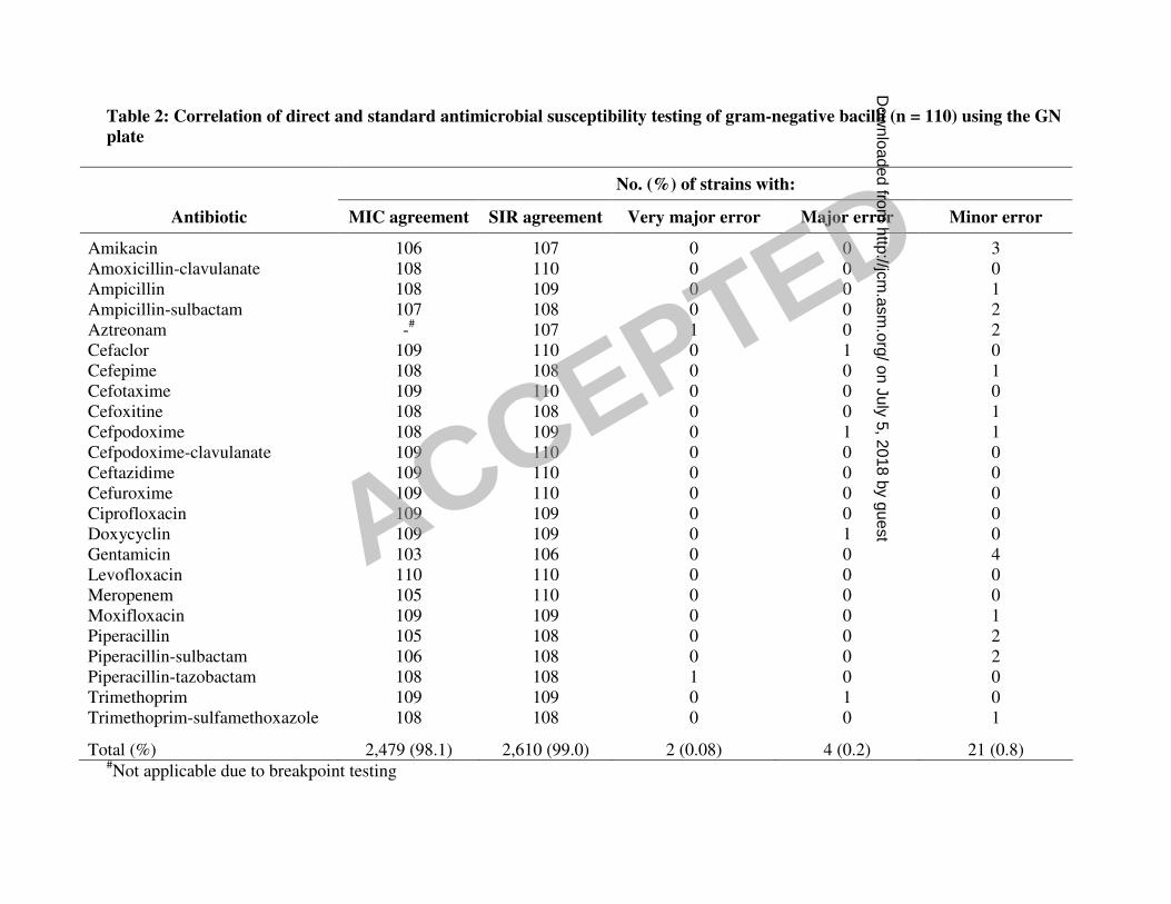
Table 2: Correlation of direct and standard antimicrobial susceptibility testing of gram-negative bacilli (n = 110) using the GN
plate
No. (%) of strains with:
Antibiotic MIC agreement SIR agreement Very major error Major error Minor error
Amikacin 106 107 0 0 3
Amoxicillin-clavulanate 108 110 0 0 0
Ampicillin 108 109 0 0 1
Ampicillin-sulbactam 107 108 0 0 2
Aztreonam -# 107 1 0 2
Cefaclor 109 110 0 1 0
Cefepime 108 108 0 0 1
Cefotaxime 109 110 0 0 0
Cefoxitine 108 108 0 0 1
Cefpodoxime 108 109 0 1 1
Cefpodoxime-clavulanate 109 110 0 0 0
Ceftazidime 109 110 0 0 0
Cefuroxime 109 110 0 0 0
Ciprofloxacin 109 109 0 0 0
Doxycyclin 109 109 0 1 0
Gentamicin 103 106 0 0 4
Levofloxacin 110 110 0 0 0
Meropenem 105 110 0 0 0
Moxifloxacin 109 109 0 0 1
Piperacillin 105 108 0 0 2
Piperacillin-sulbactam 106 108 0 0 2
Piperacillin-tazobactam 108 108 1 0 0
Trimethoprim 109 109 0 1 0
Trimethoprim-sulfamethoxazole 108 108 0 0 1
Total (%) 2,479 (98.1) 2,610 (99.0) 2 (0.08) 4 (0.2) 21 (0.8) #Not applicable due to breakpoint testing
ACCEPTED on July 5, 2018 by guest
http://jcm.asm
.org/D
ownloaded from

Table 3: Correlation of direct and standard antimicrobial susceptibility testing of gram-positive cocci (n = 373) using the GP
plate
No. (%) of strains with:
Substance MIC agreement SIR agreement Very major error Major error Minor error
Amoxicillin-clavulanate 340 372 0 1 0
Ampicillin 322 372 0 1 0
Cefazolin 349 370 1 2 0
Cefuroxime-axetil 352 371 1 1 0
Ciprofloxacin 364 369 2 2 0
Clindamycin 365 371 1 1 0
Doxycyclin 361 366 0 1 6
Erythromycin 362 365 2 5 1
Fosfomycin 363 367 3 3 0
Fusidic acid -# 369 0 3 2
Gentamicin 353 364 1 3 5
Levofloxacin 364 367 1 1 4
Linezolide 357 373 0 0 0
Meropenem 348 370 1 2 0
Moxifloxacin 364 366 0 2 5
Mupirocin 372 372 1 0 0
Oxacillin 348 368 3 2 0
Penicillin 337 370 1 2 0
Quinupristin/dalfopristin 366 367 4 0 0
Rifampicin 368 368 0 2 3
Teicoplanin 347 356 0 0 17
Telithromycin 359 370 0 3 0
Trimethoprim-sulfamethoxazole 355 366 1 1 5
Vancomycin 368 373 0 0 0
Total 8,553 (95.6) 8,841 (98.8) 23 (0.3) 38 (0.4) 48 (0.5)
ACCEPTED on July 5, 2018 by guest
http://jcm.asm
.org/D
ownloaded from

#Not applicable due to breakpoint testing
ACCEPTED on July 5, 2018 by guest
http://jcm.asm
.org/D
ownloaded from

Table 4: Correlation of direct and standard antimicrobial susceptibility testing of streptococci (n = 21) using the Strep plate
No. (%) of strains with:
Substance MIC agreement SIR agreement Very major error Major error Minor error
Amoxicillin-clavulanate 20 21 0 0 0
Ampicillin 21 21 0 0 0
Ceftriaxone 21 21 0 0 0
Cefuroxime 21 21 0 0 0
Ciprofloxacin 20 20 0 0 1
Clarithromycin 19 20 1 0 0
Clindamycin 20 21 0 0 0
Doxycyclin 20 21 0 0 0
Erythromycin 20 20 1 0 0
Penicillin 21 21 0 0 0
Trimethoprim-sulfamethoxazole 19 19 2 0 0
Total 223 (96.5) 226 (97.8) 4 (1.7) 0 (0) 1 (0.4)
ACCEPTED on July 5, 2018 by guest
http://jcm.asm
.org/D
ownloaded from