Embed Size (px)
Citation preview
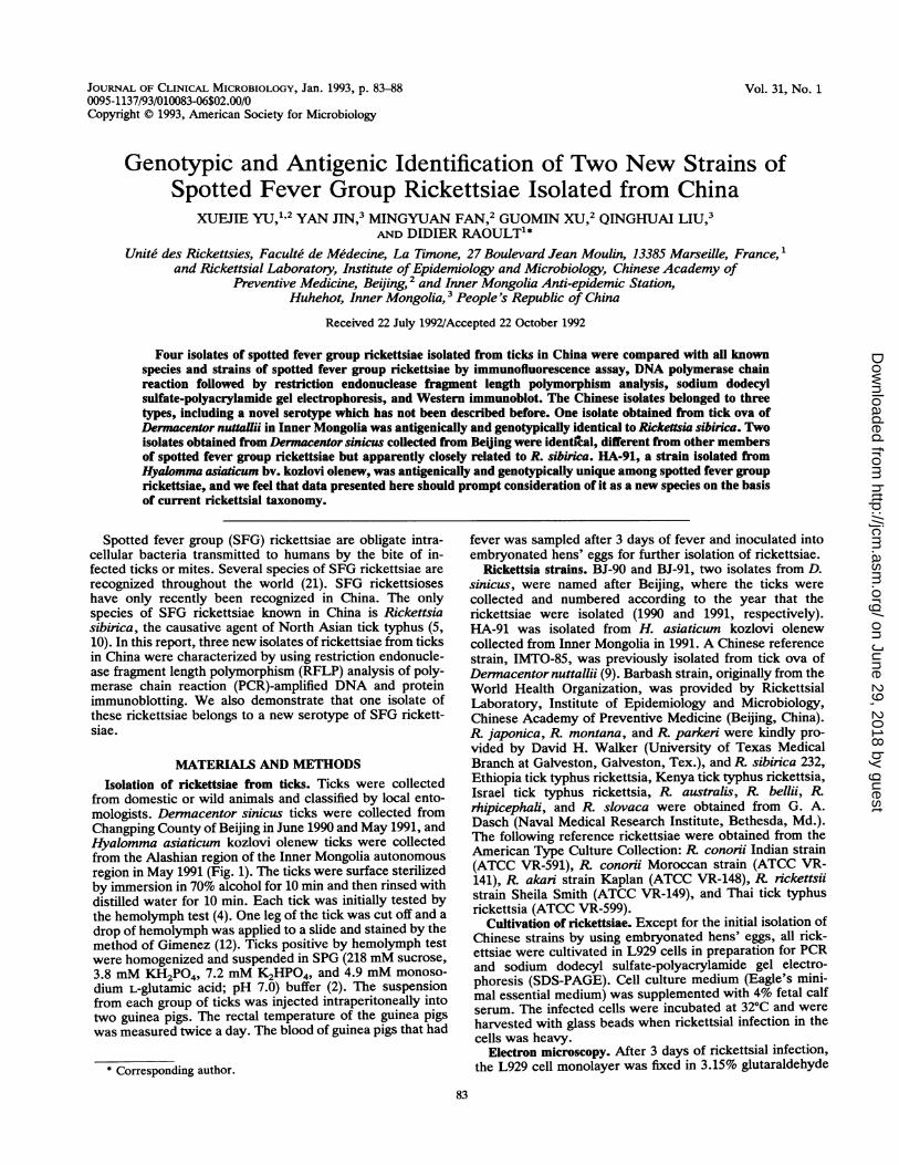
Vol. 31, No. 1JOURNAL OF CLINICAL MICROBIOLOGY, Jan. 1993, p. 83-880095-1137/93/010083-06$02.00/0Copyright © 1993, American Society for Microbiology
Genotypic and Antigenic Identification of Two New Strains ofSpotted Fever Group Rickettsiae Isolated from China
XUEJIE YU,1,2 YAN JIN,3 MINGYUAN FAN,2 GUOMIN XU,2 QINGHUAI LIU,3AND DIDIER RAOULT1*
Unite des Rickettsies, Faculte de Medecine, La Timone, 27 Boulevard Jean Moulin, 13385 Marseille, France,and Rickettsial Laboratory, Institute of Epidemiology and Microbiology, Chinese Academy of
Preventive Medicine, Beijing,2 and Inner Mongolia Anti-epidemic Station,Huhehot, Inner Mongolia,3 People's Republic of China
Received 22 July 1992/Accepted 22 October 1992
Four isolates of spotted fever group rickettsiae isolated from ticks in China were compared with all knownspecies and strains of spotted fever group rickettsiae by immunofluorescence assay, DNA polymerase chainreaction followed by restriction endonuclease fragment length polymorphism analysis, sodium dodecylsulfate-polyacrylamide gel electrophoresis, and Western immunoblot. The Chinese isolates belonged to threetypes, including a novel serotype which has not been described before. One isolate obtained from tick ova ofDermacentor nuttaUii in Inner Mongolia was antigenicaily and genotypicaily identical to Rickettsia sibirica. Twoisolates obtained from Dermacentor sinicus collected from Beiing were identical, different from other membersof spotted fever group rickettsiae but apparently closely related to R. sibirica. HA-91, a strain isolated fromHyalomma asiaticum bv. kozlovi olenew, was antigenically and genotypically unique among spotted fever grouprickettsiae, and we feel that data presented here should prompt consideration of it as a new species on the basisof current rickettsial taxonomy.
Spotted fever group (SFG) rickettsiae are obligate intra-cellular bacteria transmitted to humans by the bite of in-fected ticks or mites. Several species of SFG rickettsiae arerecognized throughout the world (21). SFG rickettsioseshave only recently been recognized in China. The onlyspecies of SFG rickettsiae known in China is Rickettsiasibirica, the causative agent of North Asian tick typhus (5,10). In this report, three new isolates of rickettsiae from ticksin China were characterized by using restriction endonucle-ase fragment length polymorphism (RFLP) analysis of poly-merase chain reaction (PCR)-amplified DNA and proteinimmunoblotting. We also demonstrate that one isolate ofthese rickettsiae belongs to a new serotype of SFG rickett-siae.
MATERIALS AND METHODSIsolation of rickettsiae from ticks. Ticks were collected
from domestic or wild animals and classified by local ento-mologists. Dermacentor sinicus ticks were collected fromChangping County of Beijing in June 1990 and May 1991, andHyalomma asiaticum kozlovi olenew ticks were collectedfrom the Alashian region of the Inner Mongolia autonomousregion in May 1991 (Fig. 1). The ticks were surface sterilizedby immersion in 70% alcohol for 10 min and then rinsed withdistilled water for 10 min. Each tick was initially tested bythe hemolymph test (4). One leg of the tick was cut off and adrop of hemolymph was applied to a slide and stained by themethod of Gimenez (12). Ticks positive by hemolymph testwere homogenized and suspended in SPG (218 mM sucrose,3.8 mM KH2PO4, 7.2 mM K2HPO4, and 4.9 mM monoso-dium L-glutamic acid; pH 7.0) buffer (2). The suspensionfrom each group of ticks was injected intraperitoneally intotwo guinea pigs. The rectal temperature of the guinea pigswas measured twice a day. The blood of guinea pigs that had
* Corresponding author.
fever was sampled after 3 days of fever and inoculated intoembryonated hens' eggs for further isolation of rickettsiae.
Rickettsia strains. BJ-90 and BJ-91, two isolates from D.sinicus, were named after Beijing, where the ticks werecollected and numbered according to the year that therickettsiae were isolated (1990 and 1991, respectively).HA-91 was isolated from H. asiaticum kozlovi olenewcollected from Inner Mongolia in 1991. A Chinese referencestrain, IMTO-85, was previously isolated from tick ova ofDernacentor nuttallii (9). Barbash strain, originally from theWorld Health Organization, was provided by RickettsialLaboratory, Institute of Epidemiology and Microbiology,Chinese Academy of Preventive Medicine (Beijing, China).R japonica, R. montana, and R parken were kindly pro-vided by David H. Walker (University of Texas MedicalBranch at Galveston, Galveston, Tex.), and R. sibirica 232,Ethiopia tick typhus rickettsia, Kenya tick typhus rickettsia,Israel tick typhus rickettsia, R australis, R bellii, R.rhipicephali, and R. slovaca were obtained from G. A.Dasch (Naval Medical Research Institute, Bethesda, Md.).The following reference rickettsiae were obtained from theAmerican Type Culture Collection: R. conorii Indian strain(ATCC VR-591), R. conorii Moroccan strain (ATCC VR-141), R akari strain Kaplan (ATCC VR-148), R rickettsiistrain Sheila Smith (ATCC VR-149), and Thai tick typhusrickettsia (ATCC VR-599).
Cultivation of rickettsiae. Except for the initial isolation ofChinese strains by using embryonated hens' eggs, all rick-ettsiae were cultivated in L929 cells in preparation for PCRand sodium dodecyl sulfate-polyacrylamide gel electro-phoresis (SDS-PAGE). Cell culture medium (Eagle's mini-mal essential medium) was supplemented with 4% fetal calfserum. The infected cells were incubated at 320C and were
harvested with glass beads when rickettsial infection in thecells was heavy.
Electron microscopy. After 3 days of rickettsial infection,the L929 cell monolayer was fixed in 3.15% glutaraldehyde
83
on June 29, 2018 by guesthttp://jcm
.asm.org/
Dow
nloaded from

84 YU ET AL.
FIG. 1. Locations in China where the ticks were collected forisolation of rickettsiae. 1, Alashian region of Inner Mongolia; 2,Changping County of Beijing.
for 30 min and then washed in sucrose-cacodylate buffer for30 min and fixed again in 1% osmium-1.5% potassiumferricyanide for 1 h. Dehydration was performed by usingincreasing concentrations of acetone (15 to 100%). The layerwas embedded in Epon, sectioned, stained with uranylacetate and lead citrate, and examined with a JEOL JEM1200 EX electron microscope (3).
Antisera. Mouse antisera were prepared by the method ofPhilip et al. (15); female Swiss Webster mice were injectedintravenously via the tail vein with 0.5 ml of rickettsia-infected L929 cells on day 0 and 7 and were exsanguinatedon day 10. The sera from each group were pooled and storedat -70°C. Rickettsia-infected L929 cells were used as anti-gens for immunofluorescence assay IFA. Fluorescein(DTAF)-labeled goat anti-mouse immunoglobulin G andimmunoglobulin M (heavy plus light chains) conjugate waspurchased from Jackson ImmunoResearch Laboratories,Inc. (West Grove, Pa.).The specificity difference (SPD) between each pair of
strains was calculated according to the formula SPD = (Aa+ Bb) - (Ab + Ba) (15), where Aa (or Bb) is the antibodytiter of serum A (or B) reacted with heterologous antigen b(or a). In this formula, antibody titers were expressed as thelog2 of the endpoint titer. If the SPD between two strains was.3 (P < 0.01), they were considered two distinct species(15).
Purification of rickettsiae. The rickettsia-infected cellswere centrifuged at 7,000 x g for 10 min. The pellet was
suspended in K36 buffer (0.1 M KCl, 0.05 M phosphatebuffer, 0.015 M NaCl; pH 7.0) (20) and sonicated in an icebath. The suspension was loaded onto a cushion of 30%sucrose in K36 buffer and centrifuged at 7,000 x g for 30min. The resulting pellet was purified by Renografin densitygradient centrifugation (20). The light and heavy bands wereharvested and washed with K36 buffer at 7,000 x g for 10min. The purified rickettsiae were resuspended in distilledwater. The rickettsial-protein concentrations were quantifiedby the method of Lowry et al. (14).SDS-PAGE and Western blot. SDS-PAGE was performed
as described previously (13); 4% stacking and 7.5% separat-ing gels with 2.6% cross-linking were used. The purified
rickettsiae were solubilized in Laemmli (13) solubilizer (4%SDS, 0.125 M Tris hydrochloride [pH 6.8], 25% glycerol,10% 2-mercaptoethanol, 0.5% bromophenol blue) at roomtemperature, and 10 ,ug of rickettsial protein was loaded intoeach well of the gels. The gel (6 by 8 by 0.1 cm) (Bio-Rad,Richmond, Calif.) was run at 10 mA in an ice bath. Westernblot (immunoblot) was performed as described previously(19) in an ice bath. The nitrocellulose paper was blockedwith 5% nonfat milk in TBS (20 mM Tris-HCI, 500 mMNaCl; pH 7.5). The mouse antisera were diluted 1/100 inTBS containing 1% nonfat milk. Peroxidase-conjugated goatanti-mouse immunoglobulin G (heavy plus light chains)(Jackson ImmunoResearch Laboratories) was diluted 1/500in TBS. The nitrocellulose paper was incubated with4-chloro-1-naphthol (Sigma, St. Louis, Mo.) to develop thecolor. Low-range and high-range prestained molecularweight standards (Bio-Rad) were used to estimate the mo-lecular weights of proteins.PCR amplification, DNA digestion, and electrophoresis.
The samples for PCR amplification were prepared as de-scribed previously (1, 7). A 1.5-ml sample of rickettsia-infected L929 cells was washed three times with distilledwater by centrifugation at 17,500 x g for 5 min. The resultingpellet was resuspended in 100 ,ul of distilled water and boiledfor 10 min; 10 ,ul of this material was used as DNAtemplates. DNA amplification, digestion, and electrophore-sis were carried out as described previously (16). DNAamplification was done in a thermal cycler (PREM III; LepScientific, Flobio, Courbevoie, France) with 35 cycles ofdenaturation (20 s at 95°C), annealing (30 s at 48°C), andextending (2 min at 60°C). Noninfecting L929 cells anddistilled water were used as negative controls. PCR amplifi-cation of DNA was verified by 1% agarose (Sigma Chimie,La Verpilliere, France) gel electrophoresis of 10 ,ul of PCRproduct. A 23.5-pdl volume ofPCR product was digested withrestriction endonuclease at 37°C for 2 h. The digestedproducts were separated on 8% polyacrylamide vertical gels(Bio-Rad Laboratories) by standard procedures (18). Gelswere run at 100 V for 4 h and then stained with ethidiumbromide. The gels were photographed with type 667 P/N film(Polaroid Corp., Cambridge, Mass.) and a 365-nm UV lightsource (Bioblock Scientific, Illkirch, France). DNA molec-ular marker V (Boehringer GmbH, Mannheim, Germany)was used to determine the sizes of the DNA fragments. Thesizes of DNA fragments were calculated by entering digi-tized migration data directly into a desktop computer (TheImager; Appligen, Heidelberg, Germany). PCR-RFLP wasperformed by using the following oligonucleotide primerpairs (Bioprobe Systems, Montreuil-sous-Bois, France) andrestriction endonucleases (New England Biolabs, Beverly,Mass.): Rp CS.877p and Rp CS.1258n (16) (encoding a381-bp sequence) withAluI, Rr 190.70P and Rr 190.602n (532bp) (16) with RsaI and PstI, and Rr 120.BG3 and Rr 120.BG4(615 bp) (7, 11) with AluI and RsaI.Comparison with other rickettsiae. Our laboratory is
equipped with an image system (Appligen) connected with aQGEL program retaining pictures and molecular weightsfrom previous works. R. akari, R. australis, R. conorii, R.rhipicephali, R parken, R. slovaca, R. helvetica, R. bellii,R. nckettsii, Thai tick typhus rickettsia, and R. massiliaehave been tested by both SDS-PAGE (1) and PCR-RFLP (1,7, 8), and the results constitute a data bank allowing com-parison of new strains with previously tested species.
J. CLIN. MICROBIOL.
on June 29, 2018 by guesthttp://jcm
.asm.org/
Dow
nloaded from

IDENTIFICATION OF CHINESE RICKETTSIAE 85
184-123--89-
51-
434-
2671234-184-123-89-64-
FIG. 2. Ethidium bromide-stained polyacrylamide gel electro-phoretograms of restriction endonuclease AluI-digested rickettsialDNA PCR amplified by using the Rp CS.877p and Rp CS.1258nprimer pair. Lanes: 1, Barbash strain; 2, R japonica; 3, R. sibirica;4, IMTO-85; 5, BJ-90; 6, BJ-91; 7, HA-91; M, molecular sizemarkers (sizes are indicated on the left in base pairs).
RESULTS
Isolation of the new strains. Two strains of rickettsiae wereisolated from D. sinicus and one strain was isolated from H.asiaticum kozlovi olenew. All of these rickettsiae werepathogenic to guinea pigs. All caused fever (40 to 42°C) andscrotal swelling. No deaths among guinea pigs were ob-served. The incubation time for the guinea pigs was between2 and 5 days, depending on the initial numbers of rickettsiaeinoculated. The rickettsiae were observed intracellularly onthe smear of guinea pig scrotal tissue after staining by themethod of Gimenez (12) (data not shown). All isolates grewwell in embryonated hens' eggs and cell culture.PCR-RFLP. Results of the PCR-RFLP analysis are shown
in Fig. 2 to 7. Amplification by using the Rp CS.877p and RpCS.1258n primer pair and digestion with restriction endonu-clease AluI demonstrated that all Chinese isolates (Fig. 2)had the typical profiles of the SFG rickettsiae (16) and thatthe DNA migration patterns ofR japonica were unique.When the Rr 190.70p and Rr 190.602n primer pair was
587-
184--
FIG. 3. Ethidium bromide-stained polyacrylamide gel electro-phoretograms of restriction endonuclease RsaI-digested rickettsialDNA PCR amplified by using the Rr 190.70p and Rr 190.602n primerpair. Lanes: 1, R. japonica; 2, Barbash strain; 3, R. sibinica; 4,IMTO-85; 5, BJ-90; 6, BJ-91; 7 and 10, HA-91; 8, Ethiopia ticktyphus rickettsia; 9, Israel tick typhus rickettsia; 11, R parkeri; 12,R conorii Moroccan strain; 13, R conorii Indian strain; 14, Kenyatick typhus rickettsia; M, molecular size markers (sizes are indi-cated on the left in base pairs).
FIG. 4. Ethidium bromide-stained polyacrylamide gel electro-phoretograms of restriction endonuclease PstI-digested rickettsialDNA PCR amplified by using the Rr 190.70p and Rr 190.602n primerpair. Lanes: 1, R. japonica; 2, Barbash strain; 3, R sibirica; 4,IMTO-85; 5, BJ-90; 6, BJ-91; 7 and 10, HA-91; 8, Ethiopia ticktyphus rickettsia; 9, Israel tick typhus rickettsia; 11, R. parkeri; 12,R conorii Moroccan strain; 13, R conorii Indian strain; 14, Kenyatick typhus rickettsia; M, molecular size markers (sizes are indi-cated on the left in base pairs).
used, the PCR products of all rickettsiae had the samepatterns on agarose gels except for that of Barbash strain,whose molecular weight was much less than those for otherSFG rickettsiae (data not shown). The amplification prod-ucts of all rickettsiae with these primer pairs were digestedby restriction endonucleases PstI and RsaI except for thePCR product of R. japonica, which could not be digested byRsaI (Fig. 3). Restriction of these products with RsaIdemonstrated that all Chinese strains were divided into twodistinct genotypic groups: one group including BJ-90, BJ-91,and IMTO-85 had PCR-RFLP patterns identical to those ofR sibirica; HA-91 belonged to another group whose PCR-RFLP patterns were different from those of R. sibirica butidentical to those ofR parkeri and similar to those of strainsofR cononii (Fig. 3 and 6). PstI digestion of PCR-amplifiedrickettsial DNA with the Rr 190.70p and Rr 190.602n primerpair showed that all Chinese strains, R. sibirica, R. parkeri,
434267k
184=7123 - =3
8064: - _ _51_
FIG. 5. Ethidium bromide-stained polyacrylamide gel electro-phoretograms of restriction endonuclease RsaI-digested rickettsialDNA PCR amplified by using the Rr 120.BG3 and Rr 120.BG4primer pair. Lanes: 1, R. sibinica; 2, IMTO-85; 3, BJ-90; 4, BJ-91; 5,HA-91; 6, R. parkeri; 7, Israel tick typhus rickettsia; 8, Ethiopia ticktyphus rickettsia; M, molecular size markers (sizes are indicated onthe left in base pairs).
VOL. 31, 1993
on June 29, 2018 by guesthttp://jcm
.asm.org/
Dow
nloaded from

J. CLIN. MICROBIOL.
Sib INTO-8S BJ-90 BJ-91 RA-91 Pak lr 5th Mor Ind Ken Bar Jap
E~~1-[- --E[E ~J-- 2 8E
553 553 553 553 563 563 563 574 563 592 592 389 560
FIG. 6. Schematic electrophoresis migration patterns of PCR-amplified DNA (Rr 190.70p and Rr 190.602n) of rickettsiae R.sibirnca (Sib), R. parkeri (Pak), Israel tick typhus rickettsia (Isr),Ethiopia tick typhus rickettsia (Eth), R. cononii Moroccan strain(Mor), R. cononii Indian strain (Ind), Kenya tick typhus rickettsia(Ken), Barbash strain (Bar), and R. japonica (Jap) digested withRsaI. Doublet bands are indicated with an asterisk. Comigratingfragments are connected by dashed lines. Numbers are molecularsizes in base pairs.
Ethiopia tick typhus rickettsia, and Israel tick typhus rick-ettsia had identical profiles, but HA-91 had patterns differentfrom those of R. conorni Moroccan strain and Indian strainand Kenya tick typhus rickettsia (Fig. 4).The Rr 120.BG 3 and Rr 120.BG 4 primer pair was further
used for PCR-RFLP analysis of strain HA-91 and R. parkeri,since they were not differentiated by digestions with RsaIand PstI, respectively, of PCR-amplified DNA derived fromthe Rr 190.70p and Rr 190.602n primer pair. Rickettsial DNAamplified with the Rr 120.BG3 and Rr 120.BG4 primer pairdigested with RsaI demonstrated that the profiles of strainHA-91 and R. park;eni were different from those of Ethiopiatick typhus rickettsia and Israel tick typhus rickettsia (Fig. 5and 7). AluI digestion of these PCR products did not discloseadditional differences between strain HA-91 and these rick-ettsiae (data not shown).
Sib INTO-aS BI3-90 BJ-91 5A-91 Pak Isr Zth
A B
205-1
106 - 8 8 - W - a: ..
FIG. 8. Profiles of Coomassie brilliant blue-stained, SDS-PAGE-
separated rickettsial whole proteins with low (A) and high (B)molecular masses. HA, HA-91; TO, IMTO-85; sib, R sibirica; 90,
BJ-90; 91, BJ-91. Molecular mass markers (in kilodaltons) are
indicated. Distinctive rickettsial protein bands are indicated byarrows.
SDS-PAGE and Western blot. The purity of rickettsiae was
tested by SDS-PAGE by comparison with host cell lysates.The results demonstrated that only one protein with a
molecular mass of 60 kDa was common among rickettsiae
and L929 cells (data not shown). The 60-kDa protein of
rickettsiae was probably not contaminated with host protein.It may be a heat shock protein and was shared by rickettsiae
and host cells (6). The protein profiles of the rickettsiae
showed that all strains shared many common proteins withlow molecular masses (< 106 kDa) (Fig. 8A), but the majorproteins in the high-molecular-mass range (>i106 kDa) weredistinctive among these strains of rickettsiae (Fig. 8).IMTO-85 and sibirica had protein profiles identical to
those of the major outer membrane proteins of 106, 118, and
155 kDa. The protein profiles of BJ-90 and BJ-91 were
identical to each other and slightly different from that of R.
sibirica, with a distinct protein band of 162 instead of 155
kDa. HA-91 strain had one distinct major protein band witha molecular mass of 180 kDa. Westem blot with typing serademonstrated that the major antigenic proteins among Chi-nese isolates were different from each other (Fig. 9). Mouse
1 2 3 4 5
205-
106---am. *a
80-
C5 1 --- E54 -----543 --- E3 --- 23 ---- 23 ---E --F54---
637 637 637 637 637 636 639 647
FIG. 7. Schematic electrophoresis migration patterns of PCR-amplified DNA (Rr 120.BG3 and Rr 120.BG4) of rickettsiae Rsibinca (Sib), R. parkeri (Pak), Israel tick typhus rickettsia (Isr), andEthiopia tick typhus rickettsia (Eth) digested with RsaI. Comigrat-ing fragments are connected by dashed lines. Numbers are molec-ular sizes in base pairs.
49.5-FIG. 9. Western blot of SDS-PAGE-separated 'rickettsial pro-
teins reacted with mouse antisera to strain HA-91. Lanes containrickettsial antigens as follows: lane 1, HA-91; lane 2, IMTO-85; lane3, R. sibirica; lane 4, BJ-90; lane 5, BJ-91. Molecular masses (inkilodaltons) are indicated on the left.
86 YU ET AL.
on June 29, 2018 by guesthttp://jcm
.asm.org/
Dow
nloaded from

IDENTIFICATION OF CHINESE RICKETTSIAE 87
+ i
J .:,. , .. .... .. . A . |FIG. 10. Transmission electron micrograph of L929 cell infected
with HA-91 rickettsiae (indicated by an arrow). Magnification, ca.x7,000.
antisera to strain HA-91 recognized two major antigenicproteins of all strains; one of them, with a molecular mass of106 kDa, was common among the strains, and the other wasdistinctive for each rickettsia. The distinctive antigenicproteins of rickettsiae were determined to be 155 kDa in sizefor IMTO-85 and R. sibirica, 162 kDa for BJ-90 and BJ-91,and 180 kDa for HA-91 (Fig. 9).
Electron microscopy. Electron microscopy showed thatHA-91 rickettsiae (Fig. 10) and BJ-90 rickettsiae (Fig. 11)were free in the cytoplasm and surrounded by an electron-lucent, halo-like zone which is apparently a slime layer (17).
IFA. Mouse antiserum to strain HA-91 at a titer of 1:8 hadno reaction with the following rickettsiae: R. akari, R.australis, R. beliji, R. japonica, R. montana, R. rhipiceph-ali, R. massiliae, and Thai tick typhus rickettsiae. Thisserum cross-reacted with other members of the SFG rickett-siae. The SPD between HA-91 strain and closely relatedspecies such as R. sibica, R. conoii, R. ickettsii, R.parkeFi, R. slovaca, Ethiopia tick typhus rickettsia, andother Chinese isolates we're 3 (Table 1).
DISCUSSION
With the high level of reproducibility of PCR-RFLP meth-ods and inclusion of appropriate DNA size standards, it ispossible to identify genotypes of unidentified rickettsialisolates by comparison with established reference patterns(16). The PCR-RFLP patterns of R sibitica and R conoriiMoroccan strain, which were used as positive controls inthis study, coincided with those of previous studies (16).Comparing our results with previously published data (16)and our data bank for other SF0rickettsiae that were notused in our study, we found that the Rsal and PstI digestpatters derived from the Rr 190.70p and Rr 190.602n primerpair for the Chinese isolates were different from those for R.uckettsii, R. slovaca, R. rhipicephali, R. montana, R. mas-
FIG. 11. Transmission electron micrograph of L929 cell infectedwith BJ-90 rickettsiae (indicated by an arrow). Magnification, ca.x7,000.
siliae, and Thai tick typhus rickettsia. The AluI digestpatterns derived from the Rp CS.877p and Rp CS.1258nprimer pair for the Chinese isolates were different from thoseofR akari, R. australis, and R bellii.Our study demonstrated that there are three types of SFG
rickettsiae distributed in northern China. Type 1 was anti-genically and genotypically identical to R. sibirica, whichwas represented by IMTO-85, the rickettsia previously iso-lated from tick ova (9). Type 2 was genotypically identical toand antigenically related to R sibirica, but its profile wasdifferent and the representative strain for this type is BJ-90.Type 3 was both genotypically and antigenically uniqueamong SFG rickettsiae, and the representative strain wasHA-91. This was confirmed by IFA because the SPDsbetween HA-91 and other SFG rickettsiae were .3. SFGrickettsia serotypes are considered species according tocurrent criteria (21). One of our new isolates, HA-91, shouldbe considered a new species of SFG rickettsiae. The newisolates BJ-90 and BJ-91 could probably be consideredvariant strains of R. sibirica.
Previous studies demonstrated that all SFG rickettsiaeisolated from the broad area of China from Xinjiang in thewest to Heilongjiang in the east were antigenically identicalto R sibirica (5, 10). Our study not only identified two newtypes of SFG rickettsiae but also expanded the documenteddistribution of SFG rickettsiae in China. The south boundaryof SFG rickettsiae distribution in China was moved tolatitude 40°N. Probably the geographic distribution of SFGrickettsiae is not limited to this area. D. sinicus is distributedbroadly in China, including Hebei and Shandong provincesin the south. Any area where D. sinicus is found could be anatural focus of SFG rickettsiae.These new isolates were pathogenic to guinea pigs; how-
ever, their pathogenicity for humans remained to be inves-tigated.
VOL. 31, 1993
1. f.".AA,
on June 29, 2018 by guesthttp://jcm
.asm.org/
Dow
nloaded from

88 YU ET AL.
TABLE 1. IFA titers of reciprocal reaction of mouse antisera with rickettsial antigens and SPDs between strains of rickettsiae
Mouse Titer of rickettsial antigen (SPD)antiserum
type HA-91 BJ-90 R sibirica R. slovaca R rickettsii R parkeri R. conofiia EthTIbHA-91 1,024 (0) 256 256 512 256 256 256 256BJ-90 128 (4) 512 (0) 512 256 64 128 64 64R sibirica 128 (5) 512 (1) 1,024 (0) 128 128 128 64 128R. slovaca 256 (4) 256 (4) 64 (8) 2,048 (0) 64 128 64 128R. rickettsii 16 (6) 16 (7) 32 (6) 32 (8) 256 (0) 64 32 16R. parkeri 256 (4) 64 (6) 64 (7) 32 (9) 256 (4) 1,024 (0) 128 512R. conorii 128 (5) 32 (8) 32 (9) 32 (10) 64 (7) 512 (4) 1,024 (0) 1,024EthTT 256 (4) 64 (7) 64 (7) 128 (7) 256 (6) 256 (3) 512 (1) 1,024 (0)
a Moroccan strain.b EthTT, Ethiopia tick typhus rickettsia.
ACKNOWLEDGMENTS
We are grateful to Michel Drancourt for technical help, PhilippeBrouqui for electron microscopy, James G. Olson (Centers forDisease Control, Atlanta, Ga.) for review of the manuscript, and V.Pinero and I. Domingo for secretarial assistance in the preparationof the manuscript.
This study was supported by a grant from Region PACA.
REFERENCES1. Beati, L., J. P. Finidori, B. Gilot, and D. Raoult. 1992. Compar-
ison of serologic typing, sodium dodecyl sulfate-polyacrylamidegel electrophoresis protein analysis, and genetic restrictionfragment length polymorphism analysis for identification ofrickettsiae: characterization of two new rickettsial strains. J.Clin. Microbiol. 30:1922-1930.
2. Bovarnick, M. R., J. C. Miller, and J. C. Snyder. 1950. Theinfluence of certain fats, amino acids, sugars, and proteins onthe stability of rickettsiae. J. Bacteriol. 59:509-522.
3. Brouqui, P., and D. Raoult. 1991. Effects of antibiotics on thephagolysosome fusion in Ehrlichia sennetsu infected P388 Dlcells, p. 751-757. In J. Kazar and D. Raoult (ed.), Rickettsiaeand rickettsial diseases. Publishing House of the Slovak Acad-emy of Sciences, Bratislava, Czechoslovakia.
4. Burgdorfer, W. 1970. Hemolymph test. A technique for detec-tion of rickettsiae in ticks. Am. J. Trop. Med. Hyg. 19:1010-1014.
5. Cheng, X. X., G. Q. Yu, and S. Y. Yu. 1989. Identification ofChinese strains of spotted fever group rickettsiae by usingspecies-specific monoclonal antibodies to Rickettsia sibirica.Chin. J. Microbiol. Immunol. (Beijing) 9:129-131.
6. Dasch, G. A., W. M. Ching, P. Y. Kim, H. Pham, C. K. Stover,E. V. Oaks, M. E. Dobson, and E. Weiss. 1990. A structural andimmunological comparison of rickettsial HSP60 antigens withthose of other species. Ann. N.Y. Acad. Sci. 590:352-369.
7. Drancourt, M., L. Beati, I. Tarasevich, and D. Raoult. 1992.Astrakan fever rickettsia is identical to Israel tick typhusrickettsia, a genotype of the Rickettsia conorii complex. J.Infect. Dis. 165:1167-1168.
8. Eremeeva, M., X. J. Yu, L. Beati, and D. Raoult. Differentiationamong spotted fever group rickettsiae species by analysis ofrestriction fragment length polymorphism of polymerase chainreaction amplified DNA. Submitted for publication.
9. Fan, M. Y., D. H. Walker, Q. H. Liu, L. Han, H. C. Bai, J. K.Zhang, B. Lenz, and C. Hong. 1987. Rickettsial and serologic
evidence for prevalent spotted fever rickettsiosis in Inner Mon-golia. Am. J. Trop. Med. Hyg. 36:615-620.
10. Fan, M. Y., X. J. Yu, and D. H. Walker. 1988. Antigenicanalysis of Chinese strains of spotted fever group rickettsiae byprotein immunoblotting. Am. J. Trop. Med. Hyg. 39:497-501.
11. Gilmore, R. D., Jr., N. Joste, and G. A. McDonald. 1989.Cloning, expression and sequence analysis of the gene encodingthe 120 kD surface-exposed protein of Rickettsia rickettsii. Mol.Microbiol. 3:1579-1586.
12. Gimenez, D. F. 1964. Staining rickettsiae in yolk-sac cultures.Stain Technol. 39:135-140.
13. Laemmli, U. K. 1970. Cleavage of structural proteins during theassembly of the head of bacteriophage T4. Nature (London)227:680-685.
14. Lowry, 0. H., N. J. Rosebrough, A. L. Farr, and R. J. Randall.1951. Protein measurement with the Folin phenol reagent. J.Biol. Chem. 193:265-275.
15. Philip, R. N., E. A. Casper, W. Burgdorfer, R. K. Gerloff, L. E.Hugues, and E. J. Bell. 1978. Serologic typing of rickettsiae ofthe spotted fever group by micro immunofluorescence. J. Im-munol. 121:1961-1968.
16. Regnery, R. L., C. L. Spruill, and B. D. Plikaytis. 1991.Genotypic identification of rickettsiae and estimation of in-traspecies sequence divergence for portions of two rickettsialgenes. J. Bacteriol. 173:1576-1589.
17. Silverman, D. J., C. L. Wisseman, Jr., A. D. Waddell, and M.Jones. 1978. External layers of Rickettsia prowazekii and Rick-ettsia rickettsii: occurrence of a slime layer. Infect. Immun.22:233-246.
18. Sambrook, J., E. F. Fritsch, and T. Maniatis. 1989. Molecularcloning: a laboratory manual, 2nd ed. Cold Spring HarborLaboratory, Cold Spring Harbor, N.Y.
19. Towbin, H., T. Staehelin, and J. Gordon. 1979. Electrophoretictransfer of proteins from polyacrylamide gels to nitrocellulosesheets: procedure and some applications. Proc. Natl. Acad. Sci.USA 76:4350-4354.
20. Weiss, E., J. C. Coolbaugh, and J. C. Williams. 1975. Separationof viable Rickettsia typhi from yolk sac and L cell host compo-nents by Renografin density gradient centrifugation. Appl. Mi-crobiol. 30:456-463.
21. Weiss, E., and J. W. Moulder. 1984. Order I. RickettsialesGieszczkiewicz 1939, 25AL, p. 687-701. In N. R. Krieg and J. G.Holt (ed.), Bergey's manual of systematic bacteriology, vol. 1.Williams & Wilkins, Baltimore.
J. CLIN. MICROBIOL.
on June 29, 2018 by guesthttp://jcm
.asm.org/
Dow
nloaded from