Embed Size (px)
Citation preview
Vol. 26, No. 10JOURNAL OF CLINICAL MICROBIOLOGY, Oct. 1988, p. 2025-20300095-1137/88/102025-06$02.00/0Copyright © 1988, American Society for Microbiology
Microtechnique for Serum Opacity Factor Characterization ofGroup A Streptococci Adaptable to the Use of Human Sera
DWIGHT R. JOHNSON AND EDWARD L. KAPLAN*
Department of Pediatrics and the World Health Organization Collaborating Center for Reference and Researchon Streptococci, University of Minnesota Medical School, Minneapolis, Minnesota 55455
Received 22 April 1988/Accepted 20 June 1988
We have developed a microtechnique for detection of streptococcal serum opacity factor (OF) and for typingof group A streptococci by inhibition of OF. This technique, which involves the use of single wells of standard96-well tissue culture plates, offers several advantages over previous methods: no advance test preparation isrequired, allowing tests to be quickly and easily performed; only small quantities of reagents are required;results can be determined visually (qualitative) or by using a photometric enzyme-linked immunosorbent assayplate reader (quantitative); and human serum samples may be quickly and easily screened for OF-inhibitoryantibody and subsequently used in place of difficult-to-produce and expensive hyperimmune animal sera for OFcharacterization of group A streptococci. Fifty-eight samples of normal adult human serum were tested by thisnew microtechnique for anti-OF antibodies, and 49 (84%) were found to have antibody against 1 or more ofthe 27 recognized OF-positive serotypes. OF antibodies to M-4, M-2, M-75, and M-48 were most common inthese individuals. These 58 human serum samples collectively contained antibody to 25 of the 27 differentOF-producing serotypes. Serum samples from four individuals were tested for persistence of OF antibody. 0Fantibodies to eight different serotypes present in the serum samples collected 7 to 12 years previously werepresent in the freshly collected sera, indicating that OF antibody persists in human antisera for many years.This new technique has distinct advantages and makes it possible for many laboratories to use this techniqueto characterize group A streptococci.
Although the efficacy of using streptococcal serum opacityfactor (OF) techniques for serotype characterization ofgroup A streptococci is well established (12, 16), OF typinghas not been widely used. Previously available techniquesare labor intensive, the results may be difficult to interpret,and antisera for OF typing have been difficult and expensiveto produce in laboratory animals. To eliminate these disad-vantages, we have developed a microtechnique which isquick and simple to perform, is easy to interpret, conservesmaterials (especially antisera), and permits use of readilyavailable and inexpensive human serum as a source ofanti-OF antibodies. This useful technique can now be madeavailable to more laboratories because of this simplicity.
MATERIALS AND METHODSGroup A streptococcal reference strains in our collection
representing the 27 currently recognized OF-positive Mtypes (M-2, M-4, M-8, M-9, M-11, M-13, M-22, M-25, M-28,M-48, M-49, M-58 to M-64, M-66, M-68, M-73, M-75 toM-79, and M-81) (1, 17) were obtained originally fromRebecca Lancefield, the Rockefeller University, New York,N.Y.; Richard Facklam, The Centers for Disease Control,Atlanta, Ga.; Geoffrey Colman, Central Public Health Lab-oratory, London, England; and Jiri Rotta, Institute of Hy-giene and Epidemiology, Prague, Czechoslovakia. (Notethat M-64 has been described as identical to M-52 [1]. Thestrain sent to us in 1975 as provisional M-64 [Rabinowitz 932]was not M-52 and is included in these studies as M-64.)Wild-type strains of group A streptococci were obtainedfrom previous and ongoing epidemiological studies in ourlaboratory. Long-term storage of stock strains was by lyo-philization. Short-term storage of strains was in blood brothfrozen at -20°C.
* Corresponding author.
Horse serum (GIBCO Diagnostics, Grand Island, N.Y.)was used as the "substrate" in the serum opacity reactionafter multiple lots from several suppliers were screened forsuitability. The horse serum was stored at -20°C. Single100-ml bottles were thawed as needed and heated at 56°C for30 min to inactivate endogenous enzymes. The pH of thehorse serum was not adjusted for use in these studies.Storage for daily use was at 4°C after addition of 0.2 mg ofthimerosal per ml to inhibit contamination.OF was obtained in two ways. Strains were grown for 16
to 18 h at 35°C in Todd-Hewitt broth (Difco Laboratories,Detroit, Mich.) supplemented with an additional 2% Neo-peptone (Difco). The clear supernatant broth was examinedfor presence of OF as described below. In addition, standardLancefield hot-HCI extracts (15) of the same strains werealso tested for presence of OF. In the work described here,we generally used HCI-extracted OF.The microwell method for detection of OF involved the
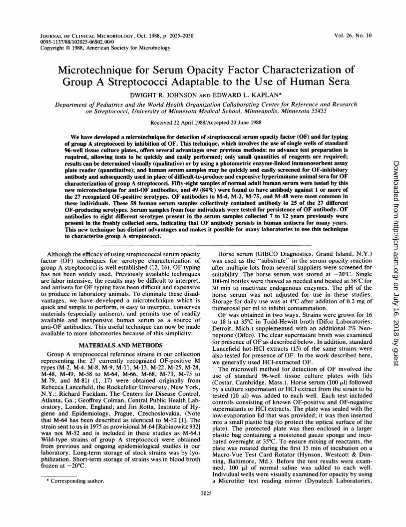
use of standard 96-well tissue culture plates with lids(Costar, Cambridge, Mass.). Horse serum (100 ,ul) followedby a culture supernatant or HCI extract from the strain to betested (10 ,ul) was added to each well. Each test includedcontrols consisting of known OF-positive and OF-negativesupernatants or HCI extracts. The plate was sealed with thelow-evaporation lid that was provided; it was then insertedinto a small plastic bag (to protect the optical surface of theplate). The protected plate was then enclosed in a largerplastic bag containing a moistened gauze sponge and incu-bated overnight at 35°C. To ensure mixing of reactants, theplate was rotated during the first 15 min of incubation on aMacro-Vue Test Card Rotator (Hynson, Westcott & Dun-ning, Baltimore, Md.). Before the test results were exam-ined, 100 ,ul of normal saline was added to each well.Individual wells were visually examined for opacity by usinga Microtiter test reading mirror (Dynatech Laboratories,
2025
on July 16, 2018 by guesthttp://jcm
.asm.org/
Dow
nloaded from
2026 JOHNSON AND KAPLAN
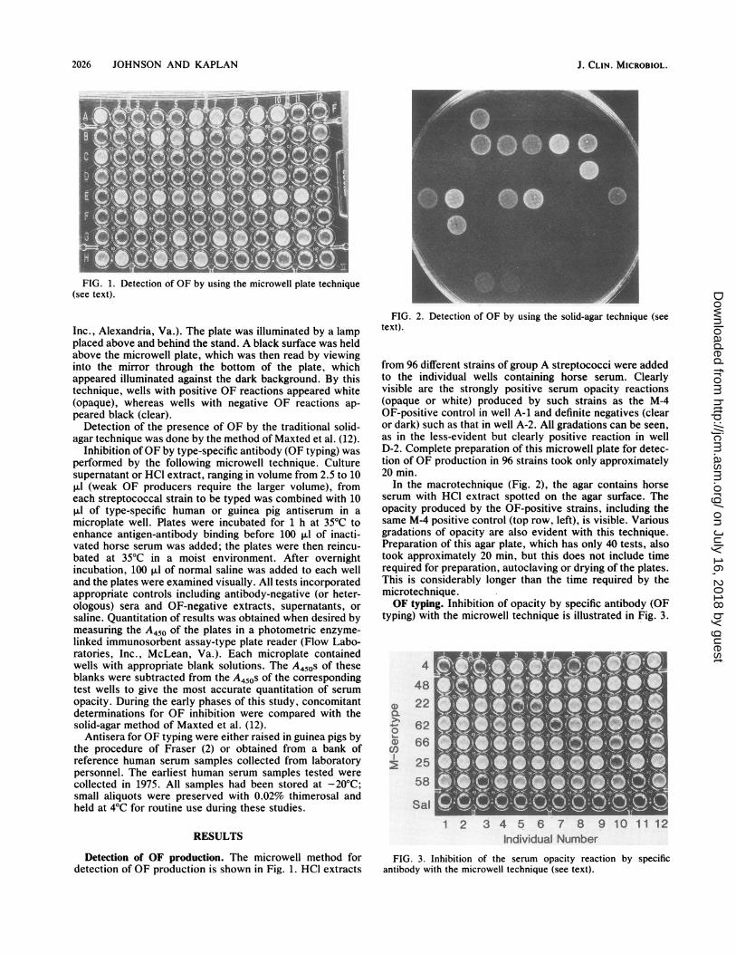
FIG. 1. Detection of OF by using the microwell plate technique(see text).
Inc., Alexandria, Va.). The plate was illuminated by a lampplaced above and behind the stand. A black surface was heldabove the microwell plate, which was then read by viewinginto the mirror through the bottom of the plate, whichappeared illuminated against the dark background. By thistechnique, wells with positive OF reactions appeared white(opaque), whereas wells with negative OF reactions ap-peared black (clear).
Detection of the presence of OF by the traditional solid-agar technique was done by the method of Maxted et al. (12).
Inhibition of OF by type-specific antibody (OF typing) wasperformed by the following microwell technique. Culturesupernatant or HCI extract, ranging in volume from 2.5 to 10pul (weak OF producers require the larger volume), fromeach streptococcal strain to be typed was combined with 10pul of type-specific human or guinea pig antiserum in amicroplate well. Plates were incubated for 1 h at 35°C toenhance antigen-antibody binding before 100 pul of inacti-vated horse serum was added; the plates were then reincu-bated at 35°C in a moist environment. After overnightincubation, 100 ,ul of normal saline was added to each welland the plates were examined visually. All tests incorporatedappropriate controls including antibody-negative (or heter-ologous) sera and OF-negative extracts, supernatants, orsaline. Quantitation of results was obtained when desired bymeasuring the A450 of the plates in a photometric enzyme-linked immunosorbent assay-type plate reader (Flow Labo-ratories, Inc., McLean, Va.). Each microplate containedwells with appropriate blank solutions. The A450s of theseblanks were subtracted from the A450s of the correspondingtest wells to give the most accurate quantitation of serumopacity. During the early phases of this study, concomitantdeterminations for OF inhibition were compared with thesolid-agar method of Maxted et al. (12).
Antisera for OF typing were either raised in guinea pigs bythe procedure of Fraser (2) or obtained from a bank ofreference human serum samples collected from laboratorypersonnel. The earliest human serum samples tested werecollected in 1975. All samples had been stored at -20°C;small aliquots were preserved with 0.02% thimerosal andheld at 4°C for routine use during these studies.
RESULTS
Detection of OF production. The microwell method fordetection of OF production is shown in Fig. 1. HCI extracts
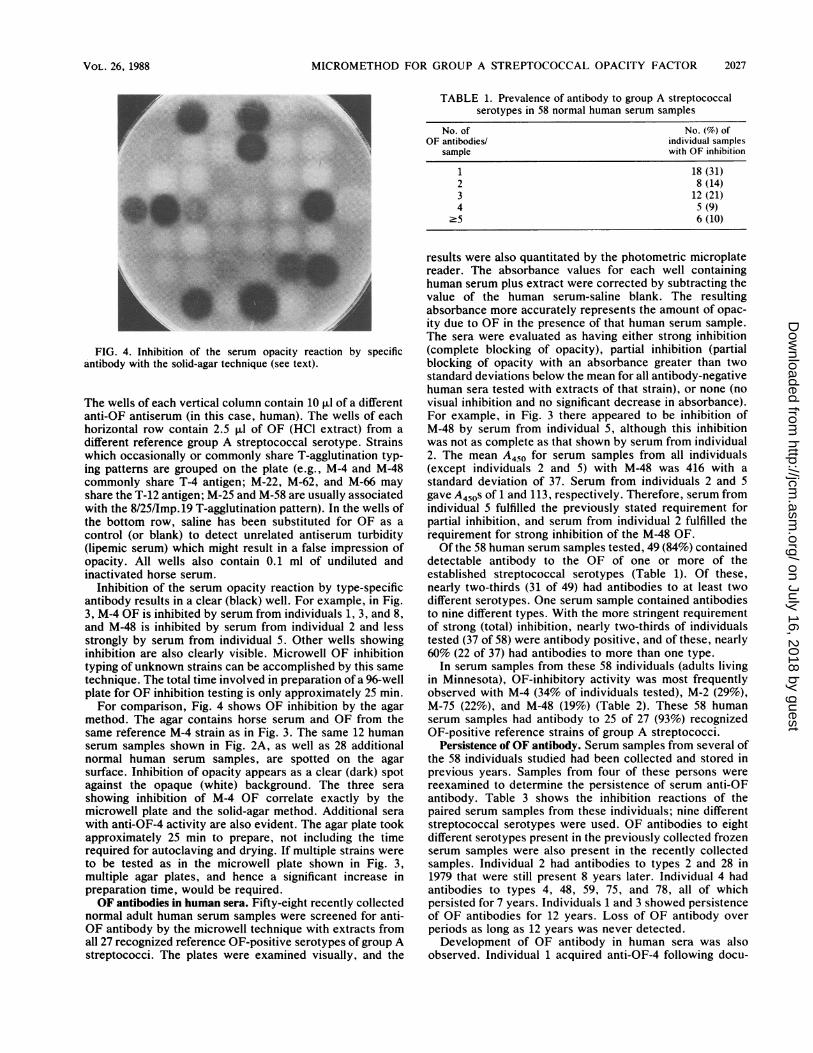
FIG. 2. Detection of OF by using the solid-agar technique (seetext).
from 96 different strains of group A streptococci were addedto the individual wells containing horse serum. Clearlyvisible are the strongly positive serum opacity reactions(opaque or white) produced by such strains as the M-4OF-positive control in well A-1 and definite negatives (clearor dark) such as that in well A-2. All gradations can be seen,as in the less-evident but clearly positive reaction in wellD-2. Complete preparation of this microwell plate for detec-tion of OF production in 96 strains took only approximately20 min.
In the macrotechnique (Fig. 2), the agar contains horseserum with HCI extract spotted on the agar surface. Theopacity produced by the OF-positive strains, including thesame M-4 positive control (top row, left), is visible. Variousgradations of opacity are also evident with this technique.Preparation of this agar plate, which has only 40 tests, alsotook approximately 20 min, but this does not include timerequired for preparation, autoclaving or drying of the plates.This is considerably longer than the time required by themicrotechnique.OF typing. Inhibition of opacity by specific antibody (OF
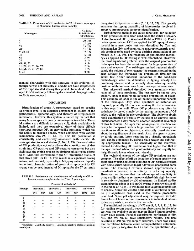
typing) with the microwell technique is illustrated in Fig. 3.
4
48
(D 22
O 62O 66
25
58
Sal
NUot~~~~~~-i1s
# -12 8 91 11;.-4ndviuaNumberU
FIG. 3. Inhibition of the serum opacity reaction by specificantibody with the microwell technique (see text).
J. CLIN. MICROBIOL.
on July 16, 2018 by guesthttp://jcm
.asm.org/
Dow
nloaded from
MICROMETHOD FOR GROUP A STREPTOCOCCAL OPACITY FACTOR
TABLE 1. Prevalence of antibody to group A streptococcalserotypes in 58 normal human serum samples
No. of No. (%) ofOF antibodies/ individual samples
sample with OF inhibition
1 18 (31)2 8 (14)3 12 (21)4 5 (9)
'5 6 (10)
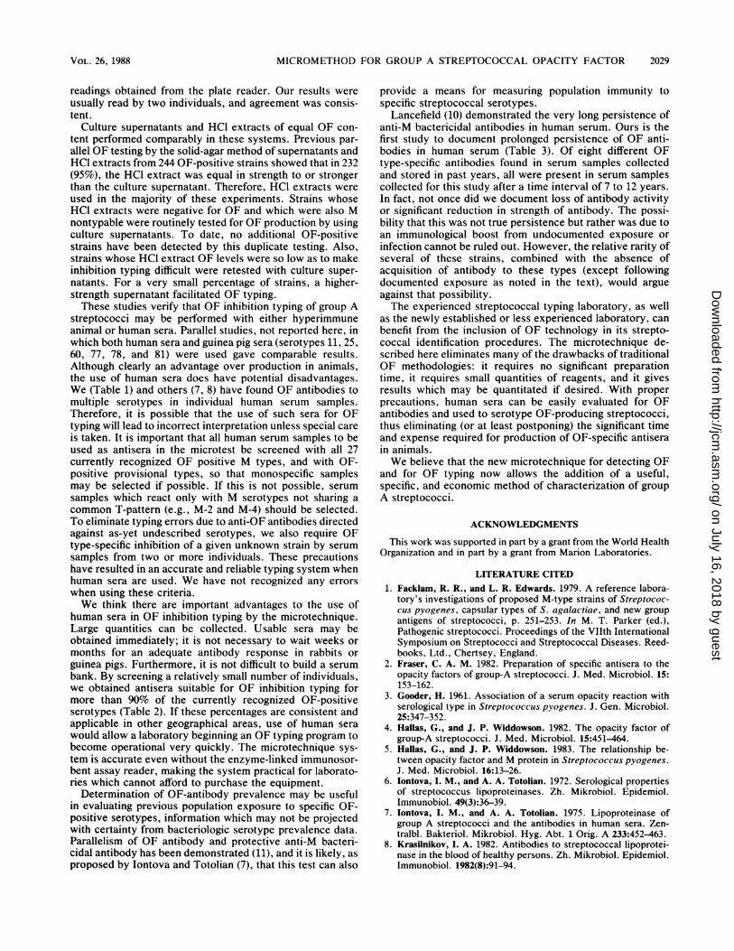
FIG. 4. Inhibition of the serum opacity reaction by specificantibody with the solid-agar technique (see text).
The wells of each vertical column contain 10 ,ul of a differentanti-OF antiserum (in this case, human). The wells of eachhorizontal row contain 2.5 ,ul of OF (HCl extract) from adifferent reference group A streptococcal serotype. Strainswhich occasionally or commonly share T-agglutination typ-ing patterns are grouped on the plate (e.g., M-4 and M-48commonly share T-4 antigen; M-22, M-62, and M-66 mayshare the T-12 antigen; M-25 and M-58 are usually associatedwith the 8/25/Imp.19 T-agglutination pattern). In the wells ofthe bottom row, saline has been substituted for OF as acontrol (or blank) to detect unrelated antiserum turbidity(lipemic serum) which might result in a false impression ofopacity. All wells also contain 0.1 ml of undiluted andinactivated horse serum.
Inhibition of the serum opacity reaction by type-specificantibody results in a clear (black) well. For example, in Fig.3, M-4 OF is inhibited by serum from individuals 1, 3, and 8,and M-48 is inhibited by serum from individual 2 and lessstrongly by serum from individual 5. Other wells showinginhibition are also clearly visible. Microwell OF inhibitiontyping of unknown strains can be accomplished by this sametechnique. The total time involved in preparation of a 96-wellplate for OF inhibition testing is only approximately 25 min.For comparison, Fig. 4 shows OF inhibition by the agar
method. The agar contains horse serum and OF from thesame reference M-4 strain as in Fig. 3. The same 12 humanserum samples shown in Fig. 2A, as well as 28 additionalnormal human serum samples, are spotted on the agarsurface. Inhibition of opacity appears as a clear (dark) spotagainst the opaque (white) background. The three serashowing inhibition of M-4 OF correlate exactly by themicrowell plate and the solid-agar method. Additional serawith anti-OF-4 activity are also evident. The agar plate tookapproximately 25 min to prepare, not including the timerequired for autoclaving and drying. If multiple strains wereto be tested as in the microwell plate shown in Fig. 3,multiple agar plates, and hence a significant increase inpreparation time, would be required.OF antibodies in human sera. Fifty-eight recently collected
normal adult human serum samples were screened for anti-OF antibody by the microwell technique with extracts fromall 27 recognized reference OF-positive serotypes ofgroup Astreptococci. The plates were examined visually, and the
results were also quantitated by the photometric microplatereader. The absorbance values for each well containinghuman serum plus extract were corrected by subtracting thevalue of the human serum-saline blank. The resultingabsorbance more accurately represents the amount of opac-ity due to OF in the presence of that human serum sample.The sera were evaluated as having either strong inhibition(complete blocking of opacity), partial inhibition (partialblocking of opacity with an absorbance greater than twostandard deviations below the mean for all antibody-negativehuman sera tested with extracts of that strain), or none (novisual inhibition and no significant decrease in absorbance).For example, in Fig. 3 there appeared to be inhibition ofM-48 by serum from individual 5, although this inhibitionwas not as complete as that shown by serum from individual2. The mean A450 for serum samples from all individuals(except individuals 2 and 5) with M-48 was 416 with astandard deviation of 37. Serum from individuals 2 and 5gave A450s of 1 and 113, respectively. Therefore, serum fromindividual 5 fulfilled the previously stated requirement forpartial inhibition, and serum from individual 2 fulfilled therequirement for strong inhibition of the M-48 OF.Of the 58 human serum samples tested, 49 (84%) contained
detectable antibody to the OF of one or more of theestablished streptococcal serotypes (Table 1). Of these,nearly two-thirds (31 of 49) had antibodies to at least twodifferent serotypes. One serum sample contained antibodiesto nine different types. With the more stringent requirementof strong (total) inhibition, nearly two-thirds of individualstested (37 of 58) were antibody positive, and of these, nearly60% (22 of 37) had antibodies to more than one type.
In serum samples from these 58 individuals (adults livingin Minnesota), OF-inhibitory activity was most frequentlyobserved with M-4 (34% of individuals tested), M-2 (29%),M-75 (22%), and M-48 (19%) (Table 2). These 58 humanserum samples had antibody to 25 of 27 (93%) recognizedOF-positive reference strains of group A streptococci.
Persistence of OF antibody. Serum samples from several ofthe 58 individuals studied had been collected and stored inprevious years. Samples from four of these persons werereexamined to determine the persistence of serum anti-OFantibody. Table 3 shows the inhibition reactions of thepaired serum samples from these individuals; nine differentstreptococcal serotypes were used. OF antibodies to eightdifferent serotypes present in the previously collected frozenserum samples were also present in the recently collectedsamples. Individual 2 had antibodies to types 2 and 28 in1979 that were still present 8 years later. Individual 4 hadantibodies to types 4, 48, 59, 75, and 78, all of whichpersisted for 7 years. Individuals 1 and 3 showed persistenceof OF antibodies for 12 years. Loss of OF antibody overperiods as long as 12 years was never detected.Development of OF antibody in human sera was also
observed. Individual 1 acquired anti-OF-4 following docu-
rVOL. 26, 1988 2027
i
on July 16, 2018 by guesthttp://jcm
.asm.org/
Dow
nloaded from
2028 JOHNSON AND KAPLAN
TABLE 2. Prevalence of OF antibodies to 27 reference serotypesin 58 normal human serum samples
No. (%) ofM serotypes individuals with
OF inhibition
4 20 (34)2 17 (29)75 13 (22)48 il (19)9, 22, 59 7 (12)78 6 (10)62 5 (9)28 4 (7)49, 58, 76 3 (5)11, 25, 60, 63, 66, 77 2 (3)61, 64, 68, 73, 79, 81 1 (2)8, 13 0 (0)
mented pharyngitis with this serotype in his children, al-though he was not clinically ill and did not have streptococciof this type isolated during this period. Individual 3 devel-oped OF-58 antibody following documented pharyngitis dueto M-58 streptococci.
DISCUSSION
Identification of group A streptococci based on specificM-protein type is an essential component in studies of thepathogenesis, epidemiology, and therapy of streptococcalinfections. However, this system is limited by the fact thatmany M serotypes are poorly immunogenic in rabbits. TheseM antisera are difficult to prepare (17), their availability islimited, and they are expensive. Many of these difficultserotypes produce OF, an enzymelike substance which hasthe ability to produce opacity when combined with variousmammalian sera (9, 13, 14, 18). This OF production isconsistently and exclusively associated with specific M-serotype streptococcal strains (3, 16, 17, 19). Demonstrationof OF production not only allows the classification of thatstrain into OF-positive and OF-negative categories but alsofacilitates the typing process by limiting initial typing effortsto M types that correspond to the OF production status ofthat strain (OF- or OF'). This results in a significant savingin time and material, especially in M typing antisera. Equallyimportant, characterization of group A streptococci by 0Ftyping can be used instead of M typing for the 27 currently
TABLE 3. Persistence and development of antibody to OF inhuman serum samples collected 7 to 12 years apart
Presence of antibody ina:
Serotype Individual 1 Individual 2 Individual 3 Individual 4
3/75 3/87 6/79 3/87 2/75 2/87 12/79 2/87
2 - - + + + + _4 - + - - + + + +28 - - + + - - - -
48 - - - - - - + +58 - - - - - + - -
59 - - - - - - + +62 + + _ _75 - - - - + + + +78 - - - - - - + +
a Symbols: +, antibody positive; -, antibody negative. Dates are given asmonth/year.
recognized OF-positive strains (6, 11, 12, 17). This greatlyenhances the typing capability of laboratories involved ingroup A streptococcal serotype identification.
Turbidimetric methods (so-called tube tests) for detectionof OF production have been used since the initial discoveryof streptococcal OF by Ward and Rudd in 1938 (18). Photo-metric quantitation of OF as applied to OF typing of strep-tococci in a macrotube test was described by Top andWannamaker (16), and quantitative macrophotometric meth-ods continue to be used by those desiring quantitation of testresults (4, 5, 11, 19). The reliability of photometric technol-ogy as applied to OF testing is well established. However,the most significant problem with the original photometrictechniques has been the requirement for large quantities ofsera and reagents. The solid-agar method reduced signifi-cantly the volume of one reagent (the reagent spotted on theagar surface) but increased the preparation time for theactual test. Other inherent limitations of the solid-agarmethodology were the difficulties in typing weakly OF-producing strains and in visually demonstrating weaklypositive inhibition reactions from true-negative reactions.The microwell method described here essentially elimi-
nates all of these problems. The test may be set up veryquickly, since it requires no advance preparation of plates.This economy of labor is one of the most important benefitsof the technique. Only small quantities of material arerequired, generally 10 ,ul or less, making the test economicalin this regard as well. Weak OF producers may often besatisfactorily tested simply by increasing the amount of OFadded to the well in the microtechnique. The ability to obtainrapid quantitation of results by the use of an enzyme-linkedimmunosorbent assay reader is another important advantageof this technique. An apparent weak-positive reaction caneasily be compared with a large number of true-negativereactions to allow an objective, statistically based decisionabout the significance of the result. Also, the opacity causedby turbid antisera (often resulting in a visually unreadabletest) can be eliminated by using a plate reader and subtract-ing appropriate blanks. The sensitivity of the microwellmethod for detecting OF production was higher than that ofthe agar method when read photometrically and slightly butinsignificantly lower when read visually.The technical aspects of the micro-OF technique are not
complex. The effect of pH on detection of serum opacity wasexamined by using doubling dilutions of OF-positive extractswith horse serum adjusted to pH 6.0 versus unadjusted horseserum. The lower-pH extracts showed approximately aone-dilution increase in sensitivity in detecting opacity.However, we believe that the advantage of simplicity inusing unadjusted horse serum outweighed this slight increasein sensitivity obtained by pH adjustment. In limited studiesexamining the effect of pH on OF typing in microwells, a pHin the range of 7.2 to 7.5 was found to give optimal inhibitionofopacity. Since this was the normal pH ofour horse serum,no pH adjustment was used in the routine proceduresdescribed. Since pH adjustment may be required with dif-ferent lots of horse serum, researchers in individual labora-tories may wish to evaluate this variable.The traditional wavelength of 475 nm (4, 5, 7, 8, 11, 12, 16)
for reading serum opacity reaction absorbance in the tubetest was not available on our enzyme-linked immunosorbentassay plate reader. Parallel experiments performed at 410,450, and 492 nm all gave satisfactory results. The finalselection of 450 nm was largely arbitrary. There was excel-lent correlation between visual semiquantitative determina-tion of opacity (negative to 4+) and the quantitative A450
J. CLIN. MICROBIOL.
on July 16, 2018 by guesthttp://jcm
.asm.org/
Dow
nloaded from
MICROMETHOD FOR GROUP A STREPTOCOCCAL OPACITY FACTOR
readings obtained from the plate reader. Our results wereusually read by two individuals, and agreement was consis-tent.
Culture supernatants and HCI extracts of equal OF con-tent performed comparably in these systems. Previous par-allel OF testing by the solid-agar method of supernatants andHCl extracts from 244 OF-positive strains showed that in 232(95%), the HCl extract was equal in strength to or strongerthan the culture supernatant. Therefore, HCl extracts wereused in the majority of these experiments. Strains whoseHCl extracts were negative for OF and which were also Mnontypable were routinely tested for OF production by usingculture supernatants. To date, no additional OF-positivestrains have been detected by this duplicate testing. Also,strains whose HCl extract OF levels were so low as to makeinhibition typing difficult were retested with culture super-natants. For a very small percentage of strains, a higher-strength supernatant facilitated OF typing.These studies verify that OF inhibition typing of group A
streptococci may be performed with either hyperimmuneanimal or human sera. Parallel studies, not reported here, inwhich both human sera and guinea pig sera (serotypes 11, 25,60, 77, 78, and 81) were used gave comparable results.Although clearly an advantage over production in animals,the use of human sera does have potential disadvantages.We (Table 1) and others (7, 8) have found OF antibodies tomultiple serotypes in individual human serum samples.Therefore, it is possible that the use of such sera for OFtyping will lead to incorrect interpretation unless special careis taken. It is important that ahl human serum samples to beused as antisera in the microtest be screened with ail 27currently recognized OF positive M types, and with OF-positive provisional types, so that monospecific samplesmay be selected if possible. If this is not possible, serumsamples which react only with M serotypes not sharing acommon T-pattern (e.g., M-2 and M-4) should be selected.To eliminate typing errors due to anti-OF antibodies directedagainst as-yet undescribed serotypes, we also require OFtype-specific inhibition of a given unknown strain by serumsamples from two or more individuals. These precautionshave resulted in an accurate and reliable typing system whenhuman sera are used. We have not recognized any errorswhen using these criteria.We think there are important advantages to the use of
human sera in OF inhibition typing by the microtechnique.Large quantities can be collected. Usable sera may beobtained immediately; it is not necessary to wait weeks ormonths for an adequate antibody response in rabbits orguinea pigs. Furthermore, it is not difficult to build a serumbank. By screening a relatively small number of individuals,we obtained antisera suitable for OF inhibition typing formore than 90% of the currently recognized OF-positiveserotypes (Table 2). If these percentages are consistent andapplicable in other geographical areas, use of human serawould allow a laboratory beginning an OF typing program tobecome operational very quickly. The microtechnique sys-tem is accurate even without the enzyme-linked immunosor-bent assay reader, making the system practical for laborato-ries which cannot afford to purchase the equipment.
Determination of OF-antibody prevalence may be usefulin evaluating previous population exposure to specific OF-positive serotypes, information which may not be projectedwith certainty from bacteriologic serotype prevalence data.Parallelism of OF antibody and protective anti-M bacteri-cidal antibody has been demonstrated (11), and it is likely, asproposed by Iontova and Totolian (7), that this test can also
provide a means for measuring population immunity tospecific streptococcal serotypes.
Lancefield (10) demonstrated the very long persistence ofanti-M bactericidal antibodies in human serum. Ours is thefirst study to document prolonged persistence of OF anti-bodies in human serum (Table 3). Of eight different OFtype-specific antibodies found in serum samples collectedand stored in past years, all were present in serum samplescollected for this study after a time interval of 7 to 12 years.In fact, not once did we document loss of antibody activityor significant reduction in strength of antibody. The possi-bility that this was not true persistence but rather was due toan immunological boost from undocumented exposure orinfection cannot be ruled out. However, the relative rarity ofseveral of these strains, combined with the absence ofacquisition of antibody to these types (except followingdocumented exposure as noted in the text), would argueagainst that possibility.The experienced streptococcal typing laboratory, as well
as the newly established or less experienced laboratory, canbenefit from the inclusion of OF technology in its strepto-coccal identification procedures. The microtechnique de-scribed here eliminates many of the drawbacks of traditionalOF methodologies: it requires no significant preparationtime, it requires small quantities of reagents, and it givesresults which may be quantitated if desired. With properprecautions, human sera can be easily evaluated for OFantibodies and used to serotype OF-producing streptococci,thus eliminating (or at least postponing) the significant timeand expense required for production of OF-specific antiserain animals.We believe that the new microtechnique for detecting OF
and for OF typing now allows the addition of a useful,specific, and economic method of characterization of groupA streptococci.
ACKNOWLEDGMENTS
This work was supported in part by a grant from the World HealthOrganization and in part by a grant from Marion Laboratories.
LITERATURE CITED1. Facklam, R. R., and L. R. Edwards. 1979. A reference labora-
tory's investigations of proposed M-type strains of Streptococ-cus pyogenes, capsular types of S. agalactiae, and new groupantigens of streptococci, p. 251-253. In M. T. Parker (ed.),Pathogenic streptococci. Proceedings of the VIIth InternationalSymposium on Streptococci and Streptococcal Diseases. Reed-books, Ltd., Chertsey, England.
2. Fraser, C. A. M. 1982. Preparation of specific antisera to theopacity factors of group-A streptococci. J. Med. Microbiol. 15:153-162.
3. Gooder, H. 1961. Association of a serum opacity reaction withserological type in Streptococcus pyogenes. J. Gen. Microbiol.25:347-352.
4. Hallas, G., and J. P. Widdowson. 1982. The opacity factor ofgroup-A streptococci. J. Med. Microbiol. 15:451-464.
5. Hallas, G., and J. P. Widdowson. 1983. The relationship be-tween opacity factor and M protein in Streptococcus pyogenes.J. Med. Microbiol. 16:13-26.
6. Jontova, I. M., and A. A. Totolian. 1972. Serological propertiesof streptococcus lipoproteinases. Zh. Mikrobiol. Epidemiol.Immunobiol. 49(3):36-39.
7. lontova, I. M., and A. A. Totolian. 1975. Lipoproteinase ofgroup A streptococci and the antibodies in human sera. Zen-tralbl. Bakteriol. Mikrobiol. Hyg. Abt. 1 Orig. A 233:452-463.
8. Krasilnikov, I. A. 1982. Antibodies to streptococcal lipoprotei-nase in the blood of healthy persons. Zh. Mikrobiol. Epidemiol.Immunobiol. 1982(8):91-94.
VOL. 26, 1988 2029
on July 16, 2018 by guesthttp://jcm
.asm.org/
Dow
nloaded from
2030 JOHNSON AND KAPLAN
9. Krumwiede, E. 1954. Studies on a lipoproteinase of group Astreptococci. J. Exp. Med. 100:629-639.
10. Lancefield, R. C. 1959. Persistence of type-specific antibodies inman following infection with group A streptococci. J. Exp.Med. 110:271-292.
11. Maxted, W. R., J. P. Widdowson, and C. A. M. Fraser. 1973.Antibody to streptococcal opacity factor in human sera. J. Hyg.71:35-42.
12. Maxted, W. R., J. P. Widdowson, C. A. M. Fraser, L. C. Ball,and D. C. J. Bassett. 1973. The use of the serum opacity reactionin the typing of group-A streptococci. J. Med. Microbiol. 6:83-90.
13. Rowen, R. 1961. The release of cholesterol esters from serumlipoproteins by extracts of certain group A streptococci. J. Exp.Med. 114:807-823.
14. Rowen, R., and J. Martin. 1963. Enhancement of cholesterolesterification in serum by an extract of group-A streptococcus.Biochim. Biophys. Acta 70:396-405.
15. Swift, H. R., A. T. Wilson, and R. C. Lancefield. 1943. Typing
J. CLIN. MICROBIOL.
group A hemolytic streptococci by M precipitin reactions incapillary pipettes. J. Exp. Med. 78:127-133.
16. Top, F. H., Jr., and L. W. Wannamaker. 1968. The serumopacity reaction of Streptococcus pyogenes: the demonstrationof multiple, strain-specific lipoproteinase antigens. J. Exp. Med.127:1013-1034.
17. Top, F. H., Jr., and L. W. Wannamaker. 1968. The serumopacity reaction of Streptococcus pyogenes: frequency of pro-duction of streptococcal lipoproteinase by strains of differentserological types and the relationship to M protein production.J. Hyg. 66:49-58.
18. Ward, H. K., and G. V. Rudd. 1938. Studies on haemolyticstreptococci from human sources. Aust. J. Exp. Biol. Med. Sci.16:181-192.
19. Widdowson, J. P., W. R. Maxted, and D. L. Grant. 1970. Theproduction of opacity in serum by group A streptococci and itsrelationship with the presence of M antigen. J. Gen. Microbiol.61:343-353.
on July 16, 2018 by guesthttp://jcm
.asm.org/
Dow
nloaded from