1
Enlarging and Reconstructing the Flexor Tendon Sheath Reduces Gliding Resistance 1,2 Simmons, S; 1,2,3 Kosmopoulos, V; 1,3 Roso, M; 4 Carlson, H; 4 Coté, R E; 4 Tayag T J; + 1,2 Bunata, R E; + 1 Bone and Joint Research Center, Dept. of Orthopaedic Surgery, University of North Texas Health Science Center, Fort Worth, TX, 2 Dept. of Orthopaedic Surgery, John Peter Smith Hospital (Tarrant County Hospital District), Fort Worth, TX, 3 Dept. of Cell Biology and Genetics, University of North Texas Health Science Center, Fort Worth, TX, 4 Dept. of Engineering, College of Science and Engineering, Texas Christian University, Fort Worth, TX Senior author [email protected] Introduction: Flexor tendon repair in zone II presents a challenge. The challenge arises from the increased tendon bulk, limiting the free gliding ability of the tendon through the snug fitting sheath. Resistance to gliding can lead to incomplete motion of the repaired tendons, to triggering or catching, and possibly to tendon rupture. Most efforts to deal with these problems have been directed at making the tendon repair stronger while trying to keep the bulk of the repair to a minimum. Historically the tendon sheath was initially dealt with by a limited resection of the sheath; then closing the sheath came into vogue, but there was concern about excessive sheath, so a period followed when the sheath was managed with benign neglect. Recently, incising (venting) portions of the sheath has become popular. Lister et. al. in 1985 suggested using a fascial graft for primary reconstruction of a deficient sheath, and Manske et. al. in 1988 conceptualized this as a way to relieve sheath tightness. The current study evaluates sheath enlargement and reconstruction during primary flexor tendon repair. The current study aims to use an established technique (Uchiyama et al. 1995) to directly measure gliding resistance under the A2 pulley for the: 1) intact tendon and sheath; 2) repaired tendon and intact sheath; 3) repaired tendon and reconstructed tendon sheath. The hypothesis is that sheath enlargement will reduce gliding resistance of repaired tendons. This information has implications for improving tendon repair rehabilitation and reducing the incidence of tendon repair rupture. Materials and Methods: Seven fresh cadaveric digits from the base of the metacarpal to the tip of the finger were tested using each of the aforementioned conditions. For the control, or intact condition 1, the tendon sheath distal to the A3 pulley was removed and a small window at the C1 level was opened. The flexor digitorum profundus (FDP) was detached from the distal phalanx and holding sutures were placed in the proximal and distal ends of FDP tendon. The FDS tendon was left in place with a holding suture in its proximal end. Vinculae were cut to allow free excursion of the FDP tendon. For the repaired tendon and intact sheath, condition 2, the FDP tendon was transected 2 mm proximal to the distal edge of the A2 pulley through a window in the C1 pulley, and the FDS tendon slips were cut 10 mm proximal to the distal edge of the A2 pulley. Both tendons were repaired with a 2 strand modified Kessler repair using 3-0 Supramid (S. Jackson, Inc.) suture and a circumferential 6-0 nylon suture. Finally, for the sheath reconstruction, condition 3, the tendon sheath was incised proximally for the length of the excursion of the FDP tendon starting 2 mm distal to the distal edge of the A2 pulley. This required cutting the C1, A2 pulley and often part of the A1 pulley. The pulley was then reconstructed by suturing a piece of extensor retinaculum, 2 to 3 mm wide and 25 to 30 mm long, into the sheath defect with a 6-0 suture, tight enough to snugly cover the tendons yet loose enough to allow free tendon excursion. The gliding resistance, the force opposing tendon motion resulting from friction with the flexor tendon sheath and angular contact with the A1-A3 pulleys, was measured using an established technique for each of the three conditions (Uchiyama et al. 1995). To do so, the specimen was mounted in a custom built frame which locked the metacarpal, proximal, and middle phalanges in place with the metacarpal-phalangeal (MP) joint at 0º and the proximal interphalangeal (IP) joint at 20º of flexion. The FDS tendon was held at 2 N of tension at 35º above the horizontal level of the digit. The proximal holding sutures in the FDP tendon were attached to a force transducer at 30º above the horizontal, and then attached to the actuator of our testing system. The distal FDP holding suture was attached to another force transducer at 20º above the horizontal and then to a 4.9 N counter-weight. Thus, the difference between the force transducers is the desired gliding resistance. The first half of a the testing cycle (flexion) consisted of the actuator pulling the tendon at a rate of 2 mm/s through its measured excursion, and the second half of the cycle (extension) had the 4.9 N mass pulling the tendon back through its sheath. The average force of the seven digits was calculated and statistically compared for each condition. Significance levels were categorized as extremely statistical significant ( = 0.001), highly statistical significant ( = 0.01), and statistically significant ( = 0.05). Results: The intact tendon/sheath system had the lowest gliding resistance (0.44±0.13 N) (Fig. 1). Cutting and repairing the flexor tendons resulted in a 246% increase in gliding resistance through the tendon sheath to an average of 1.51±0.30 N. This difference was extremely statistically significant (P<0.0001). Enlarging and reconstructing the tendon sheath reduced the gliding resistance by 31%, as compared to the tendon repair with the intact sheath, to an average of 1.04±0.18 N. This difference between the resistance of the repaired tendons through the intact sheath and the repaired tendons through the enlarged and reconstructed sheath was extremely statistically significant (P=0.0005). Figure 1. Average force and standard deviation for each condition. The standard deviation (shown after the ± in the previous paragraph), representing irregularity of resistance or roughness of movement, was significantly increased for both repair conditions as compared to the intact system (Fig. 2). The standard deviation was 38% less for condition 3 when compared to condition 2, but this difference was not found to be statistically significant (P=0.0982). Figure 2. Example from a single digit showing force fluctuations, representing movement roughness, for the three tested conditions. Discussion: This study aimed to compare the gliding resistance under the A2 pulley between the intact, repaired tendon with intact sheath, and repaired tendon through an enlarged and reconstructed sheath. As hypothesized, enlarging and reconstructing the flexor tendon sheath reduced the gliding resistance. Reduced gliding resistance should mean the patient can participate in post-operative motion regimens easier with less force required to induce tendon movement. Reduced gliding resistance also means there should be less tension on the repair site, thus reducing the likelihood of gapping or rupture. While not statistically significant, the standard deviation during gliding showed a trend. As would be expected, cutting and repairing the tendon caused more sheath interaction and roughness to tendon gliding. When the sheath was enlarged, the standard deviation was reduced indicating smoother gliding during motion. This should be perceived by the patient as less catching or snagging, and should also protect the tendon repair against sudden changes in resistance thereby decreasing the likelihood of gapping or repair rupture. The results from this study support the recommendation for primary enlargement and reconstruction of the sheath when the surgeon encounters excessive resistance to gliding of the repaired flexor tendon or when the surgeon encounters snagging or catching when the repaired tendons re-enter the tendon sheath. Poster No. 1433 55th Annual Meeting of the Orthopaedic Research Society

Enlarging and Reconstructing the Flexor Tendon … and Reconstructing the Flexor Tendon Sheath Reduces Gliding Resistance 1,2Simmons, S; 1,2,3Kosmopoulos, V; 1,3Roso, M; 4Carlson,

  • Upload
    vandang

  • View
    227

  • Download
    1

Embed Size (px)

Citation preview

Enlarging and Reconstructing the Flexor Tendon Sheath Reduces Gliding Resistance 1,2Simmons, S; 1,2,3Kosmopoulos, V; 1,3Roso, M; 4Carlson, H; 4Coté, R E; 4Tayag T J; +1,2Bunata, R E;

+1Bone and Joint Research Center, Dept. of Orthopaedic Surgery, University of North Texas Health Science Center, Fort Worth, TX, 2Dept. of Orthopaedic Surgery, John Peter Smith Hospital (Tarrant County Hospital District), Fort Worth, TX, 3Dept. of Cell Biology and Genetics, University of North Texas

Health Science Center, Fort Worth, TX, 4Dept. of Engineering, College of Science and Engineering, Texas Christian University, Fort Worth, TX Senior author [email protected]

Introduction: Flexor tendon repair in zone II presents a challenge.

The challenge arises from the increased tendon bulk, limiting the free gliding ability of the tendon through the snug fitting sheath. Resistance to gliding can lead to incomplete motion of the repaired tendons, to triggering or catching, and possibly to tendon rupture. Most efforts to deal with these problems have been directed at making the tendon repair stronger while trying to keep the bulk of the repair to a minimum. Historically the tendon sheath was initially dealt with by a limited resection of the sheath; then closing the sheath came into vogue, but there was concern about excessive sheath, so a period followed when the sheath was managed with benign neglect. Recently, incising (venting) portions of the sheath has become popular. Lister et. al. in 1985 suggested using a fascial graft for primary reconstruction of a deficient sheath, and Manske et. al. in 1988 conceptualized this as a way to relieve sheath tightness. The current study evaluates sheath enlargement and reconstruction during primary flexor tendon repair. The current study aims to use an established technique (Uchiyama et al. 1995) to directly measure gliding resistance under the A2 pulley for the: 1) intact tendon and sheath; 2) repaired tendon and intact sheath; 3) repaired tendon and reconstructed tendon sheath. The hypothesis is that sheath enlargement will reduce gliding resistance of repaired tendons. This information has implications for improving tendon repair rehabilitation and reducing the incidence of tendon repair rupture. Materials and Methods: Seven fresh cadaveric digits from the base of the metacarpal to the tip of the finger were tested using each of the aforementioned conditions. For the control, or intact condition 1, the tendon sheath distal to the A3 pulley was removed and a small window at the C1 level was opened. The flexor digitorum profundus (FDP) was detached from the distal phalanx and holding sutures were placed in the proximal and distal ends of FDP tendon. The FDS tendon was left in place with a holding suture in its proximal end. Vinculae were cut to allow free excursion of the FDP tendon. For the repaired tendon and intact sheath, condition 2, the FDP tendon was transected 2 mm proximal to the distal edge of the A2 pulley through a window in the C1 pulley, and the FDS tendon slips were cut 10 mm proximal to the distal edge of the A2 pulley. Both tendons were repaired with a 2 strand modified Kessler repair using 3-0 Supramid (S. Jackson, Inc.) suture and a circumferential 6-0 nylon suture. Finally, for the sheath reconstruction, condition 3, the tendon sheath was incised proximally for the length of the excursion of the FDP tendon starting 2 mm distal to the distal edge of the A2 pulley. This required cutting the C1, A2 pulley and often part of the A1 pulley. The pulley was then reconstructed by suturing a piece of extensor retinaculum, 2 to 3 mm wide and 25 to 30 mm long, into the sheath defect with a 6-0 suture, tight enough to snugly cover the tendons yet loose enough to allow free tendon excursion. The gliding resistance, the force opposing tendon motion resulting from friction with the flexor tendon sheath and angular contact with the A1-A3 pulleys, was measured using an established technique for each of the three conditions (Uchiyama et al. 1995). To do so, the specimen was mounted in a custom built frame which locked the metacarpal, proximal, and middle phalanges in place with the metacarpal-phalangeal (MP) joint at 0º and the proximal interphalangeal (IP) joint at 20º of flexion. The FDS tendon was held at 2 N of tension at 35º above the horizontal level of the digit. The proximal holding sutures in the FDP tendon were attached to a force transducer at 30º above the horizontal, and then attached to the actuator of our testing system. The distal FDP holding suture was attached to another force transducer at 20º above the horizontal and then to a 4.9 N counter-weight. Thus, the difference between the force transducers is the desired gliding resistance. The first half of a the testing cycle (flexion) consisted of the actuator pulling the tendon at a rate of 2 mm/s through its measured excursion, and the second half of the cycle (extension) had the 4.9 N mass pulling the tendon back through its sheath. The average force of the seven digits was calculated and statistically compared for each condition. Significance levels were categorized as extremely statistical

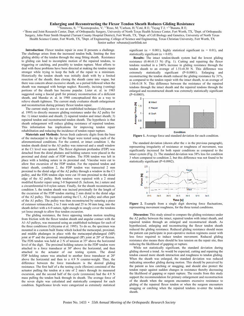
significant (α = 0.001), highly statistical significant (α = 0.01), and statistically significant (α = 0.05). Results: The intact tendon/sheath system had the lowest gliding resistance (0.44±0.13 N) (Fig. 1). Cutting and repairing the flexor tendons resulted in a 246% increase in gliding resistance through the tendon sheath to an average of 1.51±0.30 N. This difference was extremely statistically significant (P<0.0001). Enlarging and reconstructing the tendon sheath reduced the gliding resistance by 31%, as compared to the tendon repair with the intact sheath, to an average of 1.04±0.18 N. This difference between the resistance of the repaired tendons through the intact sheath and the repaired tendons through the enlarged and reconstructed sheath was extremely statistically significant (P=0.0005).

Figure 1. Average force and standard deviation for each condition.

The standard deviation (shown after the ± in the previous paragraph), representing irregularity of resistance or roughness of movement, was significantly increased for both repair conditions as compared to the intact system (Fig. 2). The standard deviation was 38% less for condition 3 when compared to condition 2, but this difference was not found to be statistically significant (P=0.0982).

Figure 2. Example from a single digit showing force fluctuations, representing movement roughness, for the three tested conditions.

Discussion: This study aimed to compare the gliding resistance under the A2 pulley between the intact, repaired tendon with intact sheath, and repaired tendon through an enlarged and reconstructed sheath. As hypothesized, enlarging and reconstructing the flexor tendon sheath reduced the gliding resistance. Reduced gliding resistance should mean the patient can participate in post-operative motion regimens easier with less force required to induce tendon movement. Reduced gliding resistance also means there should be less tension on the repair site, thus reducing the likelihood of gapping or rupture. While not statistically significant, the standard deviation during gliding showed a trend. As would be expected, cutting and repairing the tendon caused more sheath interaction and roughness to tendon gliding. When the sheath was enlarged, the standard deviation was reduced indicating smoother gliding during motion. This should be perceived by the patient as less catching or snagging, and should also protect the tendon repair against sudden changes in resistance thereby decreasing the likelihood of gapping or repair rupture. The results from this study support the recommendation for primary enlargement and reconstruction of the sheath when the surgeon encounters excessive resistance to gliding of the repaired flexor tendon or when the surgeon encounters snagging or catching when the repaired tendons re-enter the tendon sheath.

Poster No. 1433 • 55th Annual Meeting of the Orthopaedic Research Society