Embed Size (px)
Citation preview
LSHTM Research Online
Elsheikha, HM; Regan, CS; Clark, CG; (2018) Novel Entamoeba Findings in Nonhu-man Primates. Trends in parasitology, 34. pp. 283-294. ISSN 1471-4922 DOI:https://doi.org/10.1016/j.pt.2017.12.008
Downloaded from: http://researchonline.lshtm.ac.uk/4646383/
DOI: https://doi.org/10.1016/j.pt.2017.12.008
Usage Guidlines:
Please refer to usage guidelines at http://researchonline.lshtm.ac.uk/policies.html or alternativelycontact [email protected].
Available under license: http://creativecommons.org/licenses/by-nc-nd/2.5/
https://researchonline.lshtm.ac.uk
1
Review 1
2
Novel Entamoeba findings in non-human primates 3
4
5
Hany M. Elsheikha,1 Carl S. Regan,1,2 and C. Graham Clark 3, * 6
7
8
9
1 Faculty of Medicine and Health Sciences, School of Veterinary Medicine and Science, 10
University of Nottingham, Sutton Bonington Campus, Loughborough, LE12 5RD, UK 11
12
2 Current address: Vets4pets Dover Whitfield, White Cliffs Retail Park, Whitfield, Dover, 13
CT16 3PS, UK 14
15
3 Faculty of Infectious and Tropical Diseases, London School of Hygiene and Tropical 16
Medicine, London, UK 17
18
*Correspondence: 19
(C. Graham Clark) 21
22
Keywords: Entamoeba, Evolution, Non-human primates, Phylogeny, Species delineation 23
2
Abstract 24
25
In addition to well-known human-infecting species, Entamoeba species not found in humans 26
have been identified recently in non-human primates (NHPs). Importantly, it has become clear 27
that the organism identified as Entamoeba histolytica in NHPs is usually a distinct species, 28
Entamoeba nuttalli. Many DNA-based stool surveys use species-specific detection methods 29
and so may miss the full range of Entamoeba species present. In addition, authors may be using 30
the same species name to describe distinct organisms. These various shortcomings may not be 31
obvious to readers. In this review, we clarify the relationships between Entamoeba species’ 32
names based on morphological and molecular data, and highlight gaps in recently published 33
data on Entamoeba species in wild NHPs resulting from the use of variable methodology. 34
35
Humans and NHPs are both primates, but how similar are their Entamoeba species? 36
37
Humans are primates, and therefore it would be logical to assume that the parasite fauna of 38
humans and non-human primates (NHPs; see Glossary) is likely to be similar. However, this 39
simplistic view ignores the huge range of life-styles, diets and ecological specialisations 40
exhibited by NHPs, and the millions of years of independent evolution that separate us from 41
even our closest NHP relatives, the great apes. Nevertheless, humans and NHPs do appear to 42
have many parasites in common, at least when identified via microscopy. Over recent decades, 43
molecular tools have allowed us to re-examine these similarities and to challenge the 44
assumption that apparent morphological identity equates to species identity. This review 45
discusses how molecular tools provide a clearer picture of the relationships between intestinal 46
amoebae of the genus Entamoeba in humans and NHPs and where gaps in our understanding 47
remain. 48
3
49
What is causing invasive amoebiasis in humans and NHPs? 50
51
The focus on Entamoeba is largely due to Entamoeba histolytica being a significant cause of 52
morbidity and mortality in humans. Published estimates suggest this organism is responsible 53
for millions of cases of disease and over 50,000 deaths in humans annually [1]. Although these 54
numbers are extrapolated from a limited number of studies, E. histolytica is certainly 55
responsible for a significant amount of disease in some locations. Captive NHPs occasionally 56
die from a disease that is, superficially, indistinguishable from that caused by E. histolytica in 57
humans (e.g. [2]). Several other Entamoeba species that resemble E. histolytica 58
morphologically have been described in both humans and NHPs, making microscopic 59
diagnosis problematic. Morphologically distinct, non-pathogenic species of Entamoeba also 60
appear to be shared by humans and NHPs, further complicating diagnosis (see below). 61
62
The morphology era 63
64
The existence of species of Entamoeba in humans and NHPs that appear identical by 65
microscopy has been known for over a century. At that time, organisms in new hosts were often 66
given new species names, whether morphologically distinguishable or not. A major work by 67
Dobell [3] concluded that all named intestinal species of Entamoeba in humans could be 68
assigned to either E. histolytica or Entamoeba coli, but he equivocated about Entamoeba from 69
NHPs on the grounds of insufficient data; he later concluded that intestinal Entamoeba species 70
in NHPs were also E. histolytica and E. coli [4]. His two-species nomenclature stayed 71
essentially intact for 35 years. 72
73
4
In the mid-1950s, Burrows [5] resurrected the name Entamoeba hartmanni for an organism 74
that parasitologists were referring to as ‘small race E. histolytica’. Dobell [3] had viewed E. 75
hartmanni as a synonym of E. histolytica; however, Burrows showed that the sizes of E. 76
histolytica ‘large race’ and ‘small race’ cysts were not a continuum but had a clear bimodal 77
distribution. This first ‘break’ with the Dobell nomenclature was quickly adopted, because 78
parasitologists were already primed to accept it. 79
80
The molecular era 81
82
Entamoeba hartmanni was the last change to Dobell’s nomenclature scheme based on 83
morphology alone. Additional changes followed but not for many years, as the changes were 84
primarily dependent on small subunit ribosomal RNA gene (SSU-rDNA) analyses. Emile 85
Brumpt [6] proposed the existence of Entamoeba dispar, a non-pathogenic species 86
morphologically identical to E. histolytica. This proposal was rejected by most parasitologists 87
at the time (see discussion following [7]) and the name E. dispar virtually disappeared from 88
the literature. Suspicion that Brumpt had been correct followed on from studies based on both 89
lectin agglutination [8] and isoenzyme patterns [9], in which two groups within E. histolytica 90
were identified, only one of which was found in patients with invasive disease. Subsequently, 91
studies (cited in [10]) using monoclonal antibodies, DNA hybridization, SSU-rDNA restriction 92
fragment length polymorphism, and eventually DNA sequencing all identified the same two 93
groups of strains, and this led to the formal redescription of E. dispar as a species distinct from 94
E. histolytica [10]. 95
96
Other SSU-rDNA-based changes to the nomenclature of human Entamoeba species include 97
the reassignment of ‘E. histolytica-like’ amoebae to the species Entamoeba moshkovskii [11] 98
5
and the recognition that uninucleate cysts occasionally seen in humans were not always 99
immature E. histolytica but were in fact Entamoeba polecki [12]. Most recently, Entamoeba 100
bangladeshi was described as a new human species [13]; if it were not for SSU-rDNA 101
sequences this organism would have been identified as E. moshkovskii despite it being 102
genetically quite distinct. 103
104
The nomenclature for Entamoeba species in NHPs has followed suit, for the most part. 105
Entamoeba hartmanni is commonly found in NHPs. Entamoeba dispar is also widespread in 106
NHPs. Entamoeba chattoni had long been accepted as a NHP-specific species of Entamoeba 107
with uninucleate cysts. It was designated a subtype of E. polecki a few years ago [14], but this 108
change of nomenclature for E. chattoni has not been universally accepted; this will be discussed 109
further below. 110
111
Thus, for the most part, the NHP Entamoeba nomenclature changes simply mirrored those in 112
humans without any investigations to evaluate whether they were in fact the same organisms. 113
This was understandable initially because there was no reason to suspect there were differences 114
and the investigative tools were not readily available to many researchers. However, now that 115
molecular techniques are routine in most research laboratories and some diagnostic 116
laboratories, investigations into the diversity and identity of Entamoeba in NHPs have become 117
more common and are revealing some surprising and important findings. 118
119
The evidence for Entamoeba genetic diversity in NHPs is based almost exclusively on SSU-120
rDNA analyses. Analyses of other markers are rarely possible because most studies use DNA 121
extracted directly from stool samples, but when available they show the same species 122
relationships. SSU-rDNA is a multicopy gene, which makes it relatively easy to amplify from 123
6
stool samples. In addition, and in contrast to some eukaryotes, the Entamoeba SSU-rDNA is 124
relatively fast evolving (as evidenced by long branches in phylogenetic trees) meaning that 125
sufficient resolution is obtained to allow differentiation of Entamoeba taxa using this gene 126
alone. 127
128
Entamoeba nuttalli 129
Entamoeba histolytica causes disease of two main types: 1. amoebic dysentery/colitis, resulting 130
from trophozoite invasion of the colonic mucosa and leading to ulceration, bleeding and the 131
production of loose stool with blood and mucus; 2. amoebic liver abscess, resulting from 132
haematogenous spread of trophozoites from the colon via the portal system to the liver, where 133
tissue lysis leads to formation of a sterile pus-filled abscess [15]. Both types of disease have 134
been reported in NHPs, and there have been a number of reports over the years of spontaneous 135
invasive disease occurring in captive NHPs. Histologically, the diseases in humans and NHPs 136
appear identical, as do the amoebae under the microscope [e.g. 2]. Entamoeba histolytica of 137
human origin has been shown experimentally to be capable of infecting NHPs, where it can 138
cause indistinguishable pathology [e.g. 16]. The organism responsible was therefore presumed 139
to be E. histolytica in all cases of disease in NHPs. 140
141
In the last 10 years, however, molecular studies have been performed on amoebae from cases 142
of invasive amoebiasis occurring spontaneously in NHPs. The amoebae in NHPs are 143
consistently distinguishable from E. histolytica using a variety of DNA and protein markers: 144
isoenzymes, SSU-rDNA and short tandem-repeat-containing loci [17-20]. Although closely 145
related to E. histolytica – indeed it has been called “E. histolytica-like variant” [19] and “E. 146
histolytica NHP variant” [21] by some – this is clearly a distinct organism and the name E. 147
nuttalli has been revived for this amoeba [17]. Entamoeba nuttalli was originally described by 148
7
Castellani [22] in the liver abscess of a toque macaque (Macaca sinica) in Sri Lanka and is one 149
of the species considered synonymous with E. histolytica by Dobell [3, 23]. Although we 150
cannot prove after 110 years that the amoeba observed by Castellani is the same as the one now 151
being called E. nuttalli, this seems quite likely. A recent survey of wild toque macaques in Sri 152
Lanka detected asymptomatic carriage of E. nuttalli in 18.5% of the 227 animals studied [24]. 153
Entamoeba histolytica was not detected in the population. Entamoeba nuttalli has been found 154
in a variety of other NHPs – guenon, baboon, colobus and chimpanzee – in addition to other 155
species of both captive and wild macaques [19, 25, 26]. 156
157
The host and geographic ranges of E. nuttalli seem to be quite large, but so far it seems to be 158
found primarily in primates of the Old World. Invasive disease has been reported in captive 159
spider monkeys [25], but whether it infects wild New World NHPs is unknown. Only one 160
human infection with E. nuttalli has been reported to date, in a zookeeper [27]. This is despite 161
analyses of human samples that would have revealed its presence if it had been there. 162
Isoenzyme analysis, which was used widely for Entamoeba species differentiation in the 1980s 163
and early 1990s [e.g. 28], would have distinguished E. nuttalli from E. histolytica [17, 19], but 164
although many thousands of human samples were studied in order to differentiate E. dispar 165
and E. histolytica, no evidence of what is now being called E. nuttalli was reported. A second 166
human infection has apparently been identified in Iraq, based on a sequence in GenBank stated 167
to be of human origin [55; GenBank accession number: KP233837]. 168
169
Note that most DNA-based diagnostic tools cannot distinguish E. nuttalli from E. histolytica, 170
unless combined with sequencing, and neither can some commercial antigen-based diagnostic 171
kits and monoclonal antibodies [17]. Therefore, although it seems unlikely that significant 172
numbers of humans will be found to be infected with E. nuttalli, such infections may occur 173
8
occasionally among those who have close contact with NHPs, and may go unrecognized 174
depending on the diagnostic method used. Primer pairs specific for E. nuttalli do now exist [17, 175
25] so that positive identification of this species without sequencing is possible. 176
177
NHPs can be infected experimentally with E. histolytica cysts of human origin [23, 29], 178
although no invasive disease has resulted from such experiments. Captive NHP infections 179
involving E. histolytica have been confirmed by DNA sequencing [30]. Therefore, it cannot be 180
ruled out that some natural E. histolytica infections will occur in wild NHPs – most likely 181
among those that come into contact regularly with humans or human waste – although there 182
are no sequence-confirmed infections to date [e.g. 56]. It is impossible retrospectively to know 183
which organism was responsible for the invasive amoebiasis cases in NHPs reported in the 184
literature. Indeed, it is not possible to be certain that the amoeba observed was responsible for 185
the disease in some cases – the presence of an Entamoeba and dysentery in the same host does 186
not necessarily imply cause and effect. 187
188
Entamoeba polecki 189
Entamoeba polecki produces cysts with one nucleus, as does E. chattoni. Sequencing of their 190
SSU-rDNAs revealed them to be closely related organisms [31]. The former species is 191
traditionally associated with pigs and the latter with NHPs. Despite sporadic reports of E. 192
polecki infections in humans for many years [32], when uninucleated cysts were seen in 193
humans it was generally assumed that they represented immature cysts of E. histolytica rather 194
than of E. polecki or E. chattoni. Verweij et al. [12] studied human Entamoeba infections where 195
only uninucleated cysts were seen and found four distinct SSU-rDNA sequences. Two of these 196
sequences were essentially identical to those of E. polecki and E. chattoni isolated from a pig 197
and a monkey, respectively, while the other two sequences were related but distinct. This meant 198
9
that there were four closely-related organisms with two names between them and that E. 199
polecki and E. chattoni were not host-specific since all four organisms were found in humans. 200
201
Verweij et al. proposed [12] that the four should be viewed as variants of the same organism 202
and called ‘E. polecki-like’, as the name E. polecki has precedence. Later, Stensvold et al. [14] 203
proposed that they should be considered subtypes and numbered ST1-ST4, with the former E. 204
polecki becoming E. polecki ST1 and the former E. chattoni becoming E. polecki ST2. The 205
rationale for this approach is that there is no host specificity and no known difference except 206
for small amounts of sequence divergence. This subtype nomenclature has not been fully 207
accepted. One of the two ‘unnamed’ subtypes was in the interim named Entamoeba struthionis 208
[33] as it was isolated from an ostrich, but this subtype (ST3) has subsequently been found in 209
pigs [34] as well as humans. The fourth subtype has never had a species name and for a long 210
time was only known from humans, where it is the most common subtype. Recently, however, 211
ST4 was found to be the only E. polecki subtype in wild Celebes crested macaques (Macaca 212
nigra) [35], proving that E. polecki ST2 (E. chattoni) is not the only subtype found in NHPs. 213
It is possible that E. polecki ST1 and ST3 will also eventually be identified in NHP hosts. In 214
the absence of host-specificity, use of the ‘E. polecki subtype’ nomenclature seems appropriate. 215
216
Entamoeba dispar, Entamoeba hartmanni and Entamoeba coli 217
For the most part, these three species meet the original expectation that human and NHP 218
Entamoeba species are the same. Entamoeba dispar is quite a homogeneous species and there 219
is no indication to date that E. dispar from humans is in any way distinct from that in NHPs. 220
Although E. hartmanni shows a greater degree of SSU-rDNA variation than E. dispar, there is 221
no obvious clustering of sequences that reflects human or NHP origin [14, 36], suggesting it is 222
a discrete species with moderate intraspecific variation. 223
10
224
The situation in E. coli is more complex and less clear-cut. Entamoeba coli samples from 225
humans group into two clusters, which have been named ST1 and ST2 [14]; ST1 appears to be 226
slightly more common than ST2 in humans. When NHP E. coli samples are examined, the 227
same two STs are identified, with ST2 being slightly more common, although this is based on 228
relatively few samples. ST2 was recently identified in wild mountain gorillas (Gorilla beringei) 229
[36]. The degree of divergence between the SSU-rDNAs of the two subtypes is substantial and 230
distinct species names could be justified. However, other than this sequence divergence, there 231
are no known differences between the two subtypes to date. Entamoeba coli cysts can vary 232
quite dramatically in size [37, 38]. Whether this size variation is a morphological reflection of 233
the underlying sequence divergence remains to be established. 234
235
Another Entamoeba that has been detected in NHPs is Entamoeba RL7 [14]. No species name 236
has been assigned to this organism – it is simply known by its ribosomal lineage (RL) number 237
[14]. Entamoeba RL7 was originally identified in a sample from a Phayre’s leaf monkey 238
(Trachypithecus phayrei) [14], but it has subsequently been detected in humans in West Africa 239
[34]. Uniquely, this Entamoeba is most closely related to Entamoeba muris (Figure 1), which, 240
like E. coli, produces cysts with eight nuclei. Based on morphology, this organism previously 241
would have been reported as E. coli. 242
243
NHP-restricted Entamoeba Species 244
245
There are several NHP-restricted Entamoeba sequences worthy of discussion here. The first is 246
Entamoeba RL3, which to date has only been detected in langurs of various species and 247
produces cysts with a single nucleus. In the past it would likely have been reported as E. 248
11
chattoni based on microscopy. No infections with this organism have been reported in humans, 249
or indeed in any other NHP. It is closely related to, but distinct from, Entamoeba bovis and 250
related lineages that are confined to ungulates [14]. RL3 has only been found in a few samples 251
but it is notable that two lineages of Entamoeba (RL3 and RL7) have to date been detected 252
exclusively in langurs. Whether this is linked to their unusual foregut fermentative digestion is 253
unclear. 254
255
Villanueva-García et al. [39] recently reported SSU-rDNA sequences of an apparently novel 256
Entamoeba in two species of Howler monkey. Because these were only partial sequences they 257
were given a conditional lineage identifier [34] rather than a RL number. Entamoeba CL8 is 258
clearly distinct from previously sequenced Entamoeba SSU-rDNAs and, interestingly, the CL8 259
sequence branches within a cluster of sequences obtained from reptilian Entamoeba strains. 260
Villanueva-García et al. found a second Entamoeba sequence in their samples that is virtually 261
identical to Entamoeba RL6, which was originally described from the green iguana (Iguana 262
iguana) [14, 40]. The complete SSU-rDNA sequence of both these organisms would be helpful 263
in order to confirm their phylogenetic tree placement. 264
265
Finally, there has been one report of Entamoeba suis from a gorilla (Gorilla gorilla) [14], but 266
whether this is a natural host for this Entamoeba species remains to be established. This species 267
also produces cysts with a single nucleus. 268
269
Missing Entamoeba Species? 270
271
Perhaps surprisingly, there are to date no reports of E. moshkovskii from NHPs. This organism 272
is actually a species complex with substantial intra-specific sequence variation [40] and is 273
12
being reported from humans with increasing frequency now that PCR-based detection is being 274
employed [e.g. 41-43]. Entamoeba moshkovskii has also been detected in cattle, elephants [34], 275
reptiles [44] and insects [Silberman JD, personal communication], so it is likely only a matter 276
of time before it is also found in NHPs. Not all published molecular studies have tested for this 277
species and in those that did it is not clear whether the primers used would detect all variants 278
of this genetically diverse species complex. The most recently described Entamoeba of 279
humans, E. bangladeshi [13], is also yet to be reported from NHPs. 280
281
Entamoeba gingivalis, which colonises the gingival pockets in the mouth of humans, is listed 282
as having been found in NHPs [e.g. in 45]. No molecular data are available to know whether 283
the organisms reported in NHPs differ from those in humans. This may be important, as there 284
are at least two SSU-rDNA variants of E. gingivalis in humans [40] and additional diversity 285
could exist in other hosts. 286
287
A summary of the relationships between species names and identifiers can be found in Table 288
1 and an outline phylogenetic tree depicting the relationships between Entamoeba SSU-rDNA 289
sequences is depicted in Figure 1. 290
291
Captive vs. Wild NHPs 292
293
Data on the presence and prevalence of Entamoeba species in NHPs is patchy at best, and most 294
reports are based on animals in zoological parks. This is a problem when it comes to 295
interpreting the data. The first issue is how to interpret the presence of parasites in captive 296
NHPs. Animals in captivity may be exposed to organisms they would never encounter in the 297
wild. Therefore, the data only indicate that the NHP species is capable of becoming colonised 298
13
by the parasite identified, not that it is a natural host for this parasite. A second issue is the 299
impact of captivity on prevalence. It is likely that animals come in contact with faeces and 300
faecal contamination of food and water more frequently in captivity than they would in the 301
wild; this is especially true of species that are primarily or exclusively arboreal. Only by 302
studying wild NHPs can ‘natural’ infections be identified, although in the case of peri-urban 303
and urban NHPs the possibility of infection through contact with human faeces cannot be 304
excluded. It is, of course, also likely that wild NHPs will ingest faeces from other hosts, 305
accidentally or on purpose. If the ingested faeces contains Entamoeba cysts it is possible that 306
DNA of these organisms will be detected when the NHP faeces is screened by PCR. However, 307
unless the NHP species ingests faeces frequently and in significant amounts it would be 308
unlucky if the small amount of NHP faeces analysed contained detectable DNA of Entamoeba 309
cysts that were just passing through. 310
311
Relatively few studies of Entamoeba in wild NHPs have employed molecular diagnostics to 312
date, and microscopy does not differentiate most of the known Entamoeba species: only E. 313
histolytica, E. coli and E. chattoni are regularly reported in publications reliant on microscopy. 314
Each of these names actually represents a mixture of distinct organisms united only by the 315
number of nuclei in their mature cyst. Entamoeba hartmanni is the only additional species that 316
can be identified by morphology, but only if cyst diameters are measured; often this is either 317
not the case or the information is not given. As a result, only studies employing sequence-based 318
identification will be discussed below. We recognise that this excludes the vast majority of 319
studies, but if the data are not interpretable we feel they are better omitted. 320
321
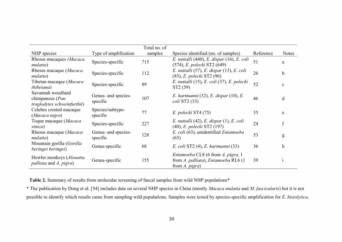
Molecular studies in wild NHPs published to date (Table 2) are few in number, mostly involve 322
Old World NHPs, and vary in the methodology used. In some studies, species-specific PCR 323
14
has been used, but often not all known species were tested for despite primers being available, 324
leaving gaps in the data (Table 2, notes). When species-specific PCR has been used, this often 325
means subtypes were not identified and potentially interesting data on sequence variation and 326
host range have been lost. Several studies did not test for E. hartmanni, leading to a false 327
impression of the distribution of this Entamoeba species in NHPs. It is notable that E. 328
histolytica was not detected in any of these studies. 329
330
The use of only species-specific primers can mean that novel Entamoeba species are missed. 331
For example, if Villanueva-García et al. [39] had used species-specific primers for Entamoeba, 332
the two novel Entamoeba species found in Howler monkeys (CL8 and RL6) would not have 333
been identified – the samples would have appeared negative even though Entamoeba 334
organisms were present. Sequencing of products amplified using genus-specific primers may 335
seem the best way forward, but there is a catch. NHPs are often carriers of multiple Entamoeba 336
species and mixed PCR products give unreadable sequences with the standard DNA 337
sequencing. The approach of Jirků-Pomajbíková et al. [46] could be a good compromise – 338
genus-specific amplification coupled with nested species-specific PCR. This allows 339
identification of species in mixed infections yet does not miss mono-infections with novel 340
Entamoeba species, as these would be positive with genus-specific but negative with all the 341
species-specific primers used. Jirků-Pomajbíková et al. [46] did not initially test for E. 342
hartmanni but through sequencing discovered that it was the Entamoeba present in the samples 343
positive with the genus-specific primers but negative with the species-specific primer pairs 344
used. However, this method will only identify the presence of novel Entamoeba species if they 345
are present as a single infection unless it is combined with cloning of the PCR products. 346
347

15
It seems likely that identification of Entamoeba in NHPs in the future will be through 348
microbiome data, whether from targeted amplification and sequencing of a portion of 349
eukaryotic SSU-rDNA or by extraction of such sequences from metagenomic data. Both 350
approaches are in use in humans and have identified Entamoeba when present, but to date have 351
rarely been applied to NHP samples. In one example, Wegener Parfrey et al. [47] identified E. 352
hartmanni (among many other eukaryotes) in captive NHPs through eukaryote-targeted SSU-353
rDNA amplification and 454 sequencing. Similarly, random sequencing of faecal DNA has the 354
potential to identify not only all the species present, but could enable assembly of partial or 355
complete genomes for the organisms identified [e.g. 48]. While such approaches are expensive 356
and likely to be available only to a few at present, the holistic information on the eukaryome 357
of NHPs likely to be obtained by such approaches makes them very attractive and we look 358
forward to seeing the data emerge in the next few years. 359
360
Concluding Remarks 361
362
Currently, at least six Entamoeba species with valid published names have been confirmed by 363
molecular analysis in NHPs: E. coli, E. polecki, E. histolytica, E. nuttalli, E. dispar and E. 364
hartmanni. However, in addition there are multiple subtypes within E. coli and E. polecki, plus 365
organisms with no name but distinct gene sequences (Entamoeba RL3, RL6, RL7 and CL8). 366
This remarkable expansion in known diversity has been driven largely by the use of molecular 367
techniques that have facilitated the identification of many novel and previously unrecognised 368
Entamoeba species in NHPs. 369
370
However, many points remain to be clarified (see “Outstanding Questions”). It is unclear 371
whether E. moshkovskii, E. bangladeshi and E. gingivalis colonise NHPs as well as humans. 372
16
Novel sequences with no linked species name are likely to continue to be detected in NHPs 373
around the world. This search for new types of Entamoeba in NHPs is essential as it remains 374
to be proven whether only E. nuttalli is responsible for morbidity and mortality in these hosts. 375
However, unless the correct approaches are used, such organisms will remain undiscovered. 376
377
We now know that NHPs are infected by both NHP-restricted and human-infective Entamoeba 378
species. Morphological diagnosis of Entamoeba species will always be problematic, but most 379
molecular approaches used to date may also be considerably underestimating the prevalence, 380
diversity, and distribution of Entamoeba in NHPs. At the same time, insufficient taxon 381
sampling and the heavy focus on humans may well have led us to inaccurate conclusions about 382
Entamoeba evolution. Fortunately, interest in the eukaryotic microbiome is growing in parallel 383
with improvements in technology, and it is likely that within the next few years a better 384
understanding of the evolution and host ranges of Entamoeba in NHPs will emerge. 385
386
Metagenomic analyses could allow the use of genes other than SSU-rDNA for phylogenetic 387
analyses. Obtaining sequence data for other genes is difficult - if not impossible - using 388
traditional molecular approaches and DNA from faecal samples. Multigene phylogenies may 389
well provide greater resolution that could confirm or refute our current views of relationships 390
within Entamoeba. Greater resolution is essential for evaluating the relative importance of 391
cospeciation and host-switching in the evolution of primate Entamoeba species. It seems likely 392
that these data will start to become available in the near future. 393
394
A recent study showed a significant reduction in the gut microbiome diversity of captive NHPs, 395
with a shift occurring from a wild NHP microbiome state toward a modern human microbiome 396
state [49]. Whether alterations in the lifestyle and diet of captive NHPs or the disruption of 397
17
normal hierarchical social behavior [50] has led to this perturbation of their gut microbiome, 398
the change may predispose captive NHPs to infection with certain Entamoeba spp, normally 399
confined to humans. Comparison of gut microbiomes across NHPs living in the wild, 400
semicaptivity and captivity using sequencing of both bacteria and Entamoeba SSU rDNA, is 401
already possible. Such data will allow us to investigate the correlation between microbiota 402
signatures and prevalence of specific Entamoeba species in NHPs. 403
404
There is much more to learn regarding both the microbiome and the eukaryome of NHPs, 405
especially those in the wild. There has been a strong focus on Old World primates, in particular 406
macaques, while New World primates are significantly underrepresented and prosimians have 407
not been studied. It is hoped that the range of species sampled will broaden, otherwise we will 408
continue to have a rather limited view of Entamoeba diversity in NHPs. 409
410
Acknowledgments 411
We would like to thank all the scientists whose research discoveries have advanced our 412
knowledge of the epidemiology of Entamoeba in non-human primates and enabled us to write 413
this review. 414
18
References 415
416
1. Lozano, R. et al. (2012) Global and regional mortality from 235 causes of death for 20 417
age groups in 1990 and 2010: a systematic analysis for the Global Burden of Disease Study 418
2010. Lancet 380, 2095–2128 419
420
2. Beaver, P.C. et al. (1988) Invasive amebiasis in naturally infected New World and Old 421
World monkeys with and without clinical disease. Am. J. Trop. Med. Hyg. 39, 343–352 422
423
3. Dobell, C. (1919) The amoebae living in Man. A zoological monograph. J. Bale, Sons, 424
and Danielson, London, UK 425
426
4. Dobell, C. (1928) Researches on the intestinal protozoa of monkeys and man. II 427
Description of the whole life-history of Entamoeba histolytica in cultures. Parasitology 20, 428
365–412 429
430
5. Burrows, R.B. (1957) Endamoeba hartmanni. Am. J. Hyg. 65, 172–188 431
432
6. Brumpt, E. (1925) Étude sommaire de l’Entamoeba dispar n. sp. Amibe à kystes 433
quadrinucléés, parasite de l’homme. Bull. Acad. Méd. (Paris) 94, 943–952 434
435
7. Brumpt, E. (1928) Differentiation of human intestinal amoebae with four-nucleated 436
cysts. Trans. R. Soc. Trop. Med. Hyg. 22, 101–114 (Discussion on pp. 115–124) 437
438
19
8. Martínez-Palomo A. et al. (1973) Selective agglutination of pathogenic strains of 439
Entamoeba histolytica induced by con A. Nat. New Biol. 245, 186–187 440
441
9. Sargeaunt, P.G. et al. (1978) The differentiation of invasive and non-invasive 442
Entamoeba histolytica by isoenzyme electrophoresis. Trans. R. Soc. Trop. Med. Hyg. 72, 519–443
521 444
445
10. Diamond, L.S. and Clark, C.G. (1993) A redescription of Entamoeba histolytica 446
Schaudinn, 1903 (emended Walker, 1911) separating it from Entamoeba dispar Brumpt, 1925. 447
J. Eukaryot. Microbiol. 40, 340–344 448
449
11. Clark, C.G. and Diamond, L.S. (1991) The Laredo strain and other Entamoeba 450
histolytica-like amoebae are Entamoeba moshkovskii. Mol. Biochem. Parasitol. 46, 11–18 451
452
12. Verweij, J.J. et al. (2001) Genetic variation among human isolates of uninucleated cyst-453
producing Entamoeba species. J. Clin. Microbiol. 39, 1644–1646 454
455
13. Royer, T.L. et al. (2012) Entamoeba bangladeshi nov. sp., Bangladesh. Emerg. Infect. 456
Dis. 18, 1543–1545 457
458
14. Stensvold, C.R. et al. (2011) Increased sampling reveals novel lineages of Entamoeba: 459
consequences of genetic diversity and host specificity for taxonomy and molecular detection. 460
Protist 162, 525–541 461
462
15. Pritt, B.S. and Clark, C.G. (2008) Amebiasis. Mayo Clin. Proc. 83, 1154–1160 463
20
464
16. Haq, A. et al. (1985) Experimental infection of rhesus monkeys with Entamoeba 465
histolytica mimics human infection. Lab. Anim. Sci. 35, 481–484 466
467
17. Tachibana, H. et al. (2007) An Entamoeba sp. strain isolated from rhesus monkey is 468
virulent but genetically different from Entamoeba histolytica. Mol. Biochem. Parasitol. 153, 469
107–114 470
471
18. Tachibana, H. et al. (2009) Isolation and characterization of a potentially virulent 472
species Entamoeba nuttalli from captive Japanese macaques. Parasitology 136, 1169–1177 473
474
19. Suzuki, J. et al. (2007) Profiles of a pathogenic Entamoeba histolytica-like variant with 475
variations in the nucleotide sequence of the small subunit ribosomal RNA isolated from a 476
primate (De Brazza's guenon). J. Zoo Wildl. Med. 38, 471–474 477
478
20. Takano, J. et al. (2009) DNA characterization of simian Entamoeba histolytica-like 479
strains to differentiate them from Entamoeba histolytica. Parasitol. Res. 10, 929–937 480
481
21. Levecke, B. et al. (2010) Molecular identification of Entamoeba spp. in captive 482
nonhuman primates. J. Clin. Microbiol. 48, 2988–2990 483
484
22. Castellani, A. (1908) Note on a liver abscess of amoebic origin in a monkey. 485
Parasitology 1, 101–102 486
487
21
23. Dobell, C. (1931) Researches on the intestinal protozoa of monkeys and man. IV An 488
experimental study of the histolytica-like species of Entamoeba living naturally in macaques 489
in cultures. Parasitology 23, 1–72 490
491
24. Tachibana, H. et al. (2016) Isolation and molecular characterization of Entamoeba 492
nuttalli strains showing novel isoenzyme patterns from wild Toque Macaques in Sri Lanka. J. 493
Eukaryot. Microbiol. 6, 171–180 494
495
25. Suzuki, J. et al. (2008) A survey of amoebic infections and differentiation of an 496
Entamoeba histolytica-like variant (JSK2004) in nonhuman primates by a multiplex 497
polymerase chain reaction. J. Zoo Wildl. Med. 39, 370–379 498
499
26. Tachibana, H. et al. (2013) Prevalence of Entamoeba nuttalli infection in wild rhesus 500
macaques in Nepal and characterization of the parasite isolates. Parasitol. Int. 62, 230–235 501
502
27. Levecke, B. et al. (2015) Transmission of Entamoeba nuttalli and Trichuris trichiura 503
from Nonhuman Primates to Humans. Emerg. Infect. Dis. 21, 1871–1872 504
505
28. Sargeaunt, P.G. (1987) The reliability of Entamoeba histolytica zymodemes in clinical 506
diagnosis. Parasitol. Today 3, 40–43 507
508
29. Abd Alla, M.D. et al. (2012) Efficacy of a Gal-lectin subunit vaccine against 509
experimental Entamoeba histolytica infection and colitis in baboons (Papio sp.). Vaccine 30, 510
3068–3075 511
512
22
30. Rivera, W.L. et al. (2010) Entamoeba histolytica and E. dispar infections in captive 513
macaques (Macaca fascicularis) in the Philippines. Primates 51, 69–74 514
515
31. Silberman, J.D. et al. (1999) Phylogeny of the genera Entamoeba and Endolimax as 516
deduced from small subunit ribosomal RNA gene sequence analysis. Mol. Biol. Evol. 16, 1740–517
1751 518
519
32. Burrows, R.B. (1959) Morphological differentiation of Entamoeba hartmanni and E. 520
polecki from E. histolytica. Am. J. Trop. Med. Hyg. 8, 583–589 521
522
33. Ponce Gordo, F. et al. (2004) Entamoeba struthionis n.sp. (Sarcomastigophora: 523
Endamoebidae) from ostriches (Struthio camelus). Vet. Parasitol. 119, 327–335 524
525
34. Jacob, A.S. et al. (2016) Expanding the Entamoeba universe: new hosts yield novel 526
ribosomal lineages. J. Eukaryot. Microbiol. 63, 69–78 527
528
35. Tuda, J. et al. (2016) Identification of Entamoeba polecki with unique 18S rRNA gene 529
sequences from Celebes Crested Macaques and pigs in Tangkoko Nature Reserve, North 530
Sulawesi, Indonesia. J. Eukaryot. Microbiol. 63, 572–577 531
532
36. Nolan, M.J. et al. (2017) Molecular characterisation of protist parasites in human-533
habituated mountain gorillas (Gorilla beringei beringei), humans and livestock, from Bwindi 534
impenetrable National Park, Uganda. Parasit. Vectors 10, 340 535
536
23
37. Matthews, J.R. (1919) A mensurative study of the cysts of Entamoeba coli. Ann. Trop. 537
Med. Parasitol. 12, 259–272 538
539
38. Dobell, C. (1936) Researches on the intestinal protozoa of monkeys and man. VIII. An 540
experimental study of some simian strains of “Entamoeba coli”. Parasitology 28, 541–593 541
542
39. Villanueva-García, C. et al. (2017). New Entamoeba group in howler monkeys 543
(Alouatta spp.) associated with parasites of reptiles. Parasitol. Res. 116, 2341–2346 544
545
40. Clark, C.G. and Diamond, L.S. (1997) Intraspecific variation and phylogenetic 546
relationships in the genus Entamoeba as revealed by riboprinting. J. Eukaryot. Microbiol. 44, 547
142–154 548
549
41. Al-Areeqi, M.A. et al. (2017) First molecular epidemiology of Entamoeba histolytica, 550
E. dispar and E. moshkovskii infections in Yemen: different species-specific associated risk 551
factors. Trop. Med. Int. Health 22, 493–504 552
553
42. López, M.C. et al. (2015) Molecular epidemiology of Entamoeba: first description of 554
Entamoeba moshkovskii in a rural area from central Colombia. PLoS One 10, e0140302 555
556
43. Nath, J. et al. (2015) Molecular epidemiology of amoebiasis: a cross-sectional study 557
among North East Indian population. PLoS Negl. Trop. Dis. 9, e0004225 558
559
44. Garcia, G. et al. (2014) Molecular epidemiology and genetic diversity of Entamoeba 560
species in a chelonian collection. J. Med. Microbiol. 63, 271–283 561
24
562
563
45. Levine, N.D. (1973) Protozoan parasites of domestic animals and of man. Second 564
Edition. Burgess Pub. Co., Minneapolis, MN, USA 565
566
46. Jirků-Pomajbíková, K. et al. (2016) Molecular identification of Entamoeba species in 567
savanna woodland chimpanzees (Pan troglodytes schweinfurthii). Parasitology 143, 741–748 568
569
47. Wegener Parfrey, L. et al. (2014) Communities of microbial eukaryotes in the 570
mammalian gut within the context of environmental eukaryotic diversity. Front. Microbiol. 5, 571
298 572
573
48. Beghini, F. et al. (2017) Large-scale comparative metagenomics of Blastocystis, a 574
common member of the human gut microbiome. ISME J. 11, 2848–2863 575
576
49. Clayton, J.B. et al. (2016) Captivity humanizes the primate microbiome. Proc, Natl, 577
Acad, Sci, USA 113, 10376–10381 578
579
50. Perofsky, A.C. et al. (2017) Hierarchical social networks shape gut microbial 580
composition in wild Verreaux's sifaka. Proc. R. Soc. B 284, 20172274 581
582
51. Feng, M. et al. (2013) Prevalence and genetic diversity of Entamoeba species infecting 583
macaques in southwest China. Parasitol. Res. 112, 1529–1536 584
585
25
52. Guan, Y. et al. (2016) Comparative analysis of genotypic diversity in Entamoeba 586
nuttalli isolates from Tibetan macaques and rhesus macaques in China. Infect. Genet. Evol. 38. 587
126–131 588
589
53. Debenham, J.J. et al. (2017) Occurrence of Giardia, Cryptosporidium, and Entamoeba 590
in wild rhesus macaques (Macaca mulatta) living in urban and semi-rural North-West India. 591
Int. J. Parasitol. Parasites Wildl. 6, 29–34 592
593
54. Dong, H. et al. (2017) Prevalence, molecular epidemiology, and zoonotic potential of 594
Entamoeba spp. in nonhuman primates in China. Infect. Genet. Evol. 54, 216–220 595
596
55. Al-Mayali, H.M.H. and Al-Abodi, H.R.J. (2017) Molecular characterization of 597
Entamoeba spp. in Al-Qadisiya province, Iraq. Al-Kufa Univ. J. Biol. 9, 374–386 598
599
56. Ragazzo, L.J. et al. (2018) Entamoeba histolytica infection in wild lemurs associated 600
with proximity to humans. Vet. Parasitol. 249, 98–101 601
602
26
Glossary 603
604
Conditional lineage (CL): an Entamoeba identified as likely to be distinct based on 605
sequencing of partial SSU-rDNA, but for which sufficient data are not yet available. See RL, 606
below. 607
608
Isoenzymes: each of two or more sequence variants of an enzyme that exhibit different 609
migration in electrophoresis gels due to charge differences. 610
611
Non-human primates (NHPs): all members of the order Primates other than humans; NHPs 612
share many similarities with humans in terms of physiology, anatomy, immunology, and 613
neurology, but are very diverse in their ecology, diet, etc. The split between humans and NHPs 614
is an artificial one, as humans are much more closely related to some NHPs than others. 615
616
Ribosomal lineage (RL): an Entamoeba taxon identified as distinct by sequencing of its 617
complete SSU-rDNA gene. Often no corresponding morphological data are available. In 618
other groups of organisms these are often called operational taxonomic units (OTUs) but in 619
this case, it is clear that they belong to the genus Entamoeba. 620
621
Small subunit ribosomal RNA gene (SSU-rDNA): the gene encoding the smaller of the two 622
major RNA components of the ribosome, also known as 18S rDNA. This gene is the most 623
widely used single locus for phylogenetic analyses in eukaryotes and bacteria. In Entamoeba, 624
the gene size generally falls between 1800 and 2200 bases. 625
626
Subtype: a discrete genetic clade within a named species. 627
27
Figure 1: Phylogenetic relationships among Entamoeba species. The phylogenetic tree 628
shown is modified from Figure 1 in Jacob et al. [34]. Names in bold lettering are those that 629
have been identified by sequencing of SSU-rDNA in NHPs. Adjacent to the Entamoeba names 630
are those of the NHP species (wild or captive) in which the Entamoeba has been identified. 631
632
633
28
Dobell nomenclaturea Current species names Identified in primates (incl. humans) Molecular identification in NHPs
E. histolytica E. histolytica E. histolytica Yb
E. dispar E. dispar Y
E. hartmanni E. hartmanni Y
E. nuttalli E. nuttalli Y
E. moshkovskii E. moshkovskii (complex) N
E. polecki E. polecki ST1 c N
E. polecki ST4 Y
E. chattoni E. polecki ST2 Y
E. struthionis E. polecki ST3 N
E. bangladeshi E. bangladeshi N
E. suis E. suis Yb
E. coli E. coli E. coli ST1 Yb
E. coli ST2 Y
E. gingivalis E. gingivalis E. gingivalis ribodeme 1 d N
E. gingivalis ribodeme 2 N
None Entamoeba RL3 e Y
Entamoeba RL6 Y
Entamoeba RL7 Y
Entamoeba CL8 f Y
Table 1. Correspondence between historic, binomial, and sequence-based nomenclature for Entamoeba species in primates.
a Dobell’s nomenclature is that proposed in his 1919 monograph [3].
bIdentified in captive NHPs only, to date.
29
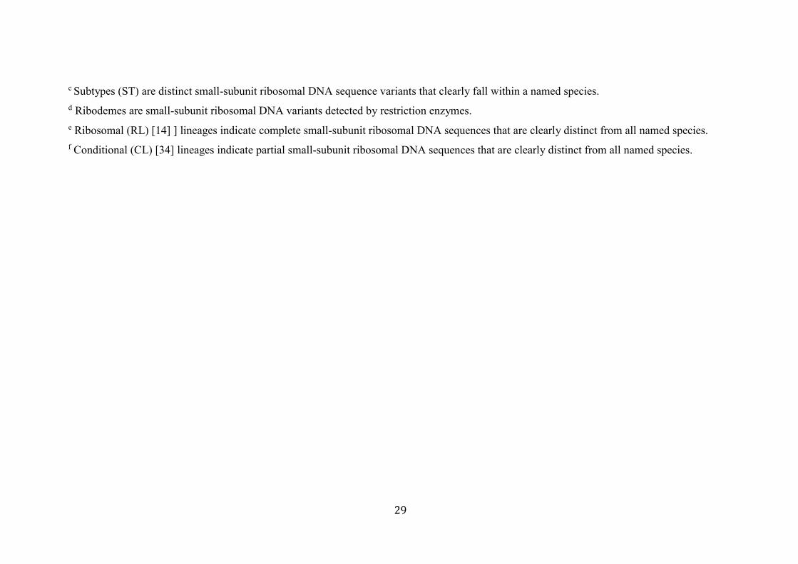
c Subtypes (ST) are distinct small-subunit ribosomal DNA sequence variants that clearly fall within a named species.
d Ribodemes are small-subunit ribosomal DNA variants detected by restriction enzymes.
e Ribosomal (RL) [14] ] lineages indicate complete small-subunit ribosomal DNA sequences that are clearly distinct from all named species.
f Conditional (CL) [34] lineages indicate partial small-subunit ribosomal DNA sequences that are clearly distinct from all named species.
30
NHP species Type of amplification
Total no. of
samples Species identified (no. of samples) Reference Notes
Rhesus macaques (Macaca
mulatta) Species-specific 715
E. nuttalli (440), E. dispar (16), E. coli
(574), E. polecki ST2 (649) 51 a
Rhesus macaque (Macaca
mulatta) Species-specific 112
E. nuttalli (57), E. dispar (13), E. coli
(83), E. polecki ST2 (96) 26 b
Tibetan macaque (Macaca
thibetana) Species-specific 89
E. nuttalli (15), E. coli (37), E. polecki
ST2 (59) 52 c
Savannah woodland
chimpanzee (Pan
troglodytes schweinfurthii)
Genus- and species-
specific 107
E. hartmanni (32), E. dispar (10), E.
coli ST2 (33) 46 d
Celebes crested macaque
(Macaca nigra)
Species/subtype-
specific 77 E. polecki ST4 (75) 35 e
Toque macaque (Macaca
sinica) Species-specific 227
E. nuttalli (42), E. dispar (1), E. coli
(40), E. polecki ST2 (197) 24 f
Rhesus macaque (Macaca
mulatta)
Genus- and species-
specific 128
E. coli (63), unidentified Entamoeba
(65) 53 g
Mountain gorilla (Gorilla
beringei beringei) Genus-specific 68 E. coli ST2 (4), E. hartmanni (33) 36 h
Howler monkeys (Alouatta
palliata and A. pigra) Genus-specific 155
Entamoeba CL8 (6 from A. pigra, 1
from A. palliata), Entamoeba RL6 (1
from A. pigra)
39 i
Table 2. Summary of results from molecular screening of faecal samples from wild NHP populations*
* The publication by Dong et al. [54] includes data on several NHP species in China (mostly Macaca mulatta and M. fascicularis) but it is not
possible to identify which results came from sampling wild populations. Samples were tested by species-specific amplification for E. histolytica,
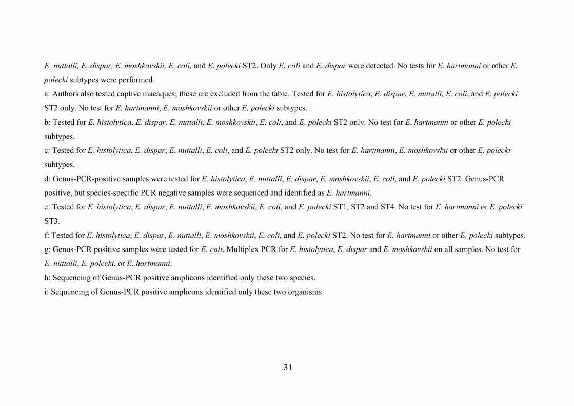
31
E. nuttalli, E. dispar, E. moshkovskii, E. coli, and E. polecki ST2. Only E. coli and E. dispar were detected. No tests for E. hartmanni or other E.
polecki subtypes were performed.
a: Authors also tested captive macaques; these are excluded from the table. Tested for E. histolytica, E. dispar, E. nuttalli, E. coli, and E. polecki
ST2 only. No test for E. hartmanni, E. moshkovskii or other E. polecki subtypes.
b: Tested for E. histolytica, E. dispar, E. nuttalli, E. moshkovskii, E. coli, and E. polecki ST2 only. No test for E. hartmanni or other E. polecki
subtypes.
c: Tested for E. histolytica, E. dispar, E. nuttalli, E. coli, and E. polecki ST2 only. No test for E. hartmanni, E. moshkovskii or other E. polecki
subtypes.
d: Genus-PCR-positive samples were tested for E. histolytica, E. nuttalli, E. dispar, E. moshkovskii, E. coli, and E. polecki ST2. Genus-PCR
positive, but species-specific PCR negative samples were sequenced and identified as E. hartmanni.
e: Tested for E. histolytica, E. dispar, E. nuttalli, E. moshkovskii, E. coli, and E. polecki ST1, ST2 and ST4. No test for E. hartmanni or E. polecki
ST3.
f: Tested for E. histolytica, E. dispar, E. nuttalli, E. moshkovskii, E. coli, and E. polecki ST2. No test for E. hartmanni or other E. polecki subtypes.
g: Genus-PCR positive samples were tested for E. coli. Multiplex PCR for E. histolytica, E. dispar and E. moshkovskii on all samples. No test for
E. nuttalli, E. polecki, or E. hartmanni.
h: Sequencing of Genus-PCR positive amplicons identified only these two species.
i: Sequencing of Genus-PCR positive amplicons identified only these two organisms.