Embed Size (px)
Citation preview
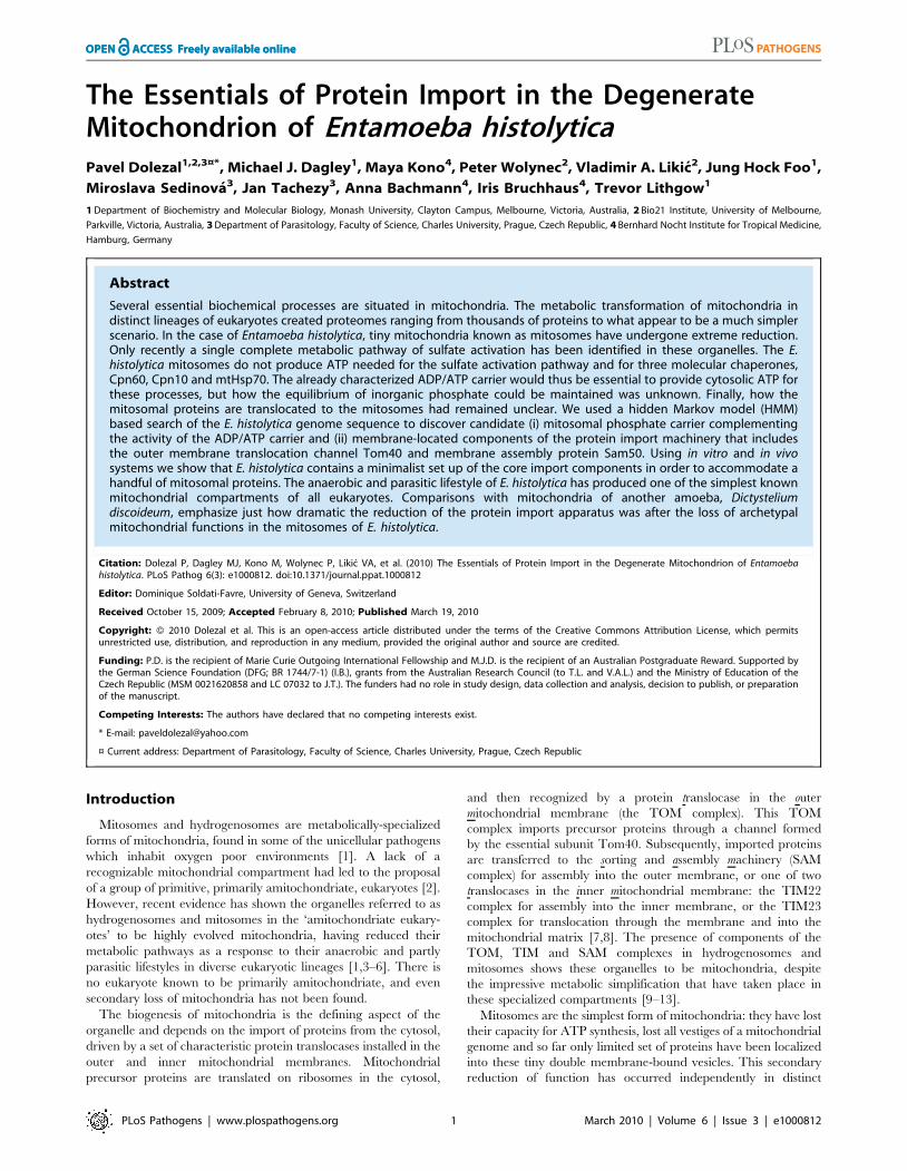
The Essentials of Protein Import in the DegenerateMitochondrion of Entamoeba histolyticaPavel Dolezal1,2,3¤*, Michael J. Dagley1, Maya Kono4, Peter Wolynec2, Vladimir A. Likic2, Jung Hock Foo1,
Miroslava Sedinova3, Jan Tachezy3, Anna Bachmann4, Iris Bruchhaus4, Trevor Lithgow1
1 Department of Biochemistry and Molecular Biology, Monash University, Clayton Campus, Melbourne, Victoria, Australia, 2 Bio21 Institute, University of Melbourne,
Parkville, Victoria, Australia, 3 Department of Parasitology, Faculty of Science, Charles University, Prague, Czech Republic, 4 Bernhard Nocht Institute for Tropical Medicine,
Hamburg, Germany
Abstract
Several essential biochemical processes are situated in mitochondria. The metabolic transformation of mitochondria indistinct lineages of eukaryotes created proteomes ranging from thousands of proteins to what appear to be a much simplerscenario. In the case of Entamoeba histolytica, tiny mitochondria known as mitosomes have undergone extreme reduction.Only recently a single complete metabolic pathway of sulfate activation has been identified in these organelles. The E.histolytica mitosomes do not produce ATP needed for the sulfate activation pathway and for three molecular chaperones,Cpn60, Cpn10 and mtHsp70. The already characterized ADP/ATP carrier would thus be essential to provide cytosolic ATP forthese processes, but how the equilibrium of inorganic phosphate could be maintained was unknown. Finally, how themitosomal proteins are translocated to the mitosomes had remained unclear. We used a hidden Markov model (HMM)based search of the E. histolytica genome sequence to discover candidate (i) mitosomal phosphate carrier complementingthe activity of the ADP/ATP carrier and (ii) membrane-located components of the protein import machinery that includesthe outer membrane translocation channel Tom40 and membrane assembly protein Sam50. Using in vitro and in vivosystems we show that E. histolytica contains a minimalist set up of the core import components in order to accommodate ahandful of mitosomal proteins. The anaerobic and parasitic lifestyle of E. histolytica has produced one of the simplest knownmitochondrial compartments of all eukaryotes. Comparisons with mitochondria of another amoeba, Dictysteliumdiscoideum, emphasize just how dramatic the reduction of the protein import apparatus was after the loss of archetypalmitochondrial functions in the mitosomes of E. histolytica.
Citation: Dolezal P, Dagley MJ, Kono M, Wolynec P, Likic VA, et al. (2010) The Essentials of Protein Import in the Degenerate Mitochondrion of Entamoebahistolytica. PLoS Pathog 6(3): e1000812. doi:10.1371/journal.ppat.1000812
Editor: Dominique Soldati-Favre, University of Geneva, Switzerland
Received October 15, 2009; Accepted February 8, 2010; Published March 19, 2010
Copyright: � 2010 Dolezal et al. This is an open-access article distributed under the terms of the Creative Commons Attribution License, which permitsunrestricted use, distribution, and reproduction in any medium, provided the original author and source are credited.
Funding: P.D. is the recipient of Marie Curie Outgoing International Fellowship and M.J.D. is the recipient of an Australian Postgraduate Reward. Supported bythe German Science Foundation (DFG; BR 1744/7-1) (I.B.), grants from the Australian Research Council (to T.L. and V.A.L.) and the Ministry of Education of theCzech Republic (MSM 0021620858 and LC 07032 to J.T.). The funders had no role in study design, data collection and analysis, decision to publish, or preparationof the manuscript.
Competing Interests: The authors have declared that no competing interests exist.
* E-mail: [email protected]
¤ Current address: Department of Parasitology, Faculty of Science, Charles University, Prague, Czech Republic
Introduction
Mitosomes and hydrogenosomes are metabolically-specialized
forms of mitochondria, found in some of the unicellular pathogens
which inhabit oxygen poor environments [1]. A lack of a
recognizable mitochondrial compartment had led to the proposal
of a group of primitive, primarily amitochondriate, eukaryotes [2].
However, recent evidence has shown the organelles referred to as
hydrogenosomes and mitosomes in the ‘amitochondriate eukary-
otes’ to be highly evolved mitochondria, having reduced their
metabolic pathways as a response to their anaerobic and partly
parasitic lifestyles in diverse eukaryotic lineages [1,3–6]. There is
no eukaryote known to be primarily amitochondriate, and even
secondary loss of mitochondria has not been found.
The biogenesis of mitochondria is the defining aspect of the
organelle and depends on the import of proteins from the cytosol,
driven by a set of characteristic protein translocases installed in the
outer and inner mitochondrial membranes. Mitochondrial
precursor proteins are translated on ribosomes in the cytosol,
and then recognized by a protein translocase in the outer
mitochondrial membrane (the TOM complex). This TOM
complex imports precursor proteins through a channel formed
by the essential subunit Tom40. Subsequently, imported proteins
are transferred to the sorting and assembly machinery (SAM
complex) for assembly into the outer membrane, or one of two
translocases in the inner mitochondrial membrane: the TIM22
complex for assembly into the inner membrane, or the TIM23
complex for translocation through the membrane and into the
mitochondrial matrix [7,8]. The presence of components of the
TOM, TIM and SAM complexes in hydrogenosomes and
mitosomes shows these organelles to be mitochondria, despite
the impressive metabolic simplification that have taken place in
these specialized compartments [9–13].
Mitosomes are the simplest form of mitochondria: they have lost
their capacity for ATP synthesis, lost all vestiges of a mitochondrial
genome and so far only limited set of proteins have been localized
into these tiny double membrane-bound vesicles. This secondary
reduction of function has occurred independently in distinct
PLoS Pathogens | www.plospathogens.org 1 March 2010 | Volume 6 | Issue 3 | e1000812
lineages of eukaryotes, being well characterized in the diplomonad
Giardia intestinalis [14], the microsporidians (such as Encephalitozoon
cuniculi [15], Antonospora locustae [12], Trachipleistophora hominis [16])
and the amoebozoan Entamoeba histolytica [17,18]. The majority of
known proteins found in the mitosomes of G. intestinalis and
microsporidia are functional counterparts of mitochondrial
proteins found in other organisms, and a unifying feature of all
these organelles is their role in the synthesis of iron-sulfur clusters
[14,19,20]. So far it is the sole metabolic process known to occur in
mitosomes of G. intestinalis and microsporidia and conflicting data
exist on the presence of the iron-sulfur clusters biosynthesis in E.
histolytica mitosomes [21,22]. In addition to being widespread in
hydrogenosomes and mitosomes, the biogenesis of iron-sulfur
centers is the only essential metabolic role of mitochondria in the
model organism Saccharomyces cerevisiae [23].
Entamoeba histolytica, the causative agent of invasive amoebiasis in
humans, seems to have taken further steps towards the extreme
reduction of the mitochondrial compartment [24]. It represents
the only known eukaryote in which the synthesis of iron-sulfur
clusters is mediated by an NIF (nitrogen fixation) system acquired
by horizontal gene transfer from an e-proteobacterium [25].
According to prediction algorithms this biosynthetic pathway is
predicted to be present in the cytosol instead of the mitochondrial
compartment [26]. Consistently, Mi-Ichi et al, did not find either
of Nif proteins in their mitosomal proteomic analysis [22] and also
no iron-sulfur cluster containing protein is known to be present in
the organelles as a candidate substrate for the NIF system [21].
However, Maralikova et al, presented data arguing for the dual
localization of both Nif proteins with their specific enrichment in
the mitosomes [21].
So far, the mitosomes of E. histolytica represent one of the
simplest mitochondria known. With the presence of sulfate
activation pathway in the mitosome the need for ATP in addition
to the molecular chaperones within the organelle is obvious
[17,27–29]. Although the ADP/ATP carrier in the mitosomal
membrane provides for ATP import [22,27], two questions are left
open. Firstly, how do mitosomes recycle inorganic phosphate (Pi)
arising from ATP hydrolysis? Secondly, how are all the mitosomal
proteins transported across the membranes of the organelle?
To address these questions, we performed hidden Markov
model (HMM) searches of the E. histolytica genome and discovered
candidate sequences for (i) a Pi carrier to complement the activity
of the ATP/ADP carrier, (ii) Tom40, a channel for substrate
protein transport across the outer mitosomal membrane, and (iii)
Sam50, an assembly machine for Tom40 in the outer membrane.
The mitosomal protein import pathway in E. histolytica is
mitochondrial in nature. Analysis of the mitochondrial protein
import machinery of the related amoeba Dictyostelium discoideum,
suggests that E. histolytica has extensively stripped the mitochon-
drial protein import machines to their essentials. This remarkable
degeneration of protein import is in keeping with the apparent
paucity of proteins imported into mitosomes in E. histolytica.
Results
Substrate transport by mitochondrial carrier proteins in E.histolytica
The family of mitochondrial carrier proteins is well character-
ized in terms of primary sequence motives and crystal structure.
According to the structure of the bovine ADP/ATP carrier, six
transmembrane segments form an a-helical bundle, consisting of
three homologous modules [30]. Each of these modules contains a
signature motif in the odd-numbered helices [P]-x-[D/E]-x-x-[K/
R]. The proline residue in the motif introduces a kink into three of
the six transmembrane helices. In order to obtain a sensitive tool
for analysis of E. histolytica genome, a hidden Markov model was
built that describes the defining features of the mitochondrial
carrier protein family.
Only two protein sequences were identified in the HMM search
of the E. histolytica genome: 269.m00084 (E-value of 2.8610294)
and 13.m00296 (E-value of 4.761025) (Accession numbers
XP_649800 and XP_656350, respectively). The first sequence
corresponds to the ADP/ATP carrier [27] and the second to the
novel, unannotated protein sequence. Additional analysis of the
sequence length and predicted topology support the possibility that
13.m00296 could encode a mitochondrial carrier protein
(Figure 1A), one of the most divergent of the carrier protein
family. Based on the functional analyses that follow, we labeled the
gene as ehpic and its translation product as EhPiC.
The 31.7 kDa protein is predicted to have six transmembrane
helices, although with the lack of signature motives in the odd-
number helices. Only the third helix has a proline residue in the
conserved position (Figure 1A). As is the case in fungi, animals and
plants, D. discoideum has a diverse set of mitochondrial carrier
proteins that can be compared in a phylogenetic analyses [31,32].
When added to the dataset from D. discoideum, the E. histolytica
ADP/ATP carrier clusters together with transporters of adenine
nucleotides and coenzyme A, while EhPiC clusters with the Pi
carrier McfN (Figure 1B). The overall sequence divergence of
EhPiC is reflected by a long branch formed in the tree and also by
low statistical support. However, a similar tree topology was
obtained using different reconstruction methods (Supplementary
information Figure S1), which indicated the affinity of EhPiC to
McfN. The amino acid residues at three contact sites in the central
pore are believed to determine the specificity of the transport
channel and their composition can therefore serve for the
estimation of the substrate nature [32]. Perhaps due to the high
degree of sequence divergence, none of the three sites of EhPiC
provided any leads towards the putative pore specificity
(Figure 1A). The proposed function of a phosphate carrier in the
mitosomes is summarized diagrammatically in Figure 1C.
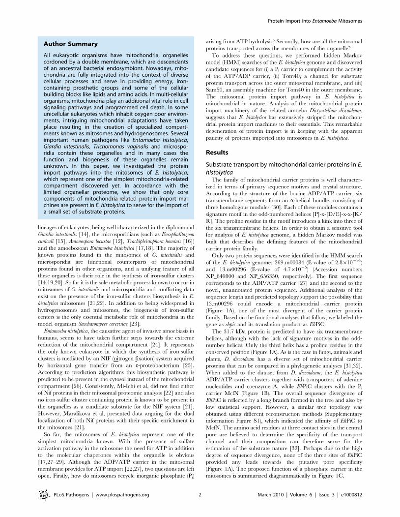
Author Summary
All eukaryotic organisms have mitochondria, organellescordoned by a double membrane, which are descendantsof an ancestral bacterial endosymbiont. Nowadays, mito-chondria are fully integrated into the context of diversecellular processes and serve in providing energy, iron-containing prosthetic groups and some of the cellularbuilding blocks like lipids and amino acids. In multi-cellularorganisms, mitochondria play an additional vital role in cellsignaling pathways and programmed cell death. In someunicellular eukaryotes which inhabit oxygen poor environ-ments, intriguing mitochondrial adaptations have takenplace resulting in the creation of specialized compart-ments known as mitosomes and hydrogenosomes. Severalimportant human pathogens like Entamoeba histolytica,Giardia intestinalis, Trichomonas vaginalis and microspo-ridia contain these organelles and in many cases thefunction and biogenesis of these organelles remainunknown. In this paper, we investigated the proteinimport pathways into the mitosomes of E. histolytica,which represent one of the simplest mitochondria-relatedcompartment discovered yet. In accordance with thelimited organellar proteome, we show that only corecomponents of mitochondria-related protein import ma-chines are present in E. histolytica to serve for the import ofa small set of substrate proteins.
Protein Import into Entamoeba Mitosomes
PLoS Pathogens | www.plospathogens.org 2 March 2010 | Volume 6 | Issue 3 | e1000812
To determine whether EhPiC could function as a phosphate
carrier protein, we made use of S. cerevisiae as a cellular assay
system. Fluorescence microscopy showed that EhPiC contains the
targeting sequences necessary for the import into mitochondria in
vivo (Figure 2A). The EhPiC protein can be translated in vitro and is
imported by mitochondria isolated from S. cerevisiae as readily as
the EhAAC (Figure 2B). Figure 2C shows that EhPiC behaves as a
typical integral membrane protein, exclusively distributed in the
pellet fraction after resisting sodium carbonate extraction of
mitochondrial membranes. Soluble and peripheral membrane
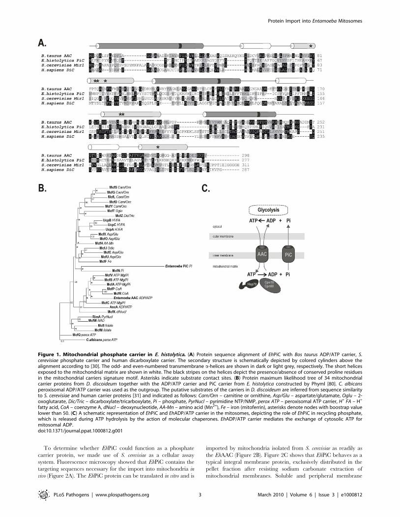
Figure 1. Mitochondrial phosphate carrier in E. histolytica. (A) Protein sequence alignment of EhPiC with Bos taurus ADP/ATP carrier, S.cerevisiae phosphate carrier and human dicarboxylate carrier. The secondary structure is schematically depicted by colored cylinders above thealignment according to [30]. The odd- and even-numbered transmembrane a-helices are shown in dark or light grey, respectively. The short helicesexposed to the mitochondrial matrix are shown in white. The black stripes on the helices depict the presence/absence of conserved proline residuesin the mitochondrial carriers signature motif. Asterisks indicate substrate contact sites. (B) Protein maximum likelihood tree of 34 mitochondrialcarrier proteins from D. discoideum together with the ADP/ATP carrier and PiC carrier from E. histolytica constructed by Phyml [80]. C. albicansperoxisomal ADP/ATP carrier was used as the outgroup. The putative substrates of the carriers in D. discoideum are inferred from sequence similarityto S. cerevisiae and human carrier proteins [31] and indicated as follows: Carn/Orn – carnitine or ornithine, Asp/Glu – aspartate/glutamate, Oglu – 2-oxoglutarate, Dic/Tric – dicarboxylate/tricarboxylate, Pi – phosphate, PyrNucl – pyrimidine NTP/NMP, perox ATP – peroxisomal ATP carrier, H+ FA – H+
fatty acid, CoA – coenzyme A, dNucl – deoxynucleotide, AA-Mn – amino acid (Mn2+), Fe – iron (mitoferrin), asterisks denote nodes with boostrap valuelower than 50. (C) A schematic representation of EhPiC and EhADP/ATP carrier in the mitosomes, depicting the role of EhPiC in recycling phosphate,which is released during ATP hydrolysis by the action of molecular chaperones. EhADP/ATP carrier mediates the exchange of cytosolic ATP formitosomal ADP.doi:10.1371/journal.ppat.1000812.g001
Protein Import into Entamoeba Mitosomes
PLoS Pathogens | www.plospathogens.org 3 March 2010 | Volume 6 | Issue 3 | e1000812
proteins, like mtHsp70, are extracted from membranes by sodium
carbonate.
To test if EhPiC functions to transport phosphate, the coding
sequence was cloned into a yeast expression vector and
transformed into S.cerevisiae strain lacking the dominant phosphate
carrier (Dmir1) [33,34]. The Dmir1 mutants grow on glucose
containing media but, due to failure to maintain phosphate
transport to sustain oxidative phosphorylation, Dmir1 cells fail to
grow on the non-fermentable carbon source glycerol (Figure 2D).
EhPiC expression complements the growth defect of these cells,
demonstrating that it functions as a phosphate carrier.
Protein import machinery encoded in the genome of E.histolytica
Both E. histolytica and the social amoeba D. discoideum are
amoebozoans, one of the six super-groups of eukaryotes [35,36].
As a sister clade to the animal and fungal lineages, it was not
surprising that the core components of the TOM and TIM
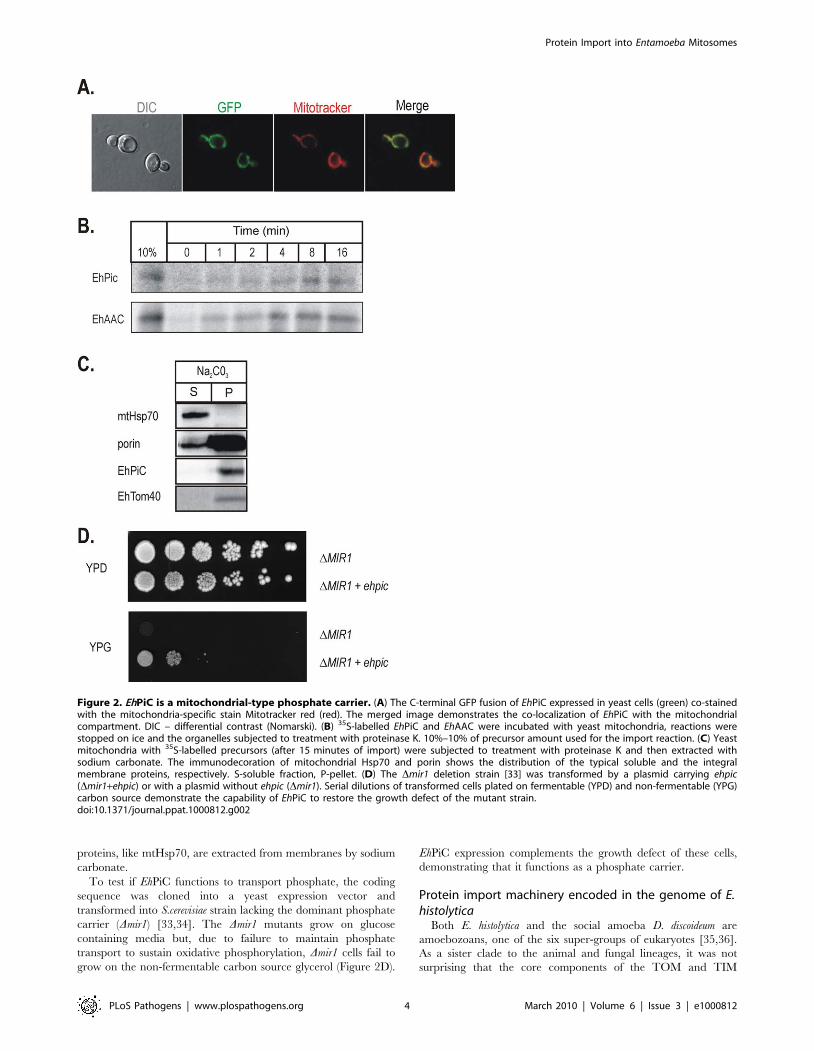
Figure 2. EhPiC is a mitochondrial-type phosphate carrier. (A) The C-terminal GFP fusion of EhPiC expressed in yeast cells (green) co-stainedwith the mitochondria-specific stain Mitotracker red (red). The merged image demonstrates the co-localization of EhPiC with the mitochondrialcompartment. DIC – differential contrast (Nomarski). (B) 35S-labelled EhPiC and EhAAC were incubated with yeast mitochondria, reactions werestopped on ice and the organelles subjected to treatment with proteinase K. 10%–10% of precursor amount used for the import reaction. (C) Yeastmitochondria with 35S-labelled precursors (after 15 minutes of import) were subjected to treatment with proteinase K and then extracted withsodium carbonate. The immunodecoration of mitochondrial Hsp70 and porin shows the distribution of the typical soluble and the integralmembrane proteins, respectively. S-soluble fraction, P-pellet. (D) The Dmir1 deletion strain [33] was transformed by a plasmid carrying ehpic(Dmir1+ehpic) or with a plasmid without ehpic (Dmir1). Serial dilutions of transformed cells plated on fermentable (YPD) and non-fermentable (YPG)carbon source demonstrate the capability of EhPiC to restore the growth defect of the mutant strain.doi:10.1371/journal.ppat.1000812.g002
Protein Import into Entamoeba Mitosomes
PLoS Pathogens | www.plospathogens.org 4 March 2010 | Volume 6 | Issue 3 | e1000812
machinery, characterized in fungi and animals, have been
identified in D. discoideum [37–40].
Given this conservation in protein import pathways between
amoebozoans, fungi and animals, it was highly surprising when the
only homologs of the mitochondrial protein import machinery
identified in the genome of E. histolytica [41] were the chaperones
mtHsp70, Cpn60 and Cpn10.
Hidden Markov models were built, representing search tools for
33 mitochondrial components known to participate during the
import process either in fungal/animal or plant mitochondria.
Each model was built from sequences clearly homologous to the
functionally characterized import component. The models were
assembled into a library and used to search the E. histolytica
genome (see Materials and Methods). In parallel we analyzed the
genome of D. discoideum (Figure 3), which is the most extensively
studied species of Amebozoa super-group thus providing a
referential dataset for our analysis of the E. histolytica genome.
Two sequences 38.m00236 and 137.m00093 (accession num-
bers XP_655014 and XP_651988) were identified by the Tom40-
and Sam50-specific HMMs, respectively. Accordingly, these two
open reading frames were named ehtom40 and ehsam50 and their
putative protein products as EhTom40 and EhSam50. The third
open reading frame identified in E. histolytica corresponds to the
previously identified gene encoding for mitochondrial-type Hsp70
[42].
A core component of the TOM complex: EhTom40The gene ehtom40 encodes a protein of 305 amino acids with a
theoretical molecular weight of 34.4 kDa, similar in size to the
Tom40 from Encephalizoon cuniculi [43]. EhTom40 is expressed in
E. histolytica with the ehtom40 mRNA detected in extracts from
amoebae by reverse transcription coupled with specific PCR
amplification (Supplementary information Figure S2). The reverse
BLASTP search with EhTom40 as a query provided hits to a
‘porin family protein’ from Arabidopsis thaliana (NP_175457.1), the
Tom40 sequence from Trimastix pyriformis (ABW76113.1) and a
hypothetical protein from Leishmania infantum (XP_001463193.1)
representing an unidentified protein predicted to be a b-barrel.
Such a structure is typical for Tom40 proteins. The mitochondrial
porin 3 superfamily of proteins (Pfam01459) encompasses both the
eukaryotic voltage-dependent anion channel (VDAC) proteins and
Tom40s [44,45]. Other mitochondrial b-barrel proteins like
Sam50 do not fall within the porin 3 superfamily. Given the
similarity of EhTom40 to b-barrel proteins predicted to be Tom40
(ABW76113.1) and VDAC (NP_175457.1), we performed a
CLANS analysis using a sequence set containing 137 VDAC
and 79 Tom40 sequences (including EhTom40). Membrane
proteins are often difficult to resolve on phylogenetic trees but
CLANS, that graphically depicts homology in large datasets of
proteins, has been previously found to be very efficient in the
classification of the b-barrel proteins from bacteria [46] and also in
the characterization of a Tom40 homologue in G. intestinalis [47].
This approach utilizes all-against-all pairwise BLAST, clustering
‘‘vertices’’ (i.e. individual protein sequences) in three-dimensional
space using an algorithm which applies a weak repulsive force to
each vertex and an attractive force between each pair of vertices.
The attractive and repulsive forces are proportional to the BLAST
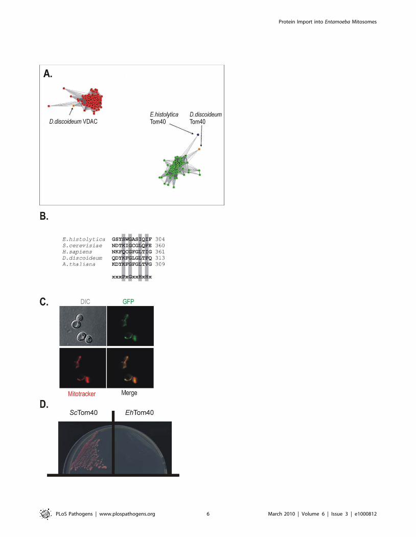
high scoring segment pair score. Figure 4A shows that cluster
analysis performed on this superset of porin3 family proteins using
a P-value cutoff of 10282 results in the sequences clustering into
two distinct groups. The clusters are either exclusively VDAC
homologues or Tom40 homologues, corresponding to their
independent phylogenies across eukaryotes. The EhTom40 is
positioned on the periphery of Tom40 cluster, clearly separated
from the compact cluster of VDAC sequences.
Mitochondrial b-barrel proteins contain a specific mitochon-
drial targeting signal (the b-signal, PxGxxHxH, where P stands for
polar, G for glycine and H for hydrophobic residue) that sits in the
last b-strand of the protein [48]. The b-signal is recognized and
bound by the SAM complex, which then mediates folding and
membrane insertion of the b-barrel. EhTom40 has a b-signal that
strictly follows this rule (Figure 4B). When EhTom40 is expressed
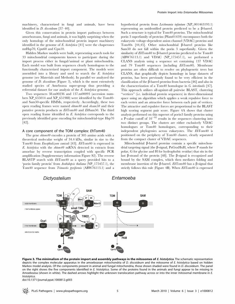
Figure 3. The minimalism of the protein import and assembly pathways in the mitosomes of E. histolytica. The schematic representationdepicts the complex molecular apparatus in the amoebozoan mitochondria of D. discoideum and the mitosome of E. histolytica based on hiddenMarkov model analysis. Of the components present in animal and fungal mitochondria, those shown shaded were found in D. discoideum. The panelon the right shows the five components identified in E. histolytica. Some of the proteins found in the animals and fungi appear to be missing inAmoebozoa (shown in white). The dashed arrows highlight the unknown translocation pathway across or into the inner mitosomal membrane in E.histolytica.doi:10.1371/journal.ppat.1000812.g003
Protein Import into Entamoeba Mitosomes
PLoS Pathogens | www.plospathogens.org 5 March 2010 | Volume 6 | Issue 3 | e1000812
Protein Import into Entamoeba Mitosomes
PLoS Pathogens | www.plospathogens.org 6 March 2010 | Volume 6 | Issue 3 | e1000812
in S. cerevisiae, it is specifically targeted to mitochondria, as judged
by fluorescence microscopy (Figure 4C). In order to test whether
EhTom40 can functionally substitute for S. cerevisiae Tom40 we
have transformed a haploid Dtom40 strain that carries the
S.cerevisiae TOM40 on a counterselectable plasmid. Upon plating
on 5-FOA media, to select against the covering plasmid, no viable
transformants were obtained (Figure 4D), while the strain
transformed with the homologous TOM40 remained viable.
Apparently the sequence divergence between the fungal and
amoebic Tom40 interferes with the correct docking and
interaction with other TOM complex subunits and thus does
not allow for functional complementation. Therefore we subse-
quently tested whether EhTom40 integrates into the native S.
cerevisiae TOM complex using the in vitro import assays. Although
the protein accumulated as a high molecular weight species in the
mitochondrial membranes (Supplementary information Figure
S3), the specific pull-down assay using the antibodies against TOM
complex subunits did not recover any of EhTom40 protein (data
not shown).
Given that TOM complex requires numerous binding sites on
several distinct subunits to assemble properly, it is not surprising
that these experiments failed. It was previously reported that even
the dual point mutations in Tom40 of S.cerevisiae can result in the
disassembly of the native TOM complex [49].
A core component of the SAM complex: EhSam50The cDNA sequence of Sam50 homologue found in E. histolytica
is deposited at NCBI under accession number XM_646896. It
encodes for a protein of 388 amino acids with a theoretical
molecular weight of 45.3 kDa. However, when aligned with the
corresponding genomic sequence, the presence of an intervening
sequence was revealed. The insertion of 72 bp starting at position
802 is limited by GAATGATT and TAG at the 59- and 39-ends.
These are conserved intron donor and acceptor splice sites
identified in E. histolytica [50], suggesting that ehsam50 gene is
interrupted by a single intron. According to the molecular weight
of the protein when episomally expressed in E. histolytica and S.
cerevisiae the mRNA is likely processed in vivo in both cellular
systems and provides for EhSam50 translation.
As shown on phylogenetic reconstruction the protein sequence
clusters with other mitochondrial Sam50 sequences [51,52] with
a-proteobacterial Omp85 sequences in a sister clade (Figure 5B)
[53], implying that EhSam50 is of direct mitochondrial origin.
Omp85 proteins contain a C–terminal b-barrel domain (the
‘surface antigen domain’), which is integrated into the outer
membrane. A characteristic N-terminal extension consists of five
POTRA (polypeptide-transport-associated) domains [54]. In
contrast, mitochondrial Sam50 proteins, including EhSam50,
contain a single POTRA domain along a b-barrel domain
(Figure 5A). Together with obtained phylogenetic data this
provides additional support that EhSam50 is a genuine mitochon-
drial homologue, not a bacterial Omp85 acquired by a horizontal
gene transfer. While the structural domains are well conserved in
EhSam50, we could not obtain a viable S.cerevisiae Dsam50 strain
expressing the E. histolytica protein (Figure 5C). As in the case of
Tom40, the primary sequence was likely too divergent to support
functional integration of EhSam50 into SAM complex of
S.cerevisiae. So far, there is no record in the literature of successful
heterologous replacement of SAM complex components, which
suggests that tight protein-protein or protein-lipid interactions are
involved in the SAM function.
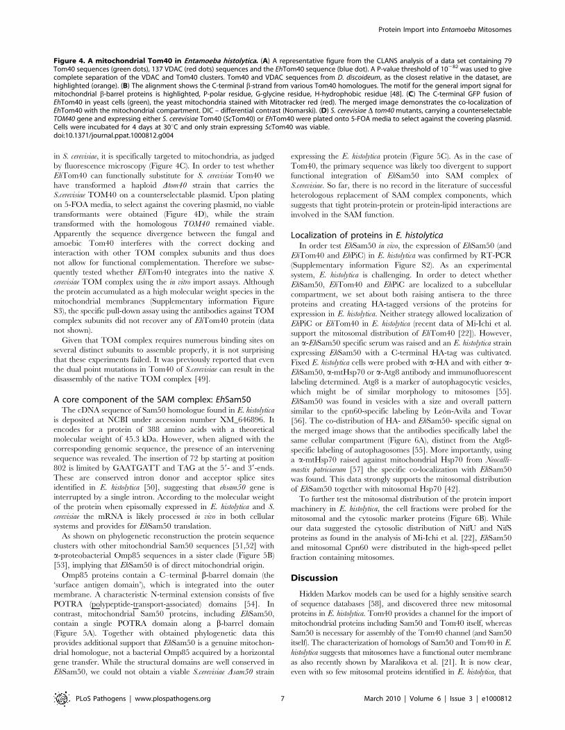
Localization of proteins in E. histolyticaIn order test EhSam50 in vivo, the expression of EhSam50 (and
EhTom40 and EhPiC) in E. histolytica was confirmed by RT-PCR
(Supplementary information Figure S2). As an experimental
system, E. histolytica is challenging. In order to detect whether
EhSam50, EhTom40 and EhPiC are localized to a subcellular
compartment, we set about both raising antisera to the three
proteins and creating HA-tagged versions of the proteins for
expression in E. histolytica. Neither strategy allowed localization of
EhPiC or EhTom40 in E. histolytica (recent data of Mi-Ichi et al.
support the mitosomal distribution of EhTom40 [22]). However,
an a-EhSam50 specific serum was raised and an E. histolytica strain
expressing EhSam50 with a C-terminal HA-tag was cultivated.
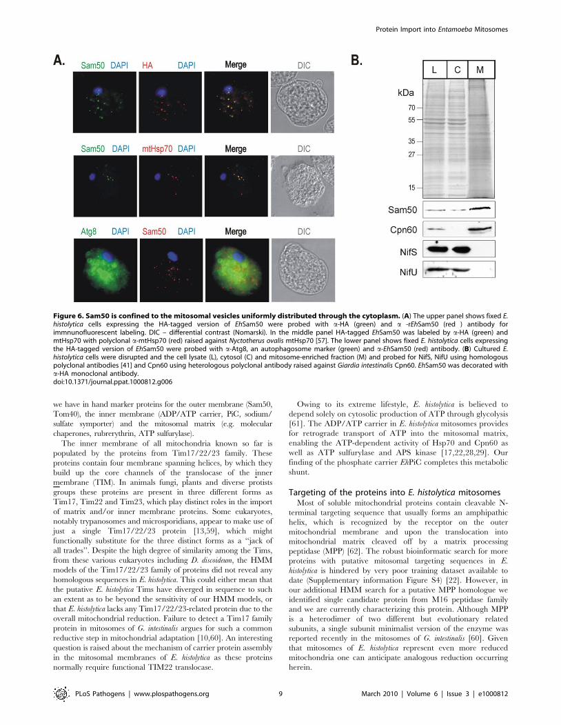
Fixed E. histolytica cells were probed with a-HA and with either a-
EhSam50, a-mtHsp70 or a-Atg8 antibody and immunofluorescent
labeling determined. Atg8 is a marker of autophagocytic vesicles,
which might be of similar morphology to mitosomes [55].
EhSam50 was found in vesicles with a size and overall pattern
similar to the cpn60-specific labeling by Leon-Avila and Tovar
[56]. The co-distribution of HA- and EhSam50- specific signal on
the merged image shows that the antibodies specifically label the
same cellular compartment (Figure 6A), distinct from the Atg8-
specific labeling of autophagosomes [55]. More importantly, using
a a-mtHsp70 raised against mitochondrial Hsp70 from Neocalli-
mastix patriciarum [57] the specific co-localization with EhSam50
was found. This data strongly supports the mitosomal distribution
of EhSam50 together with mitosomal Hsp70 [42].
To further test the mitosomal distribution of the protein import
machinery in E. histolytica, the cell fractions were probed for the
mitosomal and the cytosolic marker proteins (Figure 6B). While
our data suggested the cytosolic distribution of NifU and NifS
proteins as found in the analysis of Mi-Ichi et al. [22], EhSam50
and mitosomal Cpn60 were distributed in the high-speed pellet
fraction containing mitosomes.
Discussion
Hidden Markov models can be used for a highly sensitive search
of sequence databases [58], and discovered three new mitosomal
proteins in E. histolytica. Tom40 provides a channel for the import of
mitochondrial proteins including Sam50 and Tom40 itself, whereas
Sam50 is necessary for assembly of the Tom40 channel (and Sam50
itself). The characterization of homologs of Sam50 and Tom40 in E.
histolytica suggests that mitosomes have a functional outer membrane
as also recently shown by Maralikova et al. [21]. It is now clear,
even with so few mitosomal proteins identified in E. histolytica, that
Figure 4. A mitochondrial Tom40 in Entamoeba histolytica. (A) A representative figure from the CLANS analysis of a data set containing 79Tom40 sequences (green dots), 137 VDAC (red dots) sequences and the EhTom40 sequence (blue dot). A P-value threshold of 10282 was used to givecomplete separation of the VDAC and Tom40 clusters. Tom40 and VDAC sequences from D. discoideum, as the closest relative in the dataset, arehighlighted (orange). (B) The alignment shows the C-terminal b-strand from various Tom40 homologues. The motif for the general import signal formitochondrial b-barrel proteins is highlighted, P-polar residue, G-glycine residue, H-hydrophobic residue [48]. (C) The C-terminal GFP fusion ofEhTom40 in yeast cells (green), the yeast mitochondria stained with Mitotracker red (red). The merged image demonstrates the co-localization ofEhTom40 with the mitochondrial compartment. DIC – differential contrast (Nomarski). (D) S. cerevisiae D tom40 mutants, carrying a counterselectableTOM40 gene and expressing either S. cerevisiae Tom40 (ScTom40) or EhTom40 were plated onto 5-FOA media to select against the covering plasmid.Cells were incubated for 4 days at 30uC and only strain expressing ScTom40 was viable.doi:10.1371/journal.ppat.1000812.g004
Protein Import into Entamoeba Mitosomes
PLoS Pathogens | www.plospathogens.org 7 March 2010 | Volume 6 | Issue 3 | e1000812
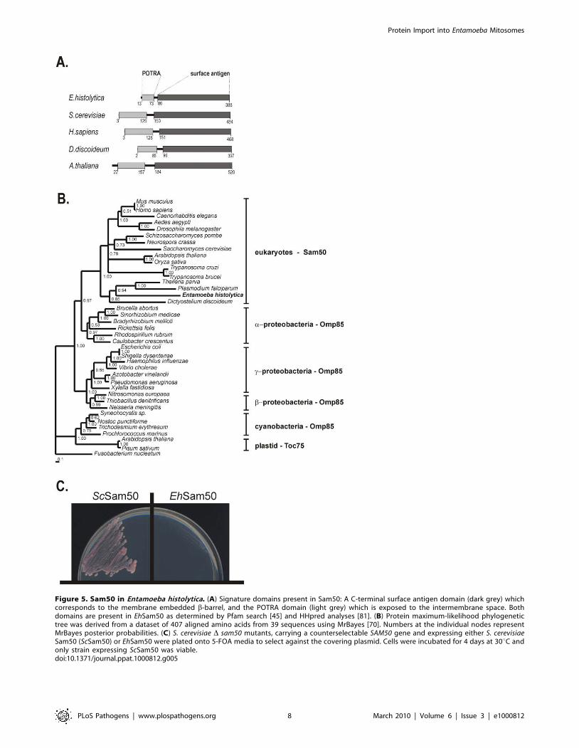
Figure 5. Sam50 in Entamoeba histolytica. (A) Signature domains present in Sam50: A C-terminal surface antigen domain (dark grey) whichcorresponds to the membrane embedded b-barrel, and the POTRA domain (light grey) which is exposed to the intermembrane space. Bothdomains are present in EhSam50 as determined by Pfam search [45] and HHpred analyses [81]. (B) Protein maximum-likelihood phylogenetictree was derived from a dataset of 407 aligned amino acids from 39 sequences using MrBayes [70]. Numbers at the individual nodes representMrBayes posterior probabilities. (C) S. cerevisiae D sam50 mutants, carrying a counterselectable SAM50 gene and expressing either S. cerevisiaeSam50 (ScSam50) or EhSam50 were plated onto 5-FOA media to select against the covering plasmid. Cells were incubated for 4 days at 30uC andonly strain expressing ScSam50 was viable.doi:10.1371/journal.ppat.1000812.g005
Protein Import into Entamoeba Mitosomes
PLoS Pathogens | www.plospathogens.org 8 March 2010 | Volume 6 | Issue 3 | e1000812
we have in hand marker proteins for the outer membrane (Sam50,
Tom40), the inner membrane (ADP/ATP carrier, PiC, sodium/
sulfate symporter) and the mitosomal matrix (e.g. molecular
chaperones, rubrerythrin, ATP sulfurylase).
The inner membrane of all mitochondria known so far is
populated by the proteins from Tim17/22/23 family. These
proteins contain four membrane spanning helices, by which they
build up the core channels of the translocase of the inner
membrane (TIM). In animals fungi, plants and diverse protists
groups these proteins are present in three different forms as
Tim17, Tim22 and Tim23, which play distinct roles in the import
of matrix and/or inner membrane proteins. Some eukaryotes,
notably trypanosomes and microsporidians, appear to make use of
just a single Tim17/22/23 protein [13,59], which might
functionally substitute for the three distinct forms as a ‘‘jack of
all trades’’. Despite the high degree of similarity among the Tims,
from these various eukaryotes including D. discoideum, the HMM
models of the Tim17/22/23 family of proteins did not reveal any
homologous sequences in E. histolytica. This could either mean that
the putative E. histolytica Tims have diverged in sequence to such
an extent as to be beyond the sensitivity of our HMM models, or
that E. histolytica lacks any Tim17/22/23-related protein due to the
overall mitochondrial reduction. Failure to detect a Tim17 family
protein in mitosomes of G. intestinalis argues for such a common
reductive step in mitochondrial adaptation [10,60]. An interesting
question is raised about the mechanism of carrier protein assembly
in the mitosomal membranes of E. histolytica as these proteins
normally require functional TIM22 translocase.
Owing to its extreme lifestyle, E. histolytica is believed to
depend solely on cytosolic production of ATP through glycolysis
[61]. The ADP/ATP carrier in E. histolytica mitosomes provides
for retrograde transport of ATP into the mitosomal matrix,
enabling the ATP-dependent activity of Hsp70 and Cpn60 as
well as ATP sulfurylase and APS kinase [17,22,28,29]. Our
finding of the phosphate carrier EhPiC completes this metabolic
shunt.
Targeting of the proteins into E. histolytica mitosomesMost of soluble mitochondrial proteins contain cleavable N-
terminal targeting sequence that usually forms an amphipathic
helix, which is recognized by the receptor on the outer
mitochondrial membrane and upon the translocation into
mitochondrial matrix cleaved off by a matrix processing
peptidase (MPP) [62]. The robust bioinformatic search for more
proteins with putative mitosomal targeting sequences in E.
histolytica is hindered by very poor training dataset available to
date (Supplementary information Figure S4) [22]. However, in
our additional HMM search for a putative MPP homologue we
identified single candidate protein from M16 peptidase family
and we are currently characterizing this protein. Although MPP
is a heterodimer of two different but evolutionary related
subunits, a single subunit minimalist version of the enzyme was
reported recently in the mitosomes of G. intestinalis [60]. Given
that mitosomes of E. histolytica represent even more reduced
mitochondria one can anticipate analogous reduction occurring
herein.
Figure 6. Sam50 is confined to the mitosomal vesicles uniformly distributed through the cytoplasm. (A) The upper panel shows fixed E.histolytica cells expressing the HA-tagged version of EhSam50 were probed with a-HA (green) and a -rEhSam50 (red ) antibody forimmnunofluorescent labeling. DIC – differential contrast (Nomarski). In the middle panel HA-tagged EhSam50 was labeled by a-HA (green) andmtHsp70 with polyclonal a-mtHsp70 (red) raised against Nyctotherus ovalis mtHsp70 [57]. The lower panel shows fixed E. histolytica cells expressingthe HA-tagged version of EhSam50 were probed with a-Atg8, an autophagosome marker (green) and a-EhSam50 (red) antibody. (B) Cultured E.histolytica cells were disrupted and the cell lysate (L), cytosol (C) and mitosome-enriched fraction (M) and probed for NifS, NifU using homologouspolyclonal antibodies [41] and Cpn60 using heterologous polyclonal antibody raised against Giardia intestinalis Cpn60. EhSam50 was decorated witha-HA monoclonal antibody.doi:10.1371/journal.ppat.1000812.g006
Protein Import into Entamoeba Mitosomes
PLoS Pathogens | www.plospathogens.org 9 March 2010 | Volume 6 | Issue 3 | e1000812
Protein import into amoebic mitochondria - a windowinto mitochondrial evolution
Phylogenetic reconstructions and the presence of flagellate cells
in the life-cycles of some Amoebozoans suggested an affiliation of
this supergroup with animals and fungi [36], so that studies on
fundamental pathways in the Amoebozoa provide important
additional information on the cell biology of the earliest animals
and fungi.
Genome sequence analysis of the highly-studied amoebozoan,
D. discoideum, revealed the presence of 14 different components
taking part in the mitochondrial protein import in animals and
fungi (see Figure 3). This analysis suggests that all four major
membrane complexes: SAM, TOM, TIM22 and TIM23 are
present in D. dictyostelium, as is the intermembrane space-located
small TIM chaperones that shuttle substrates to the SAM or
TIM22 complexes. The presence of these various complexes in D.
discoideum is consistent with them having been installed in the
earliest stages of eukaryote evolution. The components that are
missing in D. discoideum are those that appear to be specific for
fungi and animals, as has been previously discussed for receptor
subunits Tom20 [63] and Tom70 [64]. That so many TOM and
TIM components are missing from E. histolytica, is strong evidence
for a secondary gene loss having occurred, as part of the reductive
evolution impacting on the mitosomal organelle. The anaerobic
lifestyle, combined with parasitism, of E. histolytica likely selected
for a minimalist mitochondrial set up and the enormous reduction
of the import machinery. This likely reflects the even more
dramatic reduction of the overall mitochondrial metabolism. The
anaerobic, free-living amoeba Mastigamoeba balamuthi looks to have
traveled part way the same direction: 21 putative mitochondrial
proteins have been identified in the limited EST dataset, while no
TOM, TIM and SAM components were found so far [65].
The recent data on the function of E. histolytica mitosome have
revealed remarkable divergence of the processes occurring within
this mysterious mitochondrion. It now seems that while these
organelles have lost vast majority of typical mitochondrial
functions, they have accommodated several new unexpected roles
not seen in other mitochondria [21,22]. In this paper, we
uncovered the essentials of the protein import into these organelles
and it is very likely that the further research on the function and
the biogenesis of E. histolytica mitosome will bring more of the
unexpected.
Materials and Methods
Sequence search and analysisThe Hidden Markov models describing 33 mitochondrial
components were constructed and compiled into a HMM library
from manually prepared families of sequences (Oxa1, Tim10,
Tim44, Tom20opistho, Tom20plants, Tom70, Pam16, Tim13,
Tim50, Tom22opistho, Tom22plants, Hsp70, Pam18, Tim17,
Tim54, Sam35, Tim18, Tim8, Tom40, Mdm10, Sam37, Tim21,
Tim9, Tom5, Metaxin1, Sam50, Tim22, Tim9+10, Tom6,
Metaxin2, Tim23, and Tom7). The HMM library was used to
scan the genomes of E. histolytica (http://www.tigr.org/tdb/e2k1/
eha1/) and D. discoideum (NCBI). In addition, a hidden Markov
model based on 34 manually compiled MCP sequences was built
and used to scan the two genomes. The program HMMER 2.3.2
was used in all calculations [66], and the search results were
extracted with the programs prepared in-house.
The homology modeling of the mitochondrial carrier protein
was performed with SwissModel at http://swissmodel.expasy.
org//SWISS-MODEL.html [67]. The structure of bovine ATP/
ADP carrier (PDB ID 2C3E) was used as a template [30]. The
sequences were aligned using ClustalX [68] and edited manually
in BioEdit (http://www.mbio.ncsu.edu/BioEdit/bioedit.html).
For the neighbor joining analysis of mitochondrial carrier
proteins amino acid sequences were aligned and the resulting
alignment edited manually into a dataset of 33 sequences of 190
amino acid residues. SplitsTree4 [69] software was used to
calculate the bootstrapping of 500 runs and to combine the results
into a Neighbor-Net.
Protein maximum-likelihood phylogenetic tree of Omp85/
Toc75/Sam50 proteins was derived from a dataset of 407 aligned
amino acids from 39 sequences. The tree was obtained using the
program MrBayes under the JTT substitution matrix with amino
acid frequencies estimated from the dataset [70]. Site rate
variation was modeled under a discrete approximation to the Cdistribution (one invariable and four variable rate categories). Four
Monte Carlo Markov Chains, each with 2,000,000 generations,
were performed with trees sampled every 100 generations. For
compilation of Bayesian consensus topologies, a ‘‘burn-in’’ of 500
trees was used.
CLANS version 2 October 9, 2006 was obtained from http://
bioinfoserver.rsbs.anu.edu.au/programs/clans/. The EhTom40
sequence was added to a sequence set of 137 VDAC and 79
Tom40 sequences); the sequence set was derived from both the
results of BLAST and HMM searches using VDAC and Tom40
sequences. Cluster analysis was performed with a P-value cutoff
(10282) sufficient to observe complete separation of the VDAC and
Tom40 sequence clusters. As the method is non-deterministic, the
analysis was run until stable clusters formed (in excess of 200
iterations). Multiple runs were performed to ensure that the
observed clusters formed using different starting positions for the
sequences.
Entamoeba histolytica culture and preparation of RNATrophozoites of the E. histolytica isolate HM-1:IMSS were
cultured axenically in TYI-S-33 medium in plastic tissue culture
flasks [71]. For further experiments 16106 trophozoites were
cultivated for 24 h in 75 ml culture flasks. The trophozoites were
then harvested after being chilled on ice for 5 min and sedimented
at 4306g at 4uC for 5 min. The resulting pellet was washed once
in phosphate-buffered saline pH 7.2 and once in 20 mM MOPS
pH 7.2. The cell pellet was resuspended in 2 ml of 20 mM MOPS
pH 7.2 with protease inhibitors and passed 6 times through a
25 G needle until most cell were broken. The cell lysate was
diluted with 25 ml of 250 mM Sucrose, 20 mM MOPS pH 7.2
and spun down twice at 6506g for 10 min, resulting supernatant
was spun down at 2,8506g for 10 min. The final high-speed pellet
representing the mitosomal fraction was obtained after centrifu-
gation at 100,0006g for 30 min. The high-speed supernatant
corresponded to the cytosolic fraction.
For total RNA isolation 16106 E. histolytica trophozoites were
cultivated in 75 ml culture flasks for 24 h. The cells were harvested
via chilling on ice for 5 min and sedimented at 2006g for 5 min at
4uC. The cell pellet was washed twice with PBS. The trophozoites
were treated with TRIZOL reagent (Invitrogen) following the
manufacturer’s instructions. Extracted RNA was purified using the
RNeasy mini kit (Qiagen) without b-mercaptoethanol and DNA
was digested with DNase (Qiagen). cDNA synthesis was
accomplished with SuperScriptIII Reverse Transcriptase (Invitro-
gen). In a final volume of 20 ml, 1 mg of RNase-free and DNase-
treated total RNA was mixed with 56First-Strand buffer, 500 mM
dNTPs, 500 nM OdT-T71 (59-GAG AGA GGA TCC AAG TAC
TAA TAC GAC TCA CTA TAG GGA GAT24), 2 mM DTT,
40 U RnaseOut (Invitrogen) and SuperScriptIII (200 U/ml).
cDNA was synthesised for 1 h at 42uC.
Protein Import into Entamoeba Mitosomes
PLoS Pathogens | www.plospathogens.org 10 March 2010 | Volume 6 | Issue 3 | e1000812
Yeast culture and cell fractionationSaccharomyces cerevisiae strain W303a was grown in rich medium
or selective medium as previously described [72]. The Dmir1 strain
was a gift from Dr. Genevieve Dujardin (Centre de Genetique
Moleculaire, CNRS, Paris, France) [33]. For the preparation of
mitochondria for the in vitro study S. cerevisiae strain W303a was
grown in lactate media at 25uC. The mitochondria were isolated
by differential centrifugation as described previously [73]. For the
growth assays the cells were grown to a mid-logarithmic phase in a
complete media, diluted into OD600 = 0.2, spotted in the series of
fivefold dilutions on the plates and incubated at 30uC for 3–6 days.
Cloning and expression of ehpic, ehtom40 and ehsam50For GFP-tagging, the open reading frames were amplified by
PCR using E. histolytica genomic DNA as template and primers
containing 59EcoRI and 39BamHI or BglII sites (see Table S1) and
cloned into p416MET25 vector [74]. To create the C-terminal
HA-tag fusions the ORFs were cloned into a pYX143 vector with
the use of 59EcoRI and 39 MluI restriction sites. For the expression
of ehsam50 in E. histolytica, the ORF was amplified from pYX143
with the C-terminal HA-tag using the 59KpnI and 39 BglII
restriction sites. The plasmid (pNC) used for transfection is a
derivative of the expression vector EhNEO/CAT. The plasmid
contains the neomycin phosphotransferase-coding sequence
flanked by 480 bp of the 59-untranslated sequence and 600 bp
of the 39-untranslated sequence of an E. histolytica actin gene.
Transfections were performed by electroporation as described
previously [75]. Drug selection started 48 h after transfection,
using 10 mg/ml of the neomycin analogue G418. Two weeks later,
the G418 concentration was increased to 50 mg/ml.
For the production of EhSam50-derived antigen the first 300 bp
of ehsam50 were amplified by PCR and ligated into pET23a vector
(Novagen) using 59NdeI and 39 XhoI restriction sites. E. coli strain
BL21 (DE3) was used to produce the recombinant protein with C-
terminal six histidine tag. The protein was purified to homogeneity
under denaturing condition (8 M urea) on NTA-nickel column
(Qiagen). For generation of polyclonal antibodies 100 mg recom-
binant EhSam50 (rEhSam50) was injected into a mouse, followed
by two further injections.
In vitro protein importMitochondria were prepared according to the method of Daum
et al [73]. For in vitro transcription the genes were amplified by
PCR with the forward primers containing the SP6 promoter
followed by a Kozak’s sequence (ehpic, ehtom40, ehsam50) or cloned
into pSP73 vector (ehAAC), which was linearized at a unique site
downstream of the gene. [76,77]. In vitro imports were assayed
according to Gabriel and Pfanner [78].
Immunofluorescence microscopy and western blotanalysis
E. histolytica cells were analyzed with a-EhSam50 antiserum,
with an antiserum raised against the E. histolytica autophagosome
marker Atg8 (autophagy related gene 8, a gift from Dr.
Tomoyoshi Nozaki, National Institute of Infectious Diseases,
Tokyo, Japan) or with a mouse monoclonal anti-HA antibody.
Cells were fixed at room temperature for 30 min in PBS
containing 3% paraformaldehyde and subsequently permeabilized
with 0.05% saponin (PBSS). Samples were incubated at room
temperature for 1 h with antisera against EhSam50 (1:250
dilution), against Atg8 (1:500 dilution) or against the HA-tag
(1:200, Roche). Secondary antibodies were Alexa-594 coupled a-
mouse, Alexa-488 coupled a-rabbit, and Alexa-594 coupled a-
mouse antibodies. Subsequently, cells were mounted on glass
slides and examined under 63006magnification. For deconvolu-
tion microscopy images of selected cells were captured with a 636oil immersion lens in a UV equipped Leica DM RB microscope
with 0.2-mm-diameter step Z-sections. Deconvoluted Z sections
were examined for colocalisation of Atg8 and EhSam50 staining
with the Openlab 4.0.4 program. Adobe Photoshop 7.0.5 was
used for additional processing of the images. S. cerevisiae were
analyzed as previously described (Beilharz et al. 2003). ImageJ
software was used for additional image processing (http://rsbweb.
nih.gov/ij/). In the western blot analysis cell fractions were
probed with the mouse monoclonal anti-HA antibody (1:500),
rabbit polyclonal antibodies raised against Giardia intestinalis
Cpn60 (1:1000) (a kind gift from Dr. Robert Hirt, Newcastle
University, UK), E. histolytica NifS and NifU (both 1:1000) (a kind
gift from Dr.Tomoyoshi Nozaki [26]).
The distribution of EhPiC, EhTom40 and EhSam50 in S.
cerevisiae mitochondria extracted in fresh 100mM Na2CO3 was
done as previously described [79].
Supporting Information
Figure S1 Neighbor joining tree of 31 mitochondrial carrier
proteins from D. discoideum with ADP/ATP carrier and PiC carrier
from E. histolytica constructed by SplitsTree4 [86]. The carriers
cluster according to their substrate specificity. The putative
substrates of D. discoideum carriers [87] are indicated as follows:
Carn/Orn - carnitine or ornithine, Asp/Glu - aspartate/glutamate,
Oglu - 2-oxoglutarate, Dic/Tric - dicarboxylate/tricarboxylate, Pi -
phosphate, PyrNucl - pyrimidine NTP/NMP, perox ATP -
peroxisomal ATP carrier, H+ FA - H+ fatty acid, CoA - coenzyme
A, dNucl - deoxynucleotide, AA-Mn - amino acid (Mn2+), Fe - iron
(mitoferrin).
Found at: doi:10.1371/journal.ppat.1000812.s001 (0.12 MB PDF)
Figure S2 The products of specific RT are shown: 1 - ehpic, 2 -
ehtom40, 3 - ehsam50. The marker is in base pairs.
Found at: doi:10.1371/journal.ppat.1000812.s002 (0.05 MB PDF)
Figure S3 35S-labelled EhTom40 was incubated with yeast
mitochondria, solubilized with 1% digitonin and the samples
resolved on BN-PAGE. The time-dependent formation of high-
molecular weight complexes in the mitochondrial membranes is
demonstrated. The large white arrow points to disappearance of
the monomeric form of EhTom40 in the reaction, small black
arrows highlight the formations of the of high-molecular weight
complexes.
Found at: doi:10.1371/journal.ppat.1000812.s003 (0.05 MB PDF)
Figure S4 The protein sequence alignments of all three known
soluble mitosomal matrix proteins Cpn10, Cpn60, Hsp70 with
their bacterial and eukaryotic counterparts. The presence of
extremely short N-terminal targeting sequence does not allow for
creation of a prediction algorithm.
Found at: doi:10.1371/journal.ppat.1000812.s004 (0.07 MB PDF)
TableS1 Primers used in the study.
Found at: doi:10.1371/journal.ppat.1000812.s005 (0.03 MB PDF)
Acknowledgments
We thank Graham Clark (London School of Hygiene and Tropical
Medicine, UK) for E. histolytica genomic DNA, Genevieve Dujardin (Centre
de Genetique Moleculaire, CNRS, Paris, France) for a Dmir1 S. cerevisiae
strain, Tomoyoshi Nosaki (National Institute of Infectious Diseases, Tokyo,
Japan) for a-Atg8, a-NifS and a-NifU antibodies, and Robert Hirt
(Newcastle University, UK) for a-Cpn60 antibody.
Protein Import into Entamoeba Mitosomes
PLoS Pathogens | www.plospathogens.org 11 March 2010 | Volume 6 | Issue 3 | e1000812
Author Contributions
Conceived and designed the experiments: PD MJD MK IB TL. Performed
the experiments: PD MJD MK VAL JHF MS AB IB. Analyzed the data:
PD MJD MK PW VAL TL. Contributed reagents/materials/analysis
tools: PD JT. Wrote the paper: PD TL.
References
1. Embley TM, Martin W (2006) Eukaryotic evolution, changes and challenges.
Nature 440: 623–630.
2. Cavalier-Smith T (1989) Molecular phylogeny. Archaebacteria and Archezoa.
Nature 339: l00–l01.
3. Akhmanova A, Voncken F, van Alen T, van HA, Boxma B, et al. (1998) A
hydrogenosome with a genome. Nature 396: 527–528.
4. Hrdy I, Hirt RP, Dolezal P, Bardonova L, Foster PG, et al. (2004) Trichomonas
hydrogenosomes contain the NADH dehydrogenase module of mitochondrialcomplex I. Nature 432: 618–622.
5. Dyall SD, Yan W, Delgadillo-Correa MG, Lunceford A, Loo JA, et al. (2004)Non-mitochondrial complex I proteins in a hydrogenosomal oxidoreductase
complex. Nature 431: 1103–1107.
6. van derGiezen (2009) Hydrogenosomes and mitosomes: conservation and
evolution of functions. J Eukaryot Microbiol 56: 221–231.
7. Mokranjac D, Neupert W (2005) Protein import into mitochondria. Biochem
Soc Trans 33: 1019–1023.
8. Kutik S, Guiard B, Meyer HE, Wiedemann N, Pfanner N (2007) Cooperation of
translocase complexes in mitochondrial protein import. J Cell Biol 179:
585–591.
9. Dolezal P, Likic V, Tachezy J, Lithgow T (2006) Evolution of the molecular
machines for protein import into mitochondria. Science 313: 314–318.
10. Dolezal P, Smid O, Rada P, Zubacova Z, Bursac D, et al. (2005) Giardia
mitosomes and trichomonad hydrogenosomes share a common mode of proteintargeting. Proc Natl Acad Sci U S A 102: 10924–10929.
11. Regoes A, Zourmpanou D, Leon-Avila G, van derGiezen, Tovar J, et al. (2005)Protein import, replication, and inheritance of a vestigial mitochondrion. J Biol
Chem 280: 30557–30563.
12. Burri L, Williams BA, Bursac D, Lithgow T, Keeling PJ (2006) Microsporidian
mitosomes retain elements of the general mitochondrial targeting system. ProcNatl Acad Sci U S A 103: 15916–15920.
13. Burri L, Keeling PJ (2007) Protein targeting in parasites with crypticmitochondria. Int J Parasitol 37: 265–272.
14. Tovar J, Leon-Avila G, Sanchez LB, Sutak R, Tachezy J, et al. (2003)Mitochondrial remnant organelles of Giardia function in iron-sulphur protein
maturation. Nature 426: 172–176.
15. Tsaousis AD, Kunji ER, Goldberg AV, Lucocq JM, Hirt RP, et al. (2008) A
novel route for ATP acquisition by the remnant mitochondria of Encephalitozoon
cuniculi. Nature 453: 553–556.
16. Williams BA, Hirt RP, Lucocq JM, Embley TM (2002) A mitochondrial
remnant in the microsporidian Trachipleistophora hominis. Nature 418: 865–869.
17. Tovar J, Fischer A, Clark CG (1999) The mitosome, a novel organelle related to
mitochondria in the amitochondrial parasite Entamoeba histolytica. Mol Microbiol32: 1013–1021.
18. Mai Z, Ghosh S, Frisardi M, Rosenthal B, Rogers R, et al. (1999) Hsp60 istargeted to a cryptic mitochondrion-derived organelle (‘‘crypton’’) in the
microaerophilic protozoan parasite Entamoeba histolytica. Mol Cell Biol 19:2198–2205.
19. Goldberg AV, Molik S, Tsaousis AD, Neumann K, Kuhnke G, et al. (2008)Localization and functionality of microsporidian iron-sulphur cluster assembly
proteins. Nature 452: 624–628.
20. Katinka MD, Duprat S, Cornillot E, Metenier G, Thomarat F, et al. (2001)
Genome sequence and gene compaction of the eukaryote parasite Encephalitozoon
cuniculi. Nature 414: 450–453.
21. Maralikova B, Ali V, Nakada-Tsukui K, Nozaki T, van derGiezen, et al. (2009)Bacterial-type oxygen detoxification and iron-sulfur cluster assembly in amoebal
relict mitochondria. Cell Microbiol.
22. Mi-Ichi F, Yousuf MA, Nakada-Tsukui K, Nozaki T (2009) Mitosomes in
Entamoeba histolytica contain a sulfate activation pathway. Proc Natl Acad
Sci U S A.
23. Lill R, Muhlenhoff U (2006) Iron-sulfur protein biogenesis in eukaryotes:
components and mechanisms. Annu Rev Cell Dev Biol 22: 457–486.
24. Clark CG (2000) The evolution of Entamoeba, a cautionary tale. Res Microbiol
151: 599–603.
25. van der Giezen M, Cox S, Tovar J (2004) The iron-sulfur cluster assembly genes
iscS and iscU of Entamoeba histolytica were acquired by horizontal gene transfer.BMC Evol Biol 4: 7.
26. Ali V, Shigeta Y, Tokumoto U, Takahashi Y, Nozaki T (2004) An intestinalparasitic protist, Entamoeba histolytica, possesses a non-redundant nitrogen
fixation-like system for iron-sulfur cluster assembly under anaerobic conditions.J Biol Chem 279: 16863–16874.
27. Chan KW, Slotboom DJ, Cox S, Embley TM, Fabre O, et al. (2005) A novelADP/ATP transporter in the mitosome of the microaerophilic human parasite
Entamoeba histolytica. Curr Biol 15: 737–742.
28. Tovar J, Cox SS, van der Giezen M (2007) A mitosome purification protocol
based on percoll density gradients and its use in validating the mitosomal nature
of Entamoeba histolytica mitochondrial Hsp70. Methods Mol Biol 390: 167–177.
29. van der Giezen M, Leon-Avila G, Tovar J (2005) Characterization of
chaperonin 10 (Cpn10) from the intestinal human pathogen Entamoeba histolytica.Microbiology 151: 3107–3115.
30. Pebay-Peyroula E, Dahout-Gonzalez C, Kahn R, Trezeguet V, Lauquin GJ,et al. (2003) Structure of mitochondrial ADP/ATP carrier in complex with
carboxyatractyloside. Nature 426: 39–44.
31. Satre M, Mattei S, Aubry L, Gaudet P, Pelosi L, et al. (2007) Mitochondrial
carrier family: repertoire and peculiarities of the cellular slime mould Dictyostelium
discoideum. Biochimie 89: 1058–1069.
32. Kunji ER, Robinson AJ (2006) The conserved substrate binding site of
mitochondrial carriers. Biochim Biophys Acta 1757: 1237–1248.
33. Hamel P, Saint-Georges Y, de Pinto B, Lachacinski N, Altamura N, et al. (2004)
Redundancy in the function of mitochondrial phosphate transport inSaccharomyces cerevisiae and Arabidopsis thaliana. Mol Microbiol 51: 307–317.
34. Murakami H, Blobel G, Pain D (1990) Isolation and characterization of the genefor a yeast mitochondrial import receptor. Nature 347: 488–491.
35. Simpson AG, Roger AJ (2002) Eukaryotic evolution: getting to the root of theproblem. Curr Biol 12: R691–R693.
36. Nikolaev SI, Berney C, Petrov NB, Mylnikov AP, Fahrni JF, et al. (2006)Phylogenetic position of Multicilia marina and the evolution of Amoebozoa.
Int J Syst Evol Microbiol 56: 1449–1458.
37. Eichinger L, Pachebat JA, Glockner G, Rajandream MA, Sucgang R, et al.
(2005) The genome of the social amoeba Dictyostelium discoideum. Nature 435:43–57.
38. Macasev D, Whelan J, Newbigin E, Silva-Filho MC, Mulhern TD, et al. (2004)Tom229, an 8-kDa trans-site receptor in plants and protozoans, is a conserved
feature of the TOM complex that appeared early in the evolution of eukaryotes.
Mol Biol Evol 21: 1557–1564.
39. Barth C, Le P, Fisher PR (2007) Mitochondrial biology and disease in
Dictyostelium. Int Rev Cytol 263: 207–252.
40. Nagayama K, Itono S, Yoshida T, Ishiguro S, Ochiai H, et al. (2008) Antisense
RNA inhibition of the beta subunit of the Dictyostelium discoideum mitochondrialprocessing peptidase induces the expression of mitochondrial proteins. Biosci
Biotechnol Biochem 72: 1836–1846.
41. Loftus B, Anderson I, Davies R, Alsmark UC, Samuelson J, et al. (2005) The
genome of the protist parasite Entamoeba histolytica. Nature 433: 865–868.
42. Bakatselou C, Kidgell C, Graham CC (2000) A mitochondrial-type hsp70 gene
of Entamoeba histolytica. Mol Biochem Parasitol 110: 177–182.
43. Waller RF, Jabbour C, Chan NC, Celik N, Likic VA, et al. (2009) Evidence of a
reduced and modified mitochondrial protein import apparatus in microsporid-ian mitosomes. Eukaryot Cell 8: 19–26.
44. Pusnik M, Charriere F, Maser P, Waller RF, Dagley MJ, et al. (2009) The singlemitochondrial porin of Trypanosoma brucei is the main metabolite transporter in
the outer mitochondrial membrane. Mol Biol Evol 26: 671–680.
45. Finn RD, Tate J, Mistry J, Coggill PC, Sammut SJ, et al. (2008) The Pfam
protein families database. Nucleic Acids Res 36: D281–D288.
46. Frickey T, Lupas A (2004) CLANS: a Java application for visualizing protein
families based on pairwise similarity. Bioinformatics 20: 3702–3704.
47. Dagley MJ, Dolezal P, Likic VA, Smid O, Purcell AW, et al. (2009) The protein
import channel in the outer mitosomal membrane of Giardia intestinalis. Mol BiolEvol 26: 1941–1947.
48. Kutik S, Stojanovski D, Becker L, Becker T, Meinecke M, et al. (2008)Dissecting membrane insertion of mitochondrial beta-barrel proteins. Cell 132:
1011–1024.
49. Gabriel K, Egan B, Lithgow T (2003) Tom40, the import channel of the
mitochondrial outer membrane, plays an active role in sorting imported
proteins. EMBO J 22: 2380–2386.
50. Wilihoeft U, Campos-Gongora E, Touzni S, Bruchhaus I, Tannich E (2001)
Introns of Entamoeba histolytica and Entamoeba dispar. Protist 152: 149–156.
51. Gentle IE, Burri L, Lithgow T (2005) Molecular architecture and function of the
Omp85 family of proteins. Mol Microbiol 58: 1216–1225.
52. Voulhoux R, Tommassen J (2004) Omp85, an evolutionarily conserved bacterial
protein involved in outer-membrane-protein assembly. Res Microbiol 155:129–135.
53. Gentle I, Gabriel K, Beech P, Waller R, Lithgow T (2004) The Omp85 family ofproteins is essential for outer membrane biogenesis in mitochondria and
bacteria. J Cell Biol 164: 19–24.
54. Sanchez-Pulido L, Devos D, Genevrois S, Vicente M, Valencia A (2003)
POTRA: a conserved domain in the FtsQ family and a class of beta-barrel outermembrane proteins. Trends Biochem Sci 28: 523–526.
55. Picazarri K, Nakada-Tsukui K, Nozaki T (2008) Autophagy during proliferationand encystation in the protozoan parasite Entamoeba invadens. Infect Immun 76:
278–288.
56. Leon-Avila G, Tovar J (2004) Mitosomes of Entamoeba histolytica are abundant
mitochondrion-related remnant organelles that lack a detectable organellar
genome. Microbiology 150: 1245–1250.
Protein Import into Entamoeba Mitosomes
PLoS Pathogens | www.plospathogens.org 12 March 2010 | Volume 6 | Issue 3 | e1000812
57. van derGiezen, Birdsey GM, Horner DS, Lucocq J, Dyal PL, et al. (2003)
Fungal hydrogenosomes contain mitochondrial heat-shock proteins. Mol Biol
Evol 20: 1051–1061.
58. Eddy SR (1996) Hidden Markov models. Curr Opin Struct Biol 6: 361–365.
59. Schneider A, Bursac D, Lithgow T (2008) The direct route: a simplified pathway
for protein import into the mitochondrion of trypanosomes. Trends Cell Biol 18:
12–18.
60. Smid O, Matuskova A, Harris SR, Kucera T, Novotny M, et al. (2008)
Reductive evolution of the mitochondrial processing peptidases of the unicellular
parasites trichomonas vaginalis and Giardia intestinalis. PLoS Pathog 4: e1000243.
doi:10.1371/journal.ppat.1000243.
61. Saavedra E, Encalada R, Pineda E, Jasso-Chavez R, Moreno-Sanchez R (2005)
Glycolysis in Entamoeba histolytica. Biochemical characterization of recombinant
glycolytic enzymes and flux control analysis. FEBS J 272: 1767–1783.
62. Chacinska A, Koehler CM, Milenkovic D, Lithgow T, Pfanner N (2009)
Importing mitochondrial proteins: machineries and mechanisms. Cell 138:
628–644.
63. Likic VA, Perry A, Hulett J, Derby M, Traven A, et al. (2005) Patterns that
define the four domains conserved in known and novel isoforms of the protein
import receptor Tom20. J Mol Biol 347: 81–93.
64. Chan NC, Likic VA, Waller RF, Mulhern TD, Lithgow T (2006) The C-
terminal TPR domain of Tom70 defines a family of mitochondrial protein
import receptors found only in animals and fungi. J Mol Biol 358: 1010–1022.
65. Gill EE, az-Trivino S, Barbera MJ, Silberman JD, Stechmann A, et al. (2007)
Novel mitochondrion-related organelles in the anaerobic amoeba Mastigamoeba
balamuthi. Mol Microbiol 66: 1306–1320.
66. Eddy SR (1998) Profile hidden Markov models. Bioinformatics 14: 755–763.
67. Arnold K, Bordoli L, Kopp J, Schwede T (2006) The SWISS-MODEL
workspace: a web-based environment for protein structure homology modelling.
Bioinformatics 22: 195–201.
68. Thompson JD, Gibson TJ, Plewniak F, Jeanmougin F, Higgins DG (1997) The
CLUSTAL_X windows interface: flexible strategies for multiple sequence
alignment aided by quality analysis tools. Nucleic Acids Res 25: 4876–4882.
69. Huson DH, Bryant D (2006) Application of phylogenetic networks in
evolutionary studies. Mol Biol Evol 23: 254–267.70. Huelsenbeck JP, Ronquist F (2001) MRBAYES: Bayesian inference of
phylogenetic trees. Bioinformatics 17: 754–755.
71. Diamond LS, Harlow DR, Cunnick CC (1978) A new medium for the axeniccultivation of Entamoeba histolytica and other Entamoeba. Trans R Soc Trop Med
Hyg 72: 431–432.72. Lithgow T, Junne T, Wachter C, Schatz G (1994) Yeast mitochondria lacking
the two import receptors Mas20p and Mas70p can efficiently and specifically
import precursor proteins. J Biol Chem 269: 15325–15330.73. Daum G, Bohni PC, Schatz G (1982) Import of proteins into mitochondria.
Cytochrome b2 and cytochrome c peroxidase are located in the intermembranespace of yeast mitochondria. J Biol Chem 257: 13028–13033.
74. Beilharz T, Egan B, Silver PA, Hofmann K, Lithgow T (2003) Bipartite signalsmediate subcellular targeting of tail-anchored membrane proteins in Saccharo-
myces cerevisiae. J Biol Chem 278: 8219–8223.
75. Hamann L, Nickel R, Tannich E (1995) Transfection and continuous expressionof heterologous genes in the protozoan parasite Entamoeba histolytica. Proc Natl
Acad Sci U S A 92: 8975–8979.76. Nijtmans LG, Henderson NS, Holt IJ (2002) Blue Native electrophoresis to study
mitochondrial and other protein complexes. Methods 26: 327–334.
77. Wittig I, Braun HP, Schagger H (2006) Blue native PAGE. Nat Protoc 1:418–428.
78. Gabriel K, Pfanner N (2007) The mitochondrial machinery for import ofprecursor proteins. Methods Mol Biol 390: 99–117.
79. Youker RT, Walsh P, Beilharz T, Lithgow T, Brodsky JL (2004) Distinct rolesfor the Hsp40 and Hsp90 molecular chaperones during cystic fibrosis
transmembrane conductance regulator degradation in yeast. Mol Biol Cell 15:
4787–4797.80. Guindon S, Gascuel O (2003) A simple, fast, and accurate algorithm to estimate
large phylogenies by maximum likelihood. Syst Biol 52: 696–704.81. Soding J, Biegert A, Lupas AN (2005) The HHpred interactive server for protein
homology detection and structure prediction. Nucleic Acids Res 33:
W244–W248.
Protein Import into Entamoeba Mitosomes
PLoS Pathogens | www.plospathogens.org 13 March 2010 | Volume 6 | Issue 3 | e1000812