Embed Size (px)
Citation preview

University of Montana University of Montana
ScholarWorks at University of Montana ScholarWorks at University of Montana
Graduate Student Theses, Dissertations, & Professional Papers Graduate School
1960
A Study of the enzymes of Entamoeba histolytica A Study of the enzymes of Entamoeba histolytica
Philip R. Edwards The University of Montana
Follow this and additional works at: https://scholarworks.umt.edu/etd
Let us know how access to this document benefits you.
Recommended Citation Recommended Citation Edwards, Philip R., "A Study of the enzymes of Entamoeba histolytica" (1960). Graduate Student Theses, Dissertations, & Professional Papers. 4656. https://scholarworks.umt.edu/etd/4656
This Thesis is brought to you for free and open access by the Graduate School at ScholarWorks at University of Montana. It has been accepted for inclusion in Graduate Student Theses, Dissertations, & Professional Papers by an authorized administrator of ScholarWorks at University of Montana. For more information, please contact [email protected].

A STUDY OF THE ENZYMES
OFENTAMOEBA. HISTOLYTICA
PHILIP ROBINSON EDWARDS. JR.
A.B., Montana State University, 1958
Presented in partial fulfillment of the requirementsfor the Degree of Master of Science
I960
Approved by:
Chairman, Board of Examiners
Dean, Graduate School
MAR 7 I960Date

UMI Number: EP40120
All rights reserved
INFORMATION TO ALL USERS The quality of this reproduction is dependent upon the quality of the copy submitted.
In the unlikely event that the author did not send a complete manuscript and there are missing pages, these will be noted. Also, if material had to be removed,
a note will indicate the deletion.
UMIDissertation Publishing
UMI EP40120
Published by ProQuest LLC (2014). Copyright in the Dissertation held by the Author.
Microform Edition © ProQuest LLC.All rights reserved. This work is protected against
unauthorized copying under Title 17, United States Code
ProQuest’ProQuest LLC.
789 East Eisenhower Parkway P.O. Box 1346
Ann Arbor, Ml 48106- 1346

ACKNOWLEDGEMENTS
The author wishes to express his appreciation to Dro Mitsuru Nakamura, Associate Professor of Microbiology and Public Health, Montana State University, for his advice and guidance throughout the course of this research. Appreciation is also due Dr. J n J. Taylor, Dr. R. A.Faust, and Dr. R. Lo Anacker of the Department of Microbiology and Public Health, and to Dr. Co M . Senger of the Department of Zoology for assistance and advice from time to time during this investigation.
A major portion of this investigation was sponsored by the Commission on Enteric Infections, Armed Forces Epidemiological Board, Department of Defense and supported by the Office of Surgeon General, Department of the Army, Contract No. DA-*+9~007-MD-891o Additional support for some phases of this research was received from research grants to Drc Nakamura from the Brown-Hazen Fund of the Research Corporation, New York and the Baltimore Biological Laboratories, Baltimore, Maryland0
P. Ro E.

TABLE OF CONTENTS
PAGEAcknowledgements o « » . . . „ . . . . . . . . . . . . . . iiList of Tables o . . . . . . . . . . . . . . . . . . . . ivList of Figures . . . . . . . . . . . . . . . . . . . . viCHAPTER
I. INTRODUCTION. .. . . . . . . . . . . . . . . . . 1II. STATEMENT OF THE PROBLEM. . . . . . . . . . . . . 11
III. METHODS AND MATERIALS . . . . . . . . . . . . . . 12IV. RESULTS . . . . . . . . . . . . . . . . . . . . . 35V. DISCUSSION AND CONCLUSIONS. . . . . . . . . . . . 61
VI. SUMMARY o . . . o o . . . . . . . . . . . . . . „ 70BIBLIOGRAPHY*-. . * . * . * * * . * * . * . * . . . * . 71AUTOBIOGRAPHY . * . . . * * . . . * . . * * * * . . . . 78
iii

LIST OF TABLES
T A B L E
To
lieIII.
I V o
V o
V I o
VIIo VIII c
I X o
X.XI.
X I I o
XIIIo
X I V .
Data from Literature on Enzymes of Entamoeba histolytica 0 . . . . . . . . . . . . . . .
Composition of Buffer Solutions. . . . . . .Results of Catalase Activity . . . . . . . .Gelatinase Activity of E. histolytlca. . . .Effect of pH on the Gelatina.se Activity of
1° histolytica (HUS-105) . . . . . . . . .Results of Lecithinase Assay . . „ . . . . .Nuclease Activity of L histolytica. . . . .Sensitivity of the Urease Method Using
Purified Enzyme Preparations . . . . . . .Results of the Assay for Urease in E.
histolytica. . . . . . . . . . . . . . . .Results of the Citrullinase Assay. . . . . .Results of the Asparaginase Assay. . „ . • .A Survey of Bacteria for Glucuronidase
Activity , . c . . . . . . . . . . . . . .Sensitivity of the Glucuronidase Method
Using an Unidentified Gram Positive Streptococcus O C O O . O . . . . . . . . . . .
Assay for Glucuronidase in E. histolytica. .

V
TABLE PAGE
XV o Sensitivity of the Galactosidase Method UsingEscherichia coli. as the Test Organism. 0 . «, „ 56
XVIo Results of the Galactosidase Assay * . . . . . „ 58XVII o Results of the Succinic Dehydrogenase Assay«, . . 60

LIST OF FIGURES
FIGURE PAGEI o Aeration Train0 o 0 o 0 » . . <> • • © • • © o o o o 262. Gelatinase Microtesto c . . . . • • « « o © ® a 393 • Nucleic Acid Test Plate « » . . hk
Sensitivity of the Glucuronidase Method o ■ o . . o o su
5. Sensitivity of the Galactosidase Method o • . . o o cr*7y i
vi

CHAPTER I
INTRODUCTION
Relatively little is known about the metabolism of
Entamoeba histolytica, the etiological agent of amoebiasis *
Studies dealing with the physiology of this protozoan
parasite have been hindered by the inability to cultivate
this organism in axenie culture „ Metabolic studies are
difficult to perform in biological systems containing more
than one living species„ However* with adequately con
trolled experiments vital information can be obtained which
may directly or indirectly lead to the cultivation of the
amoebae in pure culture (free of other living organisms)„
Studies on the enzymology of L histolytica will shed in
formation on the metabolism of this organism,. Knowledge of
the enzymes and metabolic processes of the amoebae could
lead to a rational approach to the chemotherapy of amoe
biasis and possibly to the elucidation of the mechanism of
pathogenicityo
This approach to the physiology of E„ histolytica
has remained largely unexplored (50)9 mainly because of
the difficulties encountered in ruling out the biochemical
contributions of the bacterial associates (M+) 0 Many
workers (8, 21, V2, 50) feel that it is possible to obtain
preparations of amoebae relatively free of the associated
1

2
bacterial flora by repeated washing procedures; in such preparations the bacteria are present in such small numbers that they contribute little or nothing to the observed amoebic activity.
A number of workers have reported on the existence of certain enzymes in E» histolytica in vitro. Anderson and Hansen (1) and Reardon and Bartgis (12) reported that the amoebae were capable of hydrolyzing the gluten surrounding rice powder granules. A hemosiderin mass was observed in amoebae and this was thought to be due to residue from the dissolution of hemoglobin (1). Rees and co-workers found gelatinase in E. histolytica (6*+) „ They inoculated a gelatin medium with amoebae growing in association with bacteria unable to hydrolyze gelatin and obtained liquefaction,, Harinasuta and Maegraith (3*+, 35) reported that a washed culture of E. histolytica destroyed the gelatin on an exposed and developed photographic film when incubated at 37° C for two hours. Neal in 1956 (57) reported that extracts of washed amoebae were capable of hydrolyzing both gelatin and casein., Nakamura (53)* in a preliminary report, could not detect any casein-hydrolyzing activity in the amoebae using a plate method. Nakamura and Klein (55) found that actively growing cultures of E. histolytica did not produce collagenase; the assay system used was the rat-tail tendon dissolution technique. Bradin showed that amoebae isolated from hamster liver abscesses

3produced hyaiuronidase, but did not do so in cultures (11). DeLamater and co-workers (1.6) were unable to detect intracellular or extracellular hyaiuronidase production from amoebae in culture , nor could they detect hyaiuronidase activity in preparations of washed amoebae obtained from hamster liver abscesses 0 Carrera and Changus (13) demonstrated histochemically that there was an acid phosphatase in the amoebae, and Carrera (1^) also showed that tissues surrounding the invading amoebae in the intestinal lesions were high in acid phosphatase activity. The presence of acid phosphatase in the amoebae was also reported by Balamuth (6)o Hara and co-workers (33)9 utilizing a histochemical technique, found an alkaline phosphatase in L histolytica„ Blumenthal and co-workers (9) confirmed the presence of both acid phosphatase and alkaline phosphatase in the amoebae. These workers used p-nitro-phenyl-phosphate, a chro- mogenic substance, and measured the amount of color produced due to the hydrolysis of the chromogen-phosphate linkage.
The first and only report of any aspect of the amino acid metabolism of E. histolytica was published in 1957- Nakamura and Goldstein (56) were able to demonstrate a phosphate activated glutaminase in washed preparations of amoebaeo Succinic dehydrogenase was shown to exist in the amoebae by Seaman in 1953 (68)0 He stated that the enzyme of the amoebae differed from the mammalian counterpart in that it was inhibited by certain arsono and phosphono

analogues of succinic acid0 The existence of an esterase in Eo histolytica was shown by Hallman et al, (31)„ They titrated the acid produced from, methyl-n-butyrate by washed, lysed cells of E. histolytica 0 The only report on the presence of an oxidase-type enzyme in the amoebae was presented by Hara and his group in 195*+ (33)° They were able to demonstrate cytochemically a strong peroxidase reaction in this protozoan.
Other observations dealing with the enzymology and metabolism of the amoebae have been primarily concerned with the carbohydrate metabolism of the organisirio Amylase production has been noted by many workers (3, 29* 38).Hilker and co-workers (38) found a maltase, but no lactase nor sucrase in E. histolytica <, In 19*+3, von Brand (50)
demonstrated that the amoebae did not produce gas from glucose o Hallman et al. (30) and Loran and co-workers (*+5) also have reported that the amoebae do not directly utilize glucose. However, others have reported that the amoebae do utilize glucose (8, 22, *+2, *+9* 51)» Kun and Bra din (*+2)
presented evidence for the utilization of nine sugars by washed preparations of the amoeba, and in addition showed that Eo histolytica produced H2S from sulfur-containing compounds such as cysteineo
Hilker and White (39), utilizing disrupted cell preparations of washed amoebae, claimed to have demonstrated the presence of phosphoglucomutase, phosphohexokinase,

5'
aldolase, and glucose-6-phosphate dehydrogenase. They could
not, however, demonstrate glyceraldehyde-3-phosphate dehydrogenase or 6-phosphogluconate dehydrogenase* From these observations they concluded that the hexosemonophosphate shunt did not exist in L histolytica, and that the Entner- Doudoroff pathway does exist in the amoebae (39)® Recently, Entner (23) has confirmed the work of Hilker and White (39) with regard to the existence of the Fntner-Doudoroff pathway in Eo histolytica„
The enzymes reported to be present in Eo histolytica are listed in Table I* Included also are some reactions that could be due to enzymes or enzyme systems0 It can be seen that the metabolic studies on Eo histolytica have been very meager and little progress has been made compared to the advances in the metabolism of other microorganisms» One of the main reasons for this is the lack of a pure culture of the amoebaeo The technical difficulties so often encountered in working.with E. histolytica might also be a reason for the lack of progress (4-2, M+) „ This would account for the comparatively crude methods so far used in the studies of the enzymes of Eo histolytica.
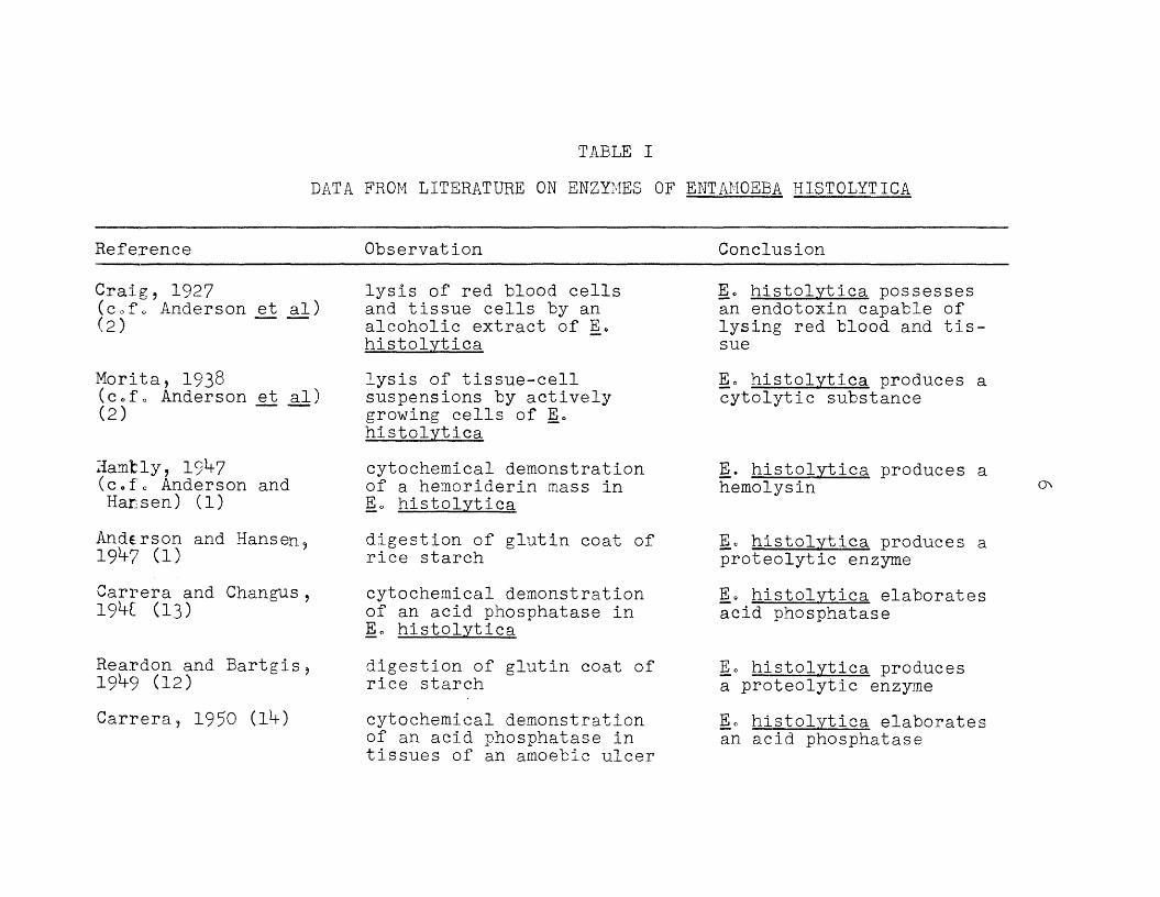
TABLE I
DATA FROM LITERATURE ON ENZYMES OF ENTAMOEBA HISTOLYTICA
Reference Observation Conclusion
Craig, 1927 (c.f. Anderson et al) (2)
Morita, 1938 (c.f. Anderson et al) (2)
Hamfcly, I9V 7 (c.f. Anderson and Hansen) (1)
Anderson and Hansen, 19V7 (1)Carrera and Changus, 1 9 W (13)
Reardon and Bartgis,19^9 (12)Carrera, 1950 (]_•+)
lysis of red blood cells and tissue cells by an alcoholic extract of E, histolyticalysis of tissue-cell suspensions by actively growing cells of E 0 histolyticacytochemical demonstration of a hemoriderin mass in Eo histolyticadigestion of glutin coat of rice starchcytochemical demonstration of an acid phosphatase in Eo histolyticadigestion of glutin coat of rice starchcytochemical demonstration of an acid phosphatase in tissues of an amoebic ulcer
E« histolytica possesses an endotoxin capable of lysing red blood and tissueEo histolytica produces a cytolytic substance
E. histolytica produces a hemolysin
Ee histolytica produces a proteolytic enzyme1° histolytica elaborates acid phosphatase
Eo histolytica produces a proteolytic enzymeEo histolytica elaboratesan acid phosphatase
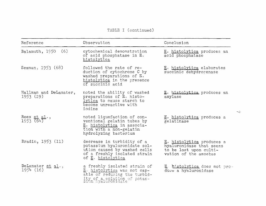
TABLE I (continued)
Reference Observation
Balamuth. 1950 (6)
Seaman? 1953 (68)
cytochemical demonstration of acid phosphatase in Eo histolyticafollowed the rate of reduction of cytochrome C by washed preparations of E 0 histolytica in the presence of succinic acid
Hallman and DeLamater,1953 (29)
noted the ability of washed preparations of L histo- lytica to cause starch to become unreactive with iodine
Rees et al«^1953 (6*0
Bradin, 1953 (11)
DeLamater et al195^ (16)
noted liquefaction of conventional gelatin tubes by 1° histolytica in association with a non-gelatin hydrolyzing bacteriumdecrease in turbidity of a potassium hyaluronidate solution caused by washed cells of a freshly isolated strain of Eo histolyticaa freshly isolated strain of Eo histolytica was not capable of reducing the turbidity of absolution of potassium hyaluronidate
Conclusion
E* histolytica produces an acid phosphatase
Eo histolytica elaborates succinic dehydrogenase
EU histolytica produces an amylase
E e histolytica produces a gelatinase
Eo histolytica produces a hyaiuronidase that seems to be lost upon cultivation of the amoebae
E_ histolytica does not produce a hyaiuronidase

TABLE I (continued)
Reference
Hara et, al o , 195^ (33)
Baernstein et ale,1955- (3)
Harinasuta and Maegraith, 195*+ (3*+)
Blumenthal eL a L , 1955 (9)
Hallman et al., 1955 ( 3 l T
Neal, 1956 (57)
Observation
histochemical demonstration of peroxidase and alkaline phosphatase in Ec histolyticabreakdown of starch to reducing sugars by acetone powders of washed E. histolyticaextracts of E. histolytica were able to hydrolyse the gelatin on exposed, developed photographic filmwashed cell preparations of Eo histolytica were able to hydrolyze p- nitrophenyl-phosphateacid production from the action of washed preparations of Eo histolytica on methyl-N-butyratecasein was hydrolyzed, and the viscosity of gelatin solutions was reduced by extracts of Eo histolytica
Conclusion
Eo histolytica elaborates an alkaline phosphatase and a peroxidase
Eo histolytica secretes an amylase
Eo histolytica produces a gelatinase
Eo histolytica produces an acid and an alkaline phosphatase
Eo histolytica elaborates an esterase
Eo histolytica elaboratesa casease and a gelatinase

TABLE I (continued
Reference
Nakamura and Goldstein, 1957 (56)
Hilker et alc 9 1957 (3^7
Nakamura and Klein,1957 (55)
Nakamura, 1957 (53)
Harinasuta and Maegraith, 1958 (35)
Observation
preparations of E« his- tolytica were capable of releasing ammonia from glutamineextracts of histolytica were able to split starch into components that would not react with iodine---- chromatographic analysis showed maltose, but not lactose or sucrose to be metabolizedE c histolytica« under bacteriostatic conditions, could not cause the dissolution of rat- tail tendonpreparations of E e his- tolytica were not capable of hydrolyzing milk-agar platesextracts of L histolytica are capable of hydrolyzing the gelatin on exposed and developed photographic film
Conclusion
Eo histolytica produces a glutaminase
Eo histolytica produces an amylase and a maltase, but not sucrase or lactase
Eo histolytica does not elaborate a collagenase
Eo histolytica does not produce a casease
Eo histolytica produces agelatinase

TABLE I (continued)
Reference Observation Conclusion
Hilker and White 1958 (39)
unreported Eo histolytica produces phosphoglucomutase, phos' phohexokinase, aldolase9 and glucose-6-phosphate dehydrogenase, hut does not contain glyceralde- hyde™3~Phosphate dehydrogenase nor 6-phospho- gluconate dehydrogenase

CHAPTER II
STATEMENT OF THE PROBLEM
Very little is known regarding the enzymes of
Entamoeba histolytica 0 Various workers have felt that
studies on the enzymes of the amoebae could lead to the
establishment of this protozoan parasite in pure culture.
The present investigation is a survey study of some
of the enzymes of E» histolytica.
11

CHAPTER III
METHODS AND MATERIALS
I. General Methods and Materials
(1) Strains of Entamoeba histolytica employed
Seven strains of E. histolytica (HUS-105* HUS-100, Washington, Griffith, ROK, Conrad and DKB) were used in these studies* The HUS-100 and HUS-105 strains, both large races, were obtained from Dr. Chia-tung Pan, Department of Tropical Public Health, Harvard School of Public Health, Boston, These strains were isolated from the stools of carriers during the South Bend, Indiana, (Singer Sewing Machine Co.) outbreak of amoebiasis in 1953 (59 9 60)0 The DFB strain, a large race growing in association with Clostridium perfringens, was obtained from Dr. J. N. DeLama- ter, Department of Medical Microbiology, University of Southern California, Los Angeles„ The DKB strain was isolated in England by Dr, J. Drbohlav in 192^, given to Professor C, Dobell in that year, and subsequently brought to the United States of America by Dr, H» E» Meleney in 1935 (8). The other strains, all small races, were obtained from Dr, R. Mo McQuay, Jr0, Mount Sinai Hospital, Chicago, Illinois » The Griffith strain was isolated by Dr „ McQuay at the Mount Sinai Hospital from the stool of a patient with
12

13symptomatic amoebiasis (70). The Washington strain was isolated in December, 1956, and the ROK strain was isolated in October, 1957, at the Mount Sinai Hospital, Chicago (^6).The Conrad strain of E. histolytica was isolated in the Parasitology Laboratory of the Hektoen Institute, Cook County Hospital, Chicago, from a patient with chronic symptomatic amoebiasis (70).
(2) Culture media and methods employed
Three different culture media were used in this study. A modified Boeck-Drbohlav medium was used earlier in the investigation, but as it was found possible to replace the serum overlay with liver extract (25), this modification was employed. Later, the Cleveland-Collier medium, overlayed with bovine serum., which produced better growth, was employed.
The Boeck-Drbohlav medium (10) was prepared in the following way: (1) One dozen fresh eggs and 120 ml ofRinger's solution were beaten with an egg beater. (2) The egg-Ringer suspension was dispersed into glass test tubes (15 by 12.5 mm) in *+o0 ml amounts/per tube. (3) The tubes were plugged, slanted and insipissated in the Arnold steam sterilizer at 75-80° C for 3 hours. This procedure coagulated the egg suspension. (*+) The tubes were then autoclaved at 15 pounds pressure for 15-20 minutes. (5)The egg slants were overlaid with 5 ml of horse serum

lb
diluted 1/5 with Ringer solution (b). The serum was
sterilized by filtration through a Selas filter (porosity
#02)* The horse serum was obtained from the Biologies
Laboratory, Department of Public Health, Antitoxin and
Vaccine Laboratory, Boston, Massachusetts. A modification
of this medium in which horse serum was replaced by 0 a5% Liver Extract "L” (Nutritional Biochemical Corp.) was
also used.
The Cleveland-Collier medium was prepared in the
following manner: (1) Thirty-three grams of Bacto End-
amoeba Medium (Difco) was dissolved in 1000 ml of cold
distilled water and heated to boiling. (2) This solution
was dispersed into test tubes in ml amounts and auto-
claved at 15 pounds pressure for 15-20 minutes. (3) The
tubes were allowed to cool in a slanted position in order
to produce a solid slant (17)* (*+) The slants were over
laid with ml of sterile bovine serum-Ringer (1/5). Bovine
blood, from which the serum was separated, was generously
supplied by Jo R. Daily, Inc., Missoula, Mont.
Immediately prior to the transfer of the amoebae,
approximately 10 mg of sterile Difco-Bacto Rice Powder was
added to the culture medium The rice powder was sterilized
by autoclaving at 15 pounds pressure for 15 minutes, dried,
and the tube vigorously agitated to obtain a fine powder.
The amoebae cultures were transferred on Mondays, Wednes
days, and Fridayso The method of transfer consisted of

15withdrawing approximately 0.5 ml of the sediment from the culture tube by means of a capillary pipette fitted with a rubber bulb. The cultures were incubated at 37° C. Every strain of amoebae utilized, except the DKB strain, had an unknown multiple bacterial associate.
(3) Cell preparations
Since the peak of the E. histolytica growth curve was found to be between ^8-72 hours by Knight et al„ (*+1) , all assays were performed with cultures incubated for this length of time. Concentrated trophozoite suspensions were obtained by centrifuging the pooled amoebae at slow speeds (approximately 650xG for 5 minutes) and resuspending them each time in fresh physiological saline. As this process was repeated, the bacteria were diluted out and the amoebae were' concentrated. The effectiveness of this method has been described by Seaman (68), and others (9, 31)-
The amoeba preparations for these studies were washed 5 times. The supernatants from the second, third and fifth washings (containing residual bacteria) and the sediment from the fifth washing (containing amoebae) were assayed for activity. In addition, plate counts were made of the supernatant from each washing step in order to check the efficiency of the washing procedure in terms of elimination of the associated bacteria. Brain-heart infusion agar (Difco) was used in these procedures to determine
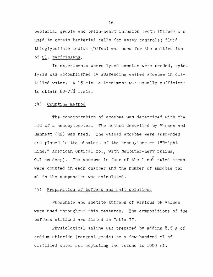
16
bacterial growth and brain-heart infusion broth (Difeo) was used to obtain bacterial cells for assay controls; fluid thioglycollate medium (Difco) was used for the cultivation of Cl. perfringens.
In experiments where lysed amoebae were needed, cyto- lysis was accomplished by suspending washed amoebae in distilled watero A 15 minute treatment was usually sufficient to obtain 60-75$ lysis.
(h) Counting method
The concentration of amoebae was determined with the aid of a hemocytometer0 The method described by Hansen and Bennett (32) was used. The washed amoebae were suspended and placed in the chambers of the hemocytometer ("Bright Line," American Optical Co0, with Neubauer-Levy ruling,
oOol mm deep). The amoebae in four of the 1 mnr ruled areas were counted in each chamber and the number of amoebae per ml in the suspension was calculated.
(5) Preparation of buffers and salt solutions
Phosphate and acetate buffers of various pH values were used throughout this research. The compositions of the buffers utilized are listed in Table II.
Physiological saline was prepared by adding 8„5 g of sodium chloride (reagent grade) to a few hundred ml of distilled water and adjusting the volume to 1000 ml.

TABLE IT
COMPOSITION OF BUFFER SOLUTIONS
(A) Phosphate Buffer Solutions
M/15 Na^HPO^. M./15 KH2P0i4
2.5 ml 97= 5 ml. 5.28
10.0 ml 90.0 ml 5.90
30.0 ml 70.0 ml 6.56
61.1 ml 38.9 ml 7.00
95.0 ml 5.0 ml 8.04
(B) Acetate Buffer Solutions
0.1 N sodium acetate 0.1 N acetic acid pH
18.0 ml 82.0 ml 5.0
70.5 ml 29.5 ml 5.0
90.5 ml 9,5 ml 5.6

18Buffered saline was prepared by adding 8*5 g of sodium
chloride to a few hundred mi of M/15 phosphate buffer (pH 7)
and bringing the volume up to 1000 ml with additional
buffer.
The composition of Ringer solution is outlined belows
NaCl 6.5 g
CaCl2 0 o12 g
KOI 0ol*f g
NaH2P0ifoH20 OoOl g
NaH2C03 0 c 20 g
Distilled water 1000 ml
IIo Experimental Methods and Materials
(1) CatalaseThe DKB strain of E 0 histolytica (growing in associ
ation with Clo perfrlngens) was used in this study0 Amoeba
preparations were prepared from *+8*=hour cultures of E» his-
tolytica grown on Cleveland-Collier mediae In all trials of
this assay, the amoeba count was at least U-00^000 amoebae
per mlo The supernatant fluid from each washing step was
pooled and centrifuged to obtain packed bacterial cells.
Purified catala.se (General Biochemicals Inc„) and packed
cells of Escherichia coll. were used as positive controls.A modification of the method described by Higgins
and Plastridge (37) was usedo The assay was carried out in
calibrated Smith fermentation tubes completely filled with

193% H2O2 ln buffered saline (pH 7 oO)„ The material to be
assayed was introduced into the Smith tubes in 1 ml amounts
by means of a curved Pasteur pipette„ The tubes were incu
bated at k-5° C for 30 minutes; at the end of the incubation
period the tubes were examined for the evolution of oxygen,,
(2) Gelatinase
Six strains of E„ histolytica were assayed for
gelatin-hydrolyzing ability,, The HUS-100, HUS-105, Wash
ington, Griffith, ROK, and Conrad, strains were tested„ All
strains were routinely grown in a modified Boeck-Drbohlav
medium,.
Two methods were used to investigate the gelatinase
of E. histolytica g (1) a sensitive microtest developed by
Thirst (75), and (2) an agar-gelatin plate test developed
by Smith (72). The microtest consisted of using a thin
film of gelatin (Difco) as substrate on a glass slide„ The
slides were prepared by dipping clean, dry microscope slides
into melted aqueous gelatin solutions {5%) containing phenol
(Ool#), an(5 allowing them, to drain and dry0 The materials
to be assayed, were placed on the slide in large drops (0o3- O A ml) and incubated at 20° C for 10-12 hours in Petri
dishes containing strips of moistened filter paper to mini
mize evaporationo The hydrolysis of gelatin was indicated
by the appearance of clear areas on the slide when stained
with dilute carbolfuchsin, a protein stain. The presence of

20
phenol prevented bacterial growth without affecting gelatinase activity.
The second method consisted of preparing 1% agar plates containing 0 ah% gelatin,, Large drops of the material to be assayed were placed on the surface of the plates, and incubated at 37° 0 for 10-12 hours. The plates were then flooded with approximately 10 ml of mercuric chloride in hydrochloric acid (15 g HgCl2/120 ml of 0.37 N HC1), a protein precipitant. A cloudy, white precipitate in the agar indicated the presence of gelatin; clear areas indicated zones of gelatin hydrolysis.
Assay materials consisted of: (a) cell preparationsof all strains were made in the manner previously described (see page 15); (b) strain HUS-105 grown in the absence ofmultiplying bacteria with the aid of growth factors, adenosine triphosphate , ribose-5-phosphate, and large concentrations of antibiotics (52), was pooled and washed; (c) preparations of pooled and washed HUS-105 grown in association with Escho collo The HUS-105 strain growing in association with Escho coll was initiated for this experiment, but could not be continued in culture for an extended period of time. Yields of amoebae varied with the method of cultivation, but at least 200,000 to 250,000 amoebae per ml were obtained.
Controls consisted of: (a) drops of sterile Ringer*ssolution on slides and plates; (b) a 12 hour broth culture of Proteus vulgaris and a 0.5% aqueous suspension of trypsin

21
(Fairchild Bros „ Sc Foster Co0) as indicators of gelatin
hydrolysis; (c) inoculation of associated flora from the
amoeba cultures into conventional gelatin tubes to detect
liquefaction of gelatin by the bacterial flora; (d) de
struction of amoebic gelatinase activity by autoclaving the
assay materials and also inhibition of the enzyme activity
with 20% copper sulfate; and (e) alteration of amoebic
enzyme activity by alternate freezing and thawing of the cell
suspensions„
An attempt was made to determine the optimum pH
range for amoebic gelatinase0 The pH of the agar-gelatin
plates was adjusted with phosphate buffers0 Finally the
effects of enzyme concentration were studied„ The washed
amoebae suspensions were diluted with saline to obtain a
1/2, l A , 1/8, 1/16, and 1/32 dilution and tested forgelatinase activity on gelatin agar plates„
\
(3) Lecithinase
A. modification of the method by Sheldon et al0 (71)
was used in this assay. The reaction is one in which the
enzyme lecithinase splits lecithovitellin to insoluble
fatty acids that appear as a cloudy precipitate (28)„The yolks of several fresh chicken eggs were stirred
with sufficient Ringer®s solution to obtain a 20% suspension
(v/v), The suspension was filtered through Whatman #1
filter paper twice. Five ml of this crude lecithovitellin

22
was added to 95 ml of a buffered agar (1%) solution that had been autoclaved and cooled to 50° C. The lecithovitellin- agar mixture was poured into sterile Petri dishes in 15 ml aliquots and allowed to harden., Antibiotic assay discs (Schleicher and Schuell, #?50-E) with a diameter of 12*7 mm, were placed on the surface of the plates; the discs were saturated with the materials to be assayed0 The plates were incubated at 20° C for 10-12 hours, and the presence of precipitates recorded<>
E. histolytica (strain HITS-105) was used in this assay for lecithinase activity. The amoeba cells were prepared as previously described (see page 15) 9 except that the amoeba were cultured on Cleveland-Collier medium. All cell preparations contained at least 5-00,000 amoebae per ml0 Washed cell preparations of Bacillus subtilis and Cl. per- fringens were used as positive controls0
(b) Desoxyrlbonuclease ana ribonucleaseA modification of the method of Jeffries et al□
(5-0) was employed to study the nuclease activity of the amoebaeo Solutions of nucleic acids, containing 10 mg per ml were prepared» The desoxyribonucleic acid (DNA) (Nutritional Biochemicals Corp.) was dissolved in distilled water, and the ribonucleic acid (RNA) (Nutritional Biochemical Corpo) was dissolved by the drcpwise addition of 1 N NaOH being careful not to allow the pH to rise above 5<>Fifty ml of the nucleic acid solutions were added to 50 ml
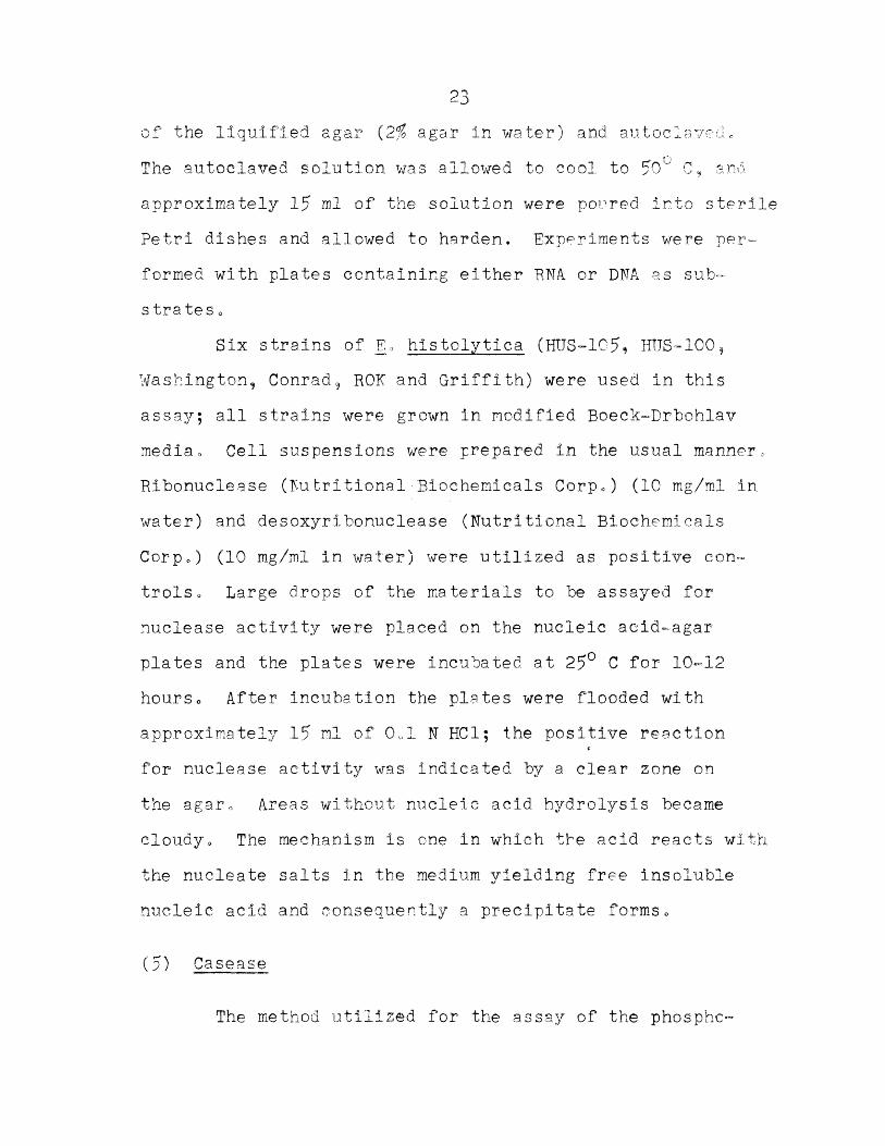
of the liquified agar (2% agar in water) and autoelaved *The autoclaved solution was allowed to cool to 50° C, and approximately 15 ml. of the solution were poured into sterile Petri dishes and allowed to harden. Experiments were performed with plates containing either RNA or DNA as sub- s trates o
Six strains of E„ histolytica (HUS-105* HIJS-100, Washington, Conrad, RON and Griffith) were used in this assay; all strains were grown in modified Boeck-Drbohlav mediao Cell suspensions were prepared in the usual manner, Ribonuclea.se (Nu tritional ■ Biochemicals Corp«) (10 mg/ml in water) and desoxyribonuclease (Nutritional Biochemicals Corp.) (10 mg/ml in water) were utilized as positive con- trolSc Large drops of the materials to be assayed for nuclease activity were placed on the nucleic aeid-agar plates and the plates were incubated at 25° C for 10-12 hourso After incubation the plates were flooded with approximately .15 ml of 0 ol N HCl; the positive reaction for nuclease activity was indicated by a clear zone on the agar0 Areas without nucleic acid hydrolysis became cloudy. The mechanism is one in which the acid reacts with the nucleate salts in the medium yielding free insoluble nucleic acid and consequently a precipitate forms8
(5) Casease
The method utilised for the assay of the phosphc-
2>+
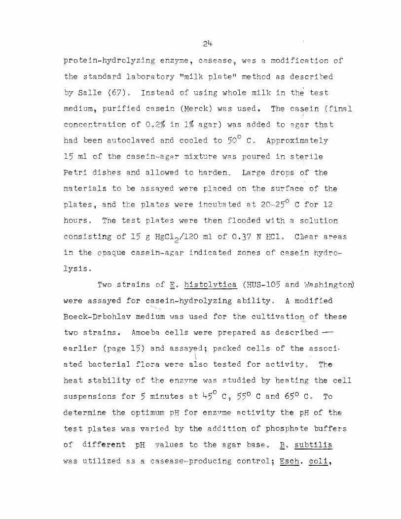
protein-hydrolyzing enzyme, casease, was a modification of the standard laboratory nmilk platet! method as described by Salle (67) » Instead of using whole milk in the test medium, purified casein (Merck) was used* The casein (final concentration of 0 o2% in 1% agar) was added to agar that had been autoclaved and cooled to 50° Co Approximately 15 nil of the casein-aggr mixture was poured in sterile Petri dishes and allowed to harden. Large drops of the materials to be assayed were placed on the surface of the plates, and the plates were incubated at 20-25° C for 12 hourso The test plates were then flooded with a solution consisting of 15 g HgCl2/120 ml of 0.37 N HC1. Clear areas in the opaque casein-agar indicated zones of casein hydrolysis o
Two strains of E. histolytica (HUS-105 and Washington) were assayed for casein-hydrolyzing ability0 A modified Boeck-Drbohlav medium was used for the cultivation of these two strainso Amoeba cells were prepared as described—earlier (page 15) and assayed; packed cells of the assoei-
\
ated bacterial•flora were also tested for activityo Theheat stability of the enzyme was studied by heating the ceil
osuspensions for 5 minutes at C 9 55° C and 65° C <» To determine the optimum pH for enzyme activity the pH of the test plates was varied by the addition of phosphate buffers of different , pH' values to the agar basec B. subtilis was utilized as a casease-producing control; Esch. coli,

25an organism unable to produce casease, was used as a control to rule out non-specific casein hydrolysis.
(6) Urease
A modified Van Slyke and Cullen (36) method for the determination of urinary ammonia was used to investigate the urease activity of Eh histolytica, An aeration train, as shown in Figure 1, was set up in the following manner:(1) 20 ml of concentrated sulfuric acid was added to tube A,the air intake tube, in order to render the incoming air ammonia-free; (2) tube B acted as a trap to prevent any accidental carryover of the sulfuric acid during aeration;(3) tube C was the reaction tube and contained 10 ml of 3% urea solution (Merck) (pH 7) and 2 ml of the material to be assayed for urease activity; (h) tube D contained 25 ml of 0.02 N sulfuric acid and served to trap the ammonia given off during the reaction. Caprylic alcohol (2 drops) was added to each tube to minimize frothing. Sintered glass impingers were used to obtain good aeration of the solutions. A water aspirator was used as a source of vacuum.
The reaction mixtures were incubated at 20-2 5° C for varied periods of time. After the incubation period, the tubes were aerated very slowly for 30 seconds, the aspirator was turned off and 10 ml of a saturated potassium carbonate solution added to tube D (reaction tube). The reaction tube was resealed and aerated for 15 minutes. At the end of

B CFIGURE 1
Aeration Train(1) air intake(2) to aspirator(3) impingers

this period, tube D was disconnected and the contents trans ferred to a 100 ml volumetric flask to which 10 ml of Nesslerfs solution (Tenso-Labo Inc., 6,8 g/100 ml of 8,5% NaOH), diluted to 30 ml with distilled ammonia-free water (boiled), was added and the volume brought up to 100 ml with ammonia-free water„ The solution was mixed, allowed to stand for five minutes, and the optical density determined at ^80 mu in a Spectronic-20 colorimeter (Bausch & Lomb) against a reagent blank*, The reagent blank was prepared by carrying a solution of substrate through the complete analytical procedure (incubation, aeration, nes- slerization) in exactly the same manner as the assay tubes0 This was done in order to correct for ammonia in the reagents, decomposition of the substrates and errors in the analysis (36).
The DKB strain of Eo histolytica, intact and lysed, was assayed for urease activity„ This strain was cultured on Cleveland-Coilier media. Washed amoebae were prepared as previously described0 A determination of the ammonia content of the washed amoeba preparations was attempted by the same analytical procedure in the absence of substrate „ The sensitivity of the method was determined by using dilutions of clinical urease tablets (Arlington Chemical Co,)*
(7) Cltrulllnase
Cit.rullina.se activity was determined using procedure

28
similar to those for the detection of urease» The only modification was that a 2% solution of dl-citrulline (Nutritional Biochemicals Corp.) (pH 7) was used as a substrate instead of urea.
In this assay the DKB strain of E. histolytica was employed. Positive controls consisted of packed cells of Cl. perfringens0 Both intact and cytolyzed amoebae were studied. Amoebae were cultured on Cleveland-Collier medium.
(8) Asparaginase
The assay for this amidase was also carried out by the Van Slyke-Cullen method (38). A 2% solution of di- asparagine (Nutritional Biochemicals Corp.) (pH 8) was utilized as substrate. Both intact and lysed cells of Eo histolytica, strain DKB, grown on Cleveland-Collier medium, were used.
(9) Glucuronidase
The glucuronidase activity of E. histolytica was investigated using a modification of the method described by Robinson et al0 (65)° Essentially, the method is based upon the colorimetric measurement of the hydrolysis of a chromogenic substrate,, phenolphthalein glucuronidate.
The assay for glucuronidase activity was carried out in 15 ml glass centrifuge tubes. Each tube received O A ml of Ool N acetate buffer of varied p H values, 0<A ml of

290.001 M phenolphthalein glucu.ronid.ate (Sigma Chemical Co.), and 0 A ml of the material to be assayed for activity.After a one hour incubation period at 37° C, 2 A ml of O.l^M sodium carbonate were added and the tubes centrifuged for 30 minutes (3000xG). Concurrently, duplicate series of tubes were treated in the same manner, except that one series did not contain substrate, and the other contained heat-inactivated (boiled) suspensions of the material assayed 0
After clearing by centrifugation, the supernatant was decanted and the optical density determined at A O mu in the Spectronic-20 colorimeter. The optical density was set at zero with the supernatant from a tube from the series lacking in substrate; the duplicate tubes (the same dilution of the same material) from the heat-inactivated series and from the enzyme series were compared with this tube, and the optical density recorded. This method of negating changes in optical density caused by bacterial cells and other non- phenolphthalein opacities was carried out with each dilution of each material assayed.
In order to become more familiar with the method and to determine the sensitivity of the method, an assay of various dilutions of a known glucuronidase-producing bacterium were deemed necessary. To obtain an organism that produced glucuronidase it was found necessary to perform a survey of several species of bacteria. Streptococcus

30liquefaciens, Streptococcus pyogenes, Staphylococcus pyogenes , var. aureus, Escherichia coli, and an unidentified Gram positive streptococcus, isolated from the HUS-105 flora, were assayed with a microtest for glucuronidase (65)=
The microtest consisted of placing a small drop of 0.01 M phenolphthalein glucuronidate in a slight depression in a sheet of aluminum foil. A. loop of the bacterial species to be tested was thoroughly mixed with the substrate, and the entire suspension drawn up into a 1 mm glass capillary tube. The suspension was incubated at 37° C for one hour. White spot plates were prepared with 0.03 ml of 0.15 M sodium carbonate in each hollow. The capillary tubes were removed from the incubator and the contents expelled into designated spots of the alkaline solution. Theprompt appearance of a distinct red color in the sodium
\carbonate solution indicated the presence of glucuronidase.
Strain DKB of E. histolytica, cultured on Cleveland- Collier medium, was used in this assay. The previous method of cell preparation was utilized; whole and lysed amoebae were assayed for activity.
(10) Beta-D-galactosidase
O-nitrophenyl-beta-d-galactopyranoside (Sigma Chemical Co.), a chromogenic substrate, was used in an assay for galactosidase (lactase) activity in E. histolytica.The method is based upon the colorimetric determination of

31chromogen produced in the supernatant by the enzymatic hydrolysis of the substrate.
The method employed was a modification of a procedure described by Lederberg (*+3). Amoeba cells, lysed and whole, were resuspended in phosphate buffer (pH 7) a.nd U-.5 ml of the suspension were pipetted into 15 ml centrifuge tubes containing 0.5 ml of M/200 substrate. The tubes were incubated at 37° C for 3° minutes, cleared by centrifugation (3000xG) and the optical density determined at b20 mpi in the Spectronic-20 colorimeter. The method of correcting for the opacities produced by factors other than the chromogen used in the assay for glucuronidase was also used in the assay for the galactosidase.
Strain DKB, cultured on Cleveland-Collier medium, was used in this study. In addition, the sensitivity of the method was assayed using Each, coli as the test organism,
(11) Succinic dehydrogenase
The Thunberg technique was used for the assay of succinic dehydrogenase in E. histolytica (HUS-105)• The method consists of determining the time necessary to decolorize methylene blue. Methylene blue acts as the final hydrogen acceptor in the oxidation (dehydrogenation) of succinic acid.
Conventional Thunberg tubes were used; 1 ml of the material to be assayed was placed in the side arms. Two

32
ml of phosphate buffer (pH 7), 1 ml of a 1:10,000 dilution of aqueous methylene blue, and 2 ml of M/?0 succinic acid (Nutritional Biochemicals Corp.) were added to each tube. Inhibitors of succinic dehydrogenase were assayed by placing 1 ml of inhibitor (M/?0) into the Thunberg tubes in addition to buffer, dye, and succinate (1 ml). The tubes were evacuated for 3 minutes with the aid of a water aspirator and
othen equillibrated for 10 minutes in a 57 water bath, and the cells were tipped into the body of the tube from the s idearm.
The tubes were incubated at 57° C and visually compared with a control tube (containing methylene blue but no substrate) at 10 minute intervals over a period of 90 min-
i iutes. The time required for the decolonization of the methylene blue to a color intensity equivalent to the control, which represented 90$ reduction, was recorded. The control tube contained 1 ml of heat-inactivated cells, 2 ml of buffer, 2 ml of substrate and a 1/100,000 dilution of methylene blue.
The amoebae, cultivated on egg slants overlayed with 0 @5fo liver extract, were prepared in the usual manner. Bacterial and endogenous (preparations of amoeba in absence of substrate) controls were also used in the prescribed fashion.
Arsono-acetate obtained from Dr. G. R. Seaman, University of Texas Medical Branch, Galveston, and malonic

33acid (Nutritional Biochemicals Co*) were used as competitive inhibitors»
The assays were performed at 77° C to inhibit therapid endogenous dehydrogenation of starch by E. histoly-
otica. The use of 77 C to inhibit the endogenous metabolism was suggested by the work of Quastel and Wooldridge (63). This temperature apparently does not affect succinic dehydrogenase activity*
(12) indole
The production of indole, a biochemical reaction dependent on an enzyme system, by E. histolytica was investigated o
For this assay, actively growing cells of the DKB strain in association with Cl. perfringens (a species unable to produce indole) were used* The Gnezda oxalic acid method and Kovac*s method as described by Salle (67) were employed.
The procedure consisted of inoculating two tubes of Cleveland-Collier media with Cl. perfringens, two tubes with Eo histolytica strain DKB-Cl. perfringens, and two tubes with strain DKB-Cl * perfringens plus Esch. coll. Strips of filter paper previously dipped in saturated oxalic acid solution and dried were then carefully placed in the necks of the tubes, avoiding contamination during the process*The tubes were then incubated at 37° C for *+8 hours and the oxalic acid papers inspected .for the presence of a pink

coloration that indicates production of indole0In order to minimize the possibility that extremely
small amounts of indole were produced that could not be detected by the Gnezda method, the tubes were centrifuged, the supernate separated from the sediment, and both tested for indole with Kovac’s reagent. The appearance of a deep cherry-red color, when 5 ml of Kovac's reagent were introduced into the tube containing the material to be analyzed, was considered to be indicative of indole production.
The composition of Kovac's reagent is indicatedbelow:
Para-dimethyl-amino-benzaldehyde Butyl alcoholHydrochloric acid (37$) c 0p.
75 ml 25 ml

CHAPTER I?
RESULTS
(.1) Catalase
In no instance was the evolution of oxygen from
hydrogen peroxide observed when this substrate was incu
bated with either whole or lysed cells of strain DKB, Entamoeba histolytica. Clostridium perfringens , an obli
gate anaerobe known not to possess catalase, was unable t
decompose the peroxide molecule. On the other hand, as
little as 7 gamma of purified catalase and as few as 300. cells of Escherichia coli produced detectable amounts of
oxygen from the substrate. When the assay vessels were
incubated at h° C there was no spontaneous breakdown of t
hydrogen peroxide solution. Results are recorded in
Table III.
(2 ) Gelatinase
A.11 six strains of E. histolytica hydrolyzed gelat.
The mixed bacterial flora of all six strains were alsocapable of splitting this protein. However, the supernal;
from the final washing did not exhibit gelatinolytic act:!
tyg indicating that the observed gelatinase was of amoebiorigin. The lytic activity of - the supernatants progress! ly decreased and finally ceased after the third washing
35

36
TABLE III
RESULTS OF CATALASE ASSAY
Preparation Oxygen Evolved (ml) Series 1 Series 2
Amoebae (500,000/ml)Amoebae, lysed (500,000/ml)Cl. perfringensEscho collTl.5 x~T0^ cells/ml)Esch. coli(3 X lO^- cells/ml)Catalase (70 gamma)Catalase (7 gamma)Endogenous (no enzyme)
0
0
0o A o
trace
2.680
0.2
0
0
0
00.50
trace
2.60
0.25
0

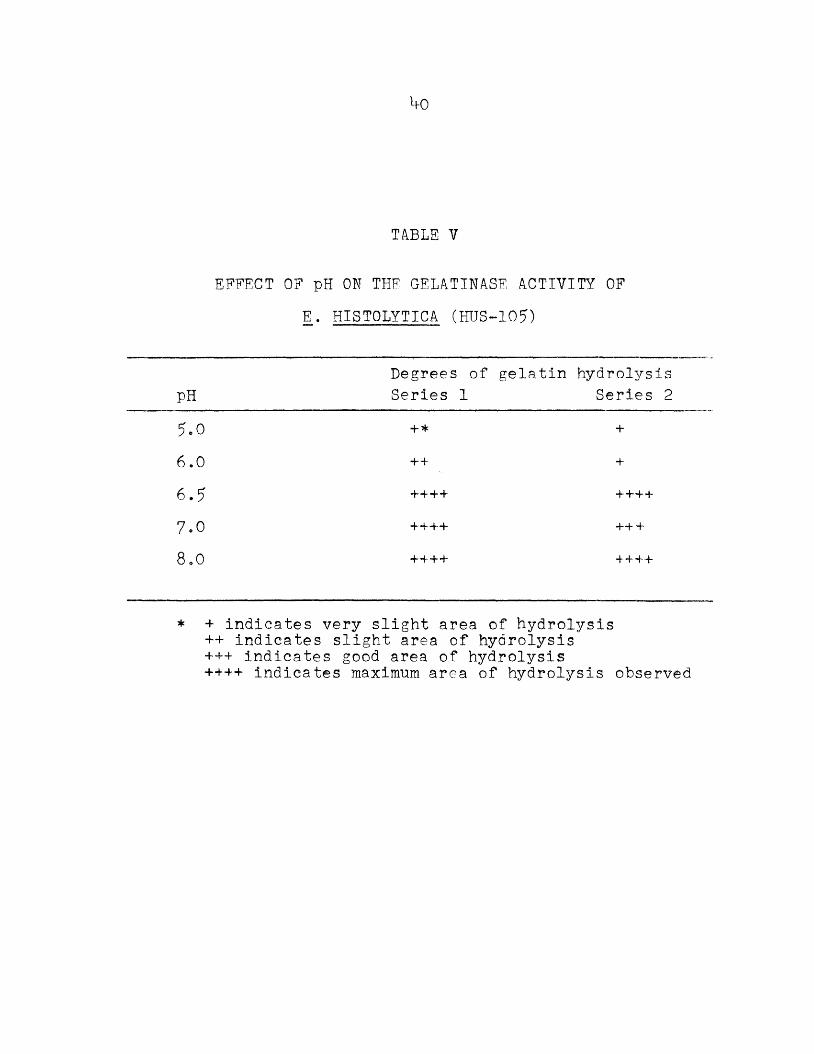
procedure. The amoebae were also able to hydrolyze gelatin when grown in a monoassociated culture with Esch. coli, a species known not to hydrolyze gelatin. The lytic activity of the amoebae was completely inhibited by auto- claving and copper sulfate and partially inhibited by freezing and thawing. The degree of hydrolysis of gelatin decreased as the amoeba preparations were diluted. A 1/1.6 dilution exhibited slight activity and a dilution of 1/32 had no activity. Amoebic gelatinase activity was not affected by pH changes from a pH of 6.5 to pH of 8. The activity was reduced at pH values of 6.0 and lower. Studle were not made at pH values of above 8 because of the degradation of gelatin at these values. Because of the limitations of the method of determining amoebic numbers, no comparison of activity between strains was attempted.
The results of the gelatinase assay (microtest) utilizing all strains is tabulated in Table IV. Figure 2 illustrates the areas of hydrolysis on the gelatin-coated slides. Table V records the results of the effects of various pH values on the amoebic gelatinase.
(3) Lecithinase
There was no lecithinase activity in the amoebae (HUS-105) even though as many as 950,000 cells/ml were usedo Cell, suspensions of Cl. perfringens and Bacillus subtilis produced large zones of flocculant, white

TABLE IV
GELATINASE ACTIVITY OF E. HISTOLYTICA
StrainsPreparation HUS -105 HUS-100 Washington Conrad ROK GriffithSupernatant (2nd wash) / / / /Supernatant (3rd wash) / - - - - -
Supernatant (Vth wash) - - - - - -
Amoebae (washed 5.x) / / / / 1 OJCO
/ indicates clear areas on the slide- indicates no clear areas on the slide

39
FIGURE 2 Gelatinase Microtest
(A) absence of gelatin hydrolysis by the supernate fluid from final wash;
(B) gelatin hydrolysis by E. histolytica;(C) gelatin hydrolysis by trypsin;(D) absence of gelatin hydrolysis by Ringer’s
solution.
^0
TABLE V
EFFECT OF pH ON THE GELATINASE ACTIVITY OF E. HISTOLYTICA (HUS-105)
pHDegrees of Series 1
gelatin hydrolysis Series 2
5,0 + * +6.0 44 +
6.5 4444 ++4+
7.0 ++++ H—b 4
8.0 +4- 44 4444
* + indicates very slight area of hydrolysis++ indicates slight area of hydrolysis 444 indicates good area of hydrolysis 4444 indicates maximum area of hydrolysis observed
bl
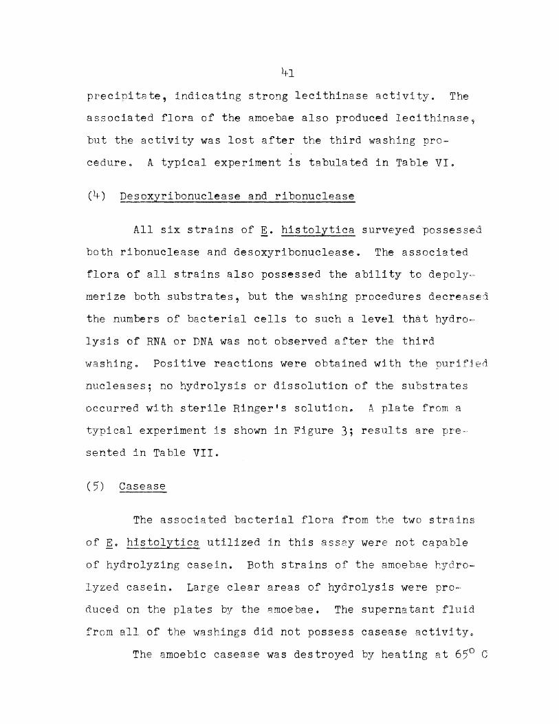
precipitate, indicating strong lecithinase activity. The associated flora of the amoebae also produced lecithinase, but the activity was lost after the third washing procedure. A typical experiment is tabulated in Table VI.
(h ) Desoxyribonuclease and ribonuclease
All six strains of E . histolytica surveyed possessed both ribonuclease and desoxyribonuclease. The associated flora of all strains also possessed the ability to depoly- merize both substrates, but the washing procedures decreased the numbers of bacterial cells to such a level that hydrolysis of RNA or DNA was not observed after the third washing. Positive reactions were obtained with the purified nucleases; no hydrolysis or dissolution of the substrates occurred with sterile Ringer1s solution. A plate from a typical experiment is shown in Figure 3? results are presented in Table VII.
(5) Gasease
The associated bacterial flora from the two strains of E . histolytica utilized in this assay were not capable of hydrolyzing casein. Both strains of the amoebae hydrolyzed casein. Large clear areas of hydrolysis were produced on the plates by the amoebae. The supernatant fluid from all of the washings did not possess casease activity.
The amoebic casease was destroyed by heating at 65° C
b2
TABLE VI
RESULTS OF LECITHINASE ASSAY
PreparationEnzyme
Series 1Activity
Series 2
Supernatantfrom 2nd washing
++ +
Supernatantfrom 3rd washing
- -
Amoebaeafter 5th washing (950,000/ml)
Bacillus subtilis +++++ +■++■+•Clostridium perfringens +++ ++++
* - indicates no detectable precipitate+ indicates slight precipitate formation ++ indicates fair precipitate.formation +++ indicates good precipitate formation ++++ indicates extensive precipitate formation +++++ indicates maximum precipitate formation
observed

T A B L E V I I
N U C L E A S E A C T I V I T Y O F E . H I S T O L Y T I C A
Preparation HUS -105 HUS-100Strain
Washington R O K Griffith ConradHydrolysis
ofHydrolysis
ofHydrolysis
ofHydrolysis
ofHydrolysis
ofHydrolysis
ofR N A D N A R N A D N A R N A D N A R N A D N A R N A D N A R N A D N A
Supernatant (2nd wash)
/ * / / / / / / / / / / /
Supernatant (3rd wash)
/ / - - - / - - -
Supernatant (*fth wash) - - - - - - - - -
Amoebae (5th wash) / / / / / / / / / / /
* / indicates formation of a clear area- indicates no detectable clear area formed

bb
FIGURE 3 Nucleic Acid Test Plate
(A) hydrolysis of ribonucleic acid (RNA) byribonuclease;
(B) absence of RNA hydrolysis by Ringer’s solution;
(C) hydrolysis of RNA by washed E. histolyticain suspension;
(D) absence of RNA hydrolysis bv the supernatantfluid from final amoeba washing preparation,,

^5for 5 minutes; the activity was not affected by heating the preparations for 5 minutes at 55° C. The experiments on the effects of pH on the amoebic casease showed that the enzyme was active over the whole pH range assayed (pH 5 to pH 8). It was not possible to quantitate any difference in hydrolytic activity over the pH range studied; all that could be determined in this case was that casein hydrolysis did occur at every pH value assayed over the mentioned range.
(6) Urease
It was not possible to detect urease activity in whole or lysed amoeba preparations. Although the preparations were incubated for as long as 12 hours, no detectable amounts of ammonia were produced from the action of the amoebae on urea solutions. The bacterial associates, however, produced increases in the optical densities of the reagent solutions in 15 minutes. Direct nesslerization of the amoeba reaction tubes did not result in the production of any detectable color. This confirmed the observation that
histolytica does not produce urease.
(7) Citrullinase
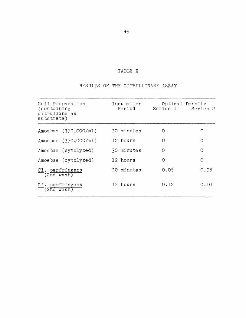
The amoebae tested did not produce ammonia from citrulline. It was not possible to obtain increases in the optical density of the reagent solutions. Direct
1+6
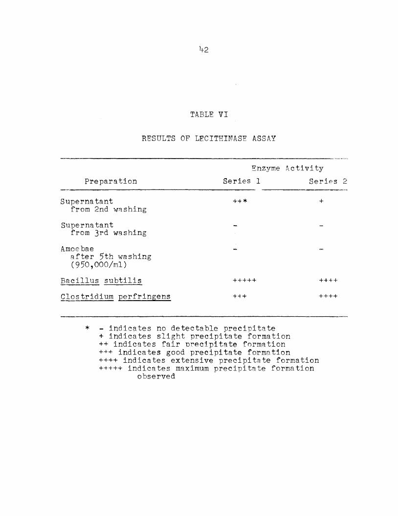
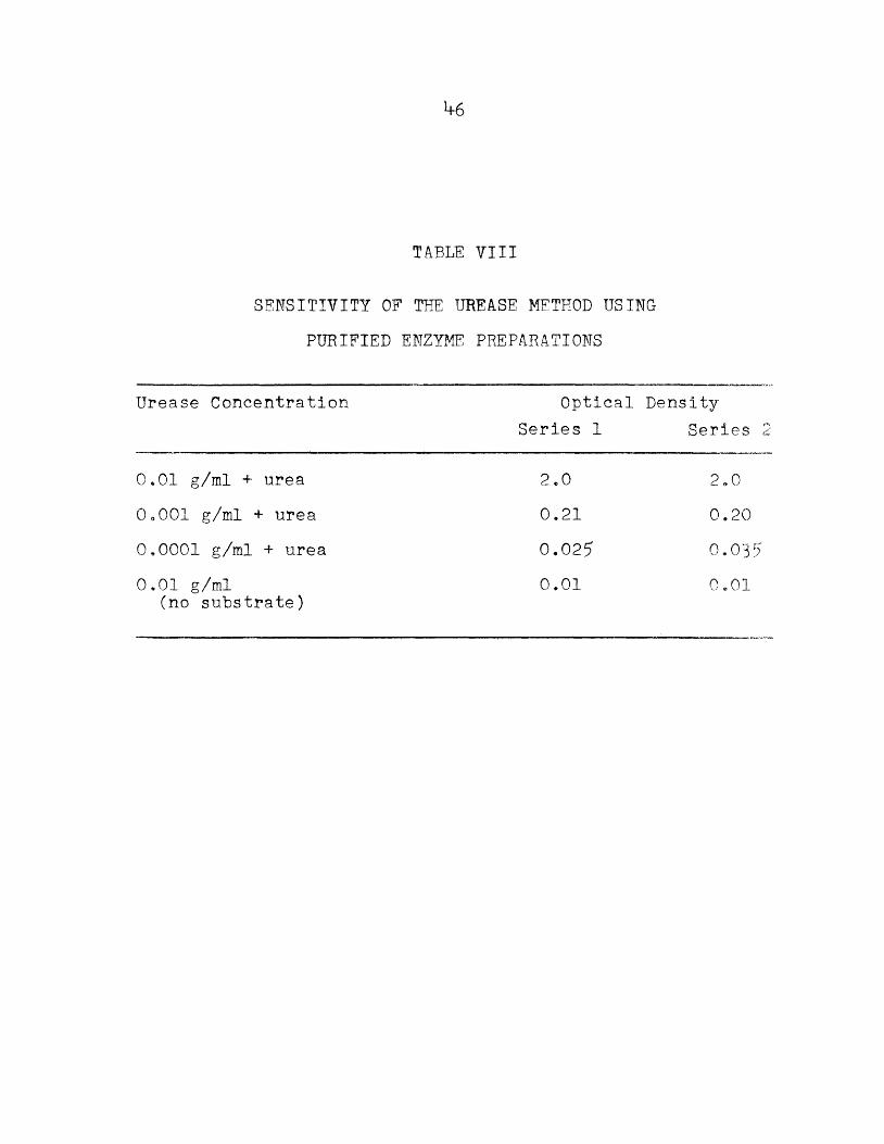
TABLE 71II
SENSITIVITY OF THE UREASE METHOD USING PURIFIED ENZYME PREPARATIONS
Urease Concentration
O.Ol g/ml + ureaOoOOl g/ml + urea0.0001 g/ml + urea0.01 g/ml
(no substrate)
Optical Density Series 1 Series
2.00.210.0250.01
2.00.200.035
o.oi

*+7
TABLE IX
RESULTS OF THE ASSAY FOR UREASE IN E. HISTOLYTICA
Cell Preparations (containing urea as substrate)
IncubationPeriod
Optical Density Series 1 Series
Amoebae (300,000/ml)Amoebae (300,000/ml)Amoebae (300,000/ml)Amoebae (lysed)Amoebae (lysed)Amoebae (lysed)Amoebae
(no substrate)C l . perfringens
(2nd wash)Cl. perfringens
(2nd wash)C l . perfringens
(2nd wash)
1? minutes 1 hour 12 hours 15 minutes 1 hour 12 hours 1 hour
15 minutes
1 hour
12 hours
0000000
0.015
0.020
0.020
0000000
0.025

**8
nesslerization of the reaction tubes indicated a lack of citrulline breakdown.
The associated bacteria, Cl. perfringens, produced citrullinase. Changes in the optical density of the reagent solutions were observed after 30 minutes of incubation.Large amounts of ammonia were produced from citrulline by these bacteria after 12 hours.
The results of the citrullinase assay appear in Table X.
(8) Asparaginase
There was an increase in the optical density of the reagent solution when the amoebae were incubated with dl-asparagine indicating that ammonia was being produced. This is evidence for the production of asparaginase by E . histolytica.
Cl. perfringens, the associated bacterium, also attacked asparagine, but there was greater ammonia production by the washed amoeba preparations than by the bacteria o The ability of the amoeba preparations to produce ammonia from asparagine was completely lost by boiling for 5 minutes. A 1/2 dilution of the amoeba suspension also produced less ammonia from the substrate. The results of this assay are presented in Table XI.
i+9
TABLE X
RESULTS OF THE CITRULLINASE ASSAY
Cell Preparation (containing citrulline as substrate)
IncubationPeriod
Optical Series 1
De n s1 tv Series 2
Amoebae (370,000/ml) 30 minutes 0 0Amoebae (370,000/ml) 12 hours 0 0Amoebae (cytolyzed) 30 minutes 0 0Amoebae (cytolyzed) 12 ’hours 0 0Cl. perfringens
(2nd wash)30 minutes 0.05 0o07
Cl. perfringens (2nd wash;
12 hours 0.12 0i—10

50
TABLE XI
RESULTS OF THE ASPARAGINASE ASSAY
Cell Preparation (containing asparagine as substrate)
IncubationPeriod
Optical DensitySeries 1 Series 2
Amoebae (200,000/ml) + substrate
Amoebae (lysed)+ substrate
Amoebae (boiled)+ substrate
Amoebae (^ dilution) + substrate
Amoebaeno substrate
Cl. perfringens (2nd wash)+ substrate
15 minutes
15 minutes
15 minutes
15 minutes
15 minutes
15 minutes
0.12
0.08
0
0.10
0
0.0^5
0.18
0 .07
0.025

(9) Glucuronidase51
The results of the assay for a glucuronidase-producing bacteria are tabulated in Table XII. The results of the tests on the sensitivity of the method are shown in Table XIII and Figure b.
Neither whole cells nor lysed amoebae were capable of hydrolyzing the substrate at pH values of *+.0, 5.0 and 5.6; free phenolphthalein was not released during the incubation periods employed. There was some activity exhibited by Cl. perfringens. The results of the glucuronidase assay are tabulated in Table XIV.
(10) Beta-D-Galactosidase
The data on the determination of the sensitivity of the method are shown in Table XV and Figure 5.
The production of galactosidase by the amoebae could not be detected using this sensitive method. There was no change in the optical density of the incubation tubes over the reagent blanks even after 12 hours of incubation. The data are tabulated in Table XVI.
(11) Succinic dehydrogenase
Washed preparations of amoebae rapidly decolorized methylene blue in the presence of the substrate, succinic acid, at 57° C« The endogenous dehydrogenating ability of the amoebae was almost completely inhibited at this

52
TABLE XII
A SURVEY OF BACTERIA FOR GLUCURONIDASE ACTIVITY
Organism ComDarative Series 1
Activity Series 2
Streptococcus liquefaciens -* -
Streptococcus pyogenes - -Staphylococcus pyogenes
var. aureus- -
Escherichia coli + +An unidentified Gram positive
s treptococcus+++ ++•+■
* - indicates no detectable color produced+ indicates slight pink color produced +++ indicates deep red color produced

53
TABLE XIII
SENSITIVITY OF THE GLUCURONIDASE METHOD USING AN UNIDENTIFIED GRAM POSITIVE STREPTOCOCCUS
Numbe r of cells/ml Optical DensitySeries 1 Series 2
2 X 10⁸ 0.2 7 0.291 X 10⁸ 0.1*f Ooi55 X Oi—1 0.06 0.083 •3 x 10? 0.05 0.062.5 x 10? 0.03 0.032 X 10? 0 0

Optical
Dens
ity
5b
0 A
0.2
2.0QNumber of Bacteria x 10 /ml
FIGURE bSensitivity of the Glucuronidase Method
Q assay 1 £ assay 2

55
TABLE XIV
ASSAY FOR GLUCURONIDASE IN E. HISTOLYTICA
Cell Preparation (containing phenolph- thalein glucuronidate as substrate)
IncubationPeriod
Optical Series 1
Density Series :
Amoebae (325,000/ml) 1 hour 0 0Amoebae (325,000/ml) 12 hours 0 0Amoebae (lysed) 1 hour 0 0Amoebae (lysed) 12 hours 0 0Cl. perfringens 1 hour 0.01 0.01Cl. perfringens 12 hours 0.025 0.02

56
TABLE XV
SENSITIVITY OF THE GALACTOSIDASE METHOD USING ESCHERICHIA COLI AS THE TEST ORGANISM
Number of cells/ml Optical Series 1
DensitySeries 2
x 108 0.32 0.282.7 x 10 0.15 0.1?1.35 x 10 0.06 0.089 x 10⁷ 0.05 0.0 76.75 x 10⁷ 0.0>+ 0.05.b x 107 0.02 0.02

Optical
Dens
ity
57
O.b
0.2
0.1
0 1.0 2.0 oNumber of Bacteria x 10 /ml
PIOURS 5Sensitivity, of the Galactosidase Method
Q assay 1. £ assay 2.

58
TABLE XVI
RESULTS OF THE GALACTOSIDASE ASSAY
Cell Preparation (containing O-nitro- phenyl beta D galacto- side as substrate)
IncubationPeriod
Optical Series 1
Density Series ;
Amoe ba e (*+00,000/ml) 30 minutes 0 0Amoebae (^00,000/ml) 12 hours 0 0Amoebae (lysed) 30 minutes 0 0Amoebae (lysed) 12 hours 0 0Cl. perfringens
(2nd wash)30 minutes 0.02 0.01
Cl. perfringens (2nd wash)
12 hours 0.06 0.06

59temperatureo After the second washing procedure, the supernatant fluid, still containing residual bacteria, exhibited no decolorizing activity. The succinic dehydrogenase of E. histolytica was completely inactivated by boiling for five minutes. Both arsono-acetate and malonic acid were shown to be powerful inhibitors of the amoebic enzyme. A typical experiment is tabulated in Table XVII.
(12) Indole
The production of indole by the amoebae plus Cl. perfringens or by Cl. perfringens alone was not detectable under the conditions of these experiments. Tubes containing metabolizing cells of Esch. coli consistently produced a positive reaction. In all cases the results of the Gnezda paper were confirmed by Kovac’s test. The Gnezda oxalic acid paper turned pink within the first 2*+ hours of incubation when E. histolytica, in association with Esch. coli, was the test organism. All tubes were inspected for amoebic growth before results were recorded. If amoebic growth was poor or absent, the tubes were discarded.

60
TABLE XVII
RESULTS OF THE SUCCINIC DEHYDROGENASE ASSAY
EnzymePreparation
Substrate Time for 90% Reduction Series 1 Series 2
Sunernate (2nd wash)
2 ml M/50 succinic acid
in excess of 90 min.
in excess of 90 min.
Amoebae(300,000/ml)
2 ml M/50 succinic acid
25 min. 15 min.
Amoebae(300,000/ml)
2 ml saline 80 min. in excess of 90 min.
Amoebae(boiled)
2 ml M/50 succinic acid
noreduction
noreduction
Amoebae(300,000/ml)
1 ml M/50 succinic acid 1 ml M/50 arsono acetate
in excess of 90 min.
in excess of 90 min.
Amoebae (300,000/ml)
1 ml M/50 succinic acid 1 ml M/50 malonic acid
80 min. 65 min.
Amoebae (300,000/ml)
1 ml M/50 succinic acid 1 ml saline
30 min. *+0 min.
CHAPTER V
DISCUSSION AND CONCLUSIONS
Two important factors involved in the enzyme assays performed in this investigation were the efficiency of the washing procedure and the numbers of amoebae utilized in each experiment. The washing procedures were efficient as indicated by the fact that plating of the fifth-wash sediments or supernatant fluids showed a great reduction in numbers of bacteria as compared with the initial population,, The fact that it was not possible to eliminate completely all of the bacteria, however, does allow some criticism of the work. Since adequate bacterial controls were utilized, there is little doubt that positive reactions for any given enzyme detected were due to amoebic activity. Since a few bacteria were present, it was not possible to eliminate completely the possibility of the existence of synergistic actions. Furthermore, it was not possible to rule out the remote possibility that the elimination of so many of the associated bacterial cells resulted in an alteration of the metabolism of Entamoeba histolytica. Perhaps the amoebae function in a different biochemical manner in the absence of supporting associated cells. These are problems not answered by this research. However, it is felt that in view of the meager knowledge
61
62
available concerning the metabolism of E. histolytica, this investigation, and others in related areas, might offer clues which may lead to a better understanding of the biochemical reactions of this protozoan parasite.
The second factor, the numbers of amoebae utilized in the assays, becomes important in the experiments where enzyme activity could not be detected in the amoebae. The inability to detect activity might have been due to the comparative paucity of cells used. Most bacterial assays are carried out with many millions of cells, not a few hundred thousand as was the case in this work with E. histolytica . Theoretical calculations indicate that a little more than a million trophozoites would have a dry weight of one milligram (26). Since at least 200,000 amoebae were used, and the method of assay was highly sensitive, it is probable that sufficient quantities of cells were employed.
Thenard (73) first observed that certain tissues of plant and animal origin were capable of decomposing hydrogen peroxide. Later workers characterized the degradation as enzymatic, and Loew, in 1901, named this enzyme catalase (73) • Catalase, an iron-containing enzyme, is concerned with the removal or utilization of hydrogen peroxide produced by biological oxidation reactions of the cell. It catalyzes the decomposition of hydrogen peroxide into water and molecular oxygen. The enzyme can be found in all

63aerobic and most .facultatively anaerobic microorganisms, but has never been found in obligate anaerobes (62, 66, 7*+).
The question of anaerobiosis as a requirement for the growth of E. histolytica has received much attention (5, 50), but many conflicting opinions have been reported (50). As recently as 1958, Elsdon-Dew (20) posed the question as to whether or not E. histolytica is a strict anaerobe.
The results of this work indicate that the amoebae do not produce catalase (in amounts detectable by the methods utilized). As an indication of the sensitivity of the catalase detection methods, it has been calculated that a single molecule of catalase can catalyze the degradation of more than five million molecules of hydrogen peroxide per minute (61). The observation that the DKB strain of E. histolytica did not produce catalase supports the view that these amoebae are probably obligate anaerobes.
The existence of proteolytic enzymes in E. histolytica have been previously reported (2, ^7, 6*0 , but until the paper by Neal in 1956 (57) and the work of Harinasuta and Maegraith (35) in 1958, some doubt existed as to whether the previously observed proteolysis was due to amoebic or bacterial enzymes<> Attempts were made to determine gela- tinase activity in the amoebae using different assay methods and also to see if casease is produced by this organism.It was found that E» histolytica elaborated gelatinase and

6*+
casease. These observations confirmed the reports of Neal and of Harinasuta and Maegraith. Although proteolysis by the amoebae has been demonstrated, with our present knowledge about the organism it cannot be incriminated as a mechanism of pathogenicity0 However, as Meleney (*+8) points out, research into the proteolytic enzymes of E. histolytica may lead to the identification of the pathogenic component of this organisnio
Very little is known concerning the nucleic acid metabolism of the amoebae. Pan and Geiman (59) recently studied the distribution of ribonucleic acid and desoxyribonucleic acid in this organism using cytochemical methods. Studies with antimetabolites have indicated the importance of nucleic acid metabolism in E . histolytica (5*+). Inhibition of the amoebae by purine ”and pyrimidine analogs and the subsequent reversal of the ‘inhibitory effects by the metabolites have established the roles of nucleic acid precursors as nutritional requirements for the amoebae.
The present work has shown that E. histolytica produced both ribonuclease and desoxyribonuclease. These enzymes split nucleic acids into oligonucleotides; possibly the amoebae possess enzymes for completing the degradation to the respective bases, pentoses, and phosphates. Additional work will be necessary to determine this. Since rapidly growing parasites synthesize large amounts of nucleic acid (12), it can be imagined that nucleases capable

65of degrading host nucleic acids to compounds readily utili- zable by the parasite might be an important aspect of the mechanism of tissue invasion by the amoebae. Perhaps, as in the case of the bacterial enzyme, streptodornase, the nucleases of the amoebae are involved in pathogenicity.
The degradation of lecithin is accomplished by some snake venoms and toxins. The hemolysis of red blood cells by some bacterial toxins, such as the Cl. perfringens "theta toxin,” is usually attributed to the action of lecithin- ase on the phospholipid of the erythrocyte stroma.Since E. histolytica engulfs and lyses red cells, it was thought that a study of lecithin hydrolysis by the amoebae would be interesting. E. histolytica, using this sensitive method (71, 77), was unable to hydrolyze lecitho- vitellin. It is possible that the dissolution of red blood cells by the amoebae is due to proteolysis and not lipo- lysis.
Since urease and citrullinase were not produced by the amoebae, the Krebs-Henseleit cycle probably does not operate in E. histolytica (58, 69). The preparations of
histolytica utilized in this assay did not produce detectable amounts of ammonia from either substrate. The method utilized for the determination of ammonia-nitrogen is considered very sensitive. Increases in optical densities are reported to occur with less than ten micrograms of ammonia-nitrogen (36). Therefore the evidence is strong

66
that urease and citrullinase were not produced by the strain of the amoebae used in these experiments.
Glutamine, the amide of glutamic acid, and asparagine, the amide of aspartic acid, are generally considered to be readily available sources of ammonia for the organism possessing glutaminase or asparaginase. These enzymes catalyze the reversible hydrolysis of the amides to release ammonia and the free amino acids (19)* Glutaminase production by E. histolytica has been previously reported by Nakamura and Goldstein (56). The present study on asparagine indicates the presence of this deaminase in the amoebae as well. Since approximately 70% of the dry protoplasmic constituents of microbial cells are nitrogenous compounds of varying complexity (62), it is not surprising that the amoebae produce deaminases which are means for obtaining utilizable sources of nitrogen for synthetic processes.These results indicate that glutaminase and asparaginase, but not urease nor citrullinase, may function in this capacity in E. histolytica.
Dolkart and Halpern (18) in 1958, reported excellent growth of the amoebae in a medium consisting entirely of gastric mucin in phosphate buffer. Mucins are enzymatically degraded to hexosamines and uronic acids. Greenberg et al. (27) found that glucosamine was an irreplacable component of their medium for the amoebae. Loran et al. (*+5a) also reported that the main action of the associated

67
bacteria on glucose was to convert it to glucosamine for use by the amoebae,, In view of these observations it was deemed of interest to learn if the amoebae were capable of splitting glucuronic acid. There has also been a report that beta-glucuronidase was able to hydrolyze hyaluronic acid (2*f) . It was thought that a study of beta-glucuronidase in the amoebae might be valuable.
The aglucuronometric method utilized in this assay is extremely sensitive (2*+, 65) and quantitative measurements can be made with only a few micrograms of phenolphthalein.On the basis of the results obtained, and on the basis of extreme sensitivity of the assay method, the conclusion reached is that E. histolytica did not produce beta-glu- curonidase under these conditions.
Some progress is now being made in the study of the carbohydrate metabolism of the amoebae (23, 38, 39), but it is obvious that a great deal of knowledge is yet to be accumulated before the diverse pathways and enzymatic reactions of E. histolytica can be elucidated. A report by Hilker et al. (38) on the absence of lactase in the amoebae was considered important because of the widespread distribution of this disaccharide and also because of previous reports on the breakdown of glucose and galactose (hydrolytic products of lactose) by the amoebae (*+2).
The extremely sensitive method of Lederberg (h-3) was used for the analysis of the lactase activity of the

68
amoebae. At one time it was thought that lactase and beta- galactosidase were entirely different enzymes, but it has been shown that lactose and lactose derivatives are hydrolyzed by beta-galactosidase (76).
The fact that the amoebae were unable to hydrolyze the glycoside, o-nitrophenyl-beta-d-galactopyranoside, indicates that the amoebae did not produce lactase. The results of this work add support to the previous findings that E. histolytica did not possess a lactase.
In 1909* Thunberg, and later, Batelli and Stern, showed that the addition of succinic acid to minced animal tissues increased the uptake of oxygen (73)• The mediator of this reaction was an enzyme, succinic dehydrogenase.This enzyme oxidizes succinic acid to fumaric acid by the removal of two hydrogen atoms. In aerobic microorganisms, the hydrogens are passed on to a cytochrome system (which act as hydrogen acceptors) until finally the hydrogens are transferred to oxygen, producing water. Oxygen is not the final hydrogen acceptor in anaerobic organisms. Instead of oxygen, various organic molecules and even portions of the substrate molecule can be utilized as the ultimate hydrogen acceptor (7, 15) •
In the Thunberg technique, an aqueous solution of methylene blue is employed as the hydrogen acceptor. Methylene blue is a complex heterocyclic compound that is blue in its oxidized form and colorless in the reduced form

69(leuco methylene blue). Thus, the dye is useful as an oxidation-reduction indicator for many biological systems.
Succinic dehydrogenase production by E. histolytica has been previously reported (68), but it seemed that the materials assayed were of questionable purity and it was probable that bacterial dehydrogenase activity was being studied. Furthermore, no bacterial controls were included in this previous work. Therefore, repetition of this phase of the work with adequate controls seemed justified. The present data indicate that succinic dehydrogenase was definitely present in E. histolytica. Since the supernatant fluids (controls) did not have any activity, the conclusion that the decolonization observed was due to amoebic enzymes seemed valid. Malonic acid, a specific competitive inhibitor of succinic dehydrogenase, inhibited the reduction of methylene blue. This is good evidence that the reduction of the dye observed in the Thunberg tubes was due to the action of succinic dehydrogenase on succinate.
Indole is a putrefactive compound produced by the action of some bacteria on tryptophane. The conversion of tryptophane to indole involves a system of enzymes rather than one specific enzyme. E. histolytica did not produce indole in Cleveland-Collier medium. A. positive reaction was obtained with Esch. coli in this medium indicating that there was sufficient tryptophane present in the medium to yield positive reactions.

CHAPTER V I
SUMMARY
1. Methods for obtaining Entamoeba histolytica relatively free of associated bacterial cells are described.
2. Procedures for controlling the activity of the residual bacteria in the enzyme assays are discussed.
3. Methods are described for the assay of catalase, gela-tinase, ribonuclease, desoxyribonuclease, lecithinase, casease, urease, citrullinase, asparaginase, glucuronidase, beta-galactosidase and succinic dehydrogenase, and for the detection of indole production.
b . The present study has shown that E. histolytica producedgelatinase, casease, ribonuclease, desoxyribonuclease, asparaginase and succinic dehydrogenase.
5. E. histolytica did not possess catalase, lecithinase, urease, citrullinase, glucuronidase and beta-galacto- sidase activity.
6. E. histolytica did not produce indole.7. The significance of the findings is discussed.
70

BIBLIOGRAPHY
1. Anderson, H. H., and Hansen, E. L., 19^7, CultivationEndamoeba histolytica. Liber Jubilaris J. Rodhain.
Soci^te Beige de Medicine Tropicale, V7-62.2. Anderson, Ho H», Bostick, W. L«, and Johnstone, H. G.,
1953, Amebiasis. Charles C. Thomas, Publisher, Springfield, 111. *+31 pp.
3 o Baernstein, H. D., Rees, C. W., and Reardon, L« V.,195^, Symbiosis in cultures of Endamoeba histolytica and single species of bacteria. Am. J. Trop. Med. Hyg., is 839-8^8.
b. Balamuth, W. , and Sandza, J. G., 19^, Simple, standardized culture medium for physiological studies on Entamoeba histolytica. Proc. Soc. Exptl. Biol. Med0<J 57T"’T^I^T63.
5. Balamuth, W., and Howard, B o , 19^6, Biological studieson Entamoeba histolytica. I. The growth cycle of populations in a mixed bacterial flora. Am. Jo Trop. Med., 26; 771-782.
6. Balamuth, W», 1950, Acid phosphatase staining reactionsin intestinal amoebae. J. Parasitol., 36: 37*
7. Baldwin, E., 1952, Dynamic Aspects of Biochemistry.Cambridge University Press, Cambridge, England, 5^+ PP°
8. Becker, C. E., and Geiman, Q« M., 1955, Utilization ofglucose by two strains of Entamoeba histolytica.Exptl. Parasitol., b: *+93-501.
9. Blumenthal, H o , Michaelson, J. B», and DeLamater, J. No,1955, Some aspects of the phosphomonoeste.rase activity of Endamoeba histolytica. Exptl. Parasitol., b : 201-207.
10. Boeck, Wc C o , and Drbohlav, Jo, 1925, The cultivation of Endamoeba histolytica. Proc0 Natl. Acad. Sci0, U.S.,11: 235-23^3 "
11 o Bradin, J Q L«, Jr., 1953, Studies on the production of hyaluronidase by Endamoeba histolytica. Exptl. Parasitol., 2: 23C-2355
71

7212o von Brand, T., 1952, Chemical Physiology of Endopara-
sitic Animals, Academic Press, Inc., New York, N.Yo 339 PPo
13o Carrera, G. M., and Changus, G. W., 19^8, Demonstration of acid phosphatase in Endamoeba histolytica . Proc. SoCo Exptl. Biolo Med„,"58: 61C-611.
1*+. Carrera, Go M 0, 1950, Acid phosphatase activity in the intestinal wall in experimental amebic colitis.ProCp SoCo Exptlo Biolo Med., 686.
15® Clifton, Co E 0, 1957, Introduction to Bacterial Physiology. McGraw-Hill Co., New York, N.yT *+1*+ pp.
16. DeLamater, J. No, Michaelson, J. B., Hallman, F. A.,and Blumenthal, H., 195*+, A.n investigation into byal- uronidase as a factor in the mechanism of tissue invas- tion by Endamoeba histolytica«, Am. J. Trop. Med.Hygo, 3: 1-8o
17. Difco Manual, 1958, Difco Laboratories, Publisher,Detroit, Michigan. 350 pp.
18. Dolkart, R. E., and Halpern, B., 1958, A new monophasicmedium for the cultivation of Entamoeba histolytica.Am. Jo Trop. Med. Hyg., 7: 595" 59^
19® Downes, H. R., 1955, The Chemistry of Living Cells. Harper and Brothers, New York, N.Y. '5,+9 pp.
20. Elsdon-Dew, R., 1958, The present status of amoebiasis.Proc. 6th Int. Cong. Trop. Med. and Malaria, Lisbon, Portugal, September, 1958.
21. Entner, N. , and Anderson, H. H. , 195*+, Lactic and succinic acid formation by Endamoeba histolytica in vitro„Exptl. Parasitol., 3: 231+~239«
22. Entner, N. , and Hall, N. C M 1955, Aspects of carbohydrate metabolism of Endamoeba histolytica» Exptl. Parasitol., hi 92-99.
23® Entner, N., 1958, On the pathway of carbohydratemetabolism in Entamoeba histolytica. J. Parasitol.,bk: 638.
2h. Fishman, W. H., 1951, Beta glucuronidase. In The Enzymes „ I_ (1) Sumner, J. B., and Myrb^ck, K.,Editors, Academic Press, Inc., New York., N. Y.1361 pp.

7325. Frye, W. W,, and Meleney, H. E., 1939, Liver extract
as a substitute for serum in the culture medium for Endamoeba histolytica. Science, 89: 569— 565-
26. Geiman, Q. M., and Becker, C. E., 1953, In vitro growthand metabolism of Endamoeba histolytica. Ann. N. Y. Acad. Sci., 56: 10 9-8-10^8.
27. Greenberg, J., Taylor, D. J., and Bond, H. W. , 1956,Glucosamine in the culture of Entamoeba histolytica with a mixed bacterial flora. Am. J. Trop. Med.Hyg., 5: 62-66.
28. Grimes, M. R., 1959, Dept, of Chemistry, Montana StateUniversity, Missoula, Montana. Personal communication.
29. Hallman, F. A., and DeLamater, J. N., 1953, Demonstration of amylolytic activity in cultures of Endamoeba histolytica. Exptl. Parasitol., 2: 170-173•
30. Hallman, F. A., Michaelson, J. B., Blumenthal, H., andDeLamater, J. M., 1959-, Studies on the carbohydrate metabolism of Endamoeba histolytica. I„ The utilization of glucose. Am. J . HygV, 59? 128-131.
31. Hallman, F. A., Delama ter, J. N., and Michaelson, J. B.,1955, Esterase activity in Endamoeba histolytica.J. Parasitol., ll: 325-326. ~~~
32. Hansen, E. L. , and Bennett, B. M. , 1952, Effects oftoxic agents on Endamoeba histolytica. Exptl. Parasitol., 1: 19-3-1 W -
33. Hara, K., Oka, S., Sawada, T., and Fuse, M., 1951,Cyto-chemical observations on Entamoeba histolytica.(1) On the research into enzymes and fatty substances in Entamoeba histolytica. Gunma J. Med. Sci., 3:29-9-256.
3l. Harinasuta, C., and Maegraith, B. G., 1959-, Proteolytic enzyme activity of Entamoeba histolytica. Trans. Rcy. Soc. Trop. Med'. Hyg.,"18: 285. (c. f. Meleney, H. E.,1957, Some unsolved problems in amebiasis. Am. J.Trop. Med. Hyg., 6: 9-87-9-98.
35. Harinasuta, C., and Maegraith, B. G., 1958, The demonstration of proteolytic enzyme activity of Entamoeba histolytica by the use of photographic gelatin film. Ann. Trop. Med. Parasitol., 52: 508-515*

7*4-36c Hawk, Po Bo, Oser, Bo L», and Summerson, Wo H„, 19h7,
Practical Physiological Chemistry0 The Blakiston Co,, Philadelphia, Pa., 1323 pp.
37. Higgins, E* M«, and Plastridge, W. N., 1956, Effect ofgaseous environment on growth and catalase content of Vo fetus cultures of bovine originG J. Bacteriol., 72s 397.
38. Hilker, Do Mo, Sherman, H. Jo, and White, L Go C„,1957* Starch hydrolysis by Entamoeba histolytica.Exptlo Parasitolo, 6: h59~h6hh
39 o Hilker, Do M., and White, A. G. C«, 1958, Pathways of carbohydrate in Entamoeba histolytica. Fed. Proc.,17 s 2h2,
h-0. Jeffries, C. D., Holtman, D. F., and Guse, D. G.,1957* Rapid method for determining the activity of microorganisms on nucleic acids, J, Bacteriolo, 73•590-591.
hi. Knight, A. Ao, Tarun, Do W«, and Miller, J., 19^9* Studies of the influence of oxyguinoline drugs on growth of Endamoeba histolytica as measured in blood- iodine levels of man. Am.. J. Tropo Med., 6: 871-880„
h2, Kun, Eo, Bradin, J. L., Jr., and Dechary, J, Mo, 1955, Effect of metabolic inhibitors on production of COp and HoS by Endamoeba histolytica., Proc. Soc. Fxptlo Biol. Me do, “B9: 'S0h-607.
h3, Lederberg, J» , 1950, The beta-D-galactosidase of Escherichia coli, strain K-12, J 0 Bacteriolo 60: 381-392t
hh 0 van der Lingen, Mo and Elsdon~Dew, R„, 1955, Someaspects of the biochemistry of Entamoeba histolvtica. South African J. Sci*, 52: 138-lhO. ~~
h5o Loran, M . R . , Kerner, M. W., and Anderson, H o H o , 1956, Cytochemicai differentiation of components of Entamoeba histolytica (with special reference to variations occurring with different associated organisms).Exptlo Parasitol., 6: 5h2~5h7.
h5a0 Loran, Mo Ro , Kerner, M« W., and Anderson, Ho Ho, 1956, Dependence of Entamoeba histolytica upon associated streptobacillus for metabolism of glucose0 Exptl0 Cell Research, 10: 2hl~2h5«
h6. McQuay, R c M», Jr0, I960, Mount Sinai Hospital, Chicago, 111. Personal communication.

75*+7• Meleney, H. E., 19*+*+, The relationship of clinical
amebiasis to various strains and growth requirements of Endamoeba histolytica. Puerto Rico J. Public He a1th Tr op. Med7, 59-70.
^8. Meleney, H. E., 1957, Some unsolved problems in amebiasis. Am. J. Trop. Med. Hyg., 6: b|_87— +98 -
)+9» Nakamura, M., and Anderson, H. H., 1952, Drug effectson the metabolism of Endamoeba histolytica. Fed „ Proc., 11: 378.
50. Nakamura, M., 1953, Nutrition and physiology of Endamoeba histolytica. Bacteriolo Reviews, 17: 189-212.
51. Nakamura, M., Hrenoff, A. K. , and Anderson, Ho H.,1953, Drug effects on the metabolism of Endamoeba histolytica; in vitro and in vivo tests of synergism. Am. J. Trop. Med. Hyg., 2: 206-211.
520 Nakamura, M., 1955, Growth factors for Endamoeba histolytica . Proc. Soc. Exptl. Biol. Me d ., 680-682.
53* Nakamura, M., 1957, Absence of casease in Entamoeba histolytica. J. Protozool., ^(suppl.)s 8.
5^. Nakamura, M., and Jonsson, S., 1957, The effect ofantimetabolites on the growth of Endamoeba histolytica 0 I. Purine and pyrimidine analogs. Arch. Biochem. Biophys., 66: l83“l8h-.
55. Nakamura, M., and Klein, L., 1957, Absence of collagen-ase activity in Endamoeba histolytica. J. Parasitol.,}£i 88. '
56. Nakamura, M., and Goldstein, L., 1957, Occurrence ofglutaminase in Endamoeba histolytica. Nature, 179: 113k.
57. Neal, R. A., 1956, Proteolytic enzymes in Entamoebahistolytica. Nature, 178: 599.
58. Neilands, J. B., and Stumpf, P. K . , 1958, Outlines ofEnzyme Chemistry. John Wiley and Sons, New York, N.Y.,^ 1 1 PPo
59. Pan, C-t., and Geiman, Q. M., 1955, Comparative studieson intestinal amebae. I. Distribution and cyclic changes of the nucleic acids in Endamoeba histolytica anc Endamoeba coli . Am. J. Hyg., 62: 66-79.

7660. Pan, C-t., 1959, Harvard Medical School, Boston, Mass.
Personal communication.61. Pfeiffer, J., 1975, Enzymes. In Physics and Chemistry
of Life. Simon and Schuster, New York, N.Y., 272 pp.62. Porter, J. R., 19^8, Bacterial Chemistry and Physiology.
John Wiley and Sons, New York., N.Y., 1073 PP*63. Quastel, J. H., and Wooldridge, W. R., 1927, The effects
of chemical and physical changes in environment of resting bacteria. Biochem. J,, 21: 19-8-168.
6 0 Rees, C. Wo, Baernstein, H. D., Reardon. L. V., and Phillips, L., 1953, Some interactions in vitro of Endamoeba histolytica and single species of microbial symbionts. Am. J. Trop. Med. Hyg., 2: 1002-1019-.
65o Robinson, J. J., Blinn, C. W . , and Frank, P. .P., 1952, Glucuronidase production by Streptococcus pyogenes.J. Bacterid., 69-: 719-723.
66. Salle, A. J., 1959-, Fundamental Principles of Bacteriology. McGraw-Hill Co., New York, N.Y. , 782 pp.
67. Salle, A. J., 195*+, Laboratory Manual on FundamentalPrinciples of Bacteriology. McGraw-Hill Co., New York, N.Y., 176 pp.
68. Seaman, G. R., 1953, Inhibition of the succinic dehydrogenase of parasitic protozoans by an arsono and a phosphono analogue of succinic acid. Exptl. Parasitol.,2: 366-373.
69. Seaman, G. R., 1959, Cytochemical evidence for ureaseactivity in Tetrahymena. J. Protozool., 6: 331-332.
70. Shaffer, J. Go, Conner, P., and Rosenthal, G., 1957,The effect of penicillin G. on two strains of small race Entamoeba histolytica in culture. Am. J. Trop.Med. Hyg., 6: 69-6-653. ~~
71. Sheldon, B . R., Moskowitz, M., and Reverell, M. W.,1959, Agar diffusion procedures for the assay of lec- ithinase from Clostridium perfringens. . J. Bacteriol., 77s 375-382.
72. Society of American Bacteriologists, Manual of Microbiological Methods, 1957* McGraw-Hill Co., New York, N.Y., 391 PP.

7773. Sumner, J. B., and Somers, G. F., 19^6, Chemistry and
Methods of Enzymes. Academic Press, Inc., New York,m u -in- i - - "J — f t 7N.Y., 365^pp.
7^. Theorell, H., 1951, The iron-containing enzymes. In The Enzymes II (1). Sumner, J. B., and Myrback, K., Editors. Academic Press, Inc., New York, N.Y., lbb-0 pp.
75. Thirst, M. L., 1957, Gelatin liquefaction: a microtest.J. Gen. Microbiol., 17: 396-^00.
76. Veibel, S., 1951, Hydrolysis of galactosides, manno-sides and thioglycosides. In The Enzymes, I (1).Sumner, J. B., and Myrback, K., Editors, Academic Press, Inc., New York, N.Y. , lH- fO pp.
77. Zeller, E. A., 1951, Enzymes as essential componentsof bacterial and animal toxins. In The Enzymes I (2). Sumner, J. B., and Myrback, K., Editors. Academic Press, Inc., New York, N.Y., 1361 pp.

AUTOBIOGRAPHY
Philip Robinson Edwards, Jr
Birthplace* Newark, N. J. Citizenship: U. S. A.Height: 5T8”Marital status: Married
Birthdate: August 2, 1932Ancestry: German-EnglishWeight: 155Children: Two
Military status: Honorably Discharged, U. S. M. C., 1951*Permanent address: 587 Woodland Avenue, Westfield, N. J.
Education:Montana State University, 1955-1958, A. B., Bacteriology
78

79Membership in Professional Societies:1. Society of American Bacteriologists 1958 - date2 0 Phi Sigma Society 1958 - dateHonors and Awards:1. Elected to National Biological Honorary
Society fPhi Sigma) 19582o Received Research Corporation Fellowship 1958 - 19593c National Science Foundation ’’Honorable
-Mention” 1959Received National Defense Education Act
Fellowship 1959 - 1962
Publications:
1, Nakamura, M., and Edwards, P. R., Jr., 1958Effect of sodium ricineoleate on Entamoeba histolytica . J. Parasitol., M+(Suppl.): 2b.
2o Nakamura, M., and Edwards, P. R., Jr., 1958Nucleic acid hydrolysis by Entamoeba histolytica. J. Protozool., 5(Suppl.): 22.
3. Nakamura, M., and Edwards, P. R., Jr., 1959Enzymes of Entamoeba histolytica. I. Gelatinase. Proc. Soc. Exptl. Biol. Med., 100: Ol-^+Oh-.
Nakamura, M., and Edwards, P. R., Jr., 1959 Casease in Entamoeba histolytica.Nature, 18^: 397.
5« Nakamura, M., and Edwards, P. R., Jr., 1959Ribonuclease and desoxyribonuclease in Entamoeba histolytica.Exptl. Cell Research, 17: 555-556.


![Entamoeba histolytica - Near East Universitydocs.neu.edu.tr/staff/serdar.susever/20entemoeba [Uyumluluk Modu]_119.pdfEntamoeba histolytica Entamoeba: cell biology, disease, and treatment](https://img.pdfslide.us/doc/110x75/5e2b9a0a3cd62723c411ced3/entamoeba-histolytica-near-east-uyumluluk-modu119pdf-entamoeba-histolytica.jpg)


![Entamoeba histolytica / E. dispar - qu.edu.iqqu.edu.iq/vmjou/wp-content/uploads/2015/01/Vol.-111-8-14.pdf · [Entamoeba histolytica / E. dispar] ... Entamoeba histolytica trophozoite](https://img.pdfslide.us/doc/110x75/5aa7ee767f8b9ab8228ce260/entamoeba-histolytica-e-dispar-queduiqqueduiqvmjouwp-contentuploads201501vol-111-8-14pdfentamoeba.jpg)