Embed Size (px)
Citation preview
antibiotics
Article
Elizabethkingia Intra-Abdominal Infection and RelatedTrimethoprim-Sulfamethoxazole Resistance:A Clinical-Genomic Study
Ling-Chiao Teng 1,†, Jiunn-Min Wang 2,†, Hsueh-Yin Lu 3, Yan-Chiao Mao 4,5 , Kuo-Lung Lai 6,Chien-Hao Tseng 1 , Yao-Ting Huang 3,* and Po-Yu Liu 1,7,8,*
�����������������
Citation: Teng, L.-C.; Wang, J.-M.;
Lu, H.-Y.; Mao, Y.-C.; Lai, K.-L.;
Tseng, C.-H.; Huang, Y.-T.; Liu, P.-Y.
Elizabethkingia Intra-Abdominal
Infection and Related
Trimethoprim-Sulfamethoxazole
Resistance: A Clinical-Genomic Study.
Antibiotics 2021, 10, 173.
https://doi.org/10.3390/
antibiotics10020173
Academic Editor: Cécile Muller
Received: 13 January 2021
Accepted: 5 February 2021
Published: 9 February 2021
Publisher’s Note: MDPI stays neutral
with regard to jurisdictional claims in
published maps and institutional affil-
iations.
Copyright: © 2021 by the authors.
Licensee MDPI, Basel, Switzerland.
This article is an open access article
distributed under the terms and
conditions of the Creative Commons
Attribution (CC BY) license (https://
creativecommons.org/licenses/by/
4.0/).
1 Section of Infectious Disease, Taichung Veterans General Hospital, Taichung 40705, Taiwan;[email protected] (L.-C.T.); [email protected] (C.-H.T.)
2 Routine Laboratory, Taichung Veterans General Hospital, Taichung 40705, Taiwan; [email protected] Department of Computer Science and Information Engineering, National Chung Cheng University,
Taichung 62102, Taiwan; [email protected] Department of Emergency Medicine, Division of Clinical Toxicology, Taichung Veterans General Hospital,
Taichung 40705, Taiwan; [email protected] National Defense Medical Center, School of Medicine, Taipei 11490, Taiwan6 Division of Allergy, Immunology and Rheumatology, Department of Internal Medicine, Taichung Veterans
General Hospital, Taichung 40705, Taiwan; [email protected] Rong Hsing Research Center for Translational Medicine, National Chung Hsing University,
Taichung 40227, Taiwan8 Ph.D. Program in Translational Medicine, National Chung Hsing University, Taichung 40227, Taiwan* Correspondence: [email protected] (Y.-T.H.); [email protected] (P.-Y.L.)† These authors contributed equally to this work.
Abstract: (1) Background: Elizabethkingia spp. is an emerging nosocomial pathogen which causesmostly blood stream infection and nosocomial pneumonia. Among Elizabethkingia species, Eliza-bethkingia anophelis is the major pathogen, but misidentification as Elizabethkingia meningoseptica is acommon problem. Elizabethkingia also possesses broad antibiotic resistance, resulting in high morbid-ity and mortality of the infection. The aim of our study was to review Elizabethkingia intra-abdominalinfections and investigate resistance mechanisms against TMP/SMX in Elizabethkingia anophelis bywhole genome sequencing. (2) Methods: We retrospectively searched records of patients with Eliz-abethkingia intra-abdominal infection between 1990 and 2019. We also conducted whole genomesequencing for a TMP/SMX-resistant Elizabethkingia anophelis to identify possible mechanisms ofresistance. (3) Results: We identified a total of nine cases of Elizabethkingia intra-abdominal infectionin a review of the literature, including our own case. The cases included three biliary tract infections,three CAPD-related infection, two with infected ascites, and two postoperation infections. Hostfactor, indwelling-catheter, and previous invasive procedure, including surgery, play important rolesin Elizabethkingia infection. Removal of the catheter is crucial for successful treatment. Genomicanalysis revealed accumulated mutations leading to TMP/SMX-resistance in folP. (4) Conclusions:Patients with underlying disease and indwelling catheter are more susceptible to Elizabethkingiaintra-abdominal infection, and successful treatment requires removal of the catheter. The emergingresistance to TMP/SMX may be related to accumulated mutations in folP.
Keywords: Elizabethkingia anopheles; trimethoprim-sulfamethoxazole; sequence alignment; wholegenome sequencing
1. Introduction
The genus Elizabethkingia was proposed by Kim in 2005 [1], and soon attracted at-tention due to nosocomial infection and broad antibiotic resistance. In the genus, Eliza-bethkingia anophelis was first discovered in 2011 from the midgut of mosquitoes in Africa [2].
Antibiotics 2021, 10, 173. https://doi.org/10.3390/antibiotics10020173 https://www.mdpi.com/journal/antibiotics
Antibiotics 2021, 10, 173 2 of 11
With advances in identification, including 16s RNA sequencing and the availability of theMALDI-ToF system with new databases, Elizabethkingia anophelis was recognized as thedominant pathogen in Elizabethkingia spp., rather than Elizabethkingia meningoseptica [3,4].
The first report of Elizabethkingia anophelis nosocomial outbreak was in a Singaporeintensive care unit in 2012. Similar outbreaks were subsequently reported in hospitalsin Wisconsin, USA, during 2015–2016 [5,6]. Most infections caused by Elizabethkingiaspecies were blood stream infections, but pneumonia, septic arthritis, infected ascites,meningitis, and eye infection were also reported [6–8]. Recently, reports have emerged ofintra-abdominal infections, such as postoperation infection, biliary tract infection, ascitesinfection, and CAPD infection. Elizabethkingia anophelis exhibit broad antibiotic resis-tance, including to most types of penicillin, cephazolin, carbapenem, aminoglycoside, andmacrolide [9,10]. Fluoroquinolones, trimethoprim-sulfamethoxazole (TMP/SMX), andpiperacillin/tazobactam were given as first-line therapy for Elizabethkingia infection, butemerging resistance has made treatment more challenging recently [9–11]. The develop-ment of sequencing technologies and genomic analyses are improving our understandingof the genetic background of resistance to antibiotics [12,13].
In this study, we present a case of Elizabethkingia anophelis infection with bacteremiaand infected ascites and review the literature on Elizabethkingia infection with intra-abdominalinfection. Whole genome sequencing and genome comparison were conducted to deter-mine the genetic factors leading to resistance against TMP/SMX in Elizabethkingia anophelis.
2. Results2.1. Case Report
A 69-year-old male who visited our hospital for severe pitting edema and dyspnea wasadmitted for autoimmune disease-related protein-losing enteropathy. A high-dose steroidwas administered. Recurrent infection developed during the hospital course, includingpneumonia, empyema, and several episodes of bacteremia.
Elizabethkingia meningoseptica bacteremia developed with fever, chills, dyspnea, andseptic shock. Blood culture yielded two sets of Elizabethkingia meningoseptica, but re-identification by whole genome sequencing detected Elizabethkingia anophelis. Levofloxacin750mg QD with TMP/SMX was administered. A survey for fever focus found asciteinfection, favoring spontaneous bacterial peritonitis, and culture from ascites also yieldedElizabethkingia anophelis.
Follow-up blood culture after three days of antibiotic treatment still found a positiveresult. Repeat culture found a change in susceptibility. A new culture report showed E.anophelis, which was resistant to trimethoprim/sulfamethoxazole (TMP/SMX) but sensitiveto piperacillin/tazobactam. Piperacillin/tazobactam was administered and TMP/SMXwith levofloxacin was discontinued. There was no improvement in fever and progressionof sepsis, and the patient expired about 10 days after piperacillin/tazobactam use, due tosepsis-related profound DIC and massive GI bleeding.
2.2. Reported Elizabethkingia Intra-Abdominal Infection in the Literature
A total of eight cases of E. anophelis- or E. meningoseptica-associated intra-abdominalinfection were identified with detailed information. With the addition of our case, a total ofnine cases are presented in this study [6,14–18] (Tables 1 and 2).
Antibiotics 2021, 10, 173 3 of 11
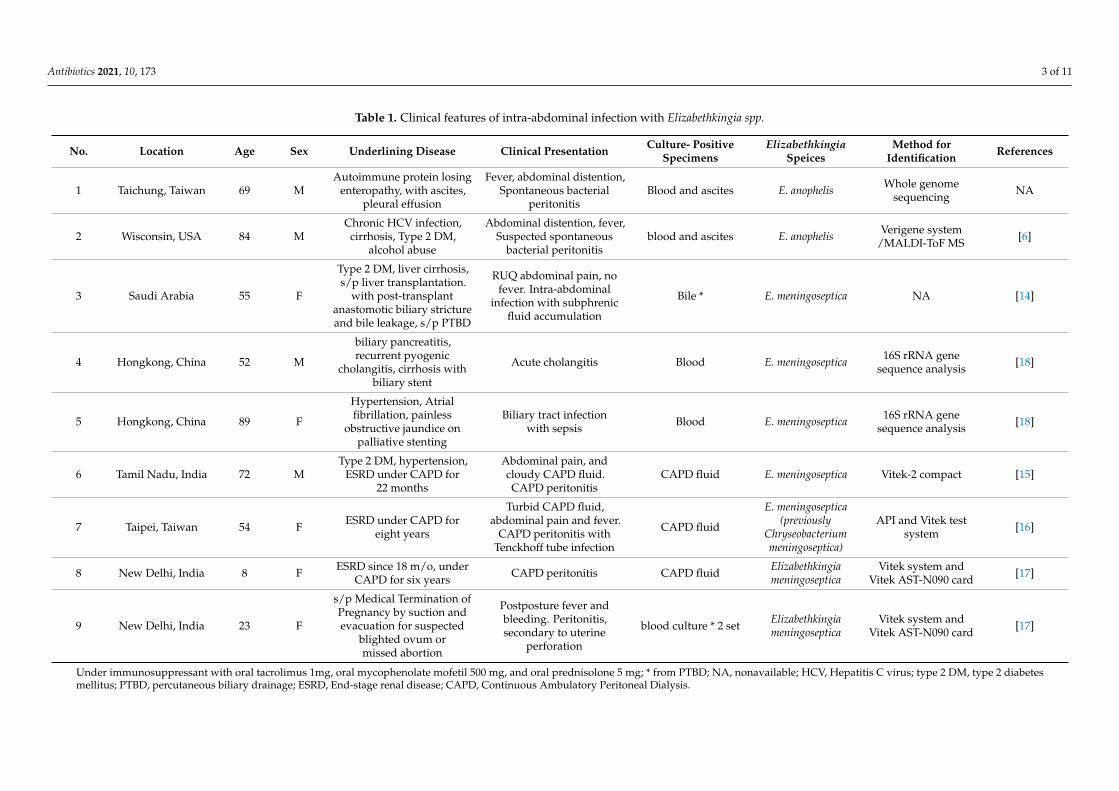
Table 1. Clinical features of intra-abdominal infection with Elizabethkingia spp.
No. Location Age Sex Underlining Disease Clinical Presentation Culture- PositiveSpecimens
ElizabethkingiaSpeices
Method forIdentification References
1 Taichung, Taiwan 69 MAutoimmune protein losing
enteropathy, with ascites,pleural effusion
Fever, abdominal distention,Spontaneous bacterial
peritonitisBlood and ascites E. anophelis Whole genome
sequencing NA
2 Wisconsin, USA 84 MChronic HCV infection,
cirrhosis, Type 2 DM,alcohol abuse
Abdominal distention, fever,Suspected spontaneous
bacterial peritonitisblood and ascites E. anophelis Verigene system
/MALDI-ToF MS [6]
3 Saudi Arabia 55 F
Type 2 DM, liver cirrhosis,s/p liver transplantation.
with post-transplantanastomotic biliary strictureand bile leakage, s/p PTBD
RUQ abdominal pain, nofever. Intra-abdominal
infection with subphrenicfluid accumulation
Bile * E. meningoseptica NA [14]
4 Hongkong, China 52 M
biliary pancreatitis,recurrent pyogenic
cholangitis, cirrhosis withbiliary stent
Acute cholangitis Blood E. meningoseptica 16S rRNA genesequence analysis [18]
5 Hongkong, China 89 F
Hypertension, Atrialfibrillation, painless
obstructive jaundice onpalliative stenting
Biliary tract infectionwith sepsis Blood E. meningoseptica 16S rRNA gene
sequence analysis [18]
6 Tamil Nadu, India 72 MType 2 DM, hypertension,
ESRD under CAPD for22 months
Abdominal pain, andcloudy CAPD fluid.CAPD peritonitis
CAPD fluid E. meningoseptica Vitek-2 compact [15]
7 Taipei, Taiwan 54 F ESRD under CAPD foreight years
Turbid CAPD fluid,abdominal pain and fever.
CAPD peritonitis withTenckhoff tube infection
CAPD fluid
E. meningoseptica(previously
Chryseobacteriummeningoseptica)
API and Vitek testsystem [16]
8 New Delhi, India 8 F ESRD since 18 m/o, underCAPD for six years CAPD peritonitis CAPD fluid Elizabethkingia
meningosepticaVitek system and
Vitek AST-N090 card [17]
9 New Delhi, India 23 F
s/p Medical Termination ofPregnancy by suction andevacuation for suspected
blighted ovum ormissed abortion
Postposture fever andbleeding. Peritonitis,secondary to uterine
perforation
blood culture * 2 set Elizabethkingiameningoseptica
Vitek system andVitek AST-N090 card [17]
Under immunosuppressant with oral tacrolimus 1mg, oral mycophenolate mofetil 500 mg, and oral prednisolone 5 mg; * from PTBD; NA, nonavailable; HCV, Hepatitis C virus; type 2 DM, type 2 diabetesmellitus; PTBD, percutaneous biliary drainage; ESRD, End-stage renal disease; CAPD, Continuous Ambulatory Peritoneal Dialysis.
Antibiotics 2021, 10, 173 4 of 11
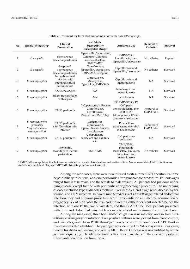
Table 2. Treatment for Intra-abdominal infection with Elizabethkingia spp.
No. Elizabethkingia spp. ClinicalPresentation
AntibioticSusceptibility
(Susceptible Drugs)Antibiotic Use Removal of
Catheter Survival
1 E. anophelis Spontaneousbacterial peritonitis
Pipercacillin/tazobactam,Cefepime, Cefopera-
zone/sulbactam;TMP/SMX *
TMP/SMX+Levofloxacin, then
Piperacillin/tazobactamNo catheter Expired
2 E. anophelisSuspected
spontaneousbacterial peritonitis
Ciprofloxacin,Piperacillin/tazobactam,TMP/SMX, Cefepime
Ciprofloxacin andPiperacillin/tazobactam No catheter Survived
3 E. meningoseptica
Intra-abdominalinfection with
subphrenic fluidaccumulation
Ciprofloxacin,Minocycline,
Tigecycline, TMP/SMX
Ciprofloxacin andmetronidazole NA Survived
4 E. meningoseptica Acute cholangitis NA Levofloxacin andmetronidazole NA Survived
5 E. meningoseptica Biliary tract infectionwith sepsis NA Levofloxacin NA Survived
6 E. meningoseptica CAPD peritonitis
Cefoperazone/sulbactam,Ciprofloxacin,Levofloxacin,
Minocycline, TMP/SMX
PO TMP/SMX + IVCefopera-
zone/sulbactam, thenshift to PO
Minocycline + IV Cef-operazone/sulbactam
Removal ofCAPD tube. Survived
7
E. meningoseptica(previously
Chryseobacteriummeningoseptica)
CAPD peritonitiswith Tenckhoff tube
infection
Gentamicin,Ciprofloxacin,
Piperacillin-tazobactam,Levofloxacin
Piperacillin-tazobactam, then shift
to Levofloxacin
Removal ofCAPD tube. Survived
8 E. meningoseptica CAPD peritonitisCefoperazone-
sulbactam and nalidixicacid
Cefoperazone-sulbactam NA Survived
9 E. meningosepticaPeritonitis,
secondary to uterineperforation
TMP/SMX
TMP/SMX,Piperacillin-
tazobactam, amikacin,teicoplanin andmetronidazole
No catheter Survived
* TMP/SMX-susceptible at first but became resistant in repeated blood culture and ascites culture; NA, nonavailable; CAPD, ContinuousAmbulatory Peritoneal Dialysis; TMP/SMX, Trimethoprim/sulfamethoxazole.
Among the nine cases, there were two infected ascites, three CAPD peritonitis, threehepato-biliary infections, and one peritonitis after gynecologic procedure. Patients agesranged from 8 to 89 years, and the female to male was 6:3. All patients had previous under-lying disease, except for one with peritonitis after gynecologic procedure. The underlyingdiseases included type II diabetes mellitus, liver cirrhosis, end-stage renal disease, hyper-tension, and HCV infection. In two of nine (22%) cases of Elizabethkingia-related abdominalinfection, they had previous procedure: liver transplantation and medical termination ofpregnancy. Six of nine cases (66.7%) had indwelling catheter or stent inserted before theinfection, with one PTBD, two biliary stent, and three CAPD tube. Most patients presentedwith fever and abdominal pain, but fever may be absent under immunosuppressant use.
Among the nine cases, three had Elizabethkingia anophelis infection and six had Eliza-bethkingia meningoseptica infection. Five positive cultures were yielded from blood culture,and bacteria growth from PTBD drainage in one case and from ascites or CAPD fluid infive cases was also identified. The pathogen was identified by Vitek 2 system in four cases,two by 16s rRNA sequencing, and one by MOLDI-ToF. Our case was re-identified by wholegenome sequencing. The identification method was unavailable in the case with postlivertransplantation infection from India.
Antibiotics 2021, 10, 173 5 of 11
Most cases with Elizabethkingia infection had broad antibiotic resistance but were sus-ceptible to ciprofloxacin, levofloxacin, Cefoperazone/sulbactam, minocycline, TMP/SMX,piperacillin, piperacillin/tazobactam, and tigecycline. Most cases survived after antibiotictreatment, but for those with catheter—especially CAPD tube—infection were controlledonly after removal of the CAPD tube.
Genomics Revealed Key Mutations Leading to TMP/SMX and Quinolone Resistance
The genome of Elizabethkingia anophelis SUE was sequenced by Nanopore and Il-lumina sequencing plateforms (Supplementary Table S1). The sequencing reads wereassembled into a circular genome of 4.2Mbp (NCBI accession number CP034247). Geneannotation revealed 3869 genes in the genome, including 3744 protein-coding genes, 73rRNAs/tRNAs/ncRNAs, and 52 pseudogenes. We compared the resistance determinantsof SUE with six other public Elizabethkingia anophelis genomes with antibiotic-resistant pro-files provided (Table 3, Supplementary Table S2). The Minimum Inhibitory Concentration(MIC) indicated three of them were resistant to TMP/SMX and to ciprofloxacin (Table 3,Supplementary Table S3), while the other four were sensitive to these agents.
Table 3. Comparison of seven Elizabethkingia anophelis genomes and antibiotic resistance.
Strains GenomeSize Genes Sequencing
Technology TMP/SMX Quinolone Status AssemblyNumber
SUE 4,201,198 bp 3869 Nanopore;Illumina MiSeq 160 R ≥4 R Circ. GCA_014702245.1
12012 4,023,312 bp 3700 Illumina MiSeq >2/38 R >2 R Linear GCA_001482795.1EM361-97 4,077,699 bp 3752 Illumina HiSeq >4/76 R >2 R Linear GCA_001703835.1
NUH1 4,334,661 bp 4031 Illumina MiSeq S S Linear GCA_000495995.1NUHP1 4,369,828 bp 4034 Illumina S S Linear GCA_000495935.2
Po0527107 4,032,057 bp 3717 IlluminaHiSeq-2000 S S Linear GCA_000689515.1
V0378064 4,036,754 bp 3804 IlluminaHiSeq-2001 S I Linear GCA_000689455.1
S: susceptible; I: intermediate; R: resistance.
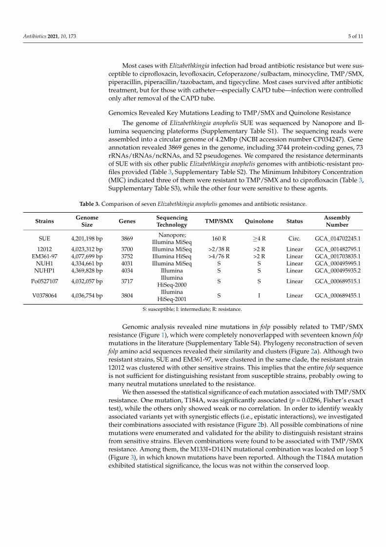
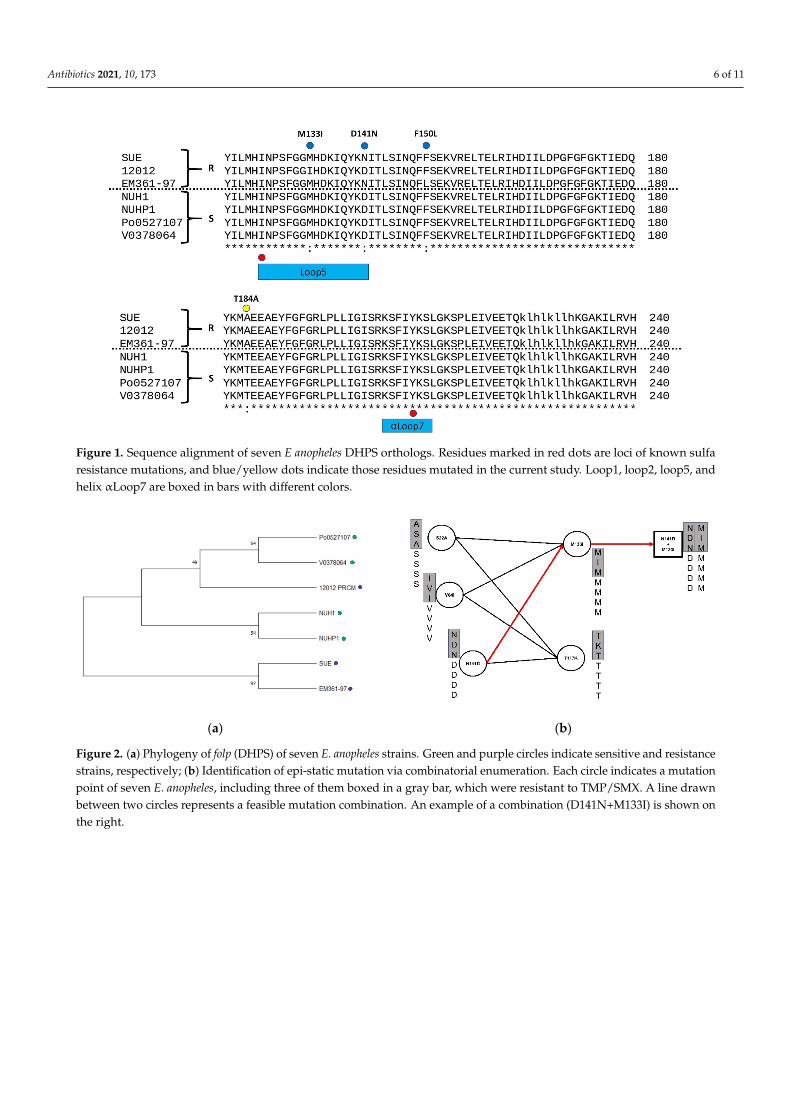
Genomic analysis revealed nine mutations in folp possibly related to TMP/SMXresistance (Figure 1), which were completely nonoverlapped with seventeen known folpmutations in the literature (Supplementary Table S4). Phylogeny reconstruction of sevenfolp amino acid sequences revealed their similarity and clusters (Figure 2a). Although tworesistant strains, SUE and EM361-97, were clustered in the same clade, the resistant strain12012 was clustered with other sensitive strains. This implies that the entire folp sequenceis not sufficient for distinguishing resistant from susceptible strains, probably owing tomany neutral mutations unrelated to the resistance.
We then assessed the statistical significance of each mutation associated with TMP/SMXresistance. One mutation, T184A, was significantly associated (p = 0.0286, Fisher’s exacttest), while the others only showed weak or no correlation. In order to identify weaklyassociated variants yet with synergistic effects (i.e., epistatic interactions), we investigatedtheir combinations associated with resistance (Figure 2b). All possible combinations of ninemutations were enumerated and validated for the ability to distinguish resistant strainsfrom sensitive strains. Eleven combinations were found to be associated with TMP/SMXresistance. Among them, the M133I+D141N mutational combination was located on loop 5(Figure 3), in which known mutations have been reported. Although the T184A mutationexhibited statistical significance, the locus was not within the conserved loop.
Antibiotics 2021, 10, 173 6 of 11Antibiotics 2021, 10, x of 11
Antibiotics 2021, 10, x. https://doi.org/10.3390/xxxxx www.mdpi.com/journal/antibiotics
Figure 1. Sequence alignment of seven E anopheles DHPS orthologs. Residues marked in red dots are loci of known sulfa resistance mutations, and blue/yellow dots indicate those residues mutated in the current study. Loop1, loop2, loop5, and helix αLoop7 are boxed in bars with different colors.
(a) (b)
Figure 2. (a) Phylogeny of folp (DHPS) of seven E. anopheles strains. Green and purple circles indicate sensitive and re-sistance strains, respectively; (b) Identification of epi-static mutation via combinatorial enumeration. Each circle indicates a mutation point of seven E. anopheles, including three of them boxed in a gray bar, which were resistant to TMP/SMX. A line drawn between two circles represents a feasible mutation combination. An example of a combination (D141N+M133I) is shown on the right.
We then assessed the statistical significance of each mutation associated with TMP/SMX resistance. One mutation, T184A, was significantly associated (p = 0.0286, Fisher’s exact test), while the others only showed weak or no correlation. In order to iden-tify weakly associated variants yet with synergistic effects (i.e., epistatic interactions), we investigated their combinations associated with resistance (Figure 2b). All possible com-binations of nine mutations were enumerated and validated for the ability to distinguish resistant strains from sensitive strains. Eleven combinations were found to be associated with TMP/SMX resistance. Among them, the M133I+D141N mutational combination was located on loop 5 (Figure 3), in which known mutations have been reported. Although the T184A mutation exhibited statistical significance, the locus was not within the conserved loop.
Figure 1. Sequence alignment of seven E anopheles DHPS orthologs. Residues marked in red dots are loci of known sulfaresistance mutations, and blue/yellow dots indicate those residues mutated in the current study. Loop1, loop2, loop5, andhelix αLoop7 are boxed in bars with different colors.
Antibiotics 2021, 10, x of 11
Antibiotics 2021, 10, x. https://doi.org/10.3390/xxxxx www.mdpi.com/journal/antibiotics
Figure 1. Sequence alignment of seven E anopheles DHPS orthologs. Residues marked in red dots are loci of known sulfa resistance mutations, and blue/yellow dots indicate those residues mutated in the current study. Loop1, loop2, loop5, and helix αLoop7 are boxed in bars with different colors.
(a) (b)
Figure 2. (a) Phylogeny of folp (DHPS) of seven E. anopheles strains. Green and purple circles indicate sensitive and re-sistance strains, respectively; (b) Identification of epi-static mutation via combinatorial enumeration. Each circle indicates a mutation point of seven E. anopheles, including three of them boxed in a gray bar, which were resistant to TMP/SMX. A line drawn between two circles represents a feasible mutation combination. An example of a combination (D141N+M133I) is shown on the right.
We then assessed the statistical significance of each mutation associated with TMP/SMX resistance. One mutation, T184A, was significantly associated (p = 0.0286, Fisher’s exact test), while the others only showed weak or no correlation. In order to iden-tify weakly associated variants yet with synergistic effects (i.e., epistatic interactions), we investigated their combinations associated with resistance (Figure 2b). All possible com-binations of nine mutations were enumerated and validated for the ability to distinguish resistant strains from sensitive strains. Eleven combinations were found to be associated with TMP/SMX resistance. Among them, the M133I+D141N mutational combination was located on loop 5 (Figure 3), in which known mutations have been reported. Although the T184A mutation exhibited statistical significance, the locus was not within the conserved loop.
Figure 2. (a) Phylogeny of folp (DHPS) of seven E. anopheles strains. Green and purple circles indicate sensitive and resistancestrains, respectively; (b) Identification of epi-static mutation via combinatorial enumeration. Each circle indicates a mutationpoint of seven E. anopheles, including three of them boxed in a gray bar, which were resistant to TMP/SMX. A line drawnbetween two circles represents a feasible mutation combination. An example of a combination (D141N+M133I) is shown onthe right.
Antibiotics 2021, 10, 173 7 of 11
Antibiotics 2021, 10, x of 11
Antibiotics 2021, 10, x. https://doi.org/10.3390/xxxxx www.mdpi.com/journal/antibiotics
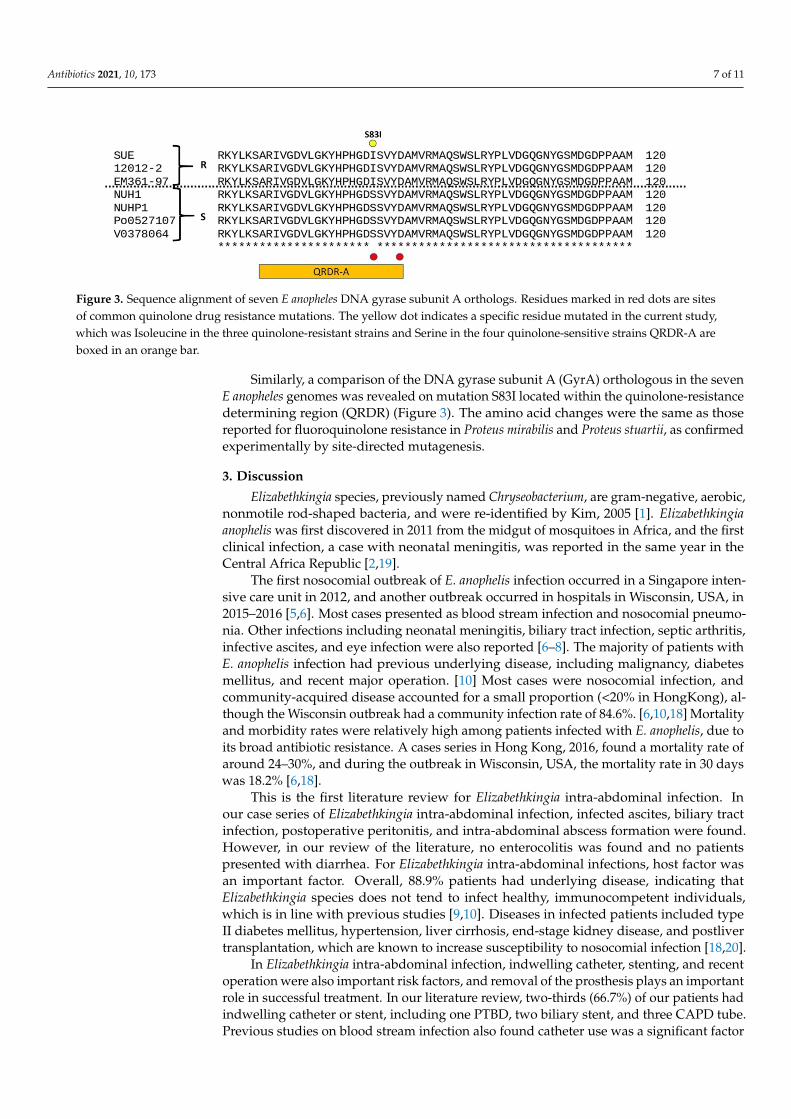
Similarly, a comparison of the DNA gyrase subunit A (GyrA) orthologous in the seven E anopheles genomes was revealed on mutation S83I located within the quinolone-resistance determining region (QRDR) (Figure 3). The amino acid changes were the same as those reported for fluoroquinolone resistance in Proteus mirabilis and Proteus stuartii, as confirmed experimentally by site-directed mutagenesis.
Figure 3. Sequence alignment of seven E anopheles DNA gyrase subunit A orthologs. Residues marked in red dots are sites of common quinolone drug resistance mutations. The yellow dot indicates a specific residue mutated in the current study, which was Isoleucine in the three quinolone-resistant strains and Serine in the four quinolone-sensitive strains QRDR-A are boxed in an orange bar.
3. Discussion Elizabethkingia species, previously named Chryseobacterium, are gram-negative, aero-
bic, nonmotile rod-shaped bacteria, and were re-identified by Kim, 2005 [1]. Elizabethkingia anophelis was first discovered in 2011 from the midgut of mosquitoes in Africa, and the first clinical infection, a case with neonatal meningitis, was reported in the same year in the Central Africa Republic [2,19].
The first nosocomial outbreak of E. anophelis infection occurred in a Singapore inten-sive care unit in 2012, and another outbreak occurred in hospitals in Wisconsin, USA, in 2015–2016 [5,6]. Most cases presented as blood stream infection and nosocomial pneumo-nia. Other infections including neonatal meningitis, biliary tract infection, septic arthritis, infective ascites, and eye infection were also reported [6–8]. The majority of patients with E. anophelis infection had previous underlying disease, including malignancy, diabetes mellitus, and recent major operation. [10] Most cases were nosocomial infection, and com-munity-acquired disease accounted for a small proportion (<20% in Hong-Kong), alt-hough the Wisconsin outbreak had a community infection rate of 84.6%. [6,10,18] Mortal-ity and morbidity rates were relatively high among patients infected with E. anophelis, due to its broad antibiotic resistance. A cases series in Hong Kong, 2016, found a mortality rate of around 24–30%, and during the outbreak in Wisconsin, USA, the mortality rate in 30 days was 18.2% [6,18].
This is the first literature review for Elizabethkingia intra-abdominal infection. In our case series of Elizabethkingia intra-abdominal infection, infected ascites, biliary tract infec-tion, postoperative peritonitis, and intra-abdominal abscess formation were found. How-ever, in our review of the literature, no enterocolitis was found and no patients presented with diarrhea. For Elizabethkingia intra-abdominal infections, host factor was an important factor. Overall, 88.9% patients had underlying disease, indicating that Elizabethkingia spe-cies does not tend to infect healthy, immunocompetent individuals, which is in line with previous studies [9,10]. Diseases in infected patients included type II diabetes mellitus, hypertension, liver cirrhosis, end-stage kidney disease, and postliver transplantation, which are known to increase susceptibility to nosocomial infection [18,20].
In Elizabethkingia intra-abdominal infection, indwelling catheter, stenting, and recent operation were also important risk factors, and removal of the prosthesis plays an im-portant role in successful treatment. In our literature review, two-thirds (66.7%) of our patients had indwelling catheter or stent, including one PTBD, two biliary stent, and three
Figure 3. Sequence alignment of seven E anopheles DNA gyrase subunit A orthologs. Residues marked in red dots are sitesof common quinolone drug resistance mutations. The yellow dot indicates a specific residue mutated in the current study,which was Isoleucine in the three quinolone-resistant strains and Serine in the four quinolone-sensitive strains QRDR-A areboxed in an orange bar.
Similarly, a comparison of the DNA gyrase subunit A (GyrA) orthologous in the sevenE anopheles genomes was revealed on mutation S83I located within the quinolone-resistancedetermining region (QRDR) (Figure 3). The amino acid changes were the same as thosereported for fluoroquinolone resistance in Proteus mirabilis and Proteus stuartii, as confirmedexperimentally by site-directed mutagenesis.
3. Discussion
Elizabethkingia species, previously named Chryseobacterium, are gram-negative, aerobic,nonmotile rod-shaped bacteria, and were re-identified by Kim, 2005 [1]. Elizabethkingiaanophelis was first discovered in 2011 from the midgut of mosquitoes in Africa, and the firstclinical infection, a case with neonatal meningitis, was reported in the same year in theCentral Africa Republic [2,19].
The first nosocomial outbreak of E. anophelis infection occurred in a Singapore inten-sive care unit in 2012, and another outbreak occurred in hospitals in Wisconsin, USA, in2015–2016 [5,6]. Most cases presented as blood stream infection and nosocomial pneumo-nia. Other infections including neonatal meningitis, biliary tract infection, septic arthritis,infective ascites, and eye infection were also reported [6–8]. The majority of patients withE. anophelis infection had previous underlying disease, including malignancy, diabetesmellitus, and recent major operation. [10] Most cases were nosocomial infection, andcommunity-acquired disease accounted for a small proportion (<20% in HongKong), al-though the Wisconsin outbreak had a community infection rate of 84.6%. [6,10,18] Mortalityand morbidity rates were relatively high among patients infected with E. anophelis, due toits broad antibiotic resistance. A cases series in Hong Kong, 2016, found a mortality rate ofaround 24–30%, and during the outbreak in Wisconsin, USA, the mortality rate in 30 dayswas 18.2% [6,18].
This is the first literature review for Elizabethkingia intra-abdominal infection. Inour case series of Elizabethkingia intra-abdominal infection, infected ascites, biliary tractinfection, postoperative peritonitis, and intra-abdominal abscess formation were found.However, in our review of the literature, no enterocolitis was found and no patientspresented with diarrhea. For Elizabethkingia intra-abdominal infections, host factor wasan important factor. Overall, 88.9% patients had underlying disease, indicating thatElizabethkingia species does not tend to infect healthy, immunocompetent individuals,which is in line with previous studies [9,10]. Diseases in infected patients included typeII diabetes mellitus, hypertension, liver cirrhosis, end-stage kidney disease, and postlivertransplantation, which are known to increase susceptibility to nosocomial infection [18,20].
In Elizabethkingia intra-abdominal infection, indwelling catheter, stenting, and recentoperation were also important risk factors, and removal of the prosthesis plays an importantrole in successful treatment. In our literature review, two-thirds (66.7%) of our patients hadindwelling catheter or stent, including one PTBD, two biliary stent, and three CAPD tube.Previous studies on blood stream infection also found catheter use was a significant factor
Antibiotics 2021, 10, 173 8 of 11
for Elizabethkingia blood-stream infection [18]. Among the cases with CAPD peritonitis,infection was finally brought under control in two out of three cases after removal of theCAPD tube, suggesting that the removal of the catheter may be an important factor insuccessful treatment [15–17]. In addition to indwelling catheter or stent, recent operationor invasive procedure was also a significant risk factor for Elizabethkingia intra-abdominalinfection. In our literature review, one patient had recent liver transplantation from aliving donor within one month and the other patients had peritonitis just after medicationtermination of pregnancy [14,17]. The first outbreak in Singapore was noted in a surgicalintensive care unit and in a cardiothoracic intensive care unit; three out of five patients werefound to have E. anophelis infection after surgery [5]. Therefore, Elizabethkingia infectionmay be an important pathogen in postoperation infection and nosocomial infection.
Elizabethkingia anophelis caused the majority of Elizabethkingia infection, but true preva-lence was underestimated because of misidentification. Most Elizabethkingia anophelis wasmisidentified as E. meningoseptica, by Vitek 2 and MOLDI-ToF [3,4,21]. E. anophelis, whichwas identified by 16s rRNA, accounted for 96.2% of Elizabethkingia infections in Singa-pore [3]. In another study in Korea, E. anopheles, re-identified by 16s rRNA sequencing,caused 59.3% of clinical Elizabethkingia infections [4]. In the study in Singapore duringthe period of 2009–2017, 76/79 of (96.2%) E. anophelis infections were misidentified as E.meningoseptica infections by MOLDI-ToF (bioMérieux) [3]. In a study by Lin. et al. in Taiwan,MALDI-TOF with knowledge base v 2.0/3.0 and Vitek 2 compact could only identify 26.5%Elizabethkingia species correctly when using 16s rRNA sequencing as the gold standard,and misidentified most Elizabethkingia species as Elizabethkingia meningoseptica [21]. In ourcase series, most cases with E. meningoseptica intra-abdominal infection were identified bythe Vitek 2 compact, and misidentification was shown to be a problem. Misidentificationcan be improved by using 16s rRNA as the current standard and using the MOLDI-ToFsystem with change in databases and inclusion of mass spectra from seven E. anophelisisolates or SARAMIS database [4,18].
Elizabethkingia anophelis has exhibited broad antibiotic resistance; fluoroquinolones,TMP/SMX and piperacillin/tazobactam have been used as the first-line therapy [9,11].However, there is large variability in susceptibility to fluoroquinolones (9.8%~70%), includ-ing ciprofloxacin and levofloxacin [11,22,23]. Elizabethkingia infection with resistance tofluoroquinolone can cause increased mortality [24]. Most resistance to fluoroquinolone inE. anophelis was caused by mutations in quinolone-resistance determining regions (QRDR),i.e., a single amino acid alteration in DNR gyrase or DNA topoisomerase IV [25,26]. Mostcommon QRDR were noted in GyrA, including Ser83Ile, Ser83Arg [11,25,26]. Lin alsoreported other nonsynonymous alteration sites in the QRDR: two in GyrA (positions95 and 102), and three in GyrB (positions 425, 452, and 470) [11]. Asp87Asn in GyrAwas also reported in E. miricola [25]. In a study by Ming-Jr Jian, a 12.7-fold increase inthe fluoroquinolone-related efflux pump AcrB was noted in fluoroquinolone-resistantElizabethkingia anophelis strains, which may play a role in fluoroquinolone resistance [26].Currently, no mutations in ParC or ParE have been reported in Elizabethkingia species.Further studies on other possible mechanisms for resistance are required to gain a moredetailed understanding of quinolone resistance in Elizabethkingia species, such as effluxpump, drug-modifying enzyme, or plasmid mediated quinolone resistance [27].
There is limited information on resistance to Trimethoprim/sulfamethoxazole in Eliza-bethkingia species. Positive dfrA12, sul I and sul II gene in Elizabethkingia with resistance toTMP/SMX was found by PCR in a previous study [28]. The Sul I gene may be associatedwith integron, but no type I nor type II integron was noted in their study. However, therewere still several strains with resistance to TMP/SMX that showed a negative finding ina previous study, suggesting other possible mechanisms than sul I, II, and dfr A1-12. Wereported mutations in folP, including T184A, M133I and D141N, which may be associ-ated with TMP/SMX resistance. However, as in previous studies, these mutations werenot present in all resistant strains. There may be multiple other mutations and mecha-
Antibiotics 2021, 10, 173 9 of 11
nisms involved in TMP/SMX resistance in Elizabethkingia, and thus, further investigationis needed.
4. Materials and Methods4.1. Literature Review
We searched the English-language medical literature using PubMed/MEDLINE andGoogle Scholar from 1990 to 2019, using the following keywords: Elizabethkingia meningosep-tica, Elizabethkingia anophelis, intra-abdominal infection, ascites infection, biliary infection.The references of articles found using this search were also reviewed to identify otherpotential cases that were not located using the search terms.
4.2. Whole Genome Sequencing and Bioinformatics Analysis
The SUE genome was deeply sequenced using Nanopore long-read sequencing andIllumina short-read sequencing (Supplementary Table S2). Adaptor sequences left inlong reads were trimmed using Porechop. The remaining reads were hybrid assembledby Unicycler (v0.4.7) into a 4.2 Mbp circular genome. Protein-coding genes, coding andnoncoding RNAs in the chromosomes, and plasmids were annotated by NCBI PGAPpipeline. Antibiotic-resistant genes were predicted by aligning protein-coding genes againstthe Comprehensive Antibiotic Resistance Database (CARD) using Diamond. Only ARGswith alignment coverage greater than 90% were retained. Efflux pumps were excludedfrom ARG analysis.
Multiple sequence alignment of DHPS (folP) and GyrA of the seven E anophelesgenomes were carried out by MEGA X in order to identity mutation loci. The multi-ple sequence alignment of DHPS was also used to generate a phylogeny tree by MEGAX. The nine mutations within DHPS were tested for association with sulfa resistance viaFisher’s exact test. All possible combinations of these nine mutations (29) were testedfor correlation with TMP resistance by solving a combinatorial problem known as theminimum test collection; that is, only the combinations able to distinguish resistant strainsfrom sensitive strains are retained as solutions.
5. Conclusions
Elizabethkingia intra-abdominal infection is a relatively uncommon condition to whichpatients with underlying disease are more susceptible. In our review of intra-abdominalinfections, infective ascites, biliary tract infection, CAPD peritonitis, and postproceduralperitonitis were reported. Indwelling catheter and recent invasive procedure may be im-portant risk factors, and removal of catheter was shown to be key to successful treatment.Elizabethkingia anophelis infection rates may be underestimated due to misidentification.We also conducted a genomic analysis to investigate antibiotic resistance genes in Eliza-bethkingia anophelis. Fol p was found to be associated with SMX/TMP resistance, and Gyr Awas related to fluroquinolone resistance. However, these mutations were not present in allresistant strains, and multiple mutations may be associated with resistance. Further studieson the mechanism of resistance to SMX/TMP in Elizabethkingia infection are needed.
Supplementary Materials: The following are available online at https://www.mdpi.com/2079-6382/10/2/173/s1, Table S1: Summary of sequencing and assembly statistics, Table S2: Genome featuresof strains analysed in the study, Table S3: Antibiotic susceptibility profile of strains analyzed in thestudy, Table S4: Identified folp mutations in SUE, Table S5: folP mutation combinations in SUE.
Author Contributions: Conceptualization, Y.-T.H. and P.-Y.L.; methodology, L.-C.T., J.-M.W., H.-Y.L. and Y.-C.M.; software, H.-Y.L., Y.-C.M. and K.-L.L.; validation, H.-Y.L., Y.-C.M., K.-L.L. andC.-H.T.; formal analysis, H.-Y.L., Y.-T.H. and P.-Y.L.; investigation, H.-Y.L., Y.-C.M., K.-L.L. andC.-H.T.; resources, Y.-C.M., K.-L.L., Y.-T.H. and P.-Y.L.; data curation, H.-Y.L., Y.-C.M. and K.-L.L.;writing—original draft preparation, L.-C.T., J.-M.W., Y.-T.H. and P.-Y.L.; writing—review and editing,L.-C.T., J.-M.W., Y.-T.H. and P.-Y.L.; visualization, L.-C.T., J.-M.W., Y.-T.H. and P.-Y.L.; supervision,J.-M.W., Y.-T.H. and P.-Y.L.; project administration, J.-M.W., Y.-T.H. and P.-Y.L.; funding acquisition,Y.-T.H. and P.-Y.L. All authors have read and agreed to the published version of the manuscript.
Antibiotics 2021, 10, 173 10 of 11
Funding: YTH was supported in part by the Ministry of Science and Technology (109-2221-E-194-038 -MY3). PYL was supported in part by the by the Ministry of Science and Technology (109-2314-B-075A-009) and Taichung Veterans General Hospital (TCVGH-1103901C).
Institutional Review Board Statement: The study was conducted according to the guidelines ofthe Declaration of Helsinki and approved by the Institutional Review Board of Taichung VeteransGeneral Hospital.
Informed Consent Statement: Informed consent was obtained from all subjects involved in the study.
Data Availability Statement: All the sequencing data have been deposited in GenBank underBioProject ID no. PRJNA507867.
Conflicts of Interest: The authors declare no conflict of interest. The funders had no role in the designof the study; in the collection, analyses, or interpretation of data; in the writing of the manuscript, orin the decision to publish the results.
References1. Kim, K.K.; Kim, M.K.; Lim, J.H.; Park, H.Y.; Lee, S.-T. Transfer of Chryseobacterium meningosepticum and Chryseobacterium miricola
to Elizabethkingia gen. nov. as Elizabethkingia meningoseptica comb. nov. and Elizabethkingia miricola comb. nov. Int. J. Syst. Evol.Microbiol. 2005, 55, 1287–1293. [CrossRef] [PubMed]
2. Kämpfer, P.; Matthews, H.; Glaeser, S.P.; Martin, K.; Lodders, N.; Faye, I. Elizabethkingia anophelis sp. nov., isolated from themidgut of the mosquito Anopheles gambiae. Int. J. Syst. Evol. Microbiol. 2011, 61, 2670–2675. [CrossRef] [PubMed]
3. Chew, K.L.; Cheng, B.; Lin, R.T.P.; Teo, J.W.P. Elizabethkingia anophelis is the dominant Elizabethkingia species found in bloodcultures in singapore. J. Clin. Microbiol. 2017, 56, e01445-17. [CrossRef]
4. Han, M.-S.; Kim, H.; Lee, Y.; Kim, M.; Ku, N.S.; Choi, J.Y.; Yong, D.; Jeong, S.H.; Lee, K.; Chong, Y. Relative prevalence andantimicrobial susceptibility of clinical isolates of Elizabethkingia species based on 16S rRNA gene sequencing. J. Clin. Microbiol.2017, 55, 274–280. [CrossRef]
5. Teo, J.; Tan, S.Y.-Y.; Tay, M.; Ding, Y.; Kjelleberg, S.; Givskov, M.; Lin, R.T.; Yang, L. First case of E anophelis outbreak in anintensive-care unit. Lancet 2013, 382, 855–856. [CrossRef]
6. Figueroa Castro, C.E.; Johnson, C.; Williams, M.; VanDerSlik, A.; Graham, M.B.; Letzer, D.; Ledeboer, N.; Buchan, B.W.; Block, T.;Borlaug, G.; et al. Elizabethkingia anophelis: Clinical experience of an academic health system in southeastern wisconsin. OpenForum Infect. Dis. 2017, 4, ofx251. [CrossRef]
7. Lin, J.-N.; Lai, C.-H.; Yang, C.-H.; Huang, Y.-H. Elizabethkingia infections in humans: From genomics to clinics. Microorganisms2019, 7, 295. [CrossRef]
8. Bulagonda, E.P.; Manivannan, B.; Mahalingam, N.; Lama, M.; Chanakya, P.P.; Khamari, B.; Jadhao, S.; Vasudevan, M.; Nagaraja, V.Comparative genomic analysis of a naturally competent Elizabethkingia anophelis isolated from an eye infection. Sci. Rep. 2018,8, 1–10. [CrossRef] [PubMed]
9. Wang, M.; Gao, H.; Lin, N.; Zhang, Y.; Huang, N.; Walker, E.D.; Ming, D.; Chen, S.; Hu, S. The antibiotic resistance andpathogenicity of a multidrug-resistant Elizabethkingia anophelis isolate. Microbiologyopen 2019, 8, e804. [CrossRef] [PubMed]
10. Janda, J.M.; Lopez, D.L. Mini review: New pathogen profiles: Elizabethkingia anophelis. Diagn. Microbiol. Infect. Dis. 2017,88, 201–205. [CrossRef]
11. Lin, J.-N.; Lai, C.-H.; Yang, C.-H.; Huang, Y.-H. Comparison of clinical manifestations, antimicrobial susceptibility patterns, andmutations of fluoroquinolone target genes between Elizabethkingia meningoseptica and Elizabethkingia anophelis isolated in Taiwan.J. Clin. Med. 2018, 7, 538. [CrossRef]
12. Burnard, D.; Gore, L.; Henderson, A.; Ranasinghe, A.; Bergh, H.; Cottrell, K.; Sarovich, D.S.; Price, E.P.; Paterson, D.L.; Harris,P. Comparative genomics and antimicrobial resistance profiling of Elizabethkingia isolates reveal nosocomial transmission andin vitro susceptibility to fluoroquinolones, tetracyclines, and trimethoprim-sulfamethoxazole. J. Clin. Microbiol. 2020, 58.[CrossRef]
13. Liang, C.-Y.; Yang, C.-H.; Lai, C.-H.; Huang, Y.-H.; Lin, J.-N. Comparative genomics of 86 whole-genome sequences in the sixspecies of the Elizabethkingia genus reveals intraspecific and interspecific divergence. Sci. Rep. 2019, 9, 1–11. [CrossRef]
14. Musalem, H.M.; Honjol, Y.N.; Tuleimat, L.M.; Al Abbad, S.I.; Alsohaibani, F.I. Elizabethkingia Meningoseptica in a case of biliarytract infection following liver transplantation. Am. J. Case Rep. 2017, 18, 1014–1019. [CrossRef]
15. Ranjan, S.; Veerappan, I.; Patil, S.; Sethuraman, R. Elizabethkingia meningoseptica peritonitis in continuous ambulatory peritonealdialysis patient: A rare case report with diagnostic challenges. Indian J. Pathol. Microbiol. 2017, 60, 626. [CrossRef] [PubMed]
16. Wu, V.-C.; Tsai, T.-J.; Wang, R.; Hsueh, P.-R. Peritonitis caused by Chryseobacterium meningosepticum in a patient undergoingcontinuous ambulatory peritoneal dialysis. J. Formos. Med. Assoc. 2003, 102, 270–272. [PubMed]
17. Khan, I.D.; Lall, M.; Sen, S.; Ninawe, S.; Chandola, P. Multiresistant Elizabethkingia meningoseptica infections in tertiary care. Med. J.Armed Forces India 2015, 71, 282–286. [CrossRef]
Antibiotics 2021, 10, 173 11 of 11
18. Lau, S.K.; Chow, W.-N.; Foo, C.-H.; Curreem, S.O.; Lo, G.C.-S.; Teng, J.L.; Chen, J.H.; Ng, R.H.; Wu, A.K.; Cheung, I.Y. Eliz-abethkingia anophelis bacteremia is associated with clinically significant infections and high mortality. Sci. Rep. 2016, 6, 1–10.[CrossRef] [PubMed]
19. Frank, T.; Gody, J.C.; Nguyen, L.B.L.; Berthet, N.; Le Fleche-Mateos, A.; Bata, P.; Rafaï, C.; Kazanji, M.; Breurec, S. First case ofElizabethkingia anophelis meningitis in the Central African Republic. Lancet 2013, 381, 734–737. [CrossRef]
20. Choi, M.H.; Kim, M.; Jeong, S.J.; Choi, J.Y.; Lee, I.-Y.; Yong, T.-S.; Yong, D.; Jeong, S.H.; Lee, K. Risk Factors for Elizabethkingiaacquisition and clinical characteristics of patients, South Korea. Emerg. Infect. Dis. 2019, 25, 42–51. [CrossRef]
21. Lin, J.-N.; Lai, C.-H.; Yang, C.-H.; Huang, Y.-H.; Lin, H.-F.; Lin, H.-H. Comparison of four automated microbiology systems with16S rRNA gene sequencing for identification of Chryseobacterium and Elizabethkingia species. Sci. Rep. 2017, 7, 1–5. [CrossRef][PubMed]
22. Perrin, A.; Larsonneur, E.; Nicholson, A.C.; Edwards, D.J.; Gundlach, K.M.; Whitney, A.M.; Gulvik, C.A.; Bell, M.E.; Rendueles,O.; Cury, J.; et al. Evolutionary dynamics and genomic features of the Elizabethkingia anophelis 2015 to 2016 Wisconsin outbreakstrain. Nat. Commun. 2017, 8, 15483. [CrossRef] [PubMed]
23. Wang, L.; Zhang, X.; Li, D.; Hu, F.; Wang, M.; Guo, Q.; Yang, F. Molecular characteristics and antimicrobial susceptibility profilesof Elizabethkingia clinical isolates in Shanghai, China. Infect. Drug Resist. 2020, 13, 247–256. [CrossRef] [PubMed]
24. Huang, Y.-C.; Lin, Y.-T.; Wang, F.-D.; Chan, Y.-J.; Yang, T.-C.; Huang, Y.-W. Risk factors and outcome of levofloxacin-resistantElizabethkingia meningoseptica bacteraemia in adult patients in Taiwan. Eur. J. Clin. Microbiol. Infect. Dis. 2017, 36, 1373–1380.[CrossRef] [PubMed]
25. Lin, J.-N.; Lai, C.-H.; Yang, C.-H.; Huang, Y.-H.; Lin, H.-H. Clinical manifestations, molecular characteristics, antimicrobialsusceptibility patterns and contributions of target gene mutation to fluoroquinolone resistance in Elizabethkingia anophelis.J. Antimicrob. Chemother. 2018, 73, 2497–2502. [CrossRef]
26. Jian, M.-J.; Cheng, Y.-H.; Chung, H.-Y.; Cheng, Y.-H.; Yang, H.-Y.; Hsu, C.-S.; Perng, C.-L.; Shang, H.-S. Fluoroquinolone re-sistancein carbapenem-resistant Elizabethkingia anophelis: Phenotypic and genotypic characteristics of clinical isolates with topoisomerasemutations and comparative genomic analysis. J. Antimicrob. Chemother. 2019, 74, 1503–1510. [CrossRef]
27. Correia, S.; Poeta, P.; Hébraud, M.; Capelo, J.L.; Igrejas, G. Mechanisms of quinolone action and resistance: Where do we stand?J. Med. Microbiol. 2017, 66, 551–559. [CrossRef] [PubMed]
28. Jiang, X.; Wang, D.; Wang, Y.; Yan, H.; Shi, L.; Zhou, L.-J. Occurrence of antimicrobial resistance genes sul and dfrA12 in hospitalenvironmental isolates of Elizabethkingia meningoseptica. World J. Microbiol. Biotechnol. 2012, 28, 3097–3102. [CrossRef]


![Intra-Abdominal and Abdominal Wall Desmoid Fibromatosis · intra-abdominal and involving the small bowel mesentery [2]. TREATMENT Surgery Margin-negative resection has historically](https://img.pdfslide.us/doc/110x75/5e5a290071d21b380f5b7e74/intra-abdominal-and-abdominal-wall-desmoid-fibromatosis-intra-abdominal-and-involving.jpg)