Embed Size (px)
Citation preview
REVIEW
Intra-Abdominal and Abdominal Wall DesmoidFibromatosis
J. Harrison Howard . Raphael E. Pollock
To view enhanced content go to www.oncologytherapy-open.comReceived: November 23, 2015 / Published online: February 3, 2016� The Author(s) 2016. This article is published with open access at Springerlink.com
ABSTRACT
Desmoid fibromatosis is a rare but locally
aggressive tumor comprised of myofibroblasts.
Desmoids do not have the ability to metastasize
but can cause significant morbidity and
mortality by local invasion. These tumors may
occur throughout the body, but are commonly
found on the abdominal wall and within the
intestinal mesentery. Desmoids in these areas
may cause unique clinical problems for
physicians and patients. Mutations in either
the b-catenin or the APC genes are usually the
cause for the development of these tumors with
the former comprising the sporadic
development of tumors and the latter being
associated with familial adenomatous polyposis
syndrome. Surgical resection with histologically
negative margins has been the cornerstone of
therapy for this disease, but this paradigm has
begun to shift. It is now common to accept a
microscopically positive margin after resection
as recurrence rates may not be significantly
affected. An even more radical evolution in
management has been the recent movement
towards ‘‘watchful waiting’’ when new desmoids
are diagnosed. As the natural history of
desmoids has become better understood, it is
evident that some tumors will not grow and
may even spontaneously regress sparing
patients the morbidity of more aggressive
therapy. Other modalities of treatment for
desmoids include radiation and systemic
therapy which both can be used adjuvantly or
as definitive therapy and have shown durable
response rates as single therapy regimens. The
decision to use radiation and/or systemic
therapies is often based on tumor biology,
tumor location, surgical morbidity, and
patient preference. Systemic therapy options
have increased to include hormonal therapies,
non-steroidal anti-inflammatory drugs and
chemotherapy, as well as targeted therapies.
Unfortunately, the rarity of this disease has
resulted in a scarcity of randomized trials to
Electronic supplementary material The onlineversion of this article (doi:10.1007/s40487-016-0017-z)contains supplementary material, which is available toauthorized users.
J. H. Howard (&) � R. E. PollockDivision of Surgical Oncology, The Ohio StateUniversity Wexner Medical Center, Arthur G. JamesCancer Hospital and Richard J. Solove ResearchInstitute, M256 Starling Loving Hall, 320 W. 10thAve, Columbus, OH 43210, USAe-mail: [email protected]
Oncol Ther (2016) 4:57–72
DOI 10.1007/s40487-016-0017-z
evaluate any of these therapies emphasizing the
need for this disease to be treated at high
volume multidisciplinary institutions.
Keywords: Abdominal desmoid tumor;
Abdominal wall desmoid tumor; Desmoid
fibromatosis; Desmoid tumor
INTRODUCTION
Desmoid tumors present a difficult problem for
both physician and patient. While many options
for treatment are available, the natural history of
this disease can be unpredictable resulting in a
complex, multidisciplinary decision-making
process with treatment algorithms that include
combinations of radical surgery, systemic
therapy and radiation, but may be as minimal
as close observation. As a disease that has been
referred to as a ‘‘benign malignancy’’ it may be
challenging for patients to understand why such
aggressive measures are needed for a disease that
does not metastasize. Conversely, patients may
be anxious about observing a tumor that has
potential to grow and become locally invasive.
These tumors can occur nearly anywhere on the
body, but this discussion will focus on the
biology and treatment of abdominal wall
desmoids and intra-abdominal desmoid tumors.
This article is based on previously conducted
studies and does not involve any new studies of
human or animal subjects performed by any of
the authors.
NATURAL HISTORY
Desmoid tumors are rare with an incidence of
0.03% of all neoplasms and 3% of soft tissue
tumors [1]. The majority of desmoid tumors (also
known as aggressive fibromatosis) occur
sporadically, but can be associated with familial
adenomatous polyposis syndrome (FAP).
FAP-associated desmoid tumors occur in 2–15%
of all desmoid patients, and are typically located
in the small bowel mesentery [2–5]. Desmoids
are seen more commonly in women than men
with a 2:1 predilection for females [4, 6, 7].
Young adult populations are most commonly
affected and this disease is frequently seen in the
25–35 year old demographic [4, 6].
Histologically, desmoid tumors are a benign
proliferation of myofibroblasts without a
capsule that locally infiltrates surrounding
tissues and may be multifocal. These
characteristics result in a tumor that is difficult
to control locally, but does not have the ability
to metastasize [8–10]. Desmoids not only often
occur on the abdominal wall or the mesentery
of the small intestine, but also affect the
shoulder girdle, chest wall and extremities.
These tumors are often thought of as a chronic
disease and require close observation regardless
of treatment strategy as they have potential for
mortality due to their locally aggressive
behavior and ability to invade adjacent critical
organs and structures [3].
RISK FACTORS AND GENETICPREDISPOSITION
The majority of cases of abdominal wall
desmoid tumor are sporadic in nature, but
there are several associated factors.
Development of desmoids is most commonly
associated with genetic predisposition,
pregnancy, hormonal exposure, and physical
factors such as trauma and/or surgery [6, 7]. A
classic presentation of desmoid includes a
post-partum woman with an abdominal wall
mass. Given the identification of estrogen
receptors on desmoid tumors, a predilection
58 Oncol Ther (2016) 4:57–72
for women and the use of anti-estrogen
therapies to manage these tumors, there does
seem to be validity in the involvement of
estrogen in disease progression [3, 11]. While
there is no clear relationship between
pregnancy and abdominal wall desmoid
tumors, a recent study of pregnancy-related
desmoid fibromatosis showed that these
tumors have an indolent course, do not
increase risk of obstetric complications and
should not be a contraindication for
subsequent pregnancies [12]. This study did
note that in women diagnosed with desmoid
tumor prior to becoming pregnant there was a
42% chance of either disease recurrence or
progression during pregnancy, suggesting a
different biology than disease that developed
during or after pregnancy. For women who
developed desmoid tumor associated with
pregnancy, 52% were managed successfully
without surgical resection, and a local relapse
of 13% was observed for those who did have
surgical resection. Ultimately, the study
concluded that desmoid fibromatosis that
develops or progresses during pregnancy is
safely managed with minimal risk to both
mother and child.
Several other patient-associated risk factors
have been studied with regard to prognosis.
Specifically patient gender, tumor size, tumor
site, and presentation (primary vs recurrent)
have been correlated with clinical outcomes. In
a multi-institutional retrospective review, Peng
et al. found that younger age and
extra-abdominal tumor location were
associated with poorer recurrence-free survival
[13]. Several studies have confirmed that young
age, large tumor size, and extra-abdominal
location are predictors of poor outcomes.
Patient sex and primary vs recurrent disease
were not prognostic for recurrence [14, 15].
Conversely, other studies have correlated
recurrent disease with having worse 5- and
10-year event-free survival when compared to
new primary tumors [16, 17]. While not
reaching statistical significance, Lev et al. also
found that age of \30 years and extremity
tumor site were associated with higher
recurrence rates [18]. As larger series becomes
available, it seems that younger age, large tumor
size, and anatomic location may play a true role
in defining recurrence risk for desmoid tumors.
Based on these data, abdominal wall desmoids
are likely to have a more favorable outcome.
Beyond these classic clinical prognostic factors,
a better understanding of the genetic
fingerprint of desmoids may be more reliable
in stratifying disease aggressiveness. Studies
have begun to identify gene expression
profiles that may better predict tumor biology
[19].
Alterations in the Wnt signaling pathway
have been identified as the likely driving
mechanism of tumorigenesis in the
development of desmoid fibromatosis [20, 21].
In general, deregulation of the Wnt pathway
results in aberrant proliferation, migration, and
differentiation of cells into human cancers [20].
Two specific genetic mutations that alter this
pathway and are associated with the
development of desmoid tumors have been
identified: the CTNNB1 gene, coding for the
b-catenin protein, and the APC gene, coding for
the adenomatous polyposis coli (APC) protein.
Both b-catenin and APC are part of the Wnt
pathway suggesting that two separate
mutations affecting the same endpoint are
involved with the development of desmoid
fibromatosis [3]. These mutations result in the
development of intranuclear accumulation of
b-catenin which subsequently stimulates DNA
transcription and cell proliferation [21].
Eighty-five percent of sporadic desmoid
tumors have been identified to have an
Oncol Ther (2016) 4:57–72 59
activating mutation in the CTNNB1 gene
coding for b-catenin. The germline mutation
of the APC gene leads to the development of
desmoid tumors for FAP patients [21, 22]. A
recent genetic analysis study by Crago et al.
looking at ‘‘wild type’’ desmoids without the
known CTNNB1 or APC mutations found that
with deep sequencing 95% of desmoids may
have mutations that affect the Wnt/b-catenin
pathway, suggesting a near universal
relationship between desmoid tumors and
Wnt signaling [23].
For sporadic disease, there is a high rate of
mutation in the gene encoding for b-catenin on
chromosome 3, CTNNB1. CTNNB1 mutations
have been found in 71–91% of sporadic
desmoid tumors with the highest rate of
mutation found in intra-abdominal tumors
[21, 24–28]. The most commonly documented
mutations found on the gene are T41A and
S45F, with the latter being nearly exclusive to
extra-abdominal desmoids. Several studies have
shown a significantly higher chance of disease
recurrence at 5 years despite complete resection
of disease for patients harboring an S45F
mutation [21, 26, 28]. Domont et al. found
higher recurrence rates for tumors with
CTNNB1 mutations, but could not correlate
this finding with any specific point mutations.
The growing amount of data suggests that
specific mutations within this gene do play a
role in disease recurrence and may influence
clinical care in the future based on the tumor’s
genetic fingerprint.
For patients with wild type b-catenin, the
mutated APC gene is suspected to be the source
of development of desmoid tumors. While
patients with FAP represent the minority of
desmoid cases, patients with this syndrome
have nearly an 850-fold increased chance of
development of desmoids compared to the
general population. Approximately 10–15% of
patients with FAP develop desmoids and it has
become the primary cause of death in patients
with FAP that have previously had a
prophylactic colectomy [22, 29]. As a tumor
suppressor gene, when APC is mutated it is
unable to properly regulate b-catenin levels
through the Wnt pathway leading to
proliferation and accumulation of b-catenin in
cell cytoplasm and nucleus. Interestingly,
despite a similar molecular endpoint between
APC and b-catenin mutations, the phenotype of
APC-mutated tumors is different as the majority
of tumors in patients with FAP are
intra-abdominal and involving the small
bowel mesentery [2].
TREATMENT
Surgery
Margin-negative resection has historically been
the gold standard for treatment of abdominal
wall and intra-abdominal desmoid tumors.
However, as a better understanding of the
natural history of desmoids developed,
strategies for surgical resection have evolved.
The initial surgical approach has transitioned
from margin-negative resection to acceptance
of microscopically-positive resection to
observation with surgery used more selectively.
Margin Status
When surgical resection is employed for
desmoid tumors, many clinicians encourage
complete microscopic resection [5, 13, 17, 30].
However, unlike soft tissue sarcoma where
positive margin status is a clear indicator of
disease recurrence, there are conflicting data on
the significance of complete microscopic
resection of desmoid tumors (Table 1). Large
retrospective studies have shown higher
60 Oncol Ther (2016) 4:57–72
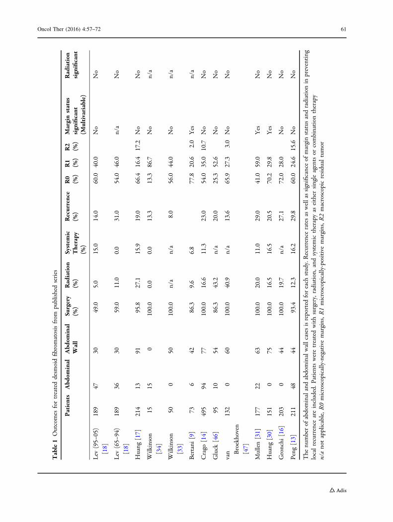
Table1
Outcomes
fortreateddesm
oidfibromatosisfrom
publishedseries
Patients
Abd
ominal
Abd
ominal
Wall
Surgery
(%)
Radiation
(%)
System
icTherapy
(%)
Recurrence
(%)
R0
(%)
R1
(%)
R2
(%)
Marginstatus
significant
(Multivariable)
Radiation
significant
Lev
(95–
05)
[18]
189
4730
49.0
5.0
15.0
14.0
60.0
40.0
No
No
Lev
(65–
94)
[18]
189
3630
59.0
11.0
0.0
31.0
54.0
46.0
n/a
No
Huang
[17]
214
1391
95.8
27.1
15.9
19.0
66.4
16.4
17.2
No
No
Wilkinson
[34]
1515
0100.0
0.0
0.0
13.3
13.3
86.7
No
n/a
Wilkinson
[33]
500
50100.0
n/a
n/a
8.0
56.0
44.0
No
n/a
Bertani
[9]
736
4286.3
9.6
6.8
77.8
20.6
2.0
Yes
n/a
Crago
[14]
495
9477
100.0
16.6
11.3
23.0
54.0
35.0
10.7
No
No
Gluck
[46]
9510
5486.3
43.2
n/a
20.0
25.3
52.6
No
No
van Broekhoven
[47]
132
060
100.0
40.9
n/a
13.6
65.9
27.3
3.0
No
No
Mullen[31]
177
2263
100.0
20.0
11.0
29.0
41.0
59.0
Yes
No
Huang
[30]
151
075
100.0
16.5
16.5
20.5
70.2
29.8
Yes
No
Gronchi
[16]
203
044
100.0
19.7
n/a
27.1
72.0
28.0
No
No
Peng
[13]
211
4844
93.4
12.3
16.2
29.8
60.0
24.6
15.6
No
No
The
numberof
abdominalandabdominalwallcasesisreported
foreach
study.Recurrenceratesaswellassignificanceof
marginstatus
andradiationin
preventing
localrecurrence
areincluded.P
atientsweretreatedwithsurgery,radiation,
andsystem
ictherapyas
either
singleagentsor
combination
therapy
n/anotapplicable,R
0microscopically-negativemargins,R
1microscopically-positivemargins,R
2macroscopicresidualtumor
Oncol Ther (2016) 4:57–72 61
recurrence rates for patients with
microscopically-positive margins (R1) when
compared to microscopically negative (R0)
margins [13, 17, 18, 30, 31]. However, some
studies have only shown margin status as a
predictor of recurrence with univariate analysis
but not on multivariate analysis [13, 31]. Other
studies show that microscopic margin status
does not affect recurrence-free survival and has
no prognostic significance when comparing R0
and R1 resections in desmoid tumors [14, 15,
17, 31]. Of particular interest in relation to this
topic is a large study from the MD Anderson
Cancer Center which compared desmoid
outcomes from two distinct time frames at the
institution: the first is 1965–1994, and the
second a more modern cohort from 1995 to
2005. Interestingly, margin status is a
significant predictor of disease recurrence in
the older cohort but that significance is lost in
the modern group despite similar rates of
margin-positive resections between the two
groups (46% vs 47 %). This adds confusion to
the true significance of margin-negative
resection for desmoid tumors [18]. Ultimately
margin status remains a controversial topic in
the management of desmoid tumors and it is
often agreed that R0 resections are ideal but that
with similar recurrence rates for R0 and R1
resections as well as the emergence of adjuvant
therapies additional surgery may be avoided
based solely on a microscopically-positive
margin. This is particularly true in patients
where re-resection may result in loss of body
function or high operative morbidity [5]. These
decisions should be made within the confines of
a multidisciplinary tumor board.
Abdominal wall and intra-abdominal desmoid
tumors present unique surgical challenges due to
their anatomic location. Abdominal wall
desmoids may require abdominal wall resection
and reconstruction while intra-abdominal and
mesenteric tumors may necessitate removal of a
significant amount of bowel (Fig. 1). Abdominal
wall desmoids have favorable outcomes when
resected and it has been suggested that these
lesions have a better prognosis than those
involving extremity based on low recurrence
rates [32]. In a study looking at 50 patients with
abdominal wall desmoid fibromatosis treated
with surgery, 92% of patients did not experience
recurrence after a median follow up of 6 years. In
this cohort 56% of patients had an R1 resection
but only tumor size ([7 cm) was associated with
recurrence. Prosthetic mesh was used in 94% of
patients to repair the surgical defect. In addition
to recurrence rates, complication rates were also
low with nomortality and only one postoperative
complication of cellulitis [33]. Peng et al. also
report good outcomes for patients with
abdominal wall or intra-abdominal desmoids
tumors and margin negative resection (R0).
Median recurrence-free survival was not reached
after follow up of 25.7 months [13]. While
intra-abdominal desmoids only represented
5.8% of their population, these patients also had
low complication rates as compared to patients
with resected extra-abdominal desmoids.
Wilkinson et al. have also confirmed good
prognosis for surgically-resected sporadic
intra-abdominal desmoids. Of 15 patients with
sporadic (non FAP associated) desmoids that were
grossly resected, 13 patients did not recur with a
median disease-free interval of 45 months. R0
resections were only accomplished in 2 of these
15 patients with the remaining 13 having R1
resections [34]. In a study looking at the use of
surgery for mesenteric desmoids in patients with
FAP, details from 16 patients were available. Small
bowel resections were necessary in 87.5% of these
cases with an average of 45.6 cm of bowel
removed [35]. When treating mesenteric
desmoid tumor, it is possible that a significant
amount of bowel may be resected resulting in
62 Oncol Ther (2016) 4:57–72
intestinal failure, chronic total parenteral
nutrition (TPN), and perhaps the need for small
bowel transplantation. Although numbers are
small (12 patients), several studies have shown
encouraging results for either intestinal or
modified multivisceral transplantation for
patients developing short gut syndrome after
resection of mesenteric desmoid tumors [36–38].
Two patients have died, but at time of publication
all other patients had intestinal rehabilitation and
were functioning without TPN. Long-term follow
up is available for 8 patients with only 2 patients
developing desmoid recurrence. Tumors recurred
either in the abdominal wall, chest wall or
incision without any patients developing
intra-abdominal recurrence [36, 38]. While the
risks of immunosuppression and graft rejection
exist for patients treated with transplant, this may
represent a reasonable long-term solution for
mesenteric desmoids necessitating life-altering
bowel resection.
Watchful Waiting
The natural biology of abdominal wall and
intra-abdominal desmoids can be
unpredictable. More indolent tumors may not
recur despite microscopically positive margins
and more aggressive tumors may recur despite
negative margins. While biologic markers are
evolving, the true nature of a desmoid cannot
be reliably predicted. In most large databases,
there are patients who receive no treatment for
their tumors and have shown no progression or
sometimes even spontaneous regression. These
observations have propelled a new strategy for
desmoid management which is gaining traction
known as ‘‘wait and see’’ or watchful waiting.
When a new patient presents with a desmoid
tumor the natural history of their disease has
likely not been declared. By employing a period
of close observation, proponents believe that it
is possible to identify more indolent tumors,
avoiding morbid procedures for tumors that
may not progress or may even spontaneously
regress. Conversely, by having frequent physical
exams and staging scans, tumors that show
more aggressive behavior can be treated with a
multidisciplinary approach before the
therapeutic window is missed [5, 10].
While evaluating the best initial therapy for
primary desmoid, Bonvalot et al. identified a
group of patients that were not treated
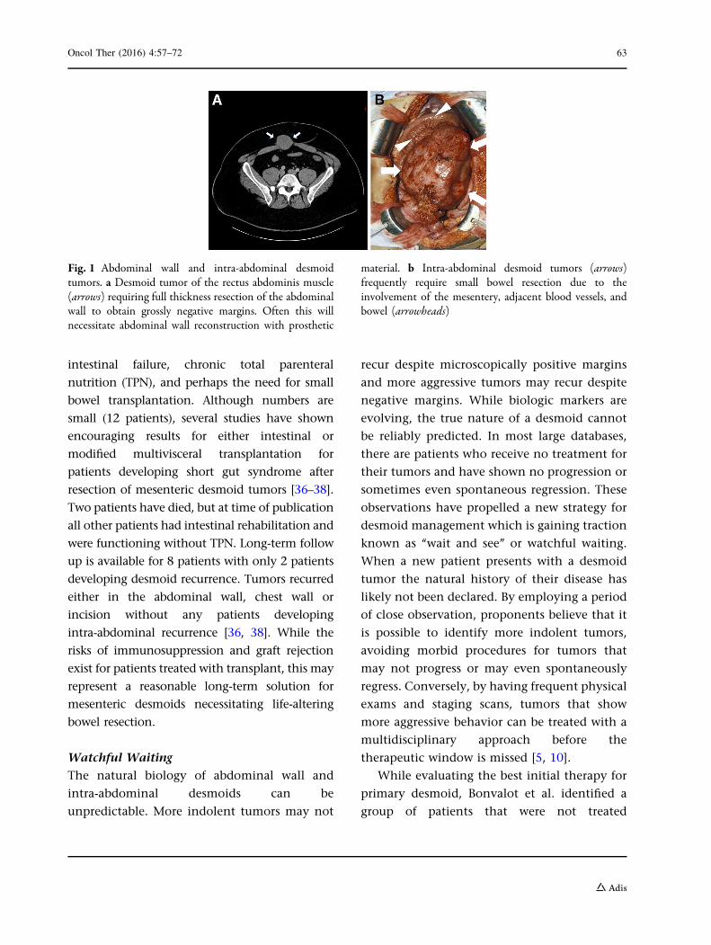
Fig. 1 Abdominal wall and intra-abdominal desmoidtumors. a Desmoid tumor of the rectus abdominis muscle(arrows) requiring full thickness resection of the abdominalwall to obtain grossly negative margins. Often this willnecessitate abdominal wall reconstruction with prosthetic
material. b Intra-abdominal desmoid tumors (arrows)frequently require small bowel resection due to theinvolvement of the mesentery, adjacent blood vessels, andbowel (arrowheads)
Oncol Ther (2016) 4:57–72 63
surgically, but had a similar outcome as patients
treated with R0 resection. Patients treated with
observation did as well as patients with
completely resected disease and better than
patients with incomplete resection [39]. In an
effort to understand how observation alone
may affect additional therapies and outcomes
for desmoid tumor, a multi-institutional study
evaluated patients not treated with surgery or
radiation and were stratified into an observation
group or a medical therapy group. Eighty-three
patients were treated with observation only and
their progression was compared to patients
treated systemically. The observation arm had
a 49.9% 5-year progression-free survival (PFS)
which was not different from the 58.6% PFS in
the systemically treated group [40]. While the
authors could not identify any prognostic
factors that would predict tumor progression,
they were able to avoid the morbidity of surgery
and/or radiation. Of patients that did progress,
89% did so in the first 2 years. Based on these
findings the authors have proposed a very close
regimen of disease surveillance for 24 months.
The authors clearly recognize the heterogeneity
of the disease and realize that proper patient
selection for this strategy is critical. In an effort
to better risk stratify sporadic desmoids and
identify patients with more indolent tumors,
Salas et al. developed a clinical risks scale based
on the primary tumor characteristics. This scale
has revealed prognostic subgroups that may
benefit from different treatment strategies
including watchful observation [15]. Several
notable European groups have made proposals
to standardize desmoid tumor management
such that a watchful waiting strategy is the
first step for all new tumors [5, 10]. Patients will
be followed closely by a multidisciplinary team
with escalation of care only if the tumor is
declared to be an aggressive phenotype, which
spares patients with indolent tumors the
morbidity of aggressive therapy. This is of
particular interest for intra-abdominal
desmoids where resection may lead to the
removal of a significant amount of bowel.
Clearly this change in management represents
an opportunity for prospective trials to define
the role of observation vs immediate
intervention.
Radiation
Radiation has been used as both adjuvant and
definitive therapy for desmoid tumors. Most
data evaluating the use of radiation are from
surgical studies with a clear selection bias
towards offering therapy to patients that have
either recurred or have had a positive margin
resection. Conversely, patients that have
radiation only, usually have tumors that
are unresectable or would require
unacceptable surgical deformity or morbidity.
While radiation has a role in treating desmoid
tumors, the highly selected patients treated
with radiation and the lack of randomized
trials make the exact role for radiation in
desmoids elusive.
A large meta-analysis of patients from 22
studies treated with surgery, surgery plus
radiation, or radiation alone suggested that all
patients with desmoid tumors should be treated
with radiation. Patients treated with radiation
or radiation plus surgery had superior local
control rates (78% and 75%, respectively) than
patients treated with surgery alone (61%) [41].
While supporting the role for radiation as a
viable treatment of desmoid tumors, the
radiation doses in this study were highly
variable and declaring that all disease should
be treated with this modality may be overly
aggressive and does not take into consideration
the morbidity associated with radiation.
Complications related to radiation for desmoid
64 Oncol Ther (2016) 4:57–72
fibromatosis are 17–23% [41–43]. Before
radiation is given for abdominal wall and
intra-abdominal desmoids the risk of
radiation-induced enteritis as well as the risk
of radiation-induced secondary malignancies
should be considered. Radiation-induced
malignancies are particularly relevant in a
disease that often affects a younger cohort of
patients [43]. Definitive radiation may be
effective for local control in patients that
would otherwise need radical and disfiguring
surgery. Radiation as the primary mode of
therapy for unresectable disease may offer a
70–80% chance of local control [42, 44, 45]. Of
particular interest is a European Organization
for Research and Treatment of Cancer (EORTC)
phase II study that standardized treatment of 44
patients with unresectable disease to 56 Gy
received in 28 fractions. At 3 years, local
control was 81.5% with 13.6% of patients
having a complete response. They additionally
report that in some patients radiation response
will continue beyond 3 years, promoting the
durability of this therapy for
unresectable disease [44].
Studies comparing local control in
surgically-resected patients based on receipt of
adjuvant radiation have not been as clear.
When Huang et al. reviewed their data on
surgically-resected desmoids they found no
overall improvement in local recurrence based
on adjuvant radiation. On subset analysis, they
did find a significant improvement in
recurrence for patients with R1 resection who
received radiation compared to those who did
not. Five-year relapse free survival rates for R1
resection followed by radiation were 75%
compared to 56.4% in patients with R1
resection alone [17]. While not statistically
significant, Lev et al. also found a trend
toward improved local control with adjuvant
radiotherapy [18]. Despite some supporting
evidence for the use of adjuvant radiation,
many other retrospective studies have found
that the use of adjuvant radiation did not
impact local recurrence rates (Table 1) [14, 30,
31, 46, 47]. While there seems to be a real
benefit of radiation for some patients, the true
utility of radiation as an adjuvant therapy in
desmoid fibromatosis has not been evaluated in
a prospective, randomized trial. Most studies are
small, retrospective and do not have
standardization of patient selection. Radiation
is not frequently used for intra-abdominal
disease and most studies are focused on its use
for disease in the extremities and soft tissues.
This is likely related to a high risk of
radiation-induced enteritis and the lack of a
distinct target for grossly resected disease. Due
to the lack of clearly defined indications for
radiation it is critical to make these decisions in
large volume multidisciplinary centers to
properly select patients and avoid unnecessary
exposure and morbidity from this treatment.
Systemic Therapy
Studies evaluating systemic therapy for desmoid
fibromatosis frequently involve patients with
mesenteric desmoid tumors due to the
difficulty treating this situation with other
therapies. In addition to high recurrence rates,
there are significant side effects and morbidity
that may arise from resection of mesenteric
desmoids due to involvement of major blood
vessels and potential need for significant
bowel resection. Many systemic therapy
treatment lines have been used for desmoids
including hormonal therapy, non-steroidal
anti-inflammatory drugs (NSAIDs),
chemotherapy, and targeted therapy with
varying degrees of success but no standard of
care. An example of the lack of standardized
systemic therapy for desmoids is shown by
Oncol Ther (2016) 4:57–72 65
recent retrospective studies showing up to 157
different combinations of therapy used to treat
these tumors [29, 48]. Unfortunately, most
studies looking at systemic therapy for treating
desmoid tumors are either retrospective or
single-armed studies. Additionally, they are
treating patients that have failed surgical
therapy or have unresectable disease,
introducing selection bias and making
comparisons to other therapies difficult;
however these studies still provide insight into
treating the disease when other options are not
feasible.
Hormonal and Nonsteroidal
Anti-Inflammatory Drugs (NSAIDs)
When effectiveness, side effects, and efficacy are
weighed, either combination or single agent
hormonal and NSAID agents are frequently
used as a first-line systemic therapy or for
unresectable, recurrent, or progressing
desmoid tumors [4, 10, 48–50]. Typically
either tamoxifen (60–120 mg/day) or raloxifen
(120–240 mg/day) is used as an anti-estrogen
agent. The NSAID most commonly used is
sulindac (150–800 mg/day). Hormone-based
therapy are effective in 40–51% of patients
[29, 51]. Hansmann et al. observed a 77%
response rate in patients with FAP-associated
mesenteric desmoids treated with first line
anti-estrogen/NSAID therapy. This compares
favorably to patients that have recurred after
surgical resection and then treated with a
similar regimen; response rates in recurrent
disease were only 50% suggesting that
anti-estrogen/NSAID therapy for patients with
FAP-associated mesenteric desmoids may be a
reasonable therapy before proceeding with
surgery [49]. A recent update from the same
center reports 134 patients treated with the
same regimen as either adjuvant or definitive
therapy. Patients with sporadic and
FAP-associated mesenteric desmoid
fibromatosis were included and equally
represented. A response rate of over 85% was
achieved (stable disease as well as responders)
using anti-estrogen/NSAID therapy.
Additionally they were able to taper therapy
and had only one long-term recurrence in
patients that had previously been resected
[52]. Such a large cohort of patients with high
response rates and durability is encouraging for
non-surgical management of these tumors. This
combination can be effective, but an objective
response may take several months to stabilize
disease and decrease associated symptoms [10,
48, 52]. These agents are often used as they are
relatively inexpensive and have a low-risk side
effect profile compared to other systemic
therapies. Despite their relatively safe
reputation it should be cautioned that
anti-estrogens have a slightly increased risk of
thromboembolic events and tamoxifen can
cause ovarian cysts in pre-menopausal women.
Chemotherapy
Several studies have found chemotherapy to be
the best systemic therapy for treating desmoid
tumors with response rates as high as 79% [29,
48, 53]. There are a lack of randomized or
controlled trials as most studies treat a small
number of patients with heterogeneous
treatment regimens. Drug combinations using
anthracyclines appear to be the most effective
treatment, but other combinations may
additionally include methotrexate, vinblastine
and cisplatin.
The anthracycline-based regimen has resulted
in long progression-free survival and even
complete responses. This has influenced some
groups to support chemotherapy as first-line
treatment for unresectable disease—particularly
of the mesentery [29, 48, 53–55]. When patients
show aggressive disease and fail hormonal
66 Oncol Ther (2016) 4:57–72
therapies, chemotherapy is often the second line
of therapy as it may show significant responses
in previously-treated patients [10, 28, 43]. A
study from the French Sarcoma Group compared
anthracycline-based therapy to other regimens
and were able to show a statistically significant
difference in response: 54% vs 12% (p = 0.0011)
favoring anthracyclines. While only 13 patients
were treated, all patients that received
anthracycline-based therapy had either
stable disease (46%) or a partial response (54%)
[56]. Other studies have confirmed a high
response rate and durable response for this
regimen with progression-free survival of
74 months [53]. As might be expected, toxicity
is higher with this regimen resulting in grade 3–4
hematological toxicities in approximately
31–43% of patients [53, 56].
Additionally, ‘‘low-dose’’ chemotherapy has
also been described for systemic treatment of
desmoids which typically includes
methotrexate and vinblastine. This has been
shown to be well tolerated and consistently
results in stable or responding disease in
67–100% of patients treated [48, 56, 57].
Importantly, this regimen has also been
associated with a prolonged response and
5-year progression-free survival as high as 67%
[57]. Unfortunately this regimen has been
associated with high toxicity rates resulting in
patient intolerance and a 50% attrition rate.
Neurotoxicity is a common side effect of
vinblastine that often results in patients not
being able to tolerate this regimen. Another
vinca alkaloid, vinorelbine, has been described
as an effective agent against desmoids with less
long-term toxicity than vinblastine and is also
given with methotrexate [58, 59]. Weiss et al.
describe a 60% response rate and improvement
of symptoms of 80% of patients treated with
vinorelbine in a small series of patients with
previously low rates of neurotoxicity [59]. This
may likely be another useful regimen
particularly in patients that do not tolerate
vinblastine.
Targeted and Evolving Therapeutics
Other evolving options include targeted
therapies using tyrosine kinase inhibitors
(TKIs) and anti-angiogenic drugs. The TKI that
have been used include imatinib and sunitinib.
A phase II trial using imatinib had modest
results with 1-year progression-free survival of
66% and an objective response rate of only 6%
[60]. Another phase II study using higher doses
of imatinib (800 mg/day) revealed a 15.7%
partial response (PR) rate (C50% tumor
shrinkage) in a heavily pre-treated group of
patients. Interestingly, all patients that
experienced a PR had intra-abdominal disease
and duration of response was greater than
1.5 years for all patients [61]. Finally, Penel
et al. treated 40 patients with imatinib
(400 mg/day) in a phase II trial and
experienced 67% progression-free survival.
Only 45.5% of these patients had abdominal
wall or mesenteric disease, but two of these
patients had a partial and durable response [62].
Toxicity for all three of these trials was
acceptable with very few grade 4 toxicities that
were treated effectively with dose reduction. In
addition to these phase II trials, other small
retrospective reviews have revealed stable or
partial response in 36–80% of patients with a
median progression-free survival of nearly
27 months by Response Evaluation Criteria In
Solid Tumors (RECIST) criteria [43].
Given the potential activity of imatinib in the
treatment of desmoid fibromatosis, another TKI,
sunitinib, has been evaluated for efficacy in
advanced disease. Sunitinib also blocks vascular
endothelial growth factor receptors adding an
anti-angiogenic effect. A phase II study treating
mostly intra-abdominal desmoids (63.2% of
Oncol Ther (2016) 4:57–72 67
participants) with sunitinib showed an overall
response rate of 26.3% and a 2-year
progression-free survival rate of 74.7%. Three of
the 12 patients with mesenteric disease developed
serious adverse events with the first dose of
treatment presenting as tumor bleeding, bowel
perforation and entero-tumoral fistula formation.
The authors postulate that all of these events
could be explained by the drugs anti-angiogenic
affect with resultant tumor necrosis [63].
Additional TKI and anti-angiogenic drugs,
sorafenib and pazopanib, have also been shown
to have efficacy in treating desmoid tumors [64,
65]. In a small, retrospective review of 26
patients treated with sorafenib, 70% of patient
reported improved symptoms, and at 6 months
95% of patients had either a partial response or
stable disease. In the 13 patients with
abdominal disease evaluated radiographically,
nearly 73% of patients had radiographic
response by RECIST criteria [64]. Following the
response seen from sorafenib the
anti-angiogenic drug pazopanib has also been
shown to be effective in treating desmoid
tumors. Two case reports show that patients
treated with pazopanib had improved
symptoms, tumor shrinkage and decreased
tumor cellularity similar to results seen with
sorafenib [65]. Conclusions regarding the
efficacy of these drugs must be interpreted
with caution until larger prospective clinical
trials are performed to validate initial findings.
As data emerge regarding the
pathophysiology of desmoid tumors, new
pathways to inhibit tumor growth are being
discovered. The NOTCH pathway has recently
been recognized as a potential therapeutic
target for desmoids. This pathway drives
several cancer-related processes in solid tumors
and can be blocked by c-secretase inhibition.
When desmoid tumor cell lines are treated with
c-secretase cell growth, migration and invasion
are inhibited [66]. In an open label phase I dose
escalation trial of a c-secretase inhibitor, five of
seven patients with desmoid tumors that were
treated showed objective and durable response
[67]. This promising data has spawned a phase II
trial evaluating a c-secretase inhibitor in adults
with desmoid tumors [68]. Recently another
target, hyaluronan (HA), a glycosaminoglycan
in the stromal microenvironment involved with
normal wound healing, has been identified and
associated with desmoid tumorigenesis [69].
This study identified overexpression of HA
levels in desmoid tumor surgical specimens as
well as immortalized cell lines. When HA
synthesis was inhibited, they found decreased
tumor proliferation rates and decreased HA
levels suggesting a novel therapeutic target in
treating desmoid fibromatosis. While new
targets in this difficult disease are exciting,
more translational studies will be required.
CONCLUSIONS
Due to the rarity and heterogeneity of this
disease, it cannot be emphasized enough that
desmoid fibromatosis should be managed within
the context of a high-volume, multidisciplinary
tumor board. Treatment recommendations
regarding surgery, radiation, and systemic
therapy are all evolving. This increases the
complexity of the decision making for this
disease and emphasizes the necessity of having
surgical oncologists, radiation oncologists and
medical oncologists involved in developing a
treatment plan for each individual patient.
While more patients are being treated with
observation for this disease, perhaps the most
exciting and game-changing developments will
come from genetic studies of these tumors. Once
the pathophysiology of this disease is better
understood, clinicians can better guide patients
68 Oncol Ther (2016) 4:57–72
in treatment recommendations and risk
stratification.
ACKNOWLEDGMENTS
No funding or sponsorship was received for this
study or publication of this article. All authors
had full access to all of the data in this study
and take complete responsibility for the
integrity of the data and accuracy of the data
analysis. All named authors meet the
International Committee of Medical Journal
Editors (ICMJE) criteria for authorship for this
manuscript, take responsibility for the integrity
of the work as a whole, and have given final
approval for the version to be published.
Disclosures. J. H. Howard and R. E. Pollock
have nothing to disclose.
Compliance with Ethics Guidelines. This
article is based on previously conducted
studies and does not involve any new studies
of human or animal subjects performed by any
of the authors.
Open Access. This article is distributed
under the terms of the Creative Commons
Attribution-NonCommercial 4.0 International
License (http://creativecommons.org/licenses/
by-nc/4.0/), which permits any noncommercial
use, distribution, and reproduction in any
medium, provided you give appropriate credit to
the original author(s) and the source, provide a
link to the Creative Commons license, and
indicate if changes were made.
REFERENCES
1. Sakorafas GH, Nissotakis C, Peros G. Abdominaldesmoid tumors. Surg Oncol. 2007;16(2):131–42.
2. Clark SK, Phillips RK. Desmoids in familialadenomatous polyposis. Br J Surg.1996;83(11):1494–504.
3. de Bree E, Keus R, Melissas J, Tsiftsis D, vanCoevorden F. Desmoid tumors: need for anindividualized approach. Expert Rev AnticancerTher. 2009;9(4):525–35.
4. Eastley NC, Hennig IM, Esler CP, Ashford RU.Nationwide trends in the current management ofdesmoid (aggressive) fibromatosis. Clin Oncol (RColl Radiol). 2015;27(6):362–8.
5. Kasper B, Baumgarten C, Bonvalot S, et al.Management of sporadic desmoid-typefibromatosis: a European consensus approachbased on patients’ and professionals’ expertise–asarcoma patients EuroNet and EuropeanOrganisation for Research and Treatment ofCancer/Soft Tissue and Bone Sarcoma Groupinitiative. Eur J Cancer. 2015;51(2):127–36.
6. Eastley N, Aujla R, Silk R, et al. Extra-abdominaldesmoid fibromatosis–a sarcoma unit review ofpractice, long term recurrence rates and survival.Eur J Surg Oncol. 2014;40(9):1125–30.
7. Walczak BE, Rose PS. Desmoid: the role of localtherapy in an era of systemic options. Curr TreatOptions Oncol. 2013;14(3):465–73.
8. Church J, Lynch C, Neary P, LaGuardia L, Elayi E. Adesmoid tumor-staging system separates patientswith intra-abdominal, familial adenomatouspolyposis-associated desmoid disease by behaviorand prognosis. Dis Colon Rectum.2008;51(6):897–901.
9. Bertani E, Testori A, Chiappa A, et al. Recurrenceand prognostic factors in patients with aggressivefibromatosis. The role of radical surgery and itslimitations. World J Surg Oncol. 2012;10:184.
10. Gronchi A, Colombo C, Le Pechoux C, et al.Sporadic desmoid-type fibromatosis: a stepwiseapproach to a non-metastasising neoplasm–aposition paper from the Italian and the FrenchSarcoma Group. Ann Oncol. 2014;25(3):578–83.
11. Deyrup AT, Tretiakova M, Montag AG. Estrogenreceptor-beta expression in extra abdominalfibromatoses: an analysis of 40 cases. Cancer.2006;106(1):208–13.
12. Fiore M, Coppola S, Cannell AJ, et al. Desmoid-typefibromatosis and pregnancy: a multi-institutionalanalysis of recurrence and obstetric risk. AnnSurgery. 2014;259(5):973–8.
13. Peng PD, Hyder O, Mavros MN, et al. Managementand recurrence patterns of desmoids tumors: a
Oncol Ther (2016) 4:57–72 69
multi-institutional analysis of 211 patients. AnnSurg Oncol. 2012;19(13):4036–42.
14. Crago AM, Denton B, Salas S, et al. A prognosticnomogram for prediction of recurrence in desmoidfibromatosis. Ann Surg. 2013;258(2):347–53.
15. Salas S, Dufresne A, Bui B, et al. Prognostic factorsinfluencing progression-free survival determinedfrom a series of sporadic desmoid tumors: await-and-see policy according to tumorpresentation. J Clin Oncol. 2011;29(26):3553–8.
16. Gronchi A, Casali PG, Mariani L, et al. Quality ofsurgery and outcome in extra-abdominal aggressivefibromatosis: a series of patients surgically treated ata single institution. J Clin Oncol.2003;21(7):1390–7.
17. Huang K, Wang CM, Chen JG, et al. Prognosticfactors influencing event-free survival andtreatments in desmoid-type fibromatosis: analysisfrom a large institution. Am J Surg.2014;207(6):847–54.
18. Lev D, Kotilingam D, Wei C, et al. Optimizingtreatment of desmoid tumors. J Clin Oncol.2007;25(13):1785–91.
19. Salas S, Brulard C, Terrier P, et al. Gene expressionprofiling of desmoid tumors by cDNA microarraysand correlation with progression-free survival. ClinCancer Res. 2015;21(18):4194–200.
20. Jilong Y, Jian W, Xiaoyan Z, Xiaoqiu L, XiongzengZ. Analysis of APC/beta-catenin genes mutationsand Wnt signalling pathway in desmoid-typefibromatosis. Pathology. 2007;39(3):319–25.
21. Lips DJ, Barker N, Clevers H, Hennipman A. Therole of APC and beta-catenin in the aetiology ofaggressive fibromatosis (desmoid tumors). Eur JSurg Oncol. 2009;35(1):3–10.
22. Colombo C, Miceli R, Lazar AJ, et al. CTNNB1 45Fmutation is a molecular prognosticator of increasedpostoperative primary desmoid tumor recurrence:an independent, multicenter validation study.Cancer. 2013;119(20):3696–702.
23. Crago AM, Chmielecki J, Rosenberg M, et al. Nearuniversal detection of alterations in CTNNB1 andWnt pathway regulators in desmoid-typefibromatosis by whole-exome sequencing andgenomic analysis. Genes Chromosomes Cancer.2015;54(10):606–15.
24. Domont J, Salas S, Lacroix L, et al. High frequencyof beta-catenin heterozygous mutations inextra-abdominal fibromatosis: a potentialmolecular tool for disease management. Br JCancer. 2010;102(6):1032–6.
25. Huss S, Nehles J, Binot E, et al. b-catenin (CTNNB1)mutations and clinicopathological features ofmesenteric desmoid-type fibromatosis.Histopathology. 2013;62(2):294–304.
26. Lazar AJ, Tuvin D, Hajibashi S, et al. Specificmutations in the beta-catenin gene (CTNNB1)correlate with local recurrence in sporadic desmoidtumors. Am J Pathol. 2008;173(5):1518–27.
27. Mullen JT, DeLaney TF, Rosenberg AE, et al.b-Catenin mutation status and outcomes insporadic desmoid tumors. Oncologist.2013;18(9):1043–9.
28. van Broekhoven DL, Verhoef C, Grunhagen DJ,et al. Prognostic value of CTNNB1 gene mutation inprimary sporadic aggressive fibromatosis. Ann SurgOncol. 2015;22(5):1464–70.
29. Desurmont T, Lefevre JH, Shields C, Colas C, TiretE, Parc Y. Desmoid tumour in familial adenomatouspolyposis patients: responses to treatments. FamCancer. 2015;14(1):31–9.
30. Huang K, Fu H, Shi YQ, Zhou Y, Du CY. Prognosticfactors for extra-abdominal and abdominal walldesmoids: a 20-year experience at a singleinstitution. J Surg Oncol. 2009;100(7):563–9.
31. Mullen JT, Delaney TF, Kobayashi WK, et al.Desmoid tumor: analysis of prognostic factors andoutcomes in a surgical series. Ann Surg Oncol.2012;19(13):4028–35.
32. Pencavel T, Strauss DC, Thomas JM, Hayes AJ. Thesurgical management of soft tissue tumours arising inthe abdominal wall. Eur J Surg Oncol.2010;36(5):489–95.
33. Wilkinson MJ, Chan KE, Hayes AJ, Strauss DC.Surgical outcomes following resection for sporadicabdominal wall fibromatosis. Ann Surg Oncol.2014;21(7):2144–9.
34. Wilkinson MJ, Fitzgerald JE, Thomas JM, Hayes AJ,Strauss DC. Surgical resection for non-familialadenomatous polyposis-related intra-abdominalfibromatosis. Br J Surg. 2012;99(5):706–13.
35. Latchford AR, Sturt NJ, Neale K, Rogers PA, PhillipsRK. A 10-year review of surgery for desmoid diseaseassociated with familial adenomatous polyposis. BrJ Surg. 2006;93(10):1258–64.
36. ChatzipetrouMA,TzakisAG,PinnaAD,etal. Intestinaltransplantation for the treatment of desmoid tumorsassociated with familial adenomatous polyposis.Surgery. 2001;129(3):277–81.
37. Jovine E, Masetti M, Cautero N, et al. Modifiedmultivisceral transplantation without a liver graft
70 Oncol Ther (2016) 4:57–72
for Gardner/desmoid syndrome and chronicintestinal pseudo-obstruction. Transplant Proc.2002;34(3):911–2.
38. Nikeghbalian S, Aliakbarian M, Shamsaeefar A,Kazemi K, Bahreini A, Malekhosseini SA.Multivisceral transplantation for the treatment ofintra-abdominal tumors. Transplant Proc.2013;45(10):3528–30.
39. Bonvalot S, Eldweny H, Haddad V, et al.Extra-abdominal primary fibromatosis: aggressivemanagement could be avoided in a subgroup ofpatients. Eur J Surg Oncol. 2008;34(4):462–8.
40. Fiore M, Rimareix F, Mariani L, et al. Desmoid-typefibromatosis: a front-line conservative approach toselect patients for surgical treatment. Ann SurgOncol. 2009;16(9):2587–93.
41. Nuyttens JJ, Rust PF, Thomas CR Jr, Turrisi AT 3rd.Surgery versus radiation therapy for patients withaggressive fibromatosis or desmoid tumors: acomparative review of 22 articles. Cancer.2000;88(7):1517–23.
42. Ballo MT, Zagars GK, Pollack A. Radiation therapyin the management of desmoid tumors. Int J RadiatOncol Biol Phys. 1998;42(5):1007–14.
43. Guadagnolo BA, Zagars GK, Ballo MT. Long-termoutcomes for desmoid tumors treated withradiation therapy. Int J Radiat Oncol Biol Phys.2008;71(2):441–7.
44. Keus RB, Nout RA, Blay JY, et al. Results of a phase IIpilot study of moderate dose radiotherapy forinoperable desmoid-type fibromatosis–an EORTCSTBSG and ROG study (EORTC 62991-22998). AnnOncol. 2013;24(10):2672–6.
45. Micke O, Seegenschmiedt MH, GermanCooperative Group on Radiotherapy for BenignDiseases. Radiation therapy for aggressivefibromatosis (desmoid tumors): results of anational patterns of care study. Int J Radiat OncolBiol Phys. 2005;61(3):882–91.
46. Gluck I, Griffith KA, Biermann JS, Feng FY, LucasDR, Ben-Josef E. Role of radiotherapy in themanagement of desmoid tumors. Int J RadiatOncol Biol Phys. 2011;80(3):787–92.
47. van Broekhoven DL, Verhoef C, Elias SG, et al. Localrecurrence after surgery for primaryextra-abdominal desmoid-type fibromatosis. Br JSurg. 2013;100(9):1214–9.
48. de Camargo VP, Keohan ML, D’Adamo DR, et al.Clinical outcomes of systemic therapy for patientswith deep fibromatosis (desmoid tumor). Cancer.2010;116(9):2258–65.
49. Hansmann A, Adolph C, Vogel T, Unger A,Moeslein G. High-dose tamoxifen and sulindac asfirst-line treatment for desmoid tumors. Cancer.2004;100(3):612–20.
50. Tanaka K, Yoshikawa R, Yanagi H, et al. Regressionof sporadic intra-abdominal desmoid tumourfollowing administration of non-steroidalanti-inflammatory drug. World J Surg Oncol.2008;6:17.
51. Bocale D, Rotelli MT, Cavallini A, Altomare DF.Anti-oestrogen therapy in the treatment of desmoidtumours: a systematic review. Colorectal Dis.2011;13(12):e388–95.
52. Quast DR, Schneider R, Burdzik E, Hoppe S, MosleinG. Long-term outcome of sporadic andFAP-associated desmoid tumors treated withhigh-dose selective estrogen receptor modulatorsand sulindac: a single-center long-termobservational study in 134 patients. Fam Cancer.2016;15(1):31–40.
53. Gega M, Yanagi H, Yoshikawa R, et al. Successfulchemotherapeutic modality of doxorubicin plusdacarbazine for the treatment of desmoid tumorsin association with familial adenomatous polyposis.J Clin Oncol. 2006;24(1):102–5.
54. Okuno SH, Edmonson JH. Combinationchemotherapy for desmoid tumors. Cancer.2003;97(4):1134–5.
55. Patel SR, Evans HL, Benjamin RS. Combinationchemotherapy in adult desmoid tumors. Cancer.1993;72(11):3244–7.
56. Garbay D, Le Cesne A, Penel N, et al. Chemotherapyin patients with desmoid tumors: a study from theFrench Sarcoma Group (FSG). Ann Oncol.2012;23(1):182–6.
57. Azzarelli A, Gronchi A, Bertulli R, et al. Low-dosechemotherapy with methotrexate and vinblastinefor patients with advanced aggressive fibromatosis.Cancer. 2001;92(5):1259–64.
58. Bertagnolli MM, Morgan JA, Fletcher CD, et al.Multimodality treatment of mesenteric desmoidtumours. Eur J Cancer. 2008;44(16):2404–10.
59. Weiss AJ, Horowitz S, Lackman RD. Therapy ofdesmoid tumors and fibromatosis usingvinorelbine. Am J Clin Oncol. 1999;22(2):193–5.
60. Chugh R, Wathen JK, Patel SR, et al. Efficacy ofimatinib in aggressive fibromatosis: results of aphase II multicenter Sarcoma Alliance for Researchthrough Collaboration (SARC) trial. Clin CancerRes. 2010;16(19):4884–91.
Oncol Ther (2016) 4:57–72 71
61. Heinrich MC, McArthur GA, Demetri GD, et al.Clinical and molecular studies of the effect ofimatinib on advanced aggressive fibromatosis(desmoid tumor). J ClinOncol. 2006;24(7):1195–203.
62. Penel N, Le Cesne A, Bui BN, et al. Imatinib forprogressive and recurrent aggressive fibromatosis(desmoid tumors): an FNCLCC/French SarcomaGroup phase II trial with a long-term follow-up.Ann Oncol. 2011;22(2):452–7.
63. Jo JC, Hong YS, Kim KP, et al. A prospectivemulticenter phase II study of sunitinib in patientswith advanced aggressive fibromatosis. Invest NewDrugs. 2014;32(2):369–76.
64. Gounder MM, Lefkowitz RA, Keohan ML, et al.Activity of Sorafenib against desmoid tumor/deepfibromatosis. Clin Cancer Res.2011;17(12):4082–90.
65. Martin-Liberal J, Benson C, McCarty H, Thway K,Messiou C, Judson I. Pazopanib is an active
treatment in desmoid tumour/aggressivefibromatosis. Clin Sarcoma Res. 2013;3(1):13.
66. Shang H, Braggio D, Lee YJ, et al. Targeting theNotch pathway: a potential therapeutic approachfor desmoid tumors. Cancer.2015;121(22):4088–96.
67. Messersmith WA, Shapiro GI, Cleary JM, et al.A Phase I, dose-finding study in patients withadvanced solid malignancies of the oralgamma-secretase inhibitor PF-03084014. ClinCancer Res. 2015;21(1):60–7.
68. Hughes DP, Kummar S, Lazar AJ. New, tolerablegamma-secretase inhibitor takes desmoid down anotch. Clin Cancer Res. 2015;21(1):7–9.
69. Briggs A, Rosenberg L, Buie JD, Rizvi H, BertagnolliMM, Cho NL. Antitumor effects of hyaluronaninhibition in desmoid tumors. Carcinogenesis.2015;36(2):272–9.
72 Oncol Ther (2016) 4:57–72


![Intra-Abdominal and Abdominal Wall Desmoid Fibromatosis · intra-abdominal and involving the small bowel mesentery [2]. TREATMENT Surgery Margin-negative resection has historically](https://img.pdfslide.us/doc/110x75/5e5a290071d21b380f5b7e74/intra-abdominal-and-abdominal-wall-desmoid-fibromatosis-intra-abdominal-and-involving.jpg)