Embed Size (px)
Citation preview
Case Report
Clinical Case Reports and Reviews
Clin Case Rep Rev, 2016 doi: 10.15761/CCRR.1000266
ISSN: 2059-0393ISSN: 2059-0393
Volume 2(8): 526-529
Case report on a unique case of intra-abdominal desmoid tumorChowdhury D*, Galea M, Malpani D and Alishahi SMDepartment of Surgery, University Hospital Ayr, Ayr, Scotland, United Kingdom
Correspondence to: Chowdhury D, Department of Surgery, University Hospital Ayr, Ayr, Scotland, United Kingdom, Tel: 00447735093645; E-mail: [email protected]
Key words: Desmoid Tumours (DT), intra-abdominal, asymptomatic patient
Received: July 02, 2016; Accepted: August 13, 2016; Published: August 15, 2016
IntroductionSymptoms from DTs may arise as the results of mass effect or
compression on abdominal organs. This effect may be manifested by intestinal obstruction, urinary obstruction or rarely arterial or venous compression FAP (Familial Adenomatous Polyposis), results from mutation of the APC gene and accounts for 1% cases of colorectal cancer and account for 0.03% of neoplasms [1]. DT account for less than 3% of all soft tissue tumours. They occur most often between 15-60 years of age with being rare in the young and in the elderly [2,3]. The incidence of DT is approximately 2-4 per million populations per year in United States. They have slightly greater preponderance in women than in men. DT occurs in 10-20% of patients with FAP [4]. A patient with FAP has an 852-fold increased risk of developing a DT [4].
Presentation of caseMr P, a 59-year old gentleman previously fit and well presented
with a 4-week history of right abdominal mass associated with mild discomfort in June 2015 to the surgical outpatient clinic. There was no reporting of any associated gastrointestinal symptoms. The mass was noted over the paramedian incisional scar through which he had undergone laparoscopic to open appendicectomy for a complicated appendicitis in 2013. There was no other related past medical history of relevance. Family history was limited from the maternal aspect as he had been adopted.
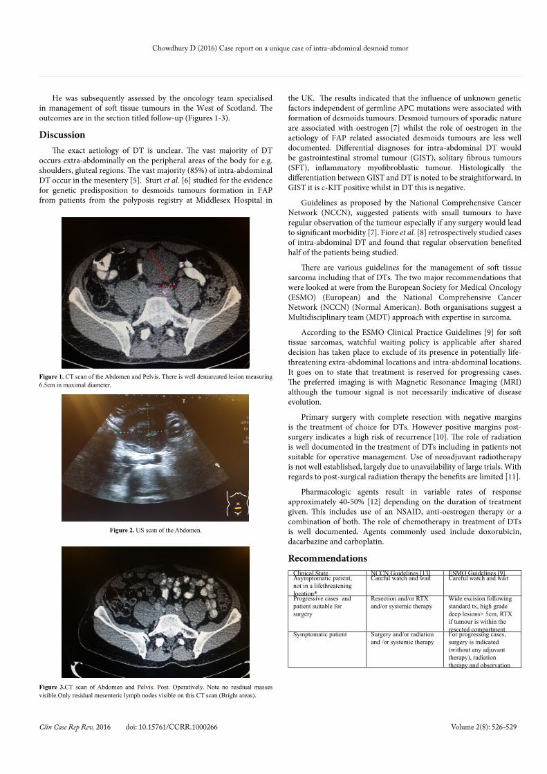
Investigation/Plan Results/OutcomeCT Abdomen+Pelvis 6.5 cm soft tissue mass in the lower abdomen below
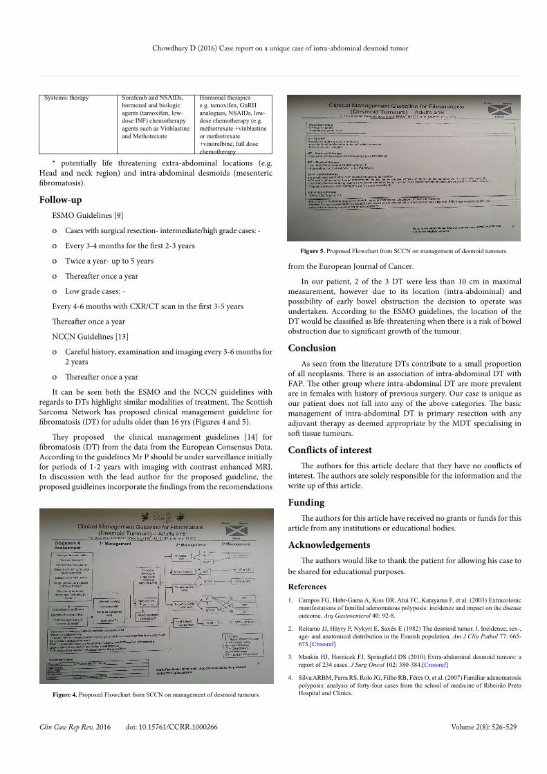
level of umbilicus (See Image.1)USS Abdomen 67mm x 63mm x 65mm, with peripheral vascularity
seen (See Image.2)Ultrasound guided biopsy of mass- transcutaneous
Fibrous connective tissue,formed of fascicles and bundles of spindled cells with spindle or comma shaped nuclei and wavy cytoplasm. There was no evidence of malignancy
Multidiscplinary Team discussion Decision for excision of lesion
Laporotomy and excision of small bowel mesentery masses
Findings intra-operatively:-10 cm mass was noted in the mesentery of the proximal ileum at juxta position to the serosa of the small bowel. Furthermore, there were a smaller (5 cm) similar mass in the distal jejunum and a 2 cm similar mass about 20 cm proximal to the largest lesion, all 3 lesions embedded in the mesentery and located at serosal juxta positions. The smallest mesenteric mass was enucleated and sent for frozen section prior to formal resection
Histology Frozen section- consistent with mesenteric fibromatosis (DT),Formal Histology-The tumours have a similar appearance of very bland fibrosis with bundles of eosinophilic cells and small blood vessels with focal keloid fibres.. No necrosis, abnormal mitoses or nuclear pleomorphism is seen throughout these masses.All lymph nodes were noted to be clear. Initial immunohistochemistry shows negative staining with CD117 and DOG1 as well as CD34. Cytokeratin, Desmin, S100 and SMA are all negative in the larger tumour while Vimentin is positive. There is some focal positivity for SMA in the smaller specimen but CD117 and DOG1 are universally negative. Beta catenin staining, carried out in Glasgow shows strong nuclear positivity.Both the light microscopic appearances and the immunohistochemistry are in keeping with mesenteric fibromatosis (desmoid tumour).
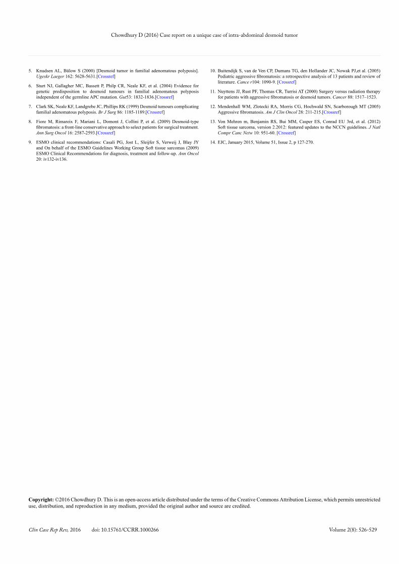
National Sarcoma MDT Post-operative CT scan, colonoscope and UGI endoscope
CT Thorax, Abdomen and Pelvis- Follow up scan
Low volume mesenteric lymph nodes but no DTs seen. (See Image. 3)
AbstractDesmoid tumours (DT) are benign tumours that arise from proliferation of well differentiated fibroblasts which have potential to be locally aggressive and recur post excision. DTs can present both intra-abdominal or extra-abdominal locations with worse prognosis intra-abdominally. We present the case of a 59-year-old gentleman with 4-week history of right abdominal mass with mild discomfort but no gastrointestinal symptoms. He underwent investigations following which underwent excision of 3 masses. Association of intra-abdominal DT with FAP is well documented.Intra-abdominal DTs are more prevalent in females with history of surgery. Our case is unique as our patient does not confer to the above categories.
Chowdhury D (2016) Case report on a unique case of intra-abdominal desmoid tumor
Volume 2(8): 526-529Clin Case Rep Rev, 2016 doi: 10.15761/CCRR.1000266
He was subsequently assessed by the oncology team specialised in management of soft tissue tumours in the West of Scotland. The outcomes are in the section titled follow-up (Figures 1-3).
DiscussionThe exact aetiology of DT is unclear. The vast majority of DT
occurs extra-abdominally on the peripheral areas of the body for e.g. shoulders, gluteal regions. The vast majority (85%) of intra-abdominal DT occur in the mesentery [5]. Sturt et al. [6] studied for the evidence for genetic predisposition to desmoids tumours formation in FAP from patients from the polyposis registry at Middlesex Hospital in
the UK. The results indicated that the influence of unknown genetic factors independent of germline APC mutations were associated with formation of desmoids tumours. Desmoid tumours of sporadic nature are associated with oestrogen [7] whilst the role of oestrogen in the aetiology of FAP related associated desmoids tumours are less well documented. Differential diagnoses for intra-abdominal DT would be gastrointestinal stromal tumour (GIST), solitary fibrous tumours (SFT), inflammatory myofibroblastic tumour. Histologically the differentiation between GIST and DT is noted to be straightforward, in GIST it is c-KIT positive whilst in DT this is negative.
Guidelines as proposed by the National Comprehensive Cancer Network (NCCN), suggested patients with small tumours to have regular observation of the tumour especially if any surgery would lead to significant morbidity [7]. Fiore et al. [8] retrospectively studied cases of intra-abdominal DT and found that regular observation benefited half of the patients being studied.
There are various guidelines for the management of soft tissue sarcoma including that of DTs. The two major recommendations that were looked at were from the European Society for Medical Oncology (ESMO) (European) and the National Comprehensive Cancer Network (NCCN) (Normal American). Both organisations suggest a Multidisciplinary team (MDT) approach with expertise in sarcoma.
According to the ESMO Clinical Practice Guidelines [9] for soft tissue sarcomas, watchful waiting policy is applicable after shared decision has taken place to exclude of its presence in potentially life-threatening extra-abdominal locations and intra-abdominal locations. It goes on to state that treatment is reserved for progressing cases. The preferred imaging is with Magnetic Resonance Imaging (MRI) although the tumour signal is not necessarily indicative of disease evolution.
Primary surgery with complete resection with negative margins is the treatment of choice for DTs. However positive margins post-surgery indicates a high risk of recurrence [10]. The role of radiation is well documented in the treatment of DTs including in patients not suitable for operative management. Use of neoadjuvant radiotherapy is not well established, largely due to unavailability of large trials. With regards to post-surgical radiation therapy the benefits are limited [11].
Pharmacologic agents result in variable rates of response approximately 40-50% [12] depending on the duration of treatment given. This includes use of an NSAID, anti-oestrogen therapy or a combination of both. The role of chemotherapy in treatment of DTs is well documented. Agents commonly used include doxorubicin, dacarbazine and carboplatin.
RecommendationsClinical State NCCN Guidelines [13] ESMO Guidelines [9]Asymptomatic patient, not in a lifethreatening location*
Careful watch and wait Careful watch and wait
Progressive cases and patient suitable for surgery
Resection and/or RTX and/or systemic therapy
Wide excision following standard tx, high grade deep lesions> 5cm, RTX if tumour is within the resected compartment
Symptomatic patient Surgery and/or radiation and /or systemic therapy
For progressing cases, surgery is indicated (without any adjuvant therapy), radiation therapy and observation
Figure 1. CT scan of the Abdomen and Pelvis. There is well demarcated lesion measuring 6.5cm in maximal diameter.
Figure 2. US scan of the Abdomen.
Figure 3.CT scan of Abdomen and Pelvis. Post. Operatively. Note no resdiual masses visible.Only residual mesenteric lymph nodes visible on this CT scan (Bright areas).
Chowdhury D (2016) Case report on a unique case of intra-abdominal desmoid tumor
Volume 2(8): 526-529Clin Case Rep Rev, 2016 doi: 10.15761/CCRR.1000266
Systemic therapy Sorafenib and NSAIDs, hormonal and biologic agents (tamoxifen, low-dose INF) chemotherapy agents such as Vinblastine and Methotrexate
Hormonal therapies e.g. tamoxifen, GnRH analogues, NSAIDs, low-dose chemotherapy (e.g. methotrexate +vinblastine or methotrexate +vinorelbine, full dose chemotherapy
* potentially life threatening extra-abdominal locations (e.g. Head and neck region) and intra-abdominal desmoids (mesenteric fibromatosis).
Follow-upESMO Guidelines [9]
o Cases with surgical resection- intermediate/high grade cases: -
o Every 3-4 months for the first 2-3 years
o Twice a year- up to 5 years
o Thereafter once a year
o Low grade cases: -
Every 4-6 months with CXR/CT scan in the first 3-5 years
Thereafter once a year
NCCN Guidelines [13]
o Careful history, examination and imaging every 3-6 months for 2 years
o Thereafter once a year
It can be seen both the ESMO and the NCCN guidelines with regards to DTs highlight similar modalities of treatment. The Scottish Sarcoma Network has proposed clinical management guideline for fibromatosis (DT) for adults older than 16 yrs (Figures 4 and 5).
They proposed the clinical management guidelines [14] for fibromatosis (DT) from the data from the European Consensus Data. According to the guidelines Mr P should be under surveillance initially for periods of 1-2 years with imaging with contrast enhanced MRI. In discussion with the lead author for the proposed guideline, the proposed guidleines incorporate the findings from the recomendations
from the European Journal of Cancer.
In our patient, 2 of the 3 DT were less than 10 cm in maximal measurement, however due to its location (intra-abdominal) and possibility of early bowel obstruction the decision to operate was undertaken. According to the ESMO guidelines, the location of the DT would be classified as life-threatening when there is a risk of bowel obstruction due to significant growth of the tumour.
ConclusionAs seen from the literature DTs contribute to a small proportion
of all neoplasms. There is an association of intra-abdominal DT with FAP. The other group where intra-abdominal DT are more prevalent are in females with history of previous surgery. Our case is unique as our patient does not fall into any of the above categories. The basic management of intra-abdominal DT is primary resection with any adjuvant therapy as deemed appropriate by the MDT specialising in soft tissue tumours.
Conflicts of interestThe authors for this article declare that they have no conflicts of
interest. The authors are solely responsible for the information and the write up of this article.
FundingThe authors for this article have received no grants or funds for this
article from any institutions or educational bodies.
AcknowledgementsThe authors would like to thank the patient for allowing his case to
be shared for educational purposes.
References1. Campos FG, Habr-Gama A, Kiss DR, Atui FC, Katayama F, et al. (2003) Extracolonic
manifestations of familial adenomatous polyposis: incidence and impact on the disease outcome. Arq Gastroenterol 40: 92-8.
2. Reitamo JJ, Häyry P, Nykyri E, Saxén E (1982) The desmoid tumor. I. Incidence, sex-, age- and anatomical distribution in the Finnish population. Am J Clin Pathol 77: 665-673.[Crossref]
3. Mankin HJ, Hornicek FJ, Springfield DS (2010) Extra-abdominal desmoid tumors: a report of 234 cases. J Surg Oncol 102: 380-384.[Crossref]
4. Silva ARBM, Parra RS, Rolo JG, Filho RB, Féres O, et al. (2007) Familiar adenomatosis polyposis: analysis of forty-four cases from the school of medicine of Ribeirão Preto Hospital and Clinics.
Figure 4. Proposed Flowchart from SCCN on management of desmoid tumours.
Figure 5. Proposed Flowchart from SCCN on management of desmoid tumours.
Chowdhury D (2016) Case report on a unique case of intra-abdominal desmoid tumor
Volume 2(8): 526-529Clin Case Rep Rev, 2016 doi: 10.15761/CCRR.1000266
5. Knudsen AL, Bülow S (2000) [Desmoid tumor in familial adenomatous polyposis]. Ugeskr Laeger 162: 5628-5631.[Crossref]
6. Sturt NJ, Gallagher MC, Bassett P, Philp CR, Neale KF, et al. (2004) Evidence for genetic predisposition to desmoid tumours in familial adenomatous polyposis independent of the germline APC mutation. Gut53: 1832-1836.[Crossref]
7. Clark SK, Neale KF, Landgrebe JC, Phillips RK (1999) Desmoid tumours complicating familial adenomatous polyposis. Br J Surg 86: 1185-1189.[Crossref]
8. Fiore M, Rimareix F, Mariani L, Domont J, Collini P, et al. (2009) Desmoid-type fibromatosis: a front-line conservative approach to select patients for surgical treatment. Ann Surg Oncol 16: 2587-2593.[Crossref]
9. ESMO clinical recommendations: Casali PG, Jost L, Sleijfer S, Verweij J, Blay JY and On behalf of the ESMO Guidelines Working Group Soft tissue sarcomas (2009) ESMO Clinical Recommendations for diagnosis, treatment and follow-up. Ann Oncol 20: iv132-iv136.
10. Buitendijk S, van de Ven CP, Dumans TG, den Hollander JC, Nowak PJ,et al. (2005) Pediatric aggressive fibromatosis: a retrospective analysis of 13 patients and review of literature. Cance r104: 1090-9. [Crossref]
11. Nuyttens JJ, Rust PF, Thomas CR, Turrisi AT (2000) Surgery versus radiation therapy for patients with aggressive fibromatosis or desmoid tumors. Cancer 88: 1517–1523.
12. Mendenhall WM, Zlotecki RA, Morris CG, Hochwald SN, Scarborough MT (2005) Aggressive fibromatosis. Am J Clin Oncol 28: 211-215.[Crossref]
13. Von Mehren m, Benjamin RS, Bui MM, Casper ES, Conrad EU 3rd, et al. (2012) Soft tissue sarcoma, version 2.2012: featured updates to the NCCN guidelines. J Natl Compr Canc Netw 10: 951-60. [Crossref]
14. EJC, January 2015, Volume 51, Issue 2, p 127-270.
Copyright: ©2016 Chowdhury D. This is an open-access article distributed under the terms of the Creative Commons Attribution License, which permits unrestricted use, distribution, and reproduction in any medium, provided the original author and source are credited.
![Intra-Abdominal and Abdominal Wall Desmoid Fibromatosis · intra-abdominal and involving the small bowel mesentery [2]. TREATMENT Surgery Margin-negative resection has historically](https://img.pdfslide.us/doc/110x75/5e5a290071d21b380f5b7e74/intra-abdominal-and-abdominal-wall-desmoid-fibromatosis-intra-abdominal-and-involving.jpg)