Embed Size (px)
Citation preview
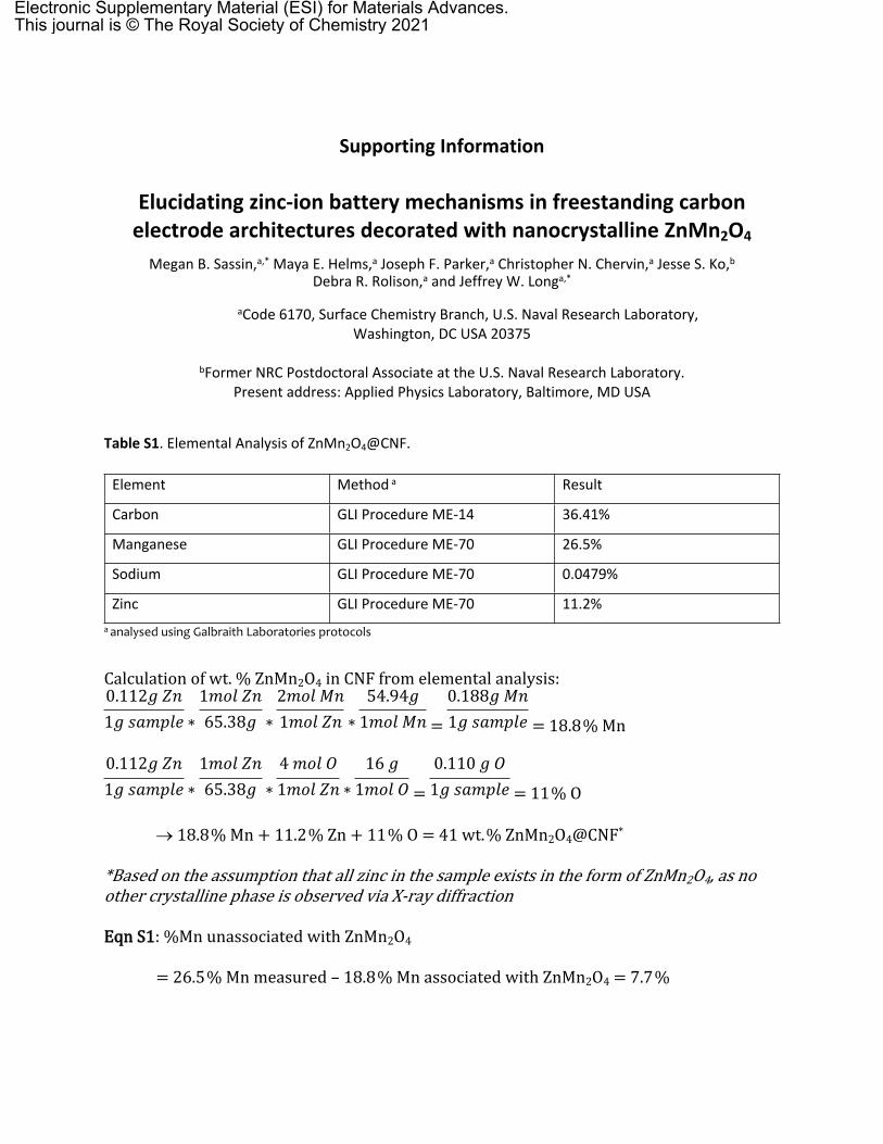
Supporting Information
Elucidating zinc-ion battery mechanisms in freestanding carbon electrode architectures decorated with nanocrystalline ZnMn2O4
Megan B. Sassin,a,* Maya E. Helms,a Joseph F. Parker,a Christopher N. Chervin,a Jesse S. Ko,b Debra R. Rolison,a and Jeffrey W. Longa,*
aCode 6170, Surface Chemistry Branch, U.S. Naval Research Laboratory, Washington, DC USA 20375
bFormer NRC Postdoctoral Associate at the U.S. Naval Research Laboratory. Present address: Applied Physics Laboratory, Baltimore, MD USA
Table S1. Elemental Analysis of ZnMn2O4@CNF.
Element Method a Result
Carbon GLI Procedure ME-14 36.41%
Manganese GLI Procedure ME-70 26.5%
Sodium GLI Procedure ME-70 0.0479%
Zinc GLI Procedure ME-70 11.2%a analysed using Galbraith Laboratories protocols
Calculation of wt. % ZnMn2O4 in CNF from elemental analysis:
* * * = = 18.8% Mn
0.112𝑔 𝑍𝑛1𝑔 𝑠𝑎𝑚𝑝𝑙𝑒
1𝑚𝑜𝑙 𝑍𝑛65.38𝑔
2𝑚𝑜𝑙 𝑀𝑛1𝑚𝑜𝑙 𝑍𝑛
54.94𝑔1𝑚𝑜𝑙 𝑀𝑛
0.188𝑔 𝑀𝑛1𝑔 𝑠𝑎𝑚𝑝𝑙𝑒
* * * = = 11% O
0.112𝑔 𝑍𝑛1𝑔 𝑠𝑎𝑚𝑝𝑙𝑒
1𝑚𝑜𝑙 𝑍𝑛65.38𝑔
4 𝑚𝑜𝑙 𝑂1𝑚𝑜𝑙 𝑍𝑛
16 𝑔1𝑚𝑜𝑙 𝑂
0.110 𝑔 𝑂1𝑔 𝑠𝑎𝑚𝑝𝑙𝑒
18.8% Mn + 11.2% Zn + 11% O = 41 wt.% ZnMn2O4@CNF*
*Based on the assumption that all zinc in the sample exists in the form of ZnMn2O4, as no other crystalline phase is observed via X-ray diffraction
Eqn S1: %Mn unassociated with ZnMn2O4
= 26.5% Mn measured – 18.8% Mn associated with ZnMn2O4 = 7.7%
Electronic Supplementary Material (ESI) for Materials Advances.This journal is © The Royal Society of Chemistry 2021
Eqn S2: % of Zn in pores for reaction
Moles Zn2+ required for reaction:
224 𝑚𝐴 ℎ𝑔𝑇𝑜𝑡𝑎𝑙
∗0.01 𝑔𝑇𝑜𝑡𝑎𝑙
𝑒𝑙𝑒𝑐𝑡𝑟𝑜𝑑𝑒∗
1 𝐴 1000 𝑚𝐴
∗ 3600 𝑠
1 ℎ ∗
1 𝑚𝑜𝑙 𝑒 ‒
96485 𝐶∗
0.5 𝑚𝑜𝑙 𝑍𝑛2 +
1 𝑚𝑜𝑙 𝑒 ‒ = 4.2 × 10 ‒ 5
Moles Zn2+ inside pores:
0.44 𝑐𝑚3 𝑔𝑇𝑜𝑡𝑎𝑙
∗0.01 𝑔𝑇𝑜𝑡𝑎𝑙
𝑒𝑙𝑒𝑐𝑡𝑟𝑜𝑑𝑒 = 0.0044 𝑐𝑚3 = 4.4 × 10 ‒ 6 𝐿 ∗
1 𝑚𝑜𝑙 𝑍𝑛𝑆𝑂4𝐿
= 4.4 × 10 ‒ 6
% of required Zn2+ inside pores
4.4 × 10 ‒ 6 𝑚𝑜𝑙 𝑍𝑛2 + 𝑖𝑛𝑠𝑖𝑑𝑒 𝑝𝑜𝑟𝑒𝑠
4.2 × 10 ‒ 5 𝑚𝑜𝑙 𝑍𝑛2 + 𝑟𝑒𝑞𝑢𝑖𝑟𝑒𝑑∗ 100 = 10.5 %
Eqn. S3: Concentration of dissolved Mn2+ inside pores
41 wt. % ZnMn2O4 in the electrodeElectrode mass: 0.01 gPore volume: 0.0044 𝑐𝑚3 = 4.4 × 10 ‒ 6 𝐿
% of electrode area internally
Geometric area of electrode: 0.000127 m2
Specific surface area of electrode: 260 m2 g-1 * 0.01 g = 2.6 m2
2.6 𝑚2 ‒ 0.000127 𝑚2
2.6 𝑚2∗ 100 = 99 %
Since 99% of the surface area is expressed internally, essentially all of the ZnMn2O4 mass exists in the interior of the electrode, so if all of the ZnMn2O4 dissolved, the [Mn2+] dissolved inside pores would be:
0.41 𝑔 𝑍𝑛𝑀𝑛2𝑂4
1 𝑔𝑇𝑜𝑡𝑎𝑙∗
0.01 𝑔𝑇𝑜𝑡𝑎𝑙
𝑒𝑙𝑒𝑐𝑡𝑟𝑜𝑑𝑒 ∗
1 𝑚𝑜𝑙 𝑍𝑛𝑀𝑛2𝑂4
239.25 𝑔 ∗
2 𝑚𝑜𝑙 𝑀𝑛1 𝑚𝑜𝑙 𝑍𝑛𝑀𝑛2𝑂4
∗𝑝𝑜𝑟𝑒 𝑣𝑜𝑙
4.4 × 10 ‒ 6 𝐿 = 7 𝑀 𝑀𝑛
DISTRIBUTION A. Approved for public release: distribution unlimited
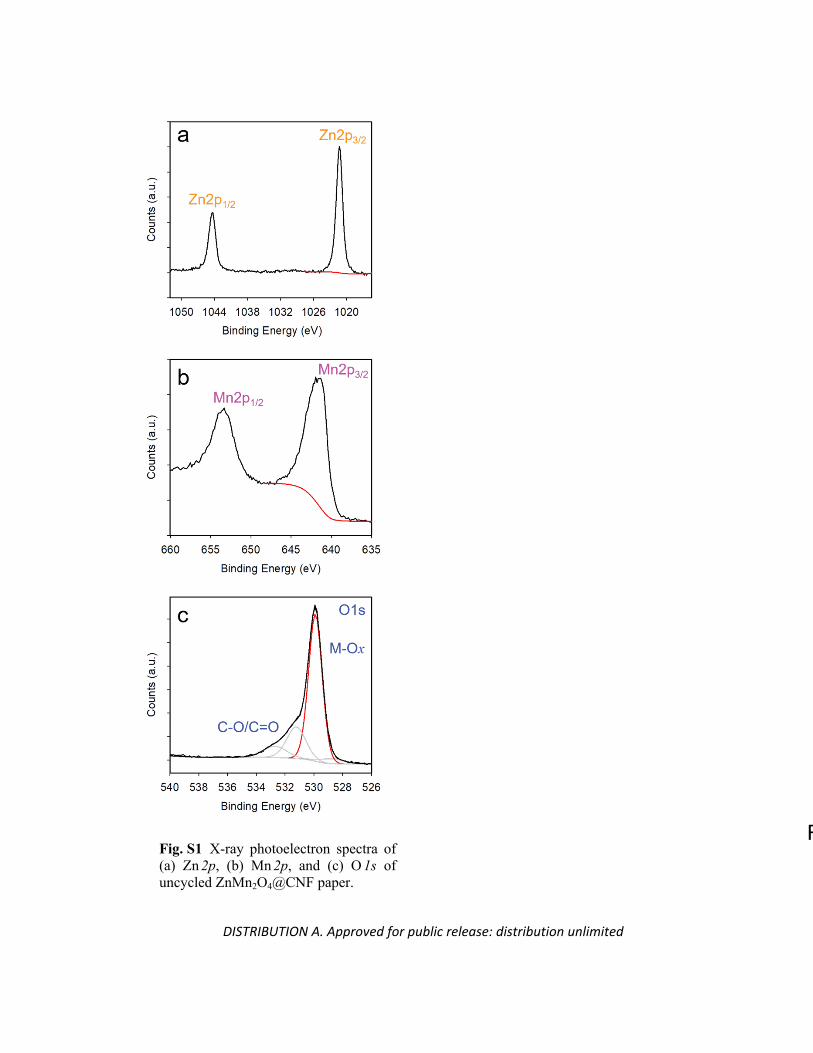
Remove the 0.9 V, 1.75 V data into its own figureFig. S1 X-ray photoelectron spectra of (a) Zn2p, (b) Mn2p, and (c) O1s of uncycled ZnMn2O4@CNF paper.
DISTRIBUTION A. Approved for public release: distribution unlimited
0 15 30 45 600
15
30
45
60
Z"
( c
m2 )
Z' ( cm2)
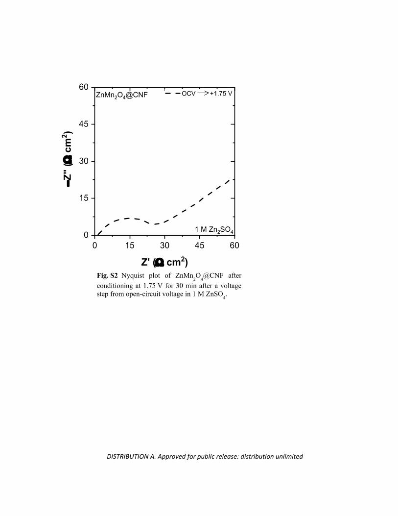
OCV +1.75 VZnMn2O4@CNF
1 M Zn2SO4
Fig. S2 Nyquist plot of ZnMn2O
4@CNF after
conditioning at 1.75 V for 30 min after a voltage step from open-circuit voltage in 1 M ZnSO
4.
DISTRIBUTION A. Approved for public release: distribution unlimited
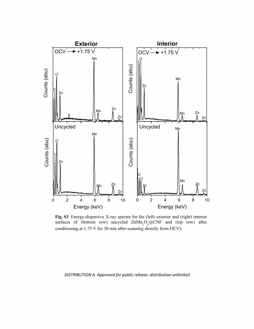
OCV +1.75 V OCV +1.75 V
Cou
nts
(abu
) CO
Zn
Mn
Mn ZnZn
0 2 4 6 8 10
Cou
nts
(abu
)
Energy (keV)
CO
Zn
Mn
MnZn
Zn
Cou
nts
(abu
)
C
O
Zn
Mn
Mn Zn
ZnS
0 2 4 6 8 10
Cou
nts
(abu
)
Energy (keV)
C
O
Zn
Mn
Mn Zn
Zn
Uncycled Uncycled
Exterior Interior
Fig. S3 Energy-dispersive X-ray spectra for the (left) exterior and (right) interior surfaces of (bottom row) uncycled ZnMn
2O
4@CNF and (top row) after
conditioning at 1.75 V for 30 min after scanning directly from OCV).
DISTRIBUTION A. Approved for public release: distribution unlimited
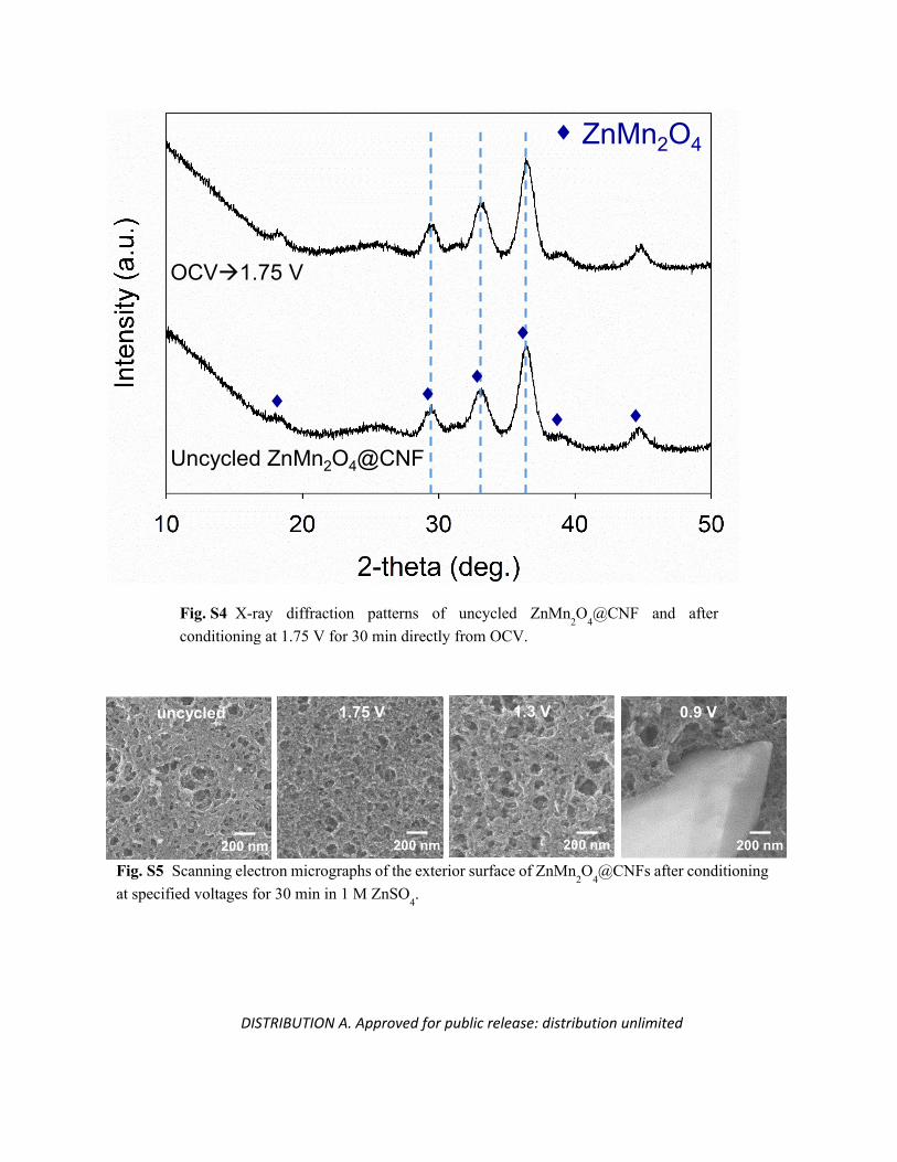
OCV1.75 V
Uncycled ZnMn2O4@CNF
ZnMn2O4
200 nm 200 nm 200 nm200 nm
uncycled 1.3 V 0.9 V1.75 V
Fig. S4 X-ray diffraction patterns of uncycled ZnMn2O
4@CNF and after
conditioning at 1.75 V for 30 min directly from OCV.
Fig. S5 Scanning electron micrographs of the exterior surface of ZnMn2O
4@CNFs after conditioning
at specified voltages for 30 min in 1 M ZnSO4.
DISTRIBUTION A. Approved for public release: distribution unlimited
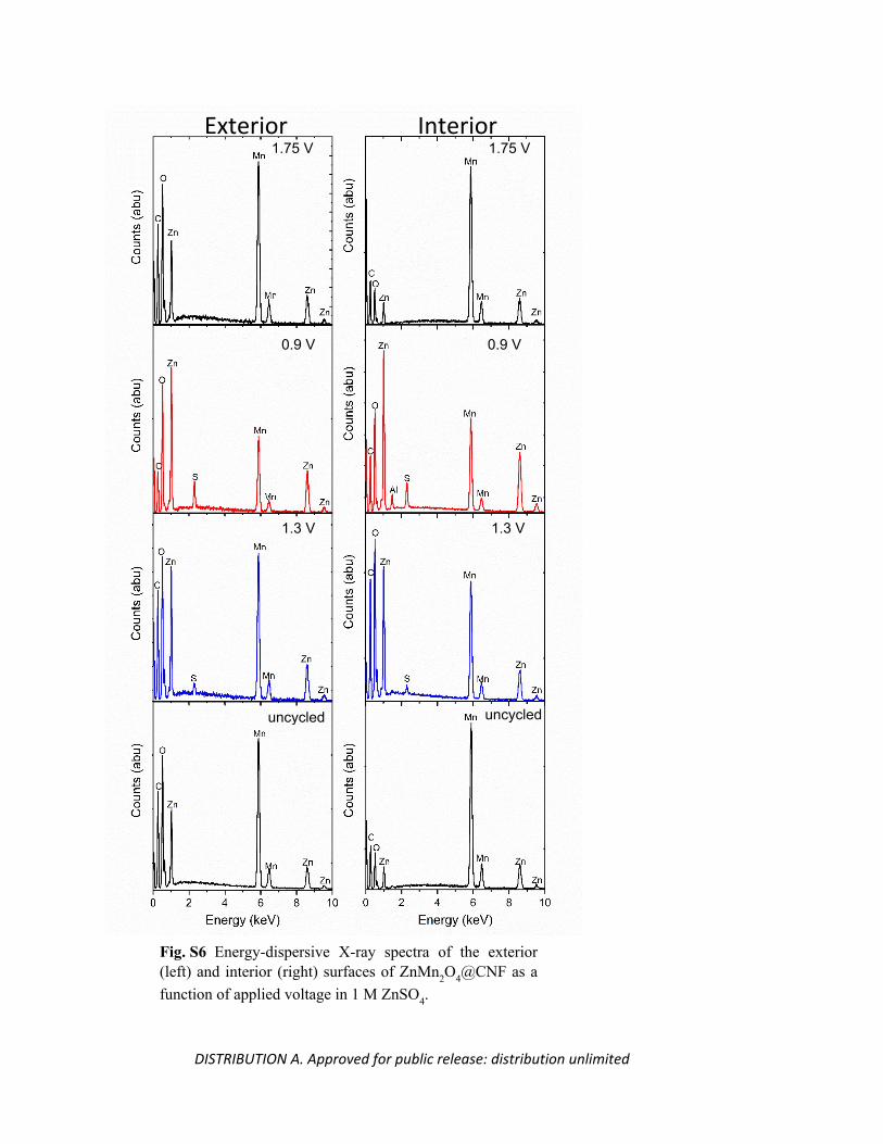
InteriorExterior
Fig. S6 Energy-dispersive X-ray spectra of the exterior (left) and interior (right) surfaces of ZnMn2O4@CNF as a function of applied voltage in 1 M ZnSO4.
1.75 V 1.75 V
0.9 V0.9 V
1.3 V 1.3 V
uncycleduncycled
DISTRIBUTION A. Approved for public release: distribution unlimited
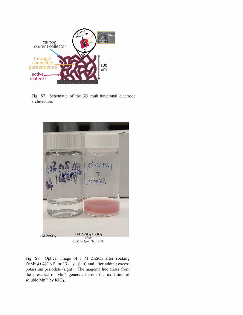
1 M ZnSO41 M ZnSO4 + KIO4
after ZnMn2O4@CNF soak
Fig. S7 Schematic of the 3D multifunctional electrode architecture.
Fig. S8 Optical image of 1 M ZnSO4 after soaking ZnMn2O4@CNF for 13 days (left) and after adding excess potassium periodate (right). The magenta hue arises from the presence of Mn7+ generated from the oxidation of soluble Mn2+ by KIO4.