Embed Size (px)
Citation preview
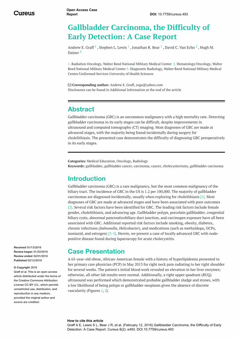
Received 01/13/2016 Review began 01/22/2016 Review ended 02/01/2016 Published 02/12/2016
© Copyright 2016Graff et al. This is an open accessarticle distributed under the terms ofthe Creative Commons AttributionLicense CC-BY 3.0., which permitsunrestricted use, distribution, andreproduction in any medium,provided the original author andsource are credited.
Gallbladder Carcinoma, the Difficulty ofEarly Detection: A Case ReportAndrew E. Graff , Stephen L. Lewis , Jonathan R. Bear , David C. Van Echo , Hugh M.Dainer
1. Radiation Oncology, Walter Reed National Military Medical Center 2. Hematology/Oncology, WalterReed National Military Medical Center 3. Diagnostic Radiology, Walter Reed National Military MedicalCenter/Uniformed Services University of Health Sciences
Corresponding author: Andrew E. Graff, [email protected] Disclosures can be found in Additional Information at the end of the article
AbstractGallbladder carcinoma (GBC) is an uncommon malignancy with a high mortality rate. Detectinggallbladder carcinoma in its early stages can be difficult, despite improvements inultrasound and computed tomography (CT) imaging. Most diagnoses of GBC are made atadvanced stages, with the majority being found incidentally during surgery forcholelithiasis. The presented case demonstrates the difficulty of diagnosing GBC preoperativelyin its early stages.
Categories: Medical Education, Oncology, RadiologyKeywords: gallbladder, gallbladder cancer, carcinoma, cancer, cholecystectomy, gallbladder carcinoma
IntroductionGallbladder carcinoma (GBC) is a rare malignancy, but the most common malignancy of thebiliary tract. The incidence of GBC in the US is 1.2 per 100,000. The majority of gallbladdercarcinomas are diagnosed incidentally, usually when exploring for cholelithiasis [1]. Mostdiagnoses of GBC are made at advanced stages and have been associated with poor outcomes[2]. Several risk factors have been identified for GBC. The leading risk factors include femalegender, cholelithiasis, and advancing age. Gallbladder polyps, porcelain gallbladder, congenitalbiliary cysts, abnormal pancreaticobiliary duct junction, and carcinogen exposure have all beenassociated with GBC. Additional reported risk factors include smoking, obesity, diabetes,chronic infections (Salmonella, Helicobacter), and medications (such as methyldopa, OCPs,isoniazid, and estrogen) [3-5]. Herein, we present a case of locally advanced GBC with node-positive disease found during laparoscopy for acute cholecystitis.
Case PresentationA 65-year-old obese, African-American female with a history of hyperlipidemia presented toher primary care physician (PCP) in May 2013 for right neck pain radiating to her right shoulderfor several weeks. The patient's initial blood work revealed an elevation in her liver enzymes;otherwise, all other lab results were normal. Additionally, a right upper quadrant (RUQ)ultrasound was performed which demonstrated probable gallbladder sludge and stones, witha low likelihood of being polyps or gallbladder neoplasm given the absence of discretevascularity (Figures 1, 2).
1 1 1 2
3
Open Access CaseReport DOI: 10.7759/cureus.493
How to cite this articleGraff A E, Lewis S L, Bear J R, et al. (February 12, 2016) Gallbladder Carcinoma, the Difficulty of EarlyDetection: A Case Report. Cureus 8(2): e493. DOI 10.7759/cureus.493

FIGURE 1: Initial right upper quadrant ultrasound.The image demonstrates intraluminal hypoechoic (red arrow) material. Given the appearanceand lack of discrete internal flow, it appears to be most likely due to gallstones or tumefactivesludge, which is a benign mimic of a neoplasm.
FIGURE 2: Initial right upper quadrant ultrasound.The image demonstrates no discrete internal flow within the gallbladder with blood flow inthe adjacent vasculature.
2016 Graff et al. Cureus 8(2): e493. DOI 10.7759/cureus.493 2 of 9

The patient was followed closely by her PCP until her symptoms resolved and elevated liverenzymes normalized over the next few months. No additional imaging was performed at thattime, and no surgery was scheduled.
In April 2014, the patient developed RUQ pain that worsened over the next four months,prompting her to seek medical help. In August 2014, she was reevaluated by her PCP andsubsequently underwent an acute abdominal series (AAS), RUQ ultrasound, and nuclearmedicine hepatobiliary (HIDA) study. The ultrasound demonstrated cholelithiasis andechogenic sludge within the gallbladder with hypoechoic and hyperemic adjacent liverparenchyma. A mildly enlarged common duct was also observed. These findingswere concerning for chronic cholecystitis, xanthogranulomatous cholecystitis, or early acutecholecystitis (Figure 3). The HIDA scan results were also consistent with acute cholecystitis(Figure 4).
FIGURE 3: Right upper quadrant ultrasound.The image demonstrates sludge/stone like material within the lumen of the gallbladder (redarrow) with a rim of hyperemia initially interpreted as outside of the gallbladder given the clinicalappearance of acute cholecystitis.
2016 Graff et al. Cureus 8(2): e493. DOI 10.7759/cureus.493 3 of 9

FIGURE 4: HIDA scan.The image demonstrates excretion through the biliary system into the duodenojejunal junction(DJJ), but with no uptake within the gallbladder/cystic duct (red arrow).
The patient was taken to surgery for a laparoscopic cholecystectomy for suspected cholecystitis.The laparoscopic approach was converted to an open procedure after the discovery of a masswithin the lumen of the gallbladder, a second mass near the hepatic flexure of the colon, andenlarged portal vein lymph nodes. During the operation, the patient underwent a radicalcholecystectomy, right hemicolectomy with primary anastomosis, and a portal vein lymphnode dissection (LND). Pathology of the gallbladder mass demonstrated infiltrating papillaryadenocarcinoma invading the perimuscular connective tissue. The pathology also revealed oneportocaval lymph node that was positive for adenocarcinoma. The patient's stage of disease wasdetermined to be IVB (pT2N2). After surgery, the patient underwent radiotherapy to 4500 cGy,targeting the gallbladder fossa and the involved lymph nodes, with concurrent chemotherapy(capecitabine 625 mg/m2 twice daily). Abdominal CT and MRI scans were performed eightmonths after the completion of chemoradiation. At that time, there was no radiologic evidenceof disease recurrence. Currently, the patient is fourteen months post treatment. Overall, she isdoing well and continues to receive close follow-up care.
DiscussionGBC is an infrequent neoplasm that is associated with a high rate of regional lymph nodemetastasis and mortality [2,6,9]. Adjuvant chemoradiation is commonly recommended fornode-positive or incompletely resected disease [7-8]. The extent of the resection correlates withsurvival. R0 resection is the most important because patients who undergo R2 resections dopoorly in spite of chemoradiation. However, there are some long-term survivors in patientstreated with adjuvant chemoradiation status post R0/R1 resections [6,9-10]. Therefore, earlydetection is important because patients found at stage T1N0 would have a greater chance forsurgical cure and spare them the potential toxicity of adjuvant therapy.
Early diagnosis can be difficult because symptoms can mimic or be caused by coexistingcholecystitis, which is a common condition [5]. Therefore, screening patients who present with
2016 Graff et al. Cureus 8(2): e493. DOI 10.7759/cureus.493 4 of 9

these symptoms is essential given the possibility for a coexisting GBC. Symptoms early inthe disease process can also be vague, often leading to a delay in diagnosis. The most commoncomplaint in the symptomatic patient with GBC is RUQ pain, specifically in the righthypochondrium. Other warning signs, which our patient did not exhibit, include weight loss,anorexia, nausea and/or vomiting, jaundice, and pruritus [11-12]. Routine laboratory tests aregenerally nondiagnostic and do not significantly improve the identification of GBCpreoperatively, as exemplified by our case [12]. Serum tumor markers, carcinoembryonicantigen (CEA), and carbohydrate antigen 19-9 (CA 19-9) are frequently elevated in patientswith GBC, but are not useful in its diagnosis because of their lack of sensitivity and specificity[13-14].
Imaging with ultrasound and CT has improved preoperative diagnosis of GBC. Despite theseadvancements, only 50% of gallbladder cancers are recognized before surgery [12]. Ultrasoundis often the initial imaging study of choice for patients presenting with symptoms consistentwith gallstone disease. The RUQ ultrasound is perhaps the single most important test inhelping lead to the diagnosis of GBC preoperatively. Ultrasound images from a group of patientsdiagnosed with GBC incidentally were reviewed retrospectively and found to have suspiciousfindings on reevaluation [15].
The most common ultrasound findings include calcified and echogenic mucosal masses, whichcan be associated with cholelithiasis or porcelain gallbladder [15]. High-risk features onultrasonography also include solitary or displaced gallstone, intraluminal mass, gallbladder-replacing or invasive mass, and discontinuity of the mucosal echo [15]. Other findings that aresuggestive of GBC include the loss of the interface between the gallbladder and liver or directliver infiltration [15]. Moreover, ultrasound abnormalities are often more subtle in early stagedisease, making detection more challenging [15]. If abnormalities or suspicious findings aredetected on ultrasound, further evaluation with other non-invasive imaging is warranted.Consideration of GBC in the differential diagnosis may help to improve detection before surgery(Figures 5-7), potentially leading to the discovery of the disease in its early stages.
FIGURE 5: Companion case. Right upper quadrant ultrasoundof a patient who presented a few months after the first patient.
2016 Graff et al. Cureus 8(2): e493. DOI 10.7759/cureus.493 5 of 9
The image demonstrates a hypoechoic lesion near the expected location of the gallbladder (redarrow). Given the suspicious ultrasound finding and recent prior GBC case, leading differentialdiagnosis was neoplasm. As a result, further evaluation with a CT scan was recommended.
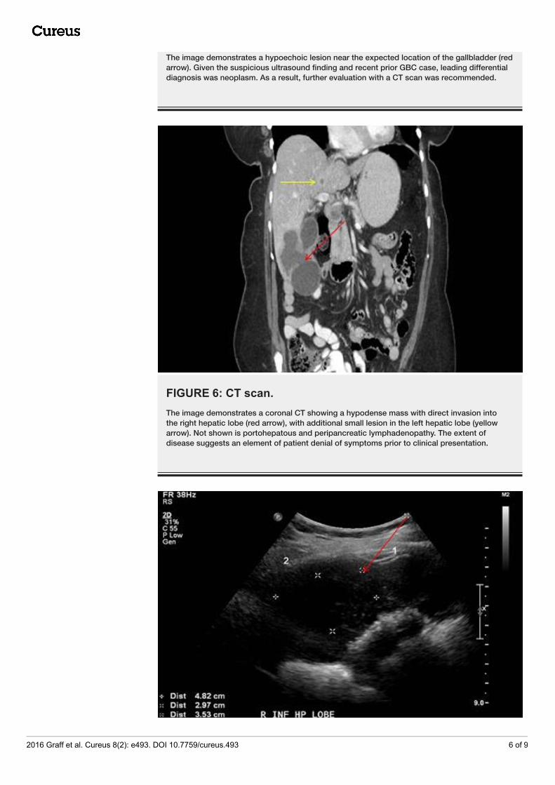
FIGURE 6: CT scan.The image demonstrates a coronal CT showing a hypodense mass with direct invasion intothe right hepatic lobe (red arrow), with additional small lesion in the left hepatic lobe (yellowarrow). Not shown is portohepatous and peripancreatic lymphadenopathy. The extent ofdisease suggests an element of patient denial of symptoms prior to clinical presentation.
2016 Graff et al. Cureus 8(2): e493. DOI 10.7759/cureus.493 6 of 9
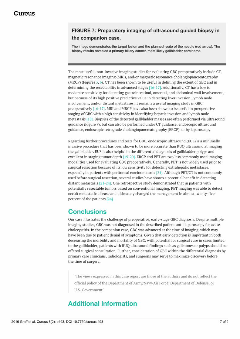
FIGURE 7: Preparatory imaging of ultrasound guided biopsy inthe companion case.The image demonstrates the target lesion and the planned route of the needle (red arrow). Thebiopsy results revealed a primary biliary cancer, most likely gallbladder carcinoma.
The most useful, non-invasive imaging studies for evaluating GBC preoperatively include CT,magnetic resonance imaging (MRI), and/or magnetic resonance cholangiopancreatography(MRCP) (Figures 5, 6). CT has been shown to be useful in defining the extent of GBC and indetermining the resectability in advanced stages [16-17]. Additionally, CT has a low tomoderate sensitivity for detecting gastrointestinal, omental, and abdominal wall involvement,but because of its high positive predictive value in detecting liver invasion, lymph nodeinvolvement, and/or distant metastases, it remains a useful imaging study in GBCpreoperatively [16-17]. MRI and MRCP have also been shown to be useful in preoperativestaging of GBC with a high sensitivity in identifying hepatic invasion and lymph nodemetastasis [18]. Biopsies of the detected gallbladder masses are often performed via ultrasoundguidance (Figure 7), but can also be performed under CT guidance, endoscopic ultrasoundguidance, endoscopic retrograde cholangiopancreatography (ERCP), or by laparoscopy.
Regarding further procedures and tests for GBC, endoscopic ultrasound (EUS) is a minimallyinvasive procedure that has been shown to be more accurate than RUQ ultrasound at imagingthe gallbladder. EUS is also helpful in the differential diagnosis of gallbladder polyps andexcellent in staging tumor depth [19-20]. ERCP and PET are two less commonly used imagingmodalities used for evaluating GBC preoperatively. Generally, PET is not widely used prior tosurgical resection because of its low sensitivity for detecting extrahepatic metastases,especially in patients with peritoneal carcinomatosis [21]. Although PET/CT is not commonlyused before surgical resection, several studies have shown a potential benefit in detectingdistant metastasis [21-24]. One retrospective study demonstrated that in patients withpotentially resectable tumors based on conventional imaging, PET imaging was able to detectoccult metastatic disease and ultimately changed the management in almost twenty-fivepercent of the patients [24].
ConclusionsOur case illustrates the challenge of preoperative, early-stage GBC diagnosis. Despite multipleimaging studies, GBC was not diagnosed in the described patient until laparoscopy for acutecholecystitis. In the companion case, GBC was advanced at the time of imaging, which mayhave been due to patient denial of symptoms. Given that early detection is important in bothdecreasing the morbidity and mortality of GBC, with potential for surgical cure in cases limitedto the gallbladder, patients with RUQ ultrasound findings such as gallstones or polyps should beoffered surgical consultation. Further, consideration of GBC within the differential diagnosis byprimary care clinicians, radiologists, and surgeons may serve to maximize discovery beforethe time of surgery.
"The views expressed in this case report are those of the authors and do not reflect the
official policy of the Department of Army/Navy/Air Force, Department of Defense, or
U.S. Government."
Additional Information
2016 Graff et al. Cureus 8(2): e493. DOI 10.7759/cureus.493 7 of 9

DisclosuresConflicts of interest: The authors have declared that no conflicts of interest exist.
References1. Carriaga MT, Henson DE: Liver, gallbladder, extrahepatic bile ducts, and pancreas. Cancer.
1995, 75:171–190. 10.1002/1097-0142(19950101)75:1+<171::AID-CNCR2820751306>3.0.CO;2-2
2. Duffy A, Capanu M, Abou-Alfa GK, et al: Gallbladder cancer (GBC): 10-year experience atMemorial Sloan-Kettering Cancer Centre (MSKCC). J Surg Oncol. 2008, 98:485–489.10.1002/jso.21141
3. Zatonski WA, Lowenfels AB, Boyle P, et al: Epidemiologic aspects of gallbladder cancer: acase-control study of the SEARCH Program of the International Agency for Research onCancer. J Natl Cancer Inst. 1997, 89:1132-1138. 10.1093/jnci/89.15.1132
4. Rustagi T, Dasanu CA: Risk factors for gallbladder cancer and cholangiocarcinoma:similarities, differences and updates. J Gastrointest Cancer. 2012, 43:137-147.10.1007/s12029-011-9284-y
5. Hsing AW, Gao YT, Han TQ, et al: Gallstones and the risk of biliary tract cancer: a population-based study in China. Br J Cancer. 2007, 97:1577–1582. 10.1038/sj.bjc.6604047
6. Groot Koerkamp B, Fong Y: Outcomes in biliary malignancy. J Surg Oncol. 2014, 110:585-591.10.1002/jso.23762
7. Horgan AM, Amir E, Walter T, Knox JJ: Adjuvant therapy in the treatment of biliary tractcancer: a systematic review and meta-analysis. J Clin Oncol. 2012, 30:1934-1940.10.1200/jco.2011.40.5381
8. Wang SJ, Lemieux A, Kalpathy-Cramer J, et al: Nomogram for predicting the benefit ofadjuvant chemoradiotherapy for resected gallbladder cancer. J Clin Oncol. 2011, 29:4627-4632. 10.1200/jco.2010.33.8020
9. Cariati A, Piromalli E, Cetta F: Gallbladder cancers: associated conditions, histological types,prognosis, and prevention. Eur J Gastroenterol Hepatol. 2014, 26:562–569.10.1097/meg.0000000000000074
10. Pilgrim CH, Groeschl RT, Turaga KK, Gamblin TC: Key factors influencing prognosis inrelation to gallbladder cancer. Dig Dis Sci. 2013, 58:2455–2462. 10.1007/s10620-013-2713-y
11. Misra S, Chaturvedi A, Misra NC, Sharma ID: Carcinoma of the gallbladder . Lancet Oncol.2003, 4:167-176. 10.1016/S1470-2045(03)01021-0
12. Löhe F, Meimarakis G, Schauer C, Angele M, Jauch KW, Schauer RJ: The time of diagnosisimpacts surgical management but not the outcome of patients with gallbladder carcinoma.Eur J Med Res. 2009, 14:345–351.
13. Strom BL, Maislin G, West SL, et al: Serum CEA and CA 19-9: potential future diagnostic orscreening tests for gallbladder cancer. Int J Cancer. 1990, 45:821-824. 10.1002/ijc.2910450505
14. Ritts RE Jr, Nagorney DM, Jacobsen DJ, Talbot RW, Zurawski VR Jr: Comparison ofpreoperative serum CA19-9 levels with results of diagnostic imaging modalities in patientsundergoing laparotomy for suspected pancreatic or gallbladder disease. Pancreas. 1994, 9:707-716.
15. Wibbenmeyer LA, Sharafuddin MJ, Wolverson MK, Heiberg EV, Wade TP, Shields JB:Sonographic diagnosis of unsuspected gallbladder cancer: imaging findings in comparisonwith benign gallbladder conditions. AJR Am J Roentgenol. 1995, 165:1169–1174.10.2214/ajr.165.5.7572497
16. Kumar A, Aggarwal S: Carcinoma of the gallbladder: CT findings in 50 cases . Abdom Imaging.1994, 19:304-308. 10.1007/BF00198184
17. Ohtani T, Shirai Y, Tsukada K, Muto T, Hatakeyama K: Spread of gallbladder carcinoma: CTevaluation with pathologic correlation. Abdom Imaging. 1996, 21:195-201.10.1007/s002619900045
18. Schwartz LH, Black J, Fong Y, et al: Gallbladder carcinoma: findings at MR imaging with MRcholangiopancreatography. J Comput Assist Tomogr. 2002, 26:405-410. 10.1097/00004728-200205000-00015
19. Sugiyama M, Atomi Y, Yamato T: Endoscopic ultrasonography for differential diagnosis ofpolypoid gall bladder lesions: analysis in surgical and follow up series. Gut. 2000, 46:250-254.
2016 Graff et al. Cureus 8(2): e493. DOI 10.7759/cureus.493 8 of 9
10.1136/gut.46.2.25020. Sadamoto Y, Kubo H, Harada N, Tanaka M, Eguchi T, Nawata H: Preoperative diagnosis and
staging of gallbladder carcinoma by EUS. Gastrointest Endosc. 2003, 58:536-541.10.1067/S0016-5107(03)01961-8
21. Anderson CD, Rice MH, Pinson CW, Chapman WC, Chari RS, Delbeke D: FluorodeoxyglucosePET imaging in the evaluation of gallbladder carcinoma and cholangiocarcinoma. JGastrointest Surg. 2004, 8:90-97. 10.1016/j.gassur.2003.10.003
22. Petrowsky H, Wildbrett P, Husarik DB, et al: Impact of integrated positron emissiontomography and computed tomography on staging and management of gallbladder cancer andcholangiocarcinoma. J Hepatol. 2006, 45:43-50. 10.1016/j.jhep.2006.03.009
23. Lee SW, Kim HJ, Park JH, et al: Clinical usefulness of 18F-FDG PET-CT for patients withgallbladder cancer and cholangiocarcinoma. J Gastroenterol. 2010, 45:560-566.10.1007/s00535-009-0188-6
24. Corvera CU, Blumgart LH, Akhurst T, et al: 18F-fluorodeoxyglucose positron emissiontomography influences management decisions in patients with biliary cancer. J Am Coll Surg.2008, 206:57-65. 10.1016/j.jamcollsurg.2007.07.002
2016 Graff et al. Cureus 8(2): e493. DOI 10.7759/cureus.493 9 of 9