Embed Size (px)
Citation preview
Non-Small Cell Lung Cancer EarlyDetection, Diagnosis, and Staging Detection and Diagnosis
Catching cancer early often allows for more treatment options. Some early cancers mayhave signs and symptoms that can be noticed, but that is not always the case.
Can Non-Small Cell Lung Cancer Be Found Early?●
Lung Cancer Prevention and Early Detection●
Non-Small Cell Lung Cancer Signs and Symptoms●
Tests for Non-Small Cell Lung Cancer●
Understanding Your Pathology Report●
Stages and Outlook (Prognosis)
After a cancer diagnosis, staging provides important information about the extent ofcancer in the body and anticipated response to treatment.
Non-Small Cell Lung Cancer Stages●
Non-Small Cell Lung Cancer Survival Rates, by Stage●
Questions to Ask About Non-Small Cell Lung Cancer
Here are some questions you can ask your cancer care team to help you betterunderstand your cancer diagnosis and treatment options.
What Should You Ask Your Health Care Team About Non-Small Cell Lung Cancer?●
Questions Worksheet [PDF]●
Can Non-Small Cell Lung Cancer Be
Found Early? Usually symptoms of lung cancer do not appear until the disease is already at anadvanced stage. Even when lung cancer does cause symptoms, many people maymistake them for other problems, such as an infection or long-term effects fromsmoking. This may delay the diagnosis.
Some lung cancers are found early by accident as a result of tests for other medicalconditions. For example, lung cancer may be found by tests done for other reasons inpeople with heart disease, pneumonia, or other lung conditions. A small portion of thesepeople do very well and may be cured of lung cancer.
Screening is the use of tests or exams to find a disease in people who don’t havesymptoms. Doctors have looked for many years for a good screening test for lungcancer, but only in recent years has a study shown that a test known as a low-dose CT(LDCT) scan can help lower the risk of dying from this disease.
The National Lung Screening Trial
The National Lung Screening Trial (NLST) was a large clinical trial that looked at usingLDCT of the chest to screen for lung cancer. CT scans of the chest provide moredetailed pictures than chest x-rays and are better at finding small abnormal areas in thelungs. Low-dose CT of the chest uses lower amounts of radiation than a standard chestCT and does not require the use of intravenous (IV) contrast dye.
The NLST compared LDCT of the chest to chest x-rays in people at high risk of lungcancer to see if these scans could help lower the risk of dying from lung cancer. Thestudy included more than 50,000 people ages 55 to 74 who were current or formersmokers and were in fairly good health. To be in the study, they had to have at least a30 pack-year history of smoking.
A pack-year is the number of cigarette packs smoked each day multiplied by thenumber of years a person has smoked. Someone who smoked a pack of cigarettes perday for 30 years has a 30 pack-year smoking history, as does someone who smoked 2packs a day for 15 years.
Former smokers could enter the study if they had quit within the past 15 years. Thestudy did not include people if they had a history of lung cancer or lung cancersymptoms, if they had part of a lung removed, if they needed to be on oxygen at hometo help them breathe, or if they had other serious medical problems.
People in the study got either 3 LDCT scans or 3 chest x-rays, each a year apart, tolook for abnormal areas in the lungs that might be cancer. After several years, the studyfound that people who got LDCT had a 20% lower chance of dying from lung cancerthan those who got chest x-rays. They were also 7% less likely to die overall (from anycause) than those who got chest x-rays.
Screening with LDCT was also shown to have some downsides that need to beconsidered. One drawback of this test is that it also finds a lot of abnormalities that haveto be checked out with more tests, but that turn out not to be cancer. (About 1 out of 4people in the NLST had such a finding.) This may lead to additional tests such as otherCT scans or more invasive tests such as needle biopsies or even surgery to remove aportion of lung in some people. These tests can sometimes lead to complications (like acollapsed lung) or rarely, death, even in people who do not have cancer (or who havevery early stage cancer).
LDCTs also expose people to a small amount of radiation with each test. It is less thanthe dose from a standard CT, but it is more than the dose from a chest x-ray. Somepeople who are screened may end up needing further CT scans, which means moreradiation exposure. When done in tens of thousands of people, this radiation may causea few people to develop breast, lung, or thyroid cancers later on.
The NLST was a large study, but it left some questions that still need to be answered.For example, it’s not clear if screening with LDCT scans would have the same effect ifdifferent people were allowed in the study, such as those who smoke less (or not at all),or people younger than age 55 or older than 74. Also, in the NLST, patients got 3 scansover 2 years. It’s not yet clear what the effect would be if people were screened forlonger than 2 years.
These factors, and others, need to be taken into account by people and their doctorswho are considering whether or not screening with LDCT scans is right for them.
American Cancer Society’s guidelines for lung cancerscreening
The American Cancer Society has thoroughly reviewed the subject of lung cancerscreening and issued guidelines that are aimed at doctors and other health careproviders:
Patients should be asked about their smoking history. Patients who meet ALL of thefollowing criteria may be candidates for lung cancer screening:
55 to 74 years old●
In fairly good health (discussed further down)●
Have at least a 30 pack-year smoking history (discussed above)●
Are either still smoking or have quit smoking within the last 15 years●
These criteria were based on what was used in the NLST.
Doctors should talk to these patients about the benefits, limitations, and potential harmsof lung cancer screening. Screening should only be done at facilities that have the righttype of CT scanner and that have a lot of experience using low-dose CT (LDCT) scansfor lung cancer screening. The facility should also have a team of specialists that canprovide the appropriate care and follow-up of patients with abnormal results on thescans.
For patients
If you fit all of the criteria listed above for lung cancer screening, you and your doctor (orother health care provider) should talk about screening, including possible benefits andharms, as well as the limitations of screening.
The main benefit is a lower chance of dying of lung cancer, which accounts for manydeaths in current and former smokers. Still, it’s important to be aware that, like with anytype of screening, not everyone who gets screened will benefit. Screening with LDCTwill not find all lung cancers, and not all of the cancers that are found will be found early.
Even if a cancer is found by screening, you may still die from lung cancer. Also, LDCToften finds things that turn out not to be cancer, but have to be checked out with moretests to know what they are. You might need more CT scans, or even invasive testssuch as a lung biopsy, in which a piece of lung tissue is removed with a needle orduring surgery. These tests have risks of their own (see above).
Screening should only be done at facilities that have the right type of CT scanner andthat have experience in LDCT scans for lung cancer screening. The facility should alsohave a team of specialists that can give patients the appropriate care and follow-up ifthere are abnormal results on the scans. You might not have the right kind of facilitynearby, so you may need to travel some distance to be screened.
If you and your doctor decide that you should be screened, you should get a LDCTevery year until you reach the age of 74, as long as you are still in good health.
If you smoke, you should get counseling about stopping. You should be told about yourrisk of lung cancer and referred to a smoking cessation program. Screening is not a
good alternative to stopping smoking. For help quitting, see our Guide to QuittingSmoking or call the American Cancer Society at 1-800-227-2345.
What does “in fairly good health” mean?
Screening is meant to find cancer in people who do not have symptoms of the disease.People who already have symptoms that might be caused by lung cancer may needtests such as CT scans to find the underlying cause, which in some cases may becancer. But this kind of testing is for diagnosis and is not the same as screening. Someof the possible symptoms of lung cancer that kept people out of the NLST werecoughing up blood and weight loss without trying.
To get the most benefit from screening, patients need to be in good health. Forexample, they need to be able to have surgery and other treatments to try to cure lungcancer if it is found. Patients who need home oxygen therapy probably couldn’twithstand having part of a lung removed, and so are not candidates for screening.Patients with other serious medical problems that would shorten their lives or keep themfrom having surgery might not benefit enough from screening for it to be worth the risks,and so should also not be screened.
Metal implants in the chest (like pacemakers) or back (like rods in the spine) caninterfere with x-rays and lead to poor quality CT images of the lungs. People with thesetypes of implants were also kept out of the NLST, and so should not be screened withCT scans for lung cancer according to the ACS guidelines.
References●
See all references for Non-Small Cell Lung Cancer
Last Medical Review: February 8, 2016 Last Revised: May 16, 2016
American Cancer Society medical information is copyrighted material. For reprintrequests, please see our Content Usage Policy.
Non-Small Cell Lung Cancer Signs andSymptoms Most lung cancers do not cause any symptoms until they have spread, but some people
with early lung cancer do have symptoms. If you go to your doctor when you first noticesymptoms, your cancer might be diagnosed at an earlier stage, when treatment is morelikely to be effective. The most common symptoms of lung cancer are:
A cough that does not go away or gets worse●
Coughing up blood or rust-colored sputum (spit or phlegm)●
Chest pain that is often worse with deep breathing, coughing, or laughing●
Hoarseness●
Weight loss and loss of appetite●
Shortness of breath●
Feeling tired or weak●
Infections such as bronchitis and pneumonia that don’t go away or keep comingback
●
New onset of wheezing●
When lung cancer spreads to distant organs, it may cause:
Bone pain (like pain in the back or hips)●
Nervous system changes (such as headache, weakness or numbness of an arm orleg, dizziness, balance problems, or seizures), from cancer spread to the brain orspinal cord
●
Yellowing of the skin and eyes (jaundice), from cancer spread to the liver●
Lumps near the surface of the body, due to cancer spreading to the skin or to lymphnodes (collections of immune system cells), such as those in the neck or above thecollarbone
●
Most of these symptoms are more likely to be caused by something other than lungcancer. Still, if you have any of these problems, it’s important to see your doctor rightaway so the cause can be found and treated, if needed.
Some lung cancers can cause syndromes, which are groups of very specific symptoms.
Horner syndrome
Cancers of the top part of the lungs (sometimes called Pancoast tumors) sometimescan affect certain nerves to the eye and part of the face, causing a group of symptomscalled Horner syndrome:
Drooping or weakness of one eyelid●
A smaller pupil (dark part in the center of the eye) in the same eye●
Reduced or absent sweating on the same side of the face●
Pancoast tumors can also sometimes cause severe shoulder pain.
Superior vena cava syndrome
The superior vena cava (SVC) is a large vein that carries blood from the head and armsback to the heart. It passes next to the upper part of the right lung and the lymph nodesinside the chest. Tumors in this area can press on the SVC, which can cause the bloodto back up in the veins. This can lead to swelling in the face, neck, arms, and upperchest (sometimes with a bluish-red skin color). It can also cause headaches, dizziness,and a change in consciousness if it affects the brain. While SVC syndrome can developgradually over time, in some cases it can become life-threatening, and needs to betreated right away.
Paraneoplastic syndromes
Some lung cancers can make hormone-like substances that enter the bloodstream andcause problems with distant tissues and organs, even though the cancer has not spreadto those tissues or organs. These problems are called paraneoplastic syndromes.Sometimes these syndromes can be the first symptoms of lung cancer. Because thesymptoms affect organs besides the lungs, patients and their doctors may suspect atfirst that a disease other than lung cancer is causing them.
Some of the more common paraneoplastic syndromes that can be caused by non-smallcell lung cancer include:
High blood calcium levels (hypercalcemia), which can cause frequent urination,thirst, constipation, nausea, vomiting, belly pain, weakness, fatigue, dizziness,confusion, and other nervous system problems
●
Excess growth/thickening of certain bones, especially those in the finger tips, whichis often painful
●
Blood clots●
Excess breast growth in men (gynecomastia)●
Again, many of these symptoms are more likely to be caused by something other thanlung cancer. Still, if you have any of these problems, it’s important to see your doctorright away so the cause can be found and treated, if needed.
References●
See all references for Non-Small Cell Lung Cancer
Last Medical Review: February 8, 2016 Last Revised: May 16, 2016
American Cancer Society medical information is copyrighted material. For reprintrequests, please see our Content Usage Policy.
Tests for Non-Small Cell Lung Cancer Some lung cancers can be found by screening, but most lung cancers are foundbecause they are causing problems. If you have possible signs or symptoms of lungcancer, see your doctor, who will examine you and may order some tests. The actualdiagnosis of lung cancer is made by looking at a sample of lung cells under amicroscope.
Medical history and physical exam
Your doctor willask about your medical history to learn about your symptoms andpossible risk factors. Your doctor will also examine you to look for signs of lung canceror other health problems.
If the results of your history and physical exam suggest you might have lung cancer,more tests will be done. These could include imaging tests and/or getting biopsies oflung tissue.
Imaging tests
Imaging tests use x-rays, magnetic fields, sound waves, or radioactive substances tocreate pictures of the inside of your body. Imaging tests may be done for a number ofreasons both before and after a diagnosis of lung cancer, including:
To look at suspicious areas that might be cancer●
To learn how far cancer may have spread●
To help determine if treatment is working●
To look for possible signs of cancer coming back after treatment●
Chest x-ray
This is often the first test your doctor will do to look for any abnormal areas in the lungs.Plain x-rays of your chest can be done at imaging centers, hospitals, and even in some
doctors’ offices. If something suspicious is seen, your doctor may order more tests.
Computed tomography (CT) scan
A CT scan uses x-rays to make detailed cross-sectional images of your body. Instead oftaking one picture, like a regular x-ray, a CT scanner takes many pictures as it rotatesaround you while you lie on a table. A computer then combines these pictures intoimages of slices of the part of your body being studied.
A CT scan is more likely to show lung tumors than routine chest x-rays. It can also showthe size, shape, and position of any lung tumors and can help find enlarged lymphnodes that might contain cancer that has spread from the lung. This test can also beused to look for masses in the adrenal glands, liver, brain, and other internal organs thatmight be due to the spread of lung cancer.
CT-guided needle biopsy: If a suspected area of cancer is deep within your body, aCT scan can be used to guide a biopsy needle into the suspected area.
Magnetic resonance imaging (MRI) scan
Like CT scans, MRI scans provide detailed images of soft tissues. But MRI scans useradio waves and strong magnets instead of x-rays. A contrast material called gadoliniumis often injected into a vein before the scan to better see details.
MRI scans are most often used to look for possible spread of lung cancer to the brain orspinal cord. Rarely, MRI of the chest may be done to see if the cancer has grown intocentral structures in the chest.
Positron emission tomography (PET) scan
For this test, a form of radioactive sugar (known as FDG) is injected into the blood.Because cancer cells in the body are growing quickly, they absorb more of theradioactive sugar. This radioactivity can be seen with a special camera.
PET/CT scan: Often a PET scan is combined with a CT scan using a special machinethat can do both at the same time. This lets the doctor compare areas of higherradioactivity on the PET scan with the more detailed appearance of that area on the CTscan. This is the type of PET scan most often used in patients with lung cancer.
If you appear to have early stage lung cancer, your doctor can use this test to help see
if the cancer has spread to nearby lymph nodes or other areas, which can helpdetermine if surgery may be an option for you. This test can also be helpful in getting abetter idea if an abnormal area on another imaging test might be cancer.
PET/CT scans can also be useful if your doctor thinks the cancer might have spread butdoesn’t know where. They can show spread of cancer to the liver, bones, adrenalglands, or some other organs. They are not as useful for looking at the brain, since allbrain cells use a lot of glucose.
PET/CT scans are often helpful in diagnosing lung cancer, but their role in checkingwhether treatment is working is unproven. Most doctors do not recommend PET/CTscans for routine follow up of patients with lung cancer after treatment.
Bone scan
For this test, a small amount of low-level radioactive material is injected into the blood.The substance settles in areas of bone changes throughout the entire skeleton. Thisradioactivity can be seen with a special camera.
A bone scan can help show if a cancer has spread to the bones. But this test isn’tneeded very often because PET scans, which are often done in patients with non-smallcell lung cancer, can usually show if cancer has spread to the bones. Bone scans aredone mainly when there is reason to think the cancer may have spread to the bones(because of symptoms such as bone pain) and other test results aren’t clear.
Tests for diagnosing lung cancer
Symptoms and the results of certain tests may strongly suggest that a person has lungcancer, but the actual diagnosis is made by looking at lung cells with a microscope.
The cells can be taken from lung secretions (sputum or phlegm), fluid removed from thearea around the lung (thoracentesis), or from a suspicious area using a needle orsurgery (known as a biopsy). The choice of which test(s) to use depends on thesituation.
Sputum cytology
A sample of mucus you cough up from the lungs (sputum) is looked at under amicroscope to see if it has cancer cells. The best way to do this is to get early morningsamples from you 3 days in a row. This test is more likely to help find cancers that start
in the major airways of the lung, such as squamous cell lung cancers. It may not be ashelpful for finding other types of non-small cell lung cancer. If your doctor suspects lungcancer, further testing will be done even if no cancer cells are found in the sputum.
Thoracentesis
If there is a buildup of fluid around the lungs (called a pleural effusion), doctors canperform thoracentesis to find out if it is caused by cancer spreading to the lining of thelungs (pleura). The buildup might also be caused by other conditions, such as heartfailure or an infection.
For this procedure, the skin is numbed and a hollow needle is inserted between the ribsto drain the fluid. (In a similar test called pericardiocentesis, fluid is removed from withinthe sac around the heart.) The fluid is checked under a microscope for cancer cells.Chemical tests of the fluid are also sometimes useful in telling a malignant (cancerous)pleural effusion from one that is not.
If a malignant pleural effusion has been diagnosed, thoracentesis may be repeated toremove more fluid. Fluid buildup can keep the lungs from filling with air, sothoracentesis can help a person breathe better.
Needle biopsy
Doctors can often use a hollow needle to get a small sample from a suspicious area(mass).
In a fine needle aspiration (FNA)biopsy, the doctor uses a syringe with a very thin,hollow needle to withdraw (aspirate) cells and small fragments of tissue.
●
In a core biopsy, a larger needle is used to remove one or more small cores oftissue. Samples from core biopsies are larger than FNA biopsies, so they are oftenpreferred.
●
An advantage of needle biopsies is that they don’t require a surgical incision. Thedrawback is that they remove only a small amount of tissue. In some cases (particularlywith FNA biopsies), the amount removed might not be enough to both make a diagnosisand to classify DNA changes in the cancer cells that can help doctors chooseanticancer drugs.
Transthoracic needle biopsy: If the suspected tumor is in the outer part of the lungs,the biopsy needle can be inserted through the skin on the chest wall. The area wherethe needle is to be inserted may be numbed with local anesthesia first. The doctor then
guides the needle into the area while looking at the lungs with either fluoroscopy (whichis like an x-ray, but creates a moving image on a screen rather than a single picture onfilm) or CT scans.
If CT is used, the needle is inserted toward the mass (tumor), a CT image is taken, andthe direction of the needle is guided based on the image. This is repeated a few timesuntil the needle is within the mass.
A possible complication of this procedure is that air may leak out of the lung at thebiopsy site and into the space between the lung and the chest wall. This is called apneumothorax. It can cause part of the lung to collapse and possibly trouble breathing.If the air leak is small, it often gets better without any treatment. Large air leaks aretreated by putting a small tube into the chest space and sucking out the air over a dayor two, after which it usually heals on its own.
Other approaches to needle biopsies: An FNA biopsy may also be done to check forcancer in the lymph nodes between the lungs:
Transtracheal FNA or transbronchial FNA is done by passing the needle throughthe wall of the trachea (windpipe) or bronchi (the large airways leading into thelungs) during bronchoscopy or endobronchial ultrasound (described below).
●
In some patients an FNA biopsy is done during endoscopic esophageal ultrasound(described below) by passing the needle through the wall of the esophagus.
●
Bronchoscopy
Bronchoscopy can help the doctor find some tumors or blockages in the larger airwaysof the lungs, which can often be biopsied during the procedure.
For this exam, a lighted, flexible fiber-optic tube (called a bronchoscope) is passedthrough the mouth or nose and down into the windpipe and bronchi. The mouth andthroat are sprayed first with a numbing medicine. You may also be given medicinethrough an intravenous (IV) line to make you feel relaxed.
Small instruments can be passed down the bronchoscope to take biopsy samples. Thedoctor can also sample cells from the lining of the airways with a small brush (bronchialbrushing) or by rinsing the airways with sterile saltwater (bronchial washing). Thesetissue and cell samples are then looked at under a microscope.
Tests to find lung cancer spread in the chest
If lung cancer has been found, it’s often important to know if it has spread to the lymphnodes in the space between the lungs (mediastinum) or other nearby areas. This canaffect a person’s treatment options. Several types of tests can be used to look for thiscancer spread.
Endobronchial ultrasound
Ultrasound is a type of imaging test that uses sound waves to create pictures of theinside of your body. For this test, a small, microphone-like instrument called atransducer gives off sound waves and picks up the echoes as they bounce off bodytissues. The echoes are converted by a computer into an image on a computer screen.
For endobronchial ultrasound, a bronchoscope is fitted with an ultrasound transducer atits tip and is passed down into the windpipe. This is done with numbing medicine (localanesthesia) and light sedation.
The transducer can be pointed in different directions to look at lymph nodes and otherstructures in the mediastinum (the area between the lungs). If suspicious areas such asenlarged lymph nodes are seen on the ultrasound, a hollow needle can be passedthrough the bronchoscope and guided into these areas to obtain a biopsy. The samplesare then sent to a lab to be looked at under a microscope.
Endoscopic esophageal ultrasound
This test is like endobronchial ultrasound, except the doctor passes an endoscope (alighted, flexible scope) down the throat and into the esophagus (the tube connecting thethroat to the stomach). This is done with numbing medicine (local anesthesia) and lightsedation.
The esophagus is just behind the windpipe and is close to some lymph nodes inside thechest to which lung cancer may spread. As with endobronchial ultrasound, thetransducer can be pointed in different directions to look at lymph nodes and otherstructures inside the chest that might contain lung cancer. If enlarged lymph nodes areseen on the ultrasound, a hollow needle can be passed through the endoscope to getbiopsy samples of them. The samples are then sent to a lab to be looked at under amicroscope.
Mediastinoscopy and mediastinotomy
These procedures may be done to look more directly at and get samples from the
structures in the mediastinum (the area between the lungs). They are done in anoperating room by a surgeon while you are under general anesthesia (in a deep sleep).The main difference between the two is in the location and size of the incision.
Mediastinoscopy: A small cut is made in the front of the neck and a thin, hollow,lighted tube is inserted behind the sternum (breast bone) and in front of the windpipe tolook at the area. Instruments can be passed through this tube to take tissue samplesfrom the lymph nodes along the windpipe and the major bronchial tube areas. Lookingat the samples under a microscope can show if they have cancer cells.
Mediastinotomy: The surgeon makes a slightly larger incision (usually about 2 incheslong) between the left second and third ribs next to the breast bone. This lets thesurgeon reach some lymph nodes that can’t be reached by mediastinoscopy.
Thoracoscopy
Thoracoscopy can be done to find out if cancer has spread to the spaces between thelungs and the chest wall, or to the linings of these spaces. It can also be used to sampletumors on the outer parts of the lungs as well as nearby lymph nodes and fluid, and toassess whether a tumor is growing into nearby tissues or organs. This procedure is notoften done just to diagnose lung cancer, unless other tests such as needle biopsies areunable to get enough samples for the diagnosis.
Thoracoscopy is done in the operating room while you are under general anesthesia (ina deep sleep). A small cut (incision) is made in the side of the chest wall. (Sometimesmore than one cut is made.) The doctor then puts a thin, lighted tube with a small videocamera on the end through the incision to view the space between the lungs and thechest wall. Using this, the doctor can see possible cancer deposits on the lining of thelung or chest wall and remove small pieces of tissue for examination. (When certainareas can’t be reached with thoracoscopy, the surgeon may need to make a largerincision in the chest wall, known as a thoracotomy.)
Thoracoscopy can also be used as part of the treatment to remove part of a lung insome early-stage lung cancers. This type of operation, known as video-assistedthoracic surgery (VATS), is described in more detail in Surgery for Non-Small Cell LungCancer.
Lab tests of biopsy and other samples
Samples that have been collected during biopsies or other tests are sent to a pathology
lab. A pathologist, a doctor who uses lab tests to diagnose diseases such as cancer, willlook at the samples with a microscope and may do other special tests to help betterclassify the cancer. (Cancers from other organs can spread to the lungs. It’s veryimportant to find out where the cancer started, because treatment is different dependingon the type of cancer.)
The results of these tests are described in a pathology report, which is usually availablewithin about a week. If you have any questions about your pathology results or anydiagnostic tests, talk to your doctor. If needed, you can get a second opinion of yourpathology report by having your tissue samples sent to a pathologist at another lab.
For more information on understanding your pathology report, see Lung Pathology.
Immunohistochemical tests
For this test, very thin slices of the samples are attached to glass microscope slides.The samples are then treated with special proteins (antibodies) that attach only to aspecific substance found in certain cancer cells. If the cancer cells have that substance,the antibody will attach to the cells. Chemicals are then added so that antibodieschange color. The doctor who looks at the sample under a microscope can see thiscolor change.
Molecular tests
In some cases, doctors may look for specific gene changes in the cancer cells thatcould mean certain targeted drugs might help treat the cancer. For example:
The epidermal growth factor receptor (EGFR) is a protein that sometimes appearsin high amounts on the surface of cancer cells and helps them grow. Some drugsthat target EGFR seem to work best against lung cancers with certain changes inthe EGFR gene, which are more common in certain groups, such as non-smokers,women, and Asians. But these drugs don’t seem to be as helpful in patients whosecancer cells have changes in the KRAS gene. Many doctors now test for changesin genes such as EGFR and KRAS to determine if these newer treatments are likelyto be helpful.
●
About 5% of non-small cell lung cancers (NSCLCs) have a change in a gene calledALK. This change is most often seen in non-smokers (or light smokers) who havethe adenocarcinoma subtype of NSCLC. Doctors may test cancers for changes inthe ALK gene to see if drugs that target this change may help them.
●
About 1% to 2% of NSCLCs have a rearrangement in the ROS1 gene, which might●
make the tumor respond to certain targeted drugs. A similar percentage have arearrangement in the RET gene. Certain drugs that target cells with RET genechanges might be options for treating these tumors.Some NSCLCs have changes in the BRAF gene. Certain drugs that target cellswith BRAF gene changes might be option for treating these tumors.
●
Newer lab tests for certain other genes or proteins may also help guide the choice oftreatment. Some of these are described in What’s New in Non-Small Cell Lung CancerResearch and Treatment?
Blood tests
Blood tests are not used to diagnose lung cancer, but they can help to get a sense of aperson’s overall health. For example, they can be used to help determine if a person ishealthy enough to have surgery.
A complete blood count (CBC) looks at whether your blood has normal numbers ofdifferent types of blood cells. For example, it can show if you are anemic (have a lownumber of red blood cells), if you could have trouble with bleeding (due to a low numberof blood platelets), or if you are at increased risk for infections (because of a lownumber of white blood cells). This test will be repeated regularly if you are treated withchemotherapy, because these drugs can affect blood-forming cells of the bone marrow.
Blood chemistry tests can help spot abnormalities in some of your organs, such asthe liver or kidneys. For example, if cancer has spread to the liver and bones, it maycause abnormal levels of certain chemicals in the blood, such as a high level of lactatedehydrogenase (LDH).
Pulmonary function tests
Pulmonary function tests (PFTs) are often done after lung cancer is diagnosed to seehow well your lungs are working (for example, how much emphysema or chronicbronchitis is present). This is especially important if surgery might be an option intreating the cancer. Surgery to remove lung cancer may mean removing part or all of alung, so it’s important to know how well the lungs are working beforehand. Some peoplewith poor lung function (like those with lung damage from smoking) don’t have enoughlung reserve to withstand removing even part of a lung. These tests can give thesurgeon an idea of whether surgery is a good option, and if so, how much lung cansafely be removed.
There are different types of PFTs, but they all basically have you breathe in and outthrough a tube that is connected to a machine that measures airflow.
Sometimes PFTs are coupled with a test called an arterial blood gas. In this test, bloodis removed from an artery (instead of from a vein, like most other blood tests) tomeasure the amount of oxygen and carbon dioxide that it contains.
References●
See all references for Non-Small Cell Lung Cancer
Last Medical Review: February 8, 2016 Last Revised: June 23, 2017
American Cancer Society medical information is copyrighted material. For reprintrequests, please see our Content Usage Policy.
Non-Small Cell Lung Cancer Stages After someone is diagnosed with non-small cell lung cancer (NSCLC), doctors will try tofigure out if it has spread, and if so, how far. This process is called staging. The stage ofa cancer describes how much cancer is in the body. It helps determine how serious thecancer is and how best to treat it. Doctors also use a cancer's stage when talking aboutsurvival statistics.
The earliest stage of NSCLC is stage 0 (also called carcinoma in situ, or CIS). Otherstages range from I (1) through IV (4). As a rule, the lower the number, the less thecancer has spread. A higher number, such as stage IV, means cancer has spreadmore. And within a stage, an earlier letter (or number) means a lower stage. Althougheach person’s cancer experience is unique, cancers with similar stages tend to have asimilar outlook and are often treated in much the same way.
How is the stage determined?
The staging system most often used for NSCLC is the American Joint Committee onCancer (AJCC) TNM system, which is based on 3 key pieces of information:
The size and extent of the main tumor (T): How large is the tumor? Has it growninto nearby structures or organs?
●
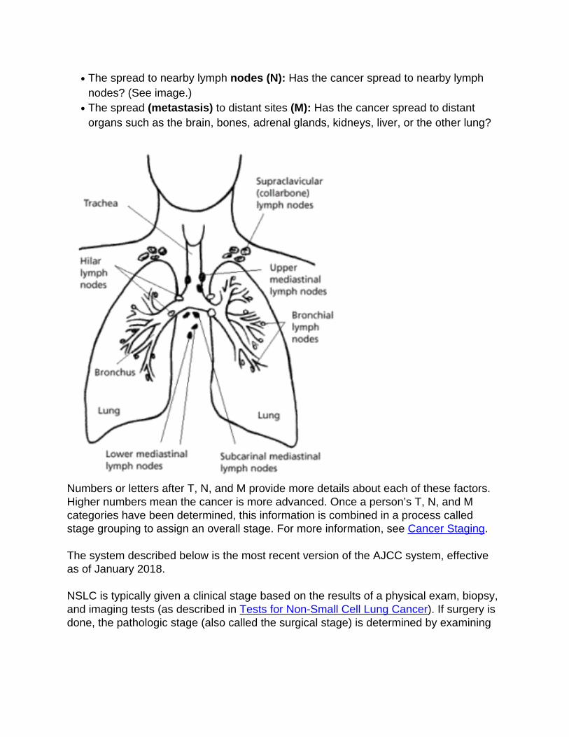
The spread to nearby lymph nodes (N): Has the cancer spread to nearby lymphnodes? (See image.)
●
The spread (metastasis) to distant sites (M): Has the cancer spread to distantorgans such as the brain, bones, adrenal glands, kidneys, liver, or the other lung?
●
Numbers or letters after T, N, and M provide more details about each of these factors.Higher numbers mean the cancer is more advanced. Once a person’s T, N, and Mcategories have been determined, this information is combined in a process calledstage grouping to assign an overall stage. For more information, see Cancer Staging.
The system described below is the most recent version of the AJCC system, effectiveas of January 2018.
NSLC is typically given a clinical stage based on the results of a physical exam, biopsy,and imaging tests (as described in Tests for Non-Small Cell Lung Cancer). If surgery isdone, the pathologic stage (also called the surgical stage) is determined by examining
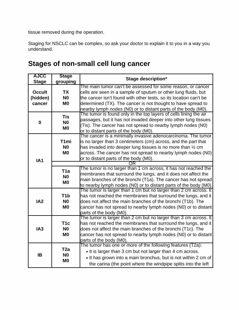
tissue removed during the operation.
Staging for NSCLC can be complex, so ask your doctor to explain it to you in a way youunderstand.
Stages of non-small cell lung cancer
AJCCStage
Stagegrouping
Stage description*
Occult(hidden)cancer
TXN0M0
The main tumor can’t be assessed for some reason, or cancercells are seen in a sample of sputum or other lung fluids, butthe cancer isn’t found with other tests, so its location can’t bedetermined (TX). The cancer is not thought to have spread tonearby lymph nodes (N0) or to distant parts of the body (M0).
0TisN0M0
The tumor is found only in the top layers of cells lining the airpassages, but it has not invaded deeper into other lung tissues(Tis). The cancer has not spread to nearby lymph nodes (N0)or to distant parts of the body (M0).
IA1
T1miN0M0
The cancer is a minimally invasive adenocarcinoma. The tumoris no larger than 3 centimeters (cm) across, and the part thathas invaded into deeper lung tissues is no more than ½ cmacross. The cancer has not spread to nearby lymph nodes (N0)or to distant parts of the body (M0).
OR
T1aN0M0
The tumor is no larger than 1 cm across, it has not reached themembranes that surround the lungs, and it does not affect themain branches of the bronchi (T1a). The cancer has not spreadto nearby lymph nodes (N0) or to distant parts of the body (M0).
IA2T1bN0M0
The tumor is larger than 1 cm but no larger than 2 cm across. Ithas not reached the membranes that surround the lungs, and itdoes not affect the main branches of the bronchi (T1b). Thecancer has not spread to nearby lymph nodes (N0) or to distantparts of the body (M0).
IA3T1cN0M0
The tumor is larger than 2 cm but no larger than 3 cm across. Ithas not reached the membranes that surround the lungs, and itdoes not affect the main branches of the bronchi (T1c). Thecancer has not spread to nearby lymph nodes (N0) or to distantparts of the body (M0).
IBT2aN0M0
The tumor has one or more of the following features (T2a):It is larger than 3 cm but not larger than 4 cm across.●
It has grown into a main bronchus, but is not within 2 cm ofthe carina (the point where the windpipe splits into the left
●
and right main bronchi) and it is not larger than 4 cmacross.It has grown into the visceral pleura (the membranessurrounding the lungs) and is not larger than 4 cm across.
●
It is partially clogging the airways (and is not larger than 4cm across).
●
The cancer has not spread to nearby lymph nodes (N0) or todistant parts of the body (M0).
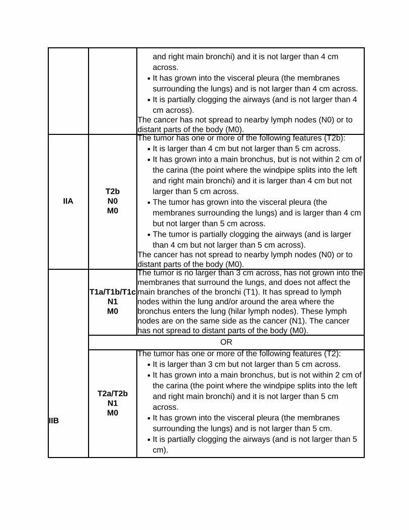
IIAT2bN0M0
The tumor has one or more of the following features (T2b):It is larger than 4 cm but not larger than 5 cm across.●
It has grown into a main bronchus, but is not within 2 cm ofthe carina (the point where the windpipe splits into the leftand right main bronchi) and it is larger than 4 cm but notlarger than 5 cm across.
●
The tumor has grown into the visceral pleura (themembranes surrounding the lungs) and is larger than 4 cmbut not larger than 5 cm across.
●
The tumor is partially clogging the airways (and is largerthan 4 cm but not larger than 5 cm across).
●
The cancer has not spread to nearby lymph nodes (N0) or todistant parts of the body (M0).
IIB
T1a/T1b/T1cN1M0
The tumor is no larger than 3 cm across, has not grown into themembranes that surround the lungs, and does not affect themain branches of the bronchi (T1). It has spread to lymphnodes within the lung and/or around the area where thebronchus enters the lung (hilar lymph nodes). These lymphnodes are on the same side as the cancer (N1). The cancerhas not spread to distant parts of the body (M0).
OR
T2a/T2bN1M0
The tumor has one or more of the following features (T2):It is larger than 3 cm but not larger than 5 cm across.●
It has grown into a main bronchus, but is not within 2 cm ofthe carina (the point where the windpipe splits into the leftand right main bronchi) and it is not larger than 5 cmacross.
●
It has grown into the visceral pleura (the membranessurrounding the lungs) and is not larger than 5 cm.
●
It is partially clogging the airways (and is not larger than 5cm).
●
The cancer has also spread to lymph nodes within the lungand/or around the area where the bronchus enters the lung(hilar lymph nodes). These lymph nodes are on the same sideas the cancer (N1). The cancer has not spread to distant partsof the body (M0).
OR
T3N0M0
The tumor has one or more of the following features (T3):It is larger than 5 cm but not larger than 7 cm across.●
It has grown into the chest wall, the inner lining of the chestwall (parietal pleura), the phrenic nerve, or membranes ofthe sac surrounding the heart (parietal pericardium).
●
There are 2 or more separate tumor nodules in the samelobe of a lung.
●
The cancer has not spread to nearby lymph nodes (N0) ordistant parts of the body (M0).
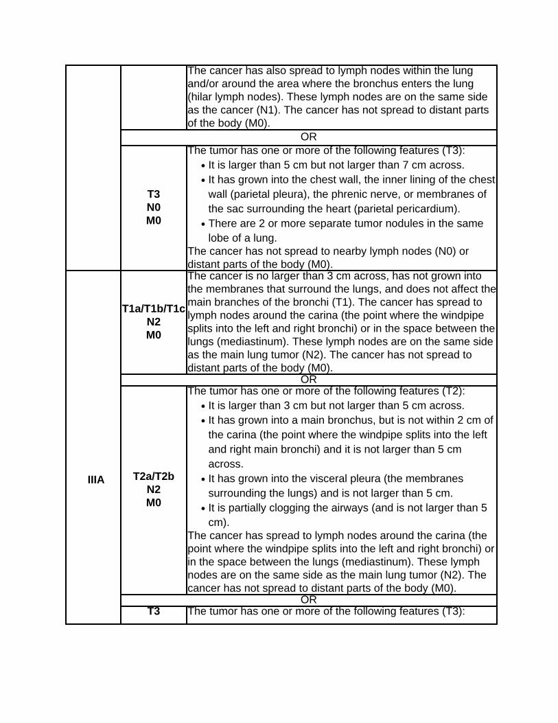
IIIA
T1a/T1b/T1cN2M0
The cancer is no larger than 3 cm across, has not grown intothe membranes that surround the lungs, and does not affect themain branches of the bronchi (T1). The cancer has spread tolymph nodes around the carina (the point where the windpipesplits into the left and right bronchi) or in the space between thelungs (mediastinum). These lymph nodes are on the same sideas the main lung tumor (N2). The cancer has not spread todistant parts of the body (M0).
OR
T2a/T2bN2M0
The tumor has one or more of the following features (T2):It is larger than 3 cm but not larger than 5 cm across.●
It has grown into a main bronchus, but is not within 2 cm ofthe carina (the point where the windpipe splits into the leftand right main bronchi) and it is not larger than 5 cmacross.
●
It has grown into the visceral pleura (the membranessurrounding the lungs) and is not larger than 5 cm.
●
It is partially clogging the airways (and is not larger than 5cm).
●
The cancer has spread to lymph nodes around the carina (thepoint where the windpipe splits into the left and right bronchi) orin the space between the lungs (mediastinum). These lymphnodes are on the same side as the main lung tumor (N2). Thecancer has not spread to distant parts of the body (M0).
ORT3 The tumor has one or more of the following features (T3):
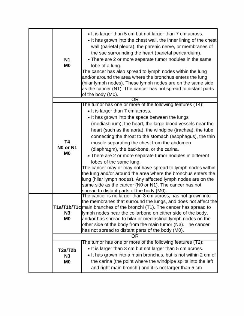
N1M0
It is larger than 5 cm but not larger than 7 cm across.●
It has grown into the chest wall, the inner lining of the chestwall (parietal pleura), the phrenic nerve, or membranes ofthe sac surrounding the heart (parietal pericardium).
●
There are 2 or more separate tumor nodules in the samelobe of a lung.
●
The cancer has also spread to lymph nodes within the lungand/or around the area where the bronchus enters the lung(hilar lymph nodes). These lymph nodes are on the same sideas the cancer (N1). The cancer has not spread to distant partsof the body (M0).
OR
T4N0 or N1
M0
The tumor has one or more of the following features (T4):It is larger than 7 cm across.●
It has grown into the space between the lungs(mediastinum), the heart, the large blood vessels near theheart (such as the aorta), the windpipe (trachea), the tubeconnecting the throat to the stomach (esophagus), the thinmuscle separating the chest from the abdomen(diaphragm), the backbone, or the carina.
●
There are 2 or more separate tumor nodules in differentlobes of the same lung.
●
The cancer may or may not have spread to lymph nodes withinthe lung and/or around the area where the bronchus enters thelung (hilar lymph nodes). Any affected lymph nodes are on thesame side as the cancer (N0 or N1). The cancer has notspread to distant parts of the body (M0).
T1a/T1b/T1cN3M0
The cancer is no larger than 3 cm across, has not grown intothe membranes that surround the lungs, and does not affect themain branches of the bronchi (T1). The cancer has spread tolymph nodes near the collarbone on either side of the body,and/or has spread to hilar or mediastinal lymph nodes on theother side of the body from the main tumor (N3). The cancerhas not spread to distant parts of the body (M0).
OR
T2a/T2bN3M0
The tumor has one or more of the following features (T2):It is larger than 3 cm but not larger than 5 cm across.●
It has grown into a main bronchus, but is not within 2 cm ofthe carina (the point where the windpipe splits into the leftand right main bronchi) and it is not larger than 5 cm
●
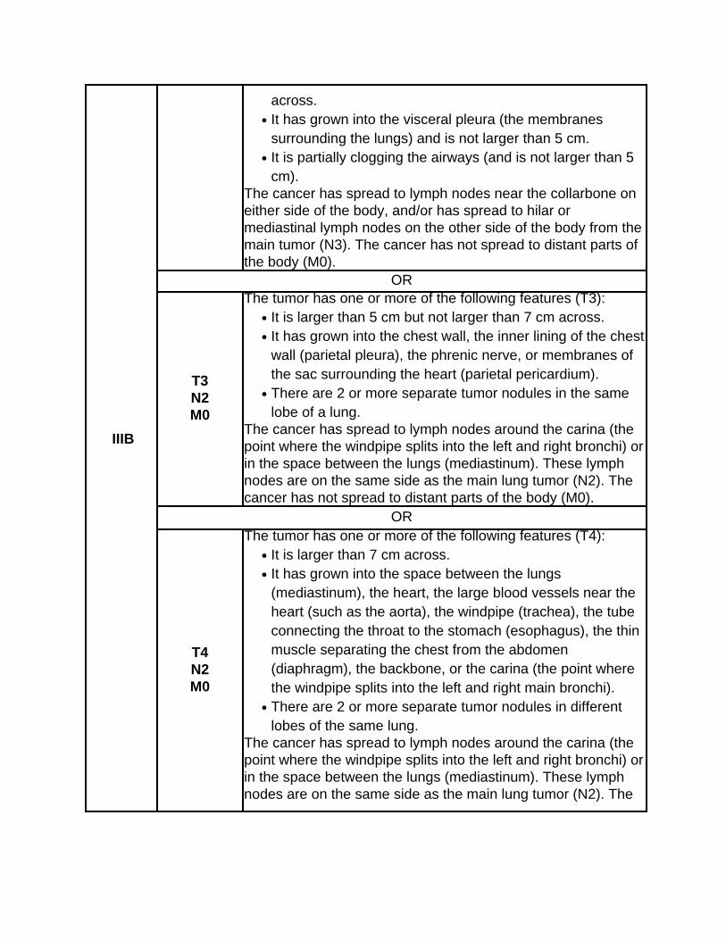
IIIB
across.It has grown into the visceral pleura (the membranessurrounding the lungs) and is not larger than 5 cm.
●
It is partially clogging the airways (and is not larger than 5cm).
●
The cancer has spread to lymph nodes near the collarbone oneither side of the body, and/or has spread to hilar ormediastinal lymph nodes on the other side of the body from themain tumor (N3). The cancer has not spread to distant parts ofthe body (M0).
OR
T3N2M0
The tumor has one or more of the following features (T3):It is larger than 5 cm but not larger than 7 cm across.●
It has grown into the chest wall, the inner lining of the chestwall (parietal pleura), the phrenic nerve, or membranes ofthe sac surrounding the heart (parietal pericardium).
●
There are 2 or more separate tumor nodules in the samelobe of a lung.
●
The cancer has spread to lymph nodes around the carina (thepoint where the windpipe splits into the left and right bronchi) orin the space between the lungs (mediastinum). These lymphnodes are on the same side as the main lung tumor (N2). Thecancer has not spread to distant parts of the body (M0).
OR
T4N2M0
The tumor has one or more of the following features (T4):It is larger than 7 cm across.●
It has grown into the space between the lungs(mediastinum), the heart, the large blood vessels near theheart (such as the aorta), the windpipe (trachea), the tubeconnecting the throat to the stomach (esophagus), the thinmuscle separating the chest from the abdomen(diaphragm), the backbone, or the carina (the point wherethe windpipe splits into the left and right main bronchi).
●
There are 2 or more separate tumor nodules in differentlobes of the same lung.
●
The cancer has spread to lymph nodes around the carina (thepoint where the windpipe splits into the left and right bronchi) orin the space between the lungs (mediastinum). These lymphnodes are on the same side as the main lung tumor (N2). The
cancer has not spread to distant parts of the body (M0).
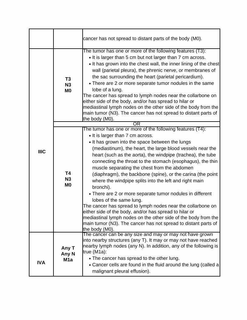
IIIC
T3N3M0
The tumor has one or more of the following features (T3):It is larger than 5 cm but not larger than 7 cm across.●
It has grown into the chest wall, the inner lining of the chestwall (parietal pleura), the phrenic nerve, or membranes ofthe sac surrounding the heart (parietal pericardium).
●
There are 2 or more separate tumor nodules in the samelobe of a lung.
●
The cancer has spread to lymph nodes near the collarbone oneither side of the body, and/or has spread to hilar ormediastinal lymph nodes on the other side of the body from themain tumor (N3). The cancer has not spread to distant parts ofthe body (M0).
OR
T4N3M0
The tumor has one or more of the following features (T4):It is larger than 7 cm across.●
It has grown into the space between the lungs(mediastinum), the heart, the large blood vessels near theheart (such as the aorta), the windpipe (trachea), the tubeconnecting the throat to the stomach (esophagus), the thinmuscle separating the chest from the abdomen(diaphragm), the backbone (spine), or the carina (the pointwhere the windpipe splits into the left and right mainbronchi).
●
There are 2 or more separate tumor nodules in differentlobes of the same lung.
●
The cancer has spread to lymph nodes near the collarbone oneither side of the body, and/or has spread to hilar ormediastinal lymph nodes on the other side of the body from themain tumor (N3). The cancer has not spread to distant parts ofthe body (M0).
IVA
Any TAny NM1a
The cancer can be any size and may or may not have growninto nearby structures (any T). It may or may not have reachednearby lymph nodes (any N). In addition, any of the following istrue (M1a):
The cancer has spread to the other lung.●
Cancer cells are found in the fluid around the lung (called amalignant pleural effusion).
●

Cancer cells are found in the fluid around the heart (calleda malignant pericardial effusion).
●
OR
Any TAny NM1b
The cancer can be any size and may or may not have growninto nearby structures (any T). It may or may not have reachednearby lymph nodes (any N). It has spread as a single tumoroutside of the chest, such as to a distant lymph node or anorgan such as the liver, bones, or brain (M1b).
IVBAny TAny NM1c
The cancer can be any size and may or may not have growninto nearby structures (any T). It may or may not have reachednearby lymph nodes (any N). It has spread as more than onetumor outside the chest, such as to distant lymph nodes and/orto other organs such as the liver, bones, or brain (M1c).
*The following additional categories are not listed in the table above:
T0: There is no evidence of a primary tumor.●
NX: Nearby lymph nodes cannot be assessed due to lack of information.●
References●
American Joint Committee on Cancer. Lung. In: AJCC Cancer Staging Manual. 8th ed.New York, NY: Springer; 2017: 431-456.
Last Medical Review: December 18, 2017 Last Revised: December 18, 2017
American Cancer Society medical information is copyrighted material. For reprintrequests, please see our Content Usage Policy.
Non-Small Cell Lung Cancer SurvivalRates, by Stage Survival rates tell you what portion of people with the same type and stage of cancerare still alive a certain amount of time (usually 5 years) after they were diagnosed.These numbers can’t tell you how long you will live, but they may help give you a betterunderstanding about how likely it is that your treatment will be successful.
What is a 5-year survival rate?
Statistics on the outlook for a certain type and stage of cancer are often given as 5-yearsurvival rates, but many people live longer – often much longer – than 5 years. The 5-year survival rate is the percentage of people who live at least 5 years after beingdiagnosed with cancer. For example, a 5-year survival rate of 80% means that anestimated 80 out of 100 people who have that cancer are still alive 5 years after beingdiagnosed. Keep in mind, however, that many of these people live much longer than 5years after diagnosis.
But remember, the 5-year survival rates are estimates – your outlook can vary based ona number of factors specific to you.
Survival rates don’t tell the whole story
Survival rates are often based on previous outcomes of large numbers of people whohad the disease, but they can’t predict what will happen in any particular person’s case.There are a number of limitations to keep in mind:
The numbers below are among the most current available. But to get 5-yearsurvival rates, doctors have to look at people who were treated at least 5 years ago.As treatments are improving over time, people who are now being diagnosed withnon-small cell lung cancer (NSCLC) may have a better outlook than these statisticsshow.
●
These statistics are based on the stage of the cancer when it was first diagnosed.They do not apply to cancers that later come back or spread, for example.
●
The outlook for people with NSCLC varies by the stage (extent) of the cancer – ingeneral, the survival rates are higher for people with earlier stage cancers. Butmany other factors can affect a person’s outlook, such as the subtype of NSCLC,gene changes in the cancer cells, the person’s age and overall health, and how wellthe cancer responds to treatment. The outlook for each person is specific to his orher circumstances.
●
Your doctor can tell you how these numbers may apply to you, as he or she is familiarwith your particular situation.
Survival rates for non-small cell lung cancer, by stage
The numbers below come from thousands of people from all over the world who were
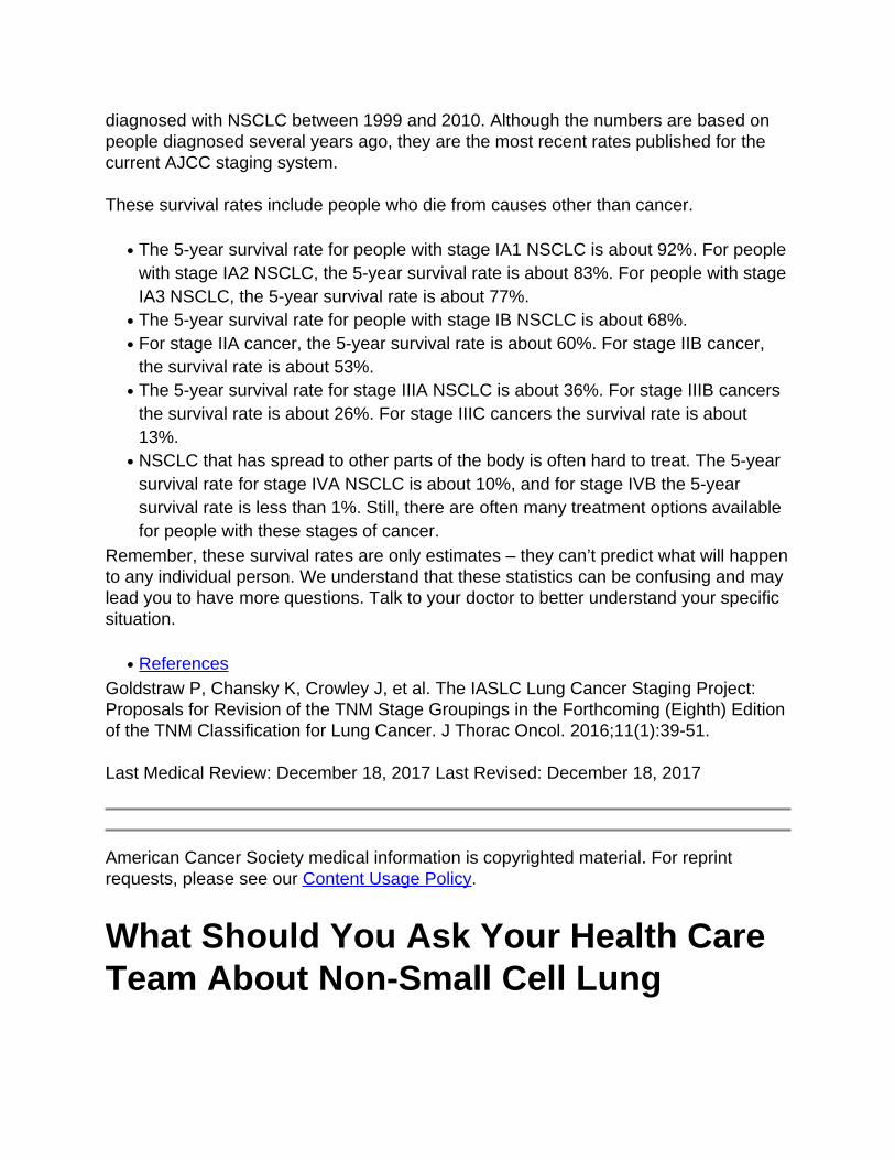
diagnosed with NSCLC between 1999 and 2010. Although the numbers are based onpeople diagnosed several years ago, they are the most recent rates published for thecurrent AJCC staging system.
These survival rates include people who die from causes other than cancer.
The 5-year survival rate for people with stage IA1 NSCLC is about 92%. For peoplewith stage IA2 NSCLC, the 5-year survival rate is about 83%. For people with stageIA3 NSCLC, the 5-year survival rate is about 77%.
●
The 5-year survival rate for people with stage IB NSCLC is about 68%.●
For stage IIA cancer, the 5-year survival rate is about 60%. For stage IIB cancer,the survival rate is about 53%.
●
The 5-year survival rate for stage IIIA NSCLC is about 36%. For stage IIIB cancersthe survival rate is about 26%. For stage IIIC cancers the survival rate is about13%.
●
NSCLC that has spread to other parts of the body is often hard to treat. The 5-yearsurvival rate for stage IVA NSCLC is about 10%, and for stage IVB the 5-yearsurvival rate is less than 1%. Still, there are often many treatment options availablefor people with these stages of cancer.
●
Remember, these survival rates are only estimates – they can’t predict what will happento any individual person. We understand that these statistics can be confusing and maylead you to have more questions. Talk to your doctor to better understand your specificsituation.
References●
Goldstraw P, Chansky K, Crowley J, et al. The IASLC Lung Cancer Staging Project:Proposals for Revision of the TNM Stage Groupings in the Forthcoming (Eighth) Editionof the TNM Classification for Lung Cancer. J Thorac Oncol. 2016;11(1):39-51.
Last Medical Review: December 18, 2017 Last Revised: December 18, 2017
American Cancer Society medical information is copyrighted material. For reprintrequests, please see our Content Usage Policy.
What Should You Ask Your Health CareTeam About Non-Small Cell Lung
Cancer? It’s important to have honest, open discussions with your cancer care team. You shouldask any question, no matter how small it might seem. Here are some questions youmight want to ask:
When you’re told you have lung cancer
What kind of lung cancer do I have?●
Where exactly is the cancer? Has it spread beyond where it started?●
What is the stage of my cancer, and what does that mean in my case?●
Will I need any other tests before we can decide on treatment?●
Have the cancer cells been checked for gene changes that could affect mytreatment options?
●
Do I need to see any other doctors or health professionals?●
If I’m concerned about the costs and insurance coverage for my diagnosis andtreatment, who can help me?
●
When deciding on a treatment plan
How much experience do you have treating this type of cancer?●
What are my treatment choices?●
What do you recommend and why?●
What is the goal of my treatment?●
Should I get a second opinion? How do I do that? Can you recommend someone?●
What are the chances my cancer can be cured with these options?●
How quickly do we need to decide on treatment?●
What should I do to be ready for treatment?●
How long will my treatment last?●
What will treatment be like?●
Where will my treatment be done?●
What are the risks and side effects with the treatments you suggest?●
Will treatment affect my daily activities?●
During treatment
Once treatment begins, you’ll need to know what to expect and what to look for. Not all
of these questions may apply to you, but asking the ones that do may be helpful.
How will we know if the treatment is working?●
Is there anything I can do to help manage side effects?●
What symptoms or side effects should I tell you about right away?●
How can I reach you on nights, holidays, or weekends?●
Do I need to change what I eat during treatment?●
Are there any limits on what I can do?●
What kind of exercise should I do, and how often?●
Can you suggest a mental health professional I can see if I start to feeloverwhelmed, depressed, or distressed?
●
After treatment
Are there any limits on what I can do?●
What symptoms should I watch for?●
What kind of exercise should I do now?●
What type of follow-up will I need after treatment?●
How often will I need to have follow-up exams and imaging tests?●
Will I need any blood tests?●
How will we know if the cancer has come back? What should I watch for?●
What will my options be if the cancer comes back?●
Along with these sample questions, be sure to write down some of your own. Forinstance, you might want more information about recovery times. Or you may want toask about getting a second opinion or about clinical trials for which you may qualify.
Keep in mind that doctors aren’t the only ones who can give you information. Otherhealth care professionals, such as nurses and social workers, can answer some of yourquestions. To find out more about speaking with your health care team, see TalkingWith Your Doctor.
References●
See all references for Non-Small Cell Lung Cancer
Last Medical Review: February 8, 2016 Last Revised: May 16, 2016
American Cancer Society medical information is copyrighted material. For reprint

requests, please see our Content Usage Policy.
2016 Copyright American Cancer Society