Embed Size (px)
Citation preview
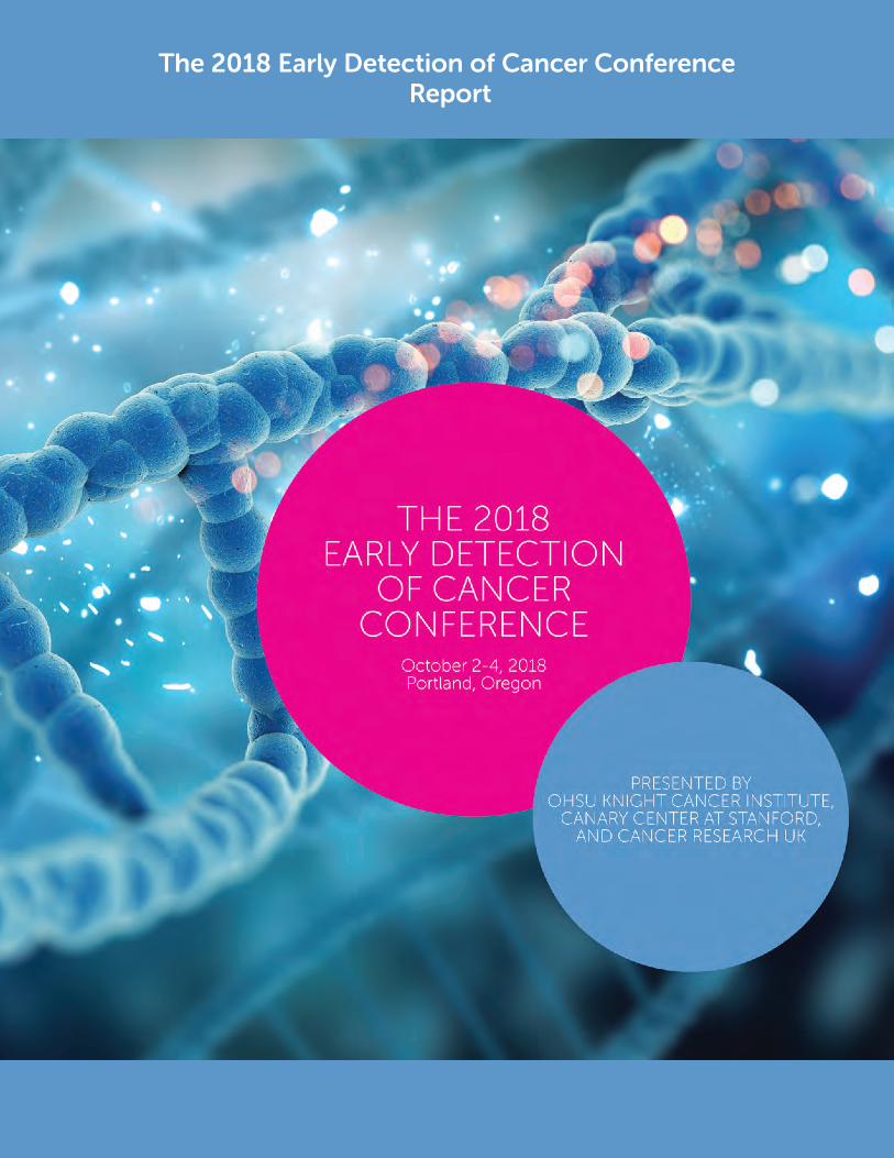
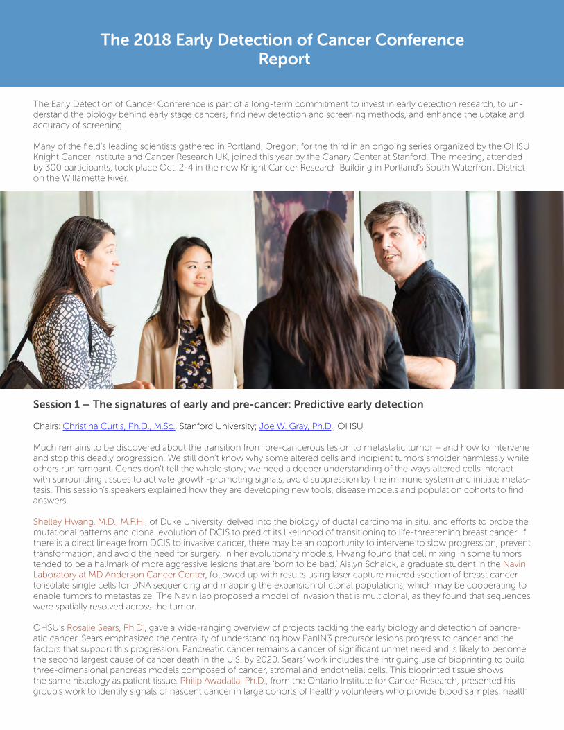
The 2018 Early Detection of Cancer Conference Report
The 2018 Early Detection of Cancer Conference Report
The Early Detection of Cancer Conference is part of a long-term commitment to invest in early detection research, to un-derstand the biology behind early stage cancers, find new detection and screening methods, and enhance the uptake and accuracy of screening.
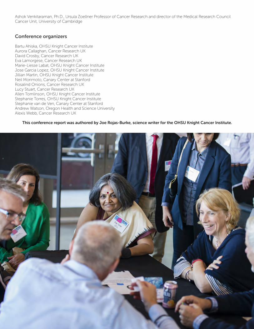
Many of the field’s leading scientists gathered in Portland, Oregon, for the third in an ongoing series organized by the OHSU Knight Cancer Institute and Cancer Research UK, joined this year by the Canary Center at Stanford. The meeting, attended by 300 participants, took place Oct. 2-4 in the new Knight Cancer Research Building in Portland’s South Waterfront District on the Willamette River.
Session 1 – The signatures of early and pre-cancer: Predictive early detection
Chairs: Christina Curtis, Ph.D., M.Sc., Stanford University; Joe W. Gray, Ph.D., OHSU
Much remains to be discovered about the transition from pre-cancerous lesion to metastatic tumor – and how to intervene and stop this deadly progression. We still don’t know why some altered cells and incipient tumors smolder harmlessly while others run rampant. Genes don’t tell the whole story; we need a deeper understanding of the ways altered cells interact with surrounding tissues to activate growth-promoting signals, avoid suppression by the immune system and initiate metas-tasis. This session’s speakers explained how they are developing new tools, disease models and population cohorts to find answers.
Shelley Hwang, M.D., M.P.H., of Duke University, delved into the biology of ductal carcinoma in situ, and efforts to probe the mutational patterns and clonal evolution of DCIS to predict its likelihood of transitioning to life-threatening breast cancer. If there is a direct lineage from DCIS to invasive cancer, there may be an opportunity to intervene to slow progression, prevent transformation, and avoid the need for surgery. In her evolutionary models, Hwang found that cell mixing in some tumors tended to be a hallmark of more aggressive lesions that are ‘born to be bad.’ Aislyn Schalck, a graduate student in the Navin Laboratory at MD Anderson Cancer Center, followed up with results using laser capture microdissection of breast cancer to isolate single cells for DNA sequencing and mapping the expansion of clonal populations, which may be cooperating to enable tumors to metastasize. The Navin lab proposed a model of invasion that is multiclonal, as they found that sequences were spatially resolved across the tumor.
OHSU’s Rosalie Sears, Ph.D., gave a wide-ranging overview of projects tackling the early biology and detection of pancre-atic cancer. Sears emphasized the centrality of understanding how PanIN3 precursor lesions progress to cancer and the factors that support this progression. Pancreatic cancer remains a cancer of significant unmet need and is likely to become the second largest cause of cancer death in the U.S. by 2020. Sears’ work includes the intriguing use of bioprinting to build three-dimensional pancreas models composed of cancer, stromal and endothelial cells. This bioprinted tissue shows the same histology as patient tissue. Philip Awadalla, Ph.D., from the Ontario Institute for Cancer Research, presented his group’s work to identify signals of nascent cancer in large cohorts of healthy volunteers who provide blood samples, health
histories and clinical test results over time. Awadalla advocated for a multidisciplinary approach and the necessity of statis-tical approaches when analyzing case cohort studies. Building cohorts of high-risk individuals, such as those with diabetes or genetic predisposition to pancreatic cancer, to test and validate candidate markers will also be crucial to enabling earlier cancer detection.
Challenges and future directions brought out in the plenary discussion:
• The positive predictive value of an early detection test is just as important, if not more so, than the sensitivity and specificity of the test as measured by area under the ROC curve. • Decision models for diagnosis should include multiple variables to make them more cost effective; we should avoid detection that relies on a single test. • To identify dangerous pre-cancerous lesions, we may need to look for signals from the microenvironment and not just cell intrinsic changes. • Health economics of test implementation should be considered more broadly in early detection research; understanding the intended circumstances of use of a test and its economics may help to determine appropriate test characteristics that should be sought during development. • With the identification of high-risk cohorts, the field must also consider what prevention and monitoring options are available to these patients.
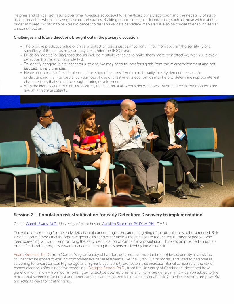
Session 2 – Population risk stratification for early Detection: Discovery to implementation
Chairs: Gareth Evans, M.D., University of Manchester; Jackilen Shannon, Ph.D., M.P.H., OHSU
The value of screening for the early detection of cancer hinges on careful targeting of the populations to be screened. Risk stratification methods that incorporate genetic risk and other factors may be able to reduce the number of people who need screening without compromising the early identification of cancers in a population. This session provided an update on the field and its progress towards cancer screening that is personalized by individual risk.
Adam Brentnall, Ph.D., from Queen Mary University of London, detailed the important role of breast density as a risk fac-tor that can be added to existing comprehensive risk assessments, like the Tyrer-Cuzick model, and used to personalize screening for breast cancer. Higher age and higher breast density are factors that increase interval cancer rate (the risk of cancer diagnosis after a negative screening). Douglas Easton, Ph.D., from the University of Cambridge, described how genetic information – from common single-nucleotide polymorphisms and from rare gene variants – can be added to the mix so that screening for breast and other cancers can be tailored to suit an individual’s risk. Genetic risk scores are powerful and reliable ways for stratifying risk.
The Fred Hutchinson Cancer Research Center’s Ruth Etzioni, Ph.D., navigated the big trade-off inherent in cancer early de-tection and screening programs: the harm of over-treating healthy people versus the benefit of saving the lives of the small fraction of the population at real risk of cancer death. Etzioni underscored the need for specific recommendations for high-er risk groups that reflect an accurate and mechanistic understanding of the difference between high- and low-risk patient diseases. Etzioni’s work is vital to shaping policy recommendations for precision early detection screening. Harm-benefit tradeoffs (over treatment vs. lives saved) depend on mechanism.
In a rousing conclusion to the day, Linda Sue Cook, Ph.D., from the University of New Mexico, challenged everyone work-ing on early detection to be mindful of the inequalities in the provision of health care services – and to seek ways to reduce those inequalities or at least not make them worse.
Challenges and future directions brought out in the plenary discussion:
• Early detection must recognize potential for overdiagnosis. Arguably, the problem can be managed by finding ways to prevent overtreatment. • Age alone is a poor risk stratifier for some types of cancer, such as breast cancer as compared with colorectal cancer. Thus, risk assessment models should be multi-factorial. • We need risk stratification to inform those who we don’t need to screen and those who require less frequent screening. • New technologies for cancer early detection could worsen health inequities unless their design and implementation explicitly factor in the needs and access limitations of marginalized and underserved populations.
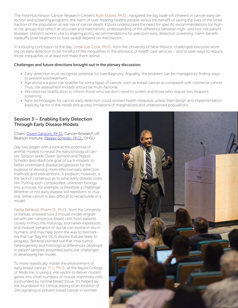
Session 3 – Enabling Early Detection Through Early Disease Models
Chairs: Owen Sansom, Ph.D., Cancer Research UK Beatson Institute; Pepper Schedin, Ph.D., OHSU
Day two began with a look at the potential of animal models to reveal the early biology of can-cer. Session leads Owen Samson and Pepper Schedin described one goal of such models: to better understand disease progression for the purpose of devising more effective early detection methods and interventions. A problem, however, is the lack of consensus as to what early disease looks like. Putting such complicated, unknown biology into a mouse, for example, is therefore a challenge. Whether or not early disease will transform to inva-sive, lethal cancer is also difficult to recapitulate in a model.
Fariba Behbod, Pharm.D., Ph.D., from the University of Kansas, showed how a mouse model engraft-ed with pre-cancerous breast cells from patients closely mimics the histology, biomarker expression, and invasive behavior of ductal carcinoma in situ in humans, and may help point the way to biomark-ers that can flag the DCIS lesions that are likely to progress. Behbod pointed out that intra-tumor heterogeneity and histological differences observed in patient samples presented particular challenges in developing her model.
To more realistically model the environment of early breast cancer, Yi Li, Ph.D., at the Baylor College of Medicine, is using a viral vector to deliver mutant genes into small numbers of mouse mammary cells surrounded by normal breast tissue. It’s helped lay the foundation for clinical testing of an inhibitor of JAK signaling to prevent breast cancer in women
with atypical hyperplasia, a type of precancerous lesion.
Turning to colorectal cancer, Simon Leedham, Ph.D., from the University of Oxford, showed how a well-designed mouse model provided evidence that targeted chemoprevention may be feasible in people with serrated polyps, the type most likely to progress to cancer. Leedham applied lineage tracing from ectopic crypts in the colon to identify the type of cells that had been proliferating in the villus, showing spatially distinct lineages. To close the morning session, Ashok Venkitaraman, Ph.D., from the University of Cambridge, described insights from mouse models of cancers spurred by BRCA2 mutations, specifically inactivation and the resulting chromosomal instability caused by disrupting homologous recombination. In carriers of truncated BRCA2 genes there may be a po-tential role of exposure to aldehydes common in household environments to act as a trigger of carcinogenesis. These are new or overlooked modulators of disease progression that could be capitalized on for prevention, treatment, or imag-ing strategies.
Challenges and future directions brought out in the plenary discussion:
• Early model systems for cancer detection and intervention remains a complex area of research which relies on iterative approaches that are constantly evolving to account for deeper understanding of human data. Models must be tailored to the scientific question they are trying to address. For example, mouse models now are being designed to replicate human cancer sub types. Other models are being developed to account for the clonal evolution time course of human tumors, and that account for the microbiome influence in human cancers. • In correlative human studies, slow-growing lesions predominate, and the dangerous fast-growing are probably not sampled; they become cancers too quickly.
Special Session – Funders’ Perspective on Early Detection Research
After lunch, Iain Foulkes, Ph.D., of Cancer Research UK and Sudhir Srivastava, Ph.D., M.S., M.P.H., of the National Cancer Institute previewed their organizations’ strategies and the range of support on the horizon for early detection research, including a new CRUK-organized international alliance of leading early detection centers, and the PreCancer Atlas in development by the NCI. Foulkes underscored the challenges in the field of early detection: the complexity of the biology, fragmented expertise, lack of visibility, low priority given by funders and research institutions, limited industry involvement, and limited knowledge exchange. He explained how CRUK set out to build a community to raise the profile of early detec-tion with funding, industry collaborations, training and infrastructure.
Session 4 – Data Science for Early Detection
Chairs: Brendan Delaney, B.M. B.Ch., M.Sc., Imperial College London; Parag Mallick, Ph.D., Stanford University
A trio of data scientists rounded out the second day. Session chair Brendan Delaney described the opportunity to learn from
the vast amounts of data becoming available to create knowledge and build infrastructure for cancer detection and diagnosis pathways. These data span many different modalities. Early detection researchers must work to improve current methods of analysis in order to make them more efficient, thorough, and reproducible.
Kinga Várnai from the Oxford University Hospitals NHS Foundation Trust, described efforts to muster routinely collected patient data and put it in shape to enable research on the early detection of cancer. Stanford University’s Sylvia Plevritis, Ph.D., talked about her application of computational models at the level of cells, tissues and whole populations – and how to think about bridging these scales through supporting data science to answer big questions about early detection. Car-ole Goble from the University of Manchester highlighted the deep level of thinking, organization, documentation and tool development needed to make in silico research reproducible and open-source for all researchers.
Finally, Imran Haque, Ph.D., former chief scientific officer at the cancer screening company Freenome, closed with some overlooked lessons from the past use of data science for biomarker discovery. He emphasized the art of knowing how much information you can reliably squeeze out of the data you have.
Challenges and future directions brought out in the plenary discussion:
• Incentives for good data practice are broken; researchers currently have little incentive to implement the data sharing principles needed to advance the field efficiently and rapidly. • Data scientists often feel like data “is just thrown over the wall” to them for analysis. The most effective collaborations begin at the beginning: experiment conceptualization and design. • Funding calls could facilitate co-discovery by teams with computer scientists and bench scientists working collaboratively. • An ideal data set for early detection would consist of a patient cohort with mode of detection and screening history, including interval diagnoses, coupled with clinical records and any available molecular data.
Session 5 – Novel Technology Accelerating Early Detection
Chairs: Utkan Demirci, Ph.D., Stanford University; Mike Heller, Ph.D., OHSU
On the final day of the conference, session co-chair Mike Heller set the scene by reminding attendees of the parable of the blind men and the elephant. The same holds true for everyone trying to detect cancer earlier – we are all grasping at differ-ent parts of the problem and need to step back and be more integrative in our approaches.
Jean Lewis, Ph.D., from the University of California San Diego gave the first talk, an update on a microarray chip for rapid analysis of biomarkers in patient blood. It uses electric current to capture nucleic acids, exosomes, vesicles and proteins shed by cancer cells, followed by on-chip immunofluorescence to identify and measure target biomarkers in 30 minutes, suggesting the platform has potential as a point of care test. Bio-Rad’s George Karlin-Neumann, Ph.D., gave a tour through the capabilities of droplet digital PCR, in which a blood sample is fractionated into thousands of droplets followed by PCR amplification of the DNA in each droplet. It has the ultra-high sensitivity needed to detect the vanishingly small amounts of circulating cancer DNA of interest for early detection. Droplet digital PCR is highly reproducible, a necessity for any clinically implemented test.
In that same theme, G. Mike Makrigiorgos, Ph.D., from Dana Farber Cancer Institute, showed how to double the amount of readable DNA in blood samples by denaturing it into single strands prior to performing digital PCR, among other clever ways to detect cancer mutations present in very low numbers in a standard blood draw. MIT’s Sangeeta Bhatia, M.D., Ph.D., gave an update on synthetic biomarker probes and inexpensive paper-based assays. Peptides on the injected probe are susceptible to cleavage by specific tumor proteases at the rate of 1,000 cleavages per hour. Each cleavage by a protease releases a reporter fragment that, once excreted in urine, can be measured and acts as signal from the tumor microenviron-ment indicating progression. In the future such probes could be loaded with a PET dye or other novel molecular imaging markers to allow for visualization of the tumor.
Challenges and future directions brought out in the plenary discussion:
• Knowing the performance of a new technology or biomarker in isolation isn’t good enough; the field would benefit from “bake-offs” comparing the performance of emerging technologies using the same patient samples. • More collaboration would allow for biomarkers from different groups to be combined, which could strength their diagnostic capability. • Funding agencies could help incentivize researchers to participate in collaborations and comparative testing of novel technologies. • As the field matures, developers of early detection tools should maintain close contact with colleagues who are developing next-generation cancer therapeutic interventions.
Final discussion
The meeting closed with a session exploring challenges and barriers to progress and how best to approach and overcome them.
High costs and unequal access are already daunting issues in cancer medicine, setting the stage for a question that arose repeatedly throughout the meeting: How to implement early detection equitably and effectively with complex and poten-tially very expensive technologies. One way forward is to explicitly include health economics as part of the development of early detection and diagnostic technologies. Understanding the intended circumstances of use and economics of imple-mentation may help to determine appropriate test characteristics that should be sought during development.
Related discussions highlighted other important knowledge gaps in the social context for early detection. New screening programs, for example, could greatly expand a population of people with “pre-cancer” and the social consequences of that have not been adequately considered. Social and behavioral science studies will be needed to inform the implementation of cancer screening programs, particularly as early detection becomes feasible for increasing numbers of different types of cancer. We will need to find ways to incentivize people to get screened, avoid screening fatigue, and support those with detected lesions that are deemed indolent or low-risk. Of all the technical challenges, perhaps none is more difficult than the problem of overdiagnosis and the consequent need to identify potentially dangerous early lesions and distinguish them from those destined to never become life-threatening, metastatic tumors. New animal models, several presented at this meeting, will be important tools to reveal the early biology of malignant transformation.
New types of patient cohorts will also be crucial. Informative cohorts will include data on patients’ screening history, mode
of detection, and pre- and post-diagnosis clinical records, blood and tissue samples, ideally including samples from early lesions that advanced to an invasive cancer. Tissue and blood samples are a limited resource, and the field needs to develop effective systems for sharing samples.
There is a comparable need for the stepped-up sharing of data and for better collaboration across disciplines. Funding agencies have an important role here. Funding and review mechanisms can create incentives for data sharing and multi-disciplinary approaches. Requests for proposals could, for example, explicitly call for co-discovery by teams with computer scientists and bench scientists working collaboratively.
Raising awareness and gathering support from patients and the general public will be crucial on every front. Patients com-mand a moral high ground for open access to data gathered from their altruistic participation in studies. Given the federal and international funding landscape, the voices of patients and survivors will also be helpful in seeking support from industry and philanthropy.
Looking ahead, participants noted several areas ripe for discussion at the next early detection conference:
• How does the immune system interact with nascent tumors, reshape the tumor microenvironment, and influence the development of invasive cancer? • How can we integrate knowledge about inherited cancer susceptibility genes with early detection efforts? • Can we use health economics and health system modeling to facilitate the implementation of discovery of cost- effective early detection biomarkers and diagnostic tests? • Do we need a road map for cancer early detection to prioritize efforts and translate discoveries to clinical and population impact faster?
It was a productive week. The conference organizers – the Knight Cancer Institute, Cancer Research UK and The Canary Center at Stanford – designed the conference to stir creative thinking, build collaborations, and move the field forward more rapidly. The fourth Early Detection of Cancer Conference will take place Sept. 24-26, 2019, in Stanford, California.
Acknowledgements
Scientific program chairs
Jack Cuzick, Ph.D., director of the Wolfson Institute of Preventive Medicine and John Snow Professor of Epidemiology, Queen Mary University of London.
Sanjiv Sam Gambhir, M.D., Ph.D., Virginia and D.K. Ludwig Professor for Clinical Investigation in Cancer Research, Stanford University and director of the Canary Center for Cancer Early Detection at Stanford
Paul Spellman, Ph.D., professor of molecular and medical genetics in the OHSU School of Medicine, co-director of the Cancer Early Detection Advanced Research Center in the Knight Cancer Institute
Session chairs
Christina Curtis, Ph.D., M.Sc., assistant professor of medicine and genetics, Stanford University School of Medicine
Brendan Delaney B.M. B.Ch., M.Sc., professor and chair in medical informatics anddecision making at Imperial College London and director of the Centre for Patient Safety and Service Quality
Utkan Demirci, Ph.D., M.S., professor, Stanford University School of Medicine
Gareth Evans, M.D., professor of medical genetics and cancer epidemiology, The University of Manchester
Joe W. Gray, Ph.D., director of the OHSU Center for Spatial Systems Biomedicine, associate director for biophysical oncolo-gy in the OHSU Knight Cancer Institute
Mike Heller, Ph.D., distinguished scientist, Cancer Early Detection Advanced Research Center, OHSU Knight Cancer Institute
Parag Mallick, Ph.D., associate Professor in the Department of Radiology at Stanford University School of Medicine
Owen Sansom, Ph.D., professor at the University of Glasgow and director of the Cancer Research UK Beatson Institute for Cancer Research
Pepper Schedin, Ph.D., professor of cell, developmental and cancer biology in the OHSU School of Medicine and co-leader of the Cancer Prevention and Control Program in the Knight Cancer Institute
Jackilen Shannon, Ph.D., M.P.H., professor, OHSU-PSU School of Public Health, associate director for community outreach and engagement in the OHSU Knight Cancer Institute
Speakers
Philip Awadalla, Ph.D., professor of population and medical genomics at the University of Toronto and director of computa-tional biology and the Ontario Health Study at the Ontario Institute for Cancer Research
Sangeeta Bhatia, M.D., Ph.D., John J. and Dorothy Wilson Professor of Health Sciences and Technology and of Electrical
Engineering and Computer Science, Massachusetts Institute of Technology; director of the Laboratory for Multiscale Re-generative Technologies; director of the Marble Center for Cancer Nanomedicine, Koch Institute
Fariba Behbod, Ph.D., associate professor in the Department of Pathology and Laboratory Medicine at the University of Kan-sas Medical Center
Adam Brentnall, Ph.D., senior statistician at the Centre for Cancer Prevention, Queen Mary University of London, and fellow of the Alan Turing Institute
Linda Sue Cook, Ph.D., professor in the Division of Epidemiology, Biostatistics, and Preventive Medicine, Department of Internal Medicine at the University of New Mexico and co-leader of the Cancer Control Research Program at the UNM Comprehensive Cancer Center
Jim Davies, M.A., M.Sc., D.Phil., professor of software engineering at the University of Oxford and director of informatics for the Oxford NIHR Biomedical Research Centre
Douglas Easton, Ph.D., professor and director of the Centre for Cancer Genetic Epidemiology, University of Cambridge
Ruth Etzioni, Ph.D., biostatistician and full member in the Division of Public Health Sciences at the Fred Hutchinson Cancer Research Center
Iain Foulkes, Ph.D., executive director of research, Cancer Research UK
Carole Goble, professor of computer science, University of Manchester
Imran Haque, Ph.D., former chief scientific officer at the cancer screening company Freenome
Eun-Sil Shelley Hwang, M.D., M.P.H., Mary and Deryl Hart Professor of Surgery in the Duke University School of Medicine, chief of breast surgery and vice chair of research in the department of surgery and the Duke University Comprehensive Cancer Center
George Karlin-Neumann, Ph.D., director of scientific affairs, Bio-Rad Digital Biology Center
Simon Leedham, Ph.D., associate professor of gastroenterology at the University of Oxford and Wellcome Trust Senior Research Fellow in Clinical Science at the Wellcome Trust Centre for Human Genetics
Jean Lewis, Ph.D., senior scientist at Biological Dynamics, San Diego
Yi Li, Ph.D., M.S., professor of molecular and cellular biology, Baylor College of Medicine
G. Mike Makrigiorgos, Ph.D., professor of radiation oncology and director of the medical physics and biophysicsdivision at Dana Farber Cancer Institute and Brigham and Women’s Hospitals, Harvard MedicalSchool.
Sylvia K. Plevritis, Ph.D., professor and chair of biomedical data science at Stanford University, co-lead of the Cancer Biology Program in the Stanford Cancer Institute
Aislyn Schalck, doctoral student in the Department of Genetics at MD Anderson Cancer Center
Rosalie Sears, Ph.D., professor of molecular and medical genetics in the OHSU School of Medicine and co-director of the Brenden-Colson Center for Pancreatic Care
Sudhir Srivastava, Ph.D., M.P.H., chief of the Cancer Biomarkers Research Group, Division of Cancer Prevention, National Cancer Institute
Ashok Venkitaraman, Ph.D., Ursula Zoellner Professor of Cancer Research and director of the Medical Research Council Cancer Unit, University of Cambridge
Conference organizers
Bartu Ahiska, OHSU Knight Cancer InstituteAurora Callaghan, Cancer Research UKDavid Crosby, Cancer Research UKEva Lamorgese, Cancer Research UKMarie-Liesse Labat, OHSU Knight Cancer InstituteJose Garcia Lopez, OHSU Knight Cancer InstituteJillian Martin, OHSU Knight Cancer InstituteNeil Morimoto, Canary Center at StanfordRosalind Onions, Cancer Research UKLucy Stuart, Cancer Research UKAllen Tomlinson, OHSU Knight Cancer InstituteStephanie Torres, OHSU Knight Cancer InstituteStephanie van de Ven, Canary Center at StanfordAndrew Watson, Oregon Health and Science UniversityAlexis Webb, Cancer Research UK
This conference report was authored by Joe Rojas-Burke, science writer for the OHSU Knight Cancer Institute.

Save the date:September 24-26, 2019
Frances C. Arrillaga Alumni Center, Stanford, California