Embed Size (px)
Citation preview
Durham Research Online
Deposited in DRO:
12 October 2009
Version of attached �le:
Published Version
Peer-review status of attached �le:
Peer-reviewed
Citation for published item:
Schultz, M. and Roberts, C. A. (2002) 'Diagnosis of leprosy in skeletons from an English later Medievalhospital using histological analysis.', in The past and present of leprosy : archaeological, historical,palaeopathological and clinical approaches. Oxford: Archaeopress, pp. 89-104. British Archaeological ReportsInternational Series. (1054).
Further information on publisher's website:
http://www.archaeopress.com/searchBar.asp?QuickSearch=manchester
Publisher's copyright statement:
Additional information:
Proceedings of the International Congress on the Evolution and Palaeoepidemiology of the Infectious Diseases 3(ICEPID), University of Bradford, 26th-31st July 1999.
Use policy
The full-text may be used and/or reproduced, and given to third parties in any format or medium, without prior permission or charge, forpersonal research or study, educational, or not-for-pro�t purposes provided that:
• a full bibliographic reference is made to the original source
• a link is made to the metadata record in DRO
• the full-text is not changed in any way
The full-text must not be sold in any format or medium without the formal permission of the copyright holders.
Please consult the full DRO policy for further details.
Durham University Library, Stockton Road, Durham DH1 3LY, United KingdomTel : +44 (0)191 334 3042 | Fax : +44 (0)191 334 2971
http://dro.dur.ac.uk
Diagnosis of leprosy in skeletons from an English later Medieval hospitalusing histological analysis
Michael Schultz' and Charlotte Roberts'
lZentrum AnatomicGeorx-August-UniversiW
Krouzbergring 361>-37075 GOttingen
Germany
Email: [email protected]
'Departmenl ofAn:baenlogyUniversity ofDurbam
South RoadDurbamDH13LE
England
Email: [email protected]
AbstrQct
The diagDosis of leprosy in skeletal materials &om .cbacologica1 sites can be problematic. Periostitis on the tibiae and fibulae isc:ormnonly seen in leprosy, in addition to other expected changes (e.B. attentions Ilrouod the anterior bony aperture of the nose).However, periosteal new boDe ConDItion could be caused by. number of conditions. This paper desaibes the histological analysis ofthin ground sections of tibiae with perioaea] new bone formation from six individuals diagnoJCd with leprosy from • later Medievalhospital site in Chichester. England. In addition. one individual from a later medieval priory site on Uhou ls1aod. Guernsey. with nocharacteristic lepromatous leprosy booe changes was examined.. The aim was to determine, by light miaoscopic analysis of thin groundsections.. whether the changes on the tibiae bad been the result of leprosy or mother condition. Results showed that there is a highprobability that • specific histological appearance is characteristic of leprosy. for differaltiaJ diagnoses. eodemic syphilis is discussed.
Keywords
Leprosy, tibial periostitis, differential diagnosis, histology
l.Introductioa
According to documentary sources, leprosy was arelatively common occurrence in later Medieval Europe(Richards, '977). However, as with so many apparentlyfrequent diseases.. the nwnber of cases diagnosed inarcbacologically derived skeletal material from all lllUS ofEurope, but particularly Eog1and, is low (sec Roberts, thisvolume). There may be many reasons for this. FirsLly, ifpeople coolnlct the !Ugh resistao. (lUb=uloid) fonn of theinfcctioo they may DOl develop any boo< changes.SccoodJy, people with leprosy may have died before therewas lime for boo< change to occur. In both these scenarios,the evidence would be Ibsenl skelctally (sec Wood .. aI.,1992 for discussioo). Thirdly iJ; as suggested inCOOlemponry sources, people with leprosy weR isolalcdfrom the rest of the COIDIDunity into leprosy hospitals, thenwe may ooly expect to see cases in the cemeteriesassociated with those institutions. Few leprosy hospitalcemeteries have heeD excavated in England (Roberts,1994) although, parwloxically, lepromatous individuals
89
have been identified in non-leprosy hospitals (Roberts, thisvolume). Fourthly, leprosy may DOl have been as commonas suggested, and this may be related to misdiagnosis.,. i.e.people with another disease may have been labeUcd asleprous. Finally, the diagnosis of leprosy in skeletalma.erial can often be hampered by poor prescrvatioo andexcavation of facial, band and foot bones, and lack ofexperience 00 the pm of the observer. In additioo, theremay be scvcnol diagnoses that could be applied 10 theindividual bone changes observed in leprosy.
When diagnosinB leprosy, most researcben follow theguidance of MaUer-ebristensen (1961), Aodersen andMancbcstcr (19g7, 1988, 1992), and Aodersen .. aI. (1992,1994). The focus is 00 the primary (or patbognomooic)changes of facie> l<proso (MllUer-cbristcnsen, 1974) orthe rhiootnaxillary syodromc (Aodersen and Manchesler,1992), seen in lepromalous or ocar lepromatousindividuals. This cooslsts of anterior nasal spine:absorptioo, nasal apertur< remodelling, inlJammalioo ofthe palate, recession of the alveolar process of the maxilla,loss of the IDterior teeth and sometimes lack of
The ParI and Pruml ofLeprosy: Archaeological. hUlorical. paJaeopathoiogical and clinical approaches
development of anterior maxillary tooth roots (IeprogenicodOIllodysplasia - Dani.I_ 1970). Se<oodarily, thehands, feel and lower legs may be affected as a ...w, ofbacterial invasion of the sensory, motor and autonomicnervous syst.ems. This can result in: absorption of the distalends of the terminal hand and foot pbalao8es, mctacarpa1sand 1tl<I8ltlmIs. knif~ed remodelling of th.metatarsals, coocentric atrophy of the metacarpals,Itl<I8ltlmIs and pbalaoges, "nicltiog" of the distal ends ofthe distal hand pbalaoges, periostitis, osteitis andosteomyelitis of hand and foot bones, septic arthritis of thefoot and band joints whicb may lead to fusion. cup andpencil deformities of the metac:arpo-phalangeal andmetatarso-phalangeal joints, associated with osteoporosisof the hand and foot booes, flexion deformities (indicatedby "grooves") in the hand and foot phalanges.. and dorsaltarsal bars of bone (indicating collapse of the arches of thefeet and consequent strain 00 the ligaments). Additiooally,the lower legs may develop a periosteal reaction in theform of new boDe formation on the tibial and fibular shafts(Lewis., w., 1995).
Because the skeleton can only react in a limited number ofways to disease, many disease processes (booe formation,booc destruction, or both) can produce similar changes inthe skeleton, and patterning of the changes is key inattempting a diagnosis. There are a number of differentialdiagnoses that need to be considered for the changes thusdescribed. Tuberculosis and the treponemaJ diseases ofyaws and venereal syphilis may affect the rbinomaxillaly.... (Manchester, 1994). The changes in the bands and feetmay also be caused by the joint diseases such as psoriaticand oon-specific septic arthritis (Rogers and Waldron,199.5), and conditions that lead to flexion deformities ofthe hands and feet such as congenital claw-band, anddonal tana1 ban caused by Oalfootodness (pe> p1anw~
pyogenic osteomyelitis, frostbite and diabetes mellitus(Aufd<rl>eid. aod Rodriguez Martin, 1998). The lower legperiostitis may also be the result of a number of conditionssuch as noo-specific infection. which may be the result of"stress" (Goodman ., w., 1988~ o-awna (Ortner andPutscbar, 1981:132~ treponemaJ disease (yaws, endemicaod venereal syphilis) - Steinbock (1976~ primary aodseeoodaJy hypertrophic osteoartbropatbY (Resoick,1995:442S~ vitamin C defic;eocy (Aufderheide andRodriguez Martin, 1998:311~ overlying soft tissueinfeetiOIl (Ortner aod Putscbar, 1981:131), includingvenous stasis (Resnick, 199.5:442.5), and infantile corticalbyperostos;s (ibid). These problems of differentialdiagnos;s emphasise the oeed to coosider the distribut;OIlpattern of abnormal lesions in the skeleton., in addition toconsidering them together, rather than in isolation.
It is tmderstood that the lower leg changes are usually aresult of the transmission of seeoodaJy infection due toleprosy from the feet up to the lower legs H.....er, ;r lb.lower legs .... affeeted, but not the rest of the sk.l.....then a specific diagnos;s ;s mon: difficult This paperdescribes ao attempt to diagnose leprosy p...1y based 00the histological changes in sections of tibiae from five
90
known leprosy cases. plus one individual with no evidenceof a specific infection.
2. Material aad Metbocb
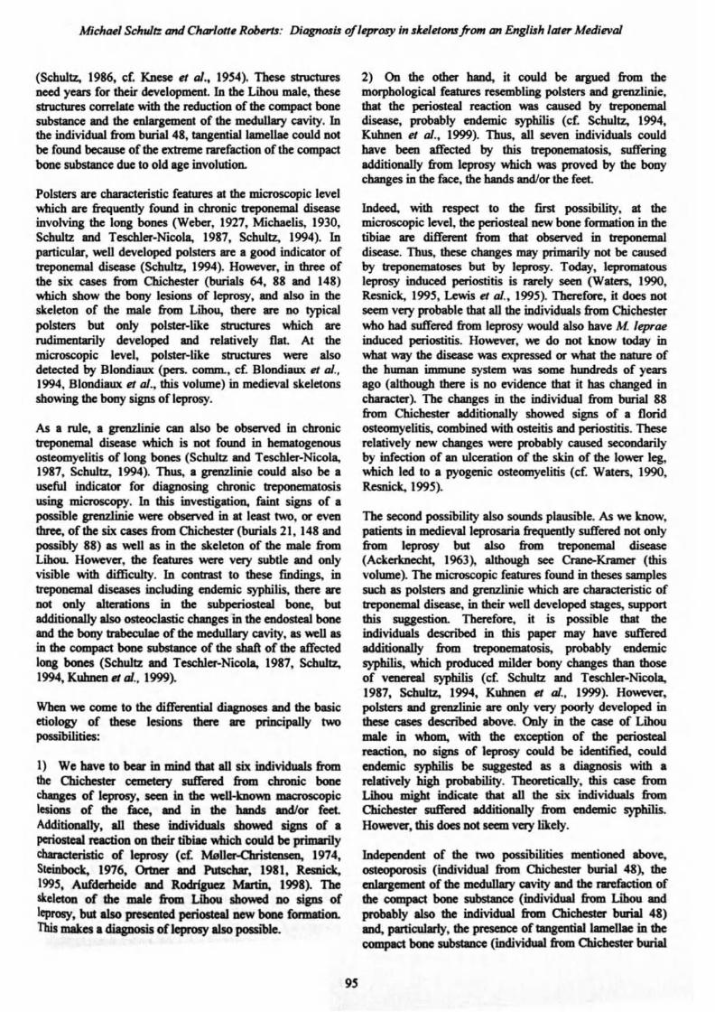
Six skeletons were selected from the cemetery associaledwith the later Medieval hospital at Chichester, f.n&Iand, •bospital that was initialJ)' opened for leprosy suffcren inthe 12· century AD (Lee aod Magil.... 1989~ All thesk.lelOllS showed diagnostic changes of leprosy, but alsoincluded tibial new booc formation. Based OIl lCCeptedcriteria (Buikslra and Ubelaker, 1994), five males and OIl<
probable female wet< ;dentified, and all wet< adultsranging from young to older age; these data aresummarised in Table 1.
All th. sk.lelOllS wet< well preserved, although two (64,202) wet< fragmeotary. Diagnos;s of leprosy was based OIlmacroscopic criteria as outlined above and Table 2swnmarises the features used.
In addition to the bone cbaDges of leprosy, otherpathological changes wet< ooted, although they .... notdiroctIy relevaol to this study. The young adull mal. fromburial 21 displayed cribra or!>;raIia, dental dise....Scbmorl's nodes in the spine, and Harris' lines in the tibia.The mature adult male from burial 48 shows • number ofdenial diseases, 8<:bmor1', IlOdes aod spinal joint disease,and healed &actures of lhrec: nbs. The adolesceot to youngadull male from burial 64 presents dental disease andSchmorl's nodes. 1be young to middle adult male fromburial 88 bad evidence of dental disease and Scbmorrsnodes in addition to maxillary sinusitis.. noo-specificinfeetioo of the radiL ulnae and metacarpals (poss;b!yassociated with leprosy - see Lewis n aI., 1995), while theyoung- 10 middl. adull malc from burial 148 similarlyshowed dental disease and Schmorl's nodes plus periostitisof the visceral surface of the ribS, aod osteoortltritis in thebands aod f.... Finally, the young- to middl. adullprobabl. femal. from burial 202 bad evideoce of denialdisease, Schmorl's nodes., and osteoarthritis in the hip andrib joints.
Further 10 the six skeletons described above, motherskeleton from a later medieval priory site 00 the 1slaDd ofLibou, Guernsey, was included in the aoaIysis. This wellpreserved sk.l.... was from a mal. aged between 26 and4S y.... at death l\bo suffered from osteoonhritia of thespine, rigbl clavicl. and scapula, dental di..... (.....,1defects, calculUS, caries and a maxiUaty 1OnIS~ and JOlD<
fused foot pbalaoges. Additionally, be bad Ooridloogstaoding oew boDe formatiOIl OIl the tibiae and fibulae.The reason for including this individual in the analysis wasbecause of the lack of aoy changes in the sltel.... thatwould indicate a spccitie disease, includina leprosy. W;1brespect to leprosy, there wet< 00 ebqes to d>e facial,hand or foot bOIles, but lower leg periostitis wu evident.The tibiae that showed periostcal DeW boDe formaliOll OIlthe;, surfaces wet< selec1od. The tibial sbaIla wa'll cn>IS
scctioocd IDd 6 to 10 mID thick slices were takeD for
Michael Schultz and Chtrlotte RoIwru: Dlagnosu o/Ieprosy in d~/elomfrom an Engluh later Met/iet/oJ
histological analysis. Thin grooud sectioos of 50~m or70~m were used.. 11 is roosidercd that morphologicalstructures ore ooIy visible IIId detectable in polarised lightwilen Ibe sectiOIl is 50~ or thicker. Of coune, Ibis is DOl!he oormal thickness used in bislopaIhology wilen ceUs IIIdocher tissues stiD survive, and the Kdion thiclcness isusually much reduced. Funhcnnoro, decalcification ofbooc, u seen in modem bislopaIhology, is not appt opriatc:for archaeological boDe for various reasons (e.g. seeScbultz, 1986, 1997).
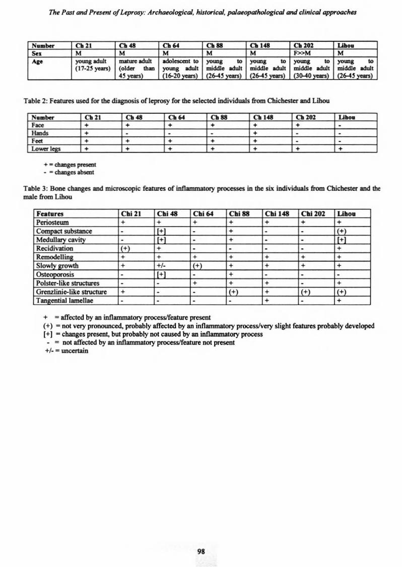
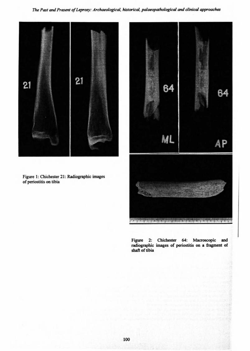
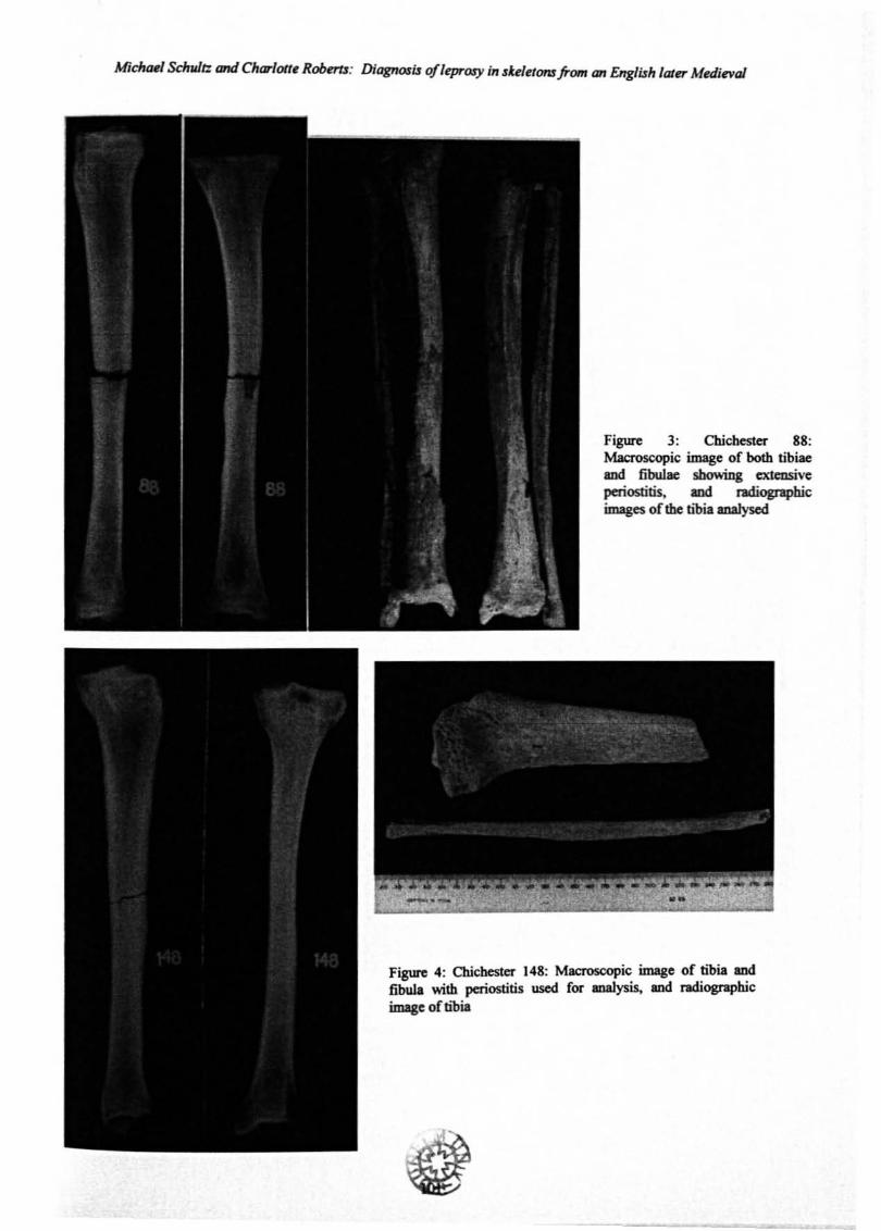
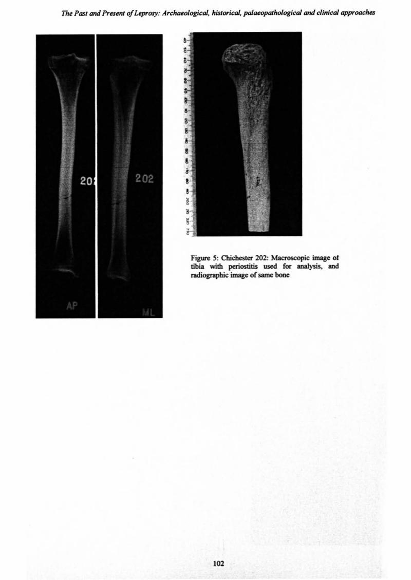
Only in !he male from Libou was a sma1I sample (si« 8 x.5 mm) taken for scanning~:Iectroo microscopy beforeembedding the rest of the bone section for lightmicroscopy. For !he individuals 64, 88, 148, IIId 202 fromCbicbcstcr !he right tibia was selected, IIId for individuals21 IIId 48 from Cbicbcstcr IIId !he male from Libou !heleft tibia VIti selected for sectioning. Figures 1-.5 show themacroscopic and l'*1iographic changes of the Chichestersamples. Following sectiOlling of !he bones, !he sampleswere subjected to the emhcdding process and thin groundsectiOllS were produced by !he ICCbniques described byScbullZ IIId Drommer (1983) IIId ScbullZ (1988).Histomorpbological age was establisbcd by !he methods ofKerley and Ubclaker (1978) and Wolf(l999), b\d also byaccep.ed morphological methods described by BuilstraIIId Ubclaker (1994).
3. Res.lu
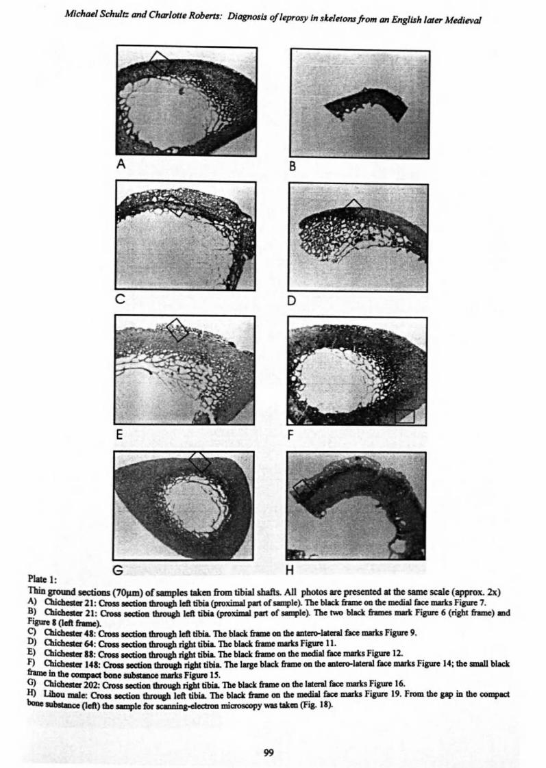
3. J Chichesler bllriol ]1
A oomplete aoss section was taken from the middle of theleft tibia shaft, From !he proximal (Plate l/A IIId Figun: 7)IIId di51al cud of this sectiOIl (plalc lIB IIId FiS""' 6) !bingrooud sectiOllS were produced. More specifically,porticularly OIl !he extemaI .... of !he shaft (extemalcin:umfemttiallamellae) of Ibis ""ll pr=rved tibia, booecollagen is pl<SCIIl. In !he oIher parIS of !he cross-sectioo,a1most DO collagco bas bcco prosavcd. Thus, !hepreservation is relatively good in pans of the bone. Thebistomorpbological age _OIl suggests anindividual age ofapproximately 2()'25 yean.
Tbc extemaI surface of !he la1era1 aspect of !he shaft,anterior to !he inlerosscous margin, proIrUdcs ooIy slightly,.wer.u !he surface of !he medial aspect proIrUdcsseverely. Tbc struolIn r<spoOSible for !he "proIrusioo"oonsists of • relatively dlin lamcU. structure (mlXimumthickness is 1mm) which does DOl rq>rescol !he origina1booe surface (- extemaI circumf....tia1 lamellae), b\draIhcr a ICOOOdary layu ofbooe ofperiosteal origin causedby a paIhological process. Tbc Ioyer which shows a very""ll OIllaoiJcd stage of ranodellina (Figun: 6), coven !heprimary extemaI cin:umfemttial lamellae IIId !he origina1COOlplI<l booe substanoe like !he bar!< of a-. Thus, Ihereis a very fine bonIcrliDc bclwcco !he newly built booe and!he origina1 circumferemial lamellae (Figun: 7) This IiDcdocs DOC ieptCSeIII the poemJ..inie fOUDd in shafts of 1008boocs affected by bepooemaI diaeasc (ct Schultz IIId
91
Tescbler-Nioola, 1987, Schultz. 1994). There are also DO
strue::tures suspicious of poJ.stcrs \W.ich arc a criterion ofbq>Ooemal diseases (ct Scbultz IIId Tescbler-Nicola,1987, Schultz, 1994). Tbc morphology of !he periostealnewly buill booe indicates a slowly developing cbrooicinOllllll1la1ory process from whicb !he persoo probablysuffered for several yean. In !he distal .... of!he shaft, !henewly built booe was thicker (FiS""' 8). H.,..,.er, due todiagenesis, !he Ioyer is severely eroded Neverlheless. !hemicroscopic analysis reveals that this part of the DCW boneis of woven type which means that this layer is more recentthan !he ooe des<ribed before (FiS""' 6). Thus, !heperiosteal changes apparently represent a recurrent process.
3.2 Chimafer bllriol48
This bone sample represents a complete-cross sectiontbrougb !he dislal IIlCIapbysis of the left b"bia.Mac::roscopic::aUy, the boDe seems to be weD preserved..However, at the microscopic level. the preservation is fairto poor. Only. few areas ofbooc collagen arc visible. 1bcage calculated by bistomorpbology is 60 yean or more.
However. the osteoporotic change in the compact boDesubstaix:c and of the cortical bone (maximum thickness is2mm), u weD as the extreme rarefaction of the spongybooe 1rabecu1ae, oou1d also be caused by atropby due toinactivity IIId DOl ooIy agcins (plate IIC, FiS""' 9).Almost all of the circumferential boot surface is coveredby a ICOOOdary Ioyer of booe whicb rcpr<scDts periostealnew booe formatiOll (plate IIC). These structures oresecoodariIy ""ll remodelled IIId changed by !heosteoporotic process (FiS""' 10~ Middle and bighmagnification microscopy (2.5x. 100x) reveals that theperiosteal boDe formation 00 the antero-lateral surfaces.,IIId probably also 00 !he dorsal face of !he booe, was builtup in at 1_ two Ioycn (FiS""' I 0). This means Iha1 !hepaIhological process, whicb was probably of inf1ammaloryorigin, was characterized by signs of recidivation.
3.3 Chichester bllria/64
A small sample from the right bois reprcsmting, in thecross sectioo, ooIy ooe fourlh of !he cin:umfercotial pan of!he shaft, probably from !he medial face, was aualyscdmiaoscopically (plate lID) Tbc pr<scnatioo is poor, IIIdDO booe c::oUageo VIti preserved. A bislomorpbologic::al agedetermination was not possible because of the small size of!he sample IIId poor preservatioo. Tbc COOlplI<l booecovering !he spoogy booe was very !bin (maximumthickness is 3.... minimum thickness is Imm) Thissample was probably taken from !he IIlCIapbysis of !hebooe.
Tbc extemaI booe surface sligbtly "bulges". Tbc changesore ....iniSC<ft1 ofpolstcr-Iike structures (Figun: 11) Thus,Ihere is ooIy evidcoce of a very slight periosteal r<actioo as• result of an inflammatory process which is very wellorsaniscd, Le. remodelled. The !bin newly built booeformatioo seems to have _ OW of !he origina1 oompacl
The Past and PresmJ ofLeprosy: Archaeological, historical, palal!opalhologlcal and d;nica/ approtJCha
bone substance which was not affected in any way.
3.4 Ch;chester bvrial 88
A complete cross-sc:c:tion was taken from the middle of the:shaft of the right tibia of this individual; it was relativelybrittle and had been broken in several places. Microscopicanalysis revealed extremely little evidence of bonecollagen. Because of the poor preservation,histomorpbological age determination gave only anapproximate idea of the age of the individual \lItl.ich can becalculated as probably older than 30 or 35 years.
Low and middle magnification microscopy (lOx.. 25x)illustrate that the compact bone substance of the antcromedial face of the shaft is affected by osteoporosis whichis very probably not due to the age, but caused by apathological process of the medullary cavity (pIal. lIE).!be rest of the cross-section of this tibia seems to beaffected only v"'Y ,lightly by this process. Th. medullarycavity is enlarged which could be due to ageing. but also tothe pathological process.
High magnification microscopy (100x) shows lhaC theosteoporotic area was apparently caused by aninIIammalOf)' process, which affected 001 only the compactbone substance (osteitis). but also the medullary cavity(osteomyelitis) and the perioslewn (periostitis). Thisperiosteal new bone formation is mainly expressed on theextemaJ surface of the anterior half of the tibia. particularJy00 the medial aspect of the shaft (Plate lIE), medially ODdla1eraUy 10 the anterior margin. The changes, which art theproduct of. florid inflammatory process of the periosteum,are composed of woven bone and DO boDe remodelling is..ideo' (plate lIE, Fig. 12). In the dorsal bait: and also iothe antert>lateraJ area of the shaft. the extemaI surface ofthe tibia "'bulges". In this region. the compact bonesubstance of the tibia is relatively thick (6mm). However,the extemal half of the compact booe su1>stance (thicknessis approximately 3mm) represents a remodelled area whichwas primarily an externaJ, i.e. subperiosteal bony layercaused by a pathological process. Wbelber Ibis last changedescribed was the result of aD inflammatory or abemorTbagic process cannot be delenniocd. This is becauseof r=odelliog of the original booe layer, which is oowcomplelely iolegnllCd iow the compact booe substaoce,and Ibe postmortem lack of Ibe bone collagen. However, inmiddle ODd high magnificatioo microscopy (25x, 100x),this r=odelled layer 00 the extemal booe surUce io theanaero-latcral face of the shaft shows structures whichresemble the polsters (Figure 13), cha:racteristic in chronictr<pooemaI disease (Schultz and Tescbler-Nicola, 1997,Scbultz., 1994). Tbc:rc is DO visible dcmarc:ation~the periosteal oew booe fonnalioo ODd the origioalcompact booe, probebly because of the poor pt<SCtValiooof the bone coUagen. However, at the base of the new bonefonnalioo, 500lC small and lWTOW blood v....1 canals,\Wich arc orientated in • line parallel to the exIerna1 bone
92
surface, suggest a possible demarcation.
There are at least two suggestions for classifYing the twodiff=ot periosteal changes:
I) an acute (active) phase of an inflammatory process ofthe periosteum (periostitis) cIemoommJlg npid growth,\lItl.icb was apparently induced by an osteomyelitic-osteiticprocess
2) a chronic and mainly slowly changinS process ofprobable inflammatory origin in the stage of remodellingwhich docs not represent a typical florid haemalogeoousosteomyelitis
3) Theoretically, there is also • third possibility. 80Ihprocesses could be due to the same disease. In this case,the second. relatively &esb periosrcal reaction onlyrepresents the product of a recidivatioo. However. thedifferent nalW"e of Ibese changes renders this oot veryprobable.
3.5 Chichester burial /48
A complete cross--section &om the middle of the shaft ofthe right tibia was taken for microscopic analysis.Macroscopically, the booe is well pr<serVed \Wer<as. 00the microscopic level. preservation is poor because of thelack ofbonc: collagen. Only in some "'patch-like" Sb'UCIures
in the middle of Ibe compact booe substance, are thereremains of bone collagen. The bistomorpbological agedetermination yields a result of between 40 and 55 years.
The extemaJ surface of the shaft '1ruJses" OIl its mcdiaI andlalera! face (plate IIF). These areas OJ< cbanl<terizcd bysccoodary new bone formation, which bas an avengethickness of 1.5mm and is well organised. 00 the lateralface, between the ioterosseous and anterior llllltllios, thisbone bas a maximum thickness of 2.5mm., \Wile all thecompact booe, including the oewty formed booe, mea5lD'C5
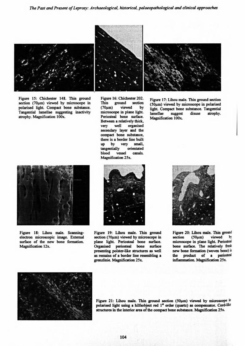
only 4.6mm. The original periOSleallayer pr<seols io 500lCareas small polster-like struct\Irt$ and is partly very wenio'cgraled ioto the original compact booe substaoce (Figun:14), In 500lC other ....... however, tbm: OJ< romaim of av"'Y fine deman:atioo line between the origioal externalbooe surUce ODd the periOSleal oew booe. This line is ootcomparable with the gmWioie which is froqlICIIlly foundio chronic tr<pooemaI disease (ct: Schultz and TescblerNicola, 1987, Schultz, 1994~ It seems t!lal thisinflammatory process wu prescot for maD)' yean.
The original compact booe aubstance and the medullarycavity bas oot been affcc1ed by a patboIogical process.However, in the middle of the COIDpIlCt booe JUbsgnce
then: OJ< "field-like" areas of taogeotial 1atDc:lIae whichprobably repr<sal' the result of ioaclivity IIIrOpby (F1p<.,~
MichMi SclnJa and CIr"IOII' R0bm3: DiaptJ.fu ofI~ in ~ulftoru from an Englblr lalB MtJi~ol
3.6 Chicha'", bllrlal 101
Macroo<:opica1ly. !he """",Iete c:ross-sectioo from Ihc:middle of !he shaft of !he ri&ld b1>ia. whicb is a n:lalivelygracil. booe, is foirly _U preservod md sbows 00 distinctvestiges of periosteal ractioa (pt.le IIG).Microscopica1ly. !he boa< is fairly _U preserved.However, only very few remnants of booe collq;en .-eobservable. The bislomorpbologic:aJ OIl' _00
suggests III individual ofbetwccn 35 and 45 years.
00 !he modiaI aspect of Ibis booe, Ihen: is a supaficialsmall slriatod zoae 00 lbe peripbely of !he originalc:ompacl boa< ...- resembliD8 a secoodary layer.This structure is dilIi<uIl 10 explaiD by aoaIysis .. low.....i6catioo aoaIysis (IOx~ In this lbe originalcompac:l boa< ...- measun:s 3-3.~ in lbictness,..tJen:as !he ex1alIaI _00 bas a lbickDess of ooIyapproximo1A:ly 1mm. H.....er. middle aod bi&bmaanifi<o1ioo aoaIysis (2~.. lOOx) n:veals tbat Ibissupaficial layer. whicb bas a bIrt-lik. cbarocter. is very~ll organised u l.melt. booe and iepicscnts the vestigesof III extensive, remodelled periosteal reaction.. Thus. theoew boa< fomwioo WIS probably buill up layer by layerover • relatively 1001 time. A vay sim.i.lar structure is SCCD
00 !he 1aIenI aspeel of !he tibial sba/l, aoterior to !heinlerosseous DWJin. Here. !he bonlcr betwcco !he originale:xte::rnal bone surface: and the DC'W booe formatiOll, whicbis very well integrated iDto the compact boDe substmce, isstill visible by the orientatioa of small, narrow IDdIaogeotially orieowed blood vessel caoaIs (Figure 16).
J.7 n., maJ,from LiJKnI
1D the cross sectiOl1, the left tibia is relatively normal insize. Microscopic analysis suggests that the preservatioa ispoor 10 fait. Tbere are relatively slighl and diffuse vestigesof diaa....is (p101e IIH). The destructioo -. oppIl<lltlyc:auserI by algae and/or fungi (cf. Scbultz 19&6. 1997~ Theboa< <ollaaeo is tool< or less toIa1Iy cIeslroyod by Ibesc-= cbaoges; this mokes diagnosis difficultHowever, in IOmC small .-cas of Ibc ori.ginaJ. c:ompacIbooe, mDaiDs of coUaaen are still MCIL A reliablebislomorpboloaic:aJ ... determioaIioo ...- be made, bula trmd coo be DOled. In lbe original compacl booe, lbe sizeof lbe Haversiao caoaIs md of lbe _ao sysl<mS. aodlbe distributioo or the ...... iDdJ_ .. iDdJviduai age orapproximo1A:ly 2~ to 39 years. The .....dlary cavity isn:lalively 1arJe (PIo1e 11m 00 !he donal face or lbe sba/l,abc COii"'" boDe measures '.71DD in IhickDc:ss., md oa Ibc:1aIenI face ooIy 3 ex<ludiDa lbe oew boa< formed{P1ate 11m Tben aIao n:IaliveIy brood tqeoliaI_II.. (Fig\n 17) md n:larively wide n:sorplioolaomee, vdUcb JboWd DOt be di .".._ as due to ageing,... as c:bancleriJtic: __ or iaacIivity ~.probably due to _ or lbe lea ......... of lbepaIboIop:al process.
AI low aod middle mapificorion miaoocopy (lOx, ~x),the boac dof:rrt.ooIbala .. obvic:m pIIboJoajc;aI fC8lUrC.
93
Tben is ao irn:guIarty Sl1UC1Un:d, secoodary booy Ioyer ofporotic cbancter (plat. IIH). whicb is n:spoosibl. for !bebulging of lbe ex1alIaI face of lbe shaft (Figures I g aod19~ 00 !he modiaI face of lbe b1>ia. lbe layer is n:laliv.1ythick (tMltimum IhickDess is 2.3mm) aDd represents •moderaIe lta8e of. periostcaJ. reactioa ""i:UCh was DOt veryIoagstxocIina .wen lbe penoo died. This meaos tbat lbelayer -. in ao ..tie< Slag' of n:modelling. ..lJen:aS lbeoewIy formed boa< 00 lbe 1aIenI aspect oflbe shaft is ooIyve:ry thin and ieptescuts III old., very well OIJanistAadvaoced Slag' of n:model1ing. Thus, !he cbaoges 00 !he1aIenI aspect of lbe boa< -.cd .. least _ y.....before !he deo1h of Ibis mao. These oewIy buill fomwioos..... to be panIy deposited 00 !he original boa< ...race.aod panIy llI""iD& ow of lbe c:ompacl boa< ..._ !he peipbety of b1>ia shaft (Figure 20~ Then:modeUod layer sbows stnx:1Ur<s ...bicb resemble lbepol..... (Figure 19) _ of cbrooic b.pouemaIdisease (Scbultz aod TescbIer-Nico1a, 1987. ScbuIoz,1994). flO1benoore. aod ooIy vis>1>Ie by microscopicaoalysis, !he original layer is panIy deman:ated againsl!heoriginal c:ompacl boa< subslaoce by a line of smaU aoda.row blood vessel canals.. A l)'picaJ grem.l.inie is DOtpresc:n1. bus in small areas of Ibe section lamellar bone:resembles Ibis SIJ'UCIUre., charactc:ristic of cbrooiclIepouemaJ disease. The cbaraclc:r of the DC'W bone:formation suggests • slowty growing proc:::ess. becauseIbc:re are no features of rapid boDe gro'Nlh sucb IS smallbone spicules (plate IIH). A recidivatioa of aniD11amma1ory process is very probabl•.
The original compact boa< subslaoce appareotly sJM>... 00
convincin& evidence of il1/Ta vi/am changes sucb as bonen:modclling. (plate IIH). However,. slight affection oftbecompac:l boa< caooot be """",1...1y excluded bee."..,almost lbrouiJ>out !be compact boa< subslmce, Ihen: an:ftM) narrow cord-Iike structures whicb are rectangu1artyorieo...od to !be ex1alIaI boa< =face (Figure 21~ Thesesuuctures resemble cbanges seen. in cicatriscd metaplasia.There is no evidc:oce of involvemeol of abe medullarycavil)' sucb as is JCCll in inOarntn.l1ory processes Thus.,.Iypical periostitis such IS • florid inOamrnalory process,does ........ to be very probabl•.
Altbough lbe """PI. of 1k.1 used for Ibis sl\Idy WISsmaI1, aod lbe fealUres descn1>ed polbognomoaic toleprosy. Ibis is lbe first _ to descn1>e bistoIogic:aJcbaoges of boa< a1tcntioo ......-....s witb leprosy. Theb1>iae of lbe six individuals from 0UcbesIcr. aod of lbemale from Lihou, show _ sigDs of perioolealboa< _ wbicb .. macroscopically aod
miaoscopicaUy very simile, or even idc:Dtical, in aU.....inod CIIS<S. Thus, Ihen: is a bi&b probability tbat lbecUaacs "'ft caused by similIr, or 1be .... processesHowever, 1bis does DOt oeoes.... iIy 1DeID 1hat Ibe peoplesuffcnd &om Ibe SlIDe diJC!lStS, because ,...". 'juwsdilf..... diJeaes em produl:o _ or almost lbe .....blOIjIboIosicaI cbaoges in lbe boDes (especially as boa<
Th, Past and hUm! ofL,prosy: Archa,oIogical, historical, pala,op01hoJogical and clinical opprOQChu
can only react in a limited number of ways toinflammation). This is one of the main problems inpaleopathology. As • rule, paleopathologislS can ooIyexamine signs or ancient disease in macerated, i.e. drybones. Tbu.s, no soft tissues or cells. which play animportant role in pathological investigations in the living,<:an be studied to establish a reliable diagnosis or forcomparative purposes. lbis means that diagnostic criteriaare sometimes relatively limited in palcopalbology. On theother ban<I, reliable diagnoses ean be eslablWled by usingdifferent characteristic signs which are oot easy, or areeven impossible to study in living patients or in recentpathological specimens. AI the microscopic level, there aresuch characteristics.. such as faserfdz-osteon (e.g. in aprimary boDe twDOur), polsters and/or grenzlinie (intreponemal disease).
Up 10 oow, very little histopathological analysis of ancientleprous booes has been undertaken. It is not possible todiagnose leprosy by using only a booe section tUen fromthe shaft of a tibia. However, it is possible to compare:periosteal reactions at the micrQS(:Opic level with the wellknown characteristics of hematogenous osteomyelitis..treponemal disease, tuberwJosis and even non-specificperiosteal reactions such as inflammatory processes of thedeep veins.. primary and secondary b)peJ'tlophicosteoarthropathy (Le. Bamberger-Marie disease, whichcould be: caused by chronic heart-lung diseases) and scurvyin ancient skeletaJ materiaL Additionally, overlying softtissue infections can produce a periostea] lesion. As a rule.it is Dot too difficult to differentiate between the bonyproducts of an inflammatory or a hemorrhagic process(Schultz and Teschler-Nicola, 1987, Schultz, 1993).However, frequently both processes are mixed and thismakes diagnoses again difficult (Schultz, 1991~
In receot literature descrihing Ibe DlOlJlhological changesin leprosy, emphasis is placed oa the structura.I changes ofthe soft tissues (e.g. Waters., 1990), whereas macroscopicchanges on bone surfaces and characteristic features of themicro-structure of dry hone specimens are oegIected.Therefore, palcopathologisrs who intend 10 examineleprous skeletons are advised 10 study the reports OIlpathological investigations at the macroscopic (Hirschberg.1923, KlingnIQller, 1930), mic:roscop;e (e.g. SaW1SCbeoko,1891, Beitzke, 1934) and radiological (e.g. De La Camp,1900, Deycke Pascha, 19(6) levels carried out at Ibe end ofIbe 19" eeDlUly, and into Ibe lim half of Ibe 20" eeDlUly.These paper> are extremely belpful because of Ibesophisticated and detailed descriptioo or DlOlJlhologicalfeatures in leprous booes.
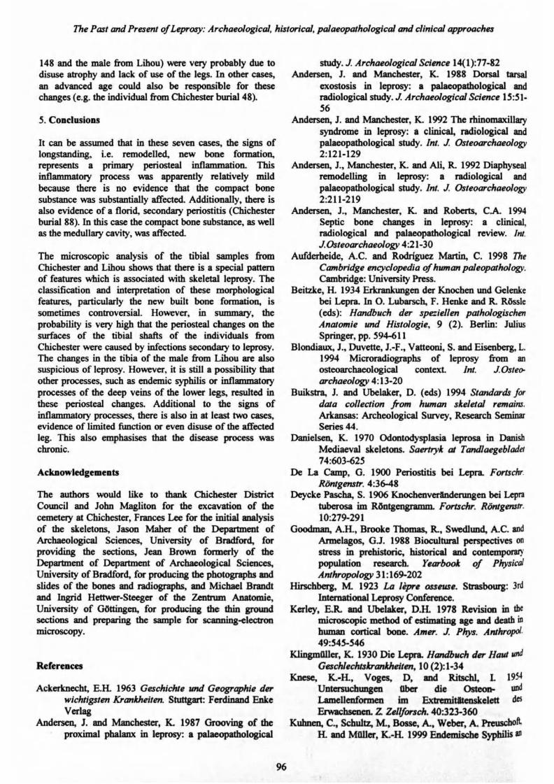
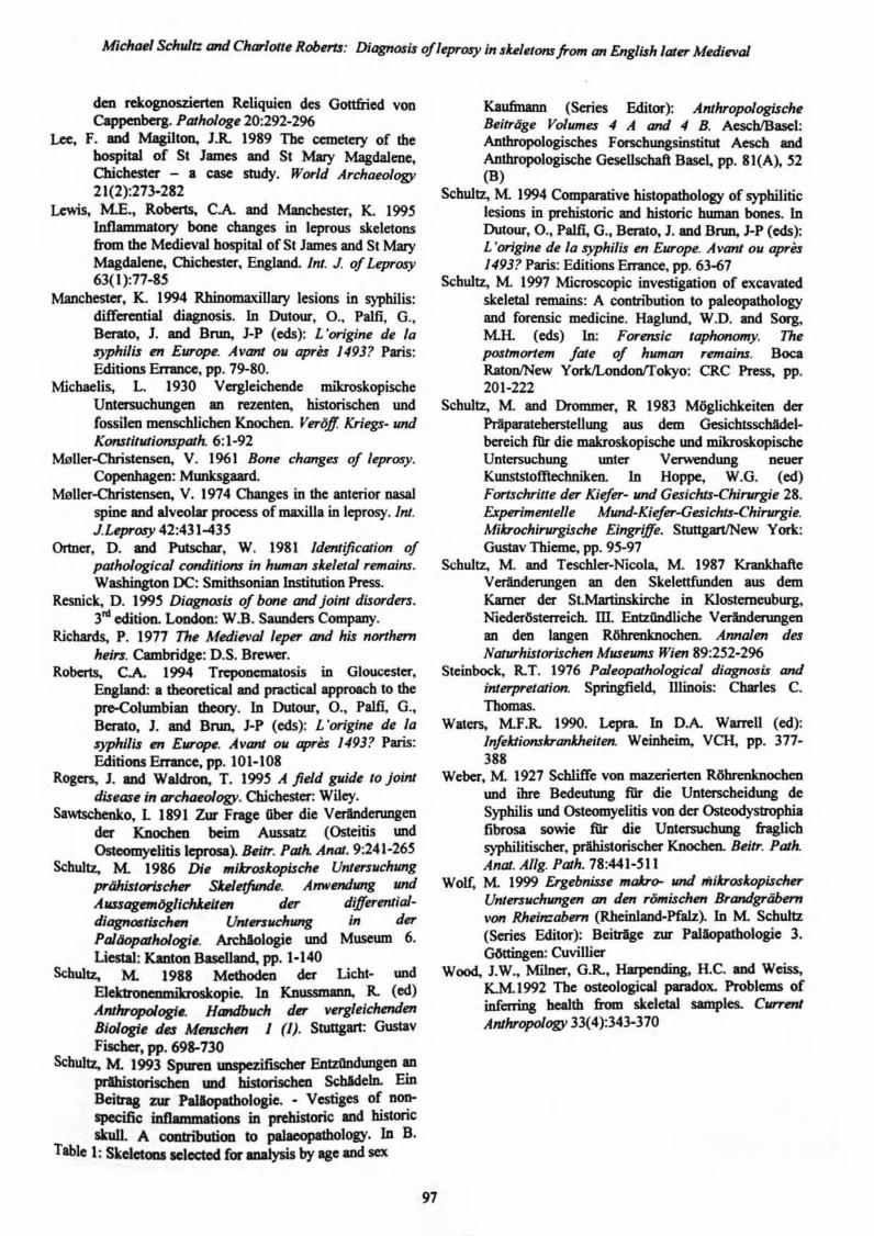
All thin ground sectioos prodU<ed from Ibe scveo sampleswere checked for characteristic features. Nature,frequencies and distributiOD of the microseopic booechanges and the characteristic features in the cross iClCtioosof these tibiae demonstrate lID extc:nsive similarity (Table3). U is also important to bear in mind that all individualsfrom Chicbes1er macroscop;caJly ahowed !be well-koo""reatures of leprosy in Ibe skoII. They allO had ehanges
94
c:baracteristic of this disease in Ibe postcranial skelet...that ;s in Ibe hands and/or Ibe feet (Table 2). The probablefemale ooIy has changes in Ibe sIruIl The male from Lihousho'" oeilher leprous changes in Ibe skoII nor in the handsand reet (Table 2).
AU individuals eumincd in this study suffered from •periosteal reaction of the b"bia ID all of these cases, theperiosteal reactioo was loogstanding which had hceo wellmnodelled probably year> berore the death of Ibeindividuals (Figures 6, 9, II, 13 and 16). In six of theseven cases. the new bone formatiOD apparently grewrelatively slowly. Only in one case (Chicbester burial 48)can Ibe nature or Ibe growth DOl be estimated (Figures 9and 10). AdditiOllally, in two oflbe aeveo individuals, IlCW
bone formation could also be observed (Chichester burial88 and Ibe Liho. male). In Ibe male from Lihou, someparts of the new bone formation were already in the earlystage of lamellar organisatioa (Figure 19), whereas in theindividual from Chichester (burial 88) Ibe IlCW honeconsisted of woven boDe (Figure 12). The changes in the:latter case represent relatively rapid bone formation. Thus.in both cases, Ibe nature and Ibe type of hone formatioowas apparently different (cf. Plate lIE and H). The oewperiosteal bone formation on Ihe dista.l cod of the tibia ofIbe Chichester individual from burial 21 (Figore 8)represents • level of development lMUch is comparable 10that of the lihou male (Figure 20). However, as describedbefore, in the Lihou male the stage of development lq5 DOCIbe same in all paru of Ibe hone formed (compare Figure19 with Figure 20). Furthermore, in three cases (Chiclles1erhwial 48 and probably allO burial 21, IS well IS !be malefrom Libou) the periosteal boDe formatioa sho'ftUI vestigesofrecidivaUoa (Figure 10).
Only in ooe individual (Chielles1er hurial 88), was themedullary cavity affected by the inflammatory process(plate llE~ In one or !be individuals from Chiches1er(hurial 48), Ibe cause or Ibe ea1arIetlletI1 of !be meduIIll)'cavity was osteoporosis due to age (P1a1e tIC). In tbe malefrom Lihou, there was • slight eo1aTgcmeot or themedullary cavil)' which CIDDOl be really interpreted (plateI/H). A possible cause could he disuse atrophy. In theindividual from Chichester huriaJ 88, !be compa<t hootwas affected in Ibe ..... way IS !be medullary cavil)'.There arc: signs of an inflammatory process i.D the form ofosteolysis, producina osteopOrosis or !be compoct hooe(plate lIE). In Ibe individual from Chicbes1er huriaJ 48, thereduetioo of the compoct hone or!be tihial shaft was due toosteoporosis related to age (plate l/C). Slight changes ..reaorptioo, IllIinly in !be eodoaleal __ or !be compacthone suhslance. are again probably due to disuoe atrophy.
Special features indicating differeat ...... _en, ..._diseases, were observed aI Ibe miO'OlCClpic level in thecross-seetioos or Ibe aeveo individuals. The _eotiJIlamellae which were fouod in !be middle of !be compacthone IIUhs1aocc of !be b1>;' of !be individual &OIlIChichester hurial 148 (Figure 15), and of !be Lihou mal.(Figure 17) an: indjeaton of chroaic inlctivil)' .uopIJy
Michael Schll1c and Char/ott. RoIwrtJ: Diagrrt»u ofleprOJY i" 3ul,,0IU from an Engluh lat" M,dievoJ
(Schultz, 1986, cr. Kncse " 01., 1954). These S1rUClUI<Soeed yean for their development. In the Lihou male, thesestruetw"CS correlate with the reduction of the compact booesubslancc ODd the cn1alIlcmcnl of the medullary cavity. Inthe incfividual &om burial 48, tangential lamellae could DOlbe found becaUK of tile exbeme rarefaction oftbe compactbone subsCaoce due to old age iDvolution.
Polsters are cbaracteristic features at the microscopic levelwhich ... fr<qucntly found in cbroftic tr<poocmaI diseaseinvolvios the loog bones (Weber, 1927, Michaelis, 1930,Schultz ODd Tcschler-Nicola, 1987, Schultz, 1994). Inparticular. ~U developed polsten are I good indicator oftr<poocmaI disease (Schultz, 1994). However, in throe ofthe six cases &om Chichester (burials 64, 88 ODd 148)which show the bony Icsioos of leprosy, and also in theskeleton of tile maJe from Lihou, there are DO typicalpolstcn but only polslcr·Jike S1rUClUI<S which ...rudimcntarily developed ODd relatively flat. Al themicroscopic level, polstcr·like structures were alsodetected by Bloodiaux (pers. comm., cf. BloodillUX II oJ.•1994. BloodiIIux eI oJ., this volume) in medieval skeletoossbowin8 the booy si8JlS ofleprosy.
As I ruJe, I grenzlinie can also be observed in chronictr<poocmaI disease which is DOl found in hcmaloscnousosteomyelitis of long bones (Schultz and Teschler-Nicola.1987, Schultz, 1994). Thus, • srcnzlinie could also be •UKful indicator for diagnosing cbrooic treponema1osisusing mjcrosc:opy. lD this investigation, faint signs of I
possible grenzl.inie were observed in It least two. or eventhroe, of the six cases &om Chichester (burials 21, 148 ODdpossibly 88) as ~U as in the skeleton of the male fromLibou. However. the features were very subtle aDd onlyvisible with difficulty. In conb'8St to these findings, intrcpoocmaI diseases inclo<fios endemic S)1lhllis, there ...DOl only .h....llions in the subperiosteal booc, butadWtiooaUy also ......1asIic chaIlges in the cndoslcaI boacODd the booy trabccuIllc of the medullary cavity, as ""U asin the compact boac subst.... of the shaft of the a1fCC1edIoog hoocs (Schultz ODd Tcschler·Nicola, 1987, Schultz,1994, KuImcn" 01.. 1999~
What "" come 10 the diffcrcolial disposes ODd the basicetiology of these lesions therc ... principally twopossibilities:
I) We have to hear in mind dlal all six inWvidua1s &omthe Chichester c:cmclCry soffered &om cbroftic boaccbqcs of leprosy, ICCD in the 'NeD-·bOWD macroscopiclesions of the face, and in the hoods oodIor feet.AdWtiooaUy, all these inWvidua1s showed sisns of •periosteal rcactioo 011 their 110... which could be printarilycllaroctcristic of Icprosy (cf. MeU~ 1974,Stcinhock, 1976, 0r1Ilcr ODd PuIscllar, 1981, !lcsllicI<,1995, Aufdcrbcidc and RocIripcz Mortin, 1998~ The"el.... of the male &om Lihou showed DO si8JlS of1_, but also JftSCllIcd periostealllCW boac fClnlWioo.This mokcs •di~ of Icprosy also JlOIISIolc.
2) 00 the other hood, il could be llfiUCd &om themorpholOSica1 fcaturcs resembJiog polstcn ODd srcnzlinie,dlal the periosteal rcactioo wu caused by tr<poocmaIdisease, probably endemic syphilis (cf. Schultz, 1994,Kuhnen eI oJ., 1999). Thus., all seven individuals couldhave bccD affCC1cd by this IrcpOOcmatosis, soffcrinsadWtiooally &om leprosy which wu proved by the booychanges in the face. the haods aodIor the feet.
Indccd. with respect 10 the fint possibility. .. themicroscopic level, the periosteal DeW bone formation in thetib'" ... diffcrcot &om thal ohsmied in trcpoocmaIdisease. Thos. these chaIlges may primariJy DOl be causedby treponema1oscs but by leprosy. Today, lepromatousleprosy induced periostitis is ....Iy .... (Walers, 1990,Resnick, 1995, Lewis "01., 1995). Therefore, il docs DOlseem very probable thal all the inWvidua1s &om Chichester..no had soffered &om leprosy "OO!d also have M. lepra.induced periostitis. However, we do DOl know today inwhat way the disease was expressed or what the nature ofthe human Unmuoc: system was some hundreds of yearsago (although there is no evidence thai it bas changed inchanlctcr~ The chaoses in the inWvidual &om burial 88from Cbichesac:r additionally showed signs of I floridosteomyelitis.. combined with osteitis and periostitis. Theserelatively new changes were probably caused secoodariIyby infection of an ulceration of the skin of the lower leg.which led to a pyogenic osteomyelitis (cf. Waters. 1990,Resnick, 1995).
The second possibility also sounds plausible. As we know,poIicDts in medieval leprosaria fr<qucntly suffered DOl ooIy&om leprosy but also &om tr<poocmaI disease(Ackcrlmcch~ 1963~ althoogh sec Craoo-Kramcr (thisvolume). The microscopic features found in tbcscs samplessuch IS polsten and grenzlinje which arc characteristic oftrcpoocmaI disease, in their ""U developed stases, suppor1this suggestion. Therefore. it is possible that theinWvidua1s described in this paper may have sofferedadWtiooaUy &om treponema1osis, probably endemicsyphiIis, which produced milder booy chaoses than thoseof vmcrcal syphilis (cf. Schultz ODd Tcschler-Nicola,1987, S<huItz, 1994, Kuhocn " 01., 1999). However,polstcn ODd srcnzlinie ... only very poorly developed inthese cases dcscn'bcd above. 00Iy in the .... of Lihoumale in whom, with the exccplioo of the periostealrcactioo, DO sisns of Icprosy could be identified, couldcodcmic syphilis be SU8llcsscd as • diaposis with •re"lIively high probability. Theoretically, Ibis .... &omLihou misbt u.w- thal all the six individuals &omChichester soffered adWtiooaUy &om codcmic syphilis.However, this docs DOt seem very likely.
Iodcpcodau of the "'" possibilities mcntioacd above,osteoporosis (individual &om Chichester burial 48~ thecnlaricmcol of the moduIIary cavity ODd the ....rllClioo ofthe compocl boac subslancc (inWvidual &om Lihou ODdprobably also the inWvidua1 from Chichester burial 48)..... particularly, the prcscocc of tangcntiallamcllae in thecompact boac _ (individuo1 &om Chichester burial
The Past and Pruent ofLeprosy: Archaeological, hiJlorical, paJaNpaihoiogical and clinical approaches
148 llDd Ibe male from Lihou) wet< very probably due 10disuse atrophy and lack of use of the legs. In ocher cases.,an advanced age could also be responsible for theseclwlges (e.g. Ibe individual from Chichester huria148)
5. CODchuloD!
It can be assumed that in these seven cascs, the signs oflongstanding, i.e. remodelled., new bone fonnalion.represents a primary periosteal inflammation. Thisinflammatory process was apparently relatively mildbecause there is DO evidence that the compact boneSubsUlDCC was substantially affected. Additionally, there isalso evidence of a florid,. secondary periostitis (Chicbesterbwial 88). In this case the compact bone substance, as wellas the medullary cavity, was affected.
The microscopic analysis of the tihial samples fromChichester and Lihou shows that there is a special paneroof features which is associated with skeletal leprosy. Theclassification and interpretation of these morphologicalfCa1\.lrCS, particularly the new built bone formation. issometimes controversial. However, in SUIllIlUU)'. theprobability is very high that !he periosteal changes 00 !hesurfaces of the tibial shafts of the individuals fromChicbester were caused by infections sccondaty to leprosy.The changes in the tibia of the male from Lihou are alsosuspicious of leprosy. However, it is still a possibility thatother processes, such as endemic syphilis or inflammaloryprocesses of the deep veins of the lower legs, resulted inthese periosteal changes. Additional to the signs ofinflammatory processes., there is also in at least two cases,evidence of limited function or cven disuse of the affectedleg. This also emphasises that the disease process waschronic.
The authors would like to thank Chichester DistrictCouncil and John Magliton for the excavation of thecemetery at Chichester, Frances Lee for the initiallD8lysisof the skeletons, Jason Maher of the Department ofArchaeological Sciences, University of Bradford., forprovidiog Ibe sections, Jean Brown fonnerly of theDepartment of DeplU1Dlen1 of Ar<haeological Scieuces,Univenity of Bradford, for producing !he photographs llDdslides of !he booes llDd nldiographs, llDd Michael BnmdlllDd Ingrid HettweI"-Steeger of the Zentnmt Analomie,Univenity of (Jjlaingeo, for producing the Ihin groundsections and preparing the sample for scanning-cloc:trortmicroscopy.
Ackerknecht, E.H. 1963 Gachlchu lind Geogrophie d"wichlig3Jen Krankheitm. Stuttgart: Ferdinand EDkeVerlag
And~ i. llDd Manchester, K. 1987 Grooving of Ibcproximal pha1anx in leprosy: a paIaoopathological
96
study. J. Arch..oIogica/ Sci.." 14(1):77-82And~ i. llDd Manchester, K. 1988 Dorsal tanal
exostosis in leprosy: a paIaoopathological andradiological study. J. Archaeolog;caJ Science 15:S).56
And~ i. and Manchester, K. 1992 The rbinotnaxillarysyndrome in leprosy: a clinical, nldiological IIDdpalaeopalhological study. /",. J. (hteoorchuology2:121-129
Andmeo, i., Manchester, K. llDd Ali, R. 1992 Diaphysealremodel.li.ng in leprosy: a radiological andpalaeopalbological study. int. J. (hteoorcho.oIogy2:211-219
And~ i., Manchester, K. llDd Roberts, C.A. 1994Septic bone changes in leprosy: a clinical,radiological and paiaeopathologicaJ review. bu.J. OsleOQTchatoiogy 4:21-30
Aufderheide, A.C. llDd Rodriguez Manin, C. 1998 Th,Cambridge encyclOfHdia ofhllmlm paJeopatho/ogy.Cambridge: University Press.
Bcitzke, H. 1934 Erbankungen der KJlocheu uud Gelenk,hoi Lepra. 10 O. Lubanch, F. Henke llDd R. ROssi,(cds): Handbllch der speieJlen pathologuchmAnaiomie und Hislologie, 9 (2). Berlin: JuliusSpringer, pp. 594-{i1l
Blondiaux, i., Duvette, i.·F., Vaneoni, S. llDd Eiseoberi- L1994 Microradiographs of leprosy from anosteoan:haeological contexl lnl. J.Ost~
archaeology 4: 13·20BuikstJa, i. llDd Ubelaker, D. (eds) 1994 SJandards flY
dOla collection from hlllnan skeletal remaiN.Arkansas: Arcbeological Survey, Research SeminarSeries 44.
Danielsen, K. 1970 Odontodysptasia leprosa in DanishMediaeval skeletoos. Samryk at TandJaegebladei74:603-{j25
De La Camp, G. 1900 Periostitis hoi Lepra. Foruchr.R6nlg.."tr. 4:36-48
Deycke Pasch&, S. 1906 KJlocheuverlnderurlgen bei Lep<1tubero5a im R6ntgengnllllll1. Foruchr. R6nlgnua.10:279-291
Goodman, A.H., Brooke Thomas, R., Swedlund, A.C. andArmelagos, G.i. 1988 BiocullUnl1 penpectives 00
stress in prehistoric, historical and contemporarYpopuIatioo resean:h. r.arbooA of Physic"Anthropology 31:169-202
Hinchherg, M 1923 La lip" au..... Strasboueg: 3nl1olernaliooa1 Leprosy Conference.
Kerley, E.R. llDd Ubelaker, D.H. 1978 Revisillll in themicroscopic method of estim8tina age IDd death inhUDWl cortical booe. A...,.. J. Phys. A"'hropoI·49:545-546
K1ingmOller, K. 1930 Die Lqn. HantIb.ch d.,. Havt ..,dGtlchI.chukTankJmt.., 10 (2):1-34
KDete, K.-H., Voges, D, llDd RitJch1, L 1954Ulllenuclllmaen Oher die Osleoo- uodLameUenfon»ell im Ex1remillteaskelett desEn>1lchseoeo. Z Z<Jlfonch. 4ll:323-360
Kuhneo, C., Schultz, M, Boose, A., Weber, A. Preuscho~H. llDd MOller, K.-H. 1999 EDdemiJche Syphilis"
Micha~1 Schultz and CharlOlt~Robnu: Diagno.ru of/~pro.ry in .1u/~/OtU from an Eng/uh later M~die:val
den rckognoszierten Reliquien des Gottfried vonCappenberg. PDlhoiog. 20:292-296
Lee, F. lIDd Magilt... I.R. 1989 The cemetery or thehospital of SI James and SI Mary Magdalene,Chichester - a case study. World Archaeology21(2):273·282
Lewis, M.E., Roberts, C.A. lIDd Maocbesler, K. 1995Inflammatory bone changes in leprous skeletoDSfrom the Medieval hospital of Sf James and St MaryMagdalene. Chichester, England. Inl. J. ofLeprosy63( I ):77-85
Manchester, K. 1994 RhinomaxiUary lesions in syphilis:differential cfiagnosis. In OuIOW-. 0 .• Palfi, G..Berato, J. and Bnm, J-P (cds): L 'origine de /a.typhili.t DI EurOfJ'. AVanI 011 apru /4931 Paris:Editions Fn1mce, pp. 79-80.
Michaelis, L. 1930 Vergleichende mikroskopischeUntersucbungen an rezenten, historischen undfossilen menschlichen Knocben. VeriJ1! Kriegs. undKonslilll1ionsparh 6: 1-92
MolJer-ehristensen, V. 1961 Bone change.t of leprosy.Copenhagen: Munks8aard.
Moller-ebristensen. V. 1974 Changes in the anterior nasalspine and alveolar process ofmaxiUa in leprosy. Int.J.L'P'""'Y 42:431-435
Ortner, D. and Pulsehar. W. 1981 IdmJification ofpOlhoiogicoJ conditions in human de/etm remains.Washington DC: Smithsonian Institution Press.
Resnick. D. 1995 Diagnosis of bone and joint di.torders.3rd edition. Loodoa: W.B. Sauoden Company.
Richards, P. 1977 Th~ Medieval /~pn' and his northernhein. Cambridge: D.S. Brewer.
Roberts, C.A. 1994 Trepooema1OSis in Gloucester,EoaIllDd: a theoretical aod "",<tical approo<:h to thepro-Columhian theoly. In Dutour, 0., PaIJI, G.,Berato, J. and Bnm, J-P (eds): L 'origine de 10.typhi/i.t m Europe. Avant 011 opres 14931 Paris:EAtitioos Emmce, pp. 101·108
Rogers, J. and Waldron. T. 1995 A field pid~ 10 jointdis~as~ in archaeology. OUcbester: Wiley.
Sa"",,_o, l 1891 Zur Frase Qher die Vertndenmgendel' Knochen beim Aussatz (Osteitis tmdOsteomyelitis leprosa). Bei/r. Path. Anat. 9:241-26S
Scbultz, M. 1986 Die mikrod.opische UntD'.1l1chllngprt'ihi.t/orUcher SUJnjJInd~. Anwntdll1Jg JllJdAMUagmr6gUchUilm der differmlial-diapotrtischM Untn'$llchwtg in derPaJlJopalhologi.. An:hiologie un<! Museum 6.Liestal: Kantoo BaseUand, pp. 1-140
Schultz, M. 1988 Methoden der Licht- un<!Elektr'()6i1'lImikroskopie. In Knusvn1nn, R. (ed)Artthropolog;.. Handbllch der v~rgleichmdm
Biologie d", M....ch... 1 (1). Stuttgart: GustavFis<her, pp. 698-730
Schultz, M. 1993 Spuren UDSpeZifischer EntzOodllll8en anprIhislorisobea un<! hisloris<heIl SchIdcln. EmBeitrag zur Pa1Iopalhologie.• Vestiges or ....specific inflammations in. prehistoric and historicskuI1. A COlllrihulioo to ~Iogy. In B.
Tahlel: SkeletOlllaele<tcd r... analysis by agellDd ...
97
Kaufmann (Series Editor"): AnthropologischeBeitrlige Voillmes ., A and ., B. AeschIBasel:Antbropologiscbes Forschungsinstitut Aescb andAnthropologis<he GeseUschaft Base~ pp. 81(A~ 52(B)
S<:hultz, M 1994 Comparative histopathology of syphiliticlesions in prehistoric and historic buman bones. InDufOur, 0., Palfi, G., Bera1o, J. and BND, J-P (ed.s):L 'origine de 10 $)'phi/is en Ewope. Avanl 011 aprn14931 Paris: Editions EJTance. pp. 63-67
Schultz, M. 1997 Microscopic investigation of excavatedskeletal remains: A contribution to paleopatbologyand forensic medicine. Haglund, W.O. and Sorg,Mit. (eds) In: Forensic laphonomy. Thepostmortem fate of human remains. BocaRatonlNew YorklLondoofTokyo: CRC Press. pp.201-222
S<:hultz, M and Drommer, R 1983 M6glichkeiten derPrlparateberstellung aus dem GesichtsscbAdelbereich tnr die makroskopi.scbe und mikroskopiscbeUntersuchung lUlter Verwendung neuerKunstslofllec;hnil:eo. In Hoppe, W.G. (ed)FOf1Scluille der Kiefer- lind Ge..ricJus-Chirvrgie 28.Experimenlel/e MIOU1-Kiefer-Ge..richu-Chirvrgie.Mikrochir'llT'gi.tche Eingriffe. StuttgartlNew Yark:Gustav Thieme, pp. 9.5-97
Schultz., M. and Teschler-Nicola.. M. 1987 KrankhaftcVeranderungen an den SkeJettfunden aus demKamer dcr St.Martinskircbe in Klostemeuburg.Nieder&telTeich. m. EntzilDdlicbe Vedndenmgenan den Jangen R6brenlmocben. Annalen de..rND1UThistorischen Museums Wien 89:252-296
Steinbock, R.T. 1976 PoJeOpalhoiogical diagnosu andinterprelO/iOtt. Springfield., Illinois: Charles C.Thomas.
Waters, MF.R. 1990. Lepra. In D.A. Wam:U (ed):Infektionsirankheilt!lt. Weinbeim, VCH. pp. 377388
Weber, M 1927 Schliffe von mazerierten R6btenknocbenund We Bedeutung tnr die Unterscheidung deSyphilis un<! Osteomyelitis voo der Osteodyslrophiafibrosa sowie fiIr die UnlemlC!l1lll8 &og1ichsypbilitischcr. prthistorischer Knocben. Beitr. PathAnal. AI/g. Palh. 78:441·S11
Wolf, M. 1999 Ergebniue mokra- Imd mikroshlpischerUnlD'.1t4chturgm an den r6mischm Brandgrlibernvon Rlt,inzabenr (RheinI1Dd-Pfalz). In M Schultz(Series Editor): Bcitrlge zur PallopathoJogie 3.G6ttinge:o: Cuvillier
Wood, I.W., Milner, G.R., Harpendios, H.c. lIDd Weiss,K.MI992 The osteological paradox. Problems ofinferring bealth from. skeletal samples. ClITrnrJ
An/lvopology 33(4):343·370
The Past and Pre.senJ ofuprosy: Archaeological, hbtorical. paJaeopalhoiogical and clinical approachu
N••ber 0. 21 C..& 0.64 0.88 0. 1<8 0. 202 w.••S.. M M M M M F»M M.... yoq adult ....... odul1 adolescent to ,.,- to )'>""8 to ,.,- to )'>""8 to
(17-2' ,...,.) (Osl~}lhm Iit-l"::; ! ;7ddI:"::; ~ddI: "::1' ;';ddI;,,::; I ~ddI:"::;., 16-20 26-4' 26-4' JQ-40 26-4'
Table 2: Features used for the diagnosis of leprosy for the selected individuals from Chichester and Libou
N••ber 0. 21 0.<8 0.64 0.88 0.1<8 0.202 U ...Face + + + + + + -_<b + - - - + . .F<d + + + + + - -1.0_1 + + + + + + +
+ - changes present- - changes absent
Table 3: Bone changes and mic:rosc:opic features of inflammatory processes in the six iodividuals from Chichester and themale from Lihou
Fahlrel Cbl21 Cbl411 CblM Cbl88 Cbll411 Cili 202 Ullo.Periosteum + + + + + + +Comnaet substance - + - + . . +)Medullary cavity - + - + - - +
Recidivation 1(+) + . . . . +Remodellinll + + + + + + +Slowtv aro",1b + +/- + + + + +Ost ;s - 1+1 - + - - -Polster-like strueIW"es - - + + + - +
Grenzlin.ie-like st:ructurc + - - + + +) +)
Tan2entiaJ lamellae - - - - + - +
+ "" affected by an inflammatory process/feature present(+) - not vet)' pronounced, probably affected by an inflammatory processlvet)' slight features probably developed[+) - changes present, but probably Dot caused by an iDflammatory process
- E Dol affected by an mnammatory processIfeatun: Dot pr<settl+/- .. Wlcertai.n
98
Micho~1 Schultz and CharlOlt~ Robuu: Diognosu o/Ieprosy in $u/elolU from an Eng/LJh later MuievoJ
D
F
Pille I:Thin grtlUIld SCCIiOll$ (701un) ofsamples Iak.. from tibial shafts. All pbolos .... pr<s<Oted Illhe some scale (opprox. 2x)A) Qicbater 21: Oots ICICtioa tbrouab left blJia (proximal P-' ofsanple). The bbck fnme 00 the mcdiaJ 6Ioe marks figure 7.8) CIai_ 21: en... ooctioo Ibn>uP left olio (pnllcimal pan or~e~ The """ bbcl< r...... "'"'" F..,.., 6 (right &.me) ondF..... I(Ieft_~C) CIai_ 41: en... -1bn>uP left tibia. Th. black &omc 00 Ihe .......1aIcn1 r..c mom F..,.., 9.OJ QUd"",cr 64: en... -1bn>uP right tibia. The blllCl< &.me mom F..,.., II.Ej CIai_II: en... _00 duou&b right o1>ia. The black &.me 00 lb. mcdiaI r..c mom F..,.., 12.F) Chicbelter 141: Croll IOCtioD tbrou&b ri&bt tibiL The large black frame OQ Cbe mt~11la'al face marks Figure 14; the mW.I black_10 the _ booe _""'_ mom FJauro 1$.Gj CIai_ 202: en... _00 duou&b npto1>ia. The bbcl< &.me 00 lb.1alcnl r..c mom F..,.., 16.H) Libou mlJe: Croa IeCIioD 1brou&h left tibia. The bIM:k frImc 00 the medial face marks Figure 19. From the pp in the~booc ....._ (left) lbc~e fur ....o;o~-..mi......"y woo tak.. (F~. 1I~
99
The Patl and Pram ofLqlrosy: Archaeo/ogicoJ. historical. polaropatho/ogicol and clinical approaches
Figure I: Chichester 21: Radiographic imagesofperiostitis on tibia
Figure 2: Chichester 64: Macroscopic andradiographic images of periostitis on • fragment ofshaft of b'bia
100
MichlNl &hldt: and Charlott~Robnu: Diagnosu ofleprosy in $ul~/oru from an English later M~dinaJ
Figure 3: Chichester 88:Macroscopic linage of both tibiaeand fibulae showing extensiveperiostitis, and radiographicimages of the tibia analysed
Figure 4: OUcbester 148: Macroscopic image of ~bia ~
fibula with periostitis used for malysis, and radiographicimage of tibia
The Pad and PraenJ ofLeprosy: Archaeological, historical, paJaeopalhoiogical and clinical approaches
Figure S: Chicbester 202: Macroscopic image oftibia with periostitis used for analysis. IDdradiographic image ofsame bone
\02
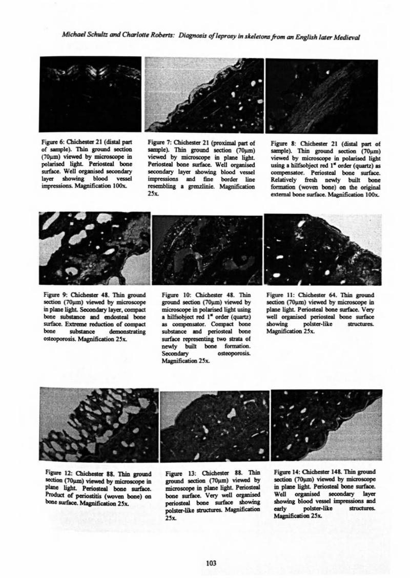
';po 6, Cl>kl>eol« 21 (diJlaI "'"of smlplc). Thin JfOUDd ICttioo(70J,un) viewed by mia'OtCOPC inpolariJed light. Periolleal booesurface. Well orp1ised IeOOOdarylayer Ibowing blood vesselimpressioos. Magnificatioo lOOx.
Fip"e 1: Olic:beste:r 21 (proximal pm of_I.~ lbiJl po.... ....... ('''''''')viewed by microscope in plane lightPerioSlel1 boDe surface. Well orpniJedICOOOdary Iayu showing blood vesselimpressions IDd fiDe border !.iDeresembling • 1faLll.in.ie. Mapi6atioD2>x.
Fipre I: 0J.icbesler 21 (diAIJ J*t ofsample). Thin pound JCJCtioa (101UU)viewed by miaosoope in polarised liBbtusing. bilfsobject red 1- order (quartz) aClOIDpCDSator. Periostea! boDe surface.Relalivcly fresh De'My buill booe(OfIDItioo (woven boae) on the originalextc:nW boDe surUce.. Mapific:aboo l00x.
........ 90 aua..- 4•• lbiJl poUDdICIttioa (70J.am) viewed by mia'OSal'pe...1aDe t;allt. ........,. ,-,"""'*'boac Jl.lt.l.Imoe IDd mdosteal boDesurtaee. Extreme reduc600 of compactbone ........... danonsIntinaosteoporosis. MipifiClOOIl. 2~ht.
Figure 10: C1lic::beslu 41. 'IbiDpoUDd ....... ('''''''') vi""'" bymiaosc:opc in polaiscd 1i&bI using• hilfJobjec:t reef I- order (quartz)u oompauIIOr. Compac:t booesubstance IDd periosteaJ bonesunace represmtma two strIl.I ofnewly buill boDe fonm:tioo.Seccodooy .............MagnifiCibOG 25x.
Ftpre 13: QUc::best« .1. 'IbiDpoUDd ....... (7"""') vi""'" bymic:roJe:OPe ill plaDe light Periostealbooe -. V", ...u ..........periostoII boDe IUI'&oe sbowina:poLstu-liko strue:t\JfC$, Mtgnificatioo2>x.
103
Figure 11: Clic:bester 64. Thin JI"O'IDdIOC:tioD (7~) viewed by miaoscope inp1aDe light. Periosteal bone surface. Verywell orpli..sed periosteal booe sur&ceshowing polscer·like ItNCnIreL
Mapific:abOD 2,x.
Fipre 14: Oaichester 14&. Thin IJ'O'Dd....... ('_) viewod by ............. pIaDc t;allt. ._booe -.
Well .......... ........,. 'sbowm, blood vesxl impreuiODl II1dearly pol.sl:a-.like structures..MapifiClll::iOD 2}x.
The Pas/ and Present ofLeprosy: Archaeologko/, his/arical, poJaeopalhoiogicaJ and clin;co/ approaches
Figure I~: Cbicbester 148. Thin groundsection (70JllD) viewed by microscope inpolarised light. Compact bone substaDce.Tangential lamellae suggesting inactivityatrophy. Magnification 100x.
Figure 16: Olicbester 202.Thin ground section(70"",) viewod bymiaoscope in plane light.Periosteal bone surface.Between • relatively thick,very weD orpnisedsccoodaty layer ad thecompact bone aubsLmce,there is • border line builtup by very small,tangentially orientlledblood vcsscl canals.Magnification 23x.
Figure 17: Libou male. Thin ground section('OJllD) viewed by microsoopc in polarisedlight. Compact bone substance. Tangentiallamellae suggest disuse atrophy.Magnificatioo 100x.
Figure 18: Lihou male. ScaDnin&electron microscopic image. Extcmalsurface of the new bone fonDlltion.Magnificstioo 12x.
Figure 19: Lihou male. Thin groUDdleCtioo (70f.UD) viewed by miaoscope Inplane light. PeriostcaJ. bone surface.(),gmUed poriosleal boo. _acepraalting polster·l.ike Ib'UctUres u weDas remains of. border tiDe rtse:mblin& •greDZliDie. Magnific.tim 23x.
Figure 20: Lihou male. Thin gromwlsection (~OJllD) viewed ~.
microlOOpC in pllDC light. Periostealbone llUl'face. The relatively &tsbDeW booe formMion (..-oval booe) is!he producI of • periostaImoammPtion. MagnifiCition 2~x.
Figure 21: Libou male. Thin JfOl.md MICtion (~p.m) villl'Wlld by mi~ illpobri>ed light...ma. hilf"'bjoct.-d ," onle< (quartz) II_. ConI-lik<ttructures in the interior area of1be 00DlpfICI booc 1UbItancc. MapifiCltioo 2~x.
104