Embed Size (px)
Citation preview
Antioxidant Systems in Poultry Biology: Superoxide DismutasePeter F. Surai1-5 *
1Department of Microbiology and Biochemistry, Faculty of Veterinary Medicine, Trakia University, Stara Zagora 6000, Bulgaria2Department of Animal Nutrition, Faculty of Agricultural and Environmental Sciences, Szent Istvan University, Gödöllo H-2103, Hungary3Department of Veterinary Expertise and Microbiology, Faculty of Veterinary Medicine, Sumy National Agrarian University, Sumy 40021,Ukraine4Odessa National Academy of Food Technology, Odessa 65039, Ukraine5Russian Academy of Science, Moscow, Russia*Corresponding author: Peter F. Surai, Department of Microbiology and Biochemistry, Faculty of Veterinary Medicine, Trakia University, StaraZagora 6000, Bulgaria, E-mail: [email protected]
Rec date: Oct 20, 2015, Acc date: Dec 24, 2015, Pub date: Dec 31, 2015
Copyright: © 2015 Peter F. Surai, This is an open-access article distributed under the terms of the Creative Commons Attribution License, whichpermits unrestricted use, distribution, and reproduction in any medium, provided the original author and source are credited.
Abstract
Commercial poultry production is associated with variousstresses responsible for decreasing productive andreproductive performance of growing chicks, breedersand commercial layers. A growing body of evidence showsthat most of stresses in poultry production at the cellularlevel are associated with oxidative stress. Recently, aconcept of the cellular antioxidant defense has beenrevised with special attention paid to cellular redox statusmaintenance and cell signaling. In fact, antioxidantsystems of the living cell are based on three major levelsof defense and superoxide dismutase (SOD) is shown tobelong to the first level of the antioxidant defensenetwork. Cellular antioxidant defenses are shown toinclude several options and vitagene activation in stressconditions is considered as a fundamental adaptivemechanism. The vitagene family includes various genesregulating synthesis of protective molecules includingthioredoxins, sirtuins, heat shock proteins and SOD.However, by the time of writing no comprehensive reviewon the roles and effects of SOD in poultry biology hasappeared. Therefore, the aim of this review is a criticalanalysis of the role of SOD in poultry biology with specificemphasis to its functions as an essential part of thevitagene network. From the analysis of the recent datarelated to SOD in poultry physiology and adaptation tostresses it is possible to conclude that: a) SOD asimportant vitagene is the main driving force in cell/bodyadaptation to various stress conditions. Indeed, in stressconditions additional synthesis of SOD is an adaptivemechanism to decrease ROS formation; b) If the stress istoo high SOD activity is decreased and apoptosis isactivated; c) there are tissue-specific differences in SODexpression which also depends on the strength of suchstress-factors as heat, heavy metals, mycotoxins and otherstressors; d) SOD is shown to provide an effectiveprotection against lipid peroxidation in chicken embryonictissues and semen; e) SOD is shown to be protective in
heat and cold stress, toxicity stress as well as in otheroxidative-stress related conditions in poultry production;f) there are complex interactions inside the antioxidantnetwork of the cell/body to ensure an effectivemaintenance of homeostasis in stress conditions. Indeed,in many cases, nutritional antioxidants (vitamin E,selenium, carotenoids, phytochemicals, etc.) in the feedcan increase SOD expression; g) nutritional means of SODupregulation in stress conditions of poultry productionand physiological and commercial consequences awaitfurther investigation; h) vitagene upregulation in stressconditions is emerging as an effective means for stressmanagement.
Key words:Antioxidant system; chicken; HSP; poultry; stress; vitagenes
Abbreviations:AO- antioxidant; ARE- antioxidant response element; CAT –
catalase; NOS- nitric oxide synthase; GSH – glutathione; GSH-Px- glutathione peroxidase; GST- glutathione transferase; HSP –heat shock protein; MDA- malondialdehyde; NF-κB- nuclearfactor-kappa B; Nrf2- Nuclear factor-erythroid-2-related factor2; SOD – superoxide dismutase.
IntroductionCommercial poultry production is associated with various
stresses responsible for decreasing productive andreproductive performance of growing chicks, breeders andcommercial layers. A growing body of evidence shows thatmost of stresses in poultry production at the cellular level areassociated with oxidative stress. Recently, a concept of thecellular antioxidant defense has been revised with specialattention paid to cellular redox status maintenance and cellsignalling. It has been suggested that the antioxidant defense
Review Article
iMedPub Journalshttp://www.imedpub.com/ Vol.1 No.1:8
2016
© Copyright iMedPub | This article is available from: http://animalnutrition.imedpub.com/ 1
Journal of Animal Research and Nutrition
ISSN 2572-5459
DOI: 10.21767/2572-5459.100008
network of the living cell is based on three major levels ofdefense and include several options [1-2]: Decreasing localizedoxygen concentration; reducing activity of pro-oxidantenzymes and improving efficiency of electron chain in themitochondria and decreasing electron leakage leading tosuperoxide production; preventing chain initiation byscavenging initial radicals due to inducing various transcriptionfactors (e.g., Nrf2, NF-κB and others) with ARE-relatedsynthesis of AO enzymes including superoxide dismutase(SOD), glutathione peroxidase (GSH-Px), catalase (CAT),glutathione reductase (GR), glutathione transferase (GST), etc.;binding metal ions (metal-binding proteins) and metalchelating; decomposing peroxides by converting them to non-radical, nontoxic products (Se-GSH-Px); chain breaking byscavenging intermediate radicals such as peroxyl and alkoxylradicals (vitamins E, C, reduced glutathione (GSH), uric acid,ubiquinol, bilirubin, etc.); repairing and removing damagedmolecules (methionine sulfoxide reductase, DNA-repairenzymes, chaperons, etc.) and vitagene activation andsynthesis and increased expression of protective molecules(GSH, thioredoxins, SOD, heat shock proteins, sirtuins, etc.).Indeed, elucidating roles of vitagenes in stress resistance ofpoultry as a background for the development of effectivestrategies to deal with stresses is an emerging topic ofresearch [1-5]. It is known that adaptive SOD synthesis is undervitagene control. However, by the time of writing nocomprehensive review on the roles and effects of SOD inpoultry biology has appeared. Therefore, the aim of thisreview is a critical analysis of the role of SOD in poultry biologywith specific emphasis to its functions as an essential part ofthe vitagene network, responsible for adaptive ability of thecells and whole organism to various stress conditions.
Free radicals and reactive oxygen andnitrogen species
Free radicals are atoms or molecules containing one or moreunpaired electrons. Free radicals are highly unstable andreactive and are capable of damaging all types of biologicallyrelevant molecules including DNA, proteins, lipids andcarbohydrates. The animal body is under constant attack fromfree radicals, formed as a natural consequence of the body’snormal metabolic activity and as part of the immune system’sstrategy for destroying invading microorganisms. Collectiveterms reactive oxygen species (ROS) and reactive nitrogenspecies (RNS) have been introduced [6] and they include notonly the oxygen or nitrogen radicals, but also some non-radicalreactive derivatives of oxygen and nitrogen.
Superoxide (O2*-) is the main free radical produced inbiological systems during normal respiration in mitochondriaand by autoxidation reactions with half-life at 37°C in therange of 1 x 10⁻⁶ second. Superoxide can inactivate someenzymes due to formation of unstable complexes withtransition metals of enzyme prosthetic groups, followed byoxidative self-destruction of the active site [7]. Depending oncondition, superoxide can act as an oxidizing or a reducingagent. It is necessary to mention that superoxide, by itself, isnot extremely dangerous and does not rapidly cross lipid
membrane bilayer [8]. However, superoxide is a precursor ofother, more powerful ROS. For example, it reacts with nitricoxide with a formation of peroxynitrite (ONOO-), a strongoxidant, which leads to formation of reactive intermediatesdue to spontaneous decomposition [9-10]. In fact, ONOO- wasshown to damage a wide variety of biomolecules, includingproteins (via nitration of tyrosine or tryptophan residues oroxidation of methionine or selenocysteine residues), DNA andlipids [11]. Superoxide can also participate in the production ofmore powerful radicals by donating an electron, and therebyreducing Cu2+ and Fe3+ to Fe2+ and Cu+, as follows:
O2- + Fe3+/Cu2+ → Fe2+/Cu+ + O2
Further reactions of Fe2+ and Cu+ with H2O2 are a source ofthe hydroxyl radical (*OH) in the Fenton reaction:
H2O2 + Fe2+/Cu+ → *OH + OH- + Fe3+/Cu2+
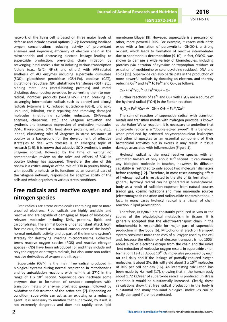
The sum of reaction of superoxide radical with transitionmetals and transition metals with hydrogen peroxide is knownas the Haber-Weiss reaction. It is necessary to underline thatsuperoxide radical is a “double-edged sword”. It is beneficialwhen produced by activated polymorphonuclear leukocytesand other phagocytes as an essential component of theirbactericidal activities but in excess it may result in tissuedamage associated with inflammation (Figure 1).
Hydroxyl radical is the most reactive species with anestimated half-life of only about 10-9 second. It can damageany biological molecule it touches, however, its diffusioncapability is restricted to only about two molecular diametersbefore reacting [12]. Therefore, in most cases damaging effectof hydroxyl radical is restricted to the site of its formation. Ingeneral, hydroxyl radical can be generated in human/animalbody as a result of radiation exposure from natural sources(radon gas, cosmic radiation) and from man-made sources(electromagnetic radiation and radionuclide contamination). Infact, in many cases hydroxyl radical is a trigger of chainreaction in lipid peroxidation.
Therefore, ROS/RNS are constantly produced in vivo in thecourse of the physiological metabolism in tissues. It isgenerally accepted that the electron-transport chain in themitochondria is responsible for major part of superoxideproduction in the body [6]. Mitochondrial electron transportsystem consumes more than 85% of all oxygen used by the celland, because the efficiency of electron transport is not 100%,about 1-3% of electrons escape from the chain and the univalent reduction of molecular oxygen results in superoxide anionformation [13-15]. About 1012 O2 molecules processed by eachrat cell daily and if the leakage of partially reduced oxygenmolecules is about 2%, this will yield about 2 x 10¹⁰ moleculesof ROS per cell per day [16]. An interesting calculation hasbeen made by Halliwell [17], showing that in the human bodyabout 1.72 kg/year of superoxide radical is produced. In stresscondition it would be substantially increased. Clearly, thesecalculations show that free radical production in the body issubstantial and many thousand biological molecules can beeasily damaged if are not protected.
Vol.1 No.1:8
2016
2 This article is available from:http://animalnutrition.imedpub.com/
Journal of Animal Research and Nutrition
ISSN 2572-5459
Figure 1: Protective roles of SOD in animal/poultryphysiology.Superoxide (O2
*) is the main free radical produced inbiological systems during normal respiration inmitochondria. In addition to mitochondria, superoxide canbe generated by other redox-active enzymes, includingxanthine oxidase, cytochrome p450, cyclooxygenase,lipoxygenase, nitric oxide synthase (NOS) and NADPHoxidases (NOXs). Superoxide, by itself, is not extremelydangerous being a signaling molecule. However, superoxideis a precursor of other, more powerful ROS, includingperoxynitrite and hydroxyl radical, which can damage alltypes of biological molecules, including proteins, lipids andnucleic acids. This results in immunosuppression, decreasedproductive and reproductive performance and developmentof various diseases. Therefore, three main forms of SOD areresponsible for conversion of superoxide into hydrogenperoxide which is further detoxified by GSH-Px and/orCatalase with water formation. This prevents damagingeffects of superoxide radical.
Three levels of antioxidant defenseDuring evolution living organisms have developed specific
antioxidant protective mechanisms to deal with ROS and RNS[6]. Therefore it is only the presence of natural antioxidants inliving organisms which enable them to survive in an oxygen-rich environment [13]. These mechanisms are described by thegeneral term “antioxidant system”. It is diverse and responsiblefor the protection of cells from the actions of free radicals. Thissystem includes [18-20]:
natural fat-soluble antioxidants (vitamins A, E, carotenoids,ubiquinones, etc.);
water-soluble antioxidants (ascorbic acid, uric acid, taurine,carnitine, etc.);
antioxidant enzymes: GSH-Px, CAT and SOD;
thiol redox system consisting of the glutathione system(glutathione/glutathione reductase/glutaredoxin/glutathioneperoxidase and a thioredoxin system (thioredoxin/thioredoxinperoxidase/thioredoxin reductase).
The protective antioxidant compounds are located inorganelles, subcellular compartments or the extracellular
space enabling maximum cellular protection to occur. Thusantioxidant systems of the living cell include three major levelsof defense [18-21].
The first level of defense is responsible for prevention offree radical formation by removing precursors of free radicalsor by inactivating catalysts and consists of three antioxidantenzymes namely SOD, GSH-Px and CAT plus metal-bindingproteins. Since the superoxide radical is the main free radicalproduced in physiological conditions in the cell [13] SOD (EC1.15.1.1) is considered to be the main element of the first levelof antioxidant defense in the cell [18]. This enzyme dismutatesthe superoxide radical in the following reaction:
2O2* +2H+ SOD → H2O2+O2
The hydrogen peroxide formed by SOD action can bedetoxified by GSH-Px or CAT which reduce it to water asfollows:
H2O2 + 2GSH+GSH-Px → GSSG+2H2O
Catalase
2H2O2 → 2H2O + O2
Transition metal ions also accelerate the decomposition oflipid hydroperoxides into cytotoxic products such as aldehydes,alkoxyl radicals and peroxyl radicals. Therefore, metal-bindingproteins (transferrin, lactoferrin, haptoglobin, hemopexin,metallothionenin, ceruloplasmin, ferritin, albumin, myoglobin,etc.) also belong to the first level of defense. It seems likelythat carnitine with its regulating functions on themitochondria free radical production [1] can also be part ofthe first level of antioxidant defense.
Unfortunately, this first level of antioxidant defense in thecell is not sufficient to completely prevent free radicalformation and some radicals do escape through the preventivefirst level of antioxidant safety screen initiating lipidperoxidation and causing damage to DNA and proteins.Therefore, the second level of defense consists of chain-breaking antioxidants - vitamin E, ubiquinol, carotenoids,vitamin A, ascorbic acid, uric acid and some otherantioxidants. Glutathione and thioredoxin systems also have asubstantial role in the second level of antioxidant defense.Chain-breaking antioxidants inhibit peroxidation by keepingthe chain length of the propagation reaction as small aspossible. Therefore, they prevent the propagation step of lipidperoxidation by scavenging peroxyl radical intermediates inthe chain reaction:
LOO* + Toc → Toc* + LOOH
(LOO* is lipid peroxyl radical; Toc - tocopherol, Toc* -tocopheroxyl radical, LOOH – lipid hydroperoxide).
However, even the second level of antioxidant defense inthe cell is not able to prevent damaging effects of ROS and RNSon lipids, proteins and DNA. In this case, the third level ofdefense is based on systems that eliminate damagedmolecules or repair them. This level of antioxidant defenseincludes lipolytic (lipases), proteolytic (peptidases orproteases) and other enzymes (DNA repair enzymes,
Vol.1 No.1:8
2016
© Copyright iMedPub 3
Journal of Animal Research and Nutrition
ISSN 2572-5459
methionine sulphoxide reductase, ligases, nucleases,polymerases, proteinases, phospholipases and varioustransferases) and chaperones, including HSPs.
Antioxidant-prooxidant balance in thebody and oxidative stress
In the body/cell a delicate critical balance exists betweenantioxidant defense and repair systems and free radicalgeneration [18-20]. In physiological conditions the right andleft parts of the so-called “balances” are in equilibrium i.e. freeradical generation is neutralised by the antioxidant system andsome free radicals and products of their metaboilismparticipate in cell signaling and transcription factor activation.Exogenous factors are among the most important elements,which increase an efficiency of the antioxidant system of theorganism. Natural and synthetic antioxidants in the feed aswell as optimal levels of Mn, Cu, Zn and Se help maintainingthe efficient levels of endogenous antioxidants in the tissues.Optimal diet composition allows the antioxidants of the foodto be efficiently absorbed and metabolised. Optimaltemperature, humidity and other environmental conditionsare also required for the effective protection against freeradical production. The prevention of different diseases byantibiotics and other drugs is an integral part of the efficientantioxidant defense as well.
Different stress conditions are associated withoverproduction of free radicals and cause oxidative stress i.e. adisturbance in the prooxidant-antioxidant balance leading topotential tissue damage [22]. Stress conditions can begenerally divided into three main categories [19]. The mostimportant part is nutritional stress conditions including highdietary levels of PUFAs, deficiencies of vitamin E, Se, Zn or Mn,Fe-overload, hypervitaminosis A and presence of differentmycotoxins and other toxic compounds in the feed. A secondgroup of stress factors includes environmental conditions:increased temperature or humidity, hyperoxia, radiation etc.Internal stress factors include various bacterial or viral diseasesas well as allergy. All the above-mentioned conditionsstimulate free radical generation in the mitochondria.
Living cells permanently balance the process of formationand inactivation of ROS and as a result ROS level remains lowbut still above zero. Adverse environmental conditions initiateattempts of organisms to resist the environment that becamemore aggressive [23]. Cells can usually tolerate mild oxidativestress by additional synthesis of various antioxidants(glutathione, antioxidant enzymes, etc.) in an attempt torestore antioxidant/oxidant balance. At the same time, energyexpenditures increased and respiration activated leading tothe increased yield of ROS [23]. However these adaptivemechanisms have limited ability. Once the free radicalproduction exceeds the ability of antioxidant system toneutralise them, oxidative stress develops and causes damageto unsaturated lipids in cell membranes, amino acids inproteins and nucleotides in DNA and as a result, membraneand cell integrity is disrupted. Membrane damage is associatedwith a decreased efficiency of absorption of different nutrients
and leads to an imbalance of vitamins, amino acids, inorganicelements and other nutrients in the organism. All these eventsresult in decreased productive and reproductive performancesof animals. Immunity incompetence and unfavourable changesin the cardio-vascular system, brain and neurones and musclesystem due to increased lipid peroxidation make the situationeven worse [20].
As it was shown above all antioxidants in the body areworking as a “team” responsible for antioxidant defense andwe call this team the antioxidant system. In this team onemember helps another one working efficiently. Therefore ifrelationships in this team are effective, which happens only inthe case of balanced diet and sufficient provision of dietaryantioxidant nutrients, then even low doses of suchantioxidants as vitamin E could be effective. On the other handwhen this team is subjected to high stress conditions, freeradical production is increased dramatically. During thesetimes, without external help it is difficult to prevent damage tomajor organs and systems. This ‘external help’ is dietarysupplementation with increased concentrations of naturalantioxidants. For nutritionist or feed formulator it is a greatchallenge to understand when the internal antioxidant team inthe body requires help, how much of this help to provide andwhat the economic return will be. Again, it is necessary toremember about essentiality of keeping right balance betweenfree radical production and antioxidant defense. Indeed, ROSand RNS have another more attractive face participating in aregulation of varieties of physiological functions.
Therefore, the antioxidant defense mechanisms includeseveral options [1-3б 19-20]:
Decreasing localized oxygen concentration;
Reducing activity of pro-oxidant enzymes and improveefficiency of electron chain in the mitochondria decreasingsuperoxide production (carnitine);
Redox-signaling with induction of various transcriptionfactors (e.g. Nrf2 and NF-kB) and gene expression with ARE-related synthesis of AO enzymes (SOD, GSH-Px, catalase, GR,GST, etc.) and other important protective molecules;
Vita-gene activation and synthesis and increased expressionof protective molecules (HSP, thioredoxins, sirtuins, SOD, etc.);
Binding metal ions (metal-binding proteins) and metalchelating;
Decomposing peroxides by converting them to non-radical,nontoxic products (Se-GSH-Px);
Chain breaking by scavenging intermediate radicals such asperoxyl and alkoxyl radicals (vitamins E, C, GSH, uric acid,carnitine, ubiquinol, bilirubin, etc.);
Antioxidant (vitamin E) recycling mechanisms (vitaminsB1,B2, Se, ascorbic acid);
Repairing and removing damaged molecules (Msr, DNA-repair enzymes, proteasomes, HSP and other chaperons, etc.);
Apoptosis activation and removal terminally damaged cellsand restriction of mutagenesis.
Vol.1 No.1:8
2016
4 This article is available from:http://animalnutrition.imedpub.com/
Journal of Animal Research and Nutrition
ISSN 2572-5459
It is important to mention once more that ROS are no longerviewed as just toxic by-products of mitochondrial respiration,but are now appreciated for their role in regulating variouscellular signaling pathways [24]. Indeed, the adaptation tostressful conditions of our life is mediated via vitagenenetwork in the body.
Superoxide Dismutase in biologicalsystems
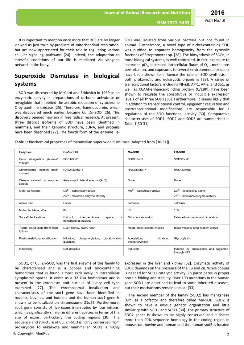
SOD was discovered by McCord and Fridovich in 1969 as anenzymatic activity in preparations of carbonic anhydrase ormyoglobin that inhibited the aerobic reduction of cytochromeC by xanthine oxidase [25]. Therefore, haemocuprein, whichwas discovered much earlier, became Cu, Zn-SOD [26]. Thisdiscovery opened new era in free radical research. At present,three distinct isoforms of SOD have been identified inmammals, and their genomic structure, cDNA, and proteinshave been described [27]. The fourth form of the enzyme Fe-
SOD was isolated from various bacteria but not found inanimal. Furthermore, a novel type of nickel-containing SODwas purified to apparent homogeneity from the cytosolicfractions of Streptomyces sp. [28]. The biosynthesis of SODs, inmost biological systems, is well controlled. In fact, exposure toincreased pO2, increased intracellular fluxes of O2-, metal ionsperturbation, and exposures to several environmental oxidantshave been shown to influence the rate of SOD synthesis inboth prokaryotic and eukaryotic organisms [29]. A range oftranscriptional factors, including NF-κB, AP-1, AP-2, and Sp1, aswell as CCAAT-enhancer-binding protein (C/EBP), have beenshown to regulate the constitutive or inducible expressionlevels of all three SODs [30]. Furthermore, it seems likely thatin addition to transcriptional control, epigenetic regulation andposttranscriptional modifications are responsible for aregulation of the SOD functional activity [30]. Comparativecharacteristics of SOD1, SOD2 and SOD3 are summarised inTable 1[30-31].
Table 1: Biochemical properties of mammalian superoxide dismutase (Adapted from [30-31]).
Enzymes CuZn-SOD Mn-SOD EC-SOD
Gene designation (human/mouse)
SOD1/Sod1 SOD2/Sod2 SOD3/Sod3
Chromosome location man/mouse)
HAS21/MMU16 HAS6/MMU17 HAS4/MMU5
Disease caused by enzymedefects
Amyotrophic lateral sclerosis(ALS) None None
Metal co-factor(s) Cu2+ - catalytically active
Zn2+ - maintains enzyme stability
Mn2+ - catalytically active Cu2+ - catalytically active
Zn2+ - maintains enzyme stability
Active form Dimer Tetramer Tetramer
Molecular Mass, kDa 88 32 135
Subcellular locations Cytosol, intermembrane space ofmitochondria, nucleus
Mitochondria matrix Extracellular matrix and circulation
Tissue distribution (from highto low)
Liver, kidney, brain, heart Heart, brain, skeletal muscle Blood vessels, lung, kidney, uterus
Post-translational modification Nitration, phosphorylation, glutathiolation,glycation
Acetylation, nitration,phosphorylation
Glycosylation
Inducibility Not inducible Inducible Induced by antioxidants and regulatedthrough NRF
SOD1, or Cu, Zn-SOD, was the first enzyme of this family tobe characterised and is a copper and zinc-containinghomodimer that is found almost exclusively in intracellularcytoplasmic spaces. It exists as a 32 kDa homodimer and ispresent in the cytoplasm and nucleus of every cell typeexamined [27]. The chromosomal localization andcharacteristics of the sod1 gene have been identified inrodents, bovines, and humans and the human sod1 gene isshown to be localized on chromosome 21q22. Furthermore,sod1 gene consists of five exons interrupted by four introns,which is significantly similar in different species in terms of thesize of exons, particularly the coding regions [30]. Thesequence and structure of Cu, Zn-SOD is highly conserved fromprokaryotes to eukaryote and mammalian SOD1 is highly
expressed in the liver and kidney [32]. Enzymatic activity ofSOD1 depends on the presence of the Cu and Zn. While copperis needed for SOD1 catalytic activity, Zn participates in properprotein folding and stability. Over 100 mutations in the humangene SOD1 are described to lead to some inherited diseases,but their mechanisms remain unclear [33].
The second member of the family (SOD2) has manganese(Mn) as a cofactor and therefore called Mn-SOD. SOD2 isshown to have a unique genetic organization and littlesimilarity with SOD1 and SOD3 [30]. The primary structure ofSOD2 genes is shown to be highly conserved and it sharesmore than 90% sequence homology in the coding region inmouse, rat, bovine and human and the human sod2 is located
Vol.1 No.1:8
2016
© Copyright iMedPub 5
Journal of Animal Research and Nutrition
ISSN 2572-5459
on chromosome 6q25.3 [30]. It was shown to be a 96 kDahomotetramer and located exclusively in the mitochondrialmatrix, a prime site of superoxide radical production [6].Therefore the expression of Mn-SOD is considered to beessential for the survival of all aerobic organisms from bacteriato humans and it participates in the development of cellularresistance to oxygen radical-mediated toxicity [34]. Indeed,Mn-SOD is shown to play a critical role in the defense againstoxidant-induced injury and apoptosis in various cells. In fact,Mn-SOD is inducible enzyme and its activity is affected bycytokines and oxidative stress. Therefore, Mn-SOD has beenshown to play a major role in promoting cellular differentiationand in protecting against hyperoxia-induced pulmonarytoxicity [34] being a crucial determinant of redox status of thecell. Furthermore, Mn-SOD influences the activity oftranscription factors (such as HIF-1α, AP-1, NF-κB and p53) andaffects DNA stability [35]. A critical role of Mn-SOD underphysiological and pathological conditions has recently beenreviewed in details and the following findings of Mn-SODconfirm the critical role of Mn-SOD in the survival of aerobiclife [36-39]:
Escherichia coli and yeasts lacking the Mn-SOD gene arehighly sensitive to oxidative stress;
Mn-SOD gene knockout mice can only survive few days afterbirth, with pathological findings of many various diseases dueto mitochondrial disorder, suggesting a critical role of theenzyme;
Cells transfected with Mn-SOD cDNAs have increasedresistance to various free radical-generating toxicants(paraquat, tumor necrosis factor, doxorubicin, mitomycin C,irradiation, ischemic reperfusion, smoking, etc.);
Human Mn-SOD gene transgenic mice show reducedseverity of free radical-induced pulmonary damage andadriamycin-induced myocardial damage.
In 1982, a third SOD isozyme was discovered by Marklundand co-workers and called extracellular superoxide dismutase(EC-SOD), due to its exclusive extracellular location. EC-SOD isa glycoprotein with a molecular weight of 135,000 kDa andhigh affinity for heparin [40]. However, there are some species-specific variations in molecular weight. The human EC-SODgene has been mapped to chromosome 4q21 and consists ofthree exons and two introns [41]. The full-length mouse EC-SOD cDNA is shown to be 82% identical to that of rat and 60%identical to the human EC-SOD [30]. EC-SOD is the onlyantioxidant enzyme that scavenges superoxide specifically inthe extracellular space. EC-SOD is present in various organismsas a tetramer or, less commonly, as a dimer and contains onecopper and one zinc atom per subunit, which are required forenzymatic activity [42]. The expression pattern of EC-SOD ishighly restricted to the specific cell type and tissues where itsactivity can exceed that of Cu,Zn-SOD or Mn- SOD. As acopper-containing enzyme, the activity of EC-SOD is regulatedby copper availability [41]. EC-SOD is comparatively resistantto high temperatures, extreme pH, and high ureaconcentrations; it can be inhibited by various agents includingazide and cyanide and inactivated by diethyldithiocarbamate
and hydrogen peroxide. Oxidative stress and post-translationalmodification of EC-SOD are shown to cause loss of EC-SODactivity [30].
SOD in avian biology
Chicken SODChicken SOD was described and purified in early 1970.
Indeed, in chicken liver two types of SOD were identified, oneof which was localized in the mitochondria while the other wasfound in the cytosol [43]. The cytosol SOD was inhibited bycyanide, whereas the mitochondrial enzyme was not. Laterthis feature was used to distinguish between two forms ofenzymes during assays. The cytosol SOD was purified tohomogeneity with apparent molecular weight in presence ofmercaptoethanol to be 30,600 Da and to contain copper andzinc, being similar to the other Cu, Zn-SOD which have beenisolated from diverse eukaryotes. In fact, purified cytosol SODfrom chicken liver contained 0.30% copper and 0.25% zinc.This corresponds to 0.9 atom of copper and 0.8 atom of zincper subunit. It was also shown that this chicken liver Cu, Zn-SOD had a tendency to polymerize [43]. In contrast, themitochondrial SOD was found in chicken liver to be amanganoprotein which has a molecular weight of 80,000 Da. Itis composed of four subunits of equal size, which are notcovalently joined. It contains 2.3 atoms of manganese permolecule and is strikingly similar to the SOD previouslyisolated from bacteria. This supports the theory thatmitochondria have evolved from aerobic prokaryotes. In fact,Mn-SOD was first isolated from the chicken liver [43]. The Mn-SOD was found primarily in the mitochondrial matrix spacewhereas the Cu,Zn-SOD, previously isolated from the cytosol,was found in the intermembrane space [44].
Cu, Zn-SOD was purified from chicken liver with a subunitMr of 16900 [45]. Low dietary copper was associated with adecrease in SOD activity and when the 10-day-old deficientchicks were injected with 0.5 mol of CuSO4 intraperitoneally,SOD activity in aorta was restored to control levels in about 8h. Indeed, dietary copper regulates SOD activity in the tissuesof young developing animals. The authors also suggested thata copper deficiency suppresses Cu, Zn-SOD activity withoutinhibiting synthesis or accumulation of the Cu, Zn-SOD proteinin this tissue [45]. Interestingly, molecular properties (aminoacid composition, molecular mass and subunit composition) ofthe chicken enzyme was shown to be similar to those of abovine erythrocyte Cu, Zn SOD [46]. Purified chicken liver Cu,Zn-SOD was confirmed to contain two subunits having Cu andZn elements with a molecular weight of 16000+/-500 for eachsubunit [47]. The optimum pH of purified Cu, Zn-SOD wasdetermined to be 8.9. The enzyme was found to have fairthermal stability up to 45oC at pH 7.4 over a 1-h incubationperiod. The SOD enzyme was not inhibited by DTT and beta-mercaptoethanol, but inhibited by CN(-) and H2O2 [47]. SODpurified from chicken heart has a molecular weight 31.0 +/- 1.0kDa and is composed of two equally sized subunits each having1.1 +/- 0.03 and 0.97 +/- 0.02 atoms of Cu and Zn elements,respectively [48]. The Mn-SOD cDNA in chicken heart was
Vol.1 No.1:8
2016
6 This article is available from:http://animalnutrition.imedpub.com/
Journal of Animal Research and Nutrition
ISSN 2572-5459
shown to be 1108 bp in length. The molecular weight of themature peptide was 22 kDa. A comparison of the deducedamino acid sequence with those of the human, rat, C. elegansand D. melanogaster showed that the amino acid homologyrates were 82.4%, 84.7%, 62.4%, and 59.3%, respectively [49].SOD activity in avian tissues depends on many different factorsincluding genetics, nutrition and various stress-related factors.For example, SOD activity in the Jungle Fowl feathermelanocytes was shown to be 2- and 4-fold higher than that inBarred Plymouth Rock and White Leghorn tissue respectively[50]. Indeed, understanding the molecular mechanisms of theregulation of SOD gene expression and the factors involved intissue- and cell-specific expression of the SOD genes are ofgreat importance for a developing novel strategies forpreventing negative consequences of various stresses inpoultry production.
SOD in chicken embryoChick embryo tissues contain a high proportion of highly
polyunsaturated fatty acids in the lipid fraction [51] andtherefore need antioxidant defense [18]. The antioxidantsystem of the newly hatched chick includes the antioxidantenzymes SOD, GSH-Px, catalase [52], fat-soluble antioxidantsvitamin E and carotenoids [53], water-soluble antioxidantsascorbic acid [53] and glutathione [52] as well as selenium[54-57]. Vitamin E [58], carotenoids [59-64] and selenium[54-57] are transferred from feed into egg and further toembryonic tissues. Glutathione and antioxidant enzymes GSH-Px, SOD and catalase are also expressed in the embryonictissues at various stages of their development [52, 65]. Ourresults indicate that there are tissue-specific features inantioxidant defense strategy during embryonic developmentof the chicken and SOD plays a crucial role as an integral partof the antioxidant network.
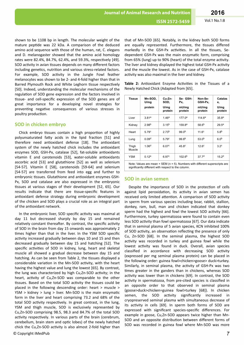
In the embryonic liver, SOD specific activity was maximal atday 11 but decreased sharply by day 15 and remainedrelatively constant thereafter. By contrast, the specific activityof SOD in the brain from day 15 onwards was approximately 2times higher than that in the liver. In the YSM SOD specificactivity increased gradually between days 10 and 15 and thendecreased gradually between day 15 and hatching [52]. Thespecific activities of SOD in kidney, lung, heart and skeletalmuscle all showed a gradual decrease between day 15 andhatching. As can be seen from Table 2, the tissues displayed aconsiderable variation in the Mn-SOD activity, with the hearthaving the highest value and lung the lowest [65]. By contrast,the lung was characterized by high Cu,Zn-SOD activity; in theheart, activity of Cu,Zn-SOD was comparable to the othertissues. Based on the total SOD activity the tissues could beplaced in the following descending order: heart > muscle >YSM > kidney > lung > liver. Mn-SOD is the main enzymaticform in the liver and heart comprising 73.2 and 68% of thetotal SOD activity respectively. In great contrast, in the lung,YSM and thigh muscle, SOD is exclusively represented byCu,Zn-SOD comprising 98.5, 98.3 and 84.7% of the total SODactivity respectively. In various parts of the brain (cerebrum,cerebellum, brain stem and optic lobes) of the newly hatchedchick the Cu,Zn-SOD activity is also almost 2-fold higher than
that of Mn-SOD [65]. Notably, in the kidney both SOD formsare equally represented. Furthermore, the tissues differedmarkedly in the GSH-Px activities. In all the tissues, Se-dependent GSH-Px was the main enzymatic form, comprisingfrom 65% (lung) up to 90% (heart) of the total enzyme activity.The liver and kidney displayed the highest total GSH-Px activityand the muscle the lowest. As in the case of GSH-Px, catalaseactivity was also maximal in the liver and kidney.
Table 2: Antioxidant Enzyme Activities in the Tissues of aNewly Hatched Chick (Adapted from [65].
Tissue Mn-SOD,U/mg
protein
Cu-Zn-SOD,
U/mgprotein
Se- GSH-Px,
mU/mgprotein
Non-Se-GSH-Px,
mU/mgprotein
Catalase,
U/mgprotein
Liver 3.81a 1.46a 177.0a 114.6a 35.8a
Kidney 2.98b 3.15b 159.8a 58.6b 29.5a
Heart 5.79c 2.73b 99.0b 11.6c 5.8b
Lung 0.09d 5.79c 99.8b 53.0b 6.0b
ThighMuscle
1.06d 6.07c 45.8c 12.6c 3.2c
YSM 0.12e 6.97c 102.6b 37.7d 15.2d
Note: Values are mean + SEM (n = 5). Numbers with different superscripts aresignificantly different with respect to the column.
SOD in avian semenDespite the importance of SOD in the protection of cells
against lipid peroxidation, its activity in avian semen hasreceived only limited attention. A comparison of SOD activityin sperm from various species including boar, rabbit, stallion,donkey, ram, bull, man and chicken indicated that donkeysperm had the highest and fowl the lowest SOD activity [66].Furthermore, turkey spermatozoa were found to contain evenless SOD activity than fowl spermatozoa [67]. Our data indicatethat in seminal plasma of 5 avian species, KCN inhibited 100%of SOD activity, an observation reflecting the presence of onlyCu, Zn-SOD [68]. In the seminal plasma, the highest SODactivity was recorded in turkey and guinea fowl while thelowest activity was found in duck. Overall, avian speciesclassified in accordance with decreasing SOD activity(expressed per mg seminal plasma protein) can be placed inthe following order: guinea fowl>chicken>goose> duck>turkey.Similarly, in seminal plasma, the activity of GSH-Px was twotimes greater in the ganders than in chickens, whereas SODactivity was lower than in chickens [69]. In contrast, the SODactivity in spermatozoa, from pre-cited species is classified inan opposite order to that observed in seminal plasma(goose>duck>chicken=guinea fowl>turkey [68]). In chickensemen, the SOD activity significantly increased incryopreserved seminal plasma with simultaneous decrease ofits activity in cells [69]. In sperm both forms of SOD areexpressed with significant species-specific differences. Forexample in goose, Cu,Zn-SOD appears twice higher than Mn-SOD and an opposite distribution between different forms ofSOD was recorded in guinea fowl where Mn-SOD was more
Vol.1 No.1:8
2016
© Copyright iMedPub 7
Journal of Animal Research and Nutrition
ISSN 2572-5459
than two-fold higher compared to Cu,Zn-SOD [68]. In chicken,about 67% of total SOD activity was detected in spermatozoaas compared to 33% in seminal plasma [70]. The biologicalmeaning and physiological consequences of such species-specific differences in SOD activity and distribution remain tobe established. Notably, in laying hens, SOD activity in theutero-vaginal junction was shown to be increased compared toother regions of the lower oviduct (vagina, uterus; [71-72]).
Dietary modulation of SOD
Mn and Cu in the dietMn-SOD is shown to be highly expressed in various organs
containing a large number of mitochondria such as the heart,liver, and kidneys. Indeed, in comparison to other tissues, theheart has the highest steady state mRNA Mn-SOD expressionlevel in chickens [73]. It has been proven that Mn availability isa regulating factor of Mn-SOD activity. For example, in primarycultured broiler myocardial cells Mn-SOD mRNA, Mn-SODprotein, and Mn-SOD activity were induced by manganese indose- and time-dependent manner. Manganese regulates Mn-SOD expression not only at transcriptional level but also attranslational and/or posttranslational levels [74]. In both heartand kidney, Mn-SOD activity was significantly depressed bydecreased dietary manganese; greatest reduction occurred inthe heart [75]. Decreased heart Mn-SOD and Cu,Zn-SODactivities, resulting from dietary Mn and Cu deficiencies, wereboth associated with increased peroxidation [76]. It seemslikely that Mn-SOD activity is very sensitive to dietary Mnlevels in commercial corn-soybean meal diets. In fact, Mndeficiency in growing chickens caused the reductions of Mnconcentrations of the liver and heart as well as Mn-SODactivity of the heart [77]. In chickens, dietary Mn contentsrequired to reach the plateau of Mn concentrations of theliver, pancreas, kidney, heart, spleen and muscle and to obtainthe maximum Mn-SOD activity of heart were calculated to be110, 111, 141, 123, 109, 99 and 121ppm respectively.Interestingly, Mn-SOD of liver and pancreas were not affected.Therefore, for broilers fed the basal corn-soybean meal diet,120ppm Mn was suggested as the required level [78] whichcorresponds to the presently recommended levels of Mnsupplementation. Chickens fed a Mn-deficient diet fromhatching had significantly lower levels of Mn-SOD activity inliver than did controls. However, activity of the Cu,Zn-SOD inthe liver was higher in Mn-deficient chickens than in controls[79]. The activity of both forms of SOD reached normal levelswhen a Mn-supplemented (1,000 ppm) diet was fed todeficient chickens, but the activity of the manganese enzymewas not affected by feeding the supplemented diet tomanganese sufficient chickens. It was shown that heart Mn-SOD activity and heart Mn-SOD mRNA levels increased linearlyas dietary Mn levels increased, confirming that dietary Mnsignificantly affected heart Mn-SOD gene transcription [80].Furthermore, birds fed supplemental Mn had lower MDAcontent in leg muscle and greater Mn-SOD activities and Mn-SOD mRNA level in breast or leg muscle than those fed thecontrol diet [81]. Compared with control chickens fed on a diet
without Mn supplementation, chickens fed Mn-supplementeddiets had higher Mn concentrations, Mn-SOD mRNA levels,Mn-SOD protein concentrations, and Mn-SOD activities withinheart tissue [82-83]. Therefore, dietary Mn can activate Mn-SOD gene expression at both the transcriptional andtranslational levels [82]. However, Mn excess can be toxic forbirds. In fact the activities of SOD and GSH-Px in chicken serumand immune organs (spleen, thymus, and bursa of Fabricius;[84]) and testes [85] were decreased due to Mn dietary excess.
It seems likely that dietary Cu is involved in regulation of theSOD activity and in the case of low Cu levels in the basic diet, itis possible to upregulate Cu,Zn-SOD in chickens by dietary Cusupplementation. For example, in the basal low-Cu group, Cu,Zn-SOD activity decreased in the liver, RBC and heart to 14, 25,and 61%, respectively, of control activities after 6 weeks'depletion [86]. On the other hand, Cu,Zn-SOD activity inchicken erythrocytes from the Cu- and vitamin C-supplemented birds was increased by 39 and 20% respectively[87]. Similarly, in the Cu-supplemented chickens, Cu,Zn-SODactivity in the liver, erythrocyte, kidney and heart significantlyincreased by 75, 40, 12, 12% respectively. Furthermore, Mn-SOD activity in the heart, liver, kidney and brain of the vitaminC –supplemented chickens was increased. In addition, in theheart of Cu-supplemented chickens Mn-SOD was found to beincreased by approximately 15%, while in liver tissue of the Cu-supplemented group it was reduced by 19% [88]. However, inan earlier study, hepatic Mn-SOD and Cu,Zn-SOD were notinfluenced by dietary Cu level or source or LPS in broiler chicks[89] probably reflecting differences in the background Culevels.
Vitamins, carnitine and amino acidsDietary vitamin A excess was shown to decrease SOD
activity in the chicken liver and brain [90]. Similarly, increasedvitamin E supplementation (40-60 mg/kg) or CCl4 injectiondecreased the activity of SOD in the chicken blood [91].However, in a recent study a higher vitamin E level (60 vs 30mg/kg) significantly increased alpha-tocopherolconcentrations and SOD activity in serum of laying hens [92].Initially, L-Carnitine dietary supplementation was shown toincrease blood SOD activity in chickens [93]. Furthermore,when chicken fed corn-soybean diets supplemented withdifferent doses of lipoic acid SOD activity in serum (300 mg/kg), liver (100, 200 and 300 mg/kg) and leg muscle (200 or 300mg/kg) was significantly increased [94]. It was shown thatincreased Lipoic acid (LA) or acetyl-l-carnitine (ALC) resulted inincreased total antioxidant capacity and SOD and GSH-Pxactivities and decreased levels of MDA in serum and liver ofbirds [95]. Notably, birds fed diets сontaining 50 mg/kg of LAand 50 mg/kg of ALC had higher serum and liver SOD activitiesthan those fed diets containing 100 mg/kg of LA or ALC alone.In laying hens reared in a hot and humid climate L-threoninesupplementation at 0.2% maximised the SOD activity in bothserum and liver [96]. Serum SOD increased linearly andquadratically in laying hens receiving excess dietarytryptophan (0·4 g/kg) [97]. Broilers given a diet containing5.9 g/kg methionine had enhanced serum SOD activity anddecreased hepatic MDA content at day 7 [98].
Vol.1 No.1:8
2016
8 This article is available from:http://animalnutrition.imedpub.com/
Journal of Animal Research and NutritionISSN 2572-5459ISSN 2572-5459
ISSN 2572-5459
SeleniumLow-Se diet caused a significant decrease in the activities of
SOD and GSH-Px, and an increased MDA content in thymus,spleen, Bursa of Fabricius and serum [99]. Interestingly, notonly Se deficiency (0.03 mg Se per kg of diet) but also Seexcess (3 mg/kg) in chickens significantly lowered SOD and CATactivities in the liver and serum [100]. It seems likely that SODin adult birds is also affected by Se status. For example, layinghens fed the Se-supplemented diet showed higher SOD andGSH-Px activity and lower MDA content in plasma comparedwith those fed the control (non-supplemented) diet [101].Positive effects of dietary Se on SOD activities in avian speciesdepend not only on Se concentration, but also on the form ofSe used, with organic Se being more effective than sodiumselenite. In fact, the activities of serum GSH-Px, SOD and totalantioxidant capacity were significantly higher in seleniumyeast than sodium selenite-fed chickens [102]. Similarly,dietary Se-Met significantly elevated T-AOC, GPX, T-SOD, CATactivities, contents of GSH and reduced carbonyl proteincontent in chicken breast muscle [103]. It was shown thatdietary organic Se significantly increased the Se content andthe activities of CAT and SOD, but decreased the MDA contentin chicken breast muscle at 42 days of age [104].
PhytochemicalsPolyphenolic compounds and various plant extracts have
received substantial attention as an important means ofdecreasing oxidative stress in vitro and in vivo. For example, incultured muscle cells of embryonic broilers, pretreatment withlow-dosage phytoestrogen equol (1µM) restored altered(decreased) by H2O2 intracellular SOD activity. However,pretreatment with high-dosage equol (10 and 100 μM)showed a synergistic effect with H2O2 in inducing cell damage,but had no effect on MDA content, SOD or GSH-Px activity[105]. Similarly, in chicken HD11 macrophages challenged withLPS activity of SOD increased in cells treated with the higherconcentration of equol (80 μmol/L or 160 μmol/L, but not in10, 20 or 40 μmol/L groups; [106]). In a chicken erythrocytemodel both curcumin and cyanidin-3-rutinoside were shownto significantly attenuate apoptosis and hemolysis, decreasingMDA content, and increasing SOD activity in a time- and dose-dependent manner [107]. Similarly, feeding diets with addedflavonoids (hesperetin and naringenin) to laying hensincreased the blood serum SOD activity [108]. There was asignificant increase in the activities of SOD chicken blood dueto Brahma Rasayana supplementation [109]. Dietaryxanthophyll (lutein+zeaxanthin) supplementation (20 or 40mg/kg) for 3 or 4 weeks was shown to increase serum SODactivity in chickens [110]. However, the SOD activity was notaffected in the chicken liver or jejunal mucosa. Inclusion intothe chicken diet of polysavone (1·5 g/kg), a natural extractfrom alfalfa, for 6 weeks increased serum and liver SODactivity, while breast muscle SOD activity at 6 weeks of agewere significantly higher and MDA content was significantlylower in 1·0 and 1·5 g/kg polysavone groups than in thecontrol group [111]. Notably, effects of plant extracts added tochicken diets on the SOD activity would depend on many
factors including polyphenol composition, concentration andbioavailability. In fact, low availability of polyphenoliccompounds for growing chickens, breeders and layers [112] isan important limiting factor of their biological efficacy andnutritive value. For example, there was no effect of dietaryturmeric rhizome powder (0.25- 0.75%) on the activities ofGSH-Px and SOD in thigh muscle [113] or serum [114]. Feedingto broiler chicks diets enriched with selected herbalsupplements failed to affect the growth performance ofchickens at 42 days of age. In addition, this supplementationhad no influence on the activities of SOD and GSH-Px,concentration of vitamin A and selected lipid metabolismindices [115].
Sod up- and down-regulation in stressconditions
Heat stressHigh environmental temperature is one of the most
important stressors causing economic losses to the poultryindustry, including poor growth performance,immunosuppression, high mortality, decreased reproductiveperformance and deterioration of meat quality [116]. SinceSOD is an inducible enzyme, depending on conditions, stressescan tissue-specifically increase or decrease SOD activity invarious avian species. For example, acute heat stress (34°C) inchickens was shown to induce a significant production of ROS,and antioxidant enzymes, including SOD, CAT and GSH-Px[117]. On exposure to chronic heat stress, GSH-Px activityremained relatively constant, though a temperature-dependent elevation in Cu,Zn-SOD activity was observed inskeletal muscle of broiler chickens [118]. Chicken exposure toheat stress increased SOD activity and MDA levels in skeletalmuscle and vitamin E or vitamin E+Se dietary supplementationfurther enhanced SOD activity in muscles in heat-stressedbirds [119]. In broiler chickens, plasma activity of SOD wasincreased, whereas GSH-Px was suppressed by heat stress (32± 1°C). Furthermore, heat exposure increased SOD andcatalase activities in breast muscle but the reverse was true inthigh muscle. On the other hand, heat stress increased SODand decreased GSH-Px activities of mitochondria regardless ofmuscle types [120]. Interestingly, in restrictedly fed broilerbreeder’s plasma MDA, protein carbonyl content, activity ofSOD and corticosterone content were not altered after acute(33°C) and prolonged heat challenges [121]. Probably thestress intensity was not high enough to upregulate SOD. Onthe other hand, if stress is too high adaptive functions of SODcan be overwhelmed with the following SOD decrease. Forexample, heat stress in black-boned chickens reduced dailyfeed intake and BW gain; decreased serum GSH and inhibitedGSH-Px, SOD and CAT activities compared with birds subjectedto thermo-neutral circumstances [122]. Similarly, in chickensheat stress induced higher levels of TNF-α, IL-4, HSP27, HSP70,and MDA levels but lower level of IFN-γ, IL-2, GSH-Px, and SODin spleen [123-124]. These responses were ameliorated by thetreatment of Se, polysaccharide of Atractylodes macrocephalaKoidz alone or in combination [124].
Vol.1 No.1:8
2016
© Copyright iMedPub 9
Journal of Animal Research and Nutrition
ISSN 2572-5459
Cold stressEnvironmental temperature either below or above the
comfort zone causes discomfort in avian species. In fact, theincrease in metabolic rate at temperatures below the comfortzone (cold stress) is a significant cause of increased mortalityfrom the pulmonary hypertension syndrome (ascites) inbroilers [125]. Initially, it was shown that when broilers wereexposed to a cool environment for 3 weeks, plasma SODactivity was decreased [126]. Similarly, cold exposure reducedchicken plasma SOD and supplemental L-carnitine (100 mg/kg)was shown to restore the SOD activity in cold-stressed birds[127]. Broilers with cold-induced ascites were characterised bya significantly decreased SOD activity in the liver [128].Opposite results were also reported. In fact, during acute coldstress, the SOD activity of the lung increased compared withtheir control group at each stress time point [129]. Similarly,there was a significant decrease in CAT and SOD in blood, butincreased SOD activity was evident in the liver [130]. Acomplexity of the SOD response to various stresses is alsoillustrated in the next two papers. In chick duodenum, underacute cold stress MDA level increased and the activity of SODand iNOS first increased and then decreased. In contrast,under chronic cold stress the activity of SOD, NO, and NOS induodenum first decreased and then increased, whereas theMDA level increased [131]. In immune organs, the activities ofSOD and GSH-Px were first increased then decreased, andactivity of total antioxidant capacity was significantlydecreased at the acute cold stress in chicks [132].
Other environmental stressesEffects of environmental stresses on SOD activity is,
probably, tissue-specific and depend on many factors,including strength and duration of the stress. For example, inbroilers corticosterone administration caused decreases inserum SOD activity as well as in the apparent digestibility ofenergy, relative weight of bursa and thymus, total antioxidantcapacity, and antibody titers to Newcastle disease virus [133].In contrast, there was an increase in SOD activity in the chickenheart during short-term corticosterone administration [134].In growing chickens exposed to high ammonia and lowhumidity blood antioxidative capacities and pectoral muscleSOD and GSH-Px activities were significantly reduced [135].Hepatic mitochondrial SOD activity decreased at 14 d in feed-restricted broiler chicks [136]. However, the plasma SODactivity of feed-restricted birds was markedly higher thanthose fed ad libitum on d 35 and d 42 [126].
Toxicological stressesAdministration of cadmium to chickens decreased SOD
activities in various tissues, including liver [137-138], kidney[139], blood [140], ovary [141], testes [142] and spleniclymphocytes in vitro [143]. Usually, decreased SOD activity wasaccompanied by decreased GSH-Px activity and increased lipidperoxidation in the same tissues. In contrast to theaforementioned results, Cd oral administration producedperoxidative damage in chickens, as indicated by increase in
TBARS, reduction in GSH concentration in liver and kidney, butincreased CAT and SOD activities were observed inerythrocytes [144]. Dietary nickel chloride is also shown tohave a negative effect on SOD and other antioxidant enzymes(GSH-Px and CAT) in the intestine [145], cecal tonsil [146] orsplenocytes [147]. Similarly, vanadium inhibited SOD activity inchicken liver and kidney [148]. The list of chicken SODinhibitors includes aluminium [149-150], fluorine [151],polychlorinated biphenyls [152-153], 4-nitrophenol [154],dioxin [155], organophosphate [156], thiram [157],furazolidone [158], valproic acid [159], oxidised oil [160]. Itseems likely that mycotoxins can also decrease SOD activity invarious chicken tissues. In particular, DON decreased SODactivity in embryo fibroblast DF-1 cells [161] and AFB1 feedcontamination was associated with decreased SOD in thechicken liver [162-163] and erythrocytes [164]. However, theactivities of SOD, GST and non-protein thiol levels in thechicken liver were not altered by the FB1-containing (100mg/kg) diet fed for 21 days [165].
Diseases and gut healthVarious avian diseases also negatively affect antioxidant
defenses including decrease SOD activity in jejunal and ilealparts of the gut challenged with Salmonella pullorum [166],brain and liver of Newcastle disease virus-infected chickens[167], erythrocytes of the Eimeria acervulina infected birds[168] and plasma of E. tenella challenged birds [169]. Sinceantioxidant-pro-oxidant balance in the gut plays an importantrole in chicken health and immunity [5], special emphasisshould be given to this area of research. For example, invitamin-D-replete chicks, Cu,Zn-SOD was shown to beassociated with the apical border (microvilli) of the duodenalabsorptive cells [170]. Furthermore, inclusion of γ-aminobutyric acid (GABA) in laying hen diet was associatedwith significant increasing the activity of SOD and GSH-Px anddecreasing MDA levels in serum [171]. Similarly, serum SODand catalase activities were significantly increased, and MDAwas decreased by dietary sodium butyrate at 0.5 or 1.0 g/kgfeeding to chickens from hatch for 21 days [172]. Broilers fed adiet supplemented with 1×10⁹ cfu Clostridium butyricum/kgdiet had greater SOD activity in the ileal mucosa on d21 and injejunal mucosa on d42 than those in the other groups fedantibiotic aureomycin or lower doses of the probiotic [173].
Clinical significance of SOD activity in differenttissues
When studying SOD, results interpretation could be achallenging task. First of all, plasma is easily obtained material;however, the meaning of increased or decreased total SOD inplasma sometimes could be misleading. Indeed, in normalhuman plasma three forms of SOD are found with the lowestamount of SOD1 (5.6-35.5 ng/ml), somehow higher amount ofSOD2 (47-150 ng/ml) and even more SOD3 (79-230 ng/ml;[174]). Therefore, ideally individual SODs should bedetermined in plasma to have maximum information toanalyse. However, practically in all studies related to SOD inavian plasma only total SOD was determined. Secondly, in
Vol.1 No.1:8
2016
10 This article is available from:http://animalnutrition.imedpub.com/
Journal of Animal Research and Nutrition
ISSN 2572-5459
tissues Mn-SOD and Cu,Zn-SOD should be distinguished.However, similar to plasma SOD, in most of poultry-relatedstudies only total SOD was analysed. Thirdly, since Mn-SOD isan inducible enzyme, an increased SOD activity in tissues couldmean an adaptive response to stress situation or couldindicate a potential of the antioxidant defense in the stressconditions. Indeed, when natural antioxidants aresupplemented with diets there could be upregulation of SODindicating an increase in antioxidant defenses ordownregulation of SOD reflecting a decreased need for SODbecause of other antioxidant mechanisms are increased.However, as mentioned above SOD is the main enzyme dealingwith superoxide production in mitochondria, a primary site ofROS formation, and most likely it cannot be replaced by otherantioxidants. Furthermore, when stress is too strong there is adecrease in SOD activity indicating that the antioxidantdefense network was overwhelmed by increased production offree radicals and the body is not able to adequately adapt tothe situation. Clearly, there is a need for additional research onindividual forms of SOD in avian species with specific emphasisto various transcription factors, including NF-κB and Nrf2,responsible for or involved in SOD activation in stressconditions.
In general, the free radical-initiated oxidative damage oflipids, proteins, and DNA as part of the unspecific immuneresponse caused by some viral (Marek’s disease, Newcastlediseases, or infectious bursal disease), bacterial diseases(Salmonella, Staphylococcus, Clostridium, or E. coli), orparasitic infections (coccidiosis) has been recently reviewed[175]. Indeed, roles of superoxide production and SOD activityin many of those diseases in poultry await investigations. Infact, it has been suggested that oxidative damage mayregulate the occurrence and development of avian infectiousbronchitis and SOD activity in the serum of chickens inoculatedwith infectious bronchitis virus significantly decreased [176].Similarly, blood SOD was shown to be significantly decreasedin broiler birds infected with Eimeria tenella [177].
Nutritional modulation of vitagenesThe aforementioned data clearly indicate that vitagenes can
be modulated by nutritional means. Indeed, vitamins E, A,carnitine, selenium and some phytochemicals can affect SODexpression and concentration in various stress conditions. It isinteresting that the same compounds can affect othervitagenes, namely thioredoxins, sirtuins and heat shockproteins [2,178]. Therefore, it would be of considerableinterest to develop an antioxidant-based composition/supplement for decreasing negative consequences of variousstresses in poultry and pig production. Such a compositionshould meet at least five important requirements [1-2]:
Vitagene activation and redox-signaling (carnitine, betaine,vitamins A, E, D, C, Se, Zn, Mn, silymarin and possibly otherphytochemicals);
Maintenance of the vitamin E recycling system (vitamin C,Se, Vitamin B1 and B2);
Provision of nutrients required for carnitine synthesis (lysineand methionine, ascorbic acid, vitamin B6 and niacin);
Supporting the liver, a main site of T-2 toxin, ochratoxins,fumonisins and aflatoxins detoxification and gut, responsibleto DON detoxification (vitamins E and C, selenium, carnitine,betaine, organic acids, methionine and lysine);
Possessing immunomodulating properties (vitamins A, E, D,C, carnitine, Se, Zn and Mn).
Inclusion of various protective compounds into the diet offarm animals and poultry to decrease negative consequencesof stress conditions is quite complicated, firstly, by a decreasedfeed consumption at time of stress. Secondly, such anapproach has a low flexibility, since existing feeding systemsdo not allow to include anything into the feed loaded into thefeed storage bins located near the poultry/pig house (usuallyseveral tons of feed for several days feeding). Therefore,before the previous feed is consumed, nothing can be addedto the feed. However, sometimes it is necessary to supplementanimals/poultry with specific additives very quickly to dealwith consequences of unexpected stresses (e.g. mycotoxins inthe feed, immunosuppression, high temperature, etc.). In sucha case, additive supplementation via drinking system is avaluable option [178]. In fact, modern commercial poultry andpig houses have water medication equipment installed, whichcan be perfectly used for the aforementionedsupplementations. For example, an attempt to address theaforementioned option was implemented in a commercialproduct PerforMax, containing a vitagene-regulating mixtureof 28 compounds, including antioxidants (vitamins E and C),carnitine, betaine, minerals (Zn and Mn) and essential aminoacids, and supplied via drinking water. Its efficacy in fightingstresses in commercial poultry production has been recentlyreviewed [4] and prospects of its use to maintain gut health inweaned piglets and newly hatched chicks was considered [5].Indeed, it is well known that commercial animal/poultryproduction is associated with a range of stress conditionsincluding environmental (high temperature), nutritional(mycotoxins and oxidized fat) or internal (vaccinations, diseasechallenges, etc.) stresses [4, 19-20]. In such conditions,supplying the PerforMax with drinking water was shown tohave protective effects in growing birds [179-180] as well as inadult birds [4] helping maintain their health, productive andreproductive performance. Therefore, the aforementionedresults are the first step to go from the development of thevitagene concept to designing a commercial product andtesting it in the commercial conditions of poultry and pigproduction. We can suggest that this idea could be realized inhuman nutrition as well. Clearly more research is needed tounderstand a fundamental role of vitagenes in adaptation tovarious stresses.
Conclusions and future directionsFrom the aforementioned analysis of the data related to
SOD in poultry physiology and adaptation to stresses it ispossible to conclude:
Vol.1 No.1:8
2016
© Copyright iMedPub 11
Journal of Animal Research and Nutrition
ISSN 2572-5459
SOD as important vitagene is the main driving force in cell/body adaptation to various stress conditions. Indeed, in stressconditions additional synthesis of SOD is an adaptivemechanism to decrease ROS formation;
If the stress is too high SOD activity is decreased andapoptosis is activated;
There are tissue-specific differences in SOD expressionwhich also depends on the strength of such stress-factors asheat, heavy metals, mycotoxins and other toxicants;
SOD is shown to provide an effective protection against lipidperoxidation in chicken embryonic tissues and in semen;
SOD is proven to be protective in heat and cold stress,toxicity stress as well as in other oxidative stress- relatedconditions in poultry production;
There are complex interactions inside the antioxidantnetwork of the cell/body to ensure an effective maintenanceof homeostasis in stress conditions. Indeed, in many casesnutritional antioxidants (vitamin E, selenium, phytochemicals,etc.) in the feed can increase SOD expression;
Regulating effects of various phytochemicals on HSPs needfurther investigation;
Nutritional means of additional SOD upregulation in stressconditions of poultry production and physiological andcommercial consequences await investigation. Indeed, inmedical sciences manipulation of SOD expression and usage ofSOD mimics are considered as an important approach indisease prevention and treatment;
Vitagene upregulation in stress conditions is emerging as aneffective means for stress management.
References1. Surai PF (2015) Antioxidant Action of Carnitine: Molecular
Mechanisms and Practical Applications. EC Veterinary Science 2:66-84.
2. Surai PF (2015) Antioxidant systems in poultry biology: HeatShock Proteins. Journal of Science 5: 1188-1222.
3. Surai PF (2015) Silymarin as a Natural Antioxidant: An Overviewof the Current Evidence and Perspectives. Antioxidants 4:204-247.
4. Shatskih E, Latipova E, Fisinin V, Denev S, Surai P, et al. (2015)Molecular mechanisms and new strategies to fight stresses inegg-producing birds. Agricultural Science and Technology 7:3-10.
5. Surai PF, Fisinin VI (2015) Antioxidant-Prooxidant Balance in theIntestine: Applications in Chick Placement and Pig Weaning. JVeter Sci Med 3: 1-16.
6. Halliwell B, Gutteridge JMC (1999) Free Radicals in Biology andMedicine. (3rdedn) Oxford University Press, Oxford.
7. Chaudière J, Ferrari-Iliou R (1999) Intracellular antioxidants:from chemical to biochemical mechanisms. Food Chem Toxicol37: 949-962.
8. Kruidenier L, Verspaget HW (2002) Review article: oxidativestress as a pathogenic factor in inflammatory bowel disease--radicals or ridiculous? Aliment Pharmacol Ther 16: 1997-2015.
9. Kontos HA (2001) Oxygen radicals in cerebral ischemia: the 2001Willis lecture. Stroke 32: 2712-2716.
10. Mruk DD, Silvestrini B, Mo MY, Cheng CY (2002) Antioxidantsuperoxide dismutase - a review: its function, regulation in thetestis, and role in male fertility. Contraception 65: 305-311.
11. Groves JT (1999) Peroxynitrite: reactive, invasive and enigmatic.Curr Opin Chem Biol 3: 226-235.
12. Yu BP (1994) Cellular defenses against damage from reactiveoxygen species. Physiol Rev 74: 139-162.
13. Halliwell B (2012) Free radicals and antioxidants: updating apersonal view. Nutr Rev 70: 257-265.
14. Singal PK, Khaper N, Palace V, Kumar D (1998) The role ofoxidative stress in the genesis of heart disease. Cardiovasc Res40: 426-432.
15. Chow CK, Ibrahim W, Wei Z, Chan AC (1999) Vitamin E regulatesmitochondrial hydrogen peroxide generation. Free Radic BiolMed 27: 580-587.
16. Chance B, Sies H, Boveris A (1979) Hydroperoxide metabolism inmammalian organs. Physiol Rev 59: 527-605.
17. Halliwell B (1994) Free radicals and antioxidants: a personalview. Nutr Rev 52: 253-265.
18. Surai PF (1999) Vitamin E in avian reproduction. Poultry andAvian Biology Reviews 10: 1-60.
19. Surai PF (2002) Natural Antioxidants in Avian Nutrition andReproduction. Nottingham University Press, Nottingham, UK.
20. Surai PF (2006) Selenium in Nutrition and Health. NottinghamUniversity Press, Nottingham, UK.
21. Niki E (2014) Antioxidants: basic principles, emerging concepts,and problems. Biomed J 37: 106-111.
22. Jaeschke H (1995) Mechanisms of oxidant stress-induced acutetissue injury. Proc Soc Exp Biol Med 209: 104-111.
23. Skulachev VP (1998) Biochemical mechanisms of evolution andthe role of oxygen. Biochemistry (Mosc) 63: 1335-1343.
24. Reczek CR, Chandel NS (2015) ROS-dependent signaltransduction. Curr Opin Cell Biol 33: 8-13.
25. McCord JM, Fridovich I (1969) Superoxide dismutase. Anenzymic function for erythrocuprein (hemocuprein). J Biol Chem244: 6049-6055.
26. Bannister WH (1988) From haemocuprein to copper-zincsuperoxide dismutase: a history on the fiftieth anniversary ofthe discovery of haemocuprein and the twentieth anniversary ofthe discovery of superoxide dismutase. Free Radic Res Commun5: 35-42.
27. Zelko IN, Mariani TJ, Folz RJ (2002) Superoxide dismutasemultigene family: a comparison of the CuZn-SOD (SOD1), Mn-SOD (SOD2), and EC-SOD (SOD3) gene structures, evolution, andexpression. Free Radic Biol Med 33: 337-349.
28. Youn HD, Kim EJ, Roe JH, Hah YC, Kang SO (1996) A novel nickel-containing superoxide dismutase from Streptomyces spp.Biochem J 318 : 889-896.
29. Hassan HM (1988) Biosynthesis and regulation of superoxidedismutases. Free Radic Biol Med 5: 377-385.
Vol.1 No.1:8
2016
12 This article is available from:http://animalnutrition.imedpub.com/
Journal of Animal Research and Nutrition
ISSN 2572-5459
30. Miao L, St Clair DK (2009) Regulation of superoxide dismutasegenes: implications in disease. Free Radic Biol Med 47: 344-356.
31. Huang TT, Zou Y, Corniola R (2012) Oxidative stress and adultneurogenesis--effects of radiation and superoxide dismutasedeficiency. Semin Cell Dev Biol 23: 738-744.
32. Culotta VC, Yang M, O'Halloran TV (2006) Activation ofsuperoxide dismutases: putting the metal to the pedal. BiochimBiophys Acta 1763: 747-758.
33. Fukai T, Ushio-Fukai M (2011) Superoxide dismutases: role inredox signaling, vascular function, and diseases. Antioxid RedoxSignal 15: 1583-1606.
34. Fridovich I (1995) Superoxide radical and superoxidedismutases. Annu Rev Biochem 64: 97-112.
35. Miriyala S, Spasojevic I, Tovmasyan A, Salvemini D, Vujaskovic Z,et al. (2012) Manganese superoxide dismutase, MnSOD and itsmimics. Biochim Biophys Acta 1822: 794-814.
36. Miriyala S, Holley AK, St Clair DK (2011) Mitochondrialsuperoxide dismutase--signals of distinction. Anticancer AgentsMed Chem 11: 181-190.
37. Holley AK, Dhar SK, Xu Y, St Clair DK (2012) Manganesesuperoxide dismutase: beyond life and death. Amino Acids 42:139-158.
38. Indo HP, Yen HC, Nakanishi I, Matsumoto K, Tamura M, et al.(2015) A mitochondrial superoxide theory for oxidative stressdiseases and aging. J Clin Biochem Nutr 56: 1-7.
39. Matés JM, Sánchez-Jiménez F (1999) Antioxidant enzymes andtheir implications in pathophysiologic processes. Front Biosci 4:D339-345.
40. Marklund SL, Holme E, Hellner L (1982) Superoxide dismutase inextracellular fluids. Clin Chim Acta 126: 41-51.
41. Nozik-Grayck E, Suliman HB, Piantadosi CA (2005) Extracellularsuperoxide dismutase. Int J Biochem Cell Biol 37: 2466-2471.
42. Fattman CL, Schaefer LM, Oury TD (2003) Extracellularsuperoxide dismutase in biology and medicine. Free Radic BiolMed 35: 236-256.
43. Weisiger RA, Fridovich I (1973) Superoxide dismutase. Organellespecificity. J Biol Chem 248: 3582-3592.
44. Weisiger RA, Fridovich I (1973) Mitochondrial superoxidesimutase. Site of synthesis and intramitochondrial localization. JBiol Chem 248: 4793-4796.
45. Dameron CT, Harris ED (1987) Regulation of aortic CuZn-superoxide dismutase with copper. Effects in vivo. Biochem J248: 663-668.
46. Michalski WP, Prowse SJ (1991) Cu,Zn superoxide dismutasefrom chicken erythrocytes. Comp Biochem Physiol B 100:371-375.
47. Oztürk-Urek R, Tarhan L (2001) Purification and characterizationof superoxide dismutase from chicken liver. Comp BiochemPhysiol B Biochem Mol Biol 128: 205-212.
48. Demirel LA, Tarhan L (2004) Dismutation properties of purifiedand GDA modified CuZnSOD from chicken heart. Artif CellsBlood Substit Immobil Biotechnol 32: 609-624.
49. Bu Y, Luo X, Li S, Lu C, Li Y, et al. (2001) Cloning and sequenceanalysis of manganese-containing superoxide dismutase(MnSOD) cDNA of chickens. Chinese Journal of Biochemistry andMolecular Biology 17: 463-467.
50. Bowers RR, Lujan J, Biboso A, Kridel S, Varkey C (1994)Premature avian melanocyte death due to low antioxidant levelsof protection: fowl model for vitiligo. Pigment Cell Res 7:409-418.
51. Speake BK, Murray AM, Noble RC (1998) Transport andtransformations of yolk lipids during development of the avianembryo. Prog Lipid Res 37: 1-32.
52. Surai PF (1999) Tissue-specific changes in the activities ofantioxidant enzymes during the development of the chickenembryo. Br Poult Sci 40: 397-405.
53. Surai PF, Noble RC, Speake BK (1996) Tissue-specific differencesin antioxidant distribution and susceptibility to lipidperoxidation during development of the chick embryo.Biochimica et Biophysica Acta 1304: 1-10.
54. Surai PF (2000) Effect of selenium and vitamin E content of thematernal diet on the antioxidant system of the yolk and thedeveloping chick. Br Poult Sci 41: 235-243.
55. Surai PF (2002) Selenium in poultry nutrition: a new look at anold element. 1. Antioxidant properties, deficiency and toxicity.World’s Poultry Science Journal 58: 333-347.
56. Surai PF (2002) Selenium in poultry nutrition: a new look at anold element. 2. Reproduction, egg and meat quality andpractical applications. World’s Poultry Science Journal 58:431-450.
57. Surai PF, Fisinin VI (2014) Selenium in poultry breeder nutrition:An update. Anim Feed Sci Technol 191: 1-15.
58. Surai PF, Speake BK (1998) Selective excretion of yolk-derivedtocotrienols into the bile of the chick embryo. Comp BiochemPhysiol B Biochem Mol Biol 121: 393-396.
59. Surai PF, Speake BK (1998) Distribution of carotenoids from theyolk to the tissues of the chick embryo. The Journal ofNutritional Biochemistry 9: 645-651.
60. Surai PF, Speake BK, Sparks NHC (2001) Carotenoids in aviannutrition and embryonic development. 1. Absorption,availability and levels in plasma and egg yolk. Journal of PoultryScience 38: 1-27.
61. Surai PF, Speake BK, Sparks NHC (2001) Carotenoids in aviannutrition and embryonic development. 2. Antioxidant propertiesand discrimination in embryonic tissues. Journal of PoultryScience 38: 117-145.
62. Surai AP, Surai PF, Steinberg W, Wakeman WG, Speake BK, et al.(2003) Effect of canthaxanthin content of the maternal diet onthe antioxidant system of the developing chick. Br Poult Sci 44:612-619.
63. Surai PF (2012) The antioxidant properties of canthaxanthin andits potential effects in the poultry eggs and on embryonicdevelopment of the chick. Part 1. World’s Poultry ScienceJournal 68: 465-475.
64. Surai PF (2012) The antioxidant properties of canthaxanthin andits potential effects in the poultry eggs and on embryonicdevelopment of the chick. Part 2. World’s Poultry ScienceJournal 68: 717- 726.
65. Surai PF, Speake BK, Noble RC, Sparks NHC (1999) Tissue-specificantioxidant profiles and susceptibility to lipid peroxidation ofthe newly hatched chick. Biological Trace Element Research 68:63-78.
66. Mannella MRT, Jones R (1980) Properties of spermatozoalsuperoxide dismutase and lack of involvement of superoxides in
Vol.1 No.1:8
2016
© Copyright iMedPub 13
Journal of Animal Research and Nutrition
ISSN 2572-5459
metal-ion-catalysed lipid peroxidation reactions in semen.Biochemical Journal 191: 289-297.
67. Froman DP, Thurston RJ (1981) Chicken and turkey spermatozoalsuperoxide dismutase: a comparative study. Biol Reprod 24:193-200.
68. Surai PF, Blesbois E, Grasseau I, Ghalah T, Brillard JP, et al. (1998)Fatty acid composition, glutathione peroxidase and superoxidedismutase activity and total antioxidant activity of avian semen.Comparative Biochemistry and Physiology 120: 527-533.
69. Partyka A, Lukaszewicz E, Niżański W (2012) Lipidperoxidation and antioxidant enzymes activity in avian semen.Anim Reprod Sci 134: 184-190.
70. Surai PF, Cerolini S, Wishart GJ, Speake BK, Noble RC, et al.(1998) Lipid and antioxidant composition of chicken semen andits susceptibility to peroxidation. Poultry and Avian BiologyReviews 9: 11-23.
71. Breque C, Surai P, Brillard JP (2006) Antioxidant status of thelower oviduct in the chicken varies with age and dietary vitaminE supplementation. Mol Reprod Dev 73: 1045-1051.
72. Bréque C, Surai P, Brillard JP (2003) Roles of antioxidants onprolonged storage of avian spermatozoa in vivo and in vitro. MolReprod Dev 66: 314-323.
73. Kong BW, Kim H, Foster DN (2003) Expression analysis andmitochondrial targeting properties of the chicken manganese-containing superoxide dismutase. Biochim Biophys Acta 1625:98-108.
74. Gao T, Wang F, Li S, Luo X, Zhang K (2011) Manganese regulatesmanganese-containing superoxide dismutase (MnSOD)expression in the primary broiler myocardial cells. Biol TraceElem Res 144: 695-704.
75. Paynter DI (1980) The role of dietary copper, manganese,selenium, and vitamin E in lipid peroxidation in tissues of therat. Biol Trace Elem Res 2: 121-135.
76. Paynter DI (1980) Changes in activity of the manganesesuperoxide dismutase enzyme in tissues of the rat with changesin dietary manganese. J Nutr 110: 437-447.
77. Luo XG, Su Q, Huang JC, Liu JX (1992) Effects of manganese (Mn)deficiency on tissue Mn-containing superoxide dismutase(MnSOD) activity and its mitochondrial ultrastructures of broilerchicks fed a practical diet. Chin. J. Anim. Vet. Sci 23: 97–101.
78. Luo X G, Su Q, Huang JC, Liu JX (1991) A study on the optimalmanganese (Mn) level in a practical diet of broiler chicks. Chin. J.Anim. Vet. Sci 22: 313–317.
79. de Rosa G, Keen CL, Leach RM, Hurley LS (1980) Regulation ofsuperoxide dismutase activity by dietary manganese. J Nutr 110:795-804.
80. Li S, Luo X, Liu B, Crenshaw TD, Kuang X, et al. (2004) Use ofchemical characteristics to predict the relative bioavailability ofsupplemental organic manganese sources for broilers. J Anim Sci82: 2352-2363.
81. Lu L, Luo XG, Ji C, Liu B, Yu SX (2007) Effect of manganesesupplementation and source on carcass traits, meat quality, andlipid oxidation in broilers. J Anim Sci 85: 812-822.
82. Li S, Lu L, Hao S, Wang Y, Zhang L, et al. (2011) Dietarymanganese modulates expression of the manganese-containingsuperoxide dismutase gene in chickens. J Nutr 141: 189-194.
83. Li S, Lin Y, Lu L, Xi L, Wang Z, et al. (2011) An estimation of themanganese requirement for broilers from 1 to 21 days of age.Biol Trace Elem Res 143: 939-948.
84. Liu XF, Li ZP, Tie F, Liu N, Zhang ZW, et al. (2013) Effects ofmanganese-toxicity on immune-related organs of cocks.Chemosphere 90: 2085-2100.
85. Liu XF, Zhang LM, Guan HN, Zhang ZW, Xu SW (2013) Effects ofoxidative stress on apoptosis in manganese-induced testiculartoxicity in cocks. Food Chem Toxicol 60: 168-176.
86. Paynter DI, Moir RJ, Underwood EJ (1979) Changes in activity ofthe Cu-Zn superoxide dismutase enzyme in tissues of the ratwith changes in dietary copper. J Nutr 109: 1570-1576.
87. Aydemir T, Oztürk R, Bozkaya LA, Tarhan L (2000) Effects ofantioxidant vitamins A, C, E and trace elements Cu, Se on CuZnSOD, GSH-Px, CAT and LPO levels in chicken erythrocytes. CellBiochem Funct 18: 109-115.
88. Oztürk-Urek R, Bozkaya LA, Tarhan L (2001) The effects of someantioxidant vitamin- and trace element-supplemented diets onactivities of SOD, CAT, GSH-Px and LPO levels in chicken tissues.Cell Biochem Funct 19: 125-132.
89. Koh TS, Peng RK, Klasing KC (1996) Dietary copper level affectscopper metabolism during lipopolysaccharide-inducedimmunological stress in chicks. Poult Sci 75: 867-872.
90. Surai PF, Kuklenko TV, Ionov IA, Noble RC, Sparks NH (2000)Effect of vitamin A on the antioxidant system of the chick duringearly postnatal development. Br Poult Sci 41: 454-458.
91. Mahmoud KZ, Hijazi AA (2007) Effect of vitamin A and/or E onplasma enzymatic antioxidant systems and total antioxidantcapacity of broiler chickens challenged with carbontetrachloride. J Anim Physiol Anim Nutr (Berl) 91: 333-340.
92. Zduńczyk Z, Drazbo A, Jankowski J, Juśkiewicz J, Czech A, et al.(2013) The effect of different dietary levels of vitamin E andselenium on antioxidant status and immunological markers inserum of laying hens. Pol J Vet Sci 16: 333-339.
93. Geng A, Guo Y, Yuan J (2004) Effects of dietary L-carnitine andcoenzyme Q10 supplementation on performance and ascitesmortality of broilers. Arch Anim Nutr 58: 473-482.
94. Chen P, Ma QG, Ji C, Zhang JY, Zhao LH, et al. (2011) Dietarylipoic acid influences antioxidant capability and oxidative statusof broilers. Int J Mol Sci 12: 8476-8488.
95. Jia R, Bao YH, Zhang Y, Ji C, Zhao LH, et al. (2014) Effects ofdietary α-lipoic acid, acetyl-l-carnitine, and sex on antioxidativeability, energy, and lipid metabolism in broilers. Poult Sci 93:2809-2817.
96. Azzam MM, Dong XY, Xie P, Zou XT (2012) Influence of L-threonine supplementation on goblet cell numbers, histologicalstructure and antioxidant enzyme activities of laying hensreared in a hot and humid climate. Br Poult Sci 53: 640-645.
97. Dong XY, Azzam MM, Rao W, Yu DY, Zou XT (2012) Evaluating theimpact of excess dietary tryptophan on laying performance andimmune function of laying hens reared under hot and humidsummer conditions. Br Poult Sci 53: 491-496.
98. Chen YP, Chen X, Zhang H, Zhou YM (2013) Effects of dietaryconcentrations of methionine on growth performance andoxidative status of broiler chickens with different hatchingweight. Br Poult Sci 54: 531-537.
99. Zhang ZW, Wang QH, Zhang JL, Li S, Wang XL, et al. (2012)Effects of oxidative stress on immunosuppression induced by
Vol.1 No.1:8
2016
14 This article is available from:http://animalnutrition.imedpub.com/
Journal of Animal Research and Nutrition
ISSN 2572-5459
selenium deficiency in chickens. Biol Trace Elem Res 149:352-361.
100. Xu JX, Cao CY, Sun YC, Wang LL, Li N, et al. (2014) Effects on liverhydrogen peroxide metabolism induced by dietary seleniumdeficiency or excess in chickens. Biol Trace Elem Res 159:174-182.
101. Jing CL, Dong XF, Wang ZM, Liu S, Tong JM (2015) Comparativestudy of DL-selenomethionine vs sodium selenite and seleno-yeast on antioxidant activity and selenium status in laying hens.Poult Sci 94: 965-975.
102. Chen G, Wu J, Li C (2014) Effect of different selenium sources onproduction performance and biochemical parameters ofbroilers. J Anim Physiol Anim Nutr (Berl) 98: 747-754.
103. Jiang Z, Lin Y, Zhou G, Luo L, Jiang S, et al. (2009) Effects ofdietary selenomethionine supplementation on growthperformance, meat quality and antioxidant property in yellowbroilers. J Agric Food Chem 57: 9769-9772.
104. Ahmad H, Tian J, Wang J, Khan MA, Wang Y, et al. (2012) Effectsof dietary sodium selenite and selenium yeast on antioxidantenzyme activities and oxidative stability of chicken breast meat.J Agric Food Chem 60: 7111-7120.
105. Wei XJ, Wu J, Ni YD, Lu LZ, Zhao RQ (2011) Antioxidant effect ofa phytoestrogen equol on cultured muscle cells of embryonicbroilers. In Vitro Cell Dev Biol Anim 47: 735-741.
106. Gou Z, Jiang S, Zheng C, Tian Z, Lin X (2015) Equol Inhibits LPS-Induced Oxidative Stress and Enhances the Immune Response inChicken HD11 Macrophages. Cell Physiol Biochem 36: 611-621.
107. Zhang J, Hou X, Ahmad H, Zhang H, Zhang L, et al. (2014)Assessment of free radicals scavenging activity of seven naturalpigments and protective effects in AAPH-challenged chickenerythrocytes. Food Chem 145: 57-65.
108. Lien TF, Yeh HS, Su WT (2008) Effect of adding extractedhesperetin, naringenin and pectin on egg cholesterol, serumtraits and antioxidant activity in laying hens. Arch Anim Nutr 62:33-43.
109. Ramnath V, Rekha PS, Sujatha KS (2008) Amelioration of heatstress induced disturbances of antioxidant defense system inchicken by brahma rasayana. Evid Based Complement AlternatMed 5: 77-84.
110. Gao YY, Xie QM, Ma JY, Zhang XB, Zhu JM, et al. (2013)Supplementation of xanthophylls increased antioxidant capacityand decreased lipid peroxidation in hens and chicks. Br J Nutr109: 977-983.
111. Dong XF, Gao WW, Su JL, Tong JM, Zhang Q (2011) Effects ofdietary polysavone (Alfalfa extract) and chlortetracyclinesupplementation on antioxidation and meat quality in broilerchickens. Br Poult Sci 52: 302-309.
112. Surai PF (2014) Polyphenol compounds in the chicken/animaldiet: from the past to the future. J Anim Physiol Anim Nutr (Berl)98: 19-31.
113. Daneshyar M (2012) Effect of dietary turmeric on antioxidantproperties of thigh meat in broiler chickens after slaughter. AnimSci J 83: 599-604.
114. Daneshyar M, Kermanshahi H, Golian A (2012) The effects ofturmeric supplementation on antioxidant status, blood gasindices and mortality in broiler chickens with T(3)-inducedascites. Br Poult Sci 53: 379-385.
115. Petrovic V, Marcincak S, Popelka P, Simkova J, Martonova M, etal. (2012) The effect of supplementation of clove and agrimonyor clove and lemon balm on growth performance, antioxidantstatus and selected indices of lipid profile of broiler chickens. JAnim Physiol Anim Nutr (Berl) 96: 970-977.
116. Lin H, Jiao HC, Buyse J, Decuypere E (2006) Strategies forpreventing heat stress in Poultry. WPSA Journal 62: 71-86.
117. Yang L, Tan GY, Fu YQ, Feng JH, Zhang MH, et al. (2010) Effects ofacute heat stress and subsequent stress removal on function ofhepatic mitochondrial respiration, ROS production and lipidperoxidation in broiler chickens. Comp Biochem Physiol C ToxicolPharmacol 151: 204-208.
118. Azad MA, Kikusato M, Maekawa T, Shirakawa H, Toyomizu M(2010) Metabolic characteristics and oxidative damage toskeletal muscle in broiler chickens exposed to chronic heatstress. Comp Biochem Physiol A Mol Integr Physiol 155:401-406.
119. Ghazi Harsini S, Habibiyan M, Moeini MM, AbdolmohammadiAR (2012) Effects of dietary selenium, vitamin E, and theircombination on growth, serum metabolites, and antioxidantdefense system in skeletal muscle of broilers under heat stress.Biol Trace Elem Res 148: 322-330.
120. Huang C, Jiao H, Song Z, Zhao J, Wang X, et al. (2015) Heat stressimpairs mitochondria functions and induces oxidative injury inbroiler chickens. J Anim Sci 93: 2144-2153.
121. Xie J, Tang L, Lu L, Zhang L, Lin X, et al. (2015) Effects of acuteand chronic heat stress on plasma metabolites, hormones andoxidant status in restrictedly fed broiler breeders. Poult Sci 94:1635-1644.
122. Liu LL, He JH, Xie HB, Yang YS, Li JC, et al. (2014) Resveratrolinduces antioxidant and heat shock protein mRNA expression inresponse to heat stress in black-boned chickens. Poult Sci 93:54-62.
123. Xu D, Tian Y (2015) Selenium and Polysaccharides ofAtractylodes macrocephala Koidz Play Different Roles inImproving the Immune Response Induced by Heat Stress inChickens. Biol Trace Elem Res 168: 235-241.
124. Xu D, Li W, Huang Y, He J, Tian Y (2014) The effect of seleniumand polysaccharide of Atractylodes macrocephala Koidz. (PAMK)on immune response in chicken spleen under heat stress. BiolTrace Elem Res 160: 232-237.
125. Julian RJ (2005) Production and growth related disorders andother metabolic diseases of poultry-a review. Vet J 169: 350-369.
126. Pan JQ, Tan X, Li JC, Sun WD, Wang XL, et al. (2005) Effects ofearly feed restriction and cold temperature on lipidperoxidation, pulmonary vascular remodelling and ascitesmorbidity in broilers under normal and cold temperature. BrPoult Sci 46: 374-381.
127. Tan X, Hu SH, Wang XL (2008) The effect of dietary l-carnitinesupplementation on pulmonary hypertension syndromemortality in broilers exposed to low temperatures. J AnimPhysiol Anim Nutr (Berl) 92: 203-210.
128. Wang Y, Guo Y, Ning D, Peng Y, Cai H, et al. (2012) Changes ofhepatic biochemical parameters and proteomics in broilers withcold-induced ascites. J Anim Sci Biotechnol 3: 41.
129. Jia HY, Li JM, Yu Q, Wang JJ, Li S (2009) [The effect of cold stresson DNA oxidative damage of lung in chicken]. Zhongguo YingYong Sheng Li Xue Za Zhi 25: 373-376.
Vol.1 No.1:8
2016
© Copyright iMedPub 15
Journal of Animal Research and Nutrition
ISSN 2572-5459
130. Ramnath V, Rekha PS (2009) Brahma Rasayana enhances in vivoantioxidant status in cold-stressed chickens (Gallus gallusdomesticus). Indian J Pharmacol 41: 115-119.
131. Zhang ZW, Lv ZH, Li JL, Li S, Xu SW, et al. (2011) Effects of coldstress on nitric oxide in duodenum of chicks. Poult Sci 90:1555-1561.
132. Zhao FQ, Zhang ZW, Qu JP, Yao HD, Li M, et al. (2014) Cold stressinduces antioxidants and Hsps in chicken immune organs. CellStress Chaperones 19: 635-648.
133. Zeng ZK, Li QY, Piao XS, Liu JD, Zhao PF, et al. (2014) Forsythiasuspensa extract attenuates corticosterone-induced growthinhibition, oxidative injury, and immune depression in broilers.Poult Sci 93: 1774-1781.
134. Lin H, Decuypere E, Buyse J (2004) Oxidative stress induced bycorticosterone administration in broiler chickens (Gallus gallusdomesticus) 2. Short-term effect. Comp Biochem Physiol BBiochem Mol Biol 139: 745-751.
135. Wei FX, Hu XF, Sa RN, Liu FZ, Li SY, et al. (2014) Antioxidantcapacity and meat quality of broilers exposed to differentambient humidity and ammonia concentrations. Genet Mol Res13: 3117-3127.
136. Yang X, Zhuang J, Rao K, Li X, Zhao R (2010) Effect of early feedrestriction on hepatic lipid metabolism and expression oflipogenic genes in broiler chickens. Res Vet Sci 89: 438-444.
137. Gupta P, Kar A (1999) Cadmium induced thyroid dysfunction inchicken: hepatic type I iodothyronine 5'-monodeiodinaseactivity and role of lipid peroxidation. Comp Biochem Physiol CPharmacol Toxicol Endocrinol 123: 39-44.
138. Li JL, Jiang CY, Li S, Xu SW (2013) Cadmium inducedhepatotoxicity in chickens (Gallus domesticus) and ameliorativeeffect by selenium. Ecotoxicol Environ Saf 96: 103-109.
139. Liu L, Yang B, Cheng Y, Lin H (2015) Ameliorative Effects ofSelenium on Cadmium-Induced Oxidative Stress andEndoplasmic Reticulum Stress in the Chicken Kidney. Biol TraceElem Res 167: 308-319.
140. Erdogan Z, Erdogan S, Celik S, Unlu A (2005) Effects of ascorbicacid on cadmium-induced oxidative stress and performance ofbroilers. Biol Trace Elem Res 104: 19-32.
141. Yang S, Zhang Z, He J, Li J, Zhang J, et al. (2012) Ovarian toxicityinduced by dietary cadmium in hen. Biol Trace Elem Res 148:53-60.
142. Li JL, Gao R, Li S, Wang JT, Tang ZX, et al. (2010) Testiculartoxicity induced by dietary cadmium in cocks and ameliorativeeffect by selenium. Biometals 23: 695-705.
143. Liu S, Xu FP, Yang ZJ, Li M, Min YH, et al. (2014) Cadmium-induced injury and the ameliorative effects of selenium onchicken splenic lymphocytes: mechanisms of oxidative stressand apoptosis. Biol Trace Elem Res 160: 340-351.
144. Bharavi K, Reddy AG, Rao GS, Reddy AR, Rao SV (2010) Reversalof Cadmium-induced Oxidative Stress in Chicken by HerbalAdaptogens Withania Somnifera and Ocimum Sanctum. ToxicolInt 17: 59-63.
145. Wu B, Cui H, Peng X, Fang J, Zuo Z, et al. (2013) Dietary nickelchloride induces oxidative intestinal damage in broilers. Int JEnviron Res Public Health 10: 2109-2119.
146. Wu B, Cui H, Peng X, Fang J, Zuo Z, et al. (2014) Dietary nickelchloride induces oxidative stress, apoptosis and alters Bax/Bcl-2
and caspase-3 mRNA expression in the cecal tonsil of broilers.Food Chem Toxicol 63: 18-29.
147. Huang J, Cui H, Peng X, Fang J, Zuo Z, et al. (2013) Theassociation between splenocyte apoptosis and alterations ofBax, Bcl-2 and caspase-3 mRNA expression, and oxidative stressinduced by dietary nickel chloride in broilers. Int J Environ ResPublic Health 10: 7310-7326.
148. Liu J, Cui H, Liu X, Peng X, Deng J, et al. (2012) Dietary highvanadium causes oxidative damage-induced renal and hepatictoxicity in broilers. Biol Trace Elem Res 145: 189-200.
149. Swain C, Chainy GB (1998) Effects of aluminum sulphate andcitric acid ingestion on lipid peroxidation and on activities ofsuperoxide dismutase and catalase in cerebral hemisphere andliver of developing young chicks. Mol Cell Biochem 187:163-172.
150. Swain C, Chainy GB (1997) Aluminum effect on lipidperoxidation and on the activities of superoxide dismutase andcatalase in the cerebral hemisphere and liver of young chicks. JTrace Elem Med Biol 11: 77-82.
151. Chen T, Cui H, Cui Y, Bai C, Gong T (2011) Decreased antioxidaseactivities and oxidative stress in the spleen of chickens fed onhigh-fluorine diets. Hum Exp Toxicol 30: 1282-1286.
152. Zhou C, Zhang C (2005) Protective effects of antioxidantvitamins on Aroclor 1254-induced toxicity in cultured chickenembryo hepatocytes. Toxicol In Vitro 19: 665-673.
153. Zhang YM (2005) Protective effect of quercetin on aroclor 1254-induced oxidative damage in cultured chicken spermatogonialcells. Toxicol Sc 88: 545-550.
154. Mi Y, Zhang C, Li CM, Taneda S, Watanabe G, et al. (2010)Protective effect of quercetin on the reproductive toxicity of 4-nitrophenol in diesel exhaust particles on male embryonicchickens. J Reprod Dev 56: 195-199.
155. Lim J, DeWitt JC, Sanders RA, Watkins JB.3rd, Henshel DS, (2007)Suppression of endogenous antioxidant enzymes by 2,3,7,8-tetrachlorodibenzo-p-dioxin-induced oxidative stress in chickenliver during development. Arch Environ Contam Toxicol 52:590-595.
156. Zhang LP, Wang QS, Guo X, Zhu YJ, Zhou GZ, et al. (2007) Time-dependent changes of lipid peroxidation and antioxidativestatus in nerve tissues of hens treated with tri-ortho-cresylphosphate (TOCP). Toxicology 239: 45-52.
157. Li J, Bi D, Pan S, Zhang Y (2007) Effect of diet with thiram on liverantioxidant capacity and tibial dyschondroplasia in broilers. BrPoult Sci 48: 724-728.
158. Sas B (1993) Contribution to the pathobiochemistry offurazolidone-induced oxidative toxicity in chickens. Acta VetHung 41: 103-121.
159. Hsieh CL, Chen KC, Ding CY, Tsai WJ, Wu JF, et al. (2013) Valproicacid substantially downregulated genes folr, IGF2R, RGS, COL6A,EDNRB, KLF6, and pax-, N-acetylcysteine alleviated most of theinduced gene alterations in chicken embryo model. Rom JMorphol Embryol 54: 993-1004.
160. Acikqoz Z, Bayraktar H, Altan O, Akhisaroglu ST, Kirkpinar F, et al.(2011) The effects of moderately oxidised dietary oil with orwithout vitamin E supplementation on performance, nutrientdigestibility, some blood traits, lipid peroxidation andantioxidant defense of male broilers. J Sci Food Agric 91:1277-1282.
Vol.1 No.1:8
2016
16 This article is available from:http://animalnutrition.imedpub.com/
Journal of Animal Research and Nutrition
ISSN 2572-5459
161. Li D, Ye Y, Lin S, Deng L, Fan X, et al. (2014) Evaluation ofdeoxynivalenol-induced toxic effects on DF-1 cells in vitro: cell-cycle arrest, oxidative stress, and apoptosis. Environ ToxicolPharmacol 37: 141-149.
162. Cao J, Wang W (2014) Effects of astaxanthin and esterifiedglucomannan on hematological and serum parameters, and liverpathological changes in broilers fed aflatoxin-B1-contaminatedfeed. Anim Sci J 85: 150-157.
163. Yarru LP, Settivari RS, Gowda NK, Antoniou E, Ledoux DR, et al.(2009) Effects of turmeric (Curcuma longa) on the expression ofhepatic genes associated with biotransformation, antioxidant,and immune systems in broiler chicks fed aflatoxin. Poult Sci 88:2620-2627.
164. Sirajudeen M, Gopi K, Tyagi JS, Moudgal RP, Mohan J, et al.(2011) Protective effects of melatonin in reduction of oxidativedamage and immunosuppression induced by aflatoxin B1-contaminated diets in young chicks. Environ Toxicol 26: 153-160.
165. Poersch AB, Trombetta F, Braga AC, Boeira SP, Oliveira MS, et al.(2014) Involvement of oxidative stress in subacute toxicityinduced by fumonisin B1 in broiler chicks. Vet Microbiol 174:180-185.
166. Wang LC, Zhang TT, Wen C, Jiang ZY, Wang T, et al. (2012)Protective effects of zinc-bearing clinoptilolite on broilerschallenged with Salmonella pullorum. Poult Sci 91: 1838-1845.
167. Subbaiah KC, Raniprameela D, Visweswari G, Rajendra W,Lokanatha V, et al. (2011) Perturbations in the antioxidantmetabolism during Newcastle disease virus (NDV) infection inchicken : protective role of vitamin E. Naturwissenschaften 98:1019-1026.
168. Georgieva NV, Gabrashanska M, Koinarski V, Yaneva Z (2011)Zinc Supplementation against Eimeria acervulina-InducedOxidative Damage in Broiler Chickens. Vet Med Int 2011:647124.
169. Wang ML, Suo X, Gu JH, Zhang WW, Fang Q, et al. (2008)Influence of grape seed proanthocyanidin extract in broilerchickens: effect on chicken coccidiosis and antioxidant status.Poult Sci 87: 2273-2280.
170. Davis WL, Matthews JL, Shibata K, Kipnis M, Farmer GR, et al.(1989) The immunocytochemical localization of superoxide
dismutase in the enterocytes of the avian intestine: the effect ofvitamin D3. Histochem J 21: 194-202.
171. Zhang M, Zou XT, Li H, Dong XY, Zhao W, et al. (2012) Effect ofdietary ?-aminobutyric acid on laying performance, egg quality,immune activity and endocrine hormone in heat-stressedRoman hens. Anim Sci J 83: 141-147.
172. Zhang WH, Jiang Y, Zhu QF, Gao F, Dai SF, et al. (2011) Sodiumbutyrate maintains growth performance by regulating theimmune response in broiler chickens. Br Poult Sci 52: 292-301.
173. Liao XD, Ma G, Cai J, Fu Y, Yan XY, et al. (2015) Effects ofClostridium butyricum on growth performance, antioxidation,and immune function of broilers. Poult Sci 94: 662-667.
174. Saitoh D, Ookawara T, Fukuzuka K, Kawakami M, Sakamoto T, etal. (2001) Characteristics of plasma extracellular SOD in burnedpatients. Burns 27: 577-581.
175. Surai PF, Fisinin VI (2012) The modern anti-stress technologies inpoultry: from antioxidants to vitagenes. Agricultural Biology(Sel’skokhozyaistvennaya Biologiya, Moscow) 4: 3-13.
176. Mezes M, Balogh K (2011) Free Radicals and Antioxidants inavian Diseases. In: Studies on Veterinary Medicine, LesterMandelker, Peter Vajdovich, Editors. Humana Press, SpringerNew York Dordrecht Heidelberg London: 175-190.
177. Wang H, Zhong X, Shi W, Guo B (2011) Study ofmalondialdehyde (MDA) content, superoxide dismutase (SOD)and glutathione peroxidase (GSH-Px) activities in chickensinfected with avian infectious bronchitis virus. African J.Biotechnol 10: 9213-9217.
178. Georgieva NV, Koinarski V, Gasdjeva V (2006) Antioxidant statusduring the course of Eimeria tenella infection in broiler chickens.Vet J 172: 488-492.
179. Fotina A, Fotina TI, Surai PF (2014) Effect of a water-solubleantistress composition on broiler chickens”. Proceedings of theXIVth European Poultry Conference, Stavanger, Norway: 555.
180. Velichko O, Surai PF (2014) Effect of an antistress compositionsupplied with water on chick growth and development”.Proceedings of the XIVth European Poultry Conference,Stavanger, Norway: 551.
Vol.1 No.1:8
2016
© Copyright iMedPub 17
Journal of Animal Research and Nutrition
ISSN 2572-5459
![Carnitine Enigma: From Antioxidant Action to Vitagene Regulation … · 2017. 4. 14. · antioxidant defence systems are also under regulation by various transcription factors [4-7]](https://img.pdfslide.us/doc/110x75/5fe5453238e8b51fb92e2290/carnitine-enigma-from-antioxidant-action-to-vitagene-regulation-2017-4-14.jpg)