Embed Size (px)
Citation preview

Does Anticoagulation After IVC Placement Prevent Clot Formation?
Vena Cava Filter Occlusion and Venous Thromboembolism Risk in Persistently Anticoagulated Patients: A Prospective,
Observational Cohort Study.
Hajduk B, Tomkowski WZ, et al:
Chest 2010; 137 (April): 877-882
Ultrasound examination of inferior vena cava (IVC) filter site can be utilized to help guide anticoagulation therapy after insertion of an IVC filter.
Background: Inferior vena cava (IVC) filters have been increasingly utilized in an attempt to prevent pulmonary embolism (PE). Despite their popularity, there have been few randomized trials to evaluate their efficacy. Objective: To evaluate the safety and efficacy of long-term anticoagulation after placement of IVC filters. Design: Prospective, observational cohort study. Methods: The authors sought to evaluate the incidence of symptomatic deep vein thrombosis (DVT) and PE. Ultrasound was used to look for clot at the IVC filter site. The rate of major bleeding was evaluated and compared to a cohort of anticoagulated patients who never had an IVC filter placed. Results: 121 patients were followed in this study. Oral anticoagulation had been instituted following initial unfractionated heparin or low molecular weight heparin. Symptomatic DVT occurred in 24 of the 121 patients. Symptomatic PE (one of which was fatal) occurred in 6 patients for an incidence of 5%. Ultrasound examination disclosed 45 episodes of filter clot that occurred in 36 patients. Only 2 cases of symptomatic lower extremity DVT were found concurrently with detected IVC abnormalities. Major bleeding occurred in 8 patients (incidence, 6.6%). In the comparison cohort (those who were anticoagulated but did not have an IVC filter), there was no difference in the rate of major bleeding. Conclusions: In 121 patients who had IVC filter placement and continued to receive anticoagulation, the incidence of symptomatic PE was 67% and the incidence of symptomatic DVT was 20%. Reviewer's Comments: Based on findings from the ultrasound examination of the IVC filter site, the authors have developed a novel algorithm for management of IVC filter thrombosis. It will be interesting to see whether many other centers choose to copy this. I wish, however, that the authors had provided more information. Were there patients seen at their institution who received IVC filters but never had anticoagulants re-started? What was the rate of recurrent PE and DVT in patients who had IVC filters placed but could not be, or were not started back on, anticoagulation? Clearly, more controlled studies are needed to evaluate the use and efficacy of IVC filters. (Reviewer-Richard A. Nusser, MD). © 2010, Oakstone Medical Publishing
Keywords: Prevention, Inferior Vena Cava Filters
Print Tag: Refer to original journal article

Asbestos Exposure Alone Does Not Cause an Obstructive Defect
Does Asbestos Exposure Cause Airway Obstruction, in the Absence of Confirmed Asbestosis?
Ameille J, Letoumeux M, et al:
Am J Respir Crit Care Med 2010; 182 (August 15): 526-530
Individuals with asbestos exposure do not develop chronic obstructive pulmonary disease due to asbestos exposure.
Objective: To evaluate lung function in relationship to cumulative asbestos exposure. Methods: Retired and unemployed workers previously exposed to asbestos were offered free examinations, high-resolution computed tomography (HRCT) and pulmonary function tests (PFTs). A standardized questionnaire was used to determine asbestos exposure. A cumulative exposure index (CEI) was developed to establish level of exposure. All patients had PFTs measuring flow–volume curve (FVC), FEV1, FEV1/FVC and forced expiratory flow between 25% and 75% (FEF25-75%.). Most patients also had total lung capacity (TLC) and residual volume (RV) measurements. Those with FEV1 >160% or <10% predicted were excluded. The same was true of those with FVC >150% or <30%. HRCTs were read by radiologists who were told to look for nonmalignant asbestos-related disease or any other pulmonary abnormality. Body mass index (BMI) was calculated and patients divided into groups of smokers, ex-smokers, or nonsmokers. Results: 3600 patients were studied; 91% had HRCT. The mean age of the patients was 63 years; 96% were male, and 75% were smokers or ex-smokers. Mean asbestos exposure was almost 28 years. Interstitial findings were noted on 6.7%, pleural plaques on 17%, and emphysema on 10%. Smokers and ex-smokers had lower flow rates and higher RV than nonsmokers. Changes in PFTs between CEI groups were small, and there was no dose-response relationship. Cumulative exposure was not related to flow rates and was inversely related to RV and TLC. Abnormal results were similar in all 5 CEI groups. Conclusions: The data do not support the claim that asbestos exposure alone leads to obstructive pulmonary defects. The American Thoracic Society guidelines should be reviewed by all physicians and related to the findings in this study. An editorial in this issue also points to the value of this article and should also be reviewed. The findings are limited to the study group and do not draw conclusions relative to those with asbestosis. Reviewer's Comments: Obstructive airways disease is not caused by asbestos exposure, period and end of story. When evaluating patients for asbestos-related disease, a full history and examination should be done and conclusions relative to the cause of symptoms should be based on all information. (Reviewer-Allan R. Goldstein, MD). © 2010, Oakstone Medical Publishing
Keywords: Asbestos Exposure, Airway Obstruction, Lung Function, Cohort Study
Print Tag: Refer to original journal article

Women With OSA at Particular Risk for Diabetes
Impact of Gender on Incident Diabetes Mellitus in Obstructive Sleep Apnea: A 16-Year Follow-Up.
Celen YT, Hedner J, et al:
J Clin Sleep Med 2010; 6 (June 15): 244-250
Women with obstructive sleep apnea are particularly at risk for diabetes mellitus in later years.
Objective: To ascertain the role of gender and obstructive sleep apnea on the development of diabetes mellitus in a sleep center population. Participants/Methods: 318 patients (254 men, 64 women) from 30 to 69 years of age were identified from the Swedish Hospital Discharge Registry. Excluded were individuals with diabetes mellitus at baseline and 47 individuals who died during follow-up. Of the remaining 261 subjects, 64% replied to a postal questionnaire regarding concomitant diseases diagnosed by a physician, including diabetes mellitus. Results: 168 of the patients (64%) replied to the questionnaire. The incidence of diabetes mellitus was 25% in patients with obstructive sleep apnea diagnosed by overnight oxygen desaturation ≥30 in 1991 compared with 11% in subjects without obstructive sleep apnea (P =0.020). Diabetes mellitus incidence in men with obstructive sleep apnea was 19.1% versus 11.1% in male subjects without obstructive sleep apnea, with P value not significant (P =0.196). The corresponding values in women were 50.0% in those with obstructive sleep apnea and 9.5% in individuals without obstructive sleep apnea (P =0.022). In a multivariate analysis, diabetes mellitus was predicted by obstructive sleep apnea in women with an odds ratio (OR) of 11.8, but not by age, body mass index, or weight change at follow-up. Only body mass index predicted diabetes mellitus in men (OR, 1.16). Conclusions: The contribution of obstructive sleep apnea to the development of diabetes mellitus appears to be female gender dependent. Reviewer's Comments: The clinical take-home message from this research is that clinicians may warn their female patients with obstructive sleep apnea that they are more likely to develop diabetes mellitus in future years. Armed with this information, patients can be proactive in taking steps to improve glucose tolerance, such as weight loss, exercise, and diet. The main drawback of this study was that continuous nocturnal oximetry pattern, not polysomnography, was the standard for diagnosis. This is understandable because otherwise, we would have never had this study to begin with. In 1991, when the study began, polysomnography was not the ubiquitous tool that it is now in the Western world of medicine. (Reviewer-A. Gray Bullard, MD). © 2010, Oakstone Medical Publishing
Keywords: Obstructive Sleep Apnea, Gender, Diabetes Mellitus
Print Tag: Refer to original journal article

Hyperinflation in COPD Compromises Cardiac Function
Decreasing Cardiac Chamber Sizes and Associated Heart Dysfunction in COPD: Role of Hyperinflation.
Watz H, Waschki B, et al:
Chest 2010; 138 (July): 32-38
Hyperinflation is a significant factor in reduced cardiac chamber size in patients with chronic obstructive pulmonary disease.
Objective: To determine the relationship between hyperinflation and cardiac chamber size and cardiac function patients with chronic obstructive pulmonary disease (COPD). Participants/Methods: 138 outpatients with COPD were studied. Excluded from the study were patients with a history of coronary heart disease, significant valvular disease of the left heart, and a left ventricular (LV) systolic ejection fraction of <50%. Lung studies included post-bronchodilator spirometry, body plethysmography, and diffusion capacity for carbon monoxide. Transthoracic echocardiography was done at end expiration. Methods of measuring chamber size, pressures, and LV function are described, as is the Tei-index. All measurements were done at least 3 times, and mean values were calculated. In most instances, echocardiography and pulmonary function tests were performed on the same morning. The 6-minute walk distance (6MWD) test was performed on a 30-meter corridor. Results: There was a relationship between lung function and cardiac chamber size. Static hyperinflation (inspiratory-to-total lung capacity ratio [IC/TLC], functional residual capacity [FRC], and residual volume [RV]) showed stronger associations with cardiac chamber size than did spirometry or diffusing capacity. IC/TLC was found to be an independent of cardiac chamber size and had the best correlation with LV end-diastolic diameter. Patients with IC/TLC ≤0.25 had a significantly impaired LV diastolic filling pattern and a significantly impaired Tei-index. An impaired LV diastolic filling defect was associated with a reduced 6MWD. Conclusions: Cardiac size decreases as COPD becomes more severe. It appears that hyperinflation is the most significant factor in this reduction of cardiac chamber size. An IC/TLC of ≤0.25 best predicts this reduction. Reviewer's Comments: Many studies have noted increased dyspnea with hyperinflation. Reduction in cardiac chamber size is one explanation for this dyspnea. It is probably not the only factor. However, earlier diagnosis of COPD and treatment aimed at reduction or prevention are key factors in improving QOL in COPD. (Reviewer-Allan R. Goldstein, MD). © 2010, Oakstone Medical Publishing
Keywords: Hyperinflation, Cardiac Chamber Size, COPD, Heart Dysfunction
Print Tag: Refer to original journal article

Melatonin -- Is it the Missing Link in Head Trauma and Sleep Disturbance?
Sleep Disturbance and Melatonin Levels Following Traumatic Brain Injury.
Shekleton JA, Parcell DL, et al:
Neurology 2010; 74 (May 25): 1732-1738
Reduced evening melatonin production may indicate circadian disruption and is associated with sleep disturbances in patients with traumatic brain injury.
Background/Objective: Sleep disturbances are common after traumatic brain injury. The authors investigated sleep-wake disturbances in traumatic brain injury patients. Methods: This observational study comparing 23 patients with traumatic brain injury to 23 healthy matched control subjects measured sleep quality and architecture with polysomnography. Salivary dim light melatonin onset time (DLMO), self-reported sleep quality, anxiety, and depression measures were obtained. Results: Patients with traumatic brain injury reported higher sleep disturbance, anxiety, and depression symptoms than the control subjects. Decreased sleep efficiency and increased wake after sleep onset (WASO) was noted in patients with traumatic brain injury. No significant differences in sleep architecture were noted when controlling for anxiety and depression scores. Patients with traumatic brain injury showed higher amounts of slow wave sleep. No differences in salivary DLMO were noted. However, patients with traumatic brain injury had significantly low evening melatonin levels. The melatonin level was correlated with REM sleep but not WASO or sleep efficiency. Conclusions: Disruption to circadian regulation of melatonin synthesis is suggested by reduced evening melatonin production measured by DLMO. Reviewer's Comments: The authors suggest that there are at least 2 factors in sleep disturbances in patients with traumatic brain injury—depression associated with reduced sleep quality and increased slow wave sleep from the effects of mechanical brain damage. In addition, it is reasonable that disrupted circadian rhythm as tracked by melatonin levels may predict disordered sleep patterns secondary to circadian dysfunction. When it is night and the central nervous system thinks it is day, insomnia results. The study also demonstrated a significant association between percentage REM sleep and melatonin production; high endogenous levels of melatonin are known to promote REM sleep, and REM sleep has been shown to be important in consolidating memory; lower levels of melatonin may diminish REM sleep and thereby contribute to the cognitive problems related to traumatic brain injury. (Reviewer-A. Gray Bullard, MD). © 2010, Oakstone Medical Publishing
Keywords: Sleep Disorders, Traumatic Brain Injury, Melatonin
Print Tag: Refer to original journal article

Aerobic Exercise Should Be a Regular Part of Asthma Treatment
Effects of Aerobic Training on Psychosocial Morbidity and Symptoms in Patients With Asthma: A Randomized Clinical
Trial.
Mendes FAR, Gonçalves RC, et al:
Chest 2010; 138 (August): 331-337
Aerobic exercise improves asthma-specific quality of life and stress-related symptoms in patients with asthma.
Objective: To evaluate the effects of an aerobic training program on asthma-specific quality of life (HRQoL), anxiety and depression scores, and asthma symptoms in asthmatics with moderate or severe persistent asthma. Participants/Methods: 101 patients from 20 to 50 years of age were recruited; 79 were women and 22 were men. Patients had been medically treated for ≥6 months and were clinically stable. Patients with cardiovascular, pulmonary, or musculoskeletal diseases that would interfere with exercise training were excluded. The patients were studied between 2 consultation visits and were randomized into a control group and a training group. Both groups received a 4-hour educational program and were taught breathing exercises. The training group completed an aerobic training program based on maximum oxygen consumption. Pulmonary function tests (PFTs) and cardiopulmonary exercise testing were completed before and after intervention. The Educational, Breathing Exercise and Aerobic Training Programs are described. Assessments included asthma-specific HRQoL, depression level, anxiety levels, clinical asthma symptoms, spirometry, and cardiopulmonary exercise testing. Methods of assessment and statistical analysis are described. Results: 89 patients completed the study. Dropout causes for the other 12 patients are listed in the article. Sex, age, body mass index, daily dose of steroids, HRQoL, anxiety and depression levels, asthma symptoms, aerobic capacity, and PFTs were similar at the start of the study. HRQoL, anxiety and depression levels, asthma symptoms including asthma-free days, emergency department and exacerbations, and aerobic capacity improved in the training group despite no PFT change between the 2 groups. Benefits seemed to be the greatest for those with the worst baseline values. Conclusions: Aerobic exercise is a valuable adjunct in the treatment of asthma. Both physiologic and psychosocial benefits are seen. Reviewer's Comments: This is a very intuitive study. Exercise is an important factor in patients with psychiatric disease, so it should come as no surprise that it is valuable and should be encouraged in asthma patients. QoL benefits are easily seen, and the reduction in exacerbations and emergency department visits should translate into reduced health-care costs and increased productivity. (Reviewer-Allan R. Goldstein, MD). © 2010, Oakstone Medical Publishing
Keywords: Asthma-Specific Quality of Life, AerobicTraining, Psychosocial Morbidity
Print Tag: Refer to original journal article

Napping Is Associated With Diabetes
Napping Is Associated With Increased Risk of Type 2 Diabetes: The Guangzhou Biobank Cohort Study.
Lam KH, Jiang CQ, et al:
Sleep 2010; 33 (March 1): 402-407
Napping is associated with increased risk of type 2 diabetes in elderly Chinese people.
Background/Objective: Napping is common, but there are only limited data regarding potential health effects. This British/Chinese study explored the possible relationship between napping and type 2 diabetes. Participants/Methods: 19,567 Chinese men and women ≥50 years of age in a community-based elderly association in Guangzhou, China were examined using cross-sectional analysis of the baseline data from the Guangzhou Biobank Cohort Study. Results: Data included questionnaire assessment of napping and fasting blood glucose measurements (and/or self-reports of physician diagnosis or treatment). Participants reporting napping 4 to 7 days per week were 42% to 52% more likely to be diabetic, with no change after adjustment for demographics, sleep habits, health status, adiposity, and metabolic markers (OR for diabetes, 1.36; 95% CI, 1.17 to 1.57 in 4 to 6 days per week nappers and 1.28; 95% CI, 1.15 to 1.44 in daily nappers). Conclusions: Napping is associated with increased prevalence of type 2 diabetes in elderly Chinese. Reviewer's Comments: Napping is common in China and other cultures (eg, siesta cultures). Two recent studies from Germany and the United States suggest a higher prevalence of diabetes in individuals reporting napping (Stang, 2007; Picasic, 2008). The present study suggests an association between frequent napping and a higher prevalence of type 2 diabetes in older Chinese. The authors suggest that this information may be very important, particularly in China, where there is an emerging diabetes epidemic in the setting of rapid socioeconomic change. It is conceivable that physicians in developed and developing countries will need to begin advising patients to avoid regular napping, but more data will be necessary to confirm these findings. It is also conceivable that daytime napping will continue to be a blessing of living in relatively undeveloped countries, where Western diets are still not readily obtainable, but the number of those countries appears to be diminishing. (Reviewer-A. Gray Bullard, MD). © 2010, Oakstone Medical Publishing
Keywords: Sleep Disorders, Diabetes Mellitus, Chinese
Print Tag: Refer to original journal article

Pirfenidone Shows Promise for Treatment of IPF
Pirfenidone in Idiopathic Pulmonary Fibrosis.
Taniguchi H, Ebina M, et al:
Eur Respir J 2010; 35 (April): 821-829
In a Japanese study, pirfenidone appeared to decrease the rate of decline of vital capacity in patients with idiopathic pulmonary fibrosis.
Background: Idiopathic pulmonary fibrosis (IPF) is the most common type of idiopathic interstitial pneumonia. Recent research suggests that abnormalities in fibrosis, rather than inflammation, may be the culprit in the genesis and progression of idiopathic pulmonary fibrosis. Pirfenidone is a promising agent for treatment of IPF. In vitro studies show that it inhibits transforming growth factor beta-stimulated collagen synthesis, decreases extracellular matrix, and blocks fibroblast proliferation. Objective: To assess the efficacy and safety of pirfenidone for treatment of IPF. Design: Multicenter, double-blind, randomized, placebo-controlled trial. Participants/Methods: Patients from age 20 to 75 years were eligible for study. A diagnosis of IPF was made by either confident diagnosis of usual interstitial pneumonia (UIP) on high-resolution computed tomography (HRCT) or surgical lung biopsy. Two criteria based on oxygen saturations measured during a 6-minute walk test had to be met for a patient to be included in this trial. First, there had to be a ≥5% difference in resting oxygen saturation and lowest oxygen saturation measured during a 6-minute walk test. Secondly, lowest oxygen saturation during the walk test had to be ≥85% while the patient was breathing room air. The 275 patients included in study were randomized to high-dose pirfenidone (1800 mg/day), low-dose pirfenidone (1200 mg/day), or placebo in a ratio of 2:1:2. The primary end point became change in vital capacity from baseline to week 52, and secondary end points were progression-free survival and the change in lowest O2 saturation during the 6-minute walk test. Results: A significant difference was found in the primary end point between the placebo and pirfenidone groups. At 52 weeks, the mean vital capacity had declined 0.16 L in the placebo group, while the decline in the high-dose pirfenidone group was >0.09 L. No significant differences were noted among the 3 groups with respect to the incidence of acute exacerbations. Treatment with pirfenidone was generally well tolerated; the major adverse event was photosensitivity. Conclusions: This study suggests that pirfenidone is beneficial in the treatment of patients with IPF. Reviewer's Comments: Data from this study led to the approval of pirfenidone for the treatment of patients with IPF in Japan. There are, however, potential limitations in this study. The primary end point of the study was changed while the study was still ongoing. Of the 275 patients, 86 did not complete the study. InterMune, the company that controls pirfenidone in the United States, sought to have the compound approved for use in this country. Ultimately, however, the Food and Drug Administration decided not to approve the use of pirfenidone for the treatment of patients with IPF and has asked for more studies. Thus, pirfenidone is currently not available in the United States. (Reviewer-Richard A. Nusser, MD). © 2010, Oakstone Medical Publishing
Keywords: Idiopathic Pulmonary Fibrosis, Pirfenidone, Efficacy, Safety
Print Tag: Refer to original journal article

Changes in EUS and GEJ Pressures May Prevent Reflux Events
Upper Esophageal Sphincter and Gastroesophageal Junction Pressure Changes Act to Prevent Gastroesophageal and
Esophagopharyngeal Reflux During Apneic Episodes in Patients With Obstructive Sleep Apnea.
Kuribayashi S, Massey BT, et al:
Chest 2010; 137 (April): 769-776
Despite a decrease in esophageal pressure during obstructive sleep apnea events, compensatory changes in upper esophageal sphincter and gastroesophageal junction pressures act to prevent reflux events.
Background/Objective: Gastroesophageal reflux is presumably induced by decreasing intraesophageal pressure during obstructive sleep apnea (OSA) events. The aim of the study was to determine upper esophageal sphincter (UES) and gastroesophageal junction (GEJ) pressure changes during OSA. Methods: 4 groups were studied: 15 controls; 9 patients with gastroesophageal reflux disease (GERD) and no OSA; 6 patients with OSA and no GERD; and 11 patients with OSA and GERD. UES pressure, GEJ pressure, esophageal body pressure, and gastric pressures were measured for 4 to 6 hours postprandially during sleep by high-resolution manometry. Pharyngeal and esophageal reflux events were measured by impedance and pH recordings. Sleep stages and respiratory events were evaluated with polysomnography. End-inspiration pressures over intervals of OSA were average in patients with OSA and compared with average values for randomly selected 10-second intervals during sleep, both in controls and in patients with GERD. Results: Esophageal body pressures decreased during OSA events. However, end-inspiratory UES and GEJ pressures progressively increased during OSA. Pressures were significantly higher at the end of OSA events than at the beginning (P <0.01). The prevalence of gastroesophageal reflux and esophagopharyngeal reflux events during sleep in patients with OSA and GERD were no different than in controls, isolated GERD, or isolated OSA. Conclusions: Compensatory changes in UES and GEJ pressures prevent reflux during OSA events. Reviewer's Comments: Being objective about what happens in the esophagus during obstructive apneas is important, because previous studies indicate that continuous positive airway pressure (CPAP) treatment can reduce the magnitude of reflux in patients with concurrent GERD and OSA (Zamagni, 1996; Kerr, 1992). In practice, we often relay hope to our OSA-GERD patients in whom we are about to start CPAP, with the encouragement that their reflux symptoms may improve also. Transient lower esophageal sphincter relaxations occur following nocturnal arousals and are most likely to be the mechanism for GERD in OSA, rather than a simple decrease in esophageal pressure during apnea events "sucking" gastric contents from the stomach: the latter mechanism is disproved by this study. (Reviewer-A. Gray Bullard, MD). © 2010, Oakstone Medical Publishing
Keywords: Obstructive Sleep Apnea, Gastroesophageal Reflux Disease
Print Tag: Refer to original journal article

No Exercise-Capacity Benefit of Sildenafil in Idiopathic Pulmonary Fibrosis
A Controlled Trial of Sildenafil in Advanced Idiopathic Pulmonary Fibrosis.
Zisman DA, Schwarz M, et al:
N Engl J Med 2010; 363 (August 12): 620-628
There is no significant benefit in exertion from sildenafil as measured by the 6-minute walk in patients with advanced idiopathic coronary fibrosis.
Objective: To test the hypothesis that treatment with sildenafil would improve walk distance, dyspnea, and quality of life in patients with advanced idiopathic pulmonary fibrosis. Design: Double-blind, randomized, placebo-controlled trial of sildenafil. Methods: Idiopathic pulmonary fibrosis was defined as a carbon monoxide diffusion capacity of <35% of the predicted value in patients with appropriate radiographic and pathological findings. The first period of study consisted of a 12-week double-blind, placebo-controlled comparison between sildenafil and placebo. The second period was a 12-week open-label analysis of all patients receiving the drug. The primary outcome was the percentage of patients with a ≥20% increase in 6-minute walk distance. Secondary outcomes included changes in oxygenation, degree of dyspnea, and quality of life. Results: Of the 180 patients enrolled in the study, 89 were in the sildenafil group (Revatio, 20 mg orally, 3 times per day), and 91 patients were in the placebo group. Ten percent of the sildenafil group and 7% of the placebo group had improvements of ≥20% in the 6-minute walk distance (P =0.39). Small but significant differences in arterial oxygenation, carbon monoxide diffusion capacity, degree of dyspnea, and quality of life favoring the sildenafil group were noted, with similar instances of serious adverse events in both groups. Conclusions: This study did not show a benefit for sildenafil in regard to the proportion of patients with an increase in the 6-minute walk distance of ≥20%. Some positive secondary outcomes were noted (arterial oxygenation, diffusion capacity, dyspnea, quality of life). Reviewer's Comments: This study showed no benefit for sildenafil for the primary outcome. Improvement in arterial oxygenation and presumably diffusion capacity are present in agonal patients undergoing cardiopulmonary resuscitation, so these improvements, although statistically significant, are of dubious clinical significance. Finally, might quality of life conceivably be enhanced because of sildenafil's other popularly known properties? A chronic lung disease may not respond in any clinically important way to a drug that simply improves blood flow to well-ventilated regions of a predominantly fibrotic end-organ. Admittedly, I have taken the most negative, albeit reasonable and objective, conclusions from the study. On the other hand, the improvement of degree of dyspnea in the secondary analysis is intriguing and is worth further investigation as a primary end point in a future study, because therapeutic intervention may at least provide palliative relief of symptoms. (Reviewer-A. Gray Bullard, MD). © 2010, Oakstone Medical Publishing
Keywords: Idiopathic Pulmonary Fibrosis, Sildenafil, Walk Distance, Dyspnea
Print Tag: Refer to original journal article

Increased Burden of COPD Over Time Has Shifted From Men to Women
Trends in Chronic Obstructive Pulmonary Disease Prevalence, Incidence, and Mortality in Ontario, Canada, 1996 to 2007:
A Population-Based Study.
Gershon AS, Wang C, et al:
Arch Intern Med 2010; 170 (March 22): 560-565
Effective clinical and public health strategies are needed to prevent COPD and manage the increasing number of people living longer with this disease.
Background: Chronic obstructive pulmonary disease (COPD) is a common chronic illness in older adults. Overall, COPD is the fourth leading cause of death among adults aged ≥65 years. The prevalence for adults worldwide is approximately 10%. For the last 2 or 3 decades, there has been a steady increase in age-adjusted office visits, hospitalizations, and mortality for COPD in men and women. Information about trends of COPD, however, has been from only a few studies. Smoking is the most important risk factor for COPD. Objective: To look at the prevalence of COPD and trends in the prevalence over time. Design/Methods: This was a longitudinal cohort study of large databases of health information containing physician claims and hospital admissions on adults aged ≥35 years from Ontario, Canada. The data from the large databases were linked to the Ontario Registered Persons Database, which contains additional information on Ontario residents, including demographic information and the date of death. The time period of the study was from 1991 until 2008. The definition of COPD was noted by one or more physician billing claims and/or one or more hospital discharge diagnoses with a diagnostic code of COPD. Results: The prevalence of adults with COPD increased by 65% from 1996 to 2007 with data showing the age- and sex-standardized prevalence to increase from 7.8% to 9.5% during that period. Women had twice the increase in age-standardized prevalence compared to men. The increase in sex-standardized prevalence was higher in adults who were aged 50 to 64 years when compared to those adults younger and older. The prevalence rate for seniors in 2007 was 22.2%. The age- and sex-standardized incidence of COPD decreased during the same time period. The age- and sex-standardized all-cause mortality rate decreased from 1996 to 2007. Conclusions: There has been an important increase in the prevalence of COPD in the last decade with trends indicating that the increased burden of this disease over time has shifted from men to women. Reviewer's Comments: This study describes the public health implications of COPD and highlights the importance of strategies needed to manage and prevent this chronic illness. Over time, we will need to develop additional strategies to address the needs of older women with COPD. (Reviewer-Michael L. Malone, MD). © 2010, Oakstone Medical Publishing
Keywords: COPD, Trends, Prevalence
Print Tag: Refer to original journal article

Therapeutic Paradox of Using Beta-Blockers in COPD Patients
β-Blockers May Reduce Mortality and Risk of Exacerbations in Patients With Chronic Obstructive Pulmonary Disease.
Rutten RH, Zuithoff NP, et al:
Arch Intern Med 2010; 170 (May 24): 880-887
β-blockers appear to reduce the risk of all-cause mortality and of exacerbation in patients with chronic obstructive pulmonary disease.
Background: Chronic obstructive pulmonary disease (COPD) is the fourth leading cause of death in older adults. In the last 25 years there has been an increase in age-adjusted COPD office visits, hospitalizations, and mortality in men and women. Cardioselective β-blockers are well tolerated in persons with COPD, even in those with some asthmatic component. In persons with COPD, a one-time dosage or long-term treatment with cardioselective β-blockers did not have a significant effect on pulmonary function tests or respiratory symptoms. There is evidence that β-blocker use decreases mortality in patients with COPD and post-myocardial infarction and in those with COPD plus major vascular surgery. There is no good evidence about the benefit of β-blockers in those with COPD without overt cardiovascular comorbidities. Objective: To look at the survival and risk of exacerbations in those who take β-blockers with COPD (without cardiovascular disease). Design/Methods: This was an observational cohort study of data from an electronic medical record database of 35 general practitioners in the Netherlands. The study involved all patients aged ≥45 years who had an incident or prevalent diagnosis of COPD from January 1995 through December 2005. The primary outcomes were all-cause mortality and the first exacerbation of COPD. In total, 2230 persons were diagnosed with COPD, and were thus included in the study. Results: The average age of the adults in this study was 65 years, and 53% were male. Overall, 25% of patients in this study had prevalent COPD, and 75% developed COPD during the follow-up. Almost 30% of all patients were prescribed β-blockers, and most of these persons were given cardioselective β-blockers. Patients who were given β-blockers had a higher survival rate than nonusers. Patients who were given β-blockers were less likely to have had an exacerbation of COPD. Conclusions: β-blockers appear to reduce the risk of all-cause mortality and of exacerbation in patients with COPD. This study was important because it included those with COPD, and without cardiovascular comorbidities. Reviewer's Comments: This was not a randomized controlled study, and the providers could have selected patients who would have the least harm for treatment. This is an important area for additional research. (Reviewer-Michael L. Malone, MD). © 2010, Oakstone Medical Publishing
Keywords: β-Blockers, Chest Pain, Drug Use
Print Tag: Refer to original journal article

Extent of Emphysema Important in COPD Evaluation
CT Scan Findings of Emphysema Predict Mortality in COPD.
Haruna A, Muro S, et al:
Chest 2010; 138 (September): 635-640
Compared with other physiologic parameters, the extent of emphysema as visualized on CT imaging may have stronger associations with mortality in patients with chronic obstructive pulmonary disease.
Background: Past studies have demonstrated that emphysematous changes visualized on CT images correlated with several known prognostic factors for chronic obstructive pulmonary disease (COPD). However, it is not known whether the emphysema is an independent prognostic factor for COPD mortality. Objective: To determine if the emphysematous changes seen on CT are a good prognostic indicator of mortality in patients with mild to severe stages of COPD. Design: Part of an ongoing COPD follow-up study from Japan. Participants: 251 regularly treated outpatients with COPD who had CT scans performed between April 1995 and April 2005. Methods: All patients underwent CT scan and pulmonary function tests at study entry. Records were reviewed, and either patients or their families were contacted to determine patient status or cause of death. All-cause mortality was assessed, as were mortality from respiratory causes (other than lung cancer) and cardiac causes. Results: The median follow-up was 8 years. Overall, 79 patients died (respiratory deaths not involving lung cancer, n=40). On univariate analysis, factors significantly related to mortality from respiratory diseases and from cardiac diseases included older age, emphysematous changes seen on CT in the upper and lower lung fields, lower body mass index, lower percent-predicted FEV1, higher residual volume/total lung capacity, and lower DLCO/alveolar volume. On multivariate analysis, only age and emphysematous changes seen on CT were predictive of death from respiratory diseases. However, body mass index, age, and emphysematous changes seen on CT were predictive for mortality from respiratory and cardiac diseases as well as all-cause mortality. Conclusions: In patients with COPD, emphysematous changes visualized on CT imaging are predictive of respiratory mortality. Therefore, CT imaging can be useful for detecting emphysema and for predicting mortality in COPD patients. Reviewer's Comments: This study confirms previously noted data that both the changes on CT scan and body mass index are indicative of the severity or emphysema. The data suggest that at least one CT scan of these patients is warranted since it appears to be predictive of earlier mortality. (Reviewer-Eric H. Gluck, MD, JD). © 2010, Oakstone Medical Publishing
Keywords: Prognosis, Effect of Emphysema
Print Tag: Refer to original journal article

COPD Screening Helps Stratify Risk Before PCI
Interactions Between COPD and Outcomes After Percutaneous Coronary Intervention.
Konecny T, Somers K, et al:
Chest 2010; 138 (September): 621-627
In patients with coronary artery disease undergoing percutaneous coronary interventions, mortality rates are increased in those with chronic obstructive pulmonary disease (COPD). Screening for COPD may help in the risk stratification of these patients.
Background: Chronic obstructive pulmonary disease (COPD) is associated with increased cardiovascular mortality rates in patients with coronary artery disease (CAD) who undergo coronary artery bypass grafting. However, little is known about the effect of COPD on patients who undergo percutaneous coronary interventions (PCI). Objective: To determine the effects of COPD and COPD severity on outcomes after PCI in patients with CAD. Design: Retrospective cross-sectional analysis of prospectively collected data from the Mayo Clinic PCI registry. Participants: 14,346 consecutive patients undergoing PCI for the treatment of CAD at a single institution during a 2.5-year study ending in August 2008. Methods: Patients were subdivided into those with and without COPD. The outcomes for these 2 groups were compared and included all-cause mortality, cardiac mortality, and the occurrence of myocardial infarction (MI). Results: 2001 patients had COPD, and 12,345 patients did not. Patients in the COPD group were older, had a greater prevalence of cardiovascular comorbidities (MI, heart failure, cerebral ischemia, and peripheral vascular disease), and were more likely to be current smokers or have a smoking history. The PCI success rates were similar for both groups. In addition, both groups had similar rates of emergency PCI, postprocedural flows, number of stents used, and rates of bifurcation lesions. At 5 years, cardiac death occurred in 7.7% of patients without COPD and 14% of those with COPD, while MI occurred in 13.2% of those without COPD and 18.9% of those with COPD. On multivariate analysis, COPD was an independent risk factor for all-cause mortality (OR, 1.79), cardiac mortality (OR, 1.57), and MI (OR, 1.30). After PCI, the 5-year survival rate was 84% for patients without COPD, 79% for mild-to-moderate COPD, 65% for severe COPD, and 56% for very severe COPD. Conclusions: In CAD patients undergoing PCI, those with COPD had an increased mortality rate and increased risks for cardiac mortality and the occurrence of MI. Survival after PCI worsened significantly as the severity of COPD increased. Reviewer's Comments: Not that there is much we can do about this, but these data suggest that patients with COPD do not fare as well after coronary procedures. There was an increased all-cause mortality, which was expected, but there was also a greater failure rate of the intervention, which is not intuitive. (Reviewer-Eric H. Gluck, MD, JD). © 2010, Oakstone Medical Publishing
Keywords: Outcomes, Percutaneous Coronary Intervention
Print Tag: Refer to original journal article

Inhaler Co-Pays Too High, Increase Risk of Noncompliance
Inhaler Costs and Medication Nonadherence Among Seniors With Chronic Pulmonary Disease.
Castaldi PJ, Rogers WH, et al:
Chest 2010; 138 (September): 614-620
In the United States, even after implementation of Medicare Part D benefits, the high inhaler co-pays may be a strong driver for medication nonadherence for Medicare beneficiaries with asthma or COPD.
Background: Cost-related medication nonadherence (CRN) patterns have changed in the United States since implementation of Medicare Part D in 2006. Previously, the rate of CRN was shown to be relatively high in patients with chronic pulmonary disease (CPD), such as asthma and chronic obstructive pulmonary disease (COPD), which was due in part to the out-of-pocket expenses associated with inhaled medications. Objective: To determine if the out-of-pocket costs associated with various inhaled medications are associated with high rates of CRN in patients with CPD. Design/Methods: Logistic regression analysis of data from a nationwide probability sample that included 13,891 Medicare beneficiaries aged ≥65 years. These data were first collected in 2003 and were updated in 2006 (after implementation of Medicare Part D benefits). Participants were classified as having a CPD if they had a diagnosis of either asthma or COPD. For this study, participants were subdivided into 1 of 4 categories: having CPD and using inhalers (inhaler+/CPD+); having CPD and not using inhalers (inhaler-/CPD+); not having CPD but using inhalers (inhaler+/CPD-); and not having CPD and not using inhalers (inhaler-/CPD-). Results: The median patient age was 75 years. In the sample analyzed, 95% had prescription medication insurance. Overall, the median monthly out-of-pocket medication cost was $63. The median monthly out-of-pocket medication cost was $45 in the inhaler-/CPD- group, $63 in the inhaler-/CPD+ group, $73 in the inhaler+/CPD- group, and $80 in the inhaler+/CPD+ group. The risk of CRN increased as the out-of-pocket medication costs increased and was significantly higher in the inhaler+/CPD+ group (31%) than in the inhaler-/CPD- group (19%). Further analysis showed that the increased risk of CRN seen in the inhaler+/CPD+ group was due, at least partly, to the inhaler costs and not just to pulmonary disease-specific factors. In addition, further analysis demonstrated that inhaler costs were an important link between CPD and CRN. Conclusions: In the United States, Medicare beneficiaries with chronic CPD who also use inhalers have an increased risk of cost-related medication nonadherence, even at modest levels of out-of-pocket medication costs. Despite adjustment for other factors, such as income level, prescription drug coverage, and level of physical function, a strong correlation is seen between CRN and inhaler use in patients with CPD. Out-of-pocket inhaler costs may be a key link between CPD and CRN. Reviewer's Comments: I am not at all surprised by these results. Out-of-pocket expenses for medications, no matter how trivial, result in a decision analysis. Can I use the money for something that is more important to me at the time? In COPD, in which the benefits are not robust, I would expect this to be a larger problem than in other disease states. (Reviewer-Eric H. Gluck, MD, JD). © 2010, Oakstone Medical Publishing
Keywords: Medication Nonadherence, Inhalers, Costs vs Compliance
Print Tag: Refer to original journal article

IOS, PLETH Assess Trough Effects in COPD Therapy
Paradoxical Trough Effects of Triple Therapy With Budesonide/Formoterol and Tiotropium Bromide on Pulmonary
Function Outcomes in COPD.
Williamson PA, Short PM, et al:
Chest 2010; 138 (September): 595-604
Impulse oscillometry and body plethysmography are sensitive for detecting the trough effects of bronchodilator therapy in chronic obstructive pulmonary disease and allow the detection of changes in airway dynamics that are not apparent with spirometry alone.
Background: Triple therapy for chronic obstructive pulmonary disease (COPD) uses a combination inhaler (budesonide plus formoterol) plus a separate anticholinergic bronchodilator (tiotropium). The standard method for assessing triple therapy has been spirometry, which focuses primarily on the effects of the medicines at their peak levels. However, some believe that it is more relevant to perform the assessment toward the end of the dose period (trough) when the effects of the different drugs are most obvious. Objective: To test the abilities of impulse oscillometry (IOS) and body plethysmography (PLETH) for assessing trough levels of tiotropium as add-on therapy to budesonide/formoterol combination therapy for COPD. Design: Randomized, double-blind, crossover trial of tiotropium versus placebo. Participants: 19 patients aged ≥50 years with an FEV1/FVC ratio <0.7 and an FEV1 <60% predicted. Methods: At baseline, spirometry was performed at 15 minutes after salbutamol administration and again 30 minutes after ipratropium bromide (IB) administration to measure additive peak bronchodilation. Participants then underwent a 4-week loading phase during which they received budesonide/formoterol 200/6 via turbohaler at a dose of 2 puffs twice daily. After the loading phase, patients were randomly assigned to either placebo or tiotropium 9 µg via a pressurized metered dose inhaler at a dose of 2 puffs once daily. Trough measurements were made at 24 hours after the first single dose of tiotropium or placebo and again after 2 weeks (tiotropium/placebo taken 24 hours before trough measurement; budesonide/formoterol taken 12 hours before trough measurement). After a 1-week washout phase, participants originally taking tiotropium were switched to placebo, and those taking placebo were switched to tiotropium. Trough measurements were again made at 24 hours and 2 weeks. Results/Conclusions: 19 of 22 randomized participants completed the study. Pretreatment with budesonide/formoterol increased airway tone predominantly in the small airways. However, budesonide/formoterol caused an unexpected worsening of IOS and PLETH outcomes (but not spirometry outcomes) compared with baseline. IOS, PLETH, and spirometry demonstrated improved lung function after a single dose of tiotropium, but only spirometry demonstrated an improvement after chronic dosing. Considering all outcomes together, there is a difference between acute and chronic responses to tiotropium as measured by IOS and PLETH compared with spirometry. Nonetheless, IOS and PLETH are sensitive to detecting trough effects of bronchodilator therapy in COPD. Tiotropium improves trough lung function and provides additive bronchodilation to budesonide/formoterol. Reviewer's Comments: These data once again support the beneficial role of tiotropium in patients with COPD. In this study, the addition of this medication provided additional benefits over only budesonide and formoterol. (Reviewer-Eric H. Gluck, MD, JD). © 2010, Oakstone Medical Publishing
Keywords: Triple Therapy, Trough vs Peak Levels, Assessment, Impulse Oscillometry
Print Tag: Refer to original journal article
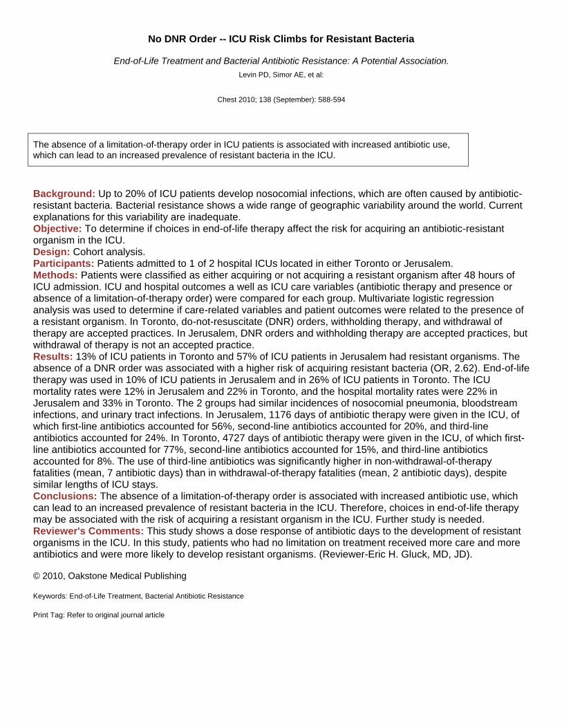
No DNR Order -- ICU Risk Climbs for Resistant Bacteria
End-of-Life Treatment and Bacterial Antibiotic Resistance: A Potential Association.
Levin PD, Simor AE, et al:
Chest 2010; 138 (September): 588-594
The absence of a limitation-of-therapy order in ICU patients is associated with increased antibiotic use, which can lead to an increased prevalence of resistant bacteria in the ICU.
Background: Up to 20% of ICU patients develop nosocomial infections, which are often caused by antibiotic-resistant bacteria. Bacterial resistance shows a wide range of geographic variability around the world. Current explanations for this variability are inadequate. Objective: To determine if choices in end-of-life therapy affect the risk for acquiring an antibiotic-resistant organism in the ICU. Design: Cohort analysis. Participants: Patients admitted to 1 of 2 hospital ICUs located in either Toronto or Jerusalem. Methods: Patients were classified as either acquiring or not acquiring a resistant organism after 48 hours of ICU admission. ICU and hospital outcomes a well as ICU care variables (antibiotic therapy and presence or absence of a limitation-of-therapy order) were compared for each group. Multivariate logistic regression analysis was used to determine if care-related variables and patient outcomes were related to the presence of a resistant organism. In Toronto, do-not-resuscitate (DNR) orders, withholding therapy, and withdrawal of therapy are accepted practices. In Jerusalem, DNR orders and withholding therapy are accepted practices, but withdrawal of therapy is not an accepted practice. Results: 13% of ICU patients in Toronto and 57% of ICU patients in Jerusalem had resistant organisms. The absence of a DNR order was associated with a higher risk of acquiring resistant bacteria (OR, 2.62). End-of-life therapy was used in 10% of ICU patients in Jerusalem and in 26% of ICU patients in Toronto. The ICU mortality rates were 12% in Jerusalem and 22% in Toronto, and the hospital mortality rates were 22% in Jerusalem and 33% in Toronto. The 2 groups had similar incidences of nosocomial pneumonia, bloodstream infections, and urinary tract infections. In Jerusalem, 1176 days of antibiotic therapy were given in the ICU, of which first-line antibiotics accounted for 56%, second-line antibiotics accounted for 20%, and third-line antibiotics accounted for 24%. In Toronto, 4727 days of antibiotic therapy were given in the ICU, of which first-line antibiotics accounted for 77%, second-line antibiotics accounted for 15%, and third-line antibiotics accounted for 8%. The use of third-line antibiotics was significantly higher in non-withdrawal-of-therapy fatalities (mean, 7 antibiotic days) than in withdrawal-of-therapy fatalities (mean, 2 antibiotic days), despite similar lengths of ICU stays. Conclusions: The absence of a limitation-of-therapy order is associated with increased antibiotic use, which can lead to an increased prevalence of resistant bacteria in the ICU. Therefore, choices in end-of-life therapy may be associated with the risk of acquiring a resistant organism in the ICU. Further study is needed. Reviewer's Comments: This study shows a dose response of antibiotic days to the development of resistant organisms in the ICU. In this study, patients who had no limitation on treatment received more care and more antibiotics and were more likely to develop resistant organisms. (Reviewer-Eric H. Gluck, MD, JD). © 2010, Oakstone Medical Publishing
Keywords: End-of-Life Treatment, Bacterial Antibiotic Resistance
Print Tag: Refer to original journal article

Obesity Alters ALI Immune Response, Not Mortality
The Association Between BMI and Plasma Cytokine Levels in Patients With Acute Lung Injury.
Stapleton RD, Dixon AE, et al:
Chest 2010; 138 (September): 568-577
In critically ill patients with acute lung injury, obesity is associated with a blunted inflammatory response and heightened endothelial injury in the lung. Perhaps due to these mixed effects, mortality remains unchanged as BMI increases.
Background: In patients with obesity, the adipose tissue is known to produce several different proinflammatory cytokines, meaning that the levels of these cytokines are elevated at baseline. In patients with acute lung injury (ALI), elevated plasma levels of inflammatory cytokines may be the potential driving force behind the over-exuberant inflammatory response. Objective: To determine if obesity affects plasma inflammatory biomarkers, mortality, ventilator-free days (VFDs), and organ-failure–free days (OFDs) in patients with ALI. Design: Analysis of data from the National Heart, Lung, and Blood Institute Acute Respiratory Distress Syndrome Network. Methods: Patients required mechanical ventilation, met the diagnostic criteria for ALI, and participated in randomized controlled trials of ALI. Based on body mass index (BMI), patients were categorized as underweight (BMI <18.5), normal (BMI, 18.5 to 24.9), overweight (BMI, 25.0 to 29.9), or obese (BMI, 30.0 to 39.9). Plasma inflammatory biomarker levels were measured at study enrollment and again at day 3. Mortality at 90 days was determined. VFDs were calculated as the number of days that a patient was alive and breathing unassisted during the first 28 days after study enrollment. OFDs were calculated as the number of days that a patient was free of nonpulmonary organ failure during the first 28 days after study enrollment. Multivariate regression analyses were performed to determine if BMI affected the relationships between baseline inflammatory cytokine levels and mortality, VFDs, and OFDs. Results: 1409 patients were included in the study. Obese patients with ALI had decreased levels of the circulating markers for inflammation (IL-8 and IL-6) and alveolar injury (surfactant protein D), which is the opposite of that found in healthy obese patients. However, a marker of endothelial injury (von Willebrand factor) increased in obese ALI patients. Therefore, obesity was associated with a mixed effect in ALI patients: a blunted inflammatory response (leading to an attenuation in alveolar injury) and heightened endothelial injury in the setting of microvascular injury. As BMI increased, so did the WBC levels. The outcomes were not significantly worse or better in obese ALI patients: BMI was not associated with increased morbidity and mortality. In addition, BMI did not modify the relationship between baseline biomarker levels and mortality, VFDs, or OFDs. Conclusions: In patients with ALI, obesity appears to be associated with an altered inflammatory response, decreased epithelial (alveolar) injury, and increased endothelial injury in the lung. However, obesity is not associated with a significant change in mortality, VFDs, or OFDs. Reviewer's Comments: Other studies have shown that patients with a marked increase in BMI or a decrease in BMI have poorer outcomes in the ICU. Patients with modest elevations do the best. This study may give us some insight into why this is the case. (Reviewer-Eric H. Gluck, MD, JD). © 2010, Oakstone Medical Publishing
Keywords: Acute Lung Injury, Effects of Obesity
Print Tag: Refer to original journal article

Airway ATP Levels Not Good Marker for Asthma
Adenosine Triphosphate Concentration of Exhaled Breath Condensate in Asthma.
Lázár S, Cervenak L, et al:
Chest 2010; 138 (September): 536-542
Adenosine triphosphate levels measured in the exhaled breath concentrate show no correlation with asthma symptom scores and bronchoconstriction. Therefore, they are probably not a useful biomarker for asthma.
Background: In patients with allergic airway inflammation, one of the proposed key mediators is adenosine triphosphate (ATP), which exerts a wide range of proinflammatory effects via P2 receptor activation. In an animal model of asthma, eosinophilic inflammation and airway hyperresponsiveness have been shown to be induced and maintained by ATP. Currently, the concentration of ATP in the exhaled breath concentrate (EBC) is not known in either healthy controls or in patients with stable asthma. Objective: To determine the concentration of ATP in the EBC of patients with asthma and in healthy controls, and to determine if ATP levels in the EBC can serve as a biomarker for asthma. Design: Cross-sectional study. Participants: 45 adults with bronchial asthma of varying severity and 32 nonatopic healthy subjects recruited from an outpatient clinic in Hungary were evaluated. Patients with acute asthma exacerbation were excluded. Methods: Each patient underwent EBC collection during tidal breathing for 10 minutes. The mean airway ATP concentration was determined using EBC samples and the luminescent technique. Exhaled nitric oxide levels (FENO) and pulmonary function were determined for each participant. Asthma symptoms were assessed via the Asthma Control Test. Results: Overall, the ATP concentrations in the EBC were similar for asthma patients and healthy controls. The ATP concentrations in the EBC did not correlate with asthma control, lung function, or FENO levels. Conclusions: Neither the ATP concentrations in the EBC nor the calculated airway ATP levels differ for asthma patients and healthy controls. There was no correlation between ATP concentrations in the EBC and asthma symptom scores, exhaled NO, and bronchoconstriction. Therefore, ATP concentrations measured in the EBC may not be a useful biomarker for asthma. Reviewer's Comments: This study suggests that we have to look elsewhere for a more reliable biomarker for airway inflammation associated with asthma. (Reviewer-Eric H. Gluck, MD, JD). © 2010, Oakstone Medical Publishing
Keywords: Airway Inflammation, Mediators, Adenosine Triphosphate, ATP
Print Tag: Refer to original journal article

NSCLC Survival Not Affected by Smoking
Long-Term Survival Outcomes by Smoking Status in Surgical and Nonsurgical Patients With Non-Small Cell Lung
Cancer: Comparing Never Smokers and Current Smokers.
Meguid RA, Hooker CM, et al:
Chest 2010; 138 (September): 500-509
Tobacco smoking is not an independent predictor of long-term survival in patients with non–small-cell lung cancer (NSCLC). The notion that NSCLC is a less-aggressive tumor in never smokers does not appear to be true.
Background: Non–small-cell lung cancer (NSCLC) can occur among smokers and never smokers, although studies have shown marked differences in the tumor biology in these 2 populations. Recent reports have depicted lung cancer in never smokers as having fewer genomic alterations, which implies that lung cancer may be less aggressive in never smokers than in smokers. Objective: To compare the prognosis of NSCLC in smokers versus never smokers. Design: Retrospective analysis of data from The Johns Hopkins Specialized Programs of Research Excellence Lung Cancer Database of patients with NSCLC evaluated from 1975 through 2004. Methods: The final study population included 4546 patients. The outcomes and characteristics of never smokers were compared to those of smokers, and a subgroup analysis was performed for patients who underwent surgery with a curative intent versus those who did not. The associations between time to death and various independent variables were analyzed. Results: In a comparison of 724 never smokers versus 3822 current smokers with NSCLC, the tumor stage at presentation was similar for both groups and did not differ by gender in either group. Adenocarcinoma occurred more frequently in never smokers (47.4%) than in smokers (34.1%; P <0.01). Bronchioloalveolar carcinoma (BAC) also occurred more often in never smokers (7.3%) than in smokers (1.9%). On multivariate analysis, overall survival was not affected by smoking status. The mortality rate was lower in women (HR, 0.78) than in men, was higher in black patients (HR, 1.22) than in white patients, and was lower in patients with BAC (HR, 0.41) than in those with adenocarcinoma. Of 1442 patients who underwent surgery with curative intent, the distribution was similar for smokers and never smokers regarding the stage of disease, type of procedure performed, proportion of the cohort undergoing surgery, and the median time between diagnosis and surgery. However, current smokers undergoing surgery tended to have higher ASA classifications than did never smokers, and the risk of death was significantly worse for ASA grades 3 and 4 than for ASA grade 1. In surgical patients, neither smoking status nor pack-year history significantly affected survival, but BAC histology was associated with a survival advantage compared with adenocarcinoma. Patient smoking status did not significantly affect the 30-day postoperative mortality rate. Conclusions: Smoking status is not an independent predictor of reduced long-term survival in patients who NSCLC. In addition, smoking status does not affect the distribution of clinical stages seen at the diagnosis of NSCLC. Reviewer's Comments: Prior studies had strongly suggested that continued smoking was associated with poorer outcomes in patients being treated for lung cancer. This study looks at a different aspect based on cigarette smoking history before the development of lung cancer. (Reviewer-Eric H. Gluck, MD, JD). © 2010, Oakstone Medical Publishing
Keywords: Non–Small-Cell Lung Cancer, Smoking Status, Survival
Print Tag: Refer to original journal article

Lung Cancer Good Surrogate Marker for Smoking
Cigarette Smoking as a Cause of Cancers Other Than Lung Cancer: An Exploratory Study Using the Surveillance,
Epidemiology, and End Results Program.
Ray G, Henson DE, Schwartz AM:
Chest 2010; 138 (September): 491-499
The epidemiologic ecologic approach to data analysis is valid for determining if cigarette smoking is an etiologic factor in various non-lung cancers by using lung cancer as a surrogate marker for smoking.
Background: In the United States, cigarette smoking is associated with 90% of lung cancer deaths in men and with 80% of lung cancer deaths in women. According to the U.S. Surgeon General, smoking is causally associated with 12 different cancers. Different approaches can be taken when trying to prove smoking as a causative agent for various cancers. Objective: To determine if an epidemiologic ecologic approach is a valid assessment tool for determining if smoking is an etiologic agent for non-lung visceral cancers by comparing the geographic trends in cancer rates for lung cancer (surrogate marker for smoking) with those for various non-lung cancers. Design: Data analysis using the Surveillance, Epidemiology, and End Results database that collects data on cancer incidence for approximately 25% of the U.S. population. Methods: The cancer incidence rate was determined and used to generate scatter plots for 21 different types of non-lung cancer versus lung cancer. After a linear regression analysis was performed for all scatter plots, then a correlation analysis was completed to determine if a statistically significant correlation existed between lung cancer and any of the 21 other cancers. The correlation was considered strong when R2 was ≥0.30. "Control cancers" included adult brain cancer, prostate cancer, and mesothelioma (smoking not a causative agent in these cancers). Results: 5 cancers had a strong correlation with lung cancer (and thus cigarette smoking), including laryngeal, esophageal, urinary bladder, kidney, and colorectal cancers. Five other cancers had a weak correlation with lung cancer, including pancreatic cancer, liver cancer, breast cancer, cancer of the lip, and cancer of the oral cavity, lip, and pharynx. In this analysis, cigarette smoking was a causative factor in approximately 40% of esophageal cancers and approximately 49% of bladder cancers. Among the 5 cancers with a weak correlation to cigarette smoking, smoking was considered a causative agent in 14% of pancreatic cancer cases, 18% of liver cancer cases, 20% of breast cancer cases, and 7% of uterine cancer cases. Conclusions: The epidemiologic ecologic approach is valid for determining if cigarette smoking is an etiologic factor in various non-lung cancers by using lung cancer as a surrogate marker for smoking. In other words, using this analytic approach, those cancers having a strong correlation with lung cancer were likely to have a strong correlation with cigarette smoking. Reviewer's Comments: This type of analysis will help us identify potential risk factors and then hopefully allow us to do something about them. This study validates the technique, but further study is in order to ensure that we won't be following false trails. (Reviewer-Eric H. Gluck, MD, JD). © 2010, Oakstone Medical Publishing
Keywords: Smoking, Causative Associations, Non-Lung Visceral Cancers
Print Tag: Refer to original journal article