Embed Size (px)
Citation preview

1
How to evaluate the patient with pelvic pain by ultrasound
Beryl Benacerraf MD
Harvard Medical School and Brigham and Women’s Hospital
Disclosures
Beryl Benacerraf
Relevant Financial Relationships: NONE
Learning ObjectivesAfter completing this presentation, the learner will be able to:
1. Use ultrasound effectively to evaluate patients with pelvic pain, using a combination of imaging, physical exam and pain guided imaging.
2. Understand how to use ultrasound to evaluate patients with all different manifestations of endometriosis.
3. Use 3D ultrasound as an essential tool in evaluating the patient who presents with pelvic pain.
Benacerraf
Lecture Outline
1. Uterine causes of pelvic pain1. Adenomyosis2. Degenerating fibroids3. Embedded IUD
2. Non-GYN causes pain1. Adhesions2. Hernia3. Appendicitis4. Ureteral stone5. Bowel diseases
3. Adnexal causes of pelvic pain1. Endometriosis
(intra and extra ovarian, deep infiltrating endometriosis)
2. Ovarian cyst3. Hydrosalpinx4. Ovarian or tubal
torsion5. Adhesions and
peritoneal inclusion cyst
6. PID, TOABenacerraf
Introduction• Pelvic pain is a public health problem
affecting ≥15% of women. • Many patients never get a diagnosis and
live with chronic pain, affecting their quality of life.
• Patients with pelvic pain deserve more than just a series of standard pictures of the uterus and ovaries. Individualizing the exam using a problem solving approach is essential for many of these patients.
Get a history during the exam
• Acute or chronic• Diffuse or focal• Cyclical or constant• Sharp or dull or cramping• ? Prior surgery• Menopausal and hormonal status• Could she be pregnant?

2
During the scan
• How tender is the patient?• Where is the tenderness? Focal?• Do organs slide past each other?• Push deliberately on each part of the
pelvis with probe and other hand to see where the pain comes from.
• Talk to the patient!
The uterus
• Adenomyosis• Degerating fibroids• Prolapsing fibroids• Abnormally placed IUD
Adenomyosis:• Endometriosis of the uterus.• Characterized by invasion of
endometrial glands into the neighboring myometrium.
• Symptoms: Dysmenorrhea abnormal bleeding, uterine enlargement and tenderness.
Adenomyosis –Ultrasound Appearance
• Mottled inhomogeneous myometrium
• Globular & asymmetrical uterus,
• Small subendometrial cysts • Indistinct endometrial stripe.
©AIUM ©AIUM

3
©AIUM
Fuzzy borders of cavity due to adenomyosis
©AIUM
This is an intracavitaryadenomyoma
Fibroids• Very common - most do not cause
pain (bleeding more likely)• Positioning of fibroid or
degeneration may make them symptomatic.
• Size of fibroid may cause discomfort due to pressure, such as hydronephrosis.
©AIUM
Many Faces of Degenerating
Fibroids
©AIUM
Degenratingfibroid
mimicking endometrioma
4
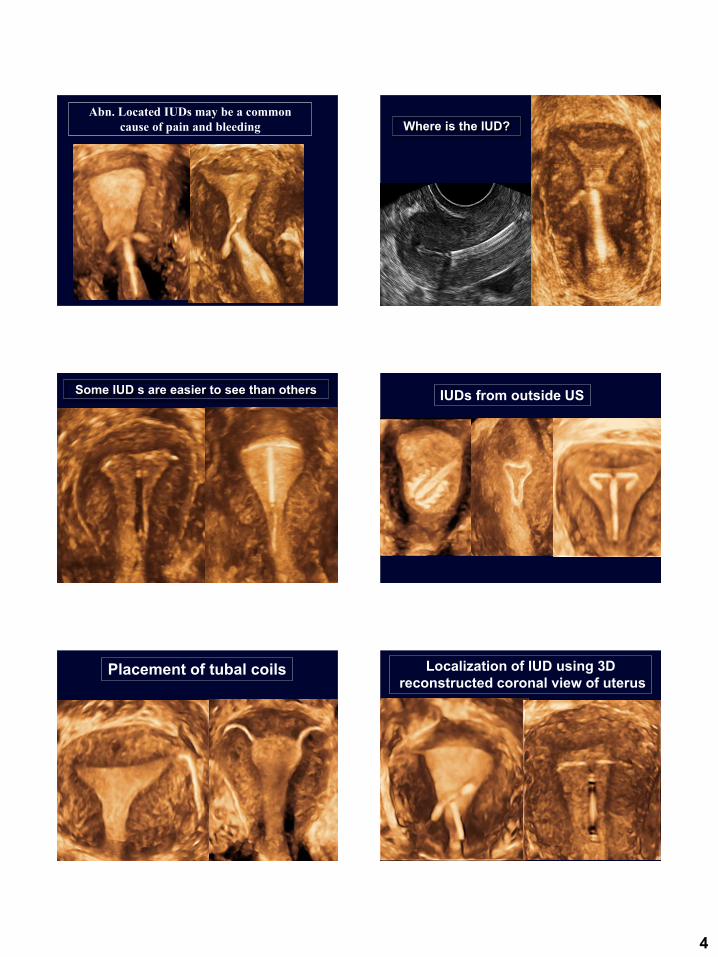
Abn. Located IUDs may be a common
cause of pain and bleeding
©AIUM
Where is the IUD?
©AIUM
Some IUD s are easier to see than others IUDs from outside US
©AIUM
Placement of tubal coils
©AIUM
Localization of IUD using 3D reconstructed coronal view of uterus

5
©AIUM
Where is the IUD?
©AIUM
Where is the IUD?
Indication for scan
Bleeding Pain Either bleeding and/or pain
IUD Imbedded
10/28
35.7%
11/28
39.2%
21/28
70.4%
IUD Not Imbedded
21/139
15.1%
27/139
19.4%
48/139
34.5%
Fisher Exact test p = 0.02 p = 0.03 p = 0.0001
Benacerraf et al. Ultrasound Obstet Gynecol 2009; 34;110.©AIUM
How to use the
IUD shaddow
to find the
device
Using the shadowUsing 3D to see that the IUD is upside down

6
©AIUM
Is the uterus too small? Does the
not IUD fit?
Size of normal uterine cavity -221 consecutive premenopausal patients
• The mean width of normal uterine cavity: 29mm • The mean is 27 mm in nulliparous women compared to 32 mm in those ≥ 1 pregnancy). • There was no appreciable relationship between the width of the uterine cavity and prior C-section or patient age (in patients who were never pregnant).
Benacerraf, et al. Obstet Gynecol
2010;116:305
©AIUM
IUD perforating left horn of septate uterus
Septate uterus – IUD stuck in lower uterine segment The Ovary - Adnexa
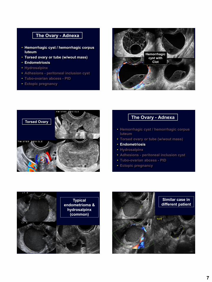
• Hemorrhagic cyst / hemorrhagic corpus luteum
• Torsed ovary or tube (w/wout mass)• Endometriosis• Hydrosalpinx• Adhesions - peritoneal inclusion cyst• Tubo-ovarian abcess – PID
7
The Ovary - Adnexa
• Hemorrhagic cyst / hemorrhagic corpus luteum
• Torsed ovary or tube (w/wout mass)• Endometriosis• Hydrosalpinx• Adhesions - peritoneal inclusion cyst• Tubo-ovarian abcess - PID• Ectopic pregnancy
©AIUM
Hemorrhagic cyst with
Clot
©AIUM
Torsed OvaryThe Ovary - Adnexa
• Hemorrhagic cyst / hemorrhagic corpus luteum
• Torsed ovary or tube (w/wout mass)• Endometriosis• Hydrosalpinx• Adhesions - peritoneal inclusion cyst• Tubo-ovarian abcess - PID• Ectopic pregnancy
©AIUM
Typical endometrioma &
hydrosalpinx(common)
©AIUM
Similar case in different patient

8
EndometriomaCystadenoma
Streaming?
Pitfall - struma ovariiIs this endometriosis?
©AIUM
Endometrioma with calcified clot
mimicking dermoid
Is this endometriosis?
©AIUM
Cul de sac endometriosis
Unusual Sites of Endometriosis
Bladder wall endometriosis
©AIUM ©AIUM
Extensive endometriosis of tube
and utero-sacral ligament in a 24 year
old patient

9
Endometriosis implant on appendix
Similar appearance to appendicitis!
Positive sliding signThe comet-sign of
endometriosis of the bowel wall
Benacerraf et al. JUM 2015;34:537
©AIUM ©AIUM
10
©AIUM
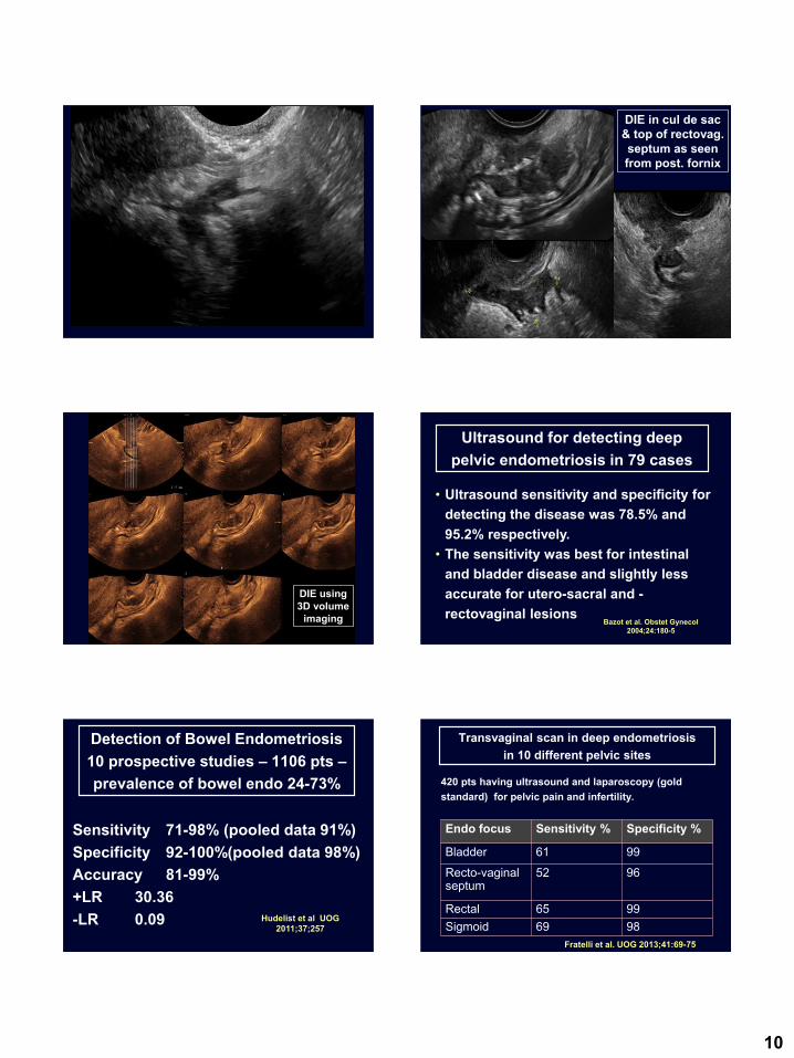
DIE in cul de sac & top of rectovag. septum as seen from post. fornix
DIE using 3D volume
imaging
• Ultrasound sensitivity and specificity for detecting the disease was 78.5% and 95.2% respectively.
• The sensitivity was best for intestinal and bladder disease and slightly less accurate for utero-sacral and -rectovaginal lesions
Ultrasound for detecting deep pelvic endometriosis in 79 cases
Bazot et al. Obstet Gynecol2004;24:180-5
Detection of Bowel Endometriosis10 prospective studies – 1106 pts –prevalence of bowel endo 24-73%
Sensitivity 71-98% (pooled data 91%)Specificity 92-100%(pooled data 98%)Accuracy 81-99%+LR 30.36-LR 0.09 Hudelist et al UOG
2011;37;257
Transvaginal scan in deep endometriosisin 10 different pelvic sites
420 pts having ultrasound and laparoscopy (gold standard) for pelvic pain and infertility.
Endo focus Sensitivity % Specificity %
Bladder 61 99
Recto-vaginalseptum
52 96
Rectal 65 99Sigmoid 69 98
Fratelli et al. UOG 2013;41:69-75
11
©AIUM
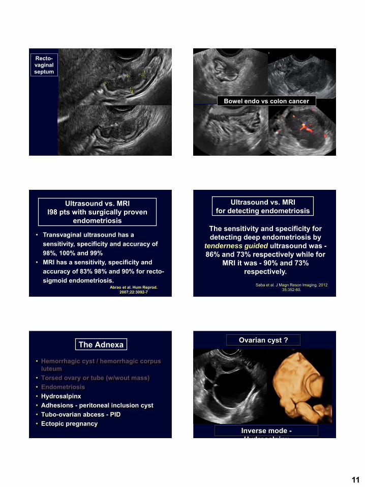
Recto-vaginal septum
©AIUM
Bowel endo vs colon cancer
Ultrasound vs. MRII98 pts with surgically proven
endometriosis
• Transvaginal ultrasound has a sensitivity, specificity and accuracy of 98%, 100% and 99%
• MRI has a sensitivity, specificity and accuracy of 83% 98% and 90% for recto-sigmoid endometriosis.
Abrao et al. Hum Reprod. 2007;22:3092-7
The sensitivity and specificity for detecting deep endometriosis by
tenderness guided ultrasound was -86% and 73% respectively while for
MRI it was - 90% and 73% respectively.
Ultrasound vs. MRIfor detecting endometriosis
Saba et al. J Magn Reson Imaging. 2012 35:352-60.
The Adnexa
• Hemorrhagic cyst / hemorrhagic corpus luteum
• Torsed ovary or tube (w/wout mass)• Endometriosis• Hydrosalpinx• Adhesions - peritoneal inclusion cyst• Tubo-ovarian abcess - PID• Ectopic pregnancy
Ovarian cyst ?
Inverse mode -Hydrosalpinx

12
©AIUM
2D and 3D images of a hydrosalpinx. 3D inverse reconstructed view shows
the lesion to best advantage
©AI
©AIUM
Peritoneal inclusion cyst with trapped
ovary
©AIUM
TOA
Focal Pain when tube is moved:Salpingitis at laparoscopy
Hemato-
salpinx in pt
with ectopic
Echogenic free
fluid (+clot)
13
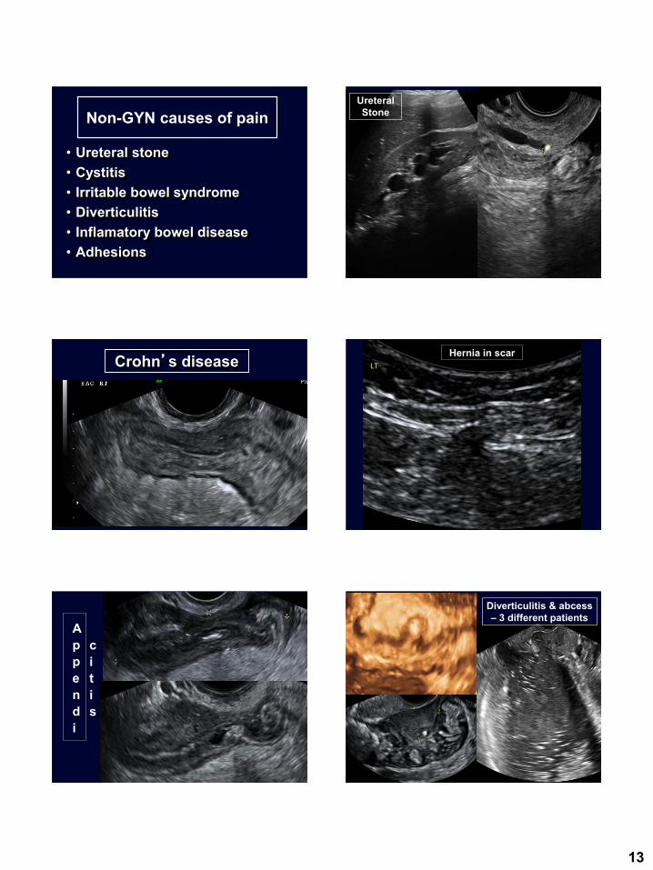
Non-GYN causes of pain
• Ureteral stone• Cystitis• Irritable bowel syndrome• Diverticulitis• Inflamatory bowel disease• Adhesions
©AIUM
Ureteral Stone
©AIUM
Crohn’s disease Hernia in scar
©AIUM
Appendi
citis
©AIUM
Diverticulitis & abcess– 3 different patients

14
Conclusions• Pelvic pain is common and impairs
quality of life.• Accurate Dx requires a combo of
ultrasound, phys. exam and history.• Patients with pelvic pain deserve
more than just a series of standard pictures of the uterus and ovaries.
• Those that we help are among the most grateful of all our patients!
References
Benacerraf
• Benacerraf BR, et al. 3D Ultrasound Detection of Embedded Intrauterine Contraceptive Devices–A source of Pelvic Pain and Abnormal Bleeding. Ultrasound ObstetGynecol 2009; 34;110-150.
• Guerriero S, et al. Accuracy of transvaginal ultrasound for diagnosis of deep endometriosis in uterosacralligaments, rectovaginal septum, vagina and bladder: systematic review and meta-analysis. Ultrasound ObstetGynecol. 2015;46:534-45.
• Reid S, Lu C, Condous G. Can we improve the prediction of pouch of Douglas obliteration in women with suspected endometriosis using ultrasound-based models? A multicenter prospective observational study. Acta Obstet Gynecol Scand. 2015;94:1297-306.