Embed Size (px)
Citation preview
OPTOM FASLU MUHAMMED
PATHOLOGY OF EYE LIDS AND ADNEXA
LESIONS OF EYE LIDS AND ADNEXA ARE OF FOLLOWING TYPES 1.Cysts 2.Inflamation /infections 3.Metabolic changes and degenaration 3.Tumorous conditions 4.Tumours
CYSTS A cyst is a closed sac-like structure that is not
a normal part of the tissue where it is found Cysts are common and can occur anywhere
in the body in people of any age. The causes of a cyst
1. wear and tear" or simple obstructions to the flow of fluid,
2. Infections,3. Tumors,4. chronic inflammatory conditions,5. genetic (inherited) conditions, and6. defects in developing organs in the embryo
CYSTS
1.Inclusion cysts 2.retention cysts 3.develpmental/Dermoid cysts
INCLUSION CYSTS Inclusion cysts occur in any part of eyelid
following trauma or infection

RETENTION CYSTS Retention cysts are the result of blockade of
ductal opening of eccrine glands rarely the piloseceous apparatus may show blockade at epidermal opening .
The cysts may contain clear or sebaceous material based on the type gland involved .the so called sebaceous cyst.

DERMOID CYSTS These cysts are encounterd in midline or
fusion lines .Dermoid cysts are frequently seen in infants or adolescents .
INFLAMMATION / INFECTIONS 1.blebharitis 2.hordeolum 3.chalazion Other infections/inflammations 1.Bacterial or myobacterial 2.Viral infections
BLEPHARITIS In this type of infection ,inflammation
effecting the eyelids Chronic inflammation of the lid margin Types: staphylococcal or seborrheic Symptoms: foreign-body sensation, burning,
mattering May predispose to chalazia,
blepharoconjunctivitis, loss of lashes

BLEPHARITIS TREATMENT Warm compresses Lid scrubs with 50/50 mixture of nonirritating
shampoo (Johnson and Johnson’s baby shampoo) and water daily
Antibiotic ointment at bedtime for 2-3 weeks (Bacitracin or erythromycin)
Resistant cases can be referred to the ophthalmologist on a non-urgent basis
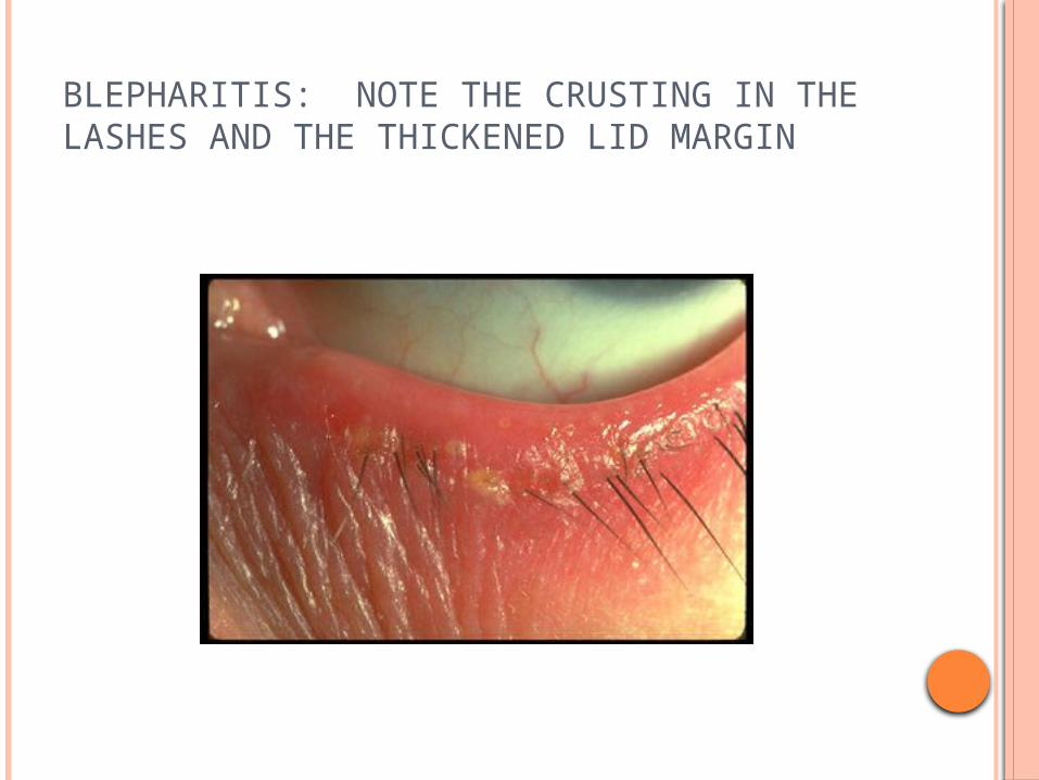
BLEPHARITIS: NOTE THE CRUSTING IN THE LASHES AND THE THICKENED LID MARGIN

HORDEOLUM Hordeolum is a suppurative nonspecific
inflammation of eye lid adnexa On the basis of anatomical site of
inflammation’ two forms are recognised 1. Hordeolum externum 2. Hordeolum internum

HORDEOLUM Usually begins as diffuse swelling followed by
localization of a nodule to the lid margin
Hordeolum – staphylococcal infection of the glands of Zeis
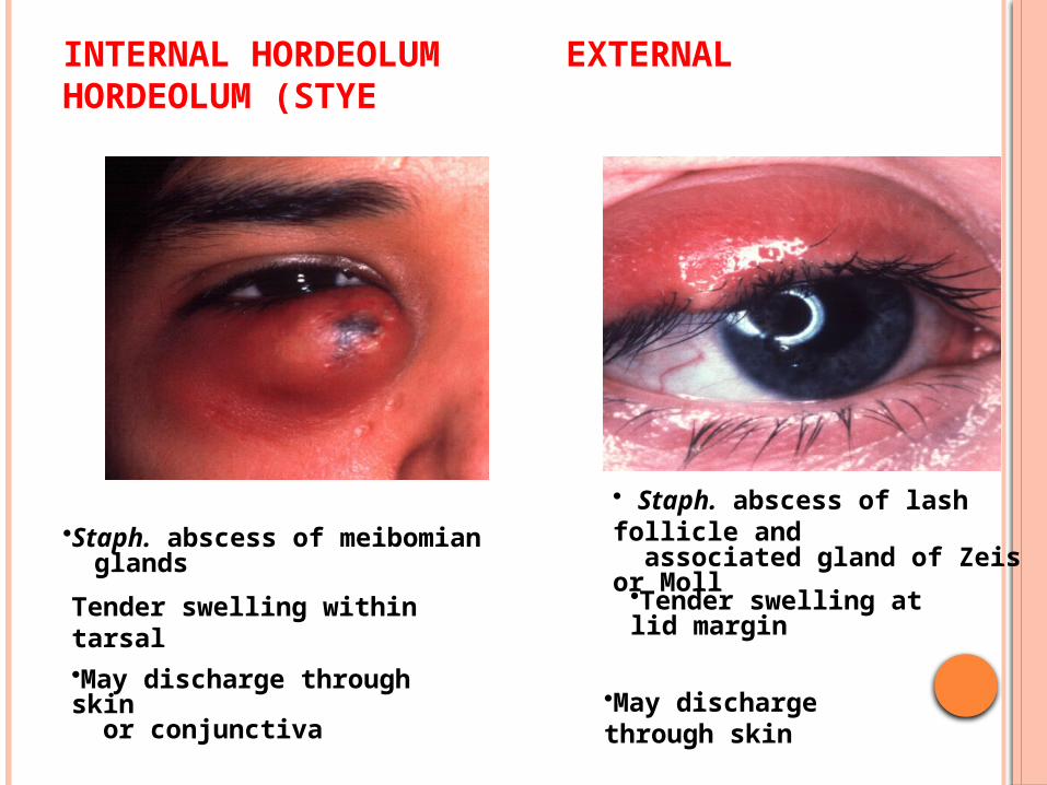
INTERNAL HORDEOLUM EXTERNAL HORDEOLUM (STYE
•Staph. abscess of meibomian glandsTender swelling within tarsal •May discharge through skin or conjunctiva
• Staph. abscess of lash follicle and associated gland of Zeis or Moll•Tender swelling at lid margin
•May discharge through skin
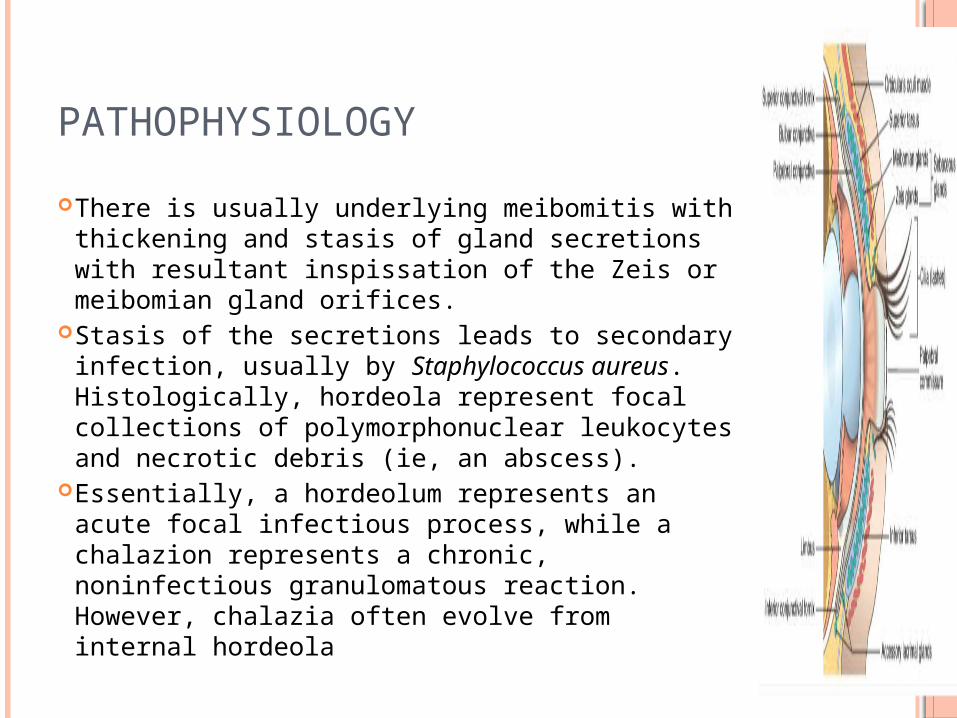
PATHOPHYSIOLOGY There is usually underlying meibomitis with
thickening and stasis of gland secretions with resultant inspissation of the Zeis or meibomian gland orifices.
Stasis of the secretions leads to secondary infection, usually by Staphylococcus aureus. Histologically, hordeola represent focal collections of polymorphonuclear leukocytes and necrotic debris (ie, an abscess).
Essentially, a hordeolum represents an acute focal infectious process, while a chalazion represents a chronic, noninfectious granulomatous reaction. However, chalazia often evolve from internal hordeola
CHALAZION Chronic non specific inflammation of
sebaceous gland of lid with or with out granulomatous reaction is called chalazion
The zies gland inflammation is termed as external chalazion
The meibomian gland inflammation is termed as internal chalazion
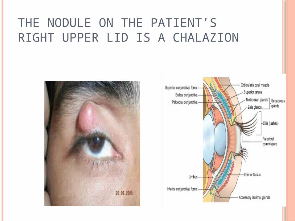
THE NODULE ON THE PATIENT’S RIGHT UPPER LID IS A CHALAZION
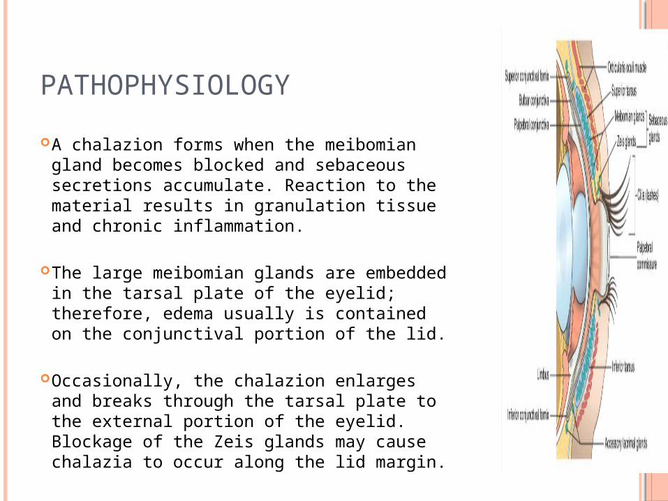
PATHOPHYSIOLOGY A chalazion forms when the meibomian gland
becomes blocked and sebaceous secretions accumulate. Reaction to the material results in granulation tissue and chronic inflammation.
The large meibomian glands are embedded in the tarsal plate of the eyelid; therefore, edema usually is contained on the conjunctival portion of the lid.
Occasionally, the chalazion enlarges and breaks through the tarsal plate to the external portion of the eyelid. Blockage of the Zeis glands may cause chalazia to occur along the lid margin.
OTHER INFECTION AND INFLAMMATIONS Bacterial infections Viral infections
MOLL SCUM CONTAGIOSUM MC is a multiple nodular or papular lesion
with umbilicated centre . The enlargement and distension of of
epidermal cells with viral inclusions

VERUCOUS LESIONS These are due to papilloma group of viruses The lesions are characterized by exophytic
proliferative warty lesions and may be multiple

END OF PART ONETHANKS