Embed Size (px)
Citation preview

Diagnosis of Alzheimer's Disease and Multiple Infarct Dementia by Tomographic Imaging of Iodine-123 IMP Marvin B. Cohen, L. Stephen Graham, Ralph Lake, E. Jeffrey Metter, Jaime Fitten, Mangala K. Kulkarni, Roger Sevrin, Les Yamada, Chia C. Chang, Nathan Woodruff, and Arthur S. Kling
Departments of Nuclear Medicine, Neurology, and Psychiatry, Veterans Administration Medical Center, Sepulveda; and UCLA School of Medicine, Los Angeles, California
Tomographic imaging of the brain was performed using a rotating slant hole collimator and [123l]A/-isopropyl p-iodoamphetamine (IMP) in normal subjects (n = 6) and patients with either Alzheimer's disease (n = 5) or multiple infarct dementia (n = 3). Four blinded observers were asked to make a diagnosis from the images. Normal subjects and patients with multiple infarct dementia were correctly identified. Alzheimer's disease was diagnosed in three of the five patients with this disease. One patient with early Alzheimer's disease was classified as normal by two of the four observers. Another patient with Alzheimer's disease had an asymmetric distribution of IMP and was incorrectly diagnosed as multiple infarct dementia by all four observers. Limited angle tomography of the cerebral distribution of 123l appears to be a useful technique for the evaluation of demented patients.
J Nucl Med 27:769-774,1986
JLositron emission tomography (PET) imaging with [18F]2-fluoro-2-deoxy-D-glucose (FDG) or oxygen-15 are the first imaging procedures to provide an accurate diagnosis of Alzheimer's disease (AD) and multiple infarct dementia (MID) (J-5). The limited availability of PET facilities restrict their clinical application. Single photon emission computed tomography (SPECT) imaging of the distribution of iodine-123 (l23I) AMsopropyl /Modoamphetamine (IMP) may also provide an accurate test for the differential diagnosis of dementia and is more clinically available (6-7). We are currently pursuing this line of investigation with a rotating gamma camera, but initial studies suggest that limited angle tomography with [123I]IMP has a definite clinical role in the differential diagnosis of dementia. We report the results obtained with a scintillation camera and a rotating slant hole collimator using IMP labeled with high purity (p,5n) 123I in a group of normal subjects and patients with either AD or MID.
Received July 31, 1985; revision accepted Jan. 21, 1986. For reprints contact: Marvin B. Cohen, MD, Chief. Nuclear
Medicine Service, V.A. Medical Center, 16111 Plummer St., Sepulveda, CA 91343.
MATERIALS AND METHODS
Subjects were normal volunteers (n = 6) or ambulatory patients with AD (n = 5) or MID (n = 3). All patients had EEGs, CAT scan, and appropriate laboratory evaluations to rule out other causes of dementia. The clinical diagnosis was made after neurologic and psychiatric evaluation. The diagnosis of MID was based on a clinical history of strokes with physical and/or CAT scan evidence of stroke and a high score on the Hachinski scale. The diagnosis of probable AD was based on the criteria of McKhann et al. (8). No attempt was made to use age-matched controls in this limited study.
"Pure" 123I (i.e., produced by a p,5n reaction and devoid of 124I or other high energy contaminants) was obtained commercially* and was utilized in our laboratory for exchange labeling of nonradioactive N-iso-propyl /Modoamphetamine+ by a melting point procedure (9). Appropriate quality control procedures were utilized to synthesize sterile, nonpyrogenic [123I]IMP with a specific activity of ~3-5 mCi/mg. The radiochemical purity was >98% as determined by thin layer chromatography on silica gel using the following two
Volume 27 • Number 6 • June 1986 769
by on April 28, 2018. For personal use only. jnm.snmjournals.org Downloaded from

FIGURE 1 A: "Cold" cube phantom. B: Images obtained with seven pinhole collimator and p,5n 1 2 3 l . C: Images obtained with rotating slant hole collimator and p,5n 12 l̂
solvent systems: (a) methanol-chloroform-glacial acetic acid (15:85:1) and (b) ethyl acetate-ethanol (1:1). Distribution and excretion studies were previously performed in rats, dogs, and monkeys for radiation dosimetry calculations. Results of distribution and excretion studies are similar to those reported by Kuhl (70), but the radiation dose to the patient was roughly half of the value reported because of the absence of 124I in our preparation. We calculate that a 5 mCi dose of IMP labeled with pure 123I (p,5n) delivered 2.5 rad to the lung, 2.2 rad to the liver, 0.36 rad to the brain, and lesser amounts to other organs. The whole body dose was 0.25 rad.
Imaging studies of phantoms were performed with a seven pinhole and a slant hole collimator to evaluate whether the slant hole collimator was superior to the seven pinhole collimator for limited angle transverse
770 Cohen, Graham, Lake et al
tomography of the brain with "pure" 123I using a vertex acquisition. The phantom consisted of a lucite box (15 cm x 15 cm x 15 cm) containing ~ 1 mCi of high purity (p,5n) 123I. Four "cold" cubes with side length 3, 4, 5, and 6 cm were centered at a depth of 5 cm inside the box. The top of the phantom was positioned 1 cm from the collimator face to simulate a vertex view and imaged with a mobile camera* using a slant hole collimator. A total of six views were taken with the collimator rotated 60° after each image was taken. The data were reconstructed with a micro-processor based computer5 using software provided by the vendor. Twelve contiguous planes were reconstructed with a separation of 1 cm between levels. In a separate study, images were also acquired under identical conditions except that 123I produced by the p,2n reaction (so-called "commercial" 123I) was used.
The Journal of Nuclear Medicine
by on April 28, 2018. For personal use only. jnm.snmjournals.org Downloaded from

A seven pinhole collimator1 mounted on a large field camera" was used to collect an image of the same phantom filled with high purity I23I. For this experiment, the phantom was positioned 14 cm from the 7.5-mm aperture and 750,000 counts were collected. These data were reconstructed using software supplied by the collimator vendor using a minicomputer (DEOPDP 11/34A).
An area of interest generator was utilized to measure contrast defined as:
Contrast = -^——-,
where CT = Counts per unit area in the target; CB = Counts per unit area in the background.
Depth resolution was evaluated qualitatively by visually comparing the sharpness of the images at progressively greater depths.
Limited angle tomographic imaging of patients and normal volunteers was performed with the mobile scintillation camera equipped with a rotating slant hole collimator. Reconstructed images were obtained using the commercially available software and the microprocessor based computer. Each of the subjects in each group was injected intravenously with 3-5 mCi (111-185 MBq) of [123I]IMP while lying quietly in a well-lighted room with ambient noise. The true administered dose was probably somewhat less than stated, because the dose calibrator reading was not corrected for the effects of characteristic x-rays as recently described by Harris et al. {11). Imaging was begun 20-30 min later with the patient's head immobilized and required 8-12 min for the acquisition of 400k counts. Data were acquired from the vertex position and then processed with an iterative backprojection algorithm to produce 12 1-cm-thick high contrast transverse tomographic images of the superficial cortex parallel to the orbital-
meatal line. Fourier smoothing and simple thresholding techniques were applied to the reconstructed images.
RESULTS
Images obtained with the "cold" cube phantom were not as distorted on the slant hole system as those obtained with the seven pinhole (Fig. 1). In addition, there were fewer artifacts and better depth resolution with the slant hole system. Computer analysis of the images obtained with the high purity m\ had ~30% greater contrast for the large cubes than those obtained with the "commercial" grade of m I . No significant difference in contrast of the smallest cubes was seen between the seven pinhole and rotating slant hole collimators.
The images were initially evaluated by two authors (M.B.C. and L.S.G.) to develop criteria for diagnosis. The most superficial slices denoting the scalp and skull, and images in the deepest planes were not considered to be suitable for analysis. The most useful diagnostic information was found in the 4-6 images encompassing the superficial cortex. Normal volunteers were found to have a relatively uniform distribution of IMP in the cerebral cortex (Fig. 2). Patients with MID had multiple, asymmetric defects involving the gray matter or both the white and gray matter (Fig. 3). Patients with AD had a diffuse, symmetric decrease of uptake in the cortex. This appeared to primarily involve the parietooccipital cortex (Fig. 4). Resolution was poorer in the deeper planes so that the inferior temporal lobe and basal ganglia frequently could not be evaluated. Only-defects seen in two or more adjacent slices were considered significant.
These diagnostic criteria were then given to four physicians who were individually blinded and asked to evaluate the images without benefit of clinical history or patient identification. Three of the physicians (R.L.,
ANT
POST.
FIGURE 2 Normal subject. Relatively symmetric uptake of IMP was observed in superficial cortex of this and other normal subjects (A). While basal ganglia are probably partially visualized (arrows) in this subject (B), spatial resolution was poorer in deep planes and prevented adequate evaluation of basal ganglia and inferior temporal lobes in most patients. All subsequent images are presented with same right to left anterior-posterior orientation
Volume 27 • Number 6 • June 1986 771
by on April 28, 2018. For personal use only. jnm.snmjournals.org Downloaded from
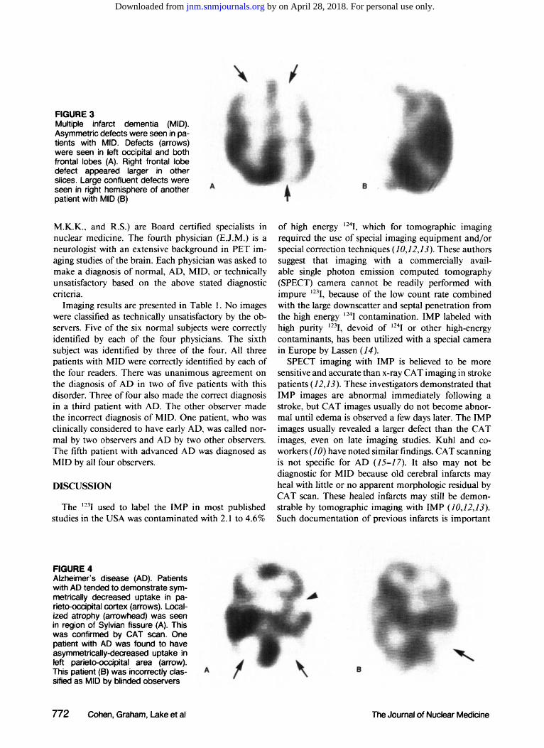
FIGURE 3 Multiple infarct dementia (MID). Asymmetric defects were seen in patients with MID. Defects (arrows) were seen in left occipital and both frontal lobes (A). Right frontal lobe defect appeared larger in other slices. Large confluent defects were seen in right hemisphere of another patient with MID (B)
M.K.K., and R.S.) are Board certified specialists in nuclear medicine. The fourth physician (E.J.M.) is a neurologist with an extensive background in PET imaging studies of the brain. Each physician was asked to make a diagnosis of normal, AD, MID, or technically unsatisfactory based on the above stated diagnostic criteria.
Imaging results are presented in Table 1. No images were classified as technically unsatisfactory by the observers. Five of the six normal subjects were correctly identified by each of the four physicians. The sixth subject was identified by three of the four. All three patients with MID were correctly identified by each of the four readers. There was unanimous agreement on the diagnosis of AD in two of five patients with this disorder. Three of four also made the correct diagnosis in a third patient with AD. The other observer made the incorrect diagnosis of MID. One patient, who was clinically considered to have early AD, was called normal by two observers and AD by two other observers. The fifth patient with advanced AD was diagnosed as MID by all four observers.
DISCUSSION
The i2il used to label the IMP in most published studies in the USA was contaminated with 2.1 to 4.6%
of high energy 124I, which for tomographic imaging required the use of special imaging equipment and/or special correction techniques (10,12,13). These authors suggest that imaging with a commercially available single photon emission computed tomography (SPECT) camera cannot be readily performed with impure 123I, because of the low count rate combined with the large downscatter and septal penetration from the high energy m I contamination. IMP labeled with high purity ,23I, devoid of 124I or other high-energy contaminants, has been utilized with a special camera in Europe by Lassen (14).
SPECT imaging with IMP is believed to be more sensitive and accurate than x-ray CAT imaging in stroke patients (12,13). These investigators demonstrated that IMP images are abnormal immediately following a stroke, but CAT images usually do not become abnormal until edema is observed a few days later. The IMP images usually revealed a larger defect than the CAT images, even on late imaging studies. Kuhl and coworkers (10) have noted similar findings. CAT scanning is not specific for AD (15-17). It also may not be diagnostic for MID because old cerebral infarcts may heal with little or no apparent morphologic residual by CAT scan. These healed infarcts may still be demonstrable by tomographic imaging with IMP (10,12,13). Such documentation of previous infarcts is important
FIGURE 4 Alzheimer's disease (AD). Patients with AD tended to demonstrate symmetrically decreased uptake in parietooccipital cortex (arrows). Localized atrophy (arrowhead) was seen in region of Sylvian fissure (A). This was confirmed by CAT scan. One patient with AD was found to have asymmetrically-decreased uptake in left parietooccipital area (arrow). This patient (B) was incorrectly classified as MID by blinded observers
772 Cohen, Graham, Lake et al The Journal of Nuclear Medicine
by on April 28, 2018. For personal use only. jnm.snmjournals.org Downloaded from

in the diagnosis of MID, because a reliable history of episodic deterioration of cognitive function may not be available when the patient is first seen. Little information has been reported on the use of p, 5n f12T]IMP for the diagnosis of AD (6-7).
Limited angle tomography has a number of inherent deficiencies, including decreased spatial resolution in deep planes and the inability to produce accurate quantitative images. However, the vertex acquisition with the collimator only millimeters away from the skull yields high resolution images of the superficial cortex. Since one of the major problems in all SPECT brain imaging with [I33I]IMP is impaired lesion detectability secondary to relatively poor counting statistics, a further improvement in lesion detectability should be possible with a longer acquisition time than the 8-12 min used in this study. Nevertheless, in this small series the accuracy of the limited angle tomography with [i23I] IMP was excellent in normal subjects and patients with MID. Of the five patients with AD, two were identified by all observers and a third was identified by three of four observers. A patient considered on clinical grounds to have early AD was called normal by two observers. Patients with early AD are also difficult to diagnose by PET imaging (18). The fifth patient with AD was called MID by all four observers, apparently due to the criteria used for image evaluation in this study. While this patient demonstrated decreased uptake of IMP in the parieto-occipital cortex bilaterally, the findings (Fig. 4) were distinctly asymmetric. This patient had a marked impairment in both memory and language function. Foster et al. (19) report symmetric impairment in uptake of [,8F]FDG in patients who present with memory failure as the predominant clinical feature. On the other hand, they found patterns of asymmetric focal changes in patients who presented with either a predominant clinical picture of language dysfunction or visuo-con-structive dysfunction. Asymmetry in uptake of [18F] FDG in patients with AD has also been reported by Friedland et al. (20), but the predominant clinical feature was not reported. Our fifth patient may be a member of such a subgroup. The four blinded observers in our study were instructed that patients with AD had a symmetric decrease in uptake of IMP in the parietooccipital cortex. This is believed to be the reason that all observers failed to identify this patient as having
AD. Qualitative images obtained with [123I]IMP by limited
angle tomography using a rotating slant hole collimator were able to identify normal subjects, patients with MID and to a lesser extent patients with AD. It will still be necessary to study patients with other forms of dementia, "pseudo-dementia," and various neurologic disorders to validate that a pattern is specific for a particular etiology. Coni and co-workers (21) found limited angle tomography with 123I (p,2n) IMP to be
TABLE 1 [123i]IMP: Limited Angle Tomography—Imaging Results
Compared with Clinical Diagnosis
Normal* MIDt AD*
True False True False True False positive positive positive positive positive positive
14§(4/4)' 5(2/4) 8(4/4) 1(4/4) 5(2/4) 11(1/4) 13 (4/4) 7(4/4) 3(1/4) 4(4/4) 12 (4/4) 6(4/4) 3(3/4) 11 (3/4) 2(4/4) 10 (4/4) 9 (4/4)
"Subjects 9-14. t Subjects 6-8. * Subjects 1-5. § Subject number. ' Number of panelists making a particular diagnosis (see text).
very useful in a group of 20 symptomatic geriatric patients for supporting the clinical diagnosis of stroke and multi-infarct disease.
The addition of a rotating slant hole collimator and associated reconstruction software to an existing camera system makes limited angle tomography a relatively inexpensive and readily available procedure. If our preliminary findings are confirmed by larger studies, it would suggest that limited angle tomography with [l23I] IMP may be especially cost effective for the differential diagnosis of dementia in large public hospitals where demented patients are frequently admitted without an accurate past history to support a clinical diagnosis of a specific etiology.
FOOTNOTES
* Crocker Laboratories, University of California at Davis, CA.
+ Medi-Physics, Inc., Emeryville, CA. * Technicare (S420), Solon, OH. § Technicare (VIP 550), Solon, OH. ' Cardiac Medical Systems Corp., Springfield, WI. "Technicare (S410), Solon, OH.
ACKNOWLEDGMENTS
This study was supported by funds from the Research Service, Veterans Administration. The editorial assistance of Patricia Shamblin is gratefully acknowledged.
REFERENCES
1. Benson DF, Kuhl DE, Hawkins RA, et al: The fluo-rodeoxyglucose 18F scan in Alzheimer's disease and multi-infarct dementia. Arch Neurol 40:711 -714,1983
2. Phelps ME, Mazziotta JC, Huang SC: Study of cerebral function with positron computed tomography. J Cerebr Blood Flow Metab 2:113-162, 1982
Volume 27 • Number 6 • June 1986 773
by on April 28, 2018. For personal use only. jnm.snmjournals.org Downloaded from

3. Metter EJ, Riege WH, Kamejama M, et al: Cerebral metabolic relationships for selected brain regions in Alzheimer's, Huntington's, and Parkinson's diseases. J Cerebr Blood Flow Metab 4:500-506, 1984
4. Phelps ME, Schelbert HR, Mazziotta JC: Positron computed tomography for studies of myocardial and cerebral function. Ann Intern Med 98:339-359, 1983
5. Alavi A, Ferris S, Wolf A, et al: Determination of cerebral metabolism in senile dementia using F-18 2-deoxyglucose and positron emission tomography. J Nucl Med 21 :P21, 1980 (abstr)
6. Cohen MB, Metter EJ, Graham LS, et al: Differential diagnosis of dementia with "pure" 1-123 iodoam-phetamine and a clinical camera. J Nucl Med 24:P 106, 1983 (abstr)
7. Cohen MB, Graham LS, Lake R, et al: SPECT imaging of 1-123 IMP in dementia. Clin Nucl Med 9:P30, 1984 (abstr)
8. McKhann G, Drachman D, Folstein M, et al: Clinical diagnosis of Alzheimer's disease: Report of the NINCDS-ADRDA work group. Neurology 34:939-944, 1984
9. Baldwin RM, Lin TH, Wu JL: Synthesis and brain uptake of isometric 1-123 iodoamphetamine derivative. J Labeled Comp Radiopharm 19:1305-1306, 1982
10. Kuhl DE, Barrio JR, Huang SC, et al: Quantifying local cerebral blood flow by 7V-isopropyl-/?-(I-123) iodoamphetamine (IMP) tomography. / Nucl Med 23:196, 1982
11. Harris CC, Jaszczak RJ, Greer KL, et al: Effects of characteristic x-rays on assay of I-123 by dose calibrator. J Nucl Med 25:1367-1370, 1984
12. Lee RGL, Hill TC. Holman BL, et al: jV-isopropyl (I-
123) ^-iodoamphetamine brain scans with single photon emission tomography: Discordance with transmission computer tomography. Radiology 145:759-799, 1984
13. Hill TC, Holman BL, Lovett R, et al: Initial experience with SPECT (single-photon computerized tomography) of the brain using 7V-isopropyl 1-123 /Modoam-phetamine. J Nucl Med 23:191-195, 1982
14. Lassen NA, Henriksen L, Holm S, et al: Cerebral blood-flow tomography: Xe-133 compared with iso-propyl-amphetamine I-123. J Nucl Med 24:17, 1983
15. Naeser MA, Gebhardt C, Levine HL: Decreased computerized tomography numbers in patients with presenile dementia. Arch Neurol 37:401, 1980
16. Gado M, Hughes CP, Danziger W, et al: Volumetric measurements of the cerebrospinal fluid spaces in demented subjects and controls. Radiology 144:535, 1982
17. Glatt SL, Lantos G, Danziger A, et al: Efficacy of CT in the diagnosis of vascular dementia. Am J Neuro-Radiol 4:703, 1983
18. Cutler NR (moderator): Brain imaging: Ageing and dementia. Ann Int Med 101:355, 1984
19. Foster NL, Chase TN, Fedio P, et al: Alzheimer's disease: Focal cortical changes shown by positron emission tomography. Neurology 33:961-965, 1983
20. Friedland RP, Budinger TF, Jaqust WJ, et al: Anterior-posterior and lateral hemisphere alterations in cortical glucose utilization in Alzheimer's disease. J Nucl Med 25:P57, 1984 (abstr)
21. Coni NK, Wraight EP, Barker RW: Regional cerebral perfusion imaging in the elderly. Age Ageing 13:214-217, 1984
774 Cohen, Graham, Lake et al The Journal of Nuclear Medicine
by on April 28, 2018. For personal use only. jnm.snmjournals.org Downloaded from

1986;27:769-774.J Nucl Med. Sevrin, Les Yamada, Chia C. Chang, Nathan Woodruff and Arthur S. KlingMarvin B. Cohen, L. Stephen Graham, Ralph Lake, E. Jeffrey Metter, Jaime Fitten, Mangala K. Kulkarni, Roger Imaging of Iodine-123 IMPDiagnosis of Alzheimer's Disease and Multiple Infarct Dementia by Tomographic
http://jnm.snmjournals.org/content/27/6/769This article and updated information are available at:
http://jnm.snmjournals.org/site/subscriptions/online.xhtml
Information about subscriptions to JNM can be found at:
http://jnm.snmjournals.org/site/misc/permission.xhtmlInformation about reproducing figures, tables, or other portions of this article can be found online at:
(Print ISSN: 0161-5505, Online ISSN: 2159-662X)1850 Samuel Morse Drive, Reston, VA 20190.SNMMI | Society of Nuclear Medicine and Molecular Imaging
is published monthly.The Journal of Nuclear Medicine
© Copyright 1986 SNMMI; all rights reserved.
by on April 28, 2018. For personal use only. jnm.snmjournals.org Downloaded from