Embed Size (px)
Citation preview

149
Diagnosing a rare case of desmoplastic small round cell tumour on liver biopsy
CHEO Fan Foon and LEOW Wei Qiang
Department of Pathology, Singapore General Hospital
Abstract
A 50-year-old male of Indian descent presented with jaundice and right hypochondrium pain. Following a computed tomography (CT) scan of the abdomen, a segment 7 liver lesion was visualized, accompanied by extensive peritoneal tumour deposits. An ultrasound guided liver biopsy was performed and histology showed loose nests and sheets of tumour cells with a small blue round cell morphology. The tumour cells showed patchy strong immunopositivity for cytokeratins (AE1/3, CK7, CK19) and synaptophysin, while showing diffuse strong perinuclear positivity for desmin. Interphase fluorescence in-situ hybridization (FISH) study using EWSR1 breakapart probe was positive for EWSR1 gene rearrangement. Desmoplastic small round cell tumour is a rare but aggressive intra-abdominal mesenchymal tumour. While the primary sites of involvement are usually the peritoneum and omentum, visceral involvement can occur. We wish to highlight the importance of considering this entity when evaluating a liver biopsy especially in a less than classical clinical context.
Key words: Desmoplastic small round cell tumour, liver biopsy, EWSR1, peritoneal tumour
Address for correspondence: Dr Cheo Fan Foon, Pathology Department, Singapore General Hospital, 20 College Road, Academia, Level 10, Diagnostics Tower, Singapore 169856. Email: [email protected]
CASE REPORT
INTRODUCTION
Desmoplastic small round cell tumour (DSRCT) is a highly aggressive and rare malignant mesenchymal tumour that usually develops in the peritoneum or omentum. It is composed of small round tumour cells associated with a prominent stromal desmoplastic response. We report a case of DSRCT diagnosed in Singapore General Hospital based on a liver biopsy.
CASE REPORT
A 50-year-old man of Indian descent with no significant medical history presented to Singapore General Hospital with worsening right hypochondrium pain, bloatedness and jaundice for one week, associated with intermittent breathlessness. Physical examination revealed jaundice and non-tender hepatomegaly. Computer tomography (CT) scan showed a 10.1cm segment VII subcapsular liver nodule compressing on the biliary tree, causing intrahepatic biliary dilatation. There were also multiple peritoneal and bilateral lung nodules (Figure 1). A CT-guided biopsy on the liver nodule was done as it was the
largest nodule and it laid in close proximity to the posterior abdominal wall, facilitating the sampling process.
PathologyHistology showed loose nests and sheets of a small round blue cell tumour composed of dyscohesive tumour cells with raised nuclear-cytoplasmic ratios, irregular nuclear contours and prominent nucleoli. Areas of surrounding desmoplastic stroma are noted. Immunohistochemistry revealed patchy strong cytoplasmic reactivity for cytokeratins (AE1/3, CK7, CK19) and synaptophysin. There was also diffuse strong perinuclear positivity for desmin. As the WT-1 immunostain clone in our laboratory only targeted the N-terminus, the tumour cells showed only cytoplasmic positivity. The tumour cells were negative for Myo-D1, Myogenin, CD45, S100, CD34, CD31, Glypican-3, SALL-4, Calretinin, PSA, TTF-1 and CD117. There was retention of INI-1 nuclear staining. The cell proliferative index (Ki-67) was approximately 70% (Figure 2). Interphase FISH studies using commercial
Malaysian J Pathol 2016; 38(2) : 149 – 152
Malaysian J Pathol August 2016
150
FIG. 1: (A) Transverse view of CT scan shows a large segment VII subcapsular nodule (red arrow). (B) Coronal view of CT scan shows multiple large peritoneal masses and nodules.
Fig 1A Fig 1B
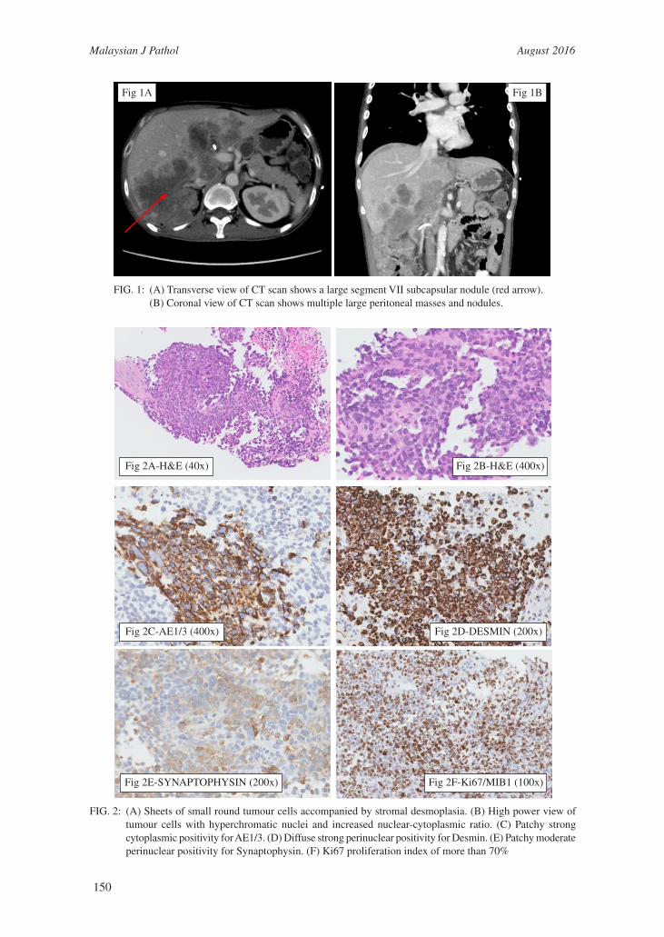
FIG. 2: (A) Sheets of small round tumour cells accompanied by stromal desmoplasia. (B) High power view of tumour cells with hyperchromatic nuclei and increased nuclear-cytoplasmic ratio. (C) Patchy strong cytoplasmic positivity for AE1/3. (D) Diffuse strong perinuclear positivity for Desmin. (E) Patchy moderate perinuclear positivity for Synaptophysin. (F) Ki67 proliferation index of more than 70%
Fig 2A-H&E (40x)
Fig 2C-AE1/3 (400x)
Fig 2E-SYNAPTOPHYSIN (200x)
Fig 2B-H&E (400x)
Fig 2D-DESMIN (200x)
Fig 2F-Ki67/MIB1 (100x)
151
DESMOPLASTIC SMALL ROUND CELL TUMOUR
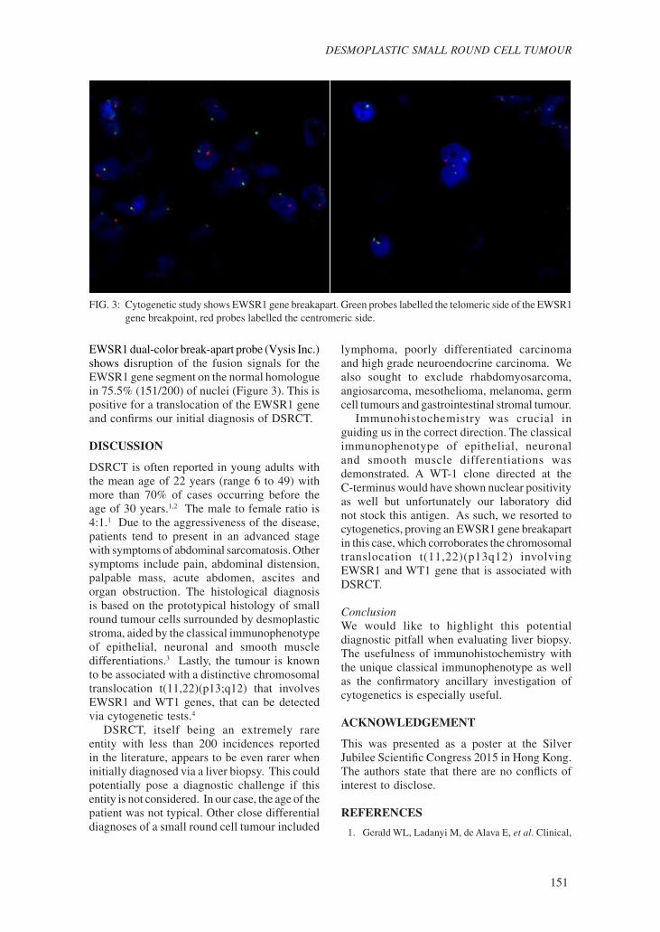
EWSR1 dual-color break-apart probe (Vysis Inc.) shows disruption of the fusion signals for the EWSR1 gene segment on the normal homologue in 75.5% (151/200) of nuclei (Figure 3). This is positive for a translocation of the EWSR1 gene and confirms our initial diagnosis of DSRCT.
DISCUSSION
DSRCT is often reported in young adults with the mean age of 22 years (range 6 to 49) with more than 70% of cases occurring before the age of 30 years.1,2 The male to female ratio is 4:1.1 Due to the aggressiveness of the disease, patients tend to present in an advanced stage with symptoms of abdominal sarcomatosis. Other symptoms include pain, abdominal distension, palpable mass, acute abdomen, ascites and organ obstruction. The histological diagnosis is based on the prototypical histology of small round tumour cells surrounded by desmoplastic stroma, aided by the classical immunophenotype of epithelial, neuronal and smooth muscle differentiations.3 Lastly, the tumour is known to be associated with a distinctive chromosomal translocation t(11,22)(p13;q12) that involves EWSR1 and WT1 genes, that can be detected via cytogenetic tests.4
DSRCT, itself being an extremely rare entity with less than 200 incidences reported in the literature, appears to be even rarer when initially diagnosed via a liver biopsy. This could potentially pose a diagnostic challenge if this entity is not considered. In our case, the age of the patient was not typical. Other close differential diagnoses of a small round cell tumour included
lymphoma, poorly differentiated carcinoma and high grade neuroendocrine carcinoma. We also sought to exclude rhabdomyosarcoma, angiosarcoma, mesothelioma, melanoma, germ cell tumours and gastrointestinal stromal tumour. Immunohistochemistry was crucial in guiding us in the correct direction. The classical immunophenotype of epithelial, neuronal and smooth muscle differentiations was demonstrated. A WT-1 clone directed at the C-terminus would have shown nuclear positivity as well but unfortunately our laboratory did not stock this antigen. As such, we resorted to cytogenetics, proving an EWSR1 gene breakapart in this case, which corroborates the chromosomal translocation t(11,22)(p13q12) involving EWSR1 and WT1 gene that is associated with DSRCT.
ConclusionWe would like to highlight this potential diagnostic pitfall when evaluating liver biopsy. The usefulness of immunohistochemistry with the unique classical immunophenotype as well as the confirmatory ancillary investigation of cytogenetics is especially useful.
ACKNOWLEDGEMENT
This was presented as a poster at the Silver Jubilee Scientific Congress 2015 in Hong Kong.The authors state that there are no conflicts of interest to disclose.
REFERENCES
1. Gerald WL, Ladanyi M, de Alava E, et al. Clinical,
FIG. 3: Cytogenetic study shows EWSR1 gene breakapart. Green probes labelled the telomeric side of the EWSR1 gene breakpoint, red probes labelled the centromeric side.
Malaysian J Pathol August 2016
152
pathologic, and molecular spectrum of tumors associated with t(11;22)(p13;q12): desmoplastic small round-cell tumor and its variants. J Clin Oncol. 1998; 16: 3028-36.
2. Ordóñez NG, el-Naggar AK, Ro JY, Silva EG, Mackay B. Intra-abdominal desmoplastic small cell tumor: a light microscopic, immunocytochemical, ultrastructural, and flow cytometric study. Hum Pathol. 1993; 24: 850-65.
3. Gerald WL, Miller HK, Battifora H, Miettinen M, Silva EG, Rosai J. Intra-abdominal desmoplastic small round-cell tumor. Report of 19 cases of a distinctive type of high-grade polyphenotypic malignancy affecting young individuals. Am J Surg Pathol. 1991; 15: 499-513.
4. Liu J, Nau MM, Yeh JC, Allegra CJ, Chu E, Wright JJ. Molecular heterogeneity and function of EWS-WT1 fusion transcripts in desmoplastic small round cell tumors. Clin Cancer Res. 2000; 6: 3522-9.