Embed Size (px)
Citation preview
To the Editor,
Abdominal desmoplastic small round cell tumor (DSRCT) is an extremely rare malignancy (1). It typically affects young men with an aggressive clinical course (2). A 23-year-old male presented with constipation and poorly localized dull abdominal pain lasting for one month. He also suffered from poor appetite and unintentional weight loss. Abdominal examination revealed a palpable lower abdominal mass. Blood
chemistry and tumor markers were unremarkable except for increased CA-125 (130 U/mL). Abdominopelvic ultrasound (US) demonstrated a macrolobulated heterogeneous pelvic mass measuring 12x10 cm with a mixed solid and cystic pattern extending to epigastrium with an unclear origin (Fig. 1A). Contrast-enhanced computed tomography (CT) depicted multiple large and lobulated masses (Fig 1B to D). Two largest masses, one in retrovesical space (Fig. 1B) and another located more superiorly in the abdomen (Fig. 1C) were
Correspondence to : Nesrin Gündüz, M.D., Dr. Erkın C. Goztepe, Kadıkoy, 34722 Istanbul, TurkeyE-mail: [email protected]
Submission date : 25/03/2017Acceptance date : 22/08/2017
Acta Gastro-Enterologica Belgica, Vol. LXXX, October-December 2017
LETTER 539
Imaging and histopathologic findings of desmoplastic small round cell tumor
A. Buz1, N. Gündüz1, N. Tekin1, Ö. Ekinci2, A. B. Ceyran3, A. Aslan1
(1) Department of Radiology, İstanbul Medeniyet University, Göztepe Training and Research Hospital, İstanbul, Turkey ; (2) Department of General Surgery, İstanbul Medeniyet University Göztepe Training and Research Hospital, İstanbul, Turkey ; (3) Department of Pathology, İstanbul Medeniyet University Göztepe Training and Research Hospital, İstanbul, Turkey.
Fig. 1. — Transabdominal US depicts a huge (12x10 cm in size), macrolobulated and mildly heterogeneous mass (arrows) with smooth borders (A). Intravenous contrast-enhanced abdominal CT shows the two dominant masses located at retrovesical region (B) (white arrows) and centrally in the abdomen (C) (white arrows). Perihepatic peritoneal implants (black arrowheads) are clearly demonstrated (D). Two distinct dominant masses (white arrows) and compression of the bladder and the rectum (white arrowheads) are demonstrated on sagittal T2-weighted MR images (E). A small amount of peritoneal fluid is clearly visible on axial T2-weighted MR images (F).
15-Gunduz.indd 539 12/12/17 14:38
540 N. Gündüz et al.
Acta Gastro-Enterologica Belgica, Vol. LXXX, October-December 2017
intestinal obstruction. However, radical surgery was deemed unsuitable due to extensive metastasis. Adjuvant chemotherapy was also administered. A large abdominal mass along with peritoneal implants on imaging in young males should raise the suspicion of DSRCT [1,2]. Multimodality imaging is helpful for the diagnosis and management and histopathological confirmation is mandatory in all cases of DSRCT..
References
1. BiSwal B.M., NaiK V.R., SHaMiM S.E. Intra-abdominal desmoplastic small round cell tumor : Presentation of four cases and review of the literature. Indian J. Med. Paediatr. Oncol., 2010, 31 : 24-27.
2. STUaRT-BUTTlE C.E., SMaRT C.J., PRiTCHaRD S., MaRTiN D., WELCH I.M. Desmoplastic small round cell tumour : a review of literature and treatment options. Surg. Oncol., 2008, 17 : 107-112.
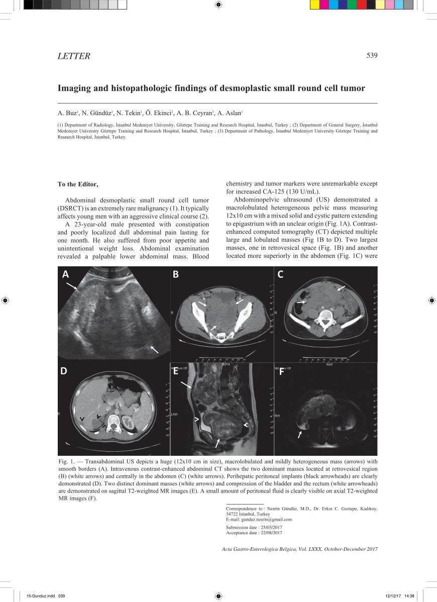
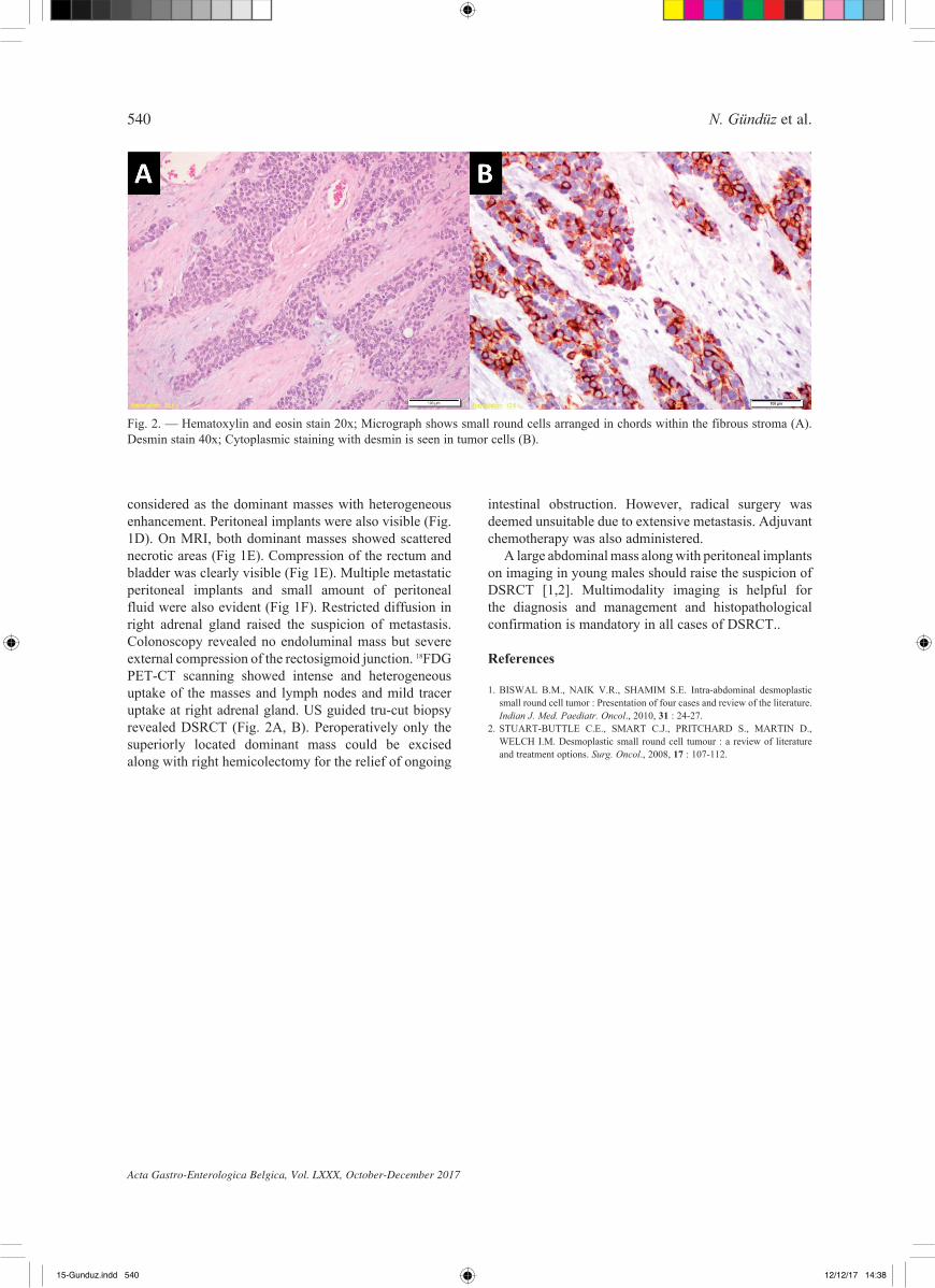
considered as the dominant masses with heterogeneous enhancement. Peritoneal implants were also visible (Fig. 1D). On MRI, both dominant masses showed scattered necrotic areas (Fig 1E). Compression of the rectum and bladder was clearly visible (Fig 1E). Multiple metastatic peritoneal implants and small amount of peritoneal fluid were also evident (Fig 1F). Restricted diffusion in right adrenal gland raised the suspicion of metastasis. Colonoscopy revealed no endoluminal mass but severe external compression of the rectosigmoid junction. 18FDG PET-CT scanning showed intense and heterogeneous uptake of the masses and lymph nodes and mild tracer uptake at right adrenal gland. US guided tru-cut biopsy revealed DSRCT (Fig. 2a, B). Peroperatively only the superiorly located dominant mass could be excised along with right hemicolectomy for the relief of ongoing
Fig. 2. — Hematoxylin and eosin stain 20x; Micrograph shows small round cells arranged in chords within the fibrous stroma (a). Desmin stain 40x; Cytoplasmic staining with desmin is seen in tumor cells (B).
15-Gunduz.indd 540 12/12/17 14:38