Embed Size (px)
Citation preview

Tierärztliche Hochschule Hannover
Degradation und Biokompatibilität der neuen Magnesiumlegierung LANd442 im
Vergleich zu LAE442 und der nicht degradablen Titanlegierung Ti6Al4V-Eli
nach intramedullärer Implantation in die Kaninchentibia
INAUGURAL-DISSERTATION
zur Erlangung des Grades einer Doktorin der Veterinärmedizin
- Doctor medicinae veterinariae -
(Dr. med. vet.)
vorgelegt von
Carolin Hampp
Heilbronn – Neckargartach
Hannover 2012

Wissenschaftliche Betreuung: Univ.-Prof. Dr. med. vet. Andrea Meyer-Lindenberg
Klinik für Kleintiere
Jetzt: Chirurgische und Gynäkologische Kleintierklinik
der Ludwig-Maximilians-Universität München
1. Gutachterin: Univ.-Prof. Dr. med. vet. Andrea Meyer-Lindenberg
2. Gutachter: Univ.-Prof. Dr. med. vet. Ingo Nolte
Tag der mündlichen Prüfung: 10.05.2012
Diese Dissertation entstand im Rahmen des Sonderforschungsbereichs 599 „Zukunftsfähige
bioresorbierbare und permanente Implantate aus metallischen und keramischen
Werkstoffen“ im Teilprojekt R6 „Degradable Implantate“, gefördert durch die Deutsche
Forschungsgemeinschaft (DFG).

Für meine Familie

Ergebnisse dieser Dissertation wurden in international anerkannten Fachzeitschriften
mit Gutachtersystem (peer review) zur Veröffentlichung angenommen oder
eingereicht:
• Advanced Engineering Materials: Advanced Biomaterials (angenommen am
29.10.2011)
DOI: 10.1002/adem.201180066
Research on the biocompatibility of the new magnesium alloy LANd442 –
an in vivo study in the rabbit tibia over 26 weeks
C. Hampp, B. Ullmann, J. Reifenrath, N. Angrisani, D. Dziuba, D. Bormann,
J.-M. Seitz, A. Meyer-Lindenberg
• Materials Science and Engineering: C (eingereicht am 07.03.2012)
Evaluation of the biocompatibility of two magnesium alloys as
degradable implant materials in comparison to titanium as non
resorbable material in the rabbit
C. Hampp, N. Angrisani, J. Reifenrath, D. Bormann, J.-M. Seitz,
A. Meyer-Lindenberg
Teilergebnisse dieser Dissertation wurden auf folgenden Fachkongressen
präsentiert:
• Euro BioMat 2011 – European Symposium on Biomaterials and related areas,
Jena, 13.-14.04.2011
In-vivo research on the biocompatibility of the new magnesium alloy
LANd442 on the basis of imaging procedures
C. Hampp, D. Rittershaus, J. Reifenrath, D. Bormann, J. Seitz,
A. Meyer-Lindenberg
• Jahrestagung der Deutschen Gesellschaft für Biomaterialien, Gießen,
10.-12.11.2011; BioMaterialien, 12, 1-4, 2011.
Untersuchung der Biokompatibilität von degradablen
Magnesiumlegierungen im Vergleich zu Titan im Kaninchenmodell
C. Hampp, J. Reifenrath, N. Angrisani, D. Bormann, J.-M. Seitz,
A. Meyer-Lindenberg


Inhaltsverzeichnis ___________________________________________________________________
Inhaltsverzeichnis
1 Einleitung ........................................................................................................... 9
2 Publikation I ..................................................................................................... 12
2.1 Abstract ...............................................................................................................13
2.2 Introduction.........................................................................................................13
2.3 Materials and Methods .......................................................................................15
2.3.1 Implants.....................................................................................................15
2.3.2 Animal model.............................................................................................15
2.3.3 Clinical investigation..................................................................................16
2.3.4 Radiological investigation ..........................................................................16
2.3.5 In vivo µ-computed tomography ................................................................16
2.3.6 Intravital staining for histologically investigating new bone growths ...........17
2.3.7 Euthanasia ................................................................................................18
2.3.8 Ex vivo µ-computed tomography ...............................................................18
2.3.9 Histological investigations .........................................................................19
2.3.10 Statistics....................................................................................................20
2.4 Results.................................................................................................................21
2.4.1 Clinical investigation..................................................................................21
2.4.2 Radiological investigations ........................................................................21
2.4.3 In vivo µ-computed tomography ................................................................22
2.4.4 Ex vivo µ-computed tomography ...............................................................25
2.4.5 Histological investigations .........................................................................27
2.5 Discussion...........................................................................................................29
2.6 Conclusions ........................................................................................................37
2.7 Acknowledgements ............................................................................................38
2.8 References ..........................................................................................................39
3 Publikation II .................................................................................................... 45
3.1 Abstract ...............................................................................................................46

Inhaltsverzeichnis ___________________________________________________________________
4 Diskussion ....................................................................................................... 47
5 Zusammenfassung.......................................................................................... 62
6 Summary.......................................................................................................... 65
7 Literaturverzeichnis ........................................................................................ 68
Einleitung ___________________________________________________________________
9
1 Einleitung
In der Orthopädie ist eine der wichtigsten Fragestellungen die nach den am besten
geeigneten Osteosynthesematerialien. Seit Jahren wird neben den etablierten
Materialien wie Titan und Stahl, die sich durch eine hohe Stabilität auszeichnen, an
resorbierbaren Implantatmaterialien geforscht. Bereits etabliert sind hierbei
verschiedene Kunststoffe wie PLA, PGA und PMMA (HOFMANN et al. 1990;
GRIFFITH 2000). Diese sind jedoch nicht stabil genug, um Anwendung am
belasteten Knochen zu finden (SONG 2007; SEALY a. GUO 2010; LICHTE et al.
2011). Daher konzentriert sich die Forschung auf degradable Metalle, wobei
Magnesium im Fokus dieser neuen Orientierung steht (WITTE et al. 2004; STAIGER
et al. 2006; FEYERABEND et al. 2010; MEYER-LINDENBERG et al. 2010;
HUEHNERSCHULTE et al. 2011). Das Leichtmetall Magnesium zeichnet sich durch
einen Elastizitätsmodul von 45 GPa aus (AVEDESIAN 1999), der im Vergleich zu
Titan (105-110 GPa (LONG a. RACK 1998)) oder Stahl (190-210 GPa (LEVESQUE
et al. 2004; MOAVENI 2010)) eine weitaus geringere Differenz zu dem des kortikalen
Knochens (18,6 GPa (RHO et al. 1993)) aufweist. Beachtet man zusätzlich die
Tatsache, dass ein Osteosyntheseimplantat zunächst die auftretenden Belastungen
aushalten muss, um den regenerierenden Knochen vor einer erneuten Fraktur zu
bewahren, erscheint ein etwas höherer Elastizitätsmodul durchaus vorteilhaft. Ist der
Unterschied jedoch zu groß, kommt es zum Phänomen des stress-shielding (SEALY
a. GUO 2010), bei dem das Implantat den heilenden Knochen von jeglicher
Belastung abschirmt. Hierdurch kommt es zu einer verminderten Mineralisierung des
Knochens (TONINO et al. 1976; BENLI et al. 2008) und die Gefahr der
Refrakturierung nach Entfernen des Implantates steigt, da das Implantatlager
zunächst wieder vom Knochen stabilisiert werden muss (WITTE et al. 2004).
Herkömmliche Implantatmaterialien haben zusätzlich den Nachteil, dass sie eine
Fremdkörperreaktion hervorrufen können, wenn sie im Körper belassen werden
(VOGGENREITER et al. 2003; LICHTE et al. 2011). Um diese Probleme zu
umgehen, werden die Implantate häufig entfernt, nachdem der erwünschte
Heilungseffekt eingetreten ist. Eine solche Zweitoperation zur Entfernung der
eingebrachten Implantate ist bei einem degradablen Osteosynthesematerial hinfällig
Einleitung ___________________________________________________________________
10
und stellt dadurch einen weiteren Vorteil von Magnesiumlegierungen dar (SONG
2007; BENLI et al. 2008). Da jede Operation Kosten, Schmerzen sowie ein nie
auszuschließendes Narkoserisiko für den Patienten bedeutet (ROKKANEN et al.
2000; WITTE et al. 2004), bietet ein degradierendes Implantat die Möglichkeit der
Verringerung dieser Faktoren.
Zusätzlich zeichnet sich Magnesium als körpereigenes Element grundsätzlich durch
eine sehr gute Verträglichkeit aus. Ein erhöhter Magnesiumspiegel, wie er im
Rahmen des Abbaus eingebrachter Implantate eventuell entstehen könnte, wird
durch den Körper selbst reguliert (VORMANN 2003) und hat in den zu erwartenden
Höchstkonzentrationen keine toxischen Folgen (SARIS et al. 2000).
Jedoch kann Magnesium nicht als alleiniges Element für Osteosyntheseimplantate
verwendet werden, da seine Primärstabilität nicht ausreicht, um den Knochen
postoperativ optimal zu stützen (MCBRIDE 1938) und diese zudem in chloridhaltigen
Medien zu schnell abnimmt (STAIGER et al. 2006; MEYER-LINDENBERG et al.
2010). Daher müssen andere Elemente zulegiert werden, um die Eigenschaften des
Materials zu verbessern. Bisherige Studien konnten zeigen, dass sich Seltene Erden
hierfür besonders eignen (WITTE et al. 2005; MINGXING et al. 2007; KRAUSE 2008;
HORT et al. 2009). In der Folge wurden verschiedene Legierungen wie LAE442,
ZEK100 und WE43 entwickelt und sowohl in vitro als auch in vivo getestet, wobei
sich LAE442 sehr vielversprechend zeigte (WITTE et al. 2005; THOMANN et al.
2009). Allen diesen Seltene Erden enthaltenden Materialien ist gemein, dass das
Mischungsverhältnis der einzelnen Elemente innerhalb der Legierungen nicht genau
bekannt ist.
Hierin liegt ein Problem für die Reproduzierbarkeit der Legierung und damit für die
Gewährleistung einer gleich bleibenden Qualität, was eine essentielle Voraussetzung
für den Einsatz in der Medizin darstellt. Daher wurde die neue Magnesiumlegierung
LANd442 entwickelt (SEITZ et al. 2011), die aufgrund der bisherigen guten
Ergebnisse auf LAE442 basiert (WITTE et al. 2005; THOMANN et al. 2009).
Verschiedene Studien stuften unter anderen Elementen der Seltenen Erden Neodym
in in vitro Versuchen als gut verträglich ein (DRYNDA et al. 2009; FEYERABEND et

Einleitung ___________________________________________________________________
11
al. 2010). Daher wurde der Anteil des in LAE442 enthaltenen Seltenen Erden-
Gemisches bei der Entwicklung der neuen Legierung durch das Einzelelement
Neodym ersetzt, um eine genau definierte Legierungszusammensetzung zu
erreichen (ROKHLIN 2003). Dieser Ansatz wurde bereits bei der Legierung
LACer442 verfolgt, bei der die Seltene Erden-Mischung durch das Element Cer
ersetzt wurde, die sich jedoch im in vivo Versuch als nicht biokompatibel
herausstellte (REIFENRATH et al. 2010). Allerdings besitzt Neodym im Gegensatz
zu Cer laut einer Verträglichkeitsstudie in vitro kein toxisches Potential
(FEYERABEND et al. 2010) bzw. erst in sehr hohen Konzentrationen (DRYNDA et
al. 2009), sodass von einer besseren Verträglichkeit ausgegangen wird.
In der zugänglichen Literatur existieren jedoch keine in vivo Studien, die die
Biokompatibilität von Neodym als Legierungselement untersuchen. Daher war es das
Ziel der vorliegenden Arbeit, die Biokompatibilität der neuen Legierung LANd442 in
vivo unter Berücksichtigung der Degradation über verschiedene Zeiträume (vier und
acht Wochen bzw. sechs Monate) im Kaninchenmodell zu prüfen. Als
Vergleichsmaterial wurde die als vielversprechend geltende Magnesiumlegierung
LAE442 herangezogen und das etablierte Osteosynthesematerial Titan diente als
Kontrolle. Zudem wurden Tibiae in die Untersuchung einbezogen, bei denen die
Operation durchgeführt wurde, die aber kein Implantat erhielten (Leertibiae), um die
Auswirkungen der Operationsmethode von den potentiellen durch die Implantate
induzierten Vorgängen am Knochen zu unterscheiden.
Publikation I ___________________________________________________________________
12
2 Publikation I
Das Manuskript wurde am 07.03.2012 im Journal „Materials Science and
Engineering: C“ zur Veröffentlichung eingereicht.
Evaluation of the biocompatibility of two magnesium alloys as
degradable implant materials in comparison to titanium as non
resorbable material in the rabbit
Carolin Hampp*, Nina Angrisani, Janin Reifenrath, Dirk Bormann, Jan-Marten Seitz,
Andrea Meyer-Lindenberg
C. Hampp, Dr. N. Angrisani, Dr. J. Reifenrath:
Small Animal Clinic, University of Veterinary Medicine Hanover, Bünteweg 9, 30559
Hanover, Germany
Dr. D. Bormann, J.-M. Seitz:
Institute of Materials Science, Leibniz University Hanover, An der Universität 2,
30823 Garbsen, Germany
Prof. Dr. A. Meyer-Lindenberg:
Clinic for Small Animal Surgery and Reproduction, Centre of Clinical Veterinary
Medicine, Faculty of Veterinary Medicine, Ludwig-Maximilians-University München,
Veterinärstraße 13, 80539 Munich, Germany
* Corresponding author

Publikation I ___________________________________________________________________
13
2.1 Abstract
The aim of this study is to compare the biocompatibility of the two magnesium based
alloys LAE442 and LANd442 with that of titanium. For this purpose, cylindrical
implants were introduced into the medullary cavity of rabbit’s tibiae for 4 and 8
weeks. Animals without any implant served as a control. In the follow-up, clinical, X-
ray and µCT-investigations were performed to evaluate the reactions of the bone
towards the implanted materials. After euthanasia, ex vivo µCT- and histological
investigations were performed to verify the results of the in vivo tests. It could be
shown that all materials induce changes in the bone. Whereas LANd442 caused the
most pronounced reactions, such as increasing bone volume and bone porosity and
decreasing bone density, titanium showed the most bone-implant contact by forming
trabeculae. The tibiae of rabbits without implants also reacted by forming cavities, it
is therefore assumed that the surgery method itself influences the bone. Compared
to LANd442, LAE442 seems to be the more qualified alloy since it demonstrated
better clinical tolerance.
2.2 Introduction
The treatment of fractures is a major field within orthopaedic surgery. Commonly
used materials are stainless steel or titanium (DISEGI a. ESCHBACH 2000;
POHLER 2000; FERRARIS et al. 2011) which have been long established and are
still considered appropriate and almost unrivalled. This is evident due to the fact that
current research focuses on the improvement of these established materials
(JAIMES et al. 2010; KERÄNEN et al. 2011) rather than developing alternatives.
However, the disadvantage of stainless steel and titanium is that both materials are
non resorbable. Therefore, they have to be removed, following complete bone
healing, in a second surgery or, when left in the organism, can cause foreign body
reactions (VOGGENREITER et al. 2003).
Competing with these durable materials are resorbable materials like polymers and
ceramics which spare the patient a second surgical procedure to remove the implant.
However, polymers can also cause foreign body reactions and neither material has
sufficient mechanical load carrying capacity for the use in weight bearing bones
(LICHTE et al. 2011). In recent years, degradable implant materials which are based
Publikation I ___________________________________________________________________
14
on magnesium were studied to a greater extent (WITTE et al. 2005; REIFENRATH et
al. 2010; ATRENS et al. 2011). They should combine the ability to degrade with
sufficient mechanical properties. The advantages of employing magnesium as the
main component are obvious: as an essential element of the human body,
magnesium is well tolerated (SARIS et al. 2000) and it prevents stress shielding
during bone healing (SEALY a. GUO 2010) due to its Young’s modulus which, of all
the osteosynthetic materials, is closest to that of bone (STAIGER et al. 2006; XU et
al. 2007). Several studies showed that the mechanical properties and the
biocompatibility of magnesium can be influenced by alloying with other elements
(STAIGER et al. 2006; KIM et al. 2008; ATRENS et al. 2011). Particularly the use of
rare earth elements (RE) led to good results (MORDIKE 2002; MINGXING et al.
2007; HORT et al. 2009). The alloy LAE442 which, besides magnesium, lithium and
aluminium, contains a mixture of various RE proved especially promising (THOMANN
et al. 2009; KRAUSE et al. 2010; WITTE et al. 2010).
Previous studies of this alloy described implantation periods of three or more months
(THOMANN et al. 2009; KRAUSE et al. 2010). However, literature on earlier time
points, which would represent the remodelling processes during fracture healing, is
currently lacking. For this reason, the present study chose implantation periods of 4
and 8 weeks to evaluate the potentially early occurrence of bone remodelling
processes. Owing to the previous good results for LAE442, this alloy was included in
the present study and was compared to LANd442, which is based on LAE442 but
only contains neodymium instead of the RE mixture. The replacement of the RE
mixture by the single element neodymium aims to achieve a better reproducibility. As
control groups, two models were chosen. On the one hand, titanium implants were
introduced; on the other hand, tibiae were used, which underwent the same surgical
procedure but without receiving an implant as performed in previous studies
(THOMANN et al. 2009; HUEHNERSCHULTE et al. 2011).
Publikation I ___________________________________________________________________
15
2.3 Materials and Methods
2.3.1 Implants
In this study, cylindrical implants (Ø 2.5 mm, 25 mm length) made of the magnesium
based alloys LAE442 and LANd442 were used as well as titanium-alloy implants of
the same geometry which were employed as a control (Ti6Al4V-Eli; S+D Spezialstahl
Handelsgesellschaft mbH, Stelle, Germany). The magnesium implants were specially
produced for this study according to a previously published method (KRAUSE et al.
2010; THOMANN et al. 2010a; HUEHNERSCHULTE et al. 2011; SEITZ et al. 2011).
Besides 90 wt% magnesium, the alloy LAE442 contains 4 wt% lithium, 4 wt%
aluminium and 2 wt% of a rare earth mixture. The alloy LANd442 is based on
LAE442 and contains the same proportions of magnesium, lithium and aluminium.
Here however, the rare earth mixture was replaced by 2 wt% of the single element
neodymium.
2.3.2 Animal model
The animal tests were approved by the Federal Office of Consumer Protection and
Food Safety, according to paragraph 8 of the animal protection law, with the
reference number 33.9-42502-04-07/1363.
For the current study 28 New Zealand White Rabbits (Charles River, Kisslegg,
Germany) were randomly placed into six groups, a 4- and an 8-week group for each
implant material (table 1). The implants of the LAE442- and LANd442-groups were
introduced according to a previously described method (HAMPP et al. 2012) into the
medullary cavity of both tibiae in four rabbits, respectively. Two rabbits only received
one implant each. In their other hind leg, surgery was carried out using the same
procedure, but no implant was inserted. The titanium implants were introduced into
two rabbits per time group on both sides at the same location. After surgery, the
rabbits received enrofloxacin (10 mg/kg, Baytril® 2.5%, Bayer HealthCare,
Leverkusen, Germany) and meloxicam (0.15 mg/kg, Metacam®, Boehringer
Ingelheim, Ingelheim, Germany) for a time period of ten days.

Publikation I ___________________________________________________________________
16
Table 1: Overview of the number of tibiae per implant material and time period
material 4-week group 8-week group LAE442 10 10
LANd442 10 10 Titanium 4 4
without implant 4 4
2.3.3 Clinical investigation
The animals were examined daily according to a previously described method
(HAMPP et al. 2012). These examinations focused on possible changes occurring in
the rabbits’ hind legs. If the rabbits showed lameness, they were treated with
meloxicam (0.15 mg/kg, Metacam®, Boehringer Ingelheim, Ingelheim, Germany)
beyond day 10 after surgery.
2.3.4 Radiological investigation
The tibiae of all rabbits were radiologically investigated once every week in two
layers (anterior-posterior, mediolateral; 48 kV and 6.3 mAs) to evaluate bone
alterations and the development of gas. All changes were assessed by a semi-
quantitative scoring according to HUEHNERSCHULTE et al. (2011) which allows
score values from 0 (no occurrence) to 3 (strong occurrence) for the individual
parameters (growths at the implantation site at the proximal tibia, growths at the
implant location at the diaphysis, gas, changes in the medullary cavity and corticalis).
Finally, all score values of the rabbits in one group were summed to a total score and
the respective group’s mean value was computed for each time point of the
investigation.
2.3.5 In vivo µ-computed tomography
Over the investigation periods of 4 and 8 weeks, µ-computed tomography
evaluations (µCT) of the rabbits were performed (resolution: 41 µm, projections: 1000
at 0-180°, integration time: 100 ms; XtremeCT, Scanco Medical, Zurich, Switzerland).
The rabbits of the 4-week groups were scanned weekly and those in the time period
Publikation I ___________________________________________________________________
17
over 8 weeks, biweekly. The investigation was done under general anaesthesia. The
evaluated area was limited by the knee joint space in the proximal direction and
reached up to about 5 mm beneath the implant in the distal direction. The analysis of
the µCT-investigations was done in both two and also three dimensions.
Two-dimensional evaluation
The overall impression of the bone including formation of cavities, periosteal and
endosteal new bone growth, the growth behaviour onto the implant as well as the
formation of gas bubbles were evaluated by an established semi-quantitative scoring
(HUEHNERSCHULTE et al. 2011) based on nine selected cross-sections
(THOMANN et al. 2010a). For every parameter, score values from 0 to 3 were given
for each cross-section (no occurrence: value 0, strong occurrence: value 3) and a
mean value per time group and per investigation time point was computed. The tibiae
of the animals with titanium implants were not investigated in this way because of an
expected insufficient resolution of detail as a consequence of the titanium’s stronger
absorption of X-rays (BERNHARDT et al. 2004).
Three-dimensional evaluation
On the basis of every in vivo µCT-investigation a three-dimensional evaluation of
each tibia was performed, including the bone area, in which the bone-implant-
compound was completely visible. Using the computational software (µCT Evaluation
Program V6.0; Scanco Medical, Zurich, Switzerland), the threshold for the evaluation
of the bone was determined to be 160. Subsequently, the computations of the tibia
areas were used to compute the bone volume (in mm³/slice), the bone density (in mg
HA/ccm) as well as the bone porosity (in %). An evaluation of mean values and
standard deviations was done for every investigation time point for every time group.
2.3.6 Intravital staining for histologically investigating new bone growths
For the fluorescent-microscopic investigation following euthanasia, the animals
received three different fluorochromes which were chosen according to RAHN (1976)
and injected subcutaneously at specific times (table 2).
Publikation I ___________________________________________________________________
18
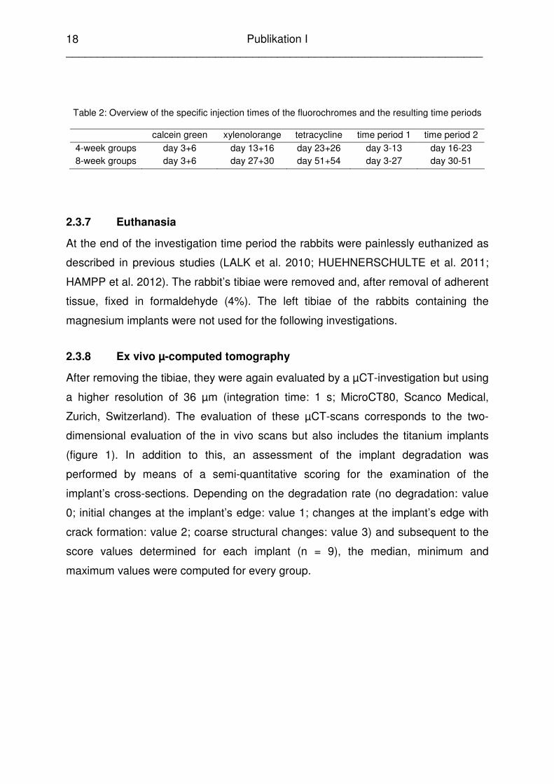
Table 2: Overview of the specific injection times of the fluorochromes and the resulting time periods
calcein green xylenolorange tetracycline time period 1 time period 2 4-week groups day 3+6 day 13+16 day 23+26 day 3-13 day 16-23 8-week groups day 3+6 day 27+30 day 51+54 day 3-27 day 30-51
2.3.7 Euthanasia
At the end of the investigation time period the rabbits were painlessly euthanized as
described in previous studies (LALK et al. 2010; HUEHNERSCHULTE et al. 2011;
HAMPP et al. 2012). The rabbit’s tibiae were removed and, after removal of adherent
tissue, fixed in formaldehyde (4%). The left tibiae of the rabbits containing the
magnesium implants were not used for the following investigations.
2.3.8 Ex vivo µ-computed tomography
After removing the tibiae, they were again evaluated by a µCT-investigation but using
a higher resolution of 36 µm (integration time: 1 s; MicroCT80, Scanco Medical,
Zurich, Switzerland). The evaluation of these µCT-scans corresponds to the two-
dimensional evaluation of the in vivo scans but also includes the titanium implants
(figure 1). In addition to this, an assessment of the implant degradation was
performed by means of a semi-quantitative scoring for the examination of the
implant’s cross-sections. Depending on the degradation rate (no degradation: value
0; initial changes at the implant’s edge: value 1; changes at the implant’s edge with
crack formation: value 2; coarse structural changes: value 3) and subsequent to the
score values determined for each implant (n = 9), the median, minimum and
maximum values were computed for every group.

Publikation I ___________________________________________________________________
19
Figure 1: Ex vivo µCT cross-sections of the bone-implant-compounds of a) LAE442, b) LANd442, c)
titanium, d) cross-section of a tibia without implant
2.3.9 Histological investigations
Following the µCT-investigations, the complete bone-implant-compound was
detached and embedded in hydroxyethylmethacrylate (Technovit 7200 VLC, Heraeus
Kulzer GmbH, Wehrheim, Germany) according to the manufacturer’s instructions. To
produce the 50 µm thick histological cross-sections of the bone, the cutting and
grinding technique was used according to DONATH (1988). At central cross-sections
of the bone, fluorescent investigations were carried out as well as transmission
microscopy investigations of histological slices stained with TRAP and toluidine blue.
Fluorescent-microscopic investigation
One cross-section per tibia was fluorescent-microscopically evaluated. Based on the
intravitally injected fluorochromes, which were visible as double bands due to two
successive injections, the mineral apposition rate (MAR) could be determined
(PARFITT 1987). Therefore, the distance between the bands of two adjacent
fluorochrome sequences was measured and divided by the number of days between
the corresponding injections (PARFITT 1987). The time points of the intravital
stainings resulted in two different time periods for the 4-week groups and the 8-week
groups, respectively (table 2). In the present study, a multichannel image of the
histological slices was generated using specific filters (FS 14, FS 18, FS 46, Carl
Zeiss AG, Jena, Germany) and the distance at twelve specific locations was
Publikation I ___________________________________________________________________
20
measured according to an established method (REIFENRATH et al. 2011; HAMPP et
al. 2012). Finally, mean values and standard deviations were computed for every
slice to determine the mean value of the individual groups.
TRAP staining
One cross-section of each tibia was subjected to a TRAP staining (Tartrate-Resistant
Acid Phosphatase; naphthol AS-MX phosphate / Fast Red TR Salt, Sigma Aldrich,
St. Louis, USA) for viewing the osteoclasts (MOSTAFA et al. 1982; LINDUNGER et
al. 1990). The counting of osteoclasts and Howship’s lacunae was carried out three
times at x 200 magnification on every stained histological slice. Afterwards, mean
values and standard deviations for all groups were determined.
Toluidine blue
The toluidine blue staining (0.1% toluidine blue O, Chroma, Münster, Germany) was
applied to two histological cross-sections of the bone per tibia. Using a previously
described semi-quantitative scoring (HAMPP et al. 2012), the slices were
investigated at x 100 magnification for the following parameters: overall impression of
the bone including cavities, periosteal bone growth and remodelling, endosteal bone
growth and remodelling, bone-implant contact area, peri-implant fibrous capsule
formation. For the evaluation, score values from 0 (no occurrence) to 3 (strong
occurrence) were assigned depending on the parameter’s occurrence. Subsequent
to this, the mean value as well as the standard deviation of all investigated cross-
sections in one group was computed (LAE442, LANd442, without implant: n = 10;
titanium: n = 8).
2.3.10 Statistics
The values determined for the present study were analyzed using the Microsoft
Office Excel program (Microsoft Office XP, Microsoft Corporation, Redmond, USA)
and SPSS version 17.0 (SPSS: an IBM Company, Chicago, USA). Firstly, they were
tested for a normal distribution. Normally distributed values were checked for
statistical significance by means of a t-test or ANOVA, respectively; non-normally
Publikation I ___________________________________________________________________
21
distributed values were tested by means of Wilcoxon- or Mann-Whitney-tests.
Statistical significance existed for p < 0.05, whereas p < 0.01 indicated a highly
significant difference.
2.4 Results
2.4.1 Clinical investigation
During the post-operative period, all rabbits showed swelling and coarse peripheral
augmentation at the implantation's location. Redness occurred in 45 of 56 legs. Only
one animal of the 4-week LAE442-group demonstrated wound dehiscence on day 15
and 16. Mild subcutaneous emphysema could be found in two legs of the 4-week
LAE442-group (lasting 1 and 16 days, respectively) and four legs of the 8-week
LANd442-group (1 to 7 days). Within this group two rabbits showed a low-grade
lameness of one hind leg which lasted one day and resulted in the administration of
meloxicam. No tibiae showed infections.
2.4.2 Radiological investigations
Within the 4-week groups, tibiae with magnesium implants showed initial alterations
in week 1 (figure 2). Contrary to this, in the titanium-group an increase in the score
could not be found before week 3, however, then an abrupt score value of 2.0 was
found which increased further to the final value 2.5 in week 4.
The final value of the LAE442-group was also 2.5 whereas the 4-week LANd442
group reached a final score of 1.5. Also, tibiae without implants showed radiographic
changes. The score values increased parallel to the LANd442-group from 0.3 in
week 1 to 1.8 in week 4.
In contrast to this, all groups over 8 weeks showed initial changes in week 3. Here, in
the titanium-group an increase to 1.0 score point was noted which, at the same time,
appeared as the maximum final value obtained. The animals with magnesium
implants showed a nearly parallel development, whereas the LAE442-group
constantly showed higher values, but already attained the final value of 1.8 score
points in week 7. The LANd442-group exhibited a stronger increase in week 8 to the

Publikation I ___________________________________________________________________
22
final value of 2.3 score points. The group without implants formed a plateau at 1.3
score points in week 5 to 7, but decreased finally to 0.7 score points.
The largest fraction of bone changes was contributed by the parameters for growth at
the implantation site and growths at the diaphysis.
Figure 2: Development of the investigated bone alterations in all experimental groups by means of
radiological investigations over a) 4 weeks, b) 8 weeks
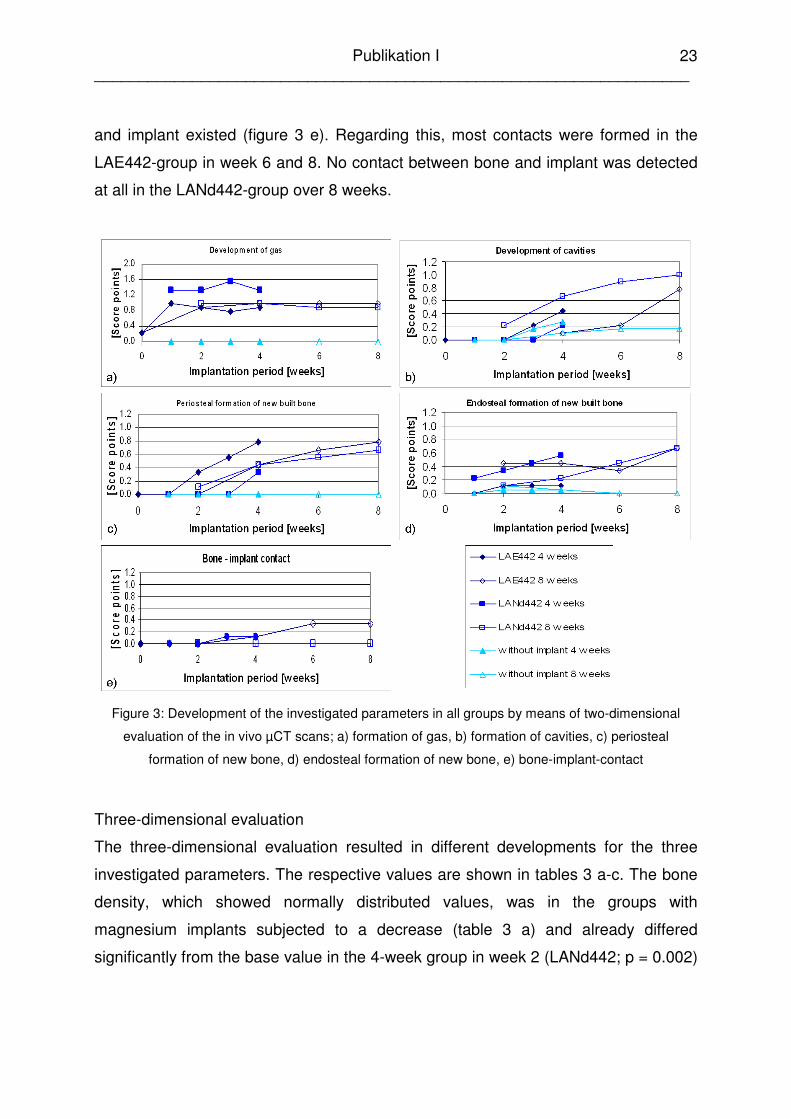
2.4.3 In vivo µ-computed tomography
Two-dimensional evaluation
In the two-dimensional evaluation of the in vivo µCT-scans, which excluded the
titanium implants because of their expected irradiation, it was shown that not all
parameters were equally pronounced. The highest score values were achieved for
the formation of gas (figure 3 a), especially in the LANd442-group over 4 weeks. In
contrast to this, gas was not even observed directly after surgery in either group
without implants. This was followed by the formation of cavities (figure 3 b), mainly in
the LANd442-group over 8 weeks (0.1 score point in week 8). Contrastingly, the
group without implants showed less cavities (0.2 score points in week 8) in the
corresponding time period. With 0.8 score points for their respective final time points,
the LAE442-groups showed over 4 and 8 weeks the most periosteal formation of new
bone which, in the groups without implants, was not detected over the entire
investigation period (figure 3 c). However, the endosteal formation of new bone was
most strongly pronounced in both LANd442-groups, while again both groups without
implants showed the least changes (figure 3 d). All in all, less contact between bone
Publikation I ___________________________________________________________________
23
and implant existed (figure 3 e). Regarding this, most contacts were formed in the
LAE442-group in week 6 and 8. No contact between bone and implant was detected
at all in the LANd442-group over 8 weeks.
Figure 3: Development of the investigated parameters in all groups by means of two-dimensional
evaluation of the in vivo µCT scans; a) formation of gas, b) formation of cavities, c) periosteal
formation of new bone, d) endosteal formation of new bone, e) bone-implant-contact
Three-dimensional evaluation
The three-dimensional evaluation resulted in different developments for the three
investigated parameters. The respective values are shown in tables 3 a-c. The bone
density, which showed normally distributed values, was in the groups with
magnesium implants subjected to a decrease (table 3 a) and already differed
significantly from the base value in the 4-week group in week 2 (LANd442; p = 0.002)
Publikation I ___________________________________________________________________
24
and week 3 (LAE442; p = 0.016). These significant changes persisted until week 4. In
the 8-week groups only those animals of the LANd442-group showed significant
changes in bone density from week 6 (p < 0.001), compared to the base value. In
contrast, both titanium-groups showed an increase of the bone density which,
however, underwent no significant changes. The groups without implants showed a
contradictory behaviour. Here, in the 4-week group a tendency of the bone density to
decrease was noted and to increase in the 8-week group. Both developments also
produced no significant changes. In comparing the individual groups, the LANd442-
group over 8 weeks already showed significant differences to the LAE442-group (p =
0.001) and the titanium-group (p = 0.003) from week 2, to the group without implants
from week 6 (p = 0.029).
The values of the bone volume also followed a normal distribution and showed, in
contrast to the bone density in the LAE442- and LANd442-groups, an increase (table
3 b). However, in both LANd442-groups significant differences to the base value
were again observable. The 4-week group differed significantly from the base value
in weeks 2 (p = 0.024) and 3 (p = 0.002), the 8-week group in week 8 (p = 0.022).
The LAE442-group over 4 weeks showed significant differences between weeks 1
and 4 (p = 0.005). Comparing the groups, significant differences were seen in week 4
between the 4-week groups of titanium and without implants (p = 0.008), and
between titanium and LANd442 (p = 0.039).
The values of bone porosity were not normally distributed and, by considering the
base and end values (table 3 c), underwent no or only minor changes. In the 8-week
groups of LAE442, LANd442 and without implants, a tendency to increase was
noted, whereas both titanium-groups as well as the LAE442-group showed a minor
decrease of the porosity over 4 weeks. From these results, significant differences
resulted from week 3 (p = 0.024) between the 4-week titanium group and the
respective LAE442- and LANd442-groups. In the corresponding time period over 8
weeks, the titanium-group differed significantly from the LAE442-group (p = 0.014)
and the LANd442-group (p = 0.024) in week 8. In the chronological sequences, no
group showed significant changes.

Publikation I ___________________________________________________________________
25
Table 3: Development of the investigated parameters in all groups by means of three-dimensional
evaluation of the in vivo µCT scans; a) bone density (in mg HA/ccm), normally distributed values; b)
bone volume (in mm³/slice), normally distributed values; c) bone porosity (in %), non-normally
distributed values. Additionally stated are significances between the individual groups (a) and within a
group (b), respectively.
2.4.4 Ex vivo µ-computed tomography
Bone
The results of the ex vivo µCT-investigations confirmed those of the in vivo
investigations to the extent that the parameters “formation of cavities” and “periosteal

Publikation I ___________________________________________________________________
26
formation of new bone” occurred most strongly, whereas bone-implant contact
developed only sporadically (table 4). The formation of cavities was demonstrated by
all groups and the deviations between them were only slight. The contact between
bone and implant only occurred distinctly in both titanium-groups (1.4 and 1.8 score
points, respectively) and was rudimentary in the LANd442-group over 8 weeks (0.1
score point). Endosteal, as well as periosteal formation of new bone could be
observed in all groups, except the groups without implants. For the endosteal
parameter, again both titanium-groups showed the strongest formation (1.2 and 0.9
score points, respectively) and the LAE442-groups the lowest (0.3 and 0.4 score
points, respectively). The results for the periosteal parameter contrasted this (both
LAE442-groups: 1.6 score points; titanium: 0.4 and 0.3 score points, respectively).
Table 4: Final values of the investigated parameters in all groups by means of two-dimensional
evaluation of the ex vivo µCT scans; stated are in each case (a) minimum value, (b) median and (c)
maximum value per group and parameter
group cavities bone-implant-contact
endosteal formation of new
bone
periosteal formation of new
bone (a) (b) (c) (a) (b) (c) (a) (b) (c) (a) (b) (c) LAE442
4 weeks 1.1 1.2 2.3 0.0 0.0 0.0 0.0 0.3 0.4 0 1.6 2.0 8 weeks 0.6 1.0 1.4 0.0 0.0 0.1 0.3 0.4 0.9 0.6 1.6 2.6
LANd442 4 weeks 0.8 1.0 1.3 0.0 0.0 0.1 0.0 0.6 0.7 0.0 0.8 2.9 8 weeks 1.0 1.3 1.8 0.0 0.1 0.3 0.4 0.8 0.8 0.2 1.0 2.8
Titanium 4 weeks 1.0 1.2 2.0 0.9 1.4 2.3 1.1 1.2 1.3 0.3 0.4 0.8 8 weeks 0.2 0.8 1.3 1.0 1.9 2.7 0.8 0.9 1.2 0.0 0.3 0.7
without implant 4 weeks 0.6 0.8 1.0 - - - 0.0 0.0 0.0 0.0 0.0 0.3 8 weeks 0.4 0.7 0.9 - - - 0.0 0.0 0.2 0.0 0.0 0.0
Implant
For the degradation of the implants, a median value of 0.0 was determined in all the
4-week groups (LANd442: min 0.0, max 1.0; LAE442: min 0.0, max 0.0; titanium: min
0.0, max 0.0). In the 8-week groups, LANd442 attained a median value of 1.0 (min
0.0, max 1.0), in the LAE442- and titanium-groups the implants appeared almost
Publikation I ___________________________________________________________________
27
unchanged (LAE442: median 0.0, min 0.0, max 1.0; titanium: median 0.0, min 0.0,
max 0.0).
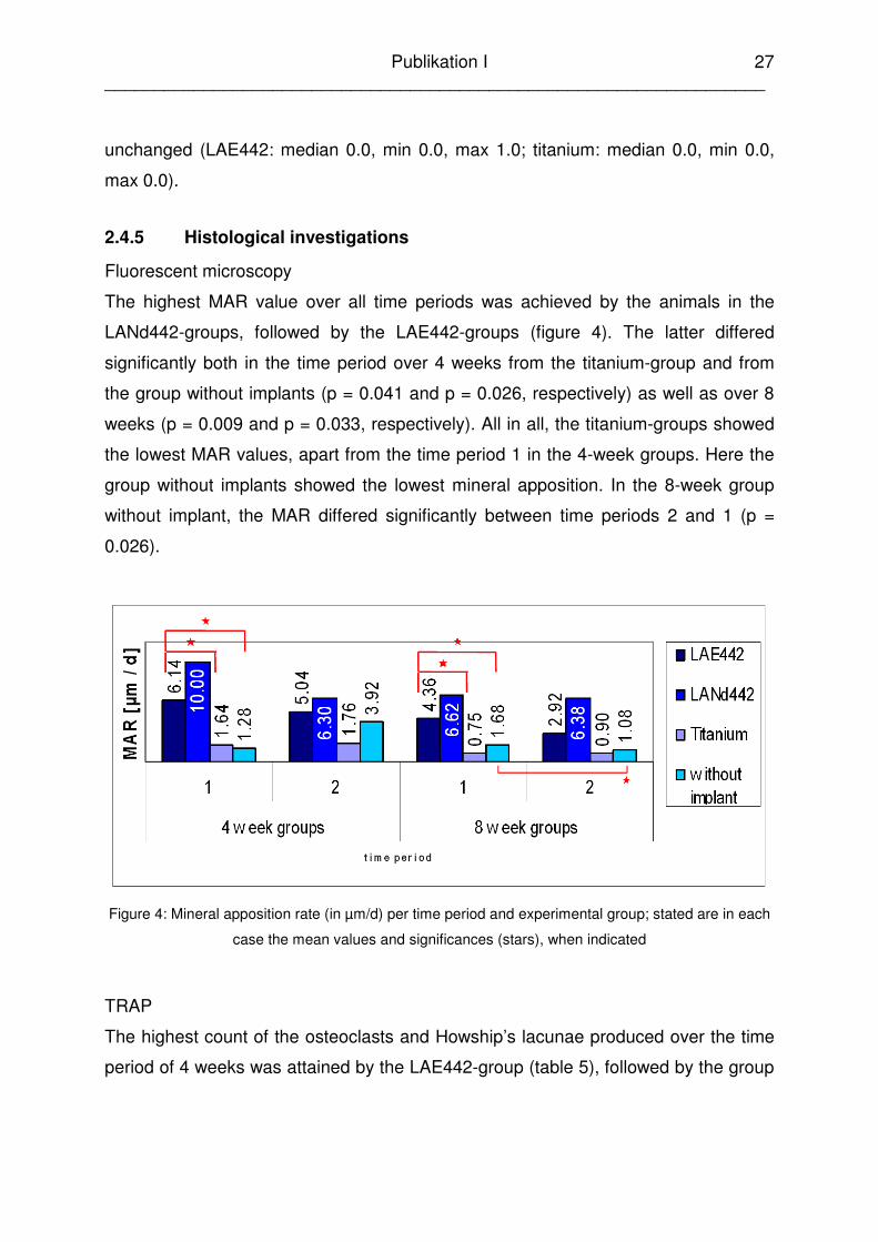
2.4.5 Histological investigations
Fluorescent microscopy
The highest MAR value over all time periods was achieved by the animals in the
LANd442-groups, followed by the LAE442-groups (figure 4). The latter differed
significantly both in the time period over 4 weeks from the titanium-group and from
the group without implants (p = 0.041 and p = 0.026, respectively) as well as over 8
weeks (p = 0.009 and p = 0.033, respectively). All in all, the titanium-groups showed
the lowest MAR values, apart from the time period 1 in the 4-week groups. Here the
group without implants showed the lowest mineral apposition. In the 8-week group
without implant, the MAR differed significantly between time periods 2 and 1 (p =
0.026).
Figure 4: Mineral apposition rate (in µm/d) per time period and experimental group; stated are in each
case the mean values and significances (stars), when indicated
TRAP
The highest count of the osteoclasts and Howship’s lacunae produced over the time
period of 4 weeks was attained by the LAE442-group (table 5), followed by the group

Publikation I ___________________________________________________________________
28
without implants, LANd442 and titanium. In contrast to this, in the time period over 8
weeks the descending order was LANd442, LAE442, titanium, without implants.
Comparing both time periods within the materials, the groups LAE442 and without
implants showed higher values over 4 weeks whereas LANd442 and titanium
implants activated more osteoclasts over 8 weeks (table 5). There were no significant
differences between the individual time groups. However, the titanium-group and the
group without implants differed significantly over 4 weeks from the respective 8-week
group (p = 0.043 and p = 0.029, respectively).
Table 5: Number of osteoclasts and Howship’s lacunae per implant material and time group; stated
are in each case the mean values and standard deviations, as well as significances between the time
groups of titanium (a) and the time groups without implant (b)
material 4-week group 8-week group MV 82 55 LAE442 SD 57 39 MV 42 61 LANd442 SD 20 39 MV 11a 42a Titanium SD 5 24 MV 48b 12b
without implant SD 25 5
Toluidine blue
With the help of the toluidine blue staining, it could be shown, that the cross-sections
of the tibiae in all groups exhibited cavities (figure 5), whereas less cavities were
formed over 4 weeks than over 8 weeks, apart from the titanium-groups. The extent
of the periosteal remodelling occurred in almost all groups equally with the score
value 1.0. The exceptions were both LAE442-groups and the LANd442-group over 8
weeks, which showed a stronger remodelling with 2.0 and 1.5 score points,
respectively. The parameter “endosteal remodelling” was assessed in all groups with
the score value 1.0. According to this, the periosteal formation of new bone occurred
more strongly than the endosteal formation. In the first case, the LANd442-groups
and the LAE442-group over 8 weeks demonstrated the most extensive changes,
whereas in the second case, only the LANd442-group over 8 weeks attained more
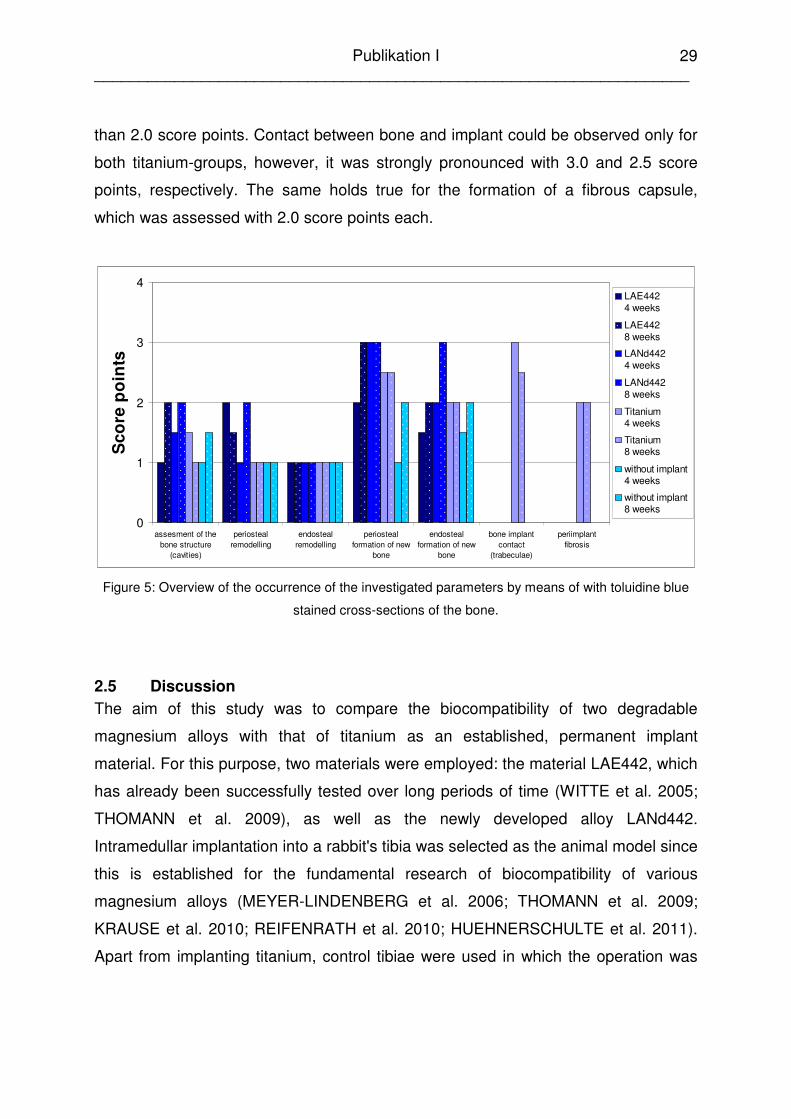
Publikation I ___________________________________________________________________
29
than 2.0 score points. Contact between bone and implant could be observed only for
both titanium-groups, however, it was strongly pronounced with 3.0 and 2.5 score
points, respectively. The same holds true for the formation of a fibrous capsule,
which was assessed with 2.0 score points each.
0
1
2
3
4
assesment of thebone structure
(cavities)
periostealremodelling
endostealremodelling
periostealformation of new
bone
endostealformation of new
bone
bone implantcontact
(trabeculae)
periimplantfibrosis
Sco
re p
oin
ts
LAE442 4 weeks
LAE442 8 weeks
LANd442 4 weeks
LANd442 8 weeks
Titanium 4 weeks
Titanium 8 weeks
without implant4 weeks
without implant8 weeks
Figure 5: Overview of the occurrence of the investigated parameters by means of with toluidine blue
stained cross-sections of the bone.
2.5 Discussion
The aim of this study was to compare the biocompatibility of two degradable
magnesium alloys with that of titanium as an established, permanent implant
material. For this purpose, two materials were employed: the material LAE442, which
has already been successfully tested over long periods of time (WITTE et al. 2005;
THOMANN et al. 2009), as well as the newly developed alloy LANd442.
Intramedullar implantation into a rabbit's tibia was selected as the animal model since
this is established for the fundamental research of biocompatibility of various
magnesium alloys (MEYER-LINDENBERG et al. 2006; THOMANN et al. 2009;
KRAUSE et al. 2010; REIFENRATH et al. 2010; HUEHNERSCHULTE et al. 2011).
Apart from implanting titanium, control tibiae were used in which the operation was

Publikation I ___________________________________________________________________
30
carried out but received no implant. This procedure has already been described in
other studies (THOMANN et al. 2010b; HUEHNERSCHULTE et al. 2011).
By means of the clinical investigations in the current study, it was possible to
establish that no differences occurred between the individual groups with regard to
the redness, swelling or peripheral augmentation. Mild subcutical emphysema
occurred in the 4-week LAE442-group as well as the 8-week LANd442-group in 2
and 4 tibiae, respectively. However, no occurrence was found in the tibiae of the
titanium-groups or the groups without implant. Since hydrogen is formed during the
degradation of magnesium (MCBRIDE 1938; WITTE et al. 2005; LI et al. 2008), it is
no surprise to discover gas bubbles during the investigations of the respective alloys.
This was also described in other studies (WITTE et al. 2005; XU et al. 2007; LI et al.
2008; ZHANG et al. 2010). Similar to the above mentioned studies, the gas bubbles
had absolutely no clinical effect on the current study.
In the 8-week LANd442-group, it was established that 2 rabbits each exhibited
lameness lasting one day. This corresponds with a previous study of LANd442, in
which the alloy was tested over a longer period of time (HAMPP et al. 2012). In that
study, a single implant degraded quicker than the other implants of the same group.
It was assumed that this was the cause of the occurring lameness. Lameness also
occurred using the unsuitable alloy LACer442 (REIFENRATH et al. 2010) since the
implants here also degraded too quickly and thereby induced pain. In the current
study, no differences could be established in the degradation behaviour of the pins,
which were implanted into the debilitated animals, compared with the other implants
in this group. It is possible that the formation of gas bubbles in the medullary cavity
leads to changed pressure ratios and therefore to short-term soreness. It may be the
case that the rabbits also received external impacts or the pain resulted from the
animal's characteristic "knocking" with their hind legs in conjunction with the existing
changes in the affected limbs. Nevertheless, lameness represents an undesirable
effect of the implanted alloy and should therefore be negatively assessed. However,
because the lameness lasts only one day, it is questionable whether this can actually
be attributable to the material or whether the totality of all the circumstances caused
the animals' pain.
Publikation I ___________________________________________________________________
31
In the current study, no greater soft-tissue reactions were established during the
clinical investigations of the LAE442 alloy than those in the groups without implant
and the titanium-groups. This corresponds to the previous studies of LAE442, where
it showed good clinical compatibility (WITTE et al. 2007a; THOMANN et al. 2009).
For all the groups, changes could be radiologically detected. The extent of the
changes is similar for all the groups, with titanium and LAE442 producing most
processes within the time period of 4 weeks whereas the LANd442-groups attained
the highest value over the 8 week time period. It is noticeable that both titanium-
groups produced changes quite late, but then to a greater extent. Besides this, even
the groups without implant also exhibited increasing curve profiles. Thus it can be
assumed that the surgical method itself has a certain influence on the bone and
leads to proliferating bone reactions. This agrees with an already existing study in
which the influence of various medullary nailing methods were investigated in rabbits
and periosteal bone regeneration was established (DANCKWARDT-LILLIESTRÖM
1969). The changes in the LANd442- and the LAE442-groups can not therefore be
attributed just to the degrading implant. The growths at the implantation site
constitute the largest contribution to the changes. This is less relevant to the
assessment of the introduced implants' biocompatibility than to assessing the
influence on the post operative healing process. The fact that the totality of changes
in the 4-week groups already occurred in the first week, whereas recognisable
changes in the 8-week groups were first seen in week 3 appears inexplicable. Such
behaviour has not previously been described in the literature. Since it is supposed to
concern the same initial material, the same behaviour in each case would be
expected. A dependency of the changes on the origin or age of the animals can be
excluded since no differences existed between the individual groups with respect to
these factors. A varying behaviour of different material charges, which can be
established using X-ray analysis, was described in a study by THOMANN (2008).
However in the current case, it concerns the same charge of each material in the
corresponding time groups. It is possible that the differing behaviour can be attributed
to a non-uniform composition of the implants within the charge used which can lead

Publikation I ___________________________________________________________________
32
to different corrosion rates (ULLMANN et al. 2011). However, this does not explain
the varying behaviour of the groups without implant.
By using the cross-sectional µCT images, gas bubbles were observable over the
entire test period in all animals carrying a magnesium implant. Here, variations were
only very slightly pronounced in each group. Other authors concluded that hydrogen
diffuses into the tissue and is therefore only visible as gas bubbles during too rapid
degradation (WITTE et al. 2007c; LI et al. 2008). In the present study, the occurrence
of gas bubbles is thus initially interpreted as more intense release of hydrogen with
which an equilibrating ratio of regeneration to resorption of the emanating gases is
associated since the total amount of gas does not increase.
Besides the formation of gas, the most pronounced change was the development of
cavities which, albeit on only a small scale, also occurred for animals without
implants. It can also be concluded from this that the implantation process produces
changes in the bone. In contrast to this, the animals without implant exhibited no
periosteal growth and only very little endosteal remodelling from week 2 to 4. On the
other hand, increasing curves exist in the LANd442- and LAE442-groups for all the
nominal parameters over the entire course of the investigation. This agrees with the
results of the investigations using LAE442 over 6 weeks (WITTE et al. 2005) as well
as the alloys LACer442 and MgCa0.8 over longer periods (REIFENRATH et al. 2010;
THOMANN et al. 2010a), which also reported periosteal bone reactions in the form of
bone growth. It is concluded from this that the osseous changes in this and in the
mentioned studies can be attributed to degradation induced influences of the
magnesium implants, which are considered to be unavoidable. The contact between
bone and implant appears to be only very slightly pronounced in the selected cross-
sectional images. From week 2, both groups were assessed with an average value of
0.1 scoring points over 4 weeks. The LANd442-group developed no trabecula over 8
weeks, whereas the LAE442-group attained an average maximum value of 0.3
scoring points over 8 weeks. This is contrary to a study of LAE442 by WITTE et al.
(2005), in which bulk trabecula formation was described in guinea pigs’ femurs after
6 weeks. In the current case, it is possible that the remodelling processes in the
endosteal region lead to reduced bone regeneration in the implant direction.

Publikation I ___________________________________________________________________
33
The relationships in the three dimensional evaluation of the in vivo µCT-scans are
represented more clearly than those in the two dimensional assessment. The fall in
the bone density for the LANd442- and the LAE442-groups can, on the one hand, be
attributed to the formation of cavities, which were already seen in the 2D cross-
sectional images. On the other hand, the drop in density could be due to
regeneration of bone in the periosteal region. According to a study by FUCHS et al.
(2008), 67% of the new bone is mineralised in rabbits after 18 days. However,
complete mineralisation only exists after 12 months. Thus, at the time of the current
investigation, the bone could not be completely mineralised and therefore exhibits a
lower density. In contrast to this, titanium induced less cavitation but significantly
increased contiguous bone growth at the implant which forms a ring of bone. These
bone braces are macroscopically denser than the periosteal bone tissue regenerated
in, for example, LANd442 and could thereby produce an increase in the bone
density.
The development of bone volume is to be seen in direct relationship to the density
changes. The increasing volume in the magnesium alloy groups, as well as that in
the titanium-group over 8 weeks, can also be attributed to the partially unrestricted
regenerated bone tissue which, for the magnesium implants, mainly occurs
periosteally and for titanium implants as peri-implant bone braces. These
observations agree with a study of WITTE et al. (2005) who have described both
periosteal as well as endosteal regenerated bone in magnesium alloys after 6 and 18
weeks in which the additional periosteal growth was significantly stronger. The
titanium-group in the current study represents an exception to this observation over 4
weeks. This group exhibits a decrease in volume between week 3 and 4. It is
possible that this observation is due to intrinsic features of the depicted µCT scan. As
already described, titanium absorbs more X-rays than bone which impedes
differentiating the implant from the bone (BERNHARDT et al. 2004) and possibly
leads to errors in the computation. Apart from the slight rise in density already
mentioned, this group shows an almost constant volume up to the 3rd week with a
slightly increasing trend.
Publikation I ___________________________________________________________________
34
The changes in the bone porosity, which can, on the whole, be referred to as very
small, can be accounted for by the factors already mentioned. This is, on the one
hand, the increasing formation of cavities, which, on the other hand, is
simultaneously balanced by the regenerated bone which is still cavity-free. This is
most significant during observations of the titanium-group, which, as already
mentioned, was only subjected to low cavitation but induced the formation of a bulk
ring of bone. Unrestricted implant-bone contact is interpreted by other authors as a
sign of good biocompatibility (WITTE et al. 2006). Since this regenerated bone
around the implant is depicted in the µCT as very dense, thus balancing the
formation of cavities, this behaviour leads on the whole to a drop in porosity.
In comparison within the two groups, the groups without implant exhibited contrary
behaviour regarding the density and volume curves and showed almost no changes
in the bone porosity. It can be concluded from this that certain changes within these
parameters are physiological and in turn not all of the changes are, in each case,
attributable to the introduced implant. However, the implants presumably reinforce
the processes of bone reconstruction since these are more pronouncedly depicted
for the rabbits with implants than in the groups without implant.
The ex vivo µCT-investigations confirmed the results of the in vivo investigations
inasmuch that the parameters cavity formation and additional periosteal formation
appeared strongly pronounced. The fact that the assessment of the bone-implant
contact differs from the in vivo investigation can be explained by the higher resolution
of the equipment used for the ex vivo investigations enabling a better ability both to
recognise structural details and also to assess the titanium implants.
The implants were almost unchanged at the end of the testing period which was to
be expected for the non-degradable titanium material. Only the LANd442-group
demonstrated low levels of degradation phenomena over the 8 weeks. However,
SEITZ et al. (2011) described the complete corrosion of LANd442 implants having
the same geometry in one in vitro test after 18 days. Here, various studies appear to
be confirmed which report that in vitro test results do not directly reflect the in vivo
behaviour of materials (WITTE et al. 2006; HUANG et al. 2007; ZHANG et al. 2010).

Publikation I ___________________________________________________________________
35
Although WITTE et al. (2010) reported significant in vivo corrosion of cylindrical
LAE442 implants after only 2 weeks.
However, Witte's implants' were, on the one hand, smaller than those used in the
present study and, on the other hand, were not intramedullarily introduced into the
rabbit's tibia but into the femoral condyle. This could explain the degradation
behaviour deviating from the present and the other investigations (THOMANN et al.
2009; KRAUSE et al. 2010). In the current case, both magnesium alloys exhibit a
promise of slow degradation which is desirable for use as osteosynthetic materials
(ATRENS et al. 2011).
By means of fluorescent microscopy, it is shown that LANd442 implants induce the
highest MAR at all points in time. This in turn confirms that bulk remodelling
processes are active in the corresponding tibiae. LAE442 also produces a high MAR,
which was significantly higher in each of the initial time intervals of the test period
than those of the titanium-groups and the animals without implant. The result that
LANd442 does not significantly differ from the other groups can be attributed to a
higher standard deviation. Besides this, it is noticeable that, for both magnesium
alloys, the MAR was larger in each of the initial time intervals than that found in the
second time interval of the test period. This corresponds with the investigation of
WITTE et al. (2007b), which also recorded falling MAR values due to the AZ91
magnesium alloy. In contrast to this, the titanium implants used in the current study
induced a relatively low increasing MAR in the curve. The low MAR values can be
explained by the bone's marked trabecula formation since little additional periosteal
but much endosteal bone was developed and the MAR was determined on the
periosteal bone. The groups without implant behaved differently but demonstrated
almost always a higher MAR than the titanium-groups. Since the cyclic remodelling
processes in the cortical bone are physiological (BALA et al. 2010), this could
indicate that titanium implants diminish the bone's natural restructuring processes in
favour of more marked trabecula formation. This would correspond to the
VOGGENREITER’s et al. (2003) assessment that titanium is not biologically inert as
has been long assumed.
Publikation I ___________________________________________________________________
36
On the other hand, the magnesium implants showed, on the whole, the most
osteoclasts in the TRAP stained histological sections in which, as a direct
comparison, more osteoclasts were counted in the group without implant over 4
weeks than in the corresponding LANd442-group. On comparing the osteoclasts'
count with the density values, which were determined using the µCT computation, it
was possible to establish a relationship since a more marked decrease in density
accompanied a higher number of osteoclasts. The single exception to this rule was
the titanium-group whose density was, as already described, subjected to an
increase despite the high osteoclast activity. This can in turn be attributable to the
formation of a bulk ring of bone around the implant in which no osteoclasts were
found and which balances or exceeds the processes in the original bone.
The results of the evaluated toluidine blue stained bone sections also confirmed the
in vivo results. Agreeing with observations from the µCT analysis, more additional
bone tissue was formed periosteally than endosteally and the bone cross-sections in
all the groups are pervaded by cavities. However, it was only possible to see contact
between bone and implant in the titanium-groups and not in the LAE442 or the
LANd442 implants, in which the latter still showed incipient trabecula formation in the
µCT. Since the contact there is also only represented as minimally pronounced, it is
assumed that the evaluated histological sections originate from other localisations at
which no trabecula had formed. In addition to this, a moderate fibrous capsule was
seen around the titanium implants by means of the histological evaluation. However,
this was not seen around the magnesium implants. One such capsule, which
separated the untreated titanium implant from the newly generated bone, was also
described by YAN et al. (1997). Whereas VAN DER POL et al. (2010) considered the
existence of fibrous tissue as unfavourable in a study of bone replacement materials,
various other authors assume that a fibrous capsule around the implant will
eventually be replaced by bone (YAN et al. 1997; WITTE et al. 2007c). This
assumption supports the results of the current study since the formation of new bone
was also only observed around the titanium implants. According to VAN DER POL et
al. (2010), the fibrous capsule around the titanium would, however, argue for a poor
biocompatibility of the introduced implant. In contrast to the current investigation,
Publikation I ___________________________________________________________________
37
magnesium implants, which are surrounded by newly generated bone, were also
observed in previous studies after various time periods (WITTE et al. 2006; XU et al.
2007; ZHANG et al. 2009; ZHANG et al. 2010), sometimes even to a larger extent
than a titanium implant used as a control (LI et al. 2008; CASTELLANI et al. 2011).
Although it must be taken into consideration that, in these studies, the implantation
was performed in the femur and, with the exception of ZHANG et al. (2010), all the
authors selected an animal model other than the rabbit. This could explain the
different growth behaviour. In addition to this, all the studies mentioned above lasted
for a time period of at least 9 weeks. Thus it can not be excluded that bone trabecula
would also have eventually formed in the current investigation.
2.6 Conclusions
The present study showed that, in principle, both tested magnesium alloys were well
tolerated. Alterations, detectable by means of imaging and histological procedures,
appeared mainly in terms of periosteal formation of newly built bone. However, it
could also be shown that in an identical experimental set-up, the material titanium,
which is long established and in clinical use, also exerts bulk influences on the
surrounding bone. Given that titanium is frequently employed as an established
implant material, the potential degree of bone changes in clinical applications seems
to be negligible. For this reason, an absence of effects on the bone should not also
be expected from magnesium based alloys. In addition to this, the animals, which
were only subjected to the surgery but received no implant in the current study, also
showed active bone remodelling processes. Hence, it is assumed that by merely
manipulating of the bone under surgical conditions leads to cell activation and
remodelling processes and can thus not be assessed as an exclusive effect of the
implant material. However, despite these observations for the two tested magnesium
alloys, LAE442 seems to be the more qualified alloy since it demonstrated better
clinical tolerance.
Publikation I ___________________________________________________________________
38
2.7 Acknowledgements
All the work for this study was carried out within the collaborative research centre 599
‘‘Sustainable bioresorbable and permanent implants of metallic and ceramic
materials’’, which is funded by the German Research Foundation (DFG).
The authors would like to thank Melanie Dahms-Büttner, Melanie Kielhorn and Diana
Strauch for excellent technical support.
Publikation I ___________________________________________________________________
39
2.8 References ATRENS, A., M. LIU a. N. I. ZAINAL ABIDIN (2011): Corrosion mechanism applicable to biodegradable magnesium implants. Materials Science and Engineering B 176, 1609-1636 BALA, Y., D. FARLAY, P. D. DELMAS, P. J. MEUNIER a. G. BOIVIN (2010): Time sequence of secondary mineralization and microhardness in cortical and cancellous bone from ewes. Bone 46, 1204-1212 BERNHARDT, R., D. SCHARNWEBER, B. MÜLLER, P. THURNER, H. SCHLIEPHAKE, P. WYSS, F. BECKMANN, J. GOEBBELS a. H. WORCH (2004): Comparison of microfocus- and synchrotron X-ray tomography for the analysis of osteointegration around Ti6Al4V implants. Eur Cell Mater 7, 42-51 CASTELLANI, C., R. A. LINDTNER, P. HAUSBRANDT, E. TSCHEGG, S. E. STANZL-TSCHEGG, G. ZANONI, S. BECK a. A.-M. WEINBERG (2011): Bone-implant interface strength and osseointegration: Biodegradable magnesium alloy versus standard titanium control. Acta Biomater 7, 432-440 DANCKWARDT-LILLIESTRÖM, G. (1969): Reaming of the medullary cavity and its effect on diaphyseal bone. A fluorochromic, microangiographic and histologic study on the rabbit tibia and dog femur. Acta Orthop Scand Suppl, 1-153 DISEGI, J. A. a. L. ESCHBACH (2000): Stainless steel in bone surgery. Injury 31, 2-6 DONATH, K. (1988): Die Trenn-Dünnschliff-Technik zur Herstellung histologischer Präparate von nicht schneidbaren Geweben und Materialien. Präparator 34, 197-206 FERRARIS, S., S. SPRIANO, G. PAN, A. VENTURELLO, C. L. BIANCHI, R. CHIESA, M. G. FAGA, G. MAINA a. E. VERNÈ (2011): Surface modification of Ti-6Al-4V alloy for biomineralization and specific biological response: Part I, inorganic modification. J Mater Sci Mater Med 22, 533-545
Publikation I ___________________________________________________________________
40
FUCHS, R. K., M. R. ALLEN, M. E. RUPPEL, T. DIAB, R. J. PHIPPS, L. M. MILLER a. D. B. BURR (2008): In situ examination of the time-course for secondary mineralization of Haversian bone using synchrotron Fourier transform infrared microspectroscopy. Matrix Biol. 27, 34-41 HAMPP, C., B. ULLMANN, J. REIFENRATH, N. ANGRISANI, D. DZIUBA, D. BORMANN, J.-M. SEITZ a. A. MEYER-LINDENBERG (2011): Research on the biocompatibility of the new magnesium alloy LANd442 - an in vivo study in the rabbit tibia over 26 weeks. Adv. Eng. Mater 14, B28–B37 HORT, N., Y. HUANG, D. FECHNER, M. STÖRMER, C. BLAWERT, F. WITTE, C. VOGT, H. DRÜCKER, R. WILLUMEIT, K. U. KAINER a. F. FEYERABEND (2009): Magnesium alloys as implant materials - Principles of property design for Mg-RE alloys. Acta Biomater 6, 1714-1725 HUANG, J., Y. REN, Y. JIANG, B. ZHANG a. K. YANG (2007): In vivo study of degradable magnesium and magnesium alloy as bone implant. Front. Mater. Sci. China 1, 405-409 HUEHNERSCHULTE, T. A., N. ANGRISANI, D. RITTERSHAUS, D. BORMANN, H. WINDHAGEN a. A. MEYER-LINDENBERG (2011): In vivo corrosion of two novel magnesium alloys ZEK100 and AX30 and their mechanical suitability as biodegradable implants. Materials 4, 1144-1167 JAIMES, R. F., M. L. AFONSO, S. O. ROGERO, S. M. AGOSTINHO a. C. A. BARBOSA (2010): New material for orthopedic implants: Electrochemical study of nickel free P558 stainless steel in minimum essential medium. Materials Letters 64, 1476-1479 KERÄNEN, P., N. MORITZ, J. J. ALM, H. YLÄNEN, B. KOMMONEN a. H. T. ARO (2011): Bioactive glass microspheres as osteopromotive inlays in macrotextured surfaces of Ti and CoCr alloy bone implants: trapezoidal surface grooves without inlay most efficient in resisting torsional forces. J Mech Behav Biomed Mater 4, 1483-1491 KIM, W. C., J. G. KIM, J. Y. LEE a. H. K. SEOK (2008): Influence of Ca on the corrosion properties of magnesium for biomaterials. Materials Letters 62, 4146-4148
Publikation I ___________________________________________________________________
41
KRAUSE, A., N. VON DER HÖH, D. BORMANN, C. KRAUSE, F.-W. BACH, H. WINDHAGEN a. A. MEYER-LINDENBERG (2010): Degradation behaviour and mechanical properties of magnesium implants in rabbit tibiae. J Mater Sci 45, 624-632 LALK, M., J. REIFENRATH, D. RITTERSHAUS, D. BORMANN a. A. MEYER-LINDENBERG (2010): Biocompatibility and degradation behaviour of degradable magnesium sponges coated with bioglass - method establishment within the framework of a pilot study. Mat Sci Eng Tech (Materials Science and Engineering Technology) 41, 1025-1034 LI, Z., X. GU, S. LOU a. Y. ZHENG (2008): The development of binary Mg-Ca alloys for use as biodegradable materials within bone. Biomaterials 29, 1329-1344 LICHTE, P., H. C. PAPE, T. PUFE, P. KOBBE a. H. FISCHER (2011): Scaffolds for bone healing: concepts, materials and evidence. Injury 42, 569-573 LINDUNGER, A., C. A. MACKAY, B. EK-RYLANDER, G. ANDERSSON a. S. C. MARKS (1990): Histochemistry and biochemistry of tartrate-resistant acid phosphatase (TRAP) and tartrate-resistant acid adenosine triphosphatase (TrATPase) in bone, bone marrow and spleen: implications for osteoclast ontogeny. Bone Miner 10, 109-119 MCBRIDE, E. D. (1938): Absorbable Metal in Bone Surgery. J Am Med Assoc 111, 2464-2467 MEYER-LINDENBERG, A., D. BORMANN, H. WINDHAGEN, C. HACKENBROICH a. A. KRAUSE (2006): Changes of the surfaces of different magnesium alloys as degradable implants during degradation in rabbit tibiae. Biomaterialien 7, 92 MINGXING, W., Z. HONG a. W. LIN (2007): Effect of Yttrium and Cerium addition on microstructure and mechanical properties of AM50 magnesium alloy. Journal of Rare Earths 25, 233-237 MORDIKE, B. L. (2002): Creep-resistant magnesium alloys. Materials Science and Engineering A, 103-112

Publikation I ___________________________________________________________________
42
MOSTAFA, Y. A., R. A. MEYER a. R. LATORRACA (1982): A simple and rapid method for osteoclast identification using a histochemical method for acid phosphatase. Histochem J 14, 409-413 PARFITT, A. M. (1987): Bone histomorphometry: standardization of nomenclature, symbols and units. Summary of proposed system. Bone Miner 2, 595-610 POHLER, O. E. M. (2000): Unalloyed titanium for implants in bone surgery. Injury 31, 7-13 RAHN, B. (1976): Die polychrome Sequenzmarkierung des Knochens. Nova Acta Leopoldina 44, 249-255 REIFENRATH, J., A. KRAUSE, D. BORMANN, B. VON RECHENBERG, H. WINDHAGEN a. A. MEYER-LINDENBERG (2010): Profound differences in the in-vivo-degradation and biocompatibility of two very similar rare-earth containing Mg-alloys in a rabbit model. Mat.-wiss. u. Werkstofftech. 41, 1054-1061 REIFENRATH, J., D. BORMANN a. A. MEYER-LINDENBERG (2011): Magnesium alloys as promising degradable implant materials in orthopaedic research in: CZERWINSKI, F. (Hrsg.): Magnesium Alloys - Corrosion and Surface Treatments, InTech Publisher, Ukraine, 93-108 SARIS, N. E., E. MERVAALA, H. KARPPANEN, J. A. KHAWAJA a. A. LEWENSTAM (2000): Magnesium. An update on physiological, clinical and analytical aspects. Clin Chim Acta 294, 1-26 SEALY, M. P. a. Y. B. GUO (2010): Surface integrity and process mechanics of laser shock peening of novel biodegradable magnesium-calcium (Mg-Ca) alloy. J Mech Behav Biomed Mater 3, 488-496 SEITZ, J.-M., K. COLLIER, E. WULF, D. BORMANN a. F.-W. BACH (2011): Comparison of the corrosion behavior of coated and uncoated magnesium alloys in an in vitro corrosion environment. Advanced Biomaterials 13, 313-323

Publikation I ___________________________________________________________________
43
STAIGER, M. P., A. M. PIETAK, J. HUADMAI a. G. DIAS (2006): Magnesium and its alloys as orthopedic biomaterials: a review. Biomaterials 27, 1728-1734 THOMANN, M., C. KRAUSE, D. BORMANN, N. VON DER HÖH, H. WINDHAGEN a. A. MEYER-LINDENBERG (2009): Comparison of the resorbable magnesium alloys LAE442 und MgCa0.8 concerning their mechanical properties, their progress of degradation and the bone-implant-contact after 12 months implantation duration in a rabbit model. Mat Sci Eng Tech 40, 82-87 THOMANN, M. (2008): Untersuchungen zur Degradation und Biokompatibilität von intramedullären Implantaten auf Magnesiumbasis im Kaninchenmodell: Prüfung der Degradation im Langzeitversuch und Untersuchung des Einflusses einer Fluoridbeschichtung. Hannover, Stiftung Tierärztliche Hochschule, Klinik für Kleintiere, Diss. THOMANN, M., C. KRAUSE, N. ANGRISANI, D. BORMANN, T. HASSEL, H. WINDHAGEN a. A. MEYER-LINDENBERG (2010a): Influence of a magnesium-fluoride coating of magnesium-based implants (MgCa0.8) on degradation in a rabbit model. J Biomed Mater Res A 93, 1609-1619 THOMANN, M., VON DER HOEH N., D. BORMANN, D. RITTERSHAUS, C. KRAUSE, H. WINDHAGEN a. A. MEYER-LINDENBERG (2010b): Comparison of the cross sectional area, the loss in volume and the mechanical properties of LAE442 and MgCa0.8 as resorbable magnesium alloy implants after 12 months implantation duration. Mater Sci Forum (Materials Science Forum) 638-642, 675-680 ULLMANN, B., J. REIFENRATH, D. DZIUBA, J.-M. SEITZ, D. BORMANN a. A. MEYER-LINDENBERG (2011): In vivo degradation behaviour of the new magnesium alloy LANd442 in rabbit tibiae. Materials 4, 2197-2218 VAN DER POL, U., L. MATHIEU, S. ZEITER, P.-E. BOURBAN, P.-Y. ZAMBELLI, S. G. PEARCE, L. P. BOURÉ a. D. P. PIOLETTI (2010): Augmentation of bone defect healing using a new biocomposite scaffold: an in vivo study in sheep. Acta Biomater 6, 3755-3762 VOGGENREITER, G., S. LEITING, H. BRAUER, P. LEITING, M. MAJETSCHAK, M. BARDENHEUER a. U. OBERTACKE (2003): Immuno-inflammatory tissue reaction to stainless-steel and titanium plates used for internal fixation of long bones. Biomaterials 24, 247-254
Publikation I ___________________________________________________________________
44
WITTE, F., I. ABELN, E. SWITZER, V. KAESE, A. MEYER-LINDENBERG a. H. WINDHAGEN (2007a): Evaluation of the skin sensitizing potential of biodegradable magnesium alloys. J Biomed Mater Res A 86, 1041-1047 WITTE, F., J. FISCHER, J. NELLESEN, H. A. CROSTACK, V. KAESE, A. PISCH, F. BECKMANN a. H. WINDHAGEN (2006): In vitro and in vivo corrosion measurements of magnesium alloys. Biomaterials 27, 1013-1018 WITTE, F., J. FISCHER, J. NELLESEN, C. VOGT, J. VOGT, T. DONATH a. F. BECKMANN (2010): In vivo corrosion and corrosion protection of magnesium alloy LAE442. Acta Biomater 6, 1792-1799 WITTE, F., V. KAESE, H. HAFERKAMP, E. SWITZER, A. MEYER-LINDENBERG, C. J. WIRTH a. H. WINDHAGEN (2005): In vivo corrosion of four magnesium alloys and the associated bone response. Biomaterials 26, 3557-3563 WITTE, F., H. ULRICH, C. PALM a. E. WILLBOLD (2007b): Biodegradable magnesium scaffolds: Part II: Peri-implant bone remodeling. J Biomed Mater Res A 81, 757-765 WITTE, F., H. ULRICH, M. RUDERT a. E. WILLBOLD (2007c): Biodegradable magnesium scaffolds: Part 1: appropriate inflammatory response. J Biomed Mater Res A 81, 748-756 XU, L., G. YU, E. ZHANG, F. PAN a. K. YANG (2007): In vivo corrosion behavior of Mg-Mn-Zn alloy for bone implant application. J Biomed Mater Res A 83, 703-711 YAN, W. Q., T. NAKAMURA, M. KOBAYASHI, H. M. KIM, F. MIYAJI a. T. KOKUBO (1997): Bonding of chemically treated titanium implants to bone. J Biomed Mater Res 37, 267-275 ZHANG, E., L. XU, G. YU, F. PAN a. K. YANG (2009): In vivo evaluation of biodegradable magnesium alloy bone implant in the first 6 months implantation. J Biomed Mater Res A 90, 882-893 ZHANG, S., X. ZHANG, C. ZHAO, J. LI, Y. SONG, C. XIE, H. TAO, Y. ZHANG, Y. HE, Y. JIANG a. Y. BIAN (2010): Research on an Mg-Zn alloy as a degradable biomaterial. Acta Biomater 6, 626-640
Publikation II ___________________________________________________________________
45
3 Publikation II
Research on the biocompatibility of the new magnesium alloy
LANd442 – an in vivo study in the rabbit tibia over 26 weeks
Carolin Hampp, Janin Reifenrath, Nina Angrisani, Dina Dziuba, Dirk Bormann,
Jan-Marten Seitz, Andrea Meyer-Lindenberg
Advanced Engineering Materials 2012
Volume 14, Issue 3, Pages: B28–B37
doi: 10.1002/adem.201180066

Publikation II ___________________________________________________________________
46
3.1 Abstract
Research on magnesium based, degradable implant materials has finally obtained
success in the field of orthopaedics. Based on the previous good results with LAE442
magnesium alloy, the new LANd442 alloy was developed for the current study. In
doing this, the single element neodymium replaces the rare earth compound. The
primary objective of this study is to assess the biocompatibility of the new alloy using
cylindrical implants in the rabbit model. During a 26 week testing period, the animals
were investigated using clinical, radiological as well as in vivo µ-computed
tomographic techniques. Following euthanasia, histological (toluidine blue and TRAP
staining), fluorescent microscopy and µ-computed tomographic investigations were
carried out. Clinically, it can be observed that additional bone forms at the implant's
location and small amounts of subcutaneous gas accumulate. Radiological
investigations show brightening of the medullary cavity as well as thickening in the
region of the diaphysis. The µ-computed tomographies reveal a reduction in the bone
density from 1226.31 mg HA/ccm to 1192.95 mg HA/ccm together with increases in
bone porosity from 4.55% to 6.6% and bone volume from 1.51 mm³/slice to 2.06
mm³/slice. By means of fluorochrome sequential marking, the determined mineral
apposition rates lie between day 93 and day 120 at 3.58 µm/d and between day 120
and day 179 at 2.25 µm/d. Elevated remodelling processes in the bone are
histologically confirmed due to the periosteal and endosteal growths as well as an
increased appearance of osteoclasts.
Owing to the established considerable bone remodelling processes following
intramedullary implantation, LANd442 appears to be a less suitable degradable
implant material for cortical bone applications.
Diskussion ___________________________________________________________________
47
4 Diskussion
Ziel der vorliegenden Arbeit war es, die Biokompatibilität der bereits als
vielversprechend beschriebenen Magnesiumbasislegierung LAE442 (WITTE et al.
2005; THOMANN et al. 2009) und der neu entwickelten Legierung LANd442 unter
Berücksichtigung der Degradation beider Materialien zu vergleichen. Als
Kontrollgruppen dienten zum einen Tiere, die Titanimplantate (Ti6Al4V-ELI) erhielten,
zum anderen wurden Sham-Tiere ohne Implantat einbezogen. Für diese
Untersuchungen wurde ein Kaninchenmodell gewählt, da es für
Biokompatibilitätsstudien von Magnesiumimplantaten etabliert ist (THOMANN et al.
2009; KRAUSE et al. 2010; REIFENRATH et al. 2010; HUEHNERSCHULTE et al.
2011) und sich durch Anwenden des gleichen Versuchsaufbaus zudem eine gute
Vergleichbarkeit mit den genannten Studien bot.
Die Biokompatibilität der Legierungen wurde im Versuchsverlauf mittels klinischer
Untersuchung der Tiere sowie bildgebender Verfahren (Röntgen, in vivo µCT) und
histologischer Methoden (Fluoreszenzmikroskopie und TRAP- sowie
Toluidinblaufärbung an Trenndünnschliffen) beurteilt. Um als degradables
Osteosynthesematerial in Betracht gezogen zu werden, muss ein Implantat die
Voraussetzung der uneingeschränkten Biokompatibilität besitzen und es darf zu
keinerlei Schädigung des Organismus führen (WITTE et al. 2004). Hierbei ist es
wichtig, ein Material über einen zeitlichen Verlauf von mehreren Monaten zu
untersuchen, wie es dem Einsatz in der orthopädischen Chirurgie entspricht
(VOGGENREITER et al. 2003). Ebenso aufschlussreich sind jedoch auch die Tage
bzw. Wochen direkt nach der Implantation, in denen sich die potentiell auftretenden
initialen Veränderungen des umgebenden Gewebes durch das eingebrachte
Implantat beobachten lassen (WITTE et al. 2010; KRAUS et al. 2012).
Als Versuchszeiträume wurden in der vorliegenden Untersuchung daher Perioden
von vier und acht Wochen gewählt, die es erlaubten, sowohl Eintreten als auch
Ausmaß der kurz nach der Implantation stattfindenden Prozesse des Knochens zu
beurteilen. Aufgrund des Umstands, dass bezüglich der neu entwickelten Legierung
LANd442 keinerlei Untersuchungsergebnisse vorhanden sind, wurde für dieses

Diskussion ___________________________________________________________________
48
Material ein weiterer Untersuchungszeitraum von sechs Monaten gewählt. Dies sollte
eine umfassendere Beurteilung bezüglich der Eignung von LANd442 im Bereich der
Osteosynthese ermöglichen, da die im Knochen stattfindenden Prozesse und die
Auswirkungen des Materials auf das umgebende Gewebe über eine längere Dauer
beobachtet werden können. LAE442, das in der vorliegenden Untersuchung als
resorbierbares Vergleichsmaterial diente, wurde hingegen bereits mit dem gleichen
Studiendesign über Zeiträume von drei und sechs (KRAUSE 2008) bzw. neun und
zwölf Monate (THOMANN 2008) untersucht, weshalb für die eigene Studie auf eine
erneute Implantation des Vergleichsmaterials LAE442 über sechs Monate verzichtet
wurde.
Bei den eigenen Kurzgruppen und der LANd442-6-Monatsgruppe zeigten sich in der
klinischen Untersuchung kaum Unterschiede bezüglich der Auswirkungen der
Implantatmaterialien auf Magnesiumbasis im Vergleich zu den Leertibiae und den
Tibiae mit Titanimplantaten. Alle Tiere zeigten Rötungen, Schwellungen und derbe
Umfangsvermehrungen an der Implantationsstelle. Diese Veränderungen
entsprachen in ihrem Ausmaß denen anderer Studien über die
Magnesiumlegierungen AX30, ZEK100, MgF und LAE442 (KRAUSE et al. 2010;
THOMANN et al. 2010; HUEHNERSCHULTE et al. 2011). Eine subkutane
Gasbildung an der Implantationsstelle trat lediglich bei sechs Tibiae der LANd442-
Gruppen zwischen der ersten und der zwölften Woche und zwei Tibiae der LAE442-
4-Wochengruppe innerhalb der ersten 16 Tage in Form leichter Gasblasen und
Emphyseme auf. Da Magnesium unter Wasserstoffbildung degradiert (LAMBOTTE
1932; TROITSKII a. TSITRIN 1944; WITTE et al. 2005; STAIGER et al. 2006), stellt
das Auftreten von Gasblasen keine Seltenheit dar, sondern wurde in zahlreichen
Studien, in denen die Magnesiumimplantate teilweise in Kontakt mit Weichgewebe
standen, bereits beschrieben (WITTE et al. 2007b; XU et al. 2007; LI et al. 2008; von
der HÖH et al. 2009; ZHANG et al. 2010; ERDMANN et al. 2011). Diese Studien
stimmen jedoch in ihrer Aussage überein, dass die Gasbildung unabhängig von
ihrem Ausmaß keine klinischen Beeinträchtigungen verursachte. In der vorliegenden
Arbeit konnte diese Beobachtung bestätigt werden.

Diskussion ___________________________________________________________________
49
Allerdings wurden klinisch in der vorliegenden Studie bei drei Kaninchen der
LANd442-Gruppen (8-Wochengruppe: n = 2; 6-Monatsgruppe: n = 1) Lahmheiten
festgestellt. Dieses wurde in bisherigen Untersuchungen nur für die Legierung
LACer442 berichtet, die sehr schnell degradierte und insgesamt als nicht
biokompatibel beurteilt wurde (REIFENRATH et al. 2010). Lahmheiten stellen
insgesamt einen unerwünschten Effekt dar und in der vorliegenden Arbeit könnte in
Erwägung gezogen werden, dass das eingebrachte Implantat die
lahmheitsauslösenden Schmerzen verursachte. Da im vorliegenden Fall in der 8-
Wochengruppe aber keine Unterschiede zwischen den Implantaten der
schmerzhaften Gliedmaßen und den übrigen Implantaten derselben Gruppe
festgestellt werden konnten und die Lahmheiten mit nur einem Tag sehr kurz
andauerten, können auch andere Ursachen wie zum Beispiel eine Distorsion beim
Freilauf in Betracht gezogen werden. In der 6-Monatsgruppe machte das Ausmaß
der Lahmheit des betroffenen Kaninchens dagegen eine medikamentöse
Behandlung über mehrere Tage erforderlich. Das Auftreten dieser Lahmheit
entsprach der Beobachtung von REIFENRATH et al. (2010) dahingehend, dass das
eingebrachte LANd442-Implantat wesentlich schneller degradierte als die übrigen
Implantate derselben Gruppe. Die Ursache für dieses unterschiedliche Verhalten ist
unklar. Möglicherweise stammte das entsprechende Implantat aus einer anderen
Lokalisation des im Herstellungsprozess verwendeten Stranges, da schon kleinste
Veränderungen in der Implantatzusammensetzung das Degradationsverhalten
beeinflussen können (SONG a. ATRENS 1999; ULLMANN et al. 2011).
Ebenso wie in der klinischen Untersuchung konnten bei den 4- und 8-
Wochengruppen auch röntgenologisch bei allen Versuchsgruppen Veränderungen
festgestellt werden, wobei deren Ausprägung zwischen den einzelnen Gruppen
ähnlich war. Zubildungen an der Implantationsstelle, die bei allen Gruppen
festgestellt wurden, sind zu vernachlässigen, da sie auf die Operationsmethode
zurückzuführen sind. Den größten Anteil an Veränderungen im diaphysären
Implantatbereich machten bei den LANd442-Gruppen über vier und acht Wochen
sowie den LAE442- bzw. Leertibiae-8-Wochengruppen periphere Zubildungen an der
Diaphyse aus, gefolgt von Aufhellungen in der Markhöhle. Zu der in der klinischen
Diskussion ___________________________________________________________________
50
Untersuchung beobachteten Bildung von subkutanen Emphysemen konnte dagegen
keine Entsprechung festgestellt werden. Es traten stattdessen an jeweils einer Tibia
in der LANd442-Gruppe über vier Wochen sowie in beiden LAE442-Gruppen und an
zwei Tibiae der LANd442-6-Monatsgruppe kleine Gasansammlungen an der
Implantationsstelle auf. Diese wurden jedoch zu Zeitpunkten festgestellt, die nicht
denen der subkutanen Emphyseme aus der klinischen Untersuchung entsprachen,
meist gegen Ende des Untersuchungszeitraums. Bei der LAE442- sowie der Titan-4-
Wochengruppe war die Verdickung der Kortikalis am stärksten ausgeprägt. Die
Titan-8-Wochengruppe zeigte keine Veränderungen. Insgesamt unterlagen die Tiere
der LANd442-Gruppen den massivsten Veränderungen vor LAE442, den Leertibiae
und Titan. In der LANd442-6-Monatsgruppe stellten Zubildungen an der Diaphyse
und periimplantäre Aufhellungen der Markhöhle die beiden am stärksten
ausgebildeten Parameter dar. Die Zubildungen an der Diaphyse und Aufhellungen
der Markhöhle sind als direkte Reaktionen auf das eingebrachte Implantat zu
interpretieren (THOMANN 2008). Diese Ergebnisse entsprechen der Arbeit von
KRAUSE (2008), die bei der Legierung LAE442 nach sechs Monaten ebenfalls
Zubildungen an der Diaphyse beobachtete. In einem Untersuchungszeitraum von
zwölf Monaten konnte THOMANN (2008) an einer Tibia diesen Befund bestätigen,
zudem zeigten in der genannten Arbeit acht von neun untersuchten Tibiae
periimplantäre Aufhellung der Markhöhle, wie sie in der vorliegenden Studie
ebenfalls gesehen wurden.
Bestätigt wurden die auftretenden röntgenologisch sichtbaren Veränderungen
zunächst durch die zweidimensionale Auswertung der in vivo µCT-Scans, wo sie
weiter differenziert werden konnten. Die Titan-Gruppen wurden jedoch von der
zweidimensionalen Auswertung der µCT-Untersuchungen ausgeschlossen, da der
Implantat-Knochen-Kontakt in den Schnittbildern durch das Titan überstrahlt und
nicht zu beurteilen war. Bei allen Tibiae mit Magnesiumimplantat war, unabhängig
von der Legierung und der Implantationsdauer, die Bildung von sowohl diffus
auftretendem Gas als auch von abgrenzbaren Gaskavernen in der Markhöhle der
Tiere über die gesamten Versuchszeiträume erkennbar, womit die anhand der
Röntgenbilder festgestellten periimplantären Aufhellungen der Markhöhle erklärt
Diskussion ___________________________________________________________________
51
werden könnten. In der LANd442-4-Wochengruppe bildete sich dabei das meiste
Gas, während die Gasmengen bei den 8-Wochengruppen der beiden
Magnesiumlegierungen nahezu gleich groß und die der LAE442-4-Wochengruppe
am geringsten waren. Schwankungen im Ausmaß des vorhandenen Gases waren in
jeder Gruppe nur sehr gering ausgeprägt. Auch KRAUSE (2008) stellte mittels µCT –
allerdings nicht in vivo, sondern post mortal – periimplantäre Aufhellungen fest und
vermutete in der Markhöhle eingeschlossene Gasblasen als Ursache. In anderen
Studien wurde geschlussfolgert, dass die Bildung von sichtbaren Gasblasen in der
Muskulatur oder subkutan nur bei zu schnell degradierenden Legierungen
beobachtet werden kann (WITTE et al. 2007b; LI et al. 2008; REIFENRATH et al.
2010). In der vorliegenden Studie wurde aufgrund der klinisch kaum sichtbaren
Gasbildung ebenfalls vermutet, dass zwar Gas gebildet wird, aber ein
ausgeglichenes Verhältnis von Bildung und Resorption des entstehenden Gases
besteht.
Die hohe Auflösung des in vivo µCTs erlaubte in der vorliegenden Arbeit zusätzlich
die Beurteilung von im kortikalen Knochen auftretender Kavitäten, wobei LANd442
über vier Wochen ähnlich geringe Werte erreichte wie die Gruppe der Leertibiae. In
der LAE442-Gruppe bildeten sich dagegen im Vergleich zu den anderen 4-Wochen-
Gruppen die meisten Kavitäten. Nach acht Wochen zeigten beide
Magnesiumlegierungen die am stärksten ausgeprägte Kavitätenbildung, die bei
LAE442 jedoch geringer ausgeprägt war, als bei LANd442. In der LANd442-6-
Monatsgruppe waren ebenfalls Kavitäten in der Kortikalis festzustellen, deren
Ausmaß dem der LANd442-8-Wochengruppe entsprach. Hier stimmen die
Ergebnisse der vorliegenden Arbeit mit einer Studie von KRAUSE (2008) überein,
die bei der Legierung LAE442 im Zeitraum über sechs Monate gleichermaßen von
der Bildung von Kavitäten im kortikalen Knochen berichtet, wenn auch in deutlich
geringerem Ausmaß als in der vorliegenden Studie bei LANd442. In einer Studie von
THOMANN (2008) konnten bei LAE442 nach zwölf Monaten dagegen keine
Kavitäten festgestellt werden.
Relevante periostale und endostale Knochenneubildungen, die den röntgenologisch
sichtbaren Veränderungen an der Diaphyse entsprachen, konnten dagegen im µCT
Diskussion ___________________________________________________________________
52
nur bei LANd442 und LAE442 über den gesamten Versuchszeitraum nachgewiesen
werden. Beide Phänomene nahmen bei den Magnesiumlegierungen in ähnlichem
Ausmaß über den zeitlichen Verlauf zu und sprechen damit für aktive
Remodellingprozesse (REIFENRATH et al. 2011). In der LANd442-6-Monatsgruppe
nahmen die periostalen Zubildungen noch geringfügig weiter zu, endostal erreichte
die Gruppe denselben Endwert wie die LANd442-8-Wochengruppe. Die Leertibiae
wiesen kein periostales Remodelling auf, nur endostal konnte neu gebildeter
Knochen in Woche 2-4 in Ansätzen beobachtet werden. Bestätigt werden die
Ergebnisse der LANd442- und LAE442-Gruppen durch die Beobachtung von
hauptsächlich vom Periost ausgehender Knochenzubildungen in früheren Studien mit
LACer442 und MgCa0,8 (REIFENRATH et al. 2010; THOMANN et al. 2010). Im
Zusammenhang mit den beobachteten Kavitäten im kortikalen Knochen könnte
vermutet werden, dass dem Knochen durch das periostale Remodelling Stabilität
verliehen werden soll, die durch die zunehmende Anzahl an Kavitäten verloren geht.
Ein direkter Kontakt zwischen Knochen und Implantat war in der vorliegenden Studie
kaum vorhanden. Im Zeitraum über vier Wochen zeigten sich bei beiden
Magnesiumlegierungen geringgradige Trabekelbildungen ab Woche 3, im Zeitraum
über acht Wochen dagegen konnte nur bei LAE442 ein beginnendes Heranwachsen
an das Implantat beobachtet werden. In der LANd442-6-Monatsgruppe wurde erst im
letzten Viertel des Untersuchungszeitraumes eine implantatgerichtete
Trabekelbildung festgestellt, die jedoch ebenfalls nur gering ausgeprägt war. Auch
THOMANN et al. (2009) konnten nach zwölf Monaten bei LAE442 ein Heranwachsen
an das Implantat beobachten und in einer Arbeit von KRAUSE (2008) wurden bei
LAE442 nach sechs Monaten implantatgerichtete Trabekel beschrieben. Andere
Autoren berichteten sogar von nahezu eingewachsenen Implantaten (WITTE et al.
2006; XU et al. 2007; LI et al. 2008; ZHANG et al. 2009). Letztere Studien bedienten
sich jedoch größtenteils anderer Tiermodelle bezüglich Tierart (Ratte,
Meerschweinchen) und Implantatlokalisation (Femur), was die bessere Integration
der Magnesiumimplantate erklären könnte.
Die dreidimensionale Berechnung des Knochens aus den in vivo µCT-
Untersuchungen war bei allen Gruppen möglich, Titan eingeschlossen. Mittels dieser
Diskussion ___________________________________________________________________
53
konnten die Beobachtungen bezüglich der aktiven Knochenumbauprozesse (z.B.
Dichte- und Volumenveränderungen) weiter konkretisiert werden. Derartige
Untersuchungen mittels in vivo µCT sind in der zugänglichen Literatur bisher nicht
beschrieben. Bei beiden Magnesiumlegierungen waren Dichteabnahmen des
Knochens über den gesamten Verlauf zu beobachten. Der stärkste Dichteverlust
fand in der LANd442-8-Wochengruppe statt, der sich in der 6-Monatsgruppe
fortsetzte. Fast ebenso hoch war die Abnahme in der LAE442-4-Wochengruppe, die
Veränderungen in den LANd442-4-Wochen- und LAE442-8-Wochengruppen waren
milder ausgeprägt. Die Abnahme der Dichte ging gleichzeitig mit einem Anstieg des
Knochenvolumens einher, die in den 4-Wochengruppen beider
Magnesiumlegierungen ein vergleichbares Ausmaß erreichte. In den 8-
Wochengruppen nahm das Knochenvolumen der LANd442-Tiere jedoch doppelt so
stark zu wie das der LAE442-Tiere. Bei der LANd442-6-Monatsgruppe war
anschließend eine weitere Verdopplung der Zunahme festzustellen. Beide
Phänomene könnten auf verschiedene Reaktionsmuster des Knochens
zurückzuführen sein: Zum einen bildet der Knochen im Verlauf der
Implantatdegradation Kavitäten aus, was in der zweidimensionalen Auswertung
sichtbar war und zur Verminderung der Knochendichte führt (GALANTE et al. 1970).
Zum anderen wird, analog zu einer Studie von WITTE et al. (2005), periostal und in
geringerem Ausmaß endostal neuer Knochen zugebildet. In beiden Fällen verleiht
das neu gebildete Knochengewebe dem kortikalen Knochengefüge die Stabilität, die
durch vorhergehende Abbauprozesse verringert wurde. Dieses neue
Knochengewebe ist jedoch während des Untersuchungszeitraums von bis zu sechs
Monaten noch nicht komplett mineralisiert. Nach einer Studie von FUCHS et al.
(2008) kann erst nach zwölf Monaten von einer vollständigen Mineralisierung
ausgegangen werden, weswegen trotz neuem, kavitätenfreiem Knochengewebe die
Dichte einem Abfall unterliegt.
Auch die Porosität des Knochens ist vermutlich durch diese Prozesse beeinflusst,
unterliegt allerdings nur sehr geringen Veränderungen, die über vier Wochen in der
LANd442-Gruppe nicht feststellbar waren. Nach acht Wochen war sie bei LANd442
etwas ausgeprägter und am deutlichsten erkennbar war die Zunahme der Porosität

Diskussion ___________________________________________________________________
54
aufgrund der Kavitätenbildung nach sechs Monaten. Bei LAE442 zeigte sich nach
vier Wochen dagegen eine leichte Porositätsabnahme, nach acht Wochen zeigte
sich aber eine Zunahme, die der von LANd442 glich. Die gleich bleibende bzw. sogar
abnehmende Porosität in den 4-Wochengruppen trotz Bildung von Kavitäten, die in
den zweidimensionalen Untersuchungen gesehen wurde, ist möglicherweise durch
das periostal in den ersten Wochen nach der Implantation neu gebildete,
kavitätenfreie Knochengewebe zu erklären. RITTERSHAUS et al. (2010)
beschrieben in einer Untersuchung über ZEK100 im zeitlichen Verlauf von zwölf
Monaten dieselben Phänomene bezüglich Dichteverlust und Volumen- sowie
Porositätszunahme des Knochens.
Demgegenüber stellten sich die Entwicklungen der Knochenveränderungen in den
Titangruppen anders dar. Die Zunahme der Knochendichte, die über vier und acht
Wochen mit einer Abnahme der Knochenporosität und nach vier Wochen mit einer
Abnahme, jedoch nach acht Wochen mit einem Anstieg des Knochenvolumens
einherging, ist zwar ebenfalls auf eine Neubildung von Knochen zurückzuführen. In
den ex vivo µCT-Untersuchungen zeigte sich aber, dass die Zubildungen im
Vergleich zu den Magnesiumimplantaten hauptsächlich im endostalen Bereich und
um das Implantat stattfand. Dieser Knochen in Form von Trabekeln und Spangen um
das Implantat stellte sich makroskopisch dichter als die periostalen Zubildungen der
Magnesiumgruppen dar und war frei von Kavitäten, weshalb er als Grund für die
Dichtezunahme zu sehen ist.
Auch die Gruppe der Leertibiae zeigte in der dreidimensionalen Auswertung
Veränderungen aller Parameter, allerdings nicht in einheitlicher Form. Die 4- und die
8-Wochengruppen verhielten sich bezüglich Knochendichte und -volumen
widersprüchlich. Die 4-Wochengruppe wies eine den LANd442- und LAE442-
Gruppen entsprechende Dichteabnahme in Verbindung mit einer Volumenzunahme
auf, während hingegen bei der 8-Wochengruppe ein Dichteanstieg mit einem
Volumenverlust ähnlich der Titan-4-Wochengruppe einherging. Die Knochenporosität
zeigte sich nahezu unverändert, obwohl anhand der zweidimensionalen Schnittbilder
in der 4-Wochengruppe ein die LANd442-Gruppe geringfügig übertreffendes Ausmaß
an Kavitäten nachgewiesen wurde. Dem liegen vermutlich physiologische
Diskussion ___________________________________________________________________
55
Umbauvorgänge zugrunde, die in den Gruppen mit Magnesiumimplantat durch die
eingebrachten Implantate verstärkt wurden, allerdings bei allen auch als Reaktion auf
die operative Manipulation gesehen werden könnten.
Insgesamt sind bei den Magnesiumgruppen vor allem die Dichteabnahme sowie die
steigende Porosität im zeitlichen Verlauf als unerwünschter Effekt zu beurteilen, der
im klinischen Einsatz eine Refrakturierung nach der vollständigen Degradation des
eingebrachten Implantates möglicherweise begünstigen würde. Die Auswirkungen
gleichen denen der Osteoporose, die ihrerseits mit einem erhöhten Frakturrisiko
einhergeht (SCHUMACHER et al. 2007). Dabei zeigte in der vorliegenden
Untersuchung die Legierung LANd442 verglichen mit LAE442 die deutlicheren
Veränderungen bezüglich Volumenzunahme und Dichteabnahme.
Abgesehen von der tatsächlichen Beurteilung der Knochendichte, konnte in der
vorliegenden Arbeit eine zusätzliche Schlussfolgerung aus der dreidimensionalen
Auswertung der in vivo µCT-Untersuchungen gezogen werden: Bei Betrachtung der
Knochendichte zeigten sich in der vorliegenden Studie in den LANd442-Gruppen in
allen drei Untersuchungszeiträumen kontinuierliche Dichteabnahmen. Wären nun
ohne in vivo Verlaufskontrolle nur die Endzustände nach Ablauf des
Versuchzeitraums untersucht worden, wie es in vielfachen Studien angewandt wurde
(YAN et al. 1997; XU et al. 2007; LI et al. 2008; ZHANG et al. 2009; WITTE et al.
2010), hätte sich anhand der vorliegenden Ergebnisse zwar ein Dichteabfall
zwischen der 4. und 8. Versuchswoche dargestellt (von 1189 auf 1145 mg HA/ccm),
anschließend jedoch eine Dichtezunahme zwischen der 8. und der 26. Woche (von
1145 auf 1193 mg HA/ccm). Diese scheinbare Dichtezunahme entspricht allerdings
nicht der tatsächlichen Situation, da sie lediglich durch tierindividuelle Unterschiede
in der Knochendichte der Einzeltiere bedingt war, was durch abweichende
Anfangswerte zwischen den einzelnen Gruppen erfasst werden konnte. Nur aufgrund
des Vorliegens der Ausgangswerte und der Werte aus den regelmäßigen
Verlaufskontrollen war es in der vorliegenden Studie möglich, den tatsächlichen
Verlauf der Knochendichte darzustellen. Eine andere Möglichkeit, diese in
individuellen Unterschieden der Tiere begründeten Fehler zu beheben, wäre die
Wahl einer größeren Stichprobenmenge. Die Ergebnisse von Studien mit kleinen
Diskussion ___________________________________________________________________
56
Gruppengrößen ohne regelmäßige in vivo Verlaufskontrolle sind daher kritisch zu
betrachten.
Die ex vivo durchgeführten µCT-Untersuchungen wurden genutzt, um die in vivo im
zeitlichen Verlauf bereits beurteilten Parameter nach Versuchsende mit einer noch
höheren Auflösung, die die Einbeziehung der Titanimplantate in die Auswertung
erlaubte, erneut zu untersuchen. Auch hier konnten Kavitätenbildung und periostale
Knochenzubildungen als vorrangige Prozesse im Rahmen des Knochenumbaus
deutlich dargestellt werden. Die Titanimplantate zeigten bezüglich der Bildung von
Kavitäten nach vier Wochen eine den Magnesiumimplantaten vergleichbare
Ausprägung, nach acht Wochen dagegen eine geringere. Der Kontakt zwischen
Knochen und Implantat sowie die endostalen Zubildungen waren jedoch stärker
ausgeprägt. Die periostalen Zubildungen traten in deutlich geringerem Maß auf. Die
Leertibiae unterlagen ebenfalls der Bildung von Kavitäten, die Parameter periostale
und endostale Zubildungen konnten nur in Einzelfällen festgestellt werden.
Abweichungen zwischen in vivo und ex vivo µCT gab es, abgesehen von der
ausschließlichen ex vivo Beurteilung der Titanimplantate, lediglich bezüglich der
Ausprägung des Knochen-Implantat-Kontaktes, der sich im ex vivo µCT
ausgeprägter darstellte. Dies ist wiederum auf die bessere Auflösung zurückzuführen
(LALK et al. 2010), wodurch feine Trabekel, die sich im in vivo µCT als
Hintergrundrauschen darstellen, erst als solche zu erkennen waren.
Mittels fluoreszenzmikroskopischer Untersuchung an histologischen
Trenndünnschliffen konnte bei allen Gruppen mit der Zeit eine Zunahme der
mineralen Appositionsrate (MAR) beobachtet werden. Vorrangig galt dies für beide
Magnesiumlegierungen, was die Ergebnisse der µCT-Untersuchungen bezüglich der
massiven periostalen Knochenzubildungen bestätigte. LAE442 unterschied sich
hierbei in allen Zeitspannen signifikant von den Titan- und Leertibiaegruppen.
Zwischen LANd442, das insgesamt die stärkste MAR erreichte, und den
Vergleichsmaterialien konnten nach vier und acht Wochen keine statistischen
Signifikanzen festgestellt werden, was auf höhere Standardabweichungen der
LANd442-Gruppen zurückgeführt werden muss. Nach sechs Monaten lagen die
Diskussion ___________________________________________________________________
57
gemessenen Werte über den von REIFENRATH et al. (2011) für LAE442
dokumentierten MAR-Werten. Sowohl LAE442 als auch LANd442 zeigten in allen
Zeitgruppen höhere MAR-Werte in der ersten Zeitspanne (4-Wochengruppen: Tag 3-
13; 8-Wochengruppen: Tag 3-27; 6-Monatsgruppe: Tag 93-120). Dies entspricht
einer Studie von WITTE et al. (2007a), die ebenfalls von fallenden mineralen
Appositionsraten berichten. LANd442 und LAE442 induzieren daher eine stärkere
initiale Knochenreaktion als Titan, die sich jedoch im zeitlichen Verlauf wieder
abschwächt. Auffällig war zudem, dass Titan mit einer Ausnahme (erste Zeitspanne
der 4-Wochengruppen) niedrigere MAR-Werte erzielte als die Gruppe der Leertibiae.
Dies könnte damit erklärt werden, dass die Tiere mit Titanimplantaten hauptsächlich
mit endostaler Knochenneubildung auf den eingebrachten Pin reagierten, die MAR
aber periostal gemessen wurde.
Die Auswertung der mit TRAP gefärbten histologischen Schnitte ergab in allen
Zeiträumen höhere Osteoklastenzahlen für beide Magnesiumlegierungen als für
Titan, was bei den Magnesiumimplantaten erneut für einen höheren Grad an
Knochenumbauprozessen spricht. Bei LAE442 wurden nach vier Wochen nahezu
doppelt so viele Osteoklasten gesehen wie bei LANd442, dagegen war die ermittelte
Anzahl bei LANd442 nach acht Wochen etwas größer als die bei LAE442. Bei den
Leertibiae wurde nach vier Wochen ein Wert erreicht, der geringfügig höher war als
bei LANd442, die für Titan beobachtete Osteoklastenzahl aber deutlich übertraf.
Nach acht Wochen wurde bei den Leertibiae aber das geringste
Osteoklastenvorkommen nachgewiesen. Insgesamt traten jedoch aufgrund der
hohen Standardabweichungen keine Signifikanzen zwischen den einzelnen Gruppen
auf.
Im Vergleich beider Zeitgruppen desselben Materials wurde dagegen bei Titan nach
acht Wochen eine signifikant höhere Osteoklastenaktivität festgestellt als nach vier
Wochen, die Leertibiae hingegen wiesen nach vier Wochen signifikant mehr
Osteoklasten auf als nach acht Wochen. Bei LAE442 wurden nach vier Wochen
mehr Osteoklasten nachgewiesen als nach acht Wochen, jedoch ohne statistische
Signifikanz. LANd442 zeigte dagegen einen statistisch ebenfalls nicht signifikanten
Anstieg der Osteoklastenzahl im Verlauf von vier über acht Wochen bis hin zu sechs

Diskussion ___________________________________________________________________
58
Monaten. Auch SCHÄFER (2011) beschrieb nach drei und sechs Monaten eine
höhere Osteoklastenaktivität bei LAE442 im Vergleich zu Titan. Die dort für LAE442
angegebene Anzahl nach sechs Monaten war hingegen deutlich geringer als die in
der vorliegenden Arbeit für LANd442 ermittelte Osteoklastenzahl. Diese war zudem
auch weitaus höher als die entsprechenden für LAE442, MgF und MgCa0,8
ermittelten Osteoklastenzahlen. Auch HÜHNERSCHULTE et al. (2012) stellten nach
sechs Monaten bei den Legierungen AX30 und ZEK100 hohe Osteoklastenzahlen
fest, die von den LANd442-Werten der vorliegenden Studie sogar fünffach bzw.
dreifach übertroffen werden. Diese Unterschiede sind möglicherweise auf
abweichende Qualitäten der TRAP-gefärbten histologischen Schnitte zwischen den
Studien aufgrund der hierfür benötigten Enzymaktivität der Osteoklasten
zurückzuführen.
Zwischen der in der vorliegenden Arbeit ermittelten Anzahl an Osteoklasten und der
Entwicklung der anhand der µCT-Untersuchungen ermittelten Dichte konnte ein
Zusammenhang festgestellt werden: Die Abnahme der Dichte ging mit hohen
Osteoklastenzahlen einher. Einzige Ausnahme stellte Titan dar, bei dem eine
Dichtezunahme zu verzeichnen war, obwohl histologisch dennoch viele Osteoklasten
sichtbar waren. Die Osteoklasten konnten aber nicht in dem vermutlich für die
Dichtezunahme verantwortlichen Knochenring festgestellt werden, sondern wurden
im kortikalen Knochen nachgewiesen.
Die mit Toluidinblau gefärbten histologischen Schnitte bestätigten in ihren
Ergebnissen vorrangig die Beobachtungen aus der zweidimensionalen Auswertung
der in vivo und ex vivo µCT-Untersuchungen. Der am stärksten ausgeprägte
Parameter war dabei die nach acht Wochen bei beiden Magnesiumimplantaten
gleichermaßen ausgeprägte periostale Zubildung von neuem Knochen, nach vier
Wochen war bei LANd442 die stärkere Knochenneubildung festzustellen. Mittels
Toluidinblau-Färbung gelang es, auch bei den Leertibiae beider Gruppen periostale
Zubildungen nachzuweisen, wenngleich deutlich geringer ausgeprägt als bei den
implantattragenden Tibiae. Gefolgt wurde dieser Parameter von den endostalen
Zubildungen, insbesondere bei LANd442, und Kavitätenbildungen. Entsprechend der
Ausmaße der Knochenneubildungen übertraf das periostale Remodelling (nach vier

Diskussion ___________________________________________________________________
59
Wochen LAE442 > LANd442; nach acht Wochen LANd442 > LAE442) das
endostale, welches bei allen Gruppen gleichermaßen ausgeprägt war. Über
periostale bzw. endostale Veränderungen am diaphysären Knochen nach
Implantation von Magnesiumimplantaten existieren in der zugänglichen Literatur
keine Hinweise.
Auch in der LANd442-6-Monatsgruppe waren die beiden Parameter
Knochenzubildungen und Remodelling periostal am stärksten ausgeprägt, gefolgt
von der Ausbildung von Kavitäten sowie Zubildungen und Remodelling endostal.
Demgegenüber wurde in der Arbeit von KRAUSE (2008) über drei und sechs Monate
bei LAE442 nur ein geringes periostales Remodelling beobachtet. THOMANN (2008)
hingegen stellte sowohl bei MgF-Implantaten nach sechs Monaten als auch bei
LAE442-Implantaten nach zwölf Monaten periostales und endostales Remodelling
fest, wobei letzteres wie in der vorliegenden Studie im direkten Vergleich deutlich
geringer ausgeprägt war. Kontakt zwischen Knochen und Implantat konnte in der
vorliegenden Arbeit histologisch nur bei den Titanzylindern und in der LANd442-6-
Monatsgruppe festgestellt werden, die auch im µCT dargestellt werden konnte. In der
vier und acht Wochengruppe war er dagegen nur im in vivo µCT erkennbar. Eine
Erklärung hierfür könnte sein, dass für die histologische Auswertung der 4- und 8-
Wochengruppen zufällig Schnitte ausgewählt wurden, die von anderen
Lokalisationen stammten, an denen keine Trabekelbildung stattgefunden hatte. Die
Ergebnisse bei LANd442 nach sechs Monaten entsprechen ebenfalls der Arbeit von
KRAUSE (2008), die bei LAE442 nach drei und sechs Monaten ebenfalls, sogar in
etwas deutlicherer Form, Knochen-Implantat-Kontakt in Form von Trabekeln
feststellen konnte. Bei THOMANN (2008) war dies nach 12 Monaten darüber hinaus
noch stärker ausgeprägt.
Auffällig war in der Toluidinblaufärbung die Bildung einer fibrösen Kapsel, die sowohl
um die Titanimplantate nach vier und acht Wochen als auch in sehr geringer
Ausprägung um die LANd442-Implantate der 6-Monatsgruppe gesehen wurde. Bei
LANd442 und LAE442 hingegen konnte weder in den 4- noch in den 8-
Wochengruppen eine fibröse Kapsel nachgewiesen werden. KRAUSE (2008)
beobachtete entsprechende Fibrosen nach sechs Monaten nur bei wenigen
Diskussion ___________________________________________________________________
60
Implantaten der Legierung LAE442 und bei der Hälfte der untersuchten
Titanimplantate. Die Bedeutung einer solchen bindegewebigen Kapsel wird in der
Literatur kontrovers diskutiert. Während VAN DER POL et al. (2010) sie als Folge
einer Entzündungsreaktion und damit als negative Auswirkung betrachten, sehen
andere Autoren in einer fibrösen Kapsel die Vorstufe zur Bildung eines
Knochenringes (YAN et al. 1997; WITTE et al. 2007b). Die vorliegende Studie könnte
letztere Aussage unterstützen, da auch hier eine fibröse Kapsel nur bei Titan und der
LANd442-Langzeitgruppe festgestellt werden konnte, die als einzige Gruppen einen
nennenswerten knöchernen Kontakt zwischen Knochen und Implantat zeigten.
Im Hinblick auf LANd442 ist allen aufgeführten Veränderungen des Knochens
gemein, dass sie keine systemische Immunantwort in Form einer Entzündung zur
Folge haben. Dies erschließt sich daraus, dass der Test aller Blutproben der 6-
Monatsgruppe auf Interleukin 6 negativ ausfiel. Es kann allerdings nicht
ausgeschlossen werden, dass es lokal zu leichten Entzündungsreaktionen kam.
Zusammenfassend kann festgestellt werden, dass in der vorliegenden Studie anhand
der bildgebenden Verfahren sowie den histologischen Methoden Veränderungen an
allen untersuchten Tibiae nachgewiesen werden konnten, was die Tibiae mit
Titanimplantaten und die Leertibiae einschließt. Daher können die verursachten
periostalen und endostalen Zubildungen und die Bildung von Kavitäten nicht in
vollem Ausmaß als Auswirkung des eingebrachten Implantates verstanden werden,
sondern müssen zum Teil als Folge der Operationsmethode gesehen werden. Diese
Effekte werden allerdings vermutlich durch das Einbringen eines Implantates
verstärkt. Dabei war es jedoch nicht ausschlaggebend, aus welchem Material das
Implantat hergestellt wurde, da die Titanimplantate ebenfalls Veränderungen am
Knochen induzierten. Diese entsprechen jedoch nicht in jeder Form den durch die
Magnesiumimplantate verursachten Prozessen. Die Knochenveränderungen, die
sich im Rahmen dieser Untersuchungen vor allem bei LANd442 im periostalen
Bereich manifestierten, erreichten trotz allem ein Ausmaß, das inakzeptabel
erscheint und zu einem Stabilitätsverlust bei abnehmender Dichte führt, weshalb eine
Anwendung dieser Legierung im lasttragenden Knochen als ungünstig erscheint.
Nach den Ergebnissen der vorliegenden Arbeit stellt sich LAE442 als das besser

Diskussion ___________________________________________________________________
61
geeignete Implantatmaterial dar, da es in vivo deutlich weniger Veränderungen
induziert als LANd442. Weitere Studien hinsichtlich der routinemäßigen Verwendung
der Legierung sind allerdings notwendig.
Zusammenfassung ___________________________________________________________________
62
5 Zusammenfassung
Carolin Hampp (2012): Degradation und Biokompatibilität der neuen
Magnesiumlegierung LANd442 im Vergleich zu LAE442 und der nicht
degradablen Titanlegierung Ti6Al4V-Eli nach intramedullärer Implantation in
die Kaninchentibia
In der vorliegenden Arbeit sollte die neu entwickelte Legierung LANd442 als
Modifizierung der vielversprechenden Legierung LAE442 in vivo im Tiermodell
Kaninchen untersucht werden. Bei dieser Legierung wurde statt der in LAE442
enthaltenen Seltenen Erden das Einzelelement Neodym verwendet. Dabei wurde
LANd442 mit der Legierung LAE442 einerseits und dem etablierten nicht
resorbierbaren Material Titan andererseits über Zeiträume von vier und acht Wochen
verglichen. Als weitere Kontrollgruppe dienten Tibiae, an denen die Operation
gleichermaßen durchgeführt wurde, ohne jedoch ein Implantat einzubringen.
Zusätzlich wurde die Legierung LANd442 über eine Untersuchungsperiode von
sechs Monaten in vivo geprüft, um Aussagen über einen längeren Zeitraum zu
erhalten.
Für die Untersuchungen wurden bei insgesamt 33 Kaninchen zylindrische Implantate
von 25 mm Länge und 2,5 mm Durchmesser aus LANd442 (n = 30), LAE442 (n = 20)
und Titan (n = 8) in die Mitte der Tibiamarkhöhle eingebracht. 25 Kaninchen erhielten
beidseits ein Implantat, bei insgesamt acht Kaninchen wurde jeweils eine Tibia als
Kontrolle leer gelassen. Im Anschluss an die Operation wurden die Tiere täglich
klinisch (v.a. auf Umfangsvermehrungen, subkutane Emphyseme und Lahmheiten),
wöchentlich röntgenologisch (v.a. auf Veränderungen der Diaphyse, Aufhellungen
der Markhöhle und Gasbildung) und je nach Untersuchungszeitraum wöchentlich
bzw. zweiwöchentlich in vivo µ-computertomographisch (v.a. auf Kavitätenbildung,
periostale und endostale Knochenneubildungen, Veränderungen von Knochendichte
und -volumen) untersucht. Nach Ablauf der Untersuchungszeiträume fand eine
erneute Untersuchung in einem höher auflösenden µCT ex vivo statt, die mit den in
vivo µCT Untersuchungen verglichen wurde. Zudem wurden Querschnitte des
Knochen-Implantat-Verbundes histologisch mit Hilfe von Fluoreszenzmikroskopie
Zusammenfassung ___________________________________________________________________
63
(Entwicklung der MAR) bzw. TRAP- (Osteoklastennachweis) und Toluidinblaufärbung
(v.a. Kavitätenbildung, periostale und endostale Knochenneubildungen, Knochen-
Implantat-Kontakt) untersucht.
Klinisch führten LANd442-Implantate bei insgesamt drei Kaninchen zu Lahmheiten.
Schwellungen, Rötungen und derbe Umfangsvermehrungen an der
Implantationsstelle traten bei allen Tieren auf. Subkutane Gasansammlungen wurden
bei LAE442 an zwei Tibiae der 4-Wochengruppe festgestellt, bei LANd442 an
insgesamt sechs Tibiae der 8-Wochen- (n = 4) und 6-Monatsgruppen (n = 2).
Röntgenologisch zeigten sich bei allen Gruppen außer Titan Zubildungen der
Kortikalis an der Diaphyse im Implantationsbereich. Die Bildung von Gas konnte über
jeweils maximal eine Woche sowohl in der LAE442-4-Wochengruppe in Woche 3 als
auch in beiden 8-Wochengruppen bei LAE442 in Woche 3 und bei LANd442 in
Woche 8 an der Implantationsstelle festgestellt werden. In der Gesamtheit der mittels
Röntgen beurteilten Veränderungen erreichte LANd442 nach acht Wochen den
höchsten Endwert aller Gruppen und zeigte über sechs Monate einen weiteren
Anstieg. Im µCT standen sowohl zwei- als auch dreidimensional deutliche periostale
Knochenneubildungen und den kortikalen Knochen durchziehende Kavitäten im
Vordergrund. LAE442 zeigte nach vier und acht Wochen vergleichbare periostale
Zubildungen wie LANd442, aber stärkere als die Leertibiae. Dagegen wies LANd442
über acht Wochen bei der Kavitätenbildung den höheren Wert auf. Bei den
Parametern Gasbildung und endostale Knochenzubildungen zeigte LANd442 nach
vier Wochen eine stärkere Ausprägung, nach acht Wochen erreichten aber beide
Magnesiumlegierungen denselben Endwert. Der Knochen-Implantat-Kontakt war
dagegen nach vier Wochen bei beiden Materialien gleichermaßen ausgebildet, nach
acht Wochen jedoch bei LAE442 stärker vorhanden. Zusätzlich wurde in allen
LANd442- und LAE442-Gruppen eine Abnahme der Knochendichte über den
gesamten Zeitraum beobachtet, welche mit einer Zunahme des Knochenvolumens
einherging. Die erneute ex vivo µCT-Untersuchung bestätigte die Ergebnisse der in
vivo Untersuchungen und die der Histologie. Als besonders geeignet zeigte sich die
höhere Auflösung bei der Beurteilung des Knochen-Implantat-Kontaktes, da hier eine
differenziertere Betrachtung der gebildeten Trabekel möglich war. Zudem konnte
Zusammenfassung ___________________________________________________________________
64
anhand der TRAP-Färbung die Aktivität von Osteoklasten beurteilt werden, die nach
vier Wochen bei LAE442 und nach acht Wochen bei LANd442 am höchsten war.
Aufgrund der auftretenden Lahmheiten und der massiven Knochenveränderungen
erscheint LANd442 für den orthopädischen Einsatz am lasttragenden Knochen
weniger geeignet. Entgegen der Erwartungen verursachte jedoch auch das etablierte
Material Titan in der vorliegenden Arbeit Knochenveränderungen, deren Ursache in
nachfolgenden Studien weitergehend überprüft werden sollte. Im direkten Vergleich
der beiden Magnesiumlegierungen zeigte sich das bereits als vielversprechend
beurteilte Material LAE422 als besser geeignet.
Summary ___________________________________________________________________
65
6 Summary
Carolin Hampp (2012): Degradation and biocompatibility of the new magnesium
alloy LANd442 in comparison to LAE442 and the non-degradable titanium alloy
Ti6Al4V-Eli after intramedullary implantation in the rabbit tibia
In the study at hand the new developed alloy LANd442 – as a modification of the
promising alloy LAE442 – should be investigated in vivo within the rabbit as animal
model. In this alloy the single element neodymium was used instead of the rare earth
composition contained in LAE442. Thereby, over time periods of four and eight
weeks LANd442 was compared to the alloy LAE442 and to the well-established non
resorbable material titanium, respectively. Furthermore, tibiae served as control
group, which underwent the same surgical procedure, however, without inserting an
implant. Additionally, the alloy LANd442 was investigated in vivo over a period of six
months to gain conclusions about a longer implantation period.
For the investigations cylindrical implants of 25 mm length and 2.5 mm diameter
made out of LANd442 (n = 30), LAE442 (n = 20) and titanium (n = 8) were inserted in
a total of 33 rabbits in the middle of the tibia’s medullary cavity. 25 rabbits received
an implant on both sides, in a total of eight rabbits one tibia, respectively, was left
empty as a control. Subsequent to the surgery, the animals were investigated
clinically (esp. for peripheral augmentation, subcutaneous emphysema, lameness)
every day, radiographically (esp. for changes of the diaphysis, brightening of the
medullary cavity, formation of gas) once a week and µ-computertomographically
(esp. for formation of cavities, periosteal and endosteal new bone growth, changes of
bone density and volume) weekly or biweekly, depending on the investigation period.
At the end of the investigation periods a second evaluation in a higher resolutive µCT
was performed ex vivo, which was compared to the in vivo investigations. Moreover,
cross sections of the bone-implant-compound were investigated histologically by
means of fluorescent microscopy (development of MAR) and TRAP- (detection of
osteoclasts) as well as toluidine blue staining (esp. formation of cavities, periosteal
and endosteal new bone growth, bone-implant-contact).
Summary ___________________________________________________________________
66
Clinically, LANd442-implants lead to lameness in a total of three rabbits. All rabbits
showed swelling, redness and coarse peripheral augmentations at the site of
implantation. Subcutaneous formations of gas were observed at two tibiae of the
LAE442-group over four weeks and in a total of six tibiae of the LANd442-groups
over eight weeks (n = 4) and six months (n = 2). Radiographically, in all groups
except titanium growths at the implant location at the diaphysis were shown. The
formation of gas could be observed at the implantation site for at most one week in
the LAE442-group over four weeks (in week 3) as well as in both groups over eight
weeks (LAE442: in week 3; LANd442: in week 8). With reference to all
radiographically evaluated changes LANd442 reached the highest final value of all
groups after eight weeks and showed a further increase over six months. In the µCT
in both two and three dimensions considerable periosteal formation of new built bone
as well as the cortical bone permeating cavities were the main findings. LAE442
showed similar periosteal new bone growth after four and eight weeks compared to
LANd442, but more than the tibiae without implant. In contrast, LANd442 reached a
higher value over eight weeks regarding the formation of cavities. In the parameters
formation of gas as well as endosteal formation of new built bone LANd442 showed a
stronger occurrence after four weeks, however, after eight weeks both magnesium
alloys reached the same final value. In contrast, the bone-implant-contact occurred in
both materials after four weeks in equal measure, however, after eight weeks it was
more pronounced at LAE442. Additionally, in all LANd442- and LAE442-groups a
loss of bone density was observed, which is attended by an increase of bone volume.
The ex vivo µCT-investigation confirmed both the findings of the in vivo µCT- and the
histological investigations. The higher resolution proved to be particularly appropriate
for the assessment of the bone-implant-contact, because it allowed a more
differentiated examination of the formed trabeculae. Furthermore, by means of the
TRAP-staining the activity of osteoclasts could be assessed, which was after four
weeks the highest at LAE442 and after eight weeks at LANd442.
Because of the occurring lameness and the massive bone changes LANd442 seems
to be less suitable for orthopaedic applications at the load-bearing bone. Contrary to
the expectations, in the study at hand the well-established material titanium induced
Summary ___________________________________________________________________
67
bone changes as well whose cause should be further investigated in future studies.
In the direct comparison of both magnesium alloys the already as promising rated
material LAE442 proved to be more suitable.

Literaturverzeichnis ___________________________________________________________________
68
7 Literaturverzeichnis
AVEDESIAN, M. M. a. BAKER, H. (1999): ASM specialty handbook, Materials Park, OH ASM International, 2. printing BENLI, S., S. AKSOY, H. HAVITCIOĞLU a. M. KUCUK (2008): Evaluation of bone plate with low-stiffness material in terms of stress distribution. J Biomech 41, 3229-3235 DRYNDA, A., N. DEINET, N. BRAUN a. M. PEUSTER (2009): Rare earth metals used in biodegradable magnesium-based stents do not interfere with proliferation of smooth muscle cells but do induce the upregulation of inflammatory genes. J Biomed Mater Res A 91, 360-369 ERDMANN, N., N. ANGRISANI, J. REIFENRATH, A. LUCAS, F. THOREY, D. BORMANN a. A. MEYER-LINDENBERG (2011): Biomechanical testing and degradation analysis of MgCa0.8 alloy screws: A comparative in vivo study in rabbits. Acta Biomater 7, 1421-1428 FEYERABEND, F., J. FISCHER, J. HOLTZ, F. WITTE, R. WILLUMEIT, H. DRÜCKER, C. VOGT a. N. HORT (2010): Evaluation of short-term effects of rare earth and other elements used in magnesium alloys on primary cells and cell lines. Acta Biomater 6, 1834-1842 FUCHS, R. K., M. R. ALLEN, M. E. RUPPEL, T. DIAB, R. J. PHIPPS, L. M. MILLER a. D. B. BURR (2008): In situ examination of the time-course for secondary mineralization of Haversian bone using synchrotron Fourier transform infrared microspectroscopy. Matrix Biol. 27, 34-41 GALANTE, J., W. ROSTOKER a. R. D. RAY (1970): Physical properties of trabecular bone. Calcif Tissue Res 5, 236-246 GRIFFITH, L. G. (2000): Polymeric biomaterials. Acta materialia 48, 263-277 HOFMANN, G. O., H. LIEDTKE, G. RUCKDESCHEL a. G. LOB (1990): The influence of bacterial contaminations on the biodegradation of PLA implants. Clin Mater 6, 137-150

Literaturverzeichnis ___________________________________________________________________
69
VON DER HÖH, N., D. BORMANN, A. LUCAS, B. DENKENA, C. HACKENBROICH a. A. MEYER-LINDENBERG (2009): Influence of different surface machining treatments of magnesium-based resorbable implants on the degradation behavior in rabbits. Adv Eng Mater 11, B47-B54 HORT, N., Y. HUANG, D. FECHNER, M. STÖRMER, C. BLAWERT, F. WITTE, C. VOGT, H. DRÜCKER, R. WILLUMEIT, K. U. KAINER a. F. FEYERABEND (2009): Magnesium alloys as implant materials - Principles of property design for Mg-RE alloys. Acta Biomater 6, 1714-1725 HUEHNERSCHULTE, T. A., J. REIFENRATH, B. VON RECHENBERG, D. DZIUBA, J. M. SEITZ, D. BORMANN, H. WINDHAGEN a. A. MEYER-LINDENBERG (2012): In vivo assessment of the host reactions to the biodegradation of the two novel magnesium alloys ZEK100 and AX30 in an animal model. Biomedical engineering online 11, 14-42 HUEHNERSCHULTE, T. A., N. ANGRISANI, D. RITTERSHAUS, D. BORMANN, H. WINDHAGEN a. A. MEYER-LINDENBERG (2011): In vivo corrosion of two novel magnesium alloys ZEK100 and AX30 and their mechanical suitability as biodegradable implants. Materials 4, 1144-1167 KRAUS, T., S. F. FISCHERAUER, A. C. HÄNZI, P. J. UGGOWITZER, J. F. LÖFFLER a. A. M. WEINBERG (2012): Magnesium alloys for temporary implants in osteosynthesis: In vivo studies of their degradation and interaction with bone. Acta Biomater 8, 1230-1238 KRAUSE, A., N. VON DER HÖH, D. BORMANN, C. KRAUSE, F.-W. BACH, H. WINDHAGEN a. A. MEYER-LINDENBERG (2010): Degradation behaviour and mechanical properties of magnesium implants in rabbit tibiae. J Mater Sci 45, 624-632 KRAUSE, A. (2008): Untersuchung der Degradation und Biokompatibilität von degradablen, intramedullären Implantaten auf Magnesiumbasis im Kaninchenmodell. Hannover, Tierärztliche Hochschule, Klinik für Kleintiere, Dissertation LALK, M., J. REIFENRATH, D. RITTERSHAUS, D. BORMANN a. A. MEYER-LINDENBERG (2010): Biocompatibility and degradation behaviour of degradable magnesium sponges coated with bioglass - method establishment within the framework of a pilot study. Mat Sci Eng Tech 41, 1025-1034

Literaturverzeichnis ___________________________________________________________________
70
LAMBOTTE, A. (1932): L’utilisation du magnesium comme materiel perdu dans l’osteosynthèse. Bull Mém Soc Nat Chir 28, 1325-1334 LEVESQUE, J., D. DUBE, M. FISET a. D. MANTOVANI (2004): Materials and Properties for Coronary Stents. Advanced materials & processes 162, 45-48 LI, Z., X. GU, S. LOU a. Y. ZHENG (2008): The development of binary Mg-Ca alloys for use as biodegradable materials within bone. Biomaterials 29, 1329-1344 LICHTE, P., H. C. PAPE, T. PUFE, P. KOBBE a. H. FISCHER (2011): Scaffolds for bone healing: concepts, materials and evidence. Injury 42, 569-573 LONG, M. a. H. J. RACK (1998): Titanium alloys in total joint replacement - a materials science perspective. Biomaterials 19, 1621-1639 MCBRIDE, E. D. (1938): Absorbable metal in bone surgery. J Am Med Assoc 111, 2464-2467 MEYER-LINDENBERG, A., M. THOMANN, A. KRAUSE, D. BORMANN, B. VON RECHENBERG u. H. WINDHAGEN (2010): Untersuchungen zum Einsatz einer Magnesiumbasislegierung als neues resorbierbares Implantatmaterial für die Osteosynthese. Kleintierpraxis 55, 349-363 MINGXING, W., Z. HONG a. W. LIN (2007): Effect of Yttrium and Cerium addition on microstructure and mechanical properties of AM50 magnesium alloy. Journal of Rare Earths 25, 233-237 MOAVENI, S. (2010): Engineering fundamentals: An introduction to engineering. 4. Aufl. Cengage Learning, Mason OH REIFENRATH, J., A. KRAUSE, D. BORMANN, B. VON RECHENBERG, H. WINDHAGEN a. A. MEYER-LINDENBERG (2010): Profound differences in the in-vivo-degradation and biocompatibility of two very similar rare-earth containing Mg-alloys in a rabbit model. Mat.-wiss. u. Werkstofftech. 41, 1054-1061
Literaturverzeichnis ___________________________________________________________________
71
REIFENRATH, J., D. BORMANN a. A. MEYER-LINDENBERG (2011): Magnesium alloys as promising degradable implant materials in orthopaedic research in: CZERWINSKI, F. (Hrsg.): Magnesium Alloys - Corrosion and Surface Treatments, InTech Publisher, Ukraine, 93-108 RHO, J. Y., R. B. ASHMAN a. C. H. TURNER (1993): Young's modulus of trabecular and cortical bone material: ultrasonic and microtensile measurements. J Biomech 26, 111-119 RITTERSHAUS, D., T. A. HUEHNERSCHULTE, N. ANGRISANI, D. BORMANN a. A. MEYER-LINDENBERG (2010): Continuous in-vivo evaluation of degradation and biocompatibility of the magnesium alloy ZEK100 by use of in-vivo micro computed tomography. Bone-tec International Bone-Tissue-Engineering Congress. Hannover, Germany ROKHLIN, L. L. (2003): Magnesium Alloys Containing Rare-earth Metals. Taylor & Francis Ltd, London ROKKANEN, P. U., O. BÖSTMAN, E. HIRVENSALO, E. A. MÄKELÄ, E. K. PARTIO, H. PÄTIÄLÄ, S. I. VAINIONPÄÄ, K. VIHTONEN a. P. TÖRMÄLÄ (2000): Bioabsorbable fixation in orthopaedic surgery and traumatology. Biomaterials 21, 2607-2613 SARIS, N. E., E. MERVAALA, H. KARPPANEN, J. A. KHAWAJA a. A. LEWENSTAM (2000): Magnesium. An update on physiological, clinical and analytical aspects. Clin Chim Acta 294, 1-26 SCHÄFER, M. (2011): Vergleichende histologische Untersuchungen von intramedullären Implantaten auf Magnesiumbasis im Kaninchenmodell. 1. Aufl. VVB Laufersweiler, Giessen SCHUMACHER, J., U. THIEM, R. SMEKTALA, J. PFEILSCHRIFTER u. L. PIENTKA (2007): Osteoporose und Sturz: medizinische Versorgung älterer Menschen mit einem erhöhten Frakturrisiko in Deutschland. ZaeFQ (101, 593-597 SEALY, M. P. a. Y. B. GUO (2010): Surface integrity and process mechanics of laser shock peening of novel biodegradable magnesium-calcium (Mg-Ca) alloy. J Mech Behav Biomed Mater 3, 488-496

Literaturverzeichnis ___________________________________________________________________
72
SEITZ, J.-M., K. COLLIER, E. WULF, D. BORMANN a. F.-W. BACH (2011): Comparison of the corrosion behavior of coated and uncoated magnesium alloys in an in vitro corrosion environment. Adv. Eng. Mater. 13, B313-B323 SONG, G. L. (2007): Control of biodegradation of biocompatable magnesium alloys. Corros Sci 49, 1696-1701 SONG, G. L. a. A. ATRENS (1999): Corrosion mechanisms of magnesium alloys. Adv Eng Mater 1, 11-33 STAIGER, M. P., A. M. PIETAK, J. HUADMAI a. G. DIAS (2006): Magnesium and its alloys as orthopedic biomaterials: a review. Biomaterials 27, 1728-1734 THOMANN, M., C. KRAUSE, D. BORMANN, N. VON DER HÖH, H. WINDHAGEN a. A. MEYER-LINDENBERG (2009): Comparison of the resorbable magnesium alloys LAE442 und MgCa0.8 concerning their mechanical properties, their progress of degradation and the bone-implant-contact after 12 months implantation duration in a rabbit model. Mat Sci Eng Tech 40, 82-87 THOMANN, M. (2008): Untersuchungen zur Degradation und Biokompatibilität von intramedullären Implantaten auf Magnesiumbasis im Kaninchenmodell: Prüfung der Degradation im Langzeitversuch und Untersuchung des Einflusses einer Fluoridbeschichtung. Hannover, Stiftung Tierärztliche Hochschule, Klinik für Kleintiere, Diss. THOMANN, M., C. KRAUSE, N. ANGRISANI, D. BORMANN, T. HASSEL, H. WINDHAGEN a. A. MEYER-LINDENBERG (2010): Influence of a magnesium-fluoride coating of magnesium-based implants (MgCa0.8) on degradation in a rabbit model. J Biomed Mater Res A 93, 1609-1619 TONINO, A. J., C. L. DAVIDSON, P. J. KLOPPER a. L. A. LINCLAU (1976): Protection from stress in bone and its effects. Experiments with stainless steel and plastic plates in dogs. J Bone Joint Surg Br 58, 107-113 TROITSKII V. V. a. D. N. TSITRIN (1944): The resorbing metallic alloy "Osteosintezit" as material for fastening broken bones. Khirurgiya 8, 41-44

Literaturverzeichnis ___________________________________________________________________
73
ULLMANN, B., J. REIFENRATH, D. DZIUBA, J.-M. SEITZ, D. BORMANN a. A. MEYER-LINDENBERG (2011): In vivo degradation behaviour of the magnesium alloy LANd442 in rabbit tibiae. Materials 4, 2197-2218 VAN DER POL, U., L. MATHIEU, S. ZEITER, P.-E. BOURBAN, P.-Y. ZAMBELLI, S. G. PEARCE, L. P. BOURÉ a. D. P. PIOLETTI (2010): Augmentation of bone defect healing using a new biocomposite scaffold: an in vivo study in sheep. Acta Biomater 6, 3755-3762 VOGGENREITER, G., S. LEITING, H. BRAUER, P. LEITING, M. MAJETSCHAK, M. BARDENHEUER a. U. OBERTACKE (2003): Immuno-inflammatory tissue reaction to stainless-steel and titanium plates used for internal fixation of long bones. Biomaterials 24, 247-254 VORMANN, J. (2003): Magnesium: nutrition and metabolism. Mol Aspects Med 24, 27-37 WITTE, F., J. FISCHER, J. NELLESEN, H. A. CROSTACK, V. KAESE, A. PISCH, F. BECKMANN a. H. WINDHAGEN (2006): In vitro and in vivo corrosion measurements of magnesium alloys. Biomaterials 27, 1013-1018 WITTE, F., J. FISCHER, J. NELLESEN, C. VOGT, J. VOGT, T. DONATH a. F. BECKMANN (2010): In vivo corrosion and corrosion protection of magnesium alloy LAE442. Acta Biomater 6, 1792-1799 WITTE, F., V. KAESE, H. HAFERKAMP, E. SWITZER, A. MEYER-LINDENBERG, C. J. WIRTH a. H. WINDHAGEN (2005): In vivo corrosion of four magnesium alloys and the associated bone response. Biomaterials 26, 3557-3563 WITTE, F., C. PODOLSKY, T. HASSEL u. A. LUCAS (2004): Fertigung von Magnesium-Implantaten. wt Werkstattstechnik online 94, 692-696 WITTE, F., H. ULRICH, C. PALM a. E. WILLBOLD (2007a): Biodegradable magnesium scaffolds: Part II: Peri-implant bone remodeling. J Biomed Mater Res A 81, 757-765
Literaturverzeichnis ___________________________________________________________________
74
WITTE, F., H. ULRICH, M. RUDERT a. E. WILLBOLD (2007b): Biodegradable magnesium scaffolds: Part 1: appropriate inflammatory response. J Biomed Mater Res A 81, 748-756 XU, L., G. YU, E. ZHANG, F. PAN a. K. YANG (2007): In vivo corrosion behavior of Mg-Mn-Zn alloy for bone implant application. J Biomed Mater Res A 83, 703-711 YAN, W. Q., T. NAKAMURA, M. KOBAYASHI, H. M. KIM, F. MIYAJI a. T. KOKUBO (1997): Bonding of chemically treated titanium implants to bone. J Biomed Mater Res 37, 267-275 ZHANG, E., L. XU, G. YU, F. PAN a. K. YANG (2009): In vivo evaluation of biodegradable magnesium alloy bone implant in the first 6 months implantation. J Biomed Mater Res A 90, 882-893 ZHANG, S., X. ZHANG, C. ZHAO, J. LI, Y. SONG, C. XIE, H. TAO, Y. ZHANG, Y. HE, Y. JIANG a. Y. BIAN (2010): Research on an Mg-Zn alloy as a degradable biomaterial. Acta Biomater 6, 626-640
___________________________________________________________________
75
Danksagung ___________________________________________________________________
76
Danksagung
Mein besonderer Dank gilt Frau Prof. Dr. med. vet. Andrea Meyer-Lindenberg für
die Überlassung des interessanten Themas und die stets herzliche und fachlich
herausragende Betreuung und Unterstützung während der Entstehung dieser Arbeit.
Ein herzliches Dankeschön geht an Dr. Nina Angrisani, Dr. Dina Dziuba und Dr.
Janin Reifenrath, die immer ein offenes Ohr für meine Fragen hatten. Ihr habt mit
euren Korrekturen und Ratschlägen wesentlich zum Gelingen dieser Arbeit
beigetragen. Meinen Mitdoktoranden Andreas, Melanie, Nina, Mareike, Berit und
Christina danke ich besonders dafür, dass wir immer ein Team waren, in dem ich
mich von Anfang an wohl gefühlt habe und auf eure Unterstützung zählen konnte.
Ebenso groß ist mein Dank an Diana, Melanie, Melanie und Rike. Neben eurer
großartigen Unterstützung bei allen Arbeiten, die so anfallen, seid ihr einfach Teil des
Teams und die Stunden im Labor und am CT werde ich nie vergessen.
Meinem Freund Rüdiger danke ich sehr dafür, dass er mich wieder aufgebaut hat,
wenn ich vor allem im Endspurt das Gefühl hatte, es würde mir alles über den Kopf
wachsen. Du warst da, als es wichtig wurde und die Wochenenden mit dir habe ich
als Ausgleich gebraucht und genossen.
Mein allergrößter Dank gilt aber meiner Familie, die mit großem Interesse und
tatkräftiger Hilfe immer für mich da war. Meiner Schwester Christina danke ich vor
allem für das Korrekturlesen in letzter Minute und das Beherrschen von
tausendundeiner Kommaregel. Insbesondere danke ich aber von ganzem Herzen
meinen Eltern Sonja und Karlheinz. Ihr habt mir sowohl das Studium als auch diese
Doktorarbeit mit eurer Geduld und eurer bedingungslosen Unterstützung in jeglicher
Hinsicht erst ermöglicht und immer an mich geglaubt. Ohne euch wär’s nicht
gegangen!!!