Embed Size (px)
Citation preview

68
DAFTAR PUSTAKA
1. Stedman’s Medical Dictionary 27th ed. Philadelphia: Lippincott Williams &
Wilkins; 2000: 56.
2. Temporal Data Relating To The Human Menstrual Cycle. In: Ferin M, Halber
F, Richart R, editors. Biorhythms and Human Reproduction (New York):
Wiley; 1974. p. 145–160.
3. Ulrike Schumacher, Jens Schumacher, Uwe Mellinger, Cristopher Gerlinger,
AndreasWienke, Jan Endrikat. Estimation Of Menstrual Blood Loss Volume
Based On Menstrual Diary And Laboratory Data. BMC Womens’s Health.
2012; 12(24): 1 – 8.
4. Arthur C. Guyton, John E. Hall. Text Book of Medical Physiology. Singapore :
Elsevier; 2008. p. 1032 – 1036.
5. Byron Asimakopoulos. Hypothalamus Pituitary Gonadal Axis: It Is Time for
Revision. Human Chromosome et Embryol 2 [Internet]. 2012 [cited 2014 Dec
15]; e160. Available From Human Genetics and Embryologi.
6. Practice Committee Of American Society For Reproductive Medicine. Current
Evaluation Of Amenorrhea. Fertil Steril. 2008; 90(5): 219 – 225.
7. Euling SY, Herman-Giddens ME, Lee PA. Examination of US Puberty Timing
Data From 1940 to 1994 For Secular Trends: Panel Findings. Pediatrics. 2008;
12(3): 172 – 191.
8. Speroff L, Fritz MA. Clinical Gynecologic Endocrinology and Infertility 7th
ed. Philadelphia: Lippincott Williams & Wilkins. 2005: 400 – 464.
69
9. Mohajertehran F, Ghodsi K, Hafizi L, Rezaee A. Frequency and The Type Of
Chromosomal AbnormalitiesIn Patients With Primary Amenorrhea In
Northeast Of Iran. Iran J Basic Med Sci. 2013; 16(4): 643 – 639.
10. Tri Indah Winarni, Dwi Intan Puspitasari. Analisis Kromosom dan Profil
Hormon Pada Pasien Amenore Primer di Semarang. Semarang: Universitas
Diponegoro; 2009.
11. Lori Homa, Semara Thomas, Joseph Sanfilippo. Primary Amenorrhea With
Transverse Vaginal Septum and Scant Hematocolpos. Open Journal of
Pediatrics. 2012; 2: 87 – 91.
12. Achermann J, Huges IA. Disorders of Sex Development. In: Kronenberg HM
editor. Williams’s Textbook of Endocrinology 11th ed. Philadelphia: Saunders
Elsevier; 2008. p. 811 – 822.
13. Verma RS, Arvind B. Human Chromosomes Principles and Techniques 2nd
ed. New York: Mc Graw Hill; 1995.
14. Lasitha Samarakoon, Nirmala D. Sirisena. Kalum T. Wettasinghe,
Kariyawasam Warnakulathanthrige Jayani, Rohan W. Jayasekara, Vajira H. W.
Dissanayake. Prevalence of Chromosomal Abnormalities In Sri Lankan
Women With Primary Amenorrhea. J. Obstet Gynaecol. 2012; 10: 1– 7.
15. Jan E. Dickinson. Increasing Maternal Age and Obstetric Outcomes. Australian
and New Zealand Journal Of Obstetrics and Gynecology. 2012; 52: 217 – 219.
16. Young Joo Kim, Jee Eun Lee, Soo Hyun Kim, Sung Shin Shim, Dong Hyun
Cha. Maternal Age Specific Rates Of Fetal Chromosomal Abnormalities In
70
Korean Pregnant Women Of Advanced Maternal Age. Obstet Gynecol Sci.
2013; 56(3): 160 – 166.
17. DA Vaughan, BJ Cleary, DJ Murphy. Delivery Outcomes For Nulliparous
Women At The Extremes Of Maternal Age. BJOG. 2014; 121: 261–268.
18. Chae Min Lee, Sun Hye Yang, Sun Pyo Lee, Byung Chul Hwang, Suk Young
Min. Clinical Factors Affecting The Timing of Delivery In Twin Pregnancies.
Obstet Gynecol. 2014; 57(6): 436 – 441.
19. Hamideh Bayrampour, Maureen Heaman, Karen A Duncan, Suzanne Tough.
Advanced Maternal Age And Risk Perceptions: A Qualitative Study. Biomed
Central Pregnancy and Childbirth. 2012; 12(100): 1 – 13.
20. Harry Fisch, Grace Hyun, Robert Golden, Terry W. Hensle, Carl A. Olsson,
Gary L. Liberson. The Influence Of Paternal Age On Down Syndrome. The
Journal Of Urology. 2003; 169: 2275–2278.
21. Lange D, Ganong WF. Review Of Medical Physiology 20 th ed. California:
McGraw Hill. 2001. p. 396 – 398.
22. Ford CE, Hamerton JL. The Chromosomes Of Man. Nature. 1956; 178(4541):
1020 – 1023.
23. Tijo JH, Levan A. The Chromosomal Number Of Man. Hereditas. 1956; 42: 1
– 6.
24. Langman L. Medical Embryology 10 th ed. Lappincot: Williams & Wilkins.
2006. p. 281 – 297.
71
25. Jirasek JE. Development Of The Genital System and Male
Pseudohermaphroditism. In: Cohen MM editor. Baltimore Johns Hopkins
Press; 1971. p. 30.
26. Jirasek JE. Morphogenesis Of The Genital System In The Human. In: Blandau
RJ, Bergsma D editors. Birth Defects: Original Article. New York: Baltimore
Johns Hopkins Press; 1971. p. 13.
27. Witschi E. Migration Of Germ Cells Of Human Embryos From The Yolk Sac
To The Primitive Gonadal Folds. Contrib Embryol. 1948; 32: 67 – 80.
28. Migeon, J Claude, Wisniewski B, Amy. Sexual Differentiation: From Genes to
Gender. Karger. 1998; 50: 245–251.
29. Kathryn McClelland, Josephine Bowles, Peter Koopman. Male Sex
Determination: Insight Into Molecular Mechanism. Asian Journal of
Andrology. 2012; 14(1): 164 – 171.
30. Pelliniemi LJ, Niemi M. Fine Structure Of The Human Fetal Testis In The
Interstitial Tissue. Z Zellforsch. 1969; 99(4): 507 – 522.
31. Siiteri PK, Wilson JD. Testosterone Formation And Metabolism During Male
Sexual Differentiation In The Human Embryo. J Clin Endocrinol Metab. 1974;
38(1): 113 – 125.
32. McLaren A. Somatic And Germ Cell Sex In Mammals. Philos Lond Biol Sci.
1988; 322(1208): 3–9.
33. Byskov AG. Differentiation Of Mammalian Embryonic Gonad. Physiol Rev.
1986; 66(1): 71–117
72
34. Jost A, Magre S. Control Mechanisms Of Testicular Differentiation. Philos
Lond Biol Sci. 1988; 322: 55–61.
35. MacLean HE, Warne GL, Zajac JD. Intersex Disorders: Shedding Light On
Male Sexual Differentiation Beyond SRY. Clin Endocrinol. 1997; 46 (1): 101
– 108.
36. Huhtaniemi I. Fetal Testis A Very Special Endocrine Organ. Eur J Endocrinol.
1994 ;130(1): 25 – 31.
37. Ogata T, Matsuo N. Testis Determining Gene(s) On The X Chromosome Short
Arm: Chromosomal Localization And Possible Role In Testis Determination. J
Med Genet. 1994; 3: 349 – 350.
38. Smith MJ. Turning on sex. Curr Biol. 1994; 4(11): 1003 – 1005.
39. Burris TP, Guo W, Le T, Mc Cabe ERB: Identification Of A Putative
Steroidogenic Factor-1 Response Element In The DAX-1 Promoter. Biochem
Biophys Res Commun. 1995; 214(2): 576 – 581.
40. Lee PA, Houk CP, Ahmed SF. Consensus Statement On Management Of
Intersex Disorders: International Consensus Conference On Intersex.
Pediatrics. 2006; 118: 488 – 500.
41. MacLaughlin DT, Donahoe PK. Sex Determination And Differentiation. N
Engl J Med. 2004; 350: 367 – 378.
42. Ostrer H. 46, XY Disorder of Sex Development and 46, XY Complete Gonadal
Dysgenesis. In: Pagon RA, Adam MP, Ardinger HH, et al., editors. Gene
Review. Seattle (WA): University of Washington, Seattle; 1993 – 2015.
73
43. Swyer syndrome. Genetics Home Reference. 2008. Available at:
http://ghr.nlm.nih.gov/condition/swyersyndrome.
44. Nesibe Akyürek, Mehmet Emre Atabek, Beray Selver Eklioğlu, Sevil Arı
Yuca. XY Gonadal Dysgenesis. Eur J Gen Med. 2012; 9(4): 292 – 294.
45. David Zangen, Yotam Kaufman, Sharon Zeligson, Shira Perlberg, Hila
Fridman, Moein Kanaan, et al,. XX Ovarian Dysgenesis Is Caused By A
PSMC3IP/HOP2 Mutation That Abolishes Coactivation Of Estrogen Driven
Transcription. The American Journal of Human Genetics. 2011; 89(4): 572 –
579.
46. Stochholm K, Juul S, Juel K, Naeraa RW, Gravholt CH. Prevalence, Incidence,
Diagnostic Delay, And Mortality In Turner Syndrome J Clin Endocrinol
Metab. 2006; 91(10): 3897 – 3902.
47. Quincy Zhong, B.S, Lawrence C. Layman. Genetic Considerations In The
Patient with Turner Syndrome 45,X With Or Without Mosaicism. Fertil Steril.
2012; 98(4): 775–779.
48. Kristen A Hahn, Lauren A Wise, Elizabeth E Hatch. Correlates Of Menstrual
Cycle Characteristic Among Nulliparous Danish Women. Clin Epidemiol.
2013; 5: 311 – 319.
49. UmbertoCornelli, Gianni Belcaro, Maria Rosaria Cesarone, Annarosa Pinco.
Analysis Of Oxidative Stress During The Menstrual Cycle. Reproductive
Biology and Endocrinology. 2013; 11(74): 2 – 6.
50. Yahalom D, Chen A, Ben-Aroya N, Rahimipour S, Kaganovsky E, Okon E, et
al. The gonadotropin Releasing Hormone Family Of Neuropeptides In The
74
51. Brain Of Human, Bovine And Rat: Identification Of A Third Isoform. FEBS
Lett. 1999; 463(3): 289 – 294.
52. Katarzyna M. Glanowska. Laura L. Burger, Suzanne M. Moenter.
Development Of Gonadotropin – Releasing Hormone Secretion And Pituitary
Response. J Neurosci. 2014; 34(45): 15060 – 15069.
53. Erhard Bieberich. Synthesis, Processing, And Function Of N – Glycans In N
Gycoproteins. Neurobiol. 2014; 9: 47 – 70.
54. Weiss J, Guendn er MJ, Halvorson LM, Jameson JL. Transcriptional
Activation Of The Follicle Stimulating Hormone Beta Subunit Gene By
Activin. Endocrinology. 1995; 136(5): 1885 – 1891.
55. Besecke LM, Guendner MJ, Schneyer AL, Bauer-Dantoin AC, Jameson JL,
Weiss J. Gonadotropin Releasing Hormone Regulates Follicle Stimulating
Hormone Beta Gene Expression Through An Activin/Follistatin Autocrine Or
Paracrine Loop. Endocrinology. 1996; 137(9): 3667 – 3673.
56. Amsterdam A, Rotmensch S. Structure Function Relationships During
Granulosa Cell Differentiation. Endocrinology. 1987; 8(3): 309 – 337.
57. Current Evaluation Of Amenorrhea. Fertil Steril. 2004; 82(1): 33 – 39.
58. Santoro N, Filicori M, Crowley WF Jr. Hypogonadotropic Disorders In Men
and Women: Diagnosis and Therapy With Pulsatile Gonadotropin Releasing
Hormone. Endocrinology. 1986; 7(1): 11 – 23.
59. Bianco SD, Kaiser UB. The Genetic And Molecular Basis Of Idiopathic
Hypogonadotropic Hypogonadism. Nature Endocrinology. 2009; 5(10): 569 –
576.
75
60. Lisa M. Caronia, Cecilia Martin, Corrine K, Gerasimos P. Sykiotis, Richard
Quinton, Apisadaporn Thambundit, et al,. A Genetic Basis For Functional
Hypothalamic Amenorrhea. N Engl J Med. 2011; 364(3): 215 – 225.
61. Catherine Dodé, Jean-Pierre Hardelin. Kallmann Syndrome. Eur J Hum Genet.
2009; 17(2): 139–146.
62. Guadalupe Maya Nun EZ, Juan Carlos Zenteno, Alfredo Ulloa Aguirre, Susana
Kofman Alfaro, Juan Pablo Mendez. A Recurrent Missense Mutation In The
KAL Gene In Patients With X-Linked Kallmann’s Syndrome. Journal of
Clinical Endocrinology and Metabolism. 1998; 83: 1650 – 1653.
63. Sultan C, Biason-Lauber A, Philibert P. Mayer Rokitansky Kuster Hauser
syndrome: Recent Clinical And Genetic Findings. Gynecol Endocrinol. 2009;
25(1): 8 – 11.
64. Kawano Y, Kamihigashi S, Nakamura S, et al,. Delayed Puberty Associated
With Hyperprolactinemia Caused By Pituitary Microadenoma. Arch Gynecol
Obstet. 2000; 264(2): 90 – 92.
65. Morcel K, Camborieux L, Müllériennes A and Guerrier D.Mayer Rokitansky-
Kuster-Hauser (MRKH) Syndrome. Orphanet J Rare Dis. 2007; 2: 13.
66. J Charania, A Khan. Cytogenetic Study In Patients With Menstruation
Disorders. The Internet Journal Of human Anatomy. 2010; 2(1): 1 – 9.
67. Sen KK, Kapoor A. Mayer Rokitansky Kuster Hauser Syndrome. Ind J Radiol
Imag. 2006; 16: 805 – 807.
68. Waad Allah S, Mula Abed, Fathima B. Pambinezhuth, Manal K. Al-Kindi,
Noor B. Al-Busaidi, Hilal N. Al-Muslahi, et al,. Adrenal Hyperplasia Due To
76
17 alpha Hydoxylase/17,20 lyase Deficiency Presenting With Hypertension
And Pseudohermaphroditism: First Case Report From Oman. Oman Med J.
2014; 29(1): 55 – 59.
69. Stephan Hamann, Jennifer Stevens, Janice Hassett Vick, Kristina Bryk,
Charmian A. Quigley, Sheri A. Berenbaumb, et al,. Brain Responses To Sexual
Images In 46, XY Women With Complete Androgen Insensitivity Syndrome
Are Female Typical. Hormones and Behavior. 2014; 66(5): 724 – 730.
70. Angeliki Galani, Sophia Kitsiou-Tzeli, Christalena Sofokleous, Emmanuel
Kanavakis, Ariadni Kalpini-Mavrou. Androgen Insensitivity Syndrome:
Clinical Features And Molecular Defects. Hormones. 2008; 7(3): 217 – 229.
71. Simpson ER, Mahendroo MS, Means GD, Kilgore MW, Hinshelwood MM,
Graham-Lorence S, et al,. Aromatase Cytochrome P450, The Enzyme
Responsible For Estrogen Biosynthesis. Endocrinology. 1994; 15: 342 – 355.
72. Hughes IA, Deeb A. Androgen Resistance. Best Pract Res Clin Endocrinol
Metab. 2006; 20(4): 577 – 598.
73. Lucia Gagliardi, Hamish S Scott, Jinghua Feng, David J Torpy. A Case Of
Aromatase Deficiency Due To A Novel CYP19A1 Mutation. BMC Endocrine
Disorders. 2014; 14: 16.
74. Seminara SB, Hayes FJ, Crowley WF Jr. Gonadotropin Releasing Hormone
Deficiency In The Human (Idiopathic Hypogonadotropic Hypogonadism and
Kallmann’s Syndrome): Pathophysiological and Genetic Considerations.
Endocrinology. 1998; 19: 39 – 52.
77
75. Sultana MHF. Pengantar Sitogenetika Klinik: Prinsip – Prinsip Teknik
Laboratorium. Semarang: Undip Press; 2015.
76. J. Vijayalakshmi, Teena Koshy, Harpreet Kaur, F. Andrea Mary, R. Selvi, V.
Deepa Parvathi, et al,. Cytogenetic Analysis Of Patients With Primary
Amenorrhea. Int J Hum Genet. 2010; 10(1-3): 71 – 76.
77. Patsalis PC, Tsaliki E, Koumbaris G, Karagrigoriou A, Velissariou
V, Papageorgiou EA. A New Non Invasive Prenatal Diagnosis Of Down
Syndrome Through Epigenetic Markers And Real Time qPCR. Expert Opin
Biol Ther. 2012; 1: 155 – 161.
78. Quigley CA, De Bellis A, Marschke KB, el-Awady MK, Wilson EM, French
FS. Androgen Receptor Defects: Historical, Clinical, and Molecular
Perspectives. Endocrinology. 1995; 16(3): 271 – 321.
79. Tanner JM. Growth At Adolescence. Oxford: Blackwell Scientific
Publications, 1962.
80. Van Weringen JC, Waffelbakker F, Verbrugge HP. Growth Diagrams, 1965.
Netherlands: Netherland Institute for Preventive Medicine; 1971.
81. Chromosome 9, Partial Monosomy 9p. In: Nataline B. Kardon, editors. NORD
Guide to Rare Disorders (Philadelphia): Lappincott Williams & Wilkins; 2003.
p. 82 – 83.
82. AI Shevchenko, IS Zakharova, SM Zakian. The Evolutionary Pathway Of X
Chromosome Inactivation In Mammals. Acta Nature. 2013; 5(2): 40 – 53.
83. Catherine E Cottrell, Annemarie Sommer, Gail D Wenger, Steven Bullard,
Tamara Busch, Katherine Nash Krahn, et al. Atypical X-Chromosome

78
Inactivation In An X;1 Translocation Patient Demonstrating Xq28 Functional
Disomy. Am J Med Genet A. 2009; 149A(3): 408 – 414.
84. Blank SK, Mc Cartney CR, Helm KD, Marshall JC: Neuroendocrine Effects Of
Androgens In adult Polycystic ovary Syndrome and Female Puberty. Seminars
in Reproductive Medicine. 2007, 25(5):352-355.
79
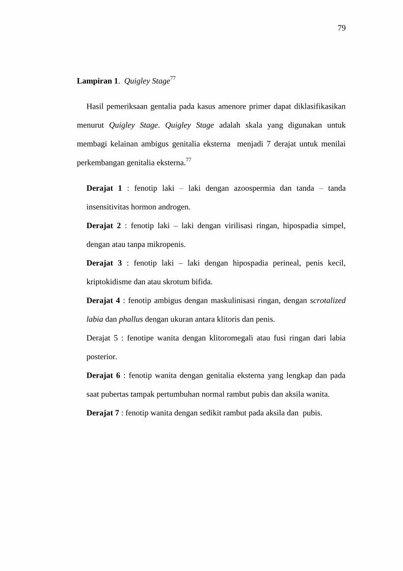
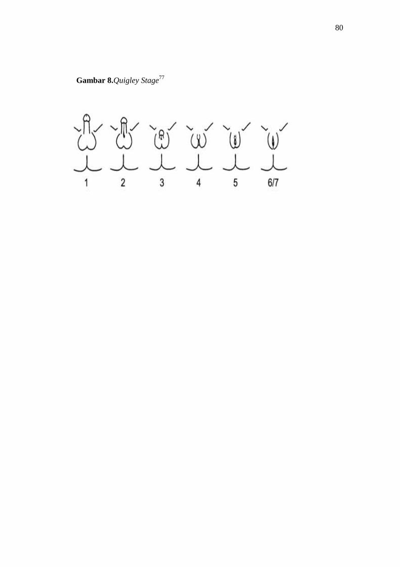
Lampiran 1. Quigley Stage77
Hasil pemeriksaan gentalia pada kasus amenore primer dapat diklasifikasikan
menurut Quigley Stage. Quigley Stage adalah skala yang digunakan untuk
membagi kelainan ambigus genitalia eksterna menjadi 7 derajat untuk menilai
perkembangan genitalia eksterna.77
Derajat 1 : fenotip laki – laki dengan azoospermia dan tanda – tanda
insensitivitas hormon androgen.
Derajat 2 : fenotip laki – laki dengan virilisasi ringan, hipospadia simpel,
dengan atau tanpa mikropenis.
Derajat 3 : fenotip laki – laki dengan hipospadia perineal, penis kecil,
kriptokidisme dan atau skrotum bifida.
Derajat 4 : fenotip ambigus dengan maskulinisasi ringan, dengan scrotalized
labia dan phallus dengan ukuran antara klitoris dan penis.
Derajat 5 : fenotipe wanita dengan klitoromegali atau fusi ringan dari labia
posterior.
Derajat 6 : fenotip wanita dengan genitalia eksterna yang lengkap dan pada
saat pubertas tampak pertumbuhan normal rambut pubis dan aksila wanita.
Derajat 7 : fenotip wanita dengan sedikit rambut pada aksila dan pubis.
80
Gambar 8.Quigley Stage77

81
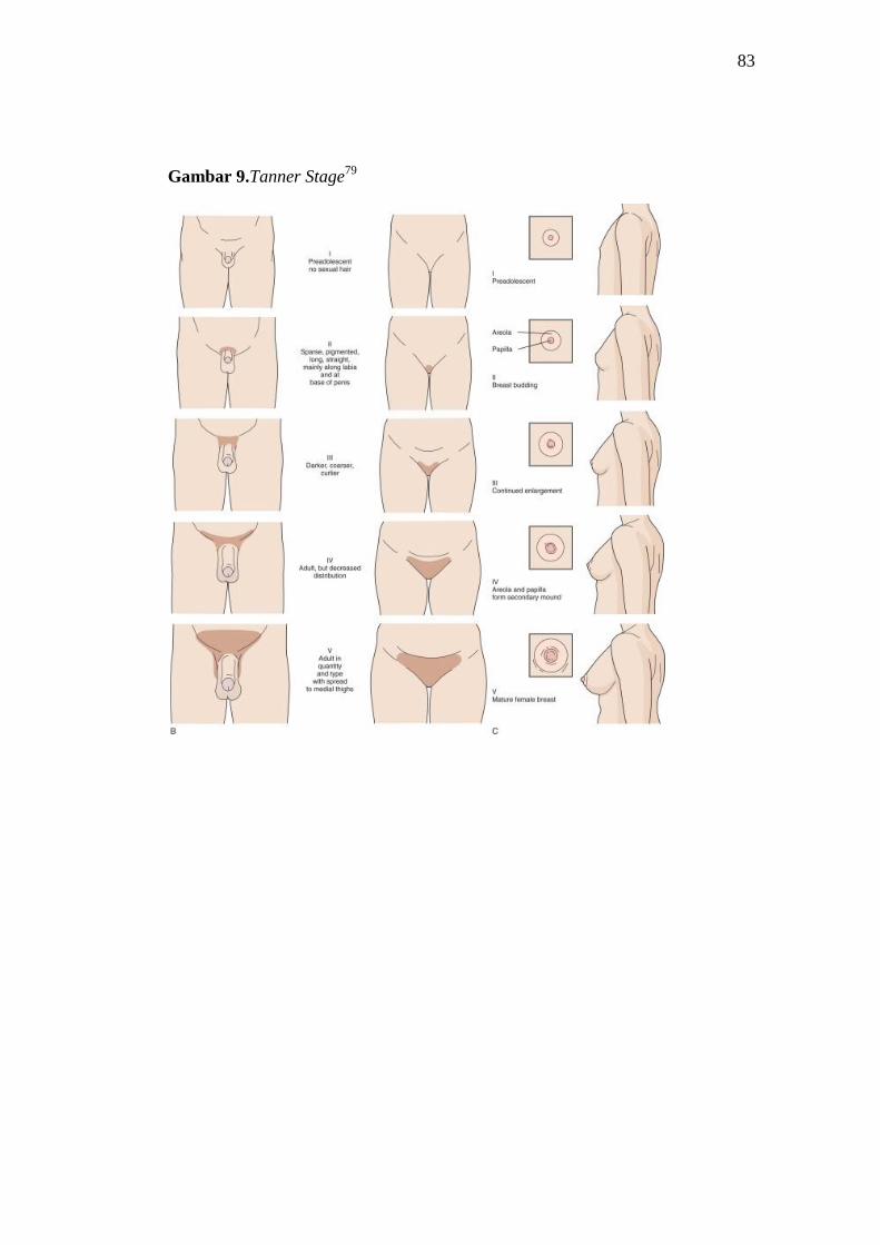
Lampiran 2. Tanner Stage78
Perkembangan seksual sekunder seperti payudara dan rambut tubuh
diklasifikasikan menurut Tanner Stage. Tanner Stage terbagi menjadi 5
klasifikasi, yaitu :78
Breast (Female)
Tanner I
Tidak ada jaringan kelenjar, terdapat elevasi papila. [≤ 10 tahun]
Tanner II
Adanyapembentukan breast bud dengan sedikit jaringan kelenjar, aerola
mulai bertambah lebar. [10 –11,5 tahun]
Tanner III
Payudara mulai meninggi, disertai degan melebarnya batas aerola mengikuti
kontur payudara. [11,5 – 13 tahun]
Tanner IV
Payudara bertambah besar dan meninggi, aerola dan papilla mammae mulai
menonjol. [13 – 15 tahun]
Tanner V
Payudara mencapai ukuran dewasa matur. [ ≥ 15 tahun ].
Pubic Hair (Female)
Tanner I
Tidak ada rambut pubis secara keseluruhan. (prepubertal state ) [≤ 10 tahun]
Tanner II
82
Terdapat pertumbuhan rambut pubis yang sedikit, halus, dan pendek pada
labia mayor. [10 – 11,5 tahun]
Tanner III
Terdapat pertumbuhan rambut pubis mulai kasar dan keriting, serta tumbuh
menyebar ke lateral. [ 11,5 – 13 tahun]
Tanner IV
Adult- like hair quality, pertumbuhan rambut mulai banyak, menyebar tetapi
tidak sampai ke medial paha. [ 13 – 14 tahun]
Tanner V
Pertumbuhan rambut menyebar sampai ke medial paha. [≥ 14 tahun].
83
Gambar 9.Tanner Stage79
84
Lampiran 3. Analisis Sitogenetika74
1. Alat
- Spuit
- Tube dispossable 15 ml, tube 1,5 ml
- Gelas ukur, botol semprot KCL
- Pipet dispossable steril 3 cc, pipet pasteur steril
- Sentrifuse, inkubator, waterbath, laminarflow
- Almari es
- Object glass
- Mikroskop cahaya, mikroskop fotografi
- Rak tabung reaksi
- Tissue
2. Bahan
Bahan yang diperiksa adalah darah vena dengan antikoagulan heparin.
3. Reagen yang dibutuhkan
- Media kultur kromosom
Media yang digunakan adalah RPMI 1640 dan MEM
- Larutan PHA-M (mixture)
- Fetal bovine Serum (FBS) 10%
- Colcemid
- KCl 0,075 M
- Larutan Carnoy’s (3 metanol : 1 asam asetat)
- Buffer Phosphat pH 6,8
85
- Phosphate Buffer Saline 0,1 M
- Giemsa
- Trypsin 1 : 250
- Minyak emersi
4. Prosedur Pengambilan Bahan
Sebelum mengambil darah pasien, dicatat identitas pasien dan alasan
pmeriksaan karena proses penanaman sel atau media yang dipilih tergantung pada
tujuan pemeriksaan kromosom dan dipastikan telah diambil informed consent.
Sampel darah diambil dari pembuluh darah vena cubiti dengan menggunakan
antikoagulan heparin.
5. Prosedur Pemeriksaan Bahan
- Penanaman
1. Meneteskan masing – masing 7 tetes buffy coat atau 10 tetes darah dalam
2 tabung yang telah berisi 2 media berbeda yaitu MEM dan RPMI 1640
(yang masing – masing tabung mengandung 10% Fetal Bovine Serum dan
100 mikroliter PHA-P)
2. Melakukan inkubasi sampel pada suhu 37º C selama 72 – 96 jam dengan
sudut kemiringan tabung 45º agar memberi peluang tumbuhnya sel di
permukaan, dalam inkubator yang mengandung 5% CO2
- Pemanenan
1. Meneteskan 3 tetes Colcemid pada setiap tabung, dan diinkubasi selama
30 menit.
86
2. Setelah dilakukan inkubasi lalu melakukan pemusingan selama 10 menit
pada 1000 RPM .
3. Membuang supernatan yang terbentuk, kemudian endapan yang terbentuk
dilakukan pemusingan dengan ditambahkan larutan hipotonik hangat KCl
0,075 M, diresuspensikan homogen dan melakukan inkubasi pada suhu
37º C selama 15 – 30 menit.
4. Melakukan pemusingan kembali pada 1000 RPM selama 10 menit, lalu
membuang supernatan yang terbentuk, dan menambahkan 5 larutan fiksasi
Carnoy’s melalui dinding tabung dan dikocok.
5. Mengulangi 3x pemberian larutan fiksasi Carnoy’s sampai didapatkan
presipitat jernih.
6. Melakukan suspensi residu dengan larutan Carnoy’s secukupnya sesuai
dengan banyaknya pelet yang terbentuk.
7. Meneteskan dan menyebarkan 2 tetes suspensi pada gelas objek.
- Pengecatan
1. Pengecatan Giemsa (Pengecatan Solid)
Melakukan pengecatan dengan Giemsa 10% dalam larutan buffer fosfat
Ph 6,8 selama 1 menit. Pengecatan dengan Giemsa 10% bertujuan untuk
melakukan skrining, tidak digunakan untuk analisis/diagnosis.
2. Pengecatan Banding dengan Trypsin tanpa penghangatan (GTG
Banding).
Mencelupkan slide yang berumur lebih dari 3 hari kedalam larutan Trypsin
0,1% yang dilarutkan dengan 90 ml PBS (Phosphat Buffer Saline) pH 6,8,
87
kemudian segera melakukan pencucian dengan PBS dan Giemsa 10% dalam
buffer fosfat.
- Analisis Kromosom
Analisis untuk seluruh kasus harus dengan pengecatan G-Banding, paling
sedikit 8 metafase dan penghitungan 20 metafase. Apabila didapatkan kelainan
mosaik, analisis dilakukan pada 100 metafase.74
- Prosedur Laporan/Diagnosis
Cara melaporkan bentuk atau konsitusi kromosom adalah mengikuti cara yang
diharuskan oleh ICSN (International System for Human Cytogenetic
Nomenclature). Standar penulisan konsitusi kromosom adalah pertama kali tulis
jumlah kromosom kemudian diikuti koma dan jenis kromosom seks, diikuti koma
lagi dan selanjutnya kelainan struktural (bila terdapat kelainan struktural). Apabila
melibatkan kelainan kromosom pada 2 kromosom maka ditulis jenis kromosom
secara urut nomor yang kecil. Seluruh metafase yang telah dianalisis, kemudian
difoto hitam putih dan dokter ahli sitogenetika akan menetukan jenis kariotipe
serta memberikan kesimpulan dari hasil pemeriksaan.74
88
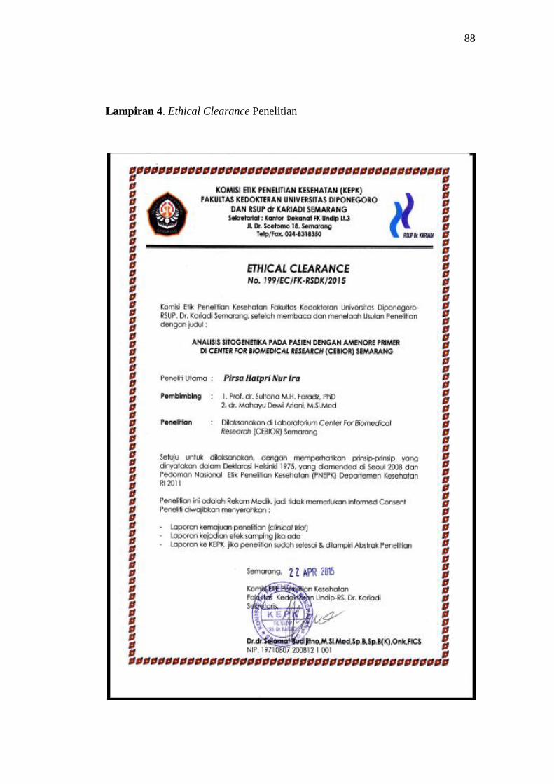
Lampiran 4. Ethical Clearance Penelitian
88
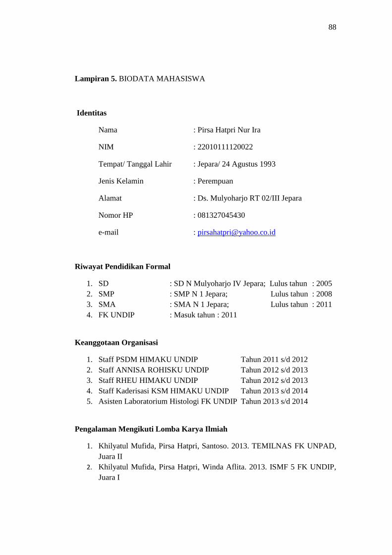
Lampiran 5. BIODATA MAHASISWA
Identitas
Nama : Pirsa Hatpri Nur Ira
NIM : 22010111120022
Tempat/ Tanggal Lahir : Jepara/ 24 Agustus 1993
Jenis Kelamin : Perempuan
Alamat : Ds. Mulyoharjo RT 02/III Jepara
Nomor HP : 081327045430
e-mail : [email protected]
Riwayat Pendidikan Formal
1. SD : SD N Mulyoharjo IV Jepara; Lulus tahun : 2005
2. SMP : SMP N 1 Jepara; Lulus tahun : 2008
3. SMA : SMA N 1 Jepara; Lulus tahun : 2011
4. FK UNDIP : Masuk tahun : 2011
Keanggotaan Organisasi
1. Staff PSDM HIMAKU UNDIP Tahun 2011 s/d 2012
2. Staff ANNISA ROHISKU UNDIP Tahun 2012 s/d 2013
3. Staff RHEU HIMAKU UNDIP Tahun 2012 s/d 2013
4. Staff Kaderisasi KSM HIMAKU UNDIP Tahun 2013 s/d 2014
5. Asisten Laboratorium Histologi FK UNDIP Tahun 2013 s/d 2014
Pengalaman Mengikuti Lomba Karya Ilmiah
1. Khilyatul Mufida, Pirsa Hatpri, Santoso. 2013. TEMILNAS FK UNPAD,
Juara II
2. Khilyatul Mufida, Pirsa Hatpri, Winda Aflita. 2013. ISMF 5 FK UNDIP,
Juara I
![DAFTAR PUSTAKA - etd.repository.ugm.ac.idetd.repository.ugm.ac.id/downloadfile/96937/potongan/S1-2016... · 90 DAFTAR PUSTAKA DAFTAR PUSTAKA [1] Badan Standardisasi Nasional. “SNI](https://img.pdfslide.us/doc/110x75/5ccf188d88c99385278e02a1/daftar-pustaka-etd-90-daftar-pustaka-daftar-pustaka-1-badan-standardisasi.jpg)